Secondary Ion Mass Spectroscopy (SIMS)
|
|
|
- Adam Cooper
- 5 years ago
- Views:
Transcription
1 Secondary Ion Mass Spectroscopy (SIMS)
2 Analyzing Inorganic Solids * = under special conditions ** = semiconductors only + = limited number of elements or groups
3 Analyzing Organic Solids * = under special conditions ** = semiconductors only + = limited number of elements or groups

4 Surface Analysis

5 Surface Analysis

6 Surface Analysis
7 Surface Analysis

8 Surface Analysis

9 Surface Analysis

10 Surface Analysis

11 Surface Analysis

12 Principle of SIMS The ion gun directs a primary beam of energetic ion at a specimen, resulting in ejection of secondary ions from the sample surface. The secondary ions are analyzed with a mass spectrometer.
13 Ejected Fragments The ejected fragments are low energy, 5-50 ev. The ejected fragments consist of positively and negatively charged ions, neutral atoms, and molecular fragments.
14 Secondary Ion Yields Primary Beam Effects Other factors affect the secondary ionization efficiencies in SIMS measurements. Oxygen bombardment increases the yield of positive ions and cesium bombardment increases the yield of negative ions. The increases can range up to four orders of magnitude. Oxygen enhancement occurs as a result of metal-oxygen bonds in an oxygen rich zone. When these bonds break in the ion emission process, the oxygen becomes negatively charged because its high electron affinity favors electron capture and its high ionization potential inhibits positive charging. The metal is left with the positive charge. Oxygen beam sputtering increases the concentration of oxygen in the surface layer. The enhanced negative ion yields produced with cesium bombardment can be explained by work functions that are reduced by implantation of cesium into the sample surface. More secondary electrons are excited over the surface potential barrier. Increased availability of electrons leads to increased negative ion formation.


15 Secondary Ion Yields Primary Beam Effects The variability in ionization efficiencies leads to different analysis conditions for different elements as indicated on the periodic table.
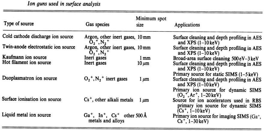
16 Ion Guns

17 Douplasmatron Ion Source Inert gases are used to form ions (He, Ar, Xe, etc.). Probe diameters between 5 m and 1 mm. Ion current densities greater than 10 ma/cm 2.
18 Liquid Metal Ion Source + + Low melting point metals are used to form ions (Cs, Ga ). Sub micron probe diameters. Ion current densities greater than 1 A/cm 2. Beam can be focused and rastered. Secondary electron images like in a SEM Elemental or molecular maps similar to EDX
19 Mass Spectrometers Quadrupole Magnetic Sector Single Focusing Double focusing Time of Flight
20 Quadrapole Mass Spectrometer Comprises of four rods to which DC and RF fields are applied. The resulting electric field allows ions of a given mass entering the analyzer to follow a stable oscillatory trajectory between the rods and reach the detector. All other ions entering the analyzer are forced to follow unstable trajectories of increasing amplitude. They collide with the rods or housing and do not reach the detector.
21 Quadrapole Mass Spectrometer

22 Quadrapole Mass Spectrometer
23 Single Focusing Magnetic Sector Mass Spectrometer Ions are diverted into circular paths by a magnetic field. The magnetic field separates the components of the total ion beam according to momentum. The individual ion beams are separated spatially and each has a unuiqe radius of curvature. Only ions with the proper trajectory will exit the slit and arrive at the detector.
24 Single Focusing Magnetic Sector Mass Spectrometer In a single focusing magnetic sector instrument there is a lack of uniformity of ion energies. The spread in ionic energies produces a spread in their radii of curvature in the magnetic field. The result is peak broadening and low to moderate mass resolution.
25 Double Focusing Magnetic Sector Mass Spectrometer Double focusing magnetic sector instruments use magnetic and electrostatic fields to disperse ions according to their momentum and translational energy. As ions pass through the electrostatic sector, they are dispersed according to their translational energy. Only those ions with the correct translational energy pass through a slit leading into the magnetic sector. Then the magnetic sector disperses the ions according to their momentum.


26 Double Focusing Magnetic Sector Mass Spectrometer


27 Double Focusing Magnetic Sector Mass Spectrometer
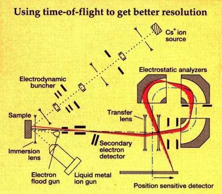
28 Time of Flight Mass Spectrometer The time of flight mass spectrometer distinguishes between ions of different mass by detecting the time taken to travel a fixed distance when accelerated by a given field. A pulsed beam of ions is incident on the sample, the ions ejected from the pulse are then accelerated, allowed to travel down a drift tube, and the different times taken for them to reach the detector produces a spectrum.

29 Time of Flight Mass Spectrometer

30 Time of Flight Mass Spectrometer

31 Time of Flight Mass Spectrometer
32 Resolution The most important parameter of a mass analyzer is its resolution. Mass peaks of ions have no natural line width so that the breath of a peak is representative of mass analyzer performance. Resolution = m/ m where m is the mass of the lighter peak.
33 Mass Interferences Mass interferences occur whenever another ion has the same nominal mass as the analyte ion. Such interferences are called isobaric. During the analysis of iron in silicon for example, 28Si2+ interferes because it has the same mass (m/z 56) as 56Fe+. Oxides are common interferences since oxygen-metal bonds are particularly stable. Thus, 40CaO+ can also interfere with 56Fe+ measurements. Low intensity hydrides of many elements appear one mass unit higher than the elements themselves. A good example is silicon-30 hydride (30SiH+) which interferes with trace phosphorous analysis. Primary ions often combine with sample elements to produce interferences. For example 133Cs32S2- is isobaric with Au- (m/z 197) during the measurement of gold in pyrite (FeS2).
34 High Mass Resolution Mass spectrometers with sufficient mass resolution can separate atomic ions from molecular ion interferences. Mass resolution is usually specified in terms of m/delta m where m is the nominal mass of the two ions and delta m is their difference. For example, 56Fe and 28Si2 (m/z and ) require m/delta m 5,600 for separation while Au and 133Cs32S2 (m/z and ) require m/delta m Each different kind of SIMS mass analyzers has a range of possible mass resolutions. For example, a (well tuned) double focusing magnetic sector instrument can have mass resolution in the range m/delta m 500 to 10,000. Loss of secondary ion intensity accompanies operation at the high end of the mass resolution range.
35 Mass Range and Resolution Analyzer Range (amu) Resolution SF Magnetic Sector DF Magnetic Sector Time of Flight Quadrupole
36 Sample Charging The SIMS primary ion beam, secondary ions, and secondary electrons produce a net electric current at the sample surface. If the sample material conducts, the current flows through the sample into the instrument. However, insulating samples undergo charge buildup. Sample charging diffuses the primary beam and diverts it from the analytical area, often eliminating the secondary ion signal entirely. Sample charging also changes the energy distribution of the secondary ions, which effects their transmission and detection by the mass spectrometer. When the sample is a thin dielectric on a conducting substrate, a strong electric field develops. Mobile ions such as sodium and lithium migrate in the electric field and depth profiles no longer reflect the original compositions of the layers. Several techniques are available to manage sample charging, and they are often used in combination.
37 Sample Charging Electron Bombardment Electrons compensate for positive charge buildup that results from positive primary ions and/or negative secondary ions and electrons. Low energy electron beams work better because higher energies produce more than one secondary electron for every incoming electron. Low energy electron beams are more easily implemented in quadrupole SIMS instruments, making quadrupoles the system of choice for insulating materials. In contrast to quadrupoles, magnetic sector instruments maintain the sample at high positive potential for positive ion spectroscopy, making it difficult to bring in a low energy electron beam. High energy electron beams, though less effective, are widely used. Adjacent Conductors Conducting grids placed over the sample reduce the effects of charging on ion optics and bring a source of electrons near to positively charged areas of the sample. When struck by a primary ion, the conductors emit secondary electrons that migrate to the charging area. Similarly, samples are often coated with conducting materials such as gold or carbon. Before starting the analysis, the coating must be sputtered away, but only in the analytical area. Negative Primary Ion Beams The most common negative primary ion beam is O, available from the same duoplasmatron sources that more commonly produce O2+. Primary O beams find wide use for insulating geological samples. Automatic Voltage Offset A continuously variable voltage offset can be applied to the accelerating voltage for samples that are only slightly charging. Automatic voltage offset procedures (called autovolt) are often incorporated into instrument control software. After every cycle in a depth profile analysis, the software invokes an energy distribution measurement and adjusts the voltage offset as needed to keep the peak of the distribution constant.
38 Types of SIMS Analysis Static SIMS Ion dosage and energy are kept low so that undamaged surface molecular fragments can be detected. Chemical structure of surfaces can be determined. Dynamic SIMS Ion dosage and sputter rates are high resulting more fragmentation. Positive charge builds up on the sample surface and may need to be neutralized with an electron flood.
39 Spectra When bombarded by electrons in the electron impact method of ionization, every substance ionizes and fragments uniquely. The uniqueness of the molecular fragmentation aids in structural identification. The mass spectrum becomes a finger print for each compound because no two molecules are fragmented and ionized in exactly the same manner on electron bombardment. This permits identification of different molecules in complex mixtures.
40 Spectra TOF-SIMS mass spectrum of an organic contaminant, Irganox 1010 a common polymer additive, on a Si wafer. The peak at m/z 219 is a unique fragment from the Irganox molecule.
41 Spectra
42 Depth Profiling
43 Depth Profiling
44 Depth Profiling TOF-SIMS depth profile of a 100 Angstrom ONO gate oxide structure.
.")
45 Secondary Electron & Secondary Ion Images Combined secondary electron (SE) and secondary ion images of a metal surface indicating localized areas rich in magnesium (Mg+). The topography of the surface is clearly shown in the secondary electron image.

46 Organic Contamination on Surfaces


47 Imaging of Long-Chain Organic Molecules Crystals of stearic acid are revealed by secondary ion imaging of molecular ions using ToF-SIMS.


48 TOF-SIMS Analysis of Device Vias
49 References J. M. Walls, Editor, Methods of Surface Analysis: Techniques and Applications, Cambridge University Press, Cambridge, J. P. Sibilia, Editor, A Guide to Materials Characterization and Chemical Analysis, VCH Publishers, Inc., New York, P. E. J. Flewitt and R. K. Wild, Physical Methods for Materials Characterization, Institute of Physics Publishing, Bristol, H. H. Willard, L. L. Merritt, Jr., J. A. Dean and F. A. Settle, Jr., Instrumental Methods of Analysis, Wadsworth Publishing Company, Belmont, California, 1988.
Secondaryionmassspectrometry
 Secondaryionmassspectrometry (SIMS) 1 Incident Ion Techniques for Surface Composition Analysis Mass spectrometric technique 1. Ionization -Electron ionization (EI) -Chemical ionization (CI) -Field ionization
Secondary Ion Mass Spectrometry (SIMS)
") CHEM53200: Lecture 10 Secondary Ion Mass Spectrometry (SIMS) Major reference: Surface Analysis Edited by J. C. Vickerman (1997). 1 Primary particles may be: Secondary particles can be e s, neutral species
CHEM53200: Lecture 10 Secondary Ion Mass Spectrometry (SIMS) Major reference: Surface Analysis Edited by J. C. Vickerman (1997). 1 Primary particles may be: Secondary particles can be e s, neutral species
Secondary ion mass spectrometry (SIMS)
") Secondary ion mass spectrometry (SIMS) ELEC-L3211 Postgraduate Course in Micro and Nanosciences Department of Micro and Nanosciences Personal motivation and experience on SIMS Offers the possibility to
Secondary ion mass spectrometry (SIMS) ELEC-L3211 Postgraduate Course in Micro and Nanosciences Department of Micro and Nanosciences Personal motivation and experience on SIMS Offers the possibility to
Secondary ion mass spectrometry (SIMS)
") Secondary ion mass spectrometry (SIMS) Lasse Vines 1 Secondary ion mass spectrometry O Zn 10000 O 2 Counts/sec 1000 100 Li Na K Cr ZnO 10 ZnO 2 1 0 20 40 60 80 100 Mass (AMU) 10 21 10 20 Si 07 Ge 0.3 Atomic
Secondary ion mass spectrometry (SIMS) Lasse Vines 1 Secondary ion mass spectrometry O Zn 10000 O 2 Counts/sec 1000 100 Li Na K Cr ZnO 10 ZnO 2 1 0 20 40 60 80 100 Mass (AMU) 10 21 10 20 Si 07 Ge 0.3 Atomic
Secondary Ion Mass Spectrometry (SIMS) Thomas Sky
 Thomas Sky") 1 Secondary Ion Mass Spectrometry (SIMS) Thomas Sky Depth (µm) 2 Characterization of solar cells 0,0 1E16 1E17 1E18 1E19 1E20 0,2 0,4 0,6 0,8 1,0 1,2 P Concentration (cm -3 ) Characterization Optimization
1 Secondary Ion Mass Spectrometry (SIMS) Thomas Sky Depth (µm) 2 Characterization of solar cells 0,0 1E16 1E17 1E18 1E19 1E20 0,2 0,4 0,6 0,8 1,0 1,2 P Concentration (cm -3 ) Characterization Optimization
Harris: Quantitative Chemical Analysis, Eight Edition
 Harris: Quantitative Chemical Analysis, Eight Edition CHAPTER 21: MASS SPECTROMETRY CHAPTER 21: Opener 21.0 Mass Spectrometry Mass Spectrometry provides information about 1) The elemental composition of
Instrumental Analysis. Mass Spectrometry. Lecturer:! Somsak Sirichai
 303351 Instrumental Analysis Mass Spectrometry Lecturer:! Somsak Sirichai Mass Spectrometry What is Mass spectrometry (MS)? An analytic method that employs ionization and mass analysis of compounds in
( 1+ A) 2 cos2 θ Incident Ion Techniques for Surface Composition Analysis Ion Scattering Spectroscopy (ISS)
 2 cos2 θ Incident Ion Techniques for Surface Composition Analysis Ion Scattering Spectroscopy (ISS)") 5.16 Incident Ion Techniques for Surface Composition Analysis 5.16.1 Ion Scattering Spectroscopy (ISS) At moderate kinetic energies (few hundred ev to few kev) ion scattered from a surface in simple kinematic
5.16 Incident Ion Techniques for Surface Composition Analysis 5.16.1 Ion Scattering Spectroscopy (ISS) At moderate kinetic energies (few hundred ev to few kev) ion scattered from a surface in simple kinematic
Chemistry Instrumental Analysis Lecture 35. Chem 4631
 Chemistry 4631 Instrumental Analysis Lecture 35 Principle components: Inlet Ion source Mass analyzer Ion transducer Pumps Signal processor Mass analyzers Quadrupole Time of Flight Double Focusing Ion
Lecture 22 Ion Beam Techniques
 Lecture 22 Ion Beam Techniques Schroder: Chapter 11.3 1/44 Announcements Homework 6/6: Will be online on later today. Due Wednesday June 6th at 10:00am. I will return it at the final exam (14 th June).
ICPMS Doherty Lecture 1
 ICPMS Doherty Lecture 1 Mass Spectrometry This material provides some background on how to measure isotope abundances by means of mass spectrometry. Mass spectrometers create and separate ionized atoms
CEE 772 Lecture #27 12/10/2014. CEE 772: Instrumental Methods in Environmental Analysis
 Updated: 10 December 2014 Print version CEE 772: Instrumental Methods in Environmental Analysis Lecture #21 Mass Spectrometry: Mass Filters & Spectrometers (Skoog, Chapt. 20, pp.511 524) (Harris, Chapt.
CEE 772: Instrumental Methods in Environmental Analysis
 Updated: 10 December 2014 Print version CEE 772: Instrumental Methods in Environmental Analysis Lecture #21 Mass Spectrometry: Mass Filters & Spectrometers (Skoog, Chapt. 20, pp.511-524) (Harris, Chapt.
Chemistry Instrumental Analysis Lecture 34. Chem 4631
 Chemistry 4631 Instrumental Analysis Lecture 34 From molecular to elemental analysis there are three major techniques used for elemental analysis: Optical spectrometry Mass spectrometry X-ray spectrometry
3.5. Accelerator Mass Spectroscopy -AMS -
 3.5. Accelerator Mass Spectroscopy -AMS - AMS is a method which counts radioactive particles ( 14 C) rather than measuring the characteristic decay activity. Comparison Traditional 14 C dating and AMS
Chemistry 311: Topic 3 - Mass Spectrometry
 Mass Spectroscopy: A technique used to measure the mass-to-charge ratio of molecules and atoms. Often characteristic ions produced by an induced unimolecular dissociation of a molecule are measured. These
Mass Analyzers. Principles of the three most common types magnetic sector, quadrupole and time of flight - will be discussed herein.
 Mass Analyzers After the production of ions in ion sources, the next critical step in mass spectrometry is to separate these gas phase ions according to their mass-to-charge ratio (m/z). Ions are extracted
Fundamentals of Mass Spectrometry. Fundamentals of Mass Spectrometry. Learning Objective. Proteomics
 Mass spectrometry (MS) is the technique for protein identification and analysis by production of charged molecular species in vacuum, and their separation by magnetic and electric fields based on mass
Introduction to the Q Trap LC/MS/MS System
 www.ietltd.com Proudly serving laboratories worldwide since 1979 CALL +1.847.913.0777 for Refurbished & Certified Lab Equipment ABI Q Trap LC/MS/MS Introduction to the Q Trap LC/MS/MS System The Q Trap
Application of Surface Analysis for Root Cause Failure Analysis
 Application of Surface Analysis for Root Cause Failure Analysis David A. Cole Evans Analytical Group East Windsor, NJ Specialists in Materials Characterization Outline Introduction X-Ray Photoelectron
MASS ANALYSER. Mass analysers - separate the ions according to their mass-to-charge ratio. sample. Vacuum pumps
 ION ANALYZERS MASS ANALYSER sample Vacuum pumps Mass analysers - separate the ions according to their mass-to-charge ratio MASS ANALYSER Separate the ions according to their mass-to-charge ratio in space
ION BEAM TECHNIQUES. Ion beam characterization techniques are illustrated in Fig
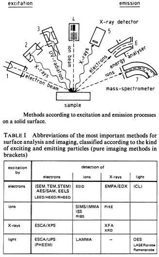
 ION BEAM TECHNIQUES Ion beam characterization techniques are illustrated in Fig. 11.21. 1 ION BEAM TECHNIQUES Incident ions are absorbed, emitted, scattered, or reflected leading to light, electron or
Lecture 8: Mass Spectrometry
 intensity Lecture 8: Mass Spectrometry Relative abundance m/z 1 Ethylbenzene experiment CH 2 CH 3 + m/z = 106 CH 2 + m/z = 91 C 8 H 10 MW = 106 CH + m/z = 77 + 2 2 What information can we get from MS spectrum?
Methods of surface analysis
 Methods of surface analysis Nanomaterials characterisation I RNDr. Věra Vodičková, PhD. Surface of solid matter: last monoatomic layer + absorbed monolayer physical properties are effected (crystal lattice
Lecture 8: Mass Spectrometry
 intensity Lecture 8: Mass Spectrometry Relative abundance m/z 1 Ethylbenzene CH 2 CH 3 + m/z = 106 CH 2 + m/z = 91 C 8 H 10 MW = 106 CH + m/z = 77 + 2 2 What information can be obtained from a MS spectrum?
Secondary-Ion Mass Spectrometry
 Principle of SIMS composition depth profiling with surface analysis techniques? Secondary-Ion Mass Spectrometry erosion of specimen surface by energetic particle bombardment sputtering two possibilities
TECHNIC A L WORK ING GROUP ITWG GUIDELINE ON SECONDARY ION MASS SPECTROMETRY (SIMS)
") NUCLE A R FORENSIC S INTERN ATION A L TECHNIC A L WORK ING GROUP ITWG GUIDELINE ON SECONDARY ION MASS SPECTROMETRY (SIMS) EXECUTIVE SUMMARY Secondary Ion Mass Spectrometry (SIMS) is used for elemental
NUCLE A R FORENSIC S INTERN ATION A L TECHNIC A L WORK ING GROUP ITWG GUIDELINE ON SECONDARY ION MASS SPECTROMETRY (SIMS) EXECUTIVE SUMMARY Secondary Ion Mass Spectrometry (SIMS) is used for elemental
ToF-SIMS or XPS? Xinqi Chen Keck-II
 ToF-SIMS or XPS? Xinqi Chen Keck-II 1 Time of Flight Secondary Ion Mass Spectrometry (ToF-SIMS) Not ToF MS (laser, solution) X-ray Photoelectron Spectroscopy (XPS) 2 3 Modes of SIMS 4 Secondary Ion Sputtering
4. How can fragmentation be useful in identifying compounds? Permits identification of branching not observed in soft ionization.
 Homework 9: Chapters 20-21 Assigned 12 April; Due 17 April 2006; Quiz on 19 April 2006 Chap. 20 (Molecular Mass Spectroscopy) Chap. 21 (Surface Analysis) 1. What are the types of ion sources in molecular
PHI 5000 Versaprobe-II Focus X-ray Photo-electron Spectroscopy
 PHI 5000 Versaprobe-II Focus X-ray Photo-electron Spectroscopy The very basic theory of XPS XPS theroy Surface Analysis Ultra High Vacuum (UHV) XPS Theory XPS = X-ray Photo-electron Spectroscopy X-ray
Ionization Techniques Part IV
 Ionization Techniques Part IV CU- Boulder CHEM 5181 Mass Spectrometry & Chromatography Presented by Prof. Jose L. Jimenez High Vacuum MS Interpretation Lectures Sample Inlet Ion Source Mass Analyzer Detector
(Refer Slide Time 00:09) (Refer Slide Time 00:13)
 (Refer Slide Time 00:13)") (Refer Slide Time 00:09) Mass Spectrometry Based Proteomics Professor Sanjeeva Srivastava Department of Biosciences and Bioengineering Indian Institute of Technology, Bombay Mod 02 Lecture Number 09 (Refer
(Refer Slide Time 00:09) Mass Spectrometry Based Proteomics Professor Sanjeeva Srivastava Department of Biosciences and Bioengineering Indian Institute of Technology, Bombay Mod 02 Lecture Number 09 (Refer
6.5 Optical-Coating-Deposition Technologies
 92 Chapter 6 6.5 Optical-Coating-Deposition Technologies The coating process takes place in an evaporation chamber with a fully controlled system for the specified requirements. Typical systems are depicted
Introduction to SIMS Basic principles Components Techniques Drawbacks Figures of Merit Variations Resources
 Introduction to SIMS Basic principles Components Techniques Drawbacks Figures of Merit Variations Resources New technique for surface chemical analysis. SIMS examines the mass of ions, instead of energy
IONTOF. Latest Developments in 2D and 3D TOF-SIMS Analysis. Surface Analysis Innovations and Solutions for Industry 2017 Coventry
 Latest Developments in 2D and 3D TOF-SIMS Analysis Surface Analysis Innovations and Solutions for Industry 2017 Coventry 12.10.2017 Matthias Kleine-Boymann Regional Sales Manager matthias.kleine-boymann@iontof.com
MS/MS .LQGVRI0606([SHULPHQWV
 0DVV6SHFWURPHWHUV Tandem Mass Spectrometry (MS/MS) :KDWLV0606" Mass spectrometers are commonly combined with separation devices such as gas chromatographs (GC) and liquid chromatographs (LC). The GC or
MS Goals and Applications. MS Goals and Applications
 MS Goals and Applications 3 Several variations on a theme, three common steps Form gas-phase ions choice of ionization method depends on sample identity and information required Separate ions on basis
Auger Electron Spectroscopy (AES) Prof. Paul K. Chu
 Prof. Paul K. Chu") Auger Electron Spectroscopy (AES) Prof. Paul K. Chu Auger Electron Spectroscopy Introduction Principles Instrumentation Qualitative analysis Quantitative analysis Depth profiling Mapping Examples The Auger
Auger Electron Spectroscopy (AES) Prof. Paul K. Chu Auger Electron Spectroscopy Introduction Principles Instrumentation Qualitative analysis Quantitative analysis Depth profiling Mapping Examples The Auger
Lecture 11 Surface Characterization of Biomaterials in Vacuum
 1 Lecture 11 Surface Characterization of Biomaterials in Vacuum The structure and chemistry of a biomaterial surface greatly dictates the degree of biocompatibility of an implant. Surface characterization
Electron Microprobe Analysis 1 Nilanjan Chatterjee, Ph.D. Principal Research Scientist
 12.141 Electron Microprobe Analysis 1 Nilanjan Chatterjee, Ph.D. Principal Research Scientist Massachusetts Institute of Technology Electron Microprobe Facility Department of Earth, Atmospheric and Planetary
Electron Microprobe Analysis 1 Nilanjan Chatterjee, Ph.D. Principal Research Scientist
 12.141 Electron Microprobe Analysis 1 Nilanjan Chatterjee, Ph.D. Principal Research Scientist Massachusetts Institute of Technology Electron Microprobe Facility Department of Earth, Atmospheric and Planetary
Imaging Methods: Scanning Force Microscopy (SFM / AFM)
") Imaging Methods: Scanning Force Microscopy (SFM / AFM) The atomic force microscope (AFM) probes the surface of a sample with a sharp tip, a couple of microns long and often less than 100 Å in diameter.
Imaging Methods: Scanning Force Microscopy (SFM / AFM) The atomic force microscope (AFM) probes the surface of a sample with a sharp tip, a couple of microns long and often less than 100 Å in diameter.
TANDEM MASS SPECTROSCOPY
 TANDEM MASS SPECTROSCOPY 1 MASS SPECTROMETER TYPES OF MASS SPECTROMETER PRINCIPLE TANDEM MASS SPECTROMETER INSTRUMENTATION QUADRAPOLE MASS ANALYZER TRIPLE QUADRAPOLE MASS ANALYZER TIME OF FLIGHT MASS ANALYSER
Photoelectron spectroscopy Instrumentation. Nanomaterials characterization 2
 Photoelectron spectroscopy Instrumentation Nanomaterials characterization 2 RNDr. Věra V Vodičkov ková,, PhD. Photoelectron Spectroscopy general scheme Impact of X-ray emitted from source to the sample
This is the total charge on an ion divided by the elementary charge (e).
.") 12.2 Fundamentals and general terms Accelerator mass spectrometry An ultra-sensitive technique using tandem accelerators employed mainly for the study of long-lived radioisotopes, and stable isotopes at
12.2 Fundamentals and general terms Accelerator mass spectrometry An ultra-sensitive technique using tandem accelerators employed mainly for the study of long-lived radioisotopes, and stable isotopes at
Mass Spectrometry in MCAL
 Mass Spectrometry in MCAL Two systems: GC-MS, LC-MS GC seperates small, volatile, non-polar material MS is detection devise (Agilent 320-MS TQ Mass Spectrometer) Full scan monitoring SIM single ion monitoring
RECOMMENDATIONS FOR NOMENCLATURE OF MASS SPECTROMETRY
 international UNION OF PURE AND APPLIED CHEMISTRY ANALYTICAL CHEMISTRY DIVISION COMMISSION ON ANALYTICAL NOMENCLATURE RECOMMENDATIONS FOR NOMENCLATURE OF MASS SPECTROMETRY RULES APPROVED 1973 LONDON BUTTER
X-Ray Photoelectron Spectroscopy (XPS) Prof. Paul K. Chu
 Prof. Paul K. Chu") X-Ray Photoelectron Spectroscopy (XPS) Prof. Paul K. Chu X-ray Photoelectron Spectroscopy Introduction Qualitative analysis Quantitative analysis Charging compensation Small area analysis and XPS imaging
X-Ray Photoelectron Spectroscopy (XPS) Prof. Paul K. Chu X-ray Photoelectron Spectroscopy Introduction Qualitative analysis Quantitative analysis Charging compensation Small area analysis and XPS imaging
20.2 Ion Sources. ions electrospray uses evaporation of a charged liquid stream to transfer high molecular mass compounds into the gas phase as MH n
 20.2 Ion Sources electron ionization produces an M + ion and extensive fragmentation chemical ionization produces an M +, MH +, M +, or M - ion with minimal fragmentation MALDI uses laser ablation to transfer
Mass Spectrometry. What is Mass Spectrometry?
 Mass Spectrometry What is Mass Spectrometry? Mass Spectrometry (MS): The generation of gaseous ions from a sample, separation of these ions by mass-to-charge ratio, and measurement of relative abundance
Chemical Analysis in TEM: XEDS, EELS and EFTEM. HRTEM PhD course Lecture 5
 Chemical Analysis in TEM: XEDS, EELS and EFTEM HRTEM PhD course Lecture 5 1 Part IV Subject Chapter Prio x-ray spectrometry 32 1 Spectra and mapping 33 2 Qualitative XEDS 34 1 Quantitative XEDS 35.1-35.4
Types of Analyzers: Quadrupole: mass filter -part1
 16 Types of Analyzers: Sector or double focusing: magnetic and electric Time-of-flight (TOF) Quadrupole (mass filter) Linear ion trap Quadrupole Ion Trap (3D trap) FTICR fourier transform ion cyclotron
Huashun Zhang. Ion Sources. With 187 Figures and 26 Tables Э SCIENCE PRESS. Springer
 Huashun Zhang Ion Sources With 187 Figures and 26 Tables Э SCIENCE PRESS Springer XI Contents 1 INTRODUCTION 1 1.1 Major Applications and Requirements 1 1.2 Performances and Research Subjects 1 1.3 Historical
Electron beam scanning
 Electron beam scanning The Electron beam scanning operates through an electro-optical system which has the task of deflecting the beam Synchronously with cathode ray tube which create the image, beam moves
Metal Deposition. Filament Evaporation E-beam Evaporation Sputter Deposition
 Metal Deposition Filament Evaporation E-beam Evaporation Sputter Deposition 1 Filament evaporation metals are raised to their melting point by resistive heating under vacuum metal pellets are placed on
Secondary Ion Mass Spectrometry (SIMS)
") OpenStax-CNX module: m50227 1 Secondary Ion Mass Spectrometry (SIMS) Kourtney Wright Andrew R. Barron This work is produced by OpenStax-CNX and licensed under the Creative Commons Attribution License 4.0
OpenStax-CNX module: m50227 1 Secondary Ion Mass Spectrometry (SIMS) Kourtney Wright Andrew R. Barron This work is produced by OpenStax-CNX and licensed under the Creative Commons Attribution License 4.0
SNMS. SNMS Applications. Combined SIMS and SNMS
 Hiden SIMS SNMS Sputtered Neutral Mass Spectrometry is a quantitative technique using essentially the same instrumentation as SIMS. However, instead of detecting the secondary ions which are formed at
Surface and Interface Characterization of Polymer Films
 Surface and Interface Characterization of Polymer Films Jeff Shallenberger, Evans Analytical Group 104 Windsor Center Dr., East Windsor NJ Copyright 2013 Evans Analytical Group Outline Introduction to
Auger Electron Spectroscopy Overview
 Auger Electron Spectroscopy Overview Also known as: AES, Auger, SAM 1 Auger Electron Spectroscopy E KLL = E K - E L - E L AES Spectra of Cu EdN(E)/dE Auger Electron E N(E) x 5 E KLL Cu MNN Cu LMM E f E
NUCLEI, RADIOACTIVITY AND NUCLEAR REACTIONS
 NUCLEI, RADIOACTIVITY AND NUCLEAR REACTIONS VERY SHORT ANSWER QUESTIONS Q-1. Which of the two is bigger 1 kwh or 1 MeV? Q-2. What should be the approximate minimum energy of a gamma ray photon for pair
Mass Analyzers. mass measurement accuracy/reproducibility. % of ions allowed through the analyzer. Highest m/z that can be analyzed
 Mass Analyzers Double Focusing Magnetic Sector Quadrupole Mass Filter Quadrupole Ion Trap Linear Time-of-Flight (TOF) Reflectron TOF Fourier Transform Ion Cyclotron Resonance (FT-ICR-MS) Mass Analyzers
Energetic particles and their detection in situ (particle detectors) Part II. George Gloeckler
 Part II. George Gloeckler") Energetic particles and their detection in situ (particle detectors) Part II George Gloeckler University of Michigan, Ann Arbor, MI University of Maryland, College Park, MD Simple particle detectors Gas-filled
Energetic particles and their detection in situ (particle detectors) Part II George Gloeckler University of Michigan, Ann Arbor, MI University of Maryland, College Park, MD Simple particle detectors Gas-filled
Gaetano L Episcopo. Scanning Electron Microscopy Focus Ion Beam and. Pulsed Plasma Deposition
 Gaetano L Episcopo Scanning Electron Microscopy Focus Ion Beam and Pulsed Plasma Deposition Hystorical background Scientific discoveries 1897: J. Thomson discovers the electron. 1924: L. de Broglie propose
History of Mass spectroscopy. Mass Spectroscopy. Introduction... Uses of Mass Spec. Where are Mass Spectrometers Used? Mass Spectroscopy Units
 History of Mass spectroscopy Mass Spectroscopy CHEM 466 Upali Siriwardane Marilyn Cox Jim Plamer http://www.chemistry.ohiostate.edu/~allen/587%20w04/587%20w0 4%20130-136%20std.pdf http://www.cem.msu.edu/~cem333/week1
Chapter 4 Scintillation Detectors
 Med Phys 4RA3, 4RB3/6R03 Radioisotopes and Radiation Methodology 4-1 4.1. Basic principle of the scintillator Chapter 4 Scintillation Detectors Scintillator Light sensor Ionizing radiation Light (visible,
2. Separate the ions based on their mass to charge (m/e) ratio. 3. Measure the relative abundance of the ions that are produced
 ratio. 3. Measure the relative abundance of the ions that are produced") I. Mass spectrometry: capable of providing both quantitative and qualitative information about samples as small as 100 pg (!) and with molar masses in the 10 4-10 5 kdalton range A. The mass spectrometer
I. Mass spectrometry: capable of providing both quantitative and qualitative information about samples as small as 100 pg (!) and with molar masses in the 10 4-10 5 kdalton range A. The mass spectrometer
THE MODERN VIEW OF ATOMIC STRUCTURE
 44 CHAPTER 2 Atoms, Molecules, and Ions GO FIGURE What is the charge on the particles that form the beam? Experiment Interpretation Incoming a particles Beam of a particles Source of a particles Nucleus
Courtesy of ESS and TheRGA web pages part of a series of application and theory notes for public use which are provided free of charge by ESS.
 ESS The RGA freenotes Theory page 1 of 14 RGA Theory Notes Courtesy of ESS and TheRGA web pages part of a series of application and theory notes for public use which are provided free of charge by ESS.
IV. Surface analysis for chemical state, chemical composition
 IV. Surface analysis for chemical state, chemical composition Probe beam Detect XPS Photon (X-ray) Photoelectron(core level electron) UPS Photon (UV) Photoelectron(valence level electron) AES electron
CHAPTER D4 ORTHOGONAL TIME OF FLIGHT OPTICS
 Back to Basics Section D: Ion Optics CHAPTER D4 ORTHOGONAL TIME OF FLIGHT OPTICS TABLE OF CONTENTS QuickGuide...413 Summary...415 Introduction...417 The physical basis of orthogonal TOF....... 419 Pulsedmainbeamsofions...421
Secondary Ion-Mass Spectroscopy (SIMS)
") Secondary Ion-Mass Spectroscopy (SIMS) Prof. Bing-Yue Tsui ( 崔秉鉞 ) Department of Electronics Engineering and Institute of Electronics National Chiao-Tung University 1 Outline Introduction to SIMS Instruments
Secondary Ion-Mass Spectroscopy (SIMS) Prof. Bing-Yue Tsui ( 崔秉鉞 ) Department of Electronics Engineering and Institute of Electronics National Chiao-Tung University 1 Outline Introduction to SIMS Instruments
= : K A
 Atoms and Nuclei. State two limitations of JJ Thomson s model of atom. 2. Write the SI unit for activity of a radioactive substance. 3. What observations led JJ Thomson to conclusion that all atoms have
Nanostructure. Materials Growth Characterization Fabrication. More see Waser, chapter 2
 Nanostructure Materials Growth Characterization Fabrication More see Waser, chapter 2 Materials growth - deposition deposition gas solid Physical Vapor Deposition Chemical Vapor Deposition Physical Vapor
Application Note GA-301E. MBMS for Preformed Ions. Extrel CMS, 575 Epsilon Drive, Pittsburgh, PA I. SAMPLING A CHEMICAL SOUP
 Application Note MBMS for Preformed Ions, 575 Epsilon Drive, Pittsburgh, PA 15238 (Poster Presented at 45th ASMS Conference on Mass Spectrometry, June 1-5, 1997) In order to accurately characterize a plasma
Introduction to GC/MS
 Why Mass Spectrometry? Introduction to GC/MS A powerful analytical technique used to: 1.Identify unknown compounds 2. Quantify known materials down to trace levels 3. Elucidate the structure of molecules
ECE Semiconductor Device and Material Characterization
 ECE 4813 Semiconductor Device and Material Characterization Dr. Alan Doolittle School of Electrical and Computer Engineering Georgia Institute of Technology As with all of these lecture slides, I am indebted
MASS SPECTROMETRY. Topics
 MASS SPECTROMETRY MALDI-TOF AND ESI-MS Topics Principle of Mass Spectrometry MALDI-TOF Determination of Mw of Proteins Structural Information by MS: Primary Sequence of a Protein 1 A. Principles Ionization:
A DIVISION OF ULVAC-PHI. Time-of-Flight Secondary Ion Mass Spectrometer with Parallel Imaging MS/MS for Confident Molecular Identification
 A DIVISION OF ULVAC-PHI Time-of-Flight Secondary Ion Mass Spectrometer with Parallel Imaging MS/MS for Confident Molecular Identification Designed for Confident Molecular Identification and Superior Imaging
Secondary Ion Mass Spectrometry (SIMS) for Surface Analysis
 for Surface Analysis") Secondary Ion Mass Spectrometry (SIMS) for Surface Analysis General overview of SIMS - principles, ionization, advantages & limitations SIMS as a surface analysis technique - operation modes, information
Secondary Ion Mass Spectrometry (SIMS) for Surface Analysis General overview of SIMS - principles, ionization, advantages & limitations SIMS as a surface analysis technique - operation modes, information
Analysis of Insulator Samples with AES
 Analysis of Insulator Samples with AES Kenichi Tsutsumi, Nobuyuki Ikeo, Akihiro Tanaka, and Toyohiko Tazawa SA Business Unit, JEL Ltd. Introduction Auger Electron Spectroscopy (AES) makes it possible to
Instrumentation and Operation
 Instrumentation and Operation 1 STM Instrumentation COMPONENTS sharp metal tip scanning system and control electronics feedback electronics (keeps tunneling current constant) image processing system data
Extrel Application Note
 Extrel Application Note Real-Time Plasma Monitoring and Detection of Trace H 2 O and HF Species in an Argon Based Plasma Jian Wei, 575 Epsilon Drive, Pittsburgh, PA 15238. (Presented at the 191st Electrochemical
Hiden SIMS Secondary Ion Mass Spectrometers. Analysers for surface, elemental and molecular analysis
 Hiden SIMS Secondary Ion Mass Spectrometers Analysers for surface, elemental and molecular analysis vacuum analysis surface science plasma diagnostics gas analysis SIMS Versatility SIMS is a high sensitivity
X-Ray Photoelectron Spectroscopy (XPS)-2
-2") X-Ray Photoelectron Spectroscopy (XPS)-2 Louis Scudiero http://www.wsu.edu/~scudiero; 5-2669 Fulmer 261A Electron Spectroscopy for Chemical Analysis (ESCA) The 3 step model: 1.Optical excitation 2.Transport
X-Ray Photoelectron Spectroscopy (XPS)-2 Louis Scudiero http://www.wsu.edu/~scudiero; 5-2669 Fulmer 261A Electron Spectroscopy for Chemical Analysis (ESCA) The 3 step model: 1.Optical excitation 2.Transport
Propose a structure for an alcohol, C4H10O, that has the following
 Propose a structure for an alcohol, C4H10O, that has the following 13CNMR spectral data: Broadband _ decoupled 13CNMR: 19.0, 31.7, 69.5 б DEPT _90: 31.7 б DEPT _ 135: positive peak at 19.0 & 31.7 б, negative
high temp ( K) Chapter 20: Atomic Spectroscopy
 Chapter 20: Atomic Spectroscopy") high temp (2000-6000K) Chapter 20: Atomic Spectroscopy 20-1. An Overview Most compounds Atoms in gas phase high temp (2000-6000K) (AES) (AAS) (AFS) sample Mass-to-charge (ICP-MS) Atomic Absorption experiment
high temp (2000-6000K) Chapter 20: Atomic Spectroscopy 20-1. An Overview Most compounds Atoms in gas phase high temp (2000-6000K) (AES) (AAS) (AFS) sample Mass-to-charge (ICP-MS) Atomic Absorption experiment
Secondary Ion Mass Spectrometry (SIMS) for Surface Analysis
 for Surface Analysis") Secondary Ion Mass Spectrometry (SIMS) for Surface Analysis General overview of SIMS - principles, ionization, advantages & limitations SIMS as a surface analysis technique - operation modes, information
Secondary Ion Mass Spectrometry (SIMS) for Surface Analysis General overview of SIMS - principles, ionization, advantages & limitations SIMS as a surface analysis technique - operation modes, information
PARTICLE ACCELERATORS
 VISUAL PHYSICS ONLINE PARTICLE ACCELERATORS Particle accelerators are used to accelerate elementary particles to very high energies for: Production of radioisotopes Probing the structure of matter There
MS Goals and Applications. MS Goals and Applications
 MS Goals and Applications 1 Several variations on a theme, three common steps Form gas-phase ions choice of ionization method depends on sample identity and information required Separate ions on basis
Nova 600 NanoLab Dual beam Focused Ion Beam IITKanpur
 Nova 600 NanoLab Dual beam Focused Ion Beam system @ IITKanpur Dual Beam Nova 600 Nano Lab From FEI company (Dual Beam = SEM + FIB) SEM: The Electron Beam for SEM Field Emission Electron Gun Energy : 500
Appendix A2. Particle Accelerators and Detectors The Large Hadron Collider (LHC) in Geneva, Switzerland on the Border of France.
 in Geneva, Switzerland on the Border of France.") Appendix A. Particle Accelerators and Detectors The Large Hadron Collider (LHC) in Geneva, Switzerland on the Border of France. Prepared by: Arash Akbari-Sharbaf Why Build Accelerators? Probe deeper From
Appendix A. Particle Accelerators and Detectors The Large Hadron Collider (LHC) in Geneva, Switzerland on the Border of France. Prepared by: Arash Akbari-Sharbaf Why Build Accelerators? Probe deeper From
FIB - SIMS. Focussed Ion Beam Secondary Ion Mass Spectrometry.
 FIB - SIMS Focussed Ion Beam Secondary Ion Mass Spectrometry Outline Introduction to Hiden Analytical Introduction to SIMS FIB-SIMS - Introduction and key features FIB-SIMS - Applications data Hiden SIMS
CHAPTER A2 LASER DESORPTION IONIZATION AND MALDI
 Back to Basics Section A: Ionization Processes CHAPTER A2 LASER DESORPTION IONIZATION AND MALDI TABLE OF CONTENTS Quick Guide...27 Summary...29 The Ionization Process...31 Other Considerations on Laser
Chapter 3 Engineering Science for Microsystems Design and Fabrication
 Lectures on MEMS and MICROSYSTEMS DESIGN and MANUFACTURE Chapter 3 Engineering Science for Microsystems Design and Fabrication In this Chapter, we will present overviews of the principles of physical and
MSE 321 Structural Characterization
 Auger Spectroscopy Auger Electron Spectroscopy (AES) Scanning Auger Microscopy (SAM) Incident Electron Ejected Electron Auger Electron Initial State Intermediate State Final State Physical Electronics
PHOTOELECTRON SPECTROSCOPY IN AIR (PESA)
") PHOTOELECTRON SPECTROSCOPY IN AIR (PESA) LEADERS IN GAS DETECTION Since 1977 Model AC-3 Features: Atmospheric pressure operation (unique in the world) Estimate work function, ionization potential, density
PHOTOELECTRON SPECTROSCOPY IN AIR (PESA) LEADERS IN GAS DETECTION Since 1977 Model AC-3 Features: Atmospheric pressure operation (unique in the world) Estimate work function, ionization potential, density
CHEM 1411 STUDY-GUIDE-for-TEST-3 (CHAPTERS 7, 8)
") CHEM 1411 STUDY-GUIDE-for-TEST-3 (CHAPTERS 7, 8) 1. In the following diagram of a wave A) (a) is amplitude and (b) is wavelength D) (a) is amplitude and (b) is frequency B) (a) is frequency and (b) is
CHEM 1411 STUDY-GUIDE-for-TEST-3 (CHAPTERS 7, 8) 1. In the following diagram of a wave A) (a) is amplitude and (b) is wavelength D) (a) is amplitude and (b) is frequency B) (a) is frequency and (b) is
= 6 (1/ nm) So what is probability of finding electron tunneled into a barrier 3 ev high?
 So what is probability of finding electron tunneled into a barrier 3 ev high?") STM STM With a scanning tunneling microscope, images of surfaces with atomic resolution can be readily obtained. An STM uses quantum tunneling of electrons to map the density of electrons on the surface
STM STM With a scanning tunneling microscope, images of surfaces with atomic resolution can be readily obtained. An STM uses quantum tunneling of electrons to map the density of electrons on the surface
~1 V ~20-40 V. Electron collector PLASMA. Ion extraction optics. Ionization zone. Mass Resolving section Ion detector. e - ~20 V Filament Heater
 RGAs and Leak detectors [Note that standard Ion Implanters are just overgrown RGAs!] RGAs or Residual Gas Analyzers are also known as Mass Spectrum Analyzers. These can sometimes be upgraded to also include
MS482 Materials Characterization ( 재료분석 ) Lecture Note 4: XRF
 Lecture Note 4: XRF") 2016 Fall Semester MS482 Materials Characterization ( 재료분석 ) Lecture Note 4: XRF Byungha Shin Dept. of MSE, KAIST 1 Course Information Syllabus 1. Overview of various characterization techniques (1 lecture)
2016 Fall Semester MS482 Materials Characterization ( 재료분석 ) Lecture Note 4: XRF Byungha Shin Dept. of MSE, KAIST 1 Course Information Syllabus 1. Overview of various characterization techniques (1 lecture)