Surrey Ion Beam Centre: the EPSRC MRF for Ion Beam Applications
|
|
|
- Cameron Charles
- 6 years ago
- Views:
Transcription
1 EPJ Web of Conferences 115, (2016) DOI: /epjconf/ Owned by the authors, published by EDP Sciences, 2016 Centre of Excellence for Nuclear Materials 2 nd Int. Workshop Irradiation of Nuclear Materials: Flux and Dose Effects November 4-6, 2015, CEA INSTN Cadarache, France Surrey Ion Beam Centre: the EPSRC MRF for Ion Beam Applications Roger P. WEBB 1 1 Surrey Ion Beam Centre, University of Surrey, (Guildford, UK) The Surrey Ion Beam Centre (SIBC) has been sponsored as a National Centre for research in the UK since It underpins more than 40M of active UK research grants at any one time on a range of subject areas but mostly to support micro-electronics radiation damage studies. It was part of large European infrastructure project (SPIRIT) providing access to ion beam equipment across Europe. In 2002 it was awarded the Queens Anniversary prize for research achievement in Ion Implantation and Optoelectronics. The citation which accompanies the award says For over thirty years the University has been recognized as a pre-eminent institution in the field of ion implantation and optoelectronics. Its innovative research into these key technologies has had an outstanding impact on the development of the modern electronics industry worldwide. The SIBC works actively with industry, developing bespoke processes and services, particularly for the photonics industry and provides ion beam facilities to ~20 companies across the world. It operates a stringent quality control program and is one of the few ion beam laboratories in the world to operate under ISO 9001 certification. The SIBC has become one of the world leaders in the field of ion beam research. In the 1970 s and early 80s it pioneered the rapid thermal annealing technique[1] which is now universally used by the semiconductor industry, in the 80 s it developed [2,3] radiation hard silicon on insulator substrates for space and military applications and for a time was the world s only supplier of these substrates. In the 1990s it developed DataFurnace [4] a computer algorithm to enable quantified analysis using ion beam techniques (one of 2 recognised by the IAEA). The Ion Beam Analysis DataFurnace is a computer code to extract elemental depth profiles from Rutherford backscattering and related ion beam analysis spectra. It is able to solve the inverse problem ("given the spectrum, what is the profile") automatically, without user intervention. In the 1990 s the SIBC also began its work on silicon photonics, beginning with research on βfesi 2 [5] in silicon and later combing this work with defect engineering [6]. This led to the formation of a spinout company SiLight Technologies and a prestigious European Research Council grant. More recently in the field of microelectronics the SIBC has worked on novel approaches to activate dopants for shallow junction devices [7]. The SIBC has also looked to widen its approach and apply its techniques to new areas of research. This has included the biomedical applications of ion beams [8], where a network led by the SIBC was instrumental in bringing the clinical community together to make a case for particle therapy in the UK three new proton therapy units are now in the process of being built in the UK after much lobbying. With colleagues in Japan, the IBC has pioneered the development of a new analytical technique MeV SIMS [9-10], this has the potential to produce high molecular mass concentration maps in ambient pressure conditions with high spatial resolution. This is not currently possible using any other technique. The IAEA are involved in promoting and developing this technique worldwide. Recently the IBC has used the capabilities afforded by its DataFurnace algorithm to initiate work with Forensic service providers, the police and Home Office and similar establishments across mainland Europe. This has led to ion beam techniques being recognised as potentially of use for high impact crime cases. In 2007 the IBC received ISO9001 accreditation and is now actively pursuing ISO certification for its Ion Beam Analysis protocol. Total IBA [11] has now been developed from DataFurnace and is a very powerful new ion beam analysis characterisation technique enabling the unambiguous depth profiling of very complex thin films, and in principle making chemical tomography of small samples feasible. This new method, which we have been instrumental in developing, fully exploits the complementary information available from the photon and particle methods. This is an Open Access article distributed under the terms of the Creative Commons Attribution License 4.0, which permits unrestricted use, distribution, and reproduction in any medium, provided the original work is properly cited.


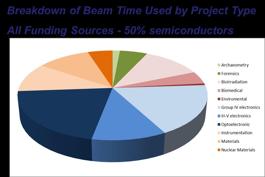
2 Centre of Excellence for Nuclear Materials 2 nd Int. Workshop Irradiation of Nuclear Materials: Flux and Dose Effects November 4-6, 2015, CEA INSTN Cadarache, France Over the years the SIBC has developed its portfolio of industrial users. Work with these companies is done via means of a non-disclosure agreement. What the company wishes to achieve is first established and then a process is developed to meet these needs. Sometimes the process is then shipped out to the company in question but more often, batches of wafers are sent to the SIBC for the bespoke implantations and processing required. A turnaround time of typically 3-5 days is achieved meaning that the IBC supplies expert services to companies across the world. The SIBC also supplies bespoke consultancy when required. It runs with a relatively robust business model attracting approximately equal funding from UK research councils, the EU and industry. Semiconductor end station The Ion Implantation Facility & Clean Room The Vertical Beam Line for Cell Irradiation Beam lines from 2MV Tandem References [1] B.J.Sealy, Rapid Thermal Annealing of Ion Implanted Semiconductors, Nuclear Physics Applications on Materials Science, 144, NATO ASI Series (Springer, Eds Recknagel, Soares), pp , [2] S.S.Gill, I.H.Wilson, RBS Analysis of Oxide Layers formed by Ion Implantation into Single Crystal Silicon Thin Solid Films 55, , [3] P.L.F.Hemment, E.A.Maydell-Ondrusz, K.G.Stephens, J.B.Butcher, D.Ioannou, J.Alderman, Formation of Buried Insulating Layers in Silicon by the Implantation of High Doses of Oxygen, Nuclear Instruments and Methods, 209/10, 157, (1983). [4] N.P.Barradas, C.Jeynes, R.P.Webb Simulated annealing analysis of Rutherford backscattering data, Applied Physics Letters, 71, 291 (1997). [5] D.Leong, M.Harry, K.J.Reeson, K.P.Homewood, A Silicon/Iron-Disilicide Light-Emitting Diode Operating at a Wavelength of 1.5 µm, Nature, 1997, Vol.387, No.6634, Pp [6] Wai Lek Ng, M. A. Lourenco, R. M. Gwilliam, S. Ledain, G. Shao & K. P. Homewood An efficient room-temperature silicon-based light-emitting diode Nature, 2001, Vol 410, [7] D.Giubertoni, G.Peppon, M.A.Sahiner, S.P.Kelty, S.Gennaro, M.Bersani, M.Kah, K.J.Kirkby, R.Doherty, M.A.Foad, F.Meirer, C.Streli, J.C.Woicik, P.Pianetta, Deactivation of submelt laser annealed arsenic ultrashallow junctions in silicon during subsequent thermal treatment, Journal of Vacuum Science and Technology B, 28(1), C1B1-C1B5, 2010 [8] N.G.Burnet, K.J.Kirkby, N.F.Kirkby, Biomedical Applications of High Energy Ion Beams, Clinical Oncology, 16, (2004). [9] B.N.Jones, V.Palitsin, R.P.Webb, Surface Analysis with High Energy ToF-SIMS Measured in Parallel with PIXE and RBS, Nuclear Instruments and Methods B, 268(11-12), , [10] Y.Wakamatsu, H.Yamada, S.Ninomiya, B.N.Jones, T.Seki, T.Aoki, R.P.Webb, J.Matsuo, Biomolecular emission by swift heavy ion bombardment, AIP Conference Proceedings, 1321, (2010). [11] C.Jeynes, M.J.Bailey, N.J.Bright, M.E.Christopher, G.W.Grime, B.N.Jones, V.V.Palitsin, R.P. Webb, Total IBA Where Are We?, Nuclear Instruments and Methods B, 269, ,
3 Ion Beams in the UK.
4 Surrey Ion Beam Centre the EPSRC MRF for Ion Beam Applications
 and MEIS facility Manchester Gamma ray and neutron irradiation emulation")
5 The Proposed UK Virtual Ion Beam Centre: Surrey Implantation and IBA facility Huddersfield Miami (in-line TEM) and MEIS facility Manchester Gamma ray and neutron irradiation emulation facility
 Prof Russell Gwilliam Prof Kevin Homewood Dr Chris Jeynes Dr Geoff Grime Liaison support staff are: Dr Julien Colaux Dr Nianhua Peng Dr Keith Heaseman Admin Support is provided")
6 Surrey Ion Beam Centre The People The core staff are: Prof. Roger Webb (director) Prof Russell Gwilliam Prof Kevin Homewood Dr Chris Jeynes Dr Geoff Grime Liaison support staff are: Dr Julien Colaux Dr Nianhua Peng Dr Keith Heaseman Admin Support is provided by: Karen Arthur Technical Support is provided by: Alex Royle Mark Browton Adrian Cansell Research Staff: Dr Vladimir Palitsin Dr Luke Antwis Dr Lucio Dos Santos Rosa Dr Ellis Moura Stori Rosa Ms Lidija Matjacic Visiting Staff: Prof Karen Kirkby Dr Brian Jones Dr Mike Merchant Dr Charlie Jeynes Dr Julien Demarche Other associated staff include: Prof Paul Sellin Prof John Watts Prof David Bradley Dr Melanie Bailey

7

8

9
10

11


12 Quality Assurance and Standards Extensive QA program ISO9001 certified since 2008 Working with both NPL and PTB on various fundamental studies ISO17025 accreditation for high accuracy RBS awarded 2015


13 Surrey Ion Beam Centre Investment Support 5M investment in machines and beam lines Approx 1.5M per annum support 2 academics 8 RAs (including 2 senior RAs) 3 technicians + 1 secretarial Technology Transfer 32 University departments around the UK 53 local and international companies 15 PhD projects Supporting >50 EPSRC grants worth > 50M
14 Surrey Ion Beam Centre Controllable Materials Modification Facilities 0.4-2MV High Energy Implanter 2-200kV High Current Implanter Implantation 2keV 4MeV (up to 10mA) Sample size mm 2 to 40cmx40cm Hot (1000 o C) or cold (~10K) Sample Chambers in class 100 clean room Plasma/thermal processing and metrology


15 Surrey Ion Beam Centre Controllable Materials Modification Applications Ion Beam Synthesis Ion Implantation Defect Engineering Proton beam lithography (on Tandem accelerator)

16 Huddersfield Miami Facility Implanter with In situ TEM o Facilities JOEL 2000FX TEM 2-100keV Implanter Temperature stage o C o Applications Nuclear Radiation damage Ion Implantation o Staff Steve Donnelly Johnathan Hinks Graeme Greaves

17 Huddersfield MEIS Facility Medium Energy Ion Scattering o Facilities keV Accelerator LEED Auger Temp range K o Applications High Resolution Depth Profiling Channelling for damage studies o Staff Jaap Van Den Berg Roger Barlow


18 Manchester Dalton Institute Cumbria Nuclear User Facility o Facilities 5MV Tandem Accelerator 60 Co gamma source 4-100Gy/min Temp range K Installing 2.5MV Tandem for dual beam work o Applications Radiation Damage Studies Nuclear Irradiation o Staff Simon Pimblott Andy Smith Nick Mason

19 The charge on the ion can be collected so that each ion can be counted as it enters the material ensuring precise control on quantity The charge on the ion enables a single isotopic mass to be selected in a magnetic field providing high purity The charge on the ion enables the particles to be accelerated by a potential gradient. The velocity of each particle is identical and when they reach the target surface will embed themselves to a well determined depth. The charge on the ion enables the ion beam to be raster scanned across a target (like an old fashioned CRT display) producing a very uniform spread across the sample under irradiation.


20 Surrey Ion Beam Centre Controllable Materials Modification Applications Ion Beam Synthesis Ion Implantation Defect Engineering Proton beam lithography (on Tandem accelerator) Synthesised compound dopant implantation distributed Ion beam synthesis dopant


21 Surrey Ion Beam Centre Controllable Materials Modification Applications Proton Beam Lithography Like e-beam lithography MeV protons set photo resist But heavy ions: don t scatter like electrons And can be focussed to ~50nm Penetrate to 40um Potential for 1000:1 aspect ratio structures Can also direct write into semiconductors to knock out carriers and decrease electro-chemical etch rate. ~28µm 720µm 45µm SEM image of a concentric circle structure produced by 2MeV protons in GaAs University Logo in GaAs

 in construction Vertical nanobeam for cell (in culture)")
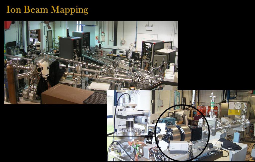
22 Surrey Ion Beam Centre Advanced Materials Analysis Facilities 2MV Tandem Techniques include RBS, EBS, ERD, PIXE, PIGE, NRA, IBIL, IBIC NEW MeV SIMS Sub micron size micro-beam with full scanning Channelling Spectroscopy for damage analysis Fully automated collection and analysis External Beam for vacuum sensitive samples Horizontal nanobeam (30nm) in construction Vertical nanobeam for cell (in culture) irradiation


23 Surrey Ion Beam Centre Advanced Materials Analysis Applications Thin Film Depth Profiling 3-D elemental composition and mapping Protein Analysis Forensics, Archaeometry Disorder Profiling of Crystals Radio biology Analysis of cells in culture

24 Bio-Irradiation Facility Facilities for bio-irradiation Targeted (~100nm) single ion irradiation End Station in Cat 2 biological clean room Vertical Arrangement to allow irradiation of living cells in liquid cultural ~10,000 cell irradiations per hour Ions up to Ca from 2MVTandem Environmental chamber, incubators etc. Facilities for Protein Analysis Analysis by micropixe To determine the heavy metal constituents in some proteins can be difficult via other methods ppm sensitivity Applications Cell Irradiation Proton/hadron therapy research Continuous mode for in-situ analysis
25 RBS Energy of scattered protons or He give light element composition and PARTICLES elemental depth profiles Fast moving, charged particles MeV ions What happens when the ion beam hits the target? MeV SIMS Using heavy ions molecular Sputtered material can be desorbed Molecules and anaylsed using ToF MS IBIC Ion Beam A Induced Charge STIM Scanning Transmission Transmitted ions Ion Microscopy PIXE Characteristic X-ray emission Simultaneous part-per-million X RAYS detection of trace elements from Na to U PIGE/NRA Nuclear reactions give GAMMA characteristic gamma rays and/or particles RAYS from light nuclei (e.g. Li, B, F) IBIL Ion Beam Induced Luminescence LIGHT ERD Forward recoil RECOILS of target atoms (particularly good for H profiling)


26 What is PIXE? Particle Induced X-ray Emission The ion beam can cause ionisation of target atoms (i.e. the removal of an electron) as shown in the figure on the left. This leaves a vacancy in one of the electron shells An electron from an outer electron shell jumps down to fill the gap An X ray is emitted, the energy of which is equal to the difference in the energy levels. Since different atoms have a different number of protons, the spacing of the energy levels for different atoms is different, and therefore the X ray energy is characteristic of the element from which it originated
 The sensitivity is at the ppm level for most elements.")
27 Benefits of PIXE Cu Pb & PIGE Fe 1000 Sn PIXE can be used to detect elements in the periodic 10 table from Na to U (Li, B, F etc. with PIGE) The sensitivity is at the ppm level for most elements. 1 The figure on the right shows how much more sensitive PIXE (blue) is to trace elements than the electron microprobe (yellow), no brehmsstrahlung The analysis can be carried out in air or in vacuum. This allows the non-destructive analysis of large and/or vacuum sensitive objects. There is usually no need for any sample preparation PIXE analysis gives absolute quantification. There is no need to compare to standards. The accuracy of the technique is ~5%. In PIXE analysis, we simultaneously collect a backscattered particle spectrum. This gives us complementary information that we can use to determine depth profiles of the major elements Sb
28 What is RBS? Ion beam Detector RBS determines compositional information about a sample: Which atoms are present and where are they? Rather than an alpha source like Rutherford, we use a more convenient ion beam from an accelerator We detect backscattered particles and their energy Ions are repelled by the positively charged nucleus, and scattered backward. The particle detector measures the energy of the backscattered particles. For high beam energies (EBS) the ion penetrates the nucleus, and the scattering probability is modified. Sample
29 How to interpret a RBS Spectra The energy of the backscattered particle depends on the depth in the sample at which it was backscattered As the particle travels through the sample, it loses energy through collisions with the target electrons These collisions don t really affect the particle s direction, just its speed (i.e. kinetic energy) The energy of the backscattered particle also depends on the mass of the atom it hit If it hits a heavy atom, it rebounds at high energy If it hits a light atom, it rebounds with lower energy
30 What is NRA/PIGE? NRA or Nuclear Reaction Analysis uses accelerated nuclei to penetrate the nucleus of a target atom causing a nuclear reaction in the target nucleus and the emission of a particle (or a gamma ray in PIGE) whose energy is characteristic of the target element. The technique is particularly useful for identifying light elements that are difficult to see with PIXE. Resonant reactions can be used which occur at particularly sharp interaction energies. As the monoenergetic beam slows gradually as it enters the target, these resonant reactions occur only at determinable depths. Hence by varying the energy of the initial beam it is possible to depth profile using this technique. Ion beam Detector Sample f:\pc_users\jpn\971118\490001g4._ : Tourmaline std: GRR573 ^3 ^2 ^1 F F Li B Al Al N a ^

31
32 What is MeV ToF- SIMS? MeV ToF-SIMS is a surface analysis technique used to look at the elemental and molecular composition on the surface of a sample For this analysis a high-energy beam of ions (MeV primary ion beam) is focussed on the surface and its energy is deposited in the uppermost layers causing atoms and molecules to be sputtered Some of these sputtered atoms become ionised The secondary ions are accelerated into a mass spectrometer and the time they take to travel through the detector is used to determine their charge-to-mass ratios This technique is used to measure a spectrum with a mass range made up of elements (and their isotopes) ranging from hydrogen to heavy molecules (e.g. proteins) consisting of thousands of atoms

33 Comparing kev and MeV-SIMS MD Simulations - Organics After 4ps plot original positions of benzene molecules moved up and broken 10keV C 60 impact in frozen benzene Ionization region MeV simulation Material flow Energized track kev Cluster SIMS Fragmentation due to nuclear collisions Nuclear energy deposition Electronic energy deposition MeV SIMS Fragmentation due to ionisation and vaporisation
34 Comparing kev and MeV-SIMS Leucine HO 2 CCH(NH 2 )CH 2 CH(CH 3 ) g/mol


35 Combining SIMS, PIXE, & RBS Because of the high primary ion energies used in MeV ToF-SIMS, we also measure PIXE and RBS spectra simultaneously PIXE gives elemental information when the primary ion excites the atoms by releasing an inner-shell electron RBS measures the backscattered particle energy to give elemental depth profile information


36 Why Use MeV Ions? Imaging Ambient Pressure Techniques MALDI Matrix Assisted Laser Desorption Ionization 10 m spatial resolution determined by spatial resolution of the laser spot Marks the sample BEFORE DESI Desorption Electro Spray Ionization 100 m spatial resolution determined by spatial resolution of the spray Marks the sample and sprays surroundings AFTER R.A.Ketola, T.Mauriala, European Journal of Pharmaceutical Sciences 46 (2012)
 To Mass")
37 Ambient Pressure SIMS 4 channel X-ray SSD detector Sweep gas flow (He) Flush (He) 100nm thick Silicon Nitride membrane. Primary beam comes through; X-Rays from target come back through to X-ray SSD detector Sweep gas flow (He) To Mass Spectrometer

38


39 s First Fully Ambient MeV-SIMS Image After demonstrating success in the vacuum system with MeV-SIMS SUR his attempting to employ MeV-SIMS in fully ambient conditions using an external beam. Using an 8.8MeV O 3+ beam at ~4um resolution we have recorded the first fully ambient MeV-SIMS molecular image PIXE Aliasing due to scanning of quadrapole mass spectrometer a) Cu PIXE Map b) PTFE molecular map

 Cu PIXE Map b) PTFE")
40 s Second Fully Ambient MeV-SIMS Image After repairing accelerator and stopping QMS from scanning. Using an 8.8MeV O 3+ beam at ~4um resolution. No aliasing, but non uniform response due to alignment issues X-ray detector only sees bottom left and SIMS sees middle brighter PIXE a) Cu PIXE Map b) PTFE molecular map 500 µm


41 s Third Fully Ambient MeV-SIMS Image After repairing accelerator and stopping QMS from scanning. Using an 8.8MeV O 3+ beam at ~4um resolution. Zoom in to avoid visibility issues and things improve PIXE a) Cu PIXE Map b) PTFE molecular map


42 And many examples.

43

44
 Manso et al (Nucl. Instr. Meth. Res.")
45 June 2008, Surrey IBC Ion beams in police casework Document from a murder investigation Ion beams were used to establish the age of the document i.e. was it current (>2006) or old (c1960?) Manso et al (Nucl. Instr. Meth. Res. B, 580 (2007), ) Concentration ( g g -1 ) Pb S Cl K Ca Ti Mn Fe Cr Zn Rb Ba S K Ca Ti Mn Fe Cu Zn Ga As Rb Sr Ba Pb The paper contained many impurities Note in particular S, K, Ba, Pb, Rb Water is now much cleaner and modern paper no longer contains such heavy elements


46

47


48


49


50


51 Art Forensics Ion beam analysis used as part of TV programme A Leonardesque painting Was it painted by Leonardo Da Vinci? Trace element analysis White pigment corresponds to 16th Century Italy Green/brown pigment corresponds to 16th Century organo-cu compounds White pigment on the back (catalogue number) was Northern European

52

53

54

55

56

57
MeV Particles, Huge Impact, Soft Desorption.
 MeV Particles, Huge Impact, Soft Desorption. S. Della-Negra Institut de Physique Nucléaire d Orsay, UMR 8608 CNRS- Univ. Paris-Sud, F-91406Orsay Cedex (dellaneg@ipno.in2p3.fr) Since the first PDMS experiment
Lecture 22 Ion Beam Techniques
 Lecture 22 Ion Beam Techniques Schroder: Chapter 11.3 1/44 Announcements Homework 6/6: Will be online on later today. Due Wednesday June 6th at 10:00am. I will return it at the final exam (14 th June).
Physics 100 PIXE F06
 Introduction: Ion Target Interaction Elastic Atomic Collisions Very low energies, typically below a few kev Surface composition and structure Ion Scattering spectrometry (ISS) Inelastic Atomic Collisions
Joint ICTP/IAEA Workshop on Advanced Simulation and Modelling for Ion Beam Analysis
 2015-19 Joint ICTP/IAEA Workshop on Advanced Simulation and Modelling for Ion Beam Analysis 23-27 February 2009 Use of the IBA DataFurnace C. Jeynes University of Surrey Ion Beam Centre U.K. IBA techniques:
Surface analysis techniques
 Experimental methods in physics Surface analysis techniques 3. Ion probes Elemental and molecular analysis Jean-Marc Bonard Academic year 10-11 3. Elemental and molecular analysis 3.1.!Secondary ion mass
The scanning microbeam PIXE analysis facility at NIRS
 Nuclear Instruments and Methods in Physics Research B 210 (2003) 42 47 www.elsevier.com/locate/nimb The scanning microbeam PIXE analysis facility at NIRS Hitoshi Imaseki a, *, Masae Yukawa a, Frank Watt
Dalton Cumbrian Facility. A state-of-the-art national user facility for nuclear research and development.
 Dalton Cumbrian Facility A state-of-the-art national user facility for nuclear research and development Dalton Cumbrian Facility The University of Manchester has established the world-leading Dalton Cumbrian
EEE4106Z Radiation Interactions & Detection
 EEE4106Z Radiation Interactions & Detection 2. Radiation Detection Dr. Steve Peterson 5.14 RW James Department of Physics University of Cape Town steve.peterson@uct.ac.za May 06, 2015 EEE4106Z :: Radiation
IAP NAN of Ukraine activity in. Possible areas of cooperation in. Roman Kholodov, PICS Meeting, Basivka, Ukraine,
 1 IAP NAN of Ukraine activity in field of High Energy Physics. Possible areas of cooperation in frame of PICS and beyond. Institute of Applied Physics National Academy of Sciences of Ukraine 2 The Institute
Ion Implantation ECE723
 Ion Implantation Topic covered: Process and Advantages of Ion Implantation Ion Distribution and Removal of Lattice Damage Simulation of Ion Implantation Range of Implanted Ions Ion Implantation is the
Trace Element Profiling of Gunshot Residues by PIXE and SEM-EDS: A Comparative Study
 Trace Element Profiling of Gunshot Residues by PIXE and SEM-EDS: A Comparative Study M. Webb, D. Cox 2, K. J. Kirkby and C. Jeynes University of Surrey Ion Beam Centre, Guildford, GU2 7XH, UK 2 Advanced
accelerating opportunities
 accelerating opportunities 9 MV Pelletron FN Tandem Accelerator for basic and applied research The 9MV Tandem Accelerator has been built by the High Voltage Engineering Corporation (HVEC) in 1973 and it
ionbeam.kigam.re.kr Beam Application Ion Group
 ionbeam.kigam.re.kr Ion Application Group http://ionbeam.kigam.re.kr Project Schedule Major subjects 88 89 9 91 92 93 94 95 96 97 98 99 1 2 3 4 5 6 7 8 9 1 11 Ion Analysis Setup of 1.7MV Tandem VDG Accelerator
Ion-beam techniques. Ion beam. Electrostatic Accelerators. Van de Graaff accelerator Pelletron Tandem Van de Graaff
 Ion-beam techniques RBS Target nucleus Ion beam STIM RBS: Rutherford backscattering ERD: Elastic recoil detection PIXE: Particle induced x-ray emission PIGE: Particle induced gamma emission NRA: Nuclear
Applications for PIXE and other Ion Beam Analysis (IBA)
") Applications for PIXE and other Ion Beam Analysis (IBA) PIXE-PAN Summer Science Program University of Notre Dame June, 2007 Larry Lamm, Research Professor Many, many facilities worldwide Some IBA Techniques
Applications for PIXE and other Ion Beam Analysis (IBA) PIXE-PAN Summer Science Program University of Notre Dame June, 2007 Larry Lamm, Research Professor Many, many facilities worldwide Some IBA Techniques
MS482 Materials Characterization ( 재료분석 ) Lecture Note 5: RBS
 Lecture Note 5: RBS") 2016 Fall Semester MS482 Materials Characterization ( 재료분석 ) Lecture Note 5: RBS Byungha Shin Dept. of MSE, KAIST 1 Course Information Syllabus 1. Overview of various characterization techniques (1 lecture)
2016 Fall Semester MS482 Materials Characterization ( 재료분석 ) Lecture Note 5: RBS Byungha Shin Dept. of MSE, KAIST 1 Course Information Syllabus 1. Overview of various characterization techniques (1 lecture)
COMPARATIVE STUDY OF PIGE, PIXE AND NAA ANALYTICAL TECHNIQUES FOR THE DETERMINATION OF MINOR ELEMENTS IN STEELS
 COMPARATIVE STUDY OF PIGE, PIXE AND NAA ANALYTICAL TECHNIQUES FOR THE DETERMINATION OF MINOR ELEMENTS IN STEELS ANTOANETA ENE 1, I. V. POPESCU 2, T. BÃDICÃ 3, C. BEªLIU 4 1 Department of Physics, Faculty
Nuclear Reactions A Z. Radioactivity, Spontaneous Decay: Nuclear Reaction, Induced Process: x + X Y + y + Q Q > 0. Exothermic Endothermic
 Radioactivity, Spontaneous Decay: Nuclear Reactions A Z 4 P D+ He + Q A 4 Z 2 Q > 0 Nuclear Reaction, Induced Process: x + X Y + y + Q Q = ( m + m m m ) c 2 x X Y y Q > 0 Q < 0 Exothermic Endothermic 2
Fission Fragment characterization with FALSTAFF at NFS
 EPJ Web of Conferences 42, 01001 (2013) DOI: 10.1051/ epjconf/ 20134201001 C Owned by the authors, published by EDP Sciences, 2013 Fission characterization with FALSTAFF at NFS D. Doré 1, F. Farget 2,
COST MP0601 Short Wavelength Laboratory Sources
 Background: Short wavelength radiation has been used in medicine and materials studies since immediately after the 1895 discovery of X-rays. The development of synchrotron sources over the last ~25 years
Interaction of ion beams with matter
 Interaction of ion beams with matter Introduction Nuclear and electronic energy loss Radiation damage process Displacements by nuclear stopping Defects by electronic energy loss Defect-free irradiation
ABNORMAL X-RAY EMISSION FROM INSULATORS BOMBARDED WITH LOW ENERGY IONS
 302 ABNORMAL X-RAY EMISSION FROM INSULATORS BOMBARDED WITH LOW ENERGY IONS M. Song 1, K. Mitsuishi 1, M. Takeguchi 1, K. Furuya 1, R. C. Birtcher 2 1 High Voltage Electron Microscopy Station, National
Ion Implantation. alternative to diffusion for the introduction of dopants essentially a physical process, rather than chemical advantages:
 Ion Implantation alternative to diffusion for the introduction of dopants essentially a physical process, rather than chemical advantages: mass separation allows wide varies of dopants dose control: diffusion
Processing of Semiconducting Materials Prof. Pallab Banerji Department of Metallurgy and Material Science Indian Institute of Technology, Kharagpur
 Processing of Semiconducting Materials Prof. Pallab Banerji Department of Metallurgy and Material Science Indian Institute of Technology, Kharagpur Lecture - 9 Diffusion and Ion Implantation III In my
Gaetano L Episcopo. Scanning Electron Microscopy Focus Ion Beam and. Pulsed Plasma Deposition
 Gaetano L Episcopo Scanning Electron Microscopy Focus Ion Beam and Pulsed Plasma Deposition Hystorical background Scientific discoveries 1897: J. Thomson discovers the electron. 1924: L. de Broglie propose
Utilization of the RBI Tandem Accelerator Facility for Analytical Applications
 Utilization of the RBI Tandem Accelerator Facility for Analytical Applications Stjepko Fazinić Laboratory for Ion Beam Interactions Rudjer Bošković Institute, Zagreb, Croatia International Topical Meeting
CHARGED PARTICLE INTERACTIONS
 CHARGED PARTICLE INTERACTIONS Background Charged Particles Heavy charged particles Charged particles with Mass > m e α, proton, deuteron, heavy ion (e.g., C +, Fe + ), fission fragment, muon, etc. α is
Applications of Nuclear Analytical Techniques in Geoscience
 Applications of Nuclear Analytical Techniques in Geoscience J. Aspiazu a,1, J. López a, J. Ramírez a, M. E. Montero b, P. Villaseñor a a Intituto Nacional de Investigaciones Nucleares (ININ), Carretera
MS482 Materials Characterization ( 재료분석 ) Lecture Note 5: RBS. Byungha Shin Dept. of MSE, KAIST
 Lecture Note 5: RBS. Byungha Shin Dept. of MSE, KAIST") 2015 Fall Semester MS482 Materials Characterization ( 재료분석 ) Lecture Note 5: RBS Byungha Shin Dept. of MSE, KAIST 1 Course Information Syllabus 1. Overview of various characterization techniques (1 lecture)
2015 Fall Semester MS482 Materials Characterization ( 재료분석 ) Lecture Note 5: RBS Byungha Shin Dept. of MSE, KAIST 1 Course Information Syllabus 1. Overview of various characterization techniques (1 lecture)
Secondaryionmassspectrometry
 Secondaryionmassspectrometry (SIMS) 1 Incident Ion Techniques for Surface Composition Analysis Mass spectrometric technique 1. Ionization -Electron ionization (EI) -Chemical ionization (CI) -Field ionization
Secondary ion mass spectrometry (SIMS)
") Secondary ion mass spectrometry (SIMS) ELEC-L3211 Postgraduate Course in Micro and Nanosciences Department of Micro and Nanosciences Personal motivation and experience on SIMS Offers the possibility to
Secondary ion mass spectrometry (SIMS) ELEC-L3211 Postgraduate Course in Micro and Nanosciences Department of Micro and Nanosciences Personal motivation and experience on SIMS Offers the possibility to
X-Ray Photoelectron Spectroscopy (XPS) Prof. Paul K. Chu
 Prof. Paul K. Chu") X-Ray Photoelectron Spectroscopy (XPS) Prof. Paul K. Chu X-ray Photoelectron Spectroscopy Introduction Qualitative analysis Quantitative analysis Charging compensation Small area analysis and XPS imaging
X-Ray Photoelectron Spectroscopy (XPS) Prof. Paul K. Chu X-ray Photoelectron Spectroscopy Introduction Qualitative analysis Quantitative analysis Charging compensation Small area analysis and XPS imaging
Secondary Ion Mass Spectrometry (SIMS) Thomas Sky
 Thomas Sky") 1 Secondary Ion Mass Spectrometry (SIMS) Thomas Sky Depth (µm) 2 Characterization of solar cells 0,0 1E16 1E17 1E18 1E19 1E20 0,2 0,4 0,6 0,8 1,0 1,2 P Concentration (cm -3 ) Characterization Optimization
1 Secondary Ion Mass Spectrometry (SIMS) Thomas Sky Depth (µm) 2 Characterization of solar cells 0,0 1E16 1E17 1E18 1E19 1E20 0,2 0,4 0,6 0,8 1,0 1,2 P Concentration (cm -3 ) Characterization Optimization
EDS User School. Principles of Electron Beam Microanalysis
 EDS User School Principles of Electron Beam Microanalysis Outline 1.) Beam-specimen interactions 2.) EDS spectra: Origin of Bremsstrahlung and characteristic peaks 3.) Moseley s law 4.) Characteristic
MSE 321 Structural Characterization
 Auger Spectroscopy Auger Electron Spectroscopy (AES) Scanning Auger Microscopy (SAM) Incident Electron Ejected Electron Auger Electron Initial State Intermediate State Final State Physical Electronics
( 1+ A) 2 cos2 θ Incident Ion Techniques for Surface Composition Analysis Ion Scattering Spectroscopy (ISS)
 2 cos2 θ Incident Ion Techniques for Surface Composition Analysis Ion Scattering Spectroscopy (ISS)") 5.16 Incident Ion Techniques for Surface Composition Analysis 5.16.1 Ion Scattering Spectroscopy (ISS) At moderate kinetic energies (few hundred ev to few kev) ion scattered from a surface in simple kinematic
5.16 Incident Ion Techniques for Surface Composition Analysis 5.16.1 Ion Scattering Spectroscopy (ISS) At moderate kinetic energies (few hundred ev to few kev) ion scattered from a surface in simple kinematic
Electron probe microanalysis - Electron microprobe analysis EPMA (EMPA) What s EPMA all about? What can you learn?
 What s EPMA all about? What can you learn?") Electron probe microanalysis - Electron microprobe analysis EPMA (EMPA) What s EPMA all about? What can you learn? EPMA - what is it? Precise and accurate quantitative chemical analyses of micron-size
Electron probe microanalysis - Electron microprobe analysis EPMA (EMPA) What s EPMA all about? What can you learn? EPMA - what is it? Precise and accurate quantitative chemical analyses of micron-size
Joint ICTP-IAEA Workshop on Nuclear Data for Analytical Applications October 2013
 2495-03 Joint ICTP-IAEA Workshop on Nuclear Data for Analytical Applications 21-25 October 2013 Ion Beam Analysis Techniques for non-destructive Profiling Studies M. Kokkoris Department of Physics National
Lecture 32 April
 Lecture 32 April 08. 2016. Hydrogen Discharge Tube and Emission of Discrete Wavelengths Description of the discrete Hydrogen Emission Spectrum by the Balmer (1884) Rydberg Ritz formula (1908) Cathode Ray
Light ion recoil detector
 Light ion recoil detector Overall design The detector for light (target-like) particles is a substantial part of the R3B setup. It allows registration of recoils in coincidence with the heavy fragments,
Secondary Ion Mass Spectrometry (SIMS)
") CHEM53200: Lecture 10 Secondary Ion Mass Spectrometry (SIMS) Major reference: Surface Analysis Edited by J. C. Vickerman (1997). 1 Primary particles may be: Secondary particles can be e s, neutral species
CHEM53200: Lecture 10 Secondary Ion Mass Spectrometry (SIMS) Major reference: Surface Analysis Edited by J. C. Vickerman (1997). 1 Primary particles may be: Secondary particles can be e s, neutral species
4. Inelastic Scattering
 1 4. Inelastic Scattering Some inelastic scattering processes A vast range of inelastic scattering processes can occur during illumination of a specimen with a highenergy electron beam. In principle, many
Overview of X-Ray Fluorescence Analysis
 Overview of X-Ray Fluorescence Analysis AMPTEK, INC., Bedford, MA 01730 Ph: +1 781 275 2242 Fax: +1 781 275 3470 sales@amptek.com 1 What is X-Ray Fluorescence (XRF)? A physical process: Emission of characteristic
Some nuclei are unstable Become stable by ejecting excess energy and often a particle in the process Types of radiation particle - particle
 Radioactivity George Starkschall, Ph.D. Lecture Objectives Identify methods for making radioactive isotopes Recognize the various types of radioactive decay Interpret an energy level diagram for radioactive
PHI 5000 Versaprobe-II Focus X-ray Photo-electron Spectroscopy
 PHI 5000 Versaprobe-II Focus X-ray Photo-electron Spectroscopy The very basic theory of XPS XPS theroy Surface Analysis Ultra High Vacuum (UHV) XPS Theory XPS = X-ray Photo-electron Spectroscopy X-ray
Quality Assurance. Purity control. Polycrystalline Ingots
 Quality Assurance Purity control Polycrystalline Ingots 1 Gamma Spectrometry Nuclide Identification Detection of Impurity Traces 1.1 Nuclides Notation: Atomic Mass Atomic Number Element Neutron Atomic
MS482 Materials Characterization ( 재료분석 ) Lecture Note 4: XRF
 Lecture Note 4: XRF") 2016 Fall Semester MS482 Materials Characterization ( 재료분석 ) Lecture Note 4: XRF Byungha Shin Dept. of MSE, KAIST 1 Course Information Syllabus 1. Overview of various characterization techniques (1 lecture)
2016 Fall Semester MS482 Materials Characterization ( 재료분석 ) Lecture Note 4: XRF Byungha Shin Dept. of MSE, KAIST 1 Course Information Syllabus 1. Overview of various characterization techniques (1 lecture)
hν' Φ e - Gamma spectroscopy - Prelab questions 1. What characteristics distinguish x-rays from gamma rays? Is either more intrinsically dangerous?
 Gamma spectroscopy - Prelab questions 1. What characteristics distinguish x-rays from gamma rays? Is either more intrinsically dangerous? 2. Briefly discuss dead time in a detector. What factors are important
Methods of surface analysis
 Methods of surface analysis Nanomaterials characterisation I RNDr. Věra Vodičková, PhD. Surface of solid matter: last monoatomic layer + absorbed monolayer physical properties are effected (crystal lattice
DETECTORS. I. Charged Particle Detectors
 DETECTORS I. Charged Particle Detectors A. Scintillators B. Gas Detectors 1. Ionization Chambers 2. Proportional Counters 3. Avalanche detectors 4. Geiger-Muller counters 5. Spark detectors C. Solid State
Electron Microprobe Analysis 1 Nilanjan Chatterjee, Ph.D. Principal Research Scientist
 12.141 Electron Microprobe Analysis 1 Nilanjan Chatterjee, Ph.D. Principal Research Scientist Massachusetts Institute of Technology Electron Microprobe Facility Department of Earth, Atmospheric and Planetary
Electron Microprobe Analysis 1 Nilanjan Chatterjee, Ph.D. Principal Research Scientist
 12.141 Electron Microprobe Analysis 1 Nilanjan Chatterjee, Ph.D. Principal Research Scientist Massachusetts Institute of Technology Electron Microprobe Facility Department of Earth, Atmospheric and Planetary
Neutron Imaging at Spallation Neutron Sources
 Neutron Imaging at Spallation Neutron Sources E.H. LEHMANN, A. KAESTNER Paul Scherrer Institut, Deptm. Spallation Neutron Source, Switzerland OUTLINE 1. Introduction: Motivation for Neutron Imaging 2.
Accelerator facilities - Ruđer Bošković Institute, Zagreb, Croatia
 Accelerator facilities - Ruđer Bošković Institute, Zagreb, Croatia Milko Jaksic, Neven Soic Our capabilities (accelerator, sources, beamlines,...) Research activities (projects, users, int. collaborations,...)
Spectroscopy on Mars!
 Spectroscopy on Mars! Pathfinder Spirit and Opportunity Real World Friday H2A The Mars Pathfinder: Geological Elemental Analysis On December 4th, 1996, the Mars Pathfinder was launched from earth to begin
Single ion implantation for nanoelectronics and the application to biological systems. Iwao Ohdomari Waseda University Tokyo, Japan
 Single ion implantation for nanoelectronics and the application to biological systems Iwao Ohdomari Waseda University Tokyo, Japan Contents 1.History of single ion implantation (SII) 2.Novel applications
State the position of protons, neutrons and electrons in the atom
 2.1 The Atom 2.1.1 - State the position of protons, neutrons and electrons in the atom Atoms are made up of a nucleus containing positively charged protons and neutral neutrons, with negatively charged
object objective lens eyepiece lens
 Advancing Physics G495 June 2015 SET #1 ANSWERS Field and Particle Pictures Seeing with electrons The compound optical microscope Q1. Before attempting this question it may be helpful to review ray diagram
Ion Beam Analysis for Forensic Science. Dr. Melanie J. Bailey Surrey Ion Beam Centre, University of Surrey, UK
 Ion Beam Analysis for Forensic Science Fighting Crime with Ion Beams Dr. Melanie J. Bailey Surrey Ion Beam Centre, University of Surrey, UK Contents Why use IBA techniques in forensic science? Example
SURFACE PROCESSING WITH HIGH-ENERGY GAS CLUSTER ION BEAMS
 SURFACE PROCESSING WITH HIGH-ENERGY GAS CLUSTER ION BEAMS Toshio Seki and Jiro Matsuo, Quantum Science and Engineering Center, Kyoto University, Gokasyo, Uji, Kyoto 611-0011, Japan Abstract Gas cluster
Surface Analysis - The Principal Techniques
 Surface Analysis - The Principal Techniques Edited by John C. Vickerman Surface Analysis Research Centre, Department of Chemistry UMIST, Manchester, UK JOHN WILEY & SONS Chichester New York Weinheim Brisbane
The interaction of radiation with matter
 Basic Detection Techniques 2009-2010 http://www.astro.rug.nl/~peletier/detectiontechniques.html Detection of energetic particles and gamma rays The interaction of radiation with matter Peter Dendooven
Ion Implant Part 1. Saroj Kumar Patra, TFE4180 Semiconductor Manufacturing Technology. Norwegian University of Science and Technology ( NTNU )
") 1 Ion Implant Part 1 Chapter 17: Semiconductor Manufacturing Technology by M. Quirk & J. Serda Spring Semester 2014 Saroj Kumar Patra,, Norwegian University of Science and Technology ( NTNU ) 2 Objectives
1 Ion Implant Part 1 Chapter 17: Semiconductor Manufacturing Technology by M. Quirk & J. Serda Spring Semester 2014 Saroj Kumar Patra,, Norwegian University of Science and Technology ( NTNU ) 2 Objectives
Xing Sheng, 微纳光电子材料与器件工艺原理. Doping 掺杂. Xing Sheng 盛兴. Department of Electronic Engineering Tsinghua University
 微纳光电子材料与器件工艺原理 Doping 掺杂 Xing Sheng 盛兴 Department of Electronic Engineering Tsinghua University xingsheng@tsinghua.edu.cn 1 Semiconductor PN Junctions Xing Sheng, EE@Tsinghua LEDs lasers detectors solar
Au-Ti THIN FILMS DEPOSITED ON GaAs
 Au-Ti THIN FILMS DEPOSITED ON GaAs R. V. GHITA *, D. PANTELICA**, M. F. LAZARESCU *, A. S. MANEA *, C. LOGOFATU *, C. NEGRILA *, V. CIUPINA *** * National Institute of Material Physics, P.O. Box MG7, Mãgurele,
Bhas Bapat MOL-PH Seminar October Ion Matter Interactions and Applications
 Ion-matter interactions and applications Bhas Bapat MOL-PH Seminar October 2013 Outline Ion-atom collisions Energy loss in matter Applications Atmospheric Science Astrophysics Material science Medicine
Secondary ion mass spectrometry (SIMS)
") Secondary ion mass spectrometry (SIMS) Lasse Vines 1 Secondary ion mass spectrometry O Zn 10000 O 2 Counts/sec 1000 100 Li Na K Cr ZnO 10 ZnO 2 1 0 20 40 60 80 100 Mass (AMU) 10 21 10 20 Si 07 Ge 0.3 Atomic
Secondary ion mass spectrometry (SIMS) Lasse Vines 1 Secondary ion mass spectrometry O Zn 10000 O 2 Counts/sec 1000 100 Li Na K Cr ZnO 10 ZnO 2 1 0 20 40 60 80 100 Mass (AMU) 10 21 10 20 Si 07 Ge 0.3 Atomic
Nuclear Decays. Alpha Decay
 Nuclear Decays The first evidence of radioactivity was a photographic plate, wrapped in black paper and placed under a piece of uranium salt by Henri Becquerel on February 26, 1896. Like many events in
Nova 600 NanoLab Dual beam Focused Ion Beam IITKanpur
 Nova 600 NanoLab Dual beam Focused Ion Beam system @ IITKanpur Dual Beam Nova 600 Nano Lab From FEI company (Dual Beam = SEM + FIB) SEM: The Electron Beam for SEM Field Emission Electron Gun Energy : 500
h p λ = mν Back to de Broglie and the electron as a wave you will learn more about this Equation in CHEM* 2060
 Back to de Broglie and the electron as a wave λ = mν h = h p you will learn more about this Equation in CHEM* 2060 We will soon see that the energies (speed for now if you like) of the electrons in the
Honors Chemistry: Chapter 4- Problem Set (with some 6)
") Honors Chemistry: Chapter 4- Problem Set (with some 6) All answers and work on a separate sheet of paper! Classify the following as always true (AT), sometimes true (ST), or never true (NT) 1. Atoms of
Honors Chemistry: Chapter 4- Problem Set (with some 6) All answers and work on a separate sheet of paper! Classify the following as always true (AT), sometimes true (ST), or never true (NT) 1. Atoms of
Radiation Quantities and Units
 Radiation Quantities and Units George Starkschall, Ph.D. Lecture Objectives Define and identify units for the following: Exposure Kerma Absorbed dose Dose equivalent Relative biological effectiveness Activity
CVD Diamond History Introduction to DDL Properties of Diamond DDL Proprietary Contact Technology Detector Applications BDD Sensors
 Diamond Detectors CVD Diamond History Introduction to DDL Properties of Diamond DDL Proprietary Contact Technology Detector Applications BDD Sensors Kevin Oliver CEO Alex Brown Sales & Marketing 20 May,
MSE 321 Structural Characterization
 Auger Spectroscopy Auger Electron Spectroscopy (AES) Scanning Auger Microscopy (SAM) Incident Electron Ejected Electron Auger Electron Initial State Intermediate State Final State Physical Electronics
Applied Nuclear Physics at the NUS Physics Department
 Applied Nuclear Physics at the NUS Physics Department Thomas Osipowicz, Department of Physics Faculty of Science, NUS A few words on the history of Nuclear Physics Early 20 st century: Various types (,,
Metrology is not a cost factor, but a profit center
 Edition February 2018 Semiconductor technology & processing Metrology is not a cost factor, but a profit center In recent years, remarkable progress has been made in the field of metrology, which is crucial
EE 5344 Introduction to MEMS CHAPTER 5 Radiation Sensors
 EE 5344 Introduction to MEMS CHAPTER 5 Radiation Sensors 5. Radiation Microsensors Radiation µ-sensors convert incident radiant signals into standard electrical out put signals. Radiant Signals Classification
Auger Electron Spectroscopy (AES) Prof. Paul K. Chu
 Prof. Paul K. Chu") Auger Electron Spectroscopy (AES) Prof. Paul K. Chu Auger Electron Spectroscopy Introduction Principles Instrumentation Qualitative analysis Quantitative analysis Depth profiling Mapping Examples The Auger
Auger Electron Spectroscopy (AES) Prof. Paul K. Chu Auger Electron Spectroscopy Introduction Principles Instrumentation Qualitative analysis Quantitative analysis Depth profiling Mapping Examples The Auger
Chemical Analysis in TEM: XEDS, EELS and EFTEM. HRTEM PhD course Lecture 5
 Chemical Analysis in TEM: XEDS, EELS and EFTEM HRTEM PhD course Lecture 5 1 Part IV Subject Chapter Prio x-ray spectrometry 32 1 Spectra and mapping 33 2 Qualitative XEDS 34 1 Quantitative XEDS 35.1-35.4
Auger Electron Spectroscopy
 Auger Electron Spectroscopy Auger Electron Spectroscopy is an analytical technique that provides compositional information on the top few monolayers of material. Detect all elements above He Detection
Serge Bouffard EMIR User days
 facilities Serge Bouffard EMIR User days October 20 th, 2011 EMIR network a national network of accelerators dedicated to material irradiation EMIR network gathers the French facilities for material irradiation
physics/ Sep 1997
 GLAS-PPE/97-6 28 August 1997 Department of Physics & Astronomy Experimental Particle Physics Group Kelvin Building, University of Glasgow, Glasgow, G12 8QQ, Scotland. Telephone: +44 - ()141 3398855 Fax:
Ion Implanter Cyclotron Apparatus System
 Ion Implanter Cyclotron Apparatus System A. Latuszyñski, K. Pyszniak, A. DroŸdziel, D. M¹czka Institute of Physics, Maria Curie-Sk³odowska University, Lublin, Poland Abstract In this paper the authors
ECE Semiconductor Device and Material Characterization
 ECE 4813 Semiconductor Device and Material Characterization Dr. Alan Doolittle School of Electrical and Computer Engineering Georgia Institute of Technology As with all of these lecture slides, I am indebted
ToF-SIMS or XPS? Xinqi Chen Keck-II
 ToF-SIMS or XPS? Xinqi Chen Keck-II 1 Time of Flight Secondary Ion Mass Spectrometry (ToF-SIMS) Not ToF MS (laser, solution) X-ray Photoelectron Spectroscopy (XPS) 2 3 Modes of SIMS 4 Secondary Ion Sputtering
Positron Annihilation Spectroscopy - A non-destructive method for material testing -
 Maik Butterling Institute of Radiation Physics http://www.hzdr.de Positron Annihilation Spectroscopy - A non-destructive method for material testing - Maik Butterling Positron Annihilation Spectroscopy
Introduction to SIMS Basic principles Components Techniques Drawbacks Figures of Merit Variations Resources
 Introduction to SIMS Basic principles Components Techniques Drawbacks Figures of Merit Variations Resources New technique for surface chemical analysis. SIMS examines the mass of ions, instead of energy
A DIVISION OF ULVAC-PHI
 A DIVISION OF ULVAC-PHI X-ray photoelectron spectroscopy (XPS/ESCA) is the most widely used surface analysis technique and has many well established industrial and research applications. XPS provides
SECTION A Quantum Physics and Atom Models
 AP Physics Multiple Choice Practice Modern Physics SECTION A Quantum Physics and Atom Models 1. Light of a single frequency falls on a photoelectric material but no electrons are emitted. Electrons may
Energetic particles and their detection in situ (particle detectors) Part II. George Gloeckler
 Part II. George Gloeckler") Energetic particles and their detection in situ (particle detectors) Part II George Gloeckler University of Michigan, Ann Arbor, MI University of Maryland, College Park, MD Simple particle detectors Gas-filled
Energetic particles and their detection in situ (particle detectors) Part II George Gloeckler University of Michigan, Ann Arbor, MI University of Maryland, College Park, MD Simple particle detectors Gas-filled
1.1 Atomic structure. The Structure of the Atom Mass Spectrometry Electronic Structure Ionisation Energies
 1.1 Atomic structure The Structure of the Atom Mass Spectrometry Electronic Structure Ionisation Energies a) Protons, neutrons and electrons THE STRUCTURE OF THE ATOM Atoms are made up of three fundamental
MT Electron microscopy Scanning electron microscopy and electron probe microanalysis
 MT-0.6026 Electron microscopy Scanning electron microscopy and electron probe microanalysis Eero Haimi Research Manager Outline 1. Introduction Basics of scanning electron microscopy (SEM) and electron
1.1 Atomic structure
 1.1 Atomic structure History of the atom The model of the atom has changed as our observations of its behavior and properties have increased. A model is used to explain observations. The model changes
Introduction to X-ray Photoelectron Spectroscopy (XPS) XPS which makes use of the photoelectric effect, was developed in the mid-1960
 XPS which makes use of the photoelectric effect, was developed in the mid-1960") Introduction to X-ray Photoelectron Spectroscopy (XPS) X-ray Photoelectron Spectroscopy (XPS), also known as Electron Spectroscopy for Chemical Analysis (ESCA) is a widely used technique to investigate
Introduction to X-ray Photoelectron Spectroscopy (XPS) X-ray Photoelectron Spectroscopy (XPS), also known as Electron Spectroscopy for Chemical Analysis (ESCA) is a widely used technique to investigate
Particle Analysis of Environmental Swipe Samples
 IAEA-SM-367/10/07 Particle Analysis of Environmental Swipe Samples D. DONOHUE, S. VOGT, A. CIURAPINSKI, F. RUEDENAUER, M. HEDBERG Safeguards Analytical Laboratory International Atomic Energy Agency Vienna,
IN THE NAME OF ALLAH, THE MOST MERCIFUL AND COMPASSIONATE
 IN THE NAME OF ALLAH, THE MOST MERCIFUL AND COMPASSIONATE Ion Beam Analysis of Diamond Thin Films Sobia Allah Rakha Experimental Physics Labs 04-03-2010 Outline Diamond Nanostructures Deposition of Diamond
A new detector for neutron beam monitoring
 A new detector for neutron beam monitoring European Organization for Nuclear Research (CERN), Geneva, Switzerland in collaboration with Commissariat à l Energie Atomique (CEA), Saclay, France, Instituto
X Rays & Crystals. Characterizing Mineral Chemistry & Structure. J.D. Price
 X Rays & Crystals Characterizing Mineral Chemistry & Structure J.D. Price Light - electromagnetic spectrum Wave behavior vs. particle behavior If atoms are on the 10-10 m scale, we need to use sufficiently
RADIOCHEMICAL METHODS OF ANALYSIS
 RADIOCHEMICAL METHODS OF ANALYSIS 1 Early Pioneers in Radioactivity Rutherfo rd: Discoverer Alpha and Beta rays 1897 Roentge n: Discoverer of X- rays 1895 The Curies: Discoverers of Radium and Polonium
Rutherford Backscattering Spectrometry
 Rutherford Backscattering Spectrometry EMSE-515 Fall 2005 F. Ernst 1 Bohr s Model of an Atom existence of central core established by single collision, large-angle scattering of alpha particles ( 4 He