Utilization of the RBI Tandem Accelerator Facility for Analytical Applications
|
|
|
- Oswald Dixon
- 5 years ago
- Views:
Transcription
1 Utilization of the RBI Tandem Accelerator Facility for Analytical Applications Stjepko Fazinić Laboratory for Ion Beam Interactions Rudjer Bošković Institute, Zagreb, Croatia International Topical Meeting on Nuclear Research Applications and Utilization of Accelerators, Vienna, 4-8 May 2009 Satellite meeting II: Particle Accelerators in Analytical and Educational Applications Ruđer Bošković Institute, Zagreb, Croatia
2 Laboratory for Ion Beam Interactions, Rudjer Boskovic Institute, Zagreb, Croatia: I. Bogdanovic Radovic, M. Bogovac, I. Bozicevic, S. Fazinic, A. Gajski, M. Jaksic (head), M. Karlusic, Z. Pastuovic, Z. Perisa, Z. Siketic, N. Skukan, D. Spanja, T.Tadic, I. Zamboni Micanovic Faculty of Engineering, University of Rijeka: L. Mandic Department for Conservation and Restoration, Academy of Fine Arts: V. Desnica Croatian Conservation Institute, National Science Laboratory M. Braun, D. Mudronja Ruđer Bošković Institute, Zagreb, Croatia


 1962 1987 6")

 2009-18 MeV")
3 History accelerators of the R. Boškovi ković Institute kev neutron generator kev neutron generator (Still in use) MV EN Tandem MV Tandetron Cyclotron (20 MeV deuterons) MeV proton cyclotron (PET isotopes)

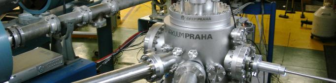
4 Tandem accelerators facility PIXE/RBS
5 RBI Laboratory for Ion Beam Interactions Interactions of accelerated ion beams with materials Fundamental processes - Stopping of ions in solids and related effects - Inner shell ionization and X-ray emission processes - Ion scattering - Nuclear reactions Developments - data acquisition and control software and hardware (SPECTOR) - accelerator computer control (including remote operation) - design and constructions of end-stations Applications - Material characterisation (analysis) by IBA - Material modifications Ruđer Bošković Institute, Zagreb, Croatia
6 Ion Beam Analysis (IBA) NRA Nuclear reaction products Charge pulse IBIC PIGE γ rays Recoil nuclei ERDA Ion beam Transmitted particles STIM PIXE X-rays Forward scattered particles RBS SEI Backscattered particles Secondary electrons Light IL TARGET Concentrations and/or depth profiles: Elements - x-rays (PIXE) - backscattered particles (RBS) - recoiled ions (ERDA) Isotopes - gamma-rays (PIGE) - nuclear reactions (NRA) Density variations transmitted ions (STIM) Charge transport ion beam induced current (IBIC) Chemical bonds light (IL), x-rays (high resolution) Crystal structure channeling RBS, STIM, PIXE Morphology secondary electrons (SEI) Ruđer Bošković Institute, Zagreb, Croatia

")


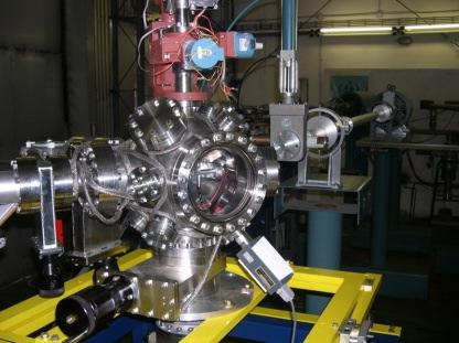
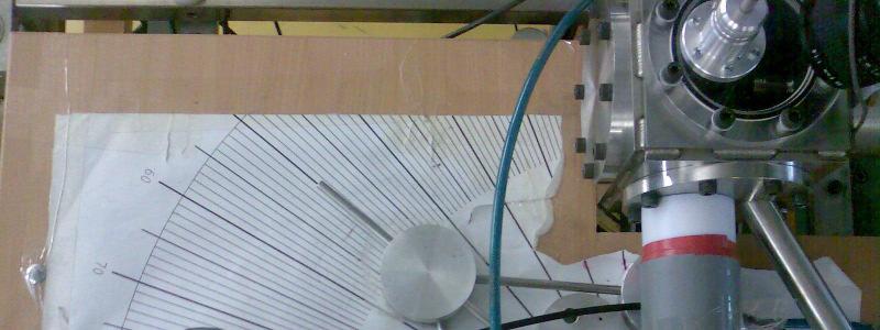
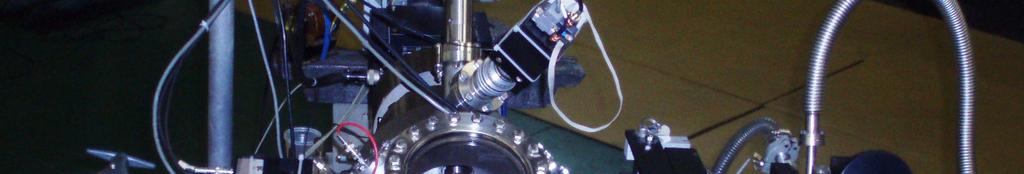
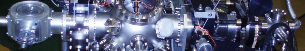
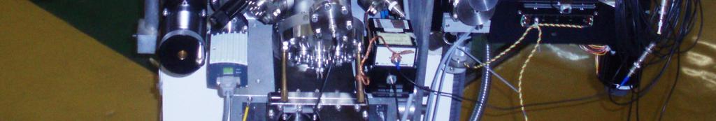
7 Ion (nuclear) microprobe PIXE, RBS, ERDA, IBIC, STIM Coincidence scattering Ion hit detection (SE & IL) Focusing system quintuplet ME/q2 < 25 Equal demagnification 0.5 µm smallest beam size Ruđer Bošković Institute, Zagreb, Croatia
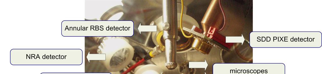
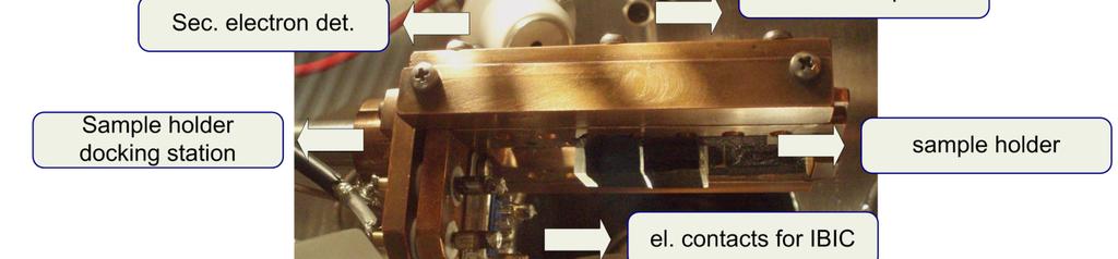
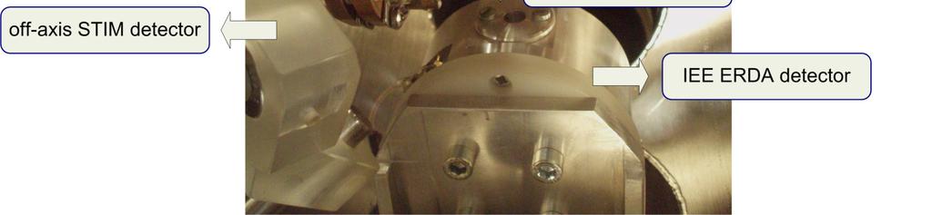
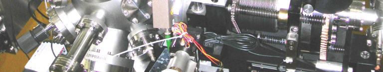
8 microprobe - inside the vacuum chamber
9 Microprobe ion beam object slits quadrupole focusing lens system sample Y scan generator X x-ray detector amplifier Y X X-ray energy spectrum Fe Ca S Pb elemental maps Ruđer Bošković Institute, Zagreb, Croatia


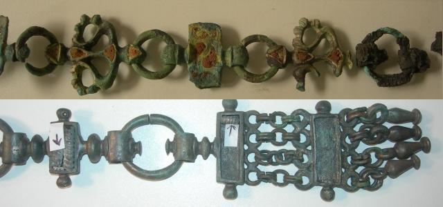
10 Example: Analysis of metal threads of a 17th century church textile with PIXE at the microprobe The left lamina contains more copper, whereas the two right ones are silver laminas. Ruđer Bošković Institute, Zagreb, Croatia
 Portal sample P1 Ba depth distribution")
11 Example: Analysis of stone samples at the microprobe Conservation of St. Marko church portal total PIXE yield total yield Ba Fe S depth (micron) Portal sample P1 Ba depth distribution (S1) Florentine method of cleaning and consolidation by soaking the stone in ammonium carbonate and barium hydroxide was used. Barium and sulfur concentration level variations with depth have been determined in samples taken from the portal after the treatment depth (micron) Sandstone sample treated in laboratory In addition, Ba depth profiles in sandstone treated by three different ways were measured. Ruđer Bošković Institute, Zagreb, Croatia


, while")

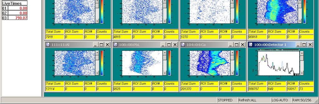
12 Analysis of pigment layers at the microprobe 2D element distribution of the pigment cross section sample taken from the red area of the painting. The light red layer exhibits high Hg and S concentrations (HgS cinnabar), while the dark red layer beneath shows presence of Pb, Al, Ca, but without Hg (either minium, or carmine).


13 Examples: Analysis of pigment layers at the microprobe Microsamples are the same as ones needed for optical mictroscopy techniques. Method is non-destructive and samples may be re-used. We analyse between 50 to 100 such samples per year. Ruđer Bošković Institute, Zagreb, Croatia

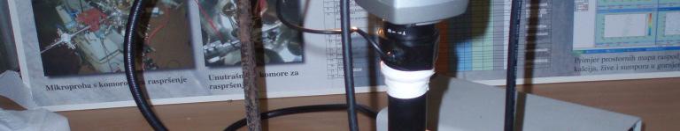
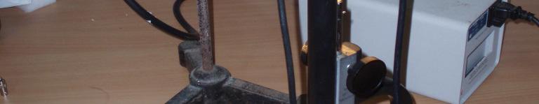
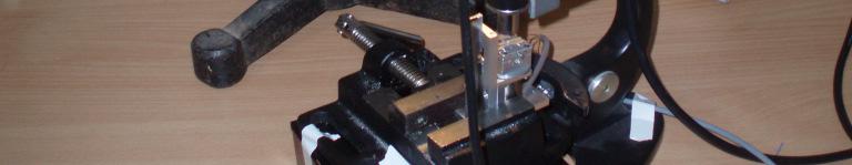
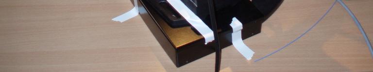
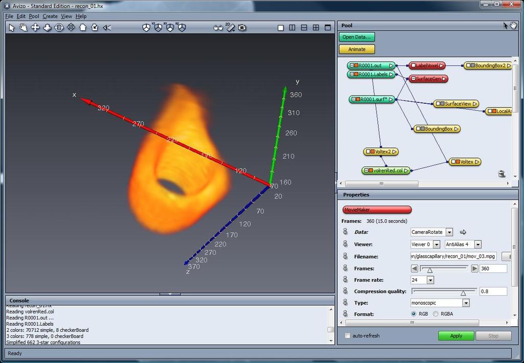
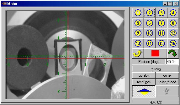
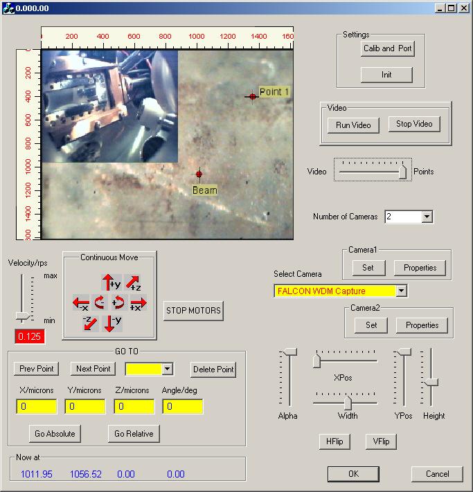
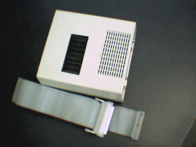
14 Example 2: STIM tomography module (1) Sample positioning on the axis of stepper motor using microscope (3) Measurement (2) Loading into the chamber (4) Reconstruction and visualization


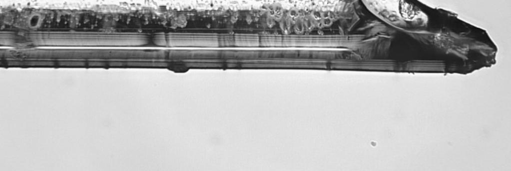
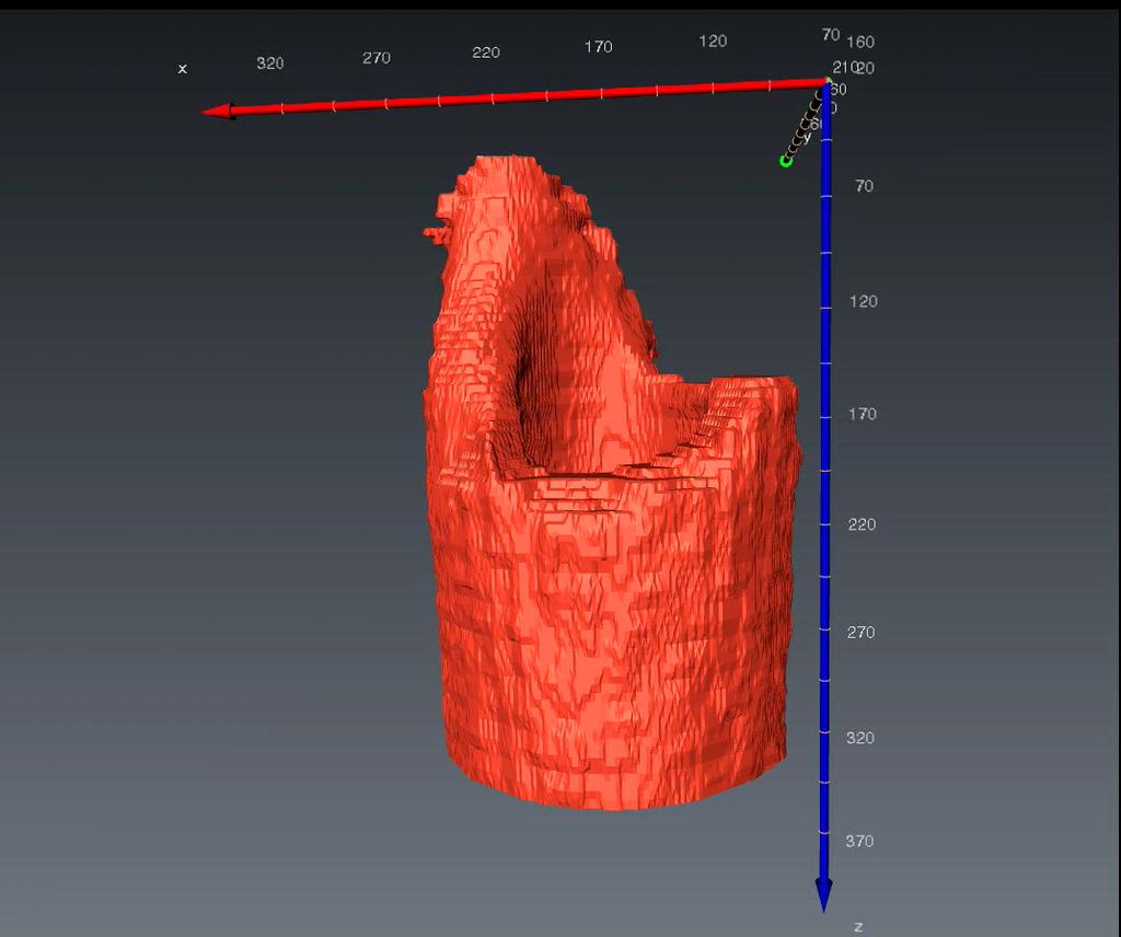
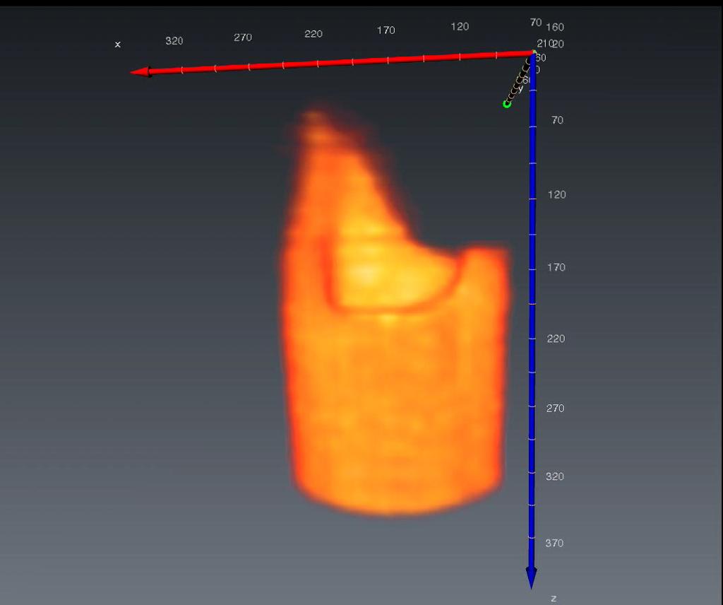
15 Object rendering using AMIRA software

 Lateral IBIC Scan area (n)")


16 Single ions tool for charge injection and probe for micro - imaging of charge transport properties Frontal IBIC Scan area Bias voltage O 12 C 7 Li alphas (Junction) Lateral IBIC Scan area (n) IBIC pulse (p) Grain boundaries Charge traps 1. Imaging of electronically active defects in Si, CVD diamond, CdTe, GaAs,... protons 2. Creation /structuring of defects change of charge transport properties: the example of Si pin diodes
 and is usually from 1 to 8 mm Faraday cup PIGE Ruđer Bošković Institute, Zagreb,")
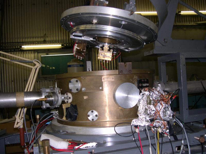
17 PIXE/RBS scattering chamber PIXE, RBS, PIGE detectors simultaneous measurements SPECTOR data acquisition + computer controled sample changer PIXE RBS Beam shape is defined by the collimators (beam defining slits) and is usually from 1 to 8 mm Faraday cup PIGE Ruđer Bošković Institute, Zagreb, Croatia


18 Example: Analysis of samples taken from the surface of the stone material of the Roman remains in Split Samples analysed in vacuum at the IBA end-station Ruđer Bošković Institute, Zagreb, Croatia


 TOF (time of")


")
19 Time of flight ERDA spectrometer x,y,z,θ θ manipulator T2 M2 TOF = T 1 T 2 = l 2 KE0 4M 1M 2 cos 2 φ K= 2 ( M1 + M 2 ) o o o o o L=523.4 mm T1 Vacuum chamber 2 Si E detector ERDA (Elastic Recoil Detection Analysis) TOF (time of flight) system for separation of scattered from recoiled ions (v,e) Depth resolution down to 5 nm Ion beams Cl, I (energies MeV) Resolution of isotopes up to M = 28 11B 10B 7Li 1H 19F 16O 4He
20 TOF-ERDA Spectrum example Coincidence spectrum (E and TOF coincidence) obtained by bombarding Si/20Å Ta/100 Å AlNO/20Å Au Round robin sample by 35 MeV 35 Cl Sample tilt angle θ in = Cl scattered from Si 56 Fe 35 Cl scattered from Fe 28 Si 14 N 12 C 16 O 27 Al 35 Cl scattered from Ta 35 Cl scattered from Au


21 High Resolution PIXE Simple high resolution wavelength dispersive crystal spectrometer with position sensitive proportional counter. Data acquisition by SPECTOR. vacuum chamber flat crystal PSPC detector He baloon
22 Example: High Resolution PIXE measured spectra of selected Ti and V compounds to investigate chemical effects on Kβ X-ray spectra


23 PIXE analysis in air For large/bulky objects and samples that cannot be exposed to vacuum Ruđer Bošković Institute, Zagreb, Croatia


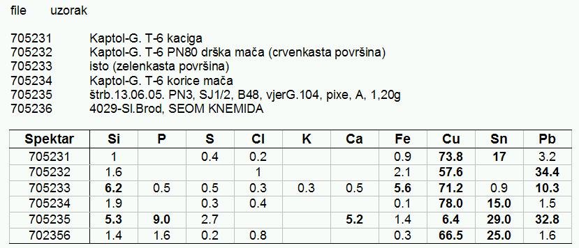
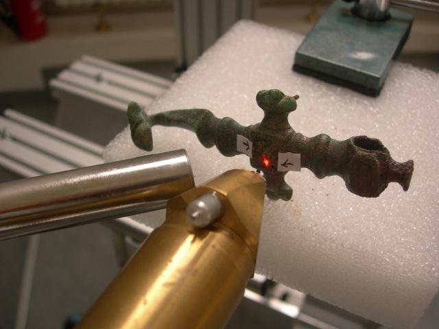
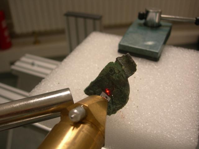
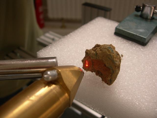
24 Examples: Analysis of alloys Analysis in air Ruđer Bošković Institute, Zagreb, Croatia
 AlK 1.66 SiK 1.05 P K 0.31 S K 0.03 ClK 3.20 K K 0.")

25 Example: Analysis of pigments at the in-air end-station Fiorentino / white Fiorentino / blue C(%) AlK 1.66 SiK 1.05 P K 0.31 S K 0.03 ClK 3.20 K K 0.91 CaK 2.34 TiK 0.00 FeK 1.22 CuK 0.06 BaLA 0.00 PbLA C(%) AlK 1.31 SiK 1.29 P K 0.36 S K 2.67 ClK 0.75 K K 0.35 CaK 1.17 TiK 0.08 FeK 1.05 CuK BaLA 5.48 PbLA 18.81


26 New dual beam chamber Beam from Tandetron Beam from EN Tandem (still under construction) simultaneous modification + analysis Load lock xyz stage

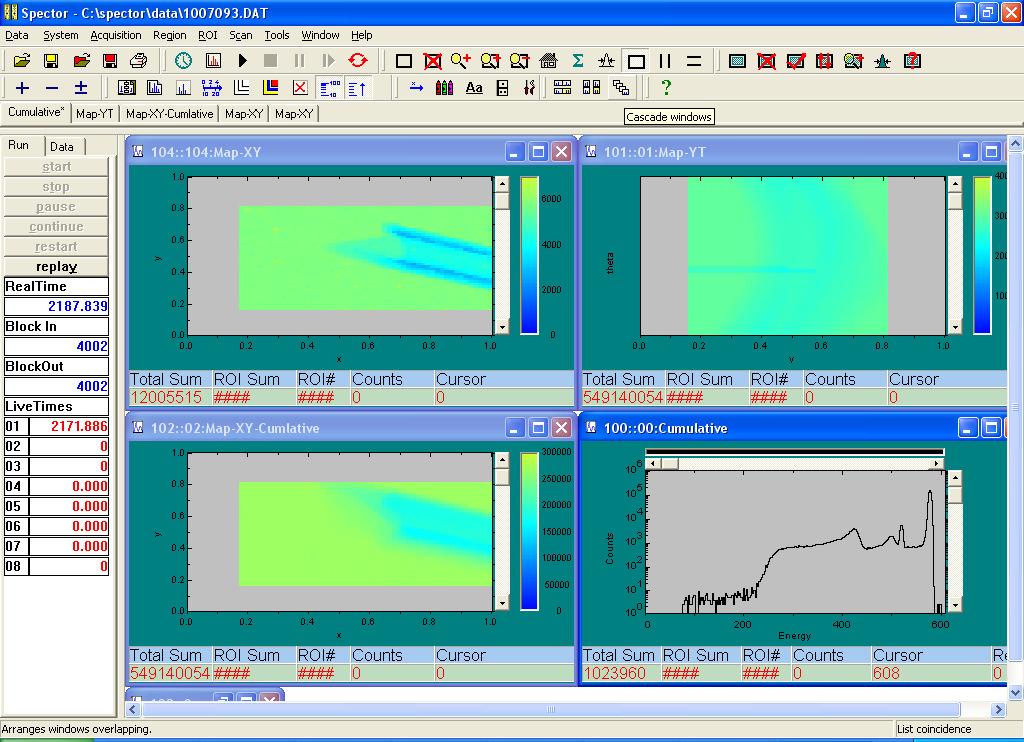
27 SPECTOR home made Data acquisition /target positioning & beam scanning software
 - Reads beam optics parameters from previous experiments - Calculates changes of parameters")
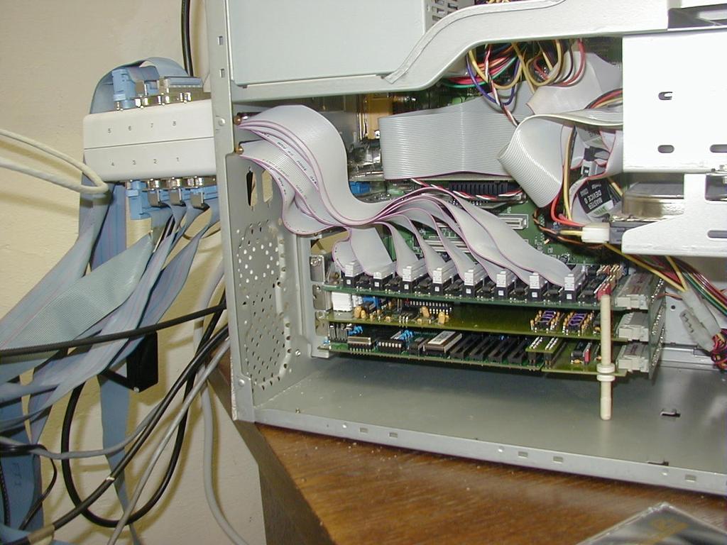
28 Computer control ACCEL6 for EN Tandem Van de Graaff and ACCEL1 for Tandetron accelerator - 16 bit AD/DA modules (controls for ion sources, accelerator and beam optics system) - 8 digital inputs, 8 digital outputs - Controls are based on TESTPOINT Capabilities: - Remote control (from remote computers) - Reads beam optics parameters from previous experiments - Calculates changes of parameters for change of energy and/or ion - Security interlock system
29 Remote experiments on accelerator Performance of experiments by expert users Access to accelerator control console Sample positioning and beam scanning Data acquisition Education and training for users unfamiliar with technique Viewer to accelerator control console Beam controls on/off Access to sample changer and data acquisition with limited access On-line tutorial Examples - IAEA trainees (in collaboration with the IAEA Seibersdorf Laboratory) - Laboratories starting PIXE - Remote Universities without experimental facilities Ruđer Bošković Institute, Zagreb, Croatia
30 Thank you
Accelerator laboratory of the Ruđer Bošković Institute
 Accelerator laboratory of the Ruđer Bošković Institute Laboratory for ion beam interactions Division of experimental physics, Zagreb, Croatia Scientific: Milko Jakšić, Tonči Tadić, Iva Bogdanović Radović,
Accelerator facilities - Ruđer Bošković Institute, Zagreb, Croatia
 Accelerator facilities - Ruđer Bošković Institute, Zagreb, Croatia Milko Jaksic, Neven Soic Our capabilities (accelerator, sources, beamlines,...) Research activities (projects, users, int. collaborations,...)
Ion-beam techniques. Ion beam. Electrostatic Accelerators. Van de Graaff accelerator Pelletron Tandem Van de Graaff
 Ion-beam techniques RBS Target nucleus Ion beam STIM RBS: Rutherford backscattering ERD: Elastic recoil detection PIXE: Particle induced x-ray emission PIGE: Particle induced gamma emission NRA: Nuclear
INTRODUCTION to ION BEAM TECHNIQUES. Marko Karlušić Ruđer Bošković Institute, Zagreb
 INTRODUCTION to ION BEAM TECHNIQUES Marko Karlušić Ruđer Bošković Institute, Zagreb INTRODUCTION to ION BEAM ANALYSIS (IBA) Electrostatic accelerators Ion beam analysis with examples RBS ERDA PIXE Nuclear
Milko Jakšić Laboratory for ion beam interactions Division for experimental physics Ruđer Bošković Institute
 Milko Jakšić Laboratory for ion beam interactions Division for experimental physics Ruđer Bošković Institute. 2. 3. Facilities IBIC Application examples Ruđer Bošković Institute 973 956 987 29 962 25 Ruđer
Massachusetts Institute of Technology. Dr. Nilanjan Chatterjee
 Massachusetts Institute of Technology Dr. Nilanjan Chatterjee Electron Probe Micro-Analysis (EPMA) Imaging and micrometer-scale chemical compositional analysis of solids Signals produced in The Electron
Applications of Nuclear Analytical Techniques in Geoscience
 Applications of Nuclear Analytical Techniques in Geoscience J. Aspiazu a,1, J. López a, J. Ramírez a, M. E. Montero b, P. Villaseñor a a Intituto Nacional de Investigaciones Nucleares (ININ), Carretera
Atomic and Nuclear Analytical Methods
 H.R. Verma Atomic and Nuclear Analytical Methods XRF, Mössbauer, XPS, NAA and Ion-Beam Spectroscopic Techniques With 128 Figures and 24 Tables Springer Contents 1 X-ray Fluorescence (XRF) and Particle-Induced
Institute of Physics & Power Engineering Current Status of the Problem of Cross Section Data for Ion Beam Analysis
 Institute of Physics & Power Engineering Current Status of the Problem of Cross Section Data for Ion Beam Analysis A.F. Gurbich IBA Methods Acronym PIXE PIGE RBS NRA NRP or r-nra ERDA or FRS Particle-Induced
IAP NAN of Ukraine activity in. Possible areas of cooperation in. Roman Kholodov, PICS Meeting, Basivka, Ukraine,
 1 IAP NAN of Ukraine activity in field of High Energy Physics. Possible areas of cooperation in frame of PICS and beyond. Institute of Applied Physics National Academy of Sciences of Ukraine 2 The Institute
Out of vacuum characterisation of surfaces: a possible approach?
 Out of vacuum characterisation of surfaces: a possible approach? Lorenzo Giuntini Dipartimento di Fisica dell Università and Sezione INFN Firenze, Italy XVIII Congresso Nazionale sulla Scienza e Tecnologia
Electron Microprobe Analysis 1 Nilanjan Chatterjee, Ph.D. Principal Research Scientist
 12.141 Electron Microprobe Analysis 1 Nilanjan Chatterjee, Ph.D. Principal Research Scientist Massachusetts Institute of Technology Electron Microprobe Facility Department of Earth, Atmospheric and Planetary
Electron Microprobe Analysis 1 Nilanjan Chatterjee, Ph.D. Principal Research Scientist
 12.141 Electron Microprobe Analysis 1 Nilanjan Chatterjee, Ph.D. Principal Research Scientist Massachusetts Institute of Technology Electron Microprobe Facility Department of Earth, Atmospheric and Planetary
Application of Ion Beam Analysis Techniques to the Study of Cultural Heritage Objects
 Application of Ion Beam Analysis Techniques to the Study of Cultural Heritage Objects Technological and Nuclear Institute - Unit of Physics and Accelerators 1 What is IBA? The IBA (Ion Beam Analysis) techniques
The scanning microbeam PIXE analysis facility at NIRS
 Nuclear Instruments and Methods in Physics Research B 210 (2003) 42 47 www.elsevier.com/locate/nimb The scanning microbeam PIXE analysis facility at NIRS Hitoshi Imaseki a, *, Masae Yukawa a, Frank Watt
Electron probe microanalysis - Electron microprobe analysis EPMA (EMPA) What s EPMA all about? What can you learn?
 What s EPMA all about? What can you learn?") Electron probe microanalysis - Electron microprobe analysis EPMA (EMPA) What s EPMA all about? What can you learn? EPMA - what is it? Precise and accurate quantitative chemical analyses of micron-size
Electron probe microanalysis - Electron microprobe analysis EPMA (EMPA) What s EPMA all about? What can you learn? EPMA - what is it? Precise and accurate quantitative chemical analyses of micron-size
Lecture 22 Ion Beam Techniques
 Lecture 22 Ion Beam Techniques Schroder: Chapter 11.3 1/44 Announcements Homework 6/6: Will be online on later today. Due Wednesday June 6th at 10:00am. I will return it at the final exam (14 th June).
Joint ICTP-IAEA Workshop on Nuclear Data for Analytical Applications October 2013
 2495-03 Joint ICTP-IAEA Workshop on Nuclear Data for Analytical Applications 21-25 October 2013 Ion Beam Analysis Techniques for non-destructive Profiling Studies M. Kokkoris Department of Physics National
W. Udo Schröder Departments of Chemistry & of Physics and Astronomy
 W. Udo Schröder Departments of Chemistry & of Physics and Astronomy ANSEL Faculty Instructors ACS NuSci Acad Infrastructure 2 Prof. Frank Wolfs Prof. Udo Schrőder Research: Large Underground Xenon (LUX)
Rutherford Backscattering Spectrometry
 Rutherford Backscattering Spectrometry EMSE-515 Fall 2005 F. Ernst 1 Bohr s Model of an Atom existence of central core established by single collision, large-angle scattering of alpha particles ( 4 He
accelerating opportunities
 accelerating opportunities 9 MV Pelletron FN Tandem Accelerator for basic and applied research The 9MV Tandem Accelerator has been built by the High Voltage Engineering Corporation (HVEC) in 1973 and it
IN THE NAME OF ALLAH, THE MOST MERCIFUL AND COMPASSIONATE
 IN THE NAME OF ALLAH, THE MOST MERCIFUL AND COMPASSIONATE Ion Beam Analysis of Diamond Thin Films Sobia Allah Rakha Experimental Physics Labs 04-03-2010 Outline Diamond Nanostructures Deposition of Diamond
EDS User School. Principles of Electron Beam Microanalysis
 EDS User School Principles of Electron Beam Microanalysis Outline 1.) Beam-specimen interactions 2.) EDS spectra: Origin of Bremsstrahlung and characteristic peaks 3.) Moseley s law 4.) Characteristic
COMPARATIVE STUDY OF PIGE, PIXE AND NAA ANALYTICAL TECHNIQUES FOR THE DETERMINATION OF MINOR ELEMENTS IN STEELS
 COMPARATIVE STUDY OF PIGE, PIXE AND NAA ANALYTICAL TECHNIQUES FOR THE DETERMINATION OF MINOR ELEMENTS IN STEELS ANTOANETA ENE 1, I. V. POPESCU 2, T. BÃDICÃ 3, C. BEªLIU 4 1 Department of Physics, Faculty
Surface analysis techniques
 Experimental methods in physics Surface analysis techniques 3. Ion probes Elemental and molecular analysis Jean-Marc Bonard Academic year 10-11 3. Elemental and molecular analysis 3.1.!Secondary ion mass
Applications for PIXE and other Ion Beam Analysis (IBA)
") Applications for PIXE and other Ion Beam Analysis (IBA) PIXE-PAN Summer Science Program University of Notre Dame June, 2007 Larry Lamm, Research Professor Many, many facilities worldwide Some IBA Techniques
Applications for PIXE and other Ion Beam Analysis (IBA) PIXE-PAN Summer Science Program University of Notre Dame June, 2007 Larry Lamm, Research Professor Many, many facilities worldwide Some IBA Techniques
ionbeam.kigam.re.kr Beam Application Ion Group
 ionbeam.kigam.re.kr Ion Application Group http://ionbeam.kigam.re.kr Project Schedule Major subjects 88 89 9 91 92 93 94 95 96 97 98 99 1 2 3 4 5 6 7 8 9 1 11 Ion Analysis Setup of 1.7MV Tandem VDG Accelerator
PHI 5000 Versaprobe-II Focus X-ray Photo-electron Spectroscopy
 PHI 5000 Versaprobe-II Focus X-ray Photo-electron Spectroscopy The very basic theory of XPS XPS theroy Surface Analysis Ultra High Vacuum (UHV) XPS Theory XPS = X-ray Photo-electron Spectroscopy X-ray
EE 5344 Introduction to MEMS CHAPTER 5 Radiation Sensors
 EE 5344 Introduction to MEMS CHAPTER 5 Radiation Sensors 5. Radiation Microsensors Radiation µ-sensors convert incident radiant signals into standard electrical out put signals. Radiant Signals Classification
RITU and the GREAT Spectrometer
 RITU and the GREAT Spectrometer Cath Scholey Department of Physics University of Jyväskylä 19 th March 2006 3rd TASCA Detector Group Meeting, GSI Darmstadt C. Scholey (JYFL, Finland) RITU and the GREAT
Particle-Induced X-Ray Emission Spectrometry (PIXE)
") Particle-Induced X-Ray Emission Spectrometry (PIXE) Edited by SVEN A. E. JOHANSSON Department of Nuclear Physics Lund Institute of Technology Lund, Sweden JOHN L. CAMPBELL Department of Physics University
Particle-Induced X-Ray Emission Spectrometry (PIXE) Edited by SVEN A. E. JOHANSSON Department of Nuclear Physics Lund Institute of Technology Lund, Sweden JOHN L. CAMPBELL Department of Physics University
Progress of the interaction between e - and molecule in Fudan University
 Progress of the interaction between e - and molecule in Fudan University B. Wei, Z. Chen, X. Wang, R. Hutton, Y. Zou Fudan University, Shanghai The 2nd Research Coordination Meeting (RCM) of the CRP, 23-25
EBSS: Hands-on Activity #8. Operation of an electromagnetic isotope separator (EMIS) Dan Stracener Jon Batchelder
 Dan Stracener Jon Batchelder") EBSS: Hands-on Activity #8 Operation of an electromagnetic isotope separator (EMIS) Dan Stracener Jon Batchelder Schematic of RIB Production at the HRIBF ISOL: Isotope Separation On-Line 2 Managed by UT-Battelle
EBSS: Hands-on Activity #8 Operation of an electromagnetic isotope separator (EMIS) Dan Stracener Jon Batchelder Schematic of RIB Production at the HRIBF ISOL: Isotope Separation On-Line 2 Managed by UT-Battelle
Optical and THz investigations of mid-ir materials exposed
 Optical and THz investigations of mid-ir materials exposed to alpha particle irradiation Dan Sporea 1*, Laura Mihai 1, Adelina Sporea 1, Ion Vâţã 2 1 National Institute for Laser, Plasma and Radiation
Refurbishment of the Electrostatic Accelerators of ithemba LABS
 Refurbishment of the Electrostatic Accelerators of ithemba LABS SNEAP 2012 INFN Legnaro, Italy, 1-5 October J.L. Conradie, G. Badenhorst, F. Balzun, A. Crombie, G. De Villiers, H. Delsink, C. Doyle, C.
Beam diagnostics: Alignment of the beam to prevent for activation. Accelerator physics: using these sensitive particle detectors.
 Beam Loss Monitors When energetic beam particles penetrates matter, secondary particles are emitted: this can be e, γ, protons, neutrons, excited nuclei, fragmented nuclei... Spontaneous radiation and
2.3 Particle Induced X-Ray Emission PIXE
 2.3 Particle Induced X-Ray Emission PIXE The previous section concentrated on X-ray fluorescence. This section discusses a different X-ray production technique that can lead to the development of 2-D/3-D
A new protocol to evaluate the charge collection efficiency degradation in semiconductor devices induced by MeV ions
 Session 12: Modification and Damage: Contribute lecture O-35 A new protocol to evaluate the charge collection efficiency degradation in semiconductor devices induced by MeV ions Ettore Vittone Physics
MT Electron microscopy Scanning electron microscopy and electron probe microanalysis
 MT-0.6026 Electron microscopy Scanning electron microscopy and electron probe microanalysis Eero Haimi Research Manager Outline 1. Introduction Basics of scanning electron microscopy (SEM) and electron
Overview of X-Ray Fluorescence Analysis
 Overview of X-Ray Fluorescence Analysis AMPTEK, INC., Bedford, MA 01730 Ph: +1 781 275 2242 Fax: +1 781 275 3470 sales@amptek.com 1 What is X-Ray Fluorescence (XRF)? A physical process: Emission of characteristic
High voltage electron microscopy facility. Jannus-Saclay facility (GIS with Jannus-Orsay) SRMA
 SRMA") SRMA Jannus-Saclay facility (GIS with Jannus-Orsay) High voltage electron microscopy facility Lucile Beck - JANNUS laboratory - SRMP Jean Henry SRMA CEA Saclay CEA 10 AVRIL 2012 PAGE 1 Programs and objectives
SRMA Jannus-Saclay facility (GIS with Jannus-Orsay) High voltage electron microscopy facility Lucile Beck - JANNUS laboratory - SRMP Jean Henry SRMA CEA Saclay CEA 10 AVRIL 2012 PAGE 1 Programs and objectives
EEE4106Z Radiation Interactions & Detection
 EEE4106Z Radiation Interactions & Detection 2. Radiation Detection Dr. Steve Peterson 5.14 RW James Department of Physics University of Cape Town steve.peterson@uct.ac.za May 06, 2015 EEE4106Z :: Radiation
II. 5. Study for NaI(Tl) and Scintillation Fiber with 80 MeV Proton Beam Toward ESPRI Experiment at NIRS-HIMAC, RIKEN-RIBF
 and Scintillation Fiber with 80 MeV Proton Beam Toward ESPRI Experiment at NIRS-HIMAC, RIKEN-RIBF") CYRIC Annual Report 2005 II. 5. Study for NaI(Tl) and Scintillation Fiber with 80 MeV Proton Beam Toward ESPRI Experiment at NIRS-HIMAC, RIKEN-RIBF Zenihiro J. 1, Matsuda Y. 2, Sakaguchi H. 3, Takeda H.
CYRIC Annual Report 2005 II. 5. Study for NaI(Tl) and Scintillation Fiber with 80 MeV Proton Beam Toward ESPRI Experiment at NIRS-HIMAC, RIKEN-RIBF Zenihiro J. 1, Matsuda Y. 2, Sakaguchi H. 3, Takeda H.
Chapter 9. Electron mean free path Microscopy principles of SEM, TEM, LEEM
 Chapter 9 Electron mean free path Microscopy principles of SEM, TEM, LEEM 9.1 Electron Mean Free Path 9. Scanning Electron Microscopy (SEM) -SEM design; Secondary electron imaging; Backscattered electron
Characterisation of SiC by IBIC and other IBA techniques
 Nuclear Instruments and Methods in Physics Research B 188 (2002) 130 134 www.elsevier.com/locate/nimb Characterisation of SiC by IBIC and other IBA techniques M. Jaksic a, *, Z. Bosnjak a, D. Gracin a,
CBE Science of Engineering Materials. Scanning Electron Microscopy (SEM)
") CBE 30361 Science of Engineering Materials Scanning Electron Microscopy (SEM) Scale of Structure Organization Units: micrometer = 10-6 m = 1µm nanometer= 10-9 m = 1nm Angstrom = 10-10 m = 1Å A hair is
CBE 30361 Science of Engineering Materials Scanning Electron Microscopy (SEM) Scale of Structure Organization Units: micrometer = 10-6 m = 1µm nanometer= 10-9 m = 1nm Angstrom = 10-10 m = 1Å A hair is
IAEA-TECDOC Instrumentation for PIXE and RBS
 IAEA-TECDOC-1190 Instrumentation for PIXE and RBS December 2000 The originating Section of this publication in the IAEA was: Physics Section International Atomic Energy Agency Wagramer Strasse 5 P.O. Box
An ultra-thin diamond membrane as a transmission particle detector and vacuum window for external microbeams
 An ultra-thin diamond membrane as a transmission particle detector and vacuum window for external microbeams V. Grilj, N. Skukan, M. Pomorski, W. Kada, N. Iwamoto, T. Kamiya, T. Ohshima, and M. Jakšić
A Comparison between Channel Selections in Heavy Ion Reactions
 Brazilian Journal of Physics, vol. 39, no. 1, March, 2009 55 A Comparison between Channel Selections in Heavy Ion Reactions S. Mohammadi Physics Department, Payame Noor University, Mashad 91735, IRAN (Received
The accelerators at LNS - INFN and diagnostics aspects of the radioactive beams
 The accelerators at LNS - INFN and diagnostics aspects of the radioactive beams L. Cosentino, P. Finocchiaro, A. Pappalardo LNS INFN Catania J. Harasimowicz Univ. of Liverpool Cockroft Institute LNS INFN
MeV Particles, Huge Impact, Soft Desorption.
 MeV Particles, Huge Impact, Soft Desorption. S. Della-Negra Institut de Physique Nucléaire d Orsay, UMR 8608 CNRS- Univ. Paris-Sud, F-91406Orsay Cedex (dellaneg@ipno.in2p3.fr) Since the first PDMS experiment
Warsaw University of Technology, Faculty of Physics. Laboratory of Nuclear Physics & Technology. Compton effect
 Warsaw University of Technology, Faculty of Physics Laboratory of Nuclear Physics & Technology Compton effect Author: MSc. Eng. Dariusz Aksamit, Dariusz.Aksamit@pw.edu.pl, Faculty of Physics on the basis
Imaging Methods: Scanning Force Microscopy (SFM / AFM)
") Imaging Methods: Scanning Force Microscopy (SFM / AFM) The atomic force microscope (AFM) probes the surface of a sample with a sharp tip, a couple of microns long and often less than 100 Å in diameter.
Imaging Methods: Scanning Force Microscopy (SFM / AFM) The atomic force microscope (AFM) probes the surface of a sample with a sharp tip, a couple of microns long and often less than 100 Å in diameter.
Secondary Ion Mass Spectrometry (SIMS)
") CHEM53200: Lecture 10 Secondary Ion Mass Spectrometry (SIMS) Major reference: Surface Analysis Edited by J. C. Vickerman (1997). 1 Primary particles may be: Secondary particles can be e s, neutral species
CHEM53200: Lecture 10 Secondary Ion Mass Spectrometry (SIMS) Major reference: Surface Analysis Edited by J. C. Vickerman (1997). 1 Primary particles may be: Secondary particles can be e s, neutral species
MS482 Materials Characterization ( 재료분석 ) Lecture Note 5: RBS
 Lecture Note 5: RBS") 2016 Fall Semester MS482 Materials Characterization ( 재료분석 ) Lecture Note 5: RBS Byungha Shin Dept. of MSE, KAIST 1 Course Information Syllabus 1. Overview of various characterization techniques (1 lecture)
2016 Fall Semester MS482 Materials Characterization ( 재료분석 ) Lecture Note 5: RBS Byungha Shin Dept. of MSE, KAIST 1 Course Information Syllabus 1. Overview of various characterization techniques (1 lecture)
Spectroscopy on Mars!
 Spectroscopy on Mars! Pathfinder Spirit and Opportunity Real World Friday H2A The Mars Pathfinder: Geological Elemental Analysis On December 4th, 1996, the Mars Pathfinder was launched from earth to begin
(10%) (c) What other peaks can appear in the pulse-height spectrum if the detector were not small? Give a sketch and explain briefly.
 (c) What other peaks can appear in the pulse-height spectrum if the detector were not small? Give a sketch and explain briefly.") Sample questions for Quiz 3, 22.101 (Fall 2006) Following questions were taken from quizzes given in previous years by S. Yip. They are meant to give you an idea of the kind of questions (what was expected
Sample questions for Quiz 3, 22.101 (Fall 2006) Following questions were taken from quizzes given in previous years by S. Yip. They are meant to give you an idea of the kind of questions (what was expected
1.5. The Tools of the Trade!
 1.5. The Tools of the Trade! Two things are required for material analysis: excitation mechanism for originating characteristic signature (radiation) radiation detection and identification system (spectroscopy)
Screens for low current beams
 Screens for low current beams P.Finocchiaro, L.Cosentino, A.Pappalardo INFN - Laboratori Nazionali del Sud Catania, Italy Status Workshop on 29-Apr-2010 on Scintillating - Preliminary Screen Applications
Benchmark measurements of non-rutherford proton elastic scattering cross section for boron
 Published in: Nuclear Instr. Meth. B (01) 0 Benchmark measurements of non-rutherford proton elastic scattering cross section for boron M. Chiari (a), M. Bianconi (b), I. Bogdanović Radović (c), M. Mayer
PROGRESS OF NUCLEAR DATA MEASUREMENT IN CHINA
 PROGRESS OF NUCLEAR DATA MEASUREMENT IN CHINA (2011-2012 ) GE Zhigang RUAN Xichao China Nuclear Data Key Laboratory China Nuclear Data Center(CNDC) China Committee of Nuclear Data(CCND) China Institute
Secondary Ion Mass Spectrometry (SIMS) Thomas Sky
 Thomas Sky") 1 Secondary Ion Mass Spectrometry (SIMS) Thomas Sky Depth (µm) 2 Characterization of solar cells 0,0 1E16 1E17 1E18 1E19 1E20 0,2 0,4 0,6 0,8 1,0 1,2 P Concentration (cm -3 ) Characterization Optimization
1 Secondary Ion Mass Spectrometry (SIMS) Thomas Sky Depth (µm) 2 Characterization of solar cells 0,0 1E16 1E17 1E18 1E19 1E20 0,2 0,4 0,6 0,8 1,0 1,2 P Concentration (cm -3 ) Characterization Optimization
11/10/2014. Chapter 1: Introduction to Medical Imaging. Projection (Transmission) vs. Emission Imaging. Emission Imaging
 vs. Emission Imaging. Emission Imaging") Chapter 1: Introduction to Medical Imaging Overview of Modalities Properties of an Image: Limitations on Information Content Contrast (both object & image): Brightness difference Sharpness (blur): Smallest
Chapter 1: Introduction to Medical Imaging Overview of Modalities Properties of an Image: Limitations on Information Content Contrast (both object & image): Brightness difference Sharpness (blur): Smallest
Materials Analysis Using Fast Ions
 A. Denker, W. Bohne, J. Rauschenberg,J. Röhrich, E. Strub Ionenstrahllabor Hahn-Meitner-Institut Berlin Materials Analysis Using Fast Ions Introduction: Energy Loss PIXE Proton Induced X-ray Emission RBS
Práctica de laboratorio número 6: Non-Rutherford scattering near the MeV 12 C(p,p) 12 C resonance
 12 C resonance") Práctica de laboratorio número 6: Non-Rutherford scattering near the 1.734 MeV 12 C(p,p) 12 C resonance 1) Scope In this experiment, the yield of protons backscattered from a thin gold foil deposited over
Práctica de laboratorio número 6: Non-Rutherford scattering near the 1.734 MeV 12 C(p,p) 12 C resonance 1) Scope In this experiment, the yield of protons backscattered from a thin gold foil deposited over
Development of a table top TW laser accelerator for medical imaging isotope production
 Development of a table top TW laser accelerator for medical imaging isotope production R U I Z, A L E X A N D R O 1 ; L E R A, R O B E R T O 1 ; T O R R E S - P E I R Ó, S A LVA D O R 1 ; B E L L I D O,
Semiconductor-Detectors
 Semiconductor-Detectors 1 Motivation ~ 195: Discovery that pn-- junctions can be used to detect particles. Semiconductor detectors used for energy measurements ( Germanium) Since ~ 3 years: Semiconductor
Materials Science. Hand axes like this one found in the United Arab Emirates indicate humans left Africa 125,000 years ago.
 Summary Materials Science Ion Beam Analysis (IBA) @ 3 MV Tandetron TM Accelerator Rutherford Backscattering Spectrometry (RBS) Elastic Recoil Detection (ERD) Nuclear Reaction Analysis (NRA) Ion implant/channeling
High-Resolution Gamma-Ray and Neutron Detectors For Nuclear Spectroscopy
 High-Resolution Gamma-Ray and Neutron Detectors For Nuclear Spectroscopy Thomas Niedermayr, I. D. Hau, S. Terracol, T. Miyazaki, S. E. Labov and S. Friedrich Former colleagues: M. F. Cunningham, J. N.
Beam Diagnostics and Instrumentation JUAS, Archamps Peter Forck Gesellschaft für Schwerionenforschnung (GSI)
") Beam Diagnostics and Instrumentation JUAS, Archamps Peter Forck Gesellschaft für Schwerionenforschnung (GSI), 2003, A dedicated proton accelerator for 1p-physics at the future GSI Demands facilities for
Beam Diagnostics and Instrumentation JUAS, Archamps Peter Forck Gesellschaft für Schwerionenforschnung (GSI), 2003, A dedicated proton accelerator for 1p-physics at the future GSI Demands facilities for
Radionuclide Imaging MII Positron Emission Tomography (PET)
") Radionuclide Imaging MII 3073 Positron Emission Tomography (PET) Positron (β + ) emission Positron is an electron with positive charge. Positron-emitting radionuclides are most commonly produced in cyclotron
Radionuclide Imaging MII 3073 Positron Emission Tomography (PET) Positron (β + ) emission Positron is an electron with positive charge. Positron-emitting radionuclides are most commonly produced in cyclotron
MS482 Materials Characterization ( 재료분석 ) Lecture Note 4: XRF
 Lecture Note 4: XRF") 2016 Fall Semester MS482 Materials Characterization ( 재료분석 ) Lecture Note 4: XRF Byungha Shin Dept. of MSE, KAIST 1 Course Information Syllabus 1. Overview of various characterization techniques (1 lecture)
2016 Fall Semester MS482 Materials Characterization ( 재료분석 ) Lecture Note 4: XRF Byungha Shin Dept. of MSE, KAIST 1 Course Information Syllabus 1. Overview of various characterization techniques (1 lecture)
Auger Electron Spectroscopy (AES)
") 1. Introduction Auger Electron Spectroscopy (AES) Silvia Natividad, Gabriel Gonzalez and Arena Holguin Auger Electron Spectroscopy (Auger spectroscopy or AES) was developed in the late 1960's, deriving
1. Introduction Auger Electron Spectroscopy (AES) Silvia Natividad, Gabriel Gonzalez and Arena Holguin Auger Electron Spectroscopy (Auger spectroscopy or AES) was developed in the late 1960's, deriving
1.4 The Tools of the Trade!
 1.4 The Tools of the Trade! Two things are required for material analysis: excitation mechanism for originating characteristic signature (radiation) radiation detection and identification system (spectroscopy)
SYSTEM FOR PIXE TRACE ELEMENT ANALYSIS AT MTF. M. Beňo, J. Dobrovodský, D. Vaňa, S. Minárik
 SYSTEM FOR PIXE TRACE ELEMENT ANALYSIS AT MTF M. Beňo, J. Dobrovodský, D. Vaňa, S. Minárik Slovak University of Technology in Bratislava, Faculty of Materials Science and Technology in Trnava (MTF), Advanced
Calculations of Neutron Yield and Gamma Rays Intensity by GEANT4
 Armenian Journal of Physics, 2016, vol. 9, issue 4, pp. 315-323 Calculations of Neutron Yield and Gamma Rays Intensity by GEANT4 R. Avagyan, R. Avetisyan, V. Ivanyan*, I. Kerobyan A.I. Alikhanyan National
Measurements of liquid xenon s response to low-energy particle interactions
 Measurements of liquid xenon s response to low-energy particle interactions Payam Pakarha Supervised by: Prof. L. Baudis May 5, 2013 1 / 37 Outline introduction Direct Dark Matter searches XENON experiment
R&T PROTON DIRECT IONIZATION
 R&T PROTON DIRECT IONIZATION Assessment of the Direct Ionization Contribution to the Proton SEU Rate N. Sukhaseum,, J. Guillermin,, N. Chatry, F. Bezerra and R. Ecoffet TRAD, Tests & Radiations Introduction
Gaetano L Episcopo. Scanning Electron Microscopy Focus Ion Beam and. Pulsed Plasma Deposition
 Gaetano L Episcopo Scanning Electron Microscopy Focus Ion Beam and Pulsed Plasma Deposition Hystorical background Scientific discoveries 1897: J. Thomson discovers the electron. 1924: L. de Broglie propose
LAB REPORT ON XRF OF POTTERY SAMPLES By BIJOY KRISHNA HALDER Mohammad Arif Ishtiaque Shuvo Jie Hong
 LAB REPORT ON XRF OF POTTERY SAMPLES By BIJOY KRISHNA HALDER Mohammad Arif Ishtiaque Shuvo Jie Hong Introduction: X-ray fluorescence (XRF) spectrometer is an x-ray instrument used for routine, relatively
Research at the Tandem Accelerator
 SI0100093 Nuclear Energy in Central Europe '98 Terme Catez, September? to 10, 1998 Research at the Tandem Accelerator M.Budnar and P.Pelicon J.Stefan Institute, SI-1001 Ljubljana, Slovenia' ABSTRACT -
h p λ = mν Back to de Broglie and the electron as a wave you will learn more about this Equation in CHEM* 2060
 Back to de Broglie and the electron as a wave λ = mν h = h p you will learn more about this Equation in CHEM* 2060 We will soon see that the energies (speed for now if you like) of the electrons in the
ECE Semiconductor Device and Material Characterization
 ECE 4813 Semiconductor Device and Material Characterization Dr. Alan Doolittle School of Electrical and Computer Engineering Georgia Institute of Technology As with all of these lecture slides, I am indebted
Contribution of Small Facilities to the study of Nuclear Reactions
 Contribution of Small Facilities to the study of Nuclear Reactions F.J. Ferrer, B. Fernández and J. Gómez Camacho Centro Nacional de Aceleradores (U. Sevilla, J. Andalucia, CSIC) Sevilla, Spain NuSPRASEN
APPLIED RADIATION PHYSICS
 A PRIMER IN APPLIED RADIATION PHYSICS F A SMITH Queen Mary & Westfield College, London fe World Scientific m Singapore * New Jersey London Hong Kong CONTENTS CHAPTER 1 : SOURCES of RADIATION 1.1 Introduction
SINGLE CRYSTAL CVD DIAMOND MEMBRANE MICRODOSIMETERS FOR HADRON THERAPY. ADAMAS2017 Zagreb 28/11/2017 Pomorski Michal
 SINGLE CRYSTAL CVD DIAMOND MEMBRANE MICRODOSIMETERS FOR HADRON THERAPY ADAMAS217 Zagreb 28/11/217 Pomorski Michal michal.pomorski@cea.fr INTERESTS FOR MICRODOSIMETRY COMMUNITY A slide from the opening
Development of a new MeV gamma-ray camera
 Development of a new MeV gamma-ray camera ICEPP Symposium February 16, 2004 Hakuba, Nagano, Japan Kyoto University Atsushi Takeda H. Kubo, K. Miuchi, T. Nagayoshi, Y. Okada, R. Orito, A. Takada, T. Tanimori,
Neutron emission asymmetries from linearly polarized γ rays on nat Cd, nat Sn, and 181 Ta
 Neutron emission asymmetries from linearly polarized γ rays on nat Cd, nat Sn, and 8 Ta Clarke Smith, Gerald Feldman, and the HIγS Collaboration George Triangle C. Smith, G. Feldman (GWU) Washington University
MSE 321 Structural Characterization
 Auger Spectroscopy Auger Electron Spectroscopy (AES) Scanning Auger Microscopy (SAM) Incident Electron Ejected Electron Auger Electron Initial State Intermediate State Final State Physical Electronics
Particle Analysis of Environmental Swipe Samples
 IAEA-SM-367/10/07 Particle Analysis of Environmental Swipe Samples D. DONOHUE, S. VOGT, A. CIURAPINSKI, F. RUEDENAUER, M. HEDBERG Safeguards Analytical Laboratory International Atomic Energy Agency Vienna,
Test Simulation of Neutron Damage to Electronic Components using Accelerator Facilities
 Test Simulation of Neutron Damage to Electronic Components using Accelerator Facilities Donald King, Patrick Griffin, Ed Bielejec, William Wampler, Chuck Hembree, Kyle McDonald, Tim Sheridan, George Vizkelethy,
International Journal of Scientific & Engineering Research, Volume 5, Issue 3, March-2014 ISSN
 316 Effective atomic number of composite materials by Compton scattering - nondestructive evaluation method Kiran K U a, Ravindraswami K b, Eshwarappa K M a and Somashekarappa H M c* a Government Science
The Use of Neutron Generators for the Detection of Illicit Materials in the Sea Transportation System
 The Use of Neutron Generators for the Detection of Illicit Materials in the Sea Transportation System G. Nebbia 1), M. Lunardon 1), S. Moretto 1), S. Pesente 1), G. Viesti 1), A. Fontana 2), A. Zenoni
Chemistry 311: Instrumentation Analysis Topic 2: Atomic Spectroscopy. Chemistry 311: Instrumentation Analysis Topic 2: Atomic Spectroscopy
 Topic 2b: X-ray Fluorescence Spectrometry Text: Chapter 12 Rouessac (1 week) 4.0 X-ray Fluorescence Download, read and understand EPA method 6010C ICP-OES Winter 2009 Page 1 Atomic X-ray Spectrometry Fundamental
DETECTORS. I. Charged Particle Detectors
 DETECTORS I. Charged Particle Detectors A. Scintillators B. Gas Detectors 1. Ionization Chambers 2. Proportional Counters 3. Avalanche detectors 4. Geiger-Muller counters 5. Spark detectors C. Solid State
Elastic Recoil Detection Method using DT Neutrons for Hydrogen Isotope Analysis in Fusion Materials. Abstract
 Elastic Recoil Detection Method using DT Neutrons for Hydrogen Isotope Analysis in Fusion Materials Naoyoshi Kubota, Kentaro Ochiai, Keitaro Kondo 2 and Takeo Nishitani. :Japan Atomic Energy Research Institute,
AP 5301/8301 Instrumental Methods of Analysis and Laboratory Lecture 11 Ion Beam Analysis
 1 AP 5301/8301 Instrumental Methods of Analysis and Laboratory Lecture 11 Ion Beam Analysis Prof YU Kin Man E-mail: kinmanyu@cityu.edu.hk Tel: 344-7813 Office: P64 Lecture 8: outline Introduction o Ion
Electron Microprobe Analysis and Scanning Electron Microscopy
 Electron Microprobe Analysis and Scanning Electron Microscopy Electron microprobe analysis (EMPA) Analytical technique in which a beam of electrons is focused on a sample surface, producing X-rays from
Mass Determination of Rn and Hg isotopes using MASHA
 Mass Determination of Rn and Hg isotopes using MASHA Alfred M. Sehone Lumkile Msebi Oleg Lishyk Stanislav Stanishevski Yuliya Brechko Supervisor Krupa Lubosh Flerov Laboratory of Nuclear reactions, JINR,
MICRO-TOMOGRAPHY AND X-RAY ANALYSIS OF GEOLOGICAL SAMPLES
 THE PUBLISHING HOUSE PROCEEDINGS OF THE ROMANIAN ACADEMY, Series A, OF THE ROMANIAN ACADEMY Volume 18, Number 1/2017, pp. 42 49 MICRO-TOMOGRAPHY AND X-RAY ANALYSIS OF GEOLOGICAL SAMPLES Ion GRUIA University