CHAPTER 4. PREPARATION AND CHARACTERIZATION OF Cr-DOPED, Co-DOPED AND Fe-DOPED NIO NANOPARTICLES
|
|
|
- Cordelia Carson
- 5 years ago
- Views:
Transcription
1 59 CHAPTER 4 PREPARATION AND CHARACTERIZATION OF Cr-DOPED, Co-DOPED AND Fe-DOPED NIO NANOPARTICLES 4.1 INTRODUCTION Extensive research has been carried out on transition metal ion doped semiconductors because of their vast technological applications in magneto electronic, opto-magneto-electronic and spintronic devices (Mishra et al. 2012). Dilute magnetic semiconductor (DMS) is a kind of novel semi conductor, which is formed using magnetic transition metal ions or rare earth metal ions which randomly replace the non-magnetic cations in semiconductors and make the semiconductor to exhibit magnetic properties (Ohno et al. 1992). Such semiconductors are potential candidates for application in spin-controlled devices. The doping of transition metal ions into NiO lattice modifies the electronic and magnetic properties. Dilute magnetic systems have attracted much attention because of their complex spin order (Ausous & Elliot 1983). The antiferromagnetic behaviour of NiO can be tuned by replacing Ni by transition metal ions. Jianfei Wang et al. (2005) have reported that Fe-doped NiO nanoparticles reveal room temperature ferromagnetism. Manna & De (2009) have reported that size reduction in NiO nanoparticles plays an important role in ferromagnetic phase due to an uncompensated spin sublattice.
2 60 Douvalis et al investigated the structural and magnetic properties of 2 at% Fe-doped NiO samples. Nowotny Janusz & Mieczysław Rekas (1984) reported that Cr-doped NiO and Fe-doped NiO are p-type materials, which exhibit ferromagnetism at room temperature (Kiattisak Noipa et al. 2014). The basic crystal structure of NiO (bunsenite) is being different from that of FeO (Wustite), Fe doping in NiO matrix is expected to create considerable strain in the latter. The associated modification in Fe-doped NiO should therefore influence its magnetic properties. Cr-doed, Co-doped and Fe-doped NiO are found to exhibit room temperature ferromagnetism (Mishra et al. 2012). In general, ferromagnetic responses of transition metal doped semiconductor oxides are mainly due to the exterior and delocalized 3d electrons in transition metals which induce strong direct exchange interactions. Transition metal ion doped semiconductor oxides have received a great interest because of their unique optical properties and promising applications in optoelectronic devices (Manna & De 2009). The magnetic studies carried out on the prepared samples (Chapter 3, Section 3.3.6) shows that the only sample prepared using 0.1 M concentration of nickel nitrate exhibits good magnetic properties. So, in the present work, Cr-doped NiO, Co-doped NiO and Fe-doped NiO nanocrystalline materials have been prepared using 0.1M nickel nitrate by chemical precipitation method and their structural, optical and magnetic properties are studied.
3 EXPERIMENTAL DETAILS Preparation of Cr-doped NiO Nanoparticles NiO nanoparticles doped with 4%, 6% and 8% chromium have been prepared by chemical precipitation method. The 4% weight of dopant material chromic nitrate(cr(no3)3.9h2o) is taken according to the stoichiometry, dissolved in 0.1M nickel nitrate solution and stirred for one hour at room temperature. Then aqueous solution of 0.1M NaOH is added drop wise to the above mixture. Finally the mixture is allowed to precipitate and the precipitate is filtered, centrifuged at 3000 rpm for 5 minutes and washed with distilled water several times. The collected precipitate is allowed to dry for 12 hours and then annealed at 350 ºC for 2 hours. The dried powder is grinded in an agate mortar to obtain 4% Cr-doped NiO nanoparticles. 6% Cr-doped and 8% Cr-doped NiO nanoparticles have been prepared using the same procedure Preparation of Co-doped NiO Nanoparticles 4%, 6% and 8% Co-doped NiO nanoparticles have been prepared by chemical precipitation method. The 4% weight of cobalt nitrate (Co (NO3)2.6H2O) is taken according to the stoichiometry, dissolved in 0.1M nickel nitrate solution and stirred for one hour at room temperature. Then aqueous solution of 0.1M NaOH is added drop wise to the above mixture. Finally the mixture is allowed to precipitate and the precipitate is filtered, centrifuged at 3000 rpm for 5 minutes and washed with distilled water several times. The collected precipitate is allowed to dry for 12 hours and then annealed at 350 ºC for 2 hours. The dried powder is grinded in an agate mortar to obtain 4% Co-doped NiO nanoparticles. Using the same procedure, 6% Co-doped and 8% Co-doped NiO nanoparticles have also been prepared.
4 Preparation of Fe-doped NiO Nanoparticles 4%, 6% and 8% Fe-doped NiO nanoparticles have been prepared by chemical precipitation method. The 4% weight of ferric nitrate (Fe (NO3)3.9H2O) is taken according to the stoichiometry, dissolved in 0.1M nickel nitrate solution and stirred for one hour at room temperature. Then aqueous solution of 0.1M NaOH is added drop wise to the above mixture. Finally the mixture is changed to precipitate and the precipitate was filtered, centrifuged at 3000 rpm for 5 minutes and washed with distilled water several times. The collected precipitate is allowed to dry for 12 hours and then annealed at 350 ºC for 2 hours. The dried powder is grinded in an agate mortar to obtain 4% Fe-doped NiO nanoparticles. Using the same procedure, 6% Fe-doped and 8% Fe-doped NiO nanoparticles have also been prepared. The flow chart showing the procedure used for preparing Cr-doped, Co-doped and Fe-doped NiO nanoparticles by chemical method is shown in Figure 4.1.
5 63 Ni(NO3)2.6H2O + Distilled water Cr(NO3)3.9H2O (or) Co(NO3)2.6H2O(or) Fe(NO3)3.9H2O + Distilled water Stirring for 1 hour Stirring for 1 hour Mixture solution Addition of NaOH solution to mixture solution Precipitate Centrifused and precipitate washed with distilled water Precipitate dried at 70 C for 12 hours Annealed at 350 C for 2 hours Cr (or) Co (or) Fe-doped NiO Nanoparticles Figure 4.1 Flow chart showing the preparation of Cr/Co/Fe-doped NiO nanoparticles
6 CHARACTERIZATION OF Cr-DOPED, Co-DOPED AND Fe-DOPED NIO NANOPARTICLES X-ray Diffraction Analysis Figure 4.2 shows the X-ray diffraction pattern of the prepared Cr-doped NiO nanoparticles. The addition of Cr-ion is observed to broaden the peak of NiO and no impurity phase is observed in Cr-doped NiO nanoparticles. It is observed that the diffraction peaks of the Cr-doped NiO shows a small shift towards higher 2θ values when compared to that of the undoped NiO which indicates that Cr ions have got accomodated at Ni site without changing the FCC structure. In Cr-doped NiO samples, the lattice parameters are observed to decrease slightly with increase in Cr 2+ concentration as shown in Table 4.1. Doping of Cr in NiO does not lead to any structural phase transformation but introduces a lattice contraction. The grain size has been estimated by using the Scherrer s equation, K D (4.1) cos where D is the grain size, K is a constant taken to be 0.94, λ is the wave length of X-rays, β is the full width at half maximum intensity and θ is the Bragg s angle. The grain size of 4%, 6% and 8% Cr-doped NiO nanoparticles is determined using the above equation and are shown in Table 4.1. The doping of Cr ions reduces the grain size of NiO nanoparticles, as the incorporation of Cr ion creates micro strain in the NiO lattice and affects the grain growth (Granqvist 1995). The reduced grain size indicates that the growth of host lattice is restricted by Cr 2+ -ion. The microstructural disorder is due to the difference in the ionic radii of Ni 2+ (0.69 Å) and Cr 2+ ion
7 65 (0.89 Å) (Moura et al. 2012). The increase of doping concentration of chromium ions occupy the interstitial voids and create stress between the Ni 2+ ion and Cr 2+ ion which increase the grain size and are shown in Table % Cr-doped NiO Intensity (a.u) (111) (200) 6% Cr-doped NiO (220) 4% Cr-doped NiO Theta (Degree) Figure 4.2 XRD pattern of 4%, 6% and 8% Cr-doped NiO nanoparticles Figure 4.3 shows the XRD diffraction pattern of cobalt doped NiO nanoparticles; the addition of cobalt ions also broadens the Bragg s peak and reduces the peak intensity. No characteristic peaks of impurity phase are observed in the X-ray diffraction pattern of the cobalt doped samples. The structural parametesr of cobalt doped NiO is given in Table 4.2. The grain size of the cobalt doped NiO nanoparticles is smaller when compared to that of the undoped NiO nanoparticles.
8 66 Table 4.1 Structural parameters of Cr-doped NiO nanoparticles S.No Samples 2 θ(200) d-spacing of (200) plane (Å) Lattice constant " a" (Ǻ) Grain size (nm) 1 Un-doped NiO % Cr-doped NiO % Cr-doped NiO % Cr-doped NiO The grain size of 4%, 6% and 8% cobalt doped NiO nanoparticles are 9.71 nm, 9.92 nm and nm respectively. The reduction in grain size with cobalt doping in the seed matrix indicates the restriction of seed lattice growth on cobalt doping. The broadening and reduced intensity of peaks show that the doping of cobalt ions increases the micro strain due to the disorder in crystalline structure. The micro structural disorder arises from the difference in the ionic radii of Ni 2+ (0.69 Å) and Co 2+ (0.745 Å) ions. The increase in weight percentage of cobalt nitrate to nickel nitrate increases the grain size of end product. Figure 4.4 shows the X-ray diffraction pattern of the prepared Fe-doped NiO nanoparticles. It is observed that the diffraction peaks of the Fe-doped NiO show a small shift towards higher 2θ values when compared to that of undoped NiO. The diffraction pattern reveals that the Fe-doped NiO also exhibits FCC structure. In Fe-doped NiO samples, the lattice parameters are observed to decrease slightly with increase in Fe concentration as shown in Table 4.3. Doping of Fe into NiO does not lead to any structural phase transformation.
9 67 8% Co-doped NiO 6% Co-doped NiO Intensity (a.u) (111) (200) (220) 4% Co-doped NiO Theta (Degree) Figure 4.3 XRD pattern of 4%, 6% and 8% Co-doped NiO nanoparticles. Table 4.2 Structural parameters of Co-doped NiO nanoparticles S.No Samples 2 θ(200) d-spacing of (200) plane (Å) Lattice constant " a" (Ǻ) Grain size (nm) 1 Un-doped NiO % Co-doped NiO % Co-doped NiO % Co-doped NiO When compare to undoped NiO, the grain size of Fe-doped NiO is reduced because of ionic radii difference between Ni 2+ (0.69 Ǻ) and Fe 2+
10 68 (0.74 Ǻ) (Moura et al. 2012). The grain size of Fe-doped NiO is found to increase with increase of Fe-ion concentration (Table 4.3). The grain size of 4% Fe, 6% Fe and 8% Fe-doped NiO is found to be 8.01 nm, 8.11 nm and 8.52 nm respectively. 8% Fe-doped NiO Intensity (a.u) (111) (200) 6% Fe-doped NiO (220) 4% Fe-doped NiO Theta (Degree) Figure 4.4 XRD pattern of 4%, 6% and 8% Fe-doped NiO nanoparticles Table 4.3 Structural parameters of Fe-doped NiO nanoparticles S. No Samples 2 θ(200) d-spacing of (200) plane (Å) Lattice constant " a" (Ǻ) Grain size (nm) 1 Un-doped NiO % Fe-doped NiO % Fe-doped NiO % Fe-doped NiO
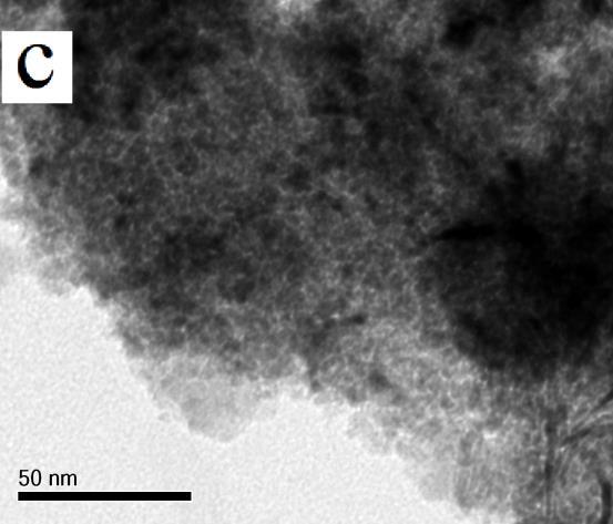
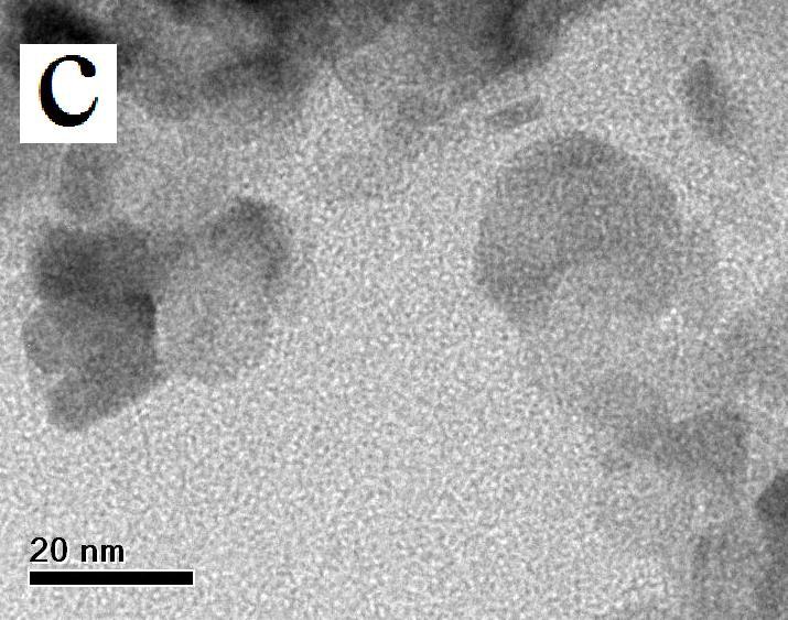
11 Surface Morphology Studies The surface morphology of the prepared samples has been studied using field emission scanning electron microscopy (FESEM). Figure 4.5 shows the FESEM images of the prepared Cr-doped (4% Cr, 6% Cr and 8% Cr) NiO samples. The image clearly shows that the samples have uniform grain size and nanoclusters have formed. The introduction of Cr-ion in NiO reduces the grain size which indicates the increase of strain in the matrix which restricts the lattice growth (Moura et al. 2012). It is well known that irrespective of the preparation method used to obtain nano-oxides, there is compelling evidence that crystallization does not follow a traditional nucleation and growth mechanism, more so in the case of increasing concentration of oxo-hydroxides to form metal oxides (José A. Rodriguez & Marcos Fernández-García, 2007). Figure 4.6 shows the FESEM images of cobalt doped NiO samples. The particles size is nearly uniform. The increase of doping concentration produces a change in the sample micro structure and a more disperse system is obtained. Figure 4.7 shows the FESEM images of Fe-doped NiO samples. The grains are of uniform size and arranged regularly. The introduction of Fe ion concentration produces homogenous distribution of grains.
 4% Cr-doped NiO nanoparticles (b) 6% Cr-doped")
12 70 Figure 4.5 FESEM images of (a) 4% Cr-doped NiO nanoparticles (b) 6% Cr-doped NiO nanoparticles and (c) 8% Cr-doped NiO nanoparticles
 4% Co-doped NiO nanoparticles (b) 6% Co-doped")
13 71 Figure 4.6 FESEM images of (a) 4% Co-doped NiO nanoparticles (b) 6% Co-doped NiO nanoparticles and (c) 8% Co-doped NiO nanoparticles.
 4% Fe-doped NiO nanoparticles (b) 6% Fe-doped")
14 72 Figure 4.7 FESEM images of (a) 4% Fe-doped NiO nanoparticles (b) 6% Fe-doped NiO nanoparticles and (c) 8% Fe-doped NiO nanoparticles
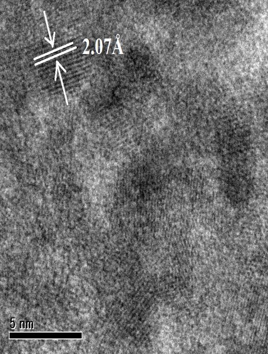
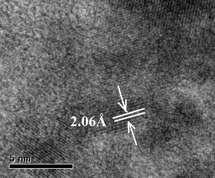
15 HRTEM Analysis High-resolution transmission electron microscopy (HRTEM) was used to investigate the microstructure of Cr-doped, Co-doped and Fe-doped NiO nanoparticles. The HRTEM images help to check out the crystallinity and phase purity of the prepared samples. For HRTEM analysis, the nanoparticles are dissolved in acetone and sonicated for 30 minutes. Table 4.4 Calculated d-spacing value from XRD pattern and HRTEM fringe pattern of Cr-doped NiO samples S.No Samples d-spacing value from XRD pattern Å d-spacing value from HRTEM fringe pattern Å 1 4% Cr-doped NiO % Cr-doped NiO % Cr-doped NiO Figure 4.8 shows the HRTEM images of Cr-doped NiO nanoparticles. The nanoparticles are an agglomeration of smaller particles forming nanoclusters. The images also exhibit lattice fringes and the lattice spacing has been determined using these fringes. Fringes of uniform width have been observed and the image reveals the crystallinity and defect free nature of the sample. The d-spacing was calculated for various concentrations of Cr-doped NiO and it is 2.08 Å, 2.07 Å and 2.06 Å respectively which corresponds to (200) plane. The d-spacing values calculated from these images are in close agreement with the values obtained from X-ray diffraction studies (Table 4.4).


 4%")
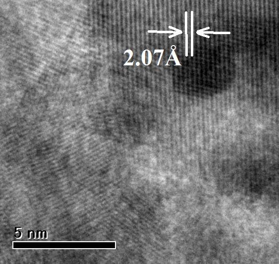
16 74 Figure 4.8 HRTEM images and fringe pattern of (a) 4% Cr-doped NiO nanoparticles(b) 6% Cr-doped NiO nanoparticles and (c) 8% Cr-doped NiO nanoparticles
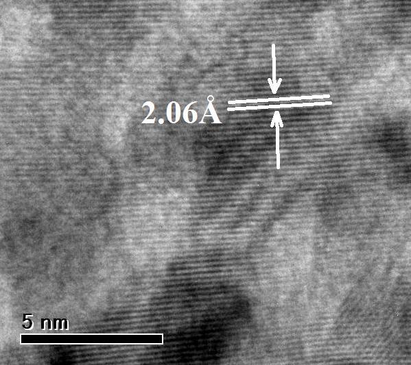
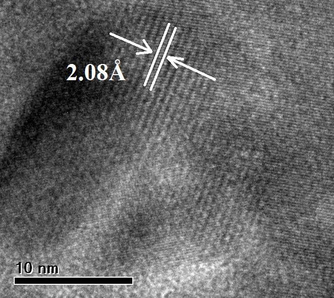
17 75 Figure 4.9 shows the HRTEM images of cobalt doped NiO nanoparticles prepared using 0.1M nickel nitrate and different weight percentage of cobalt nitrate. The HRTEM images show fringe pattern and the d-spacing is found from the fringe pattern. The d-spacing values calculated from the fringe pattern are 2.09 Å, 2.08 Å and 2.07 Å for cobalt doped NiO nanoparticles which correspond to the (200) plane of FCC NiO and are shown in Table 4.5. Table 4.5 Calculated d-spacing value from XRD pattern and HRTEM fringe pattern Co-doped samples d-spacing value S. No Samples d-spacing value from XRD pattern Å from HRTEM fringe pattern Å 1 4% Co-doped NiO % Co-doped NiO % Co-doped NiO Figure 4.10 shows the HRTEM images of Fe-doped NiO nanoparticles. The images exhibit uniform fringe pattern and using the fringe pattern d-spacing values are determined which correspond to the (200) plane and are 2.08Å, 2.07 Å and 2.06 Å respectively and are shown in Table 4.6.


 4%")

18 76 Figure 4.9 HRTEM images and fringe pattern of (a) 4% Co-doped NiO nanoparticles (b) 6% Co-doped NiO nanoparticles and (c) 8% Co-doped NiO nanoparticles
19 77 Table 4.6 Calculated d-spacing value from XRD pattern and HRTEM fringe pattern of Fe-doped samples S. No Samples d-spacing value from XRD pattern Å d-spacing value from HRTEM fringe pattern Å 1 4% Fe-doped NiO % Fe-doped NiO % Fe-doped NiO


 4%")

20 78 Figure 4.10 HRTEM images and fringe pattern of (a) 4% Fe-doped NiO nanoparticles (b) 6% Fe-doped NiO nanoparticles and (c) 8% Fe-doped NiO nanoparticles

21 Selected Area Electron Diffraction (SAED) Studies The selected area electron diffraction (SAED) pattern is used to study about the crystal properties of a particular region. The observed rings for the samples show that the samples exhibit FCC structure and are nanocrystalline in nature. Figures 4.11(a, b & c) shows the selected area electron diffraction images of Cr-doped, Co-doped and Fe-doped NiO nanoparticles. The presence of rings with discrete spots suggests that the prepared nanoparticles are made of small particles of uniform size. Ring patterns corresponding to planes (111), (200), (220), (311) and (222) are observed in the SAED pattern. Tables 4.7, 4.8 & 4.9 show the d-spacing values calculated from the selected area electron diffraction patterns of nanocrystalline Cr-doped, Co-doped and Fe-doped NiO nanoparticles and they are found to match well with the standard d-spacing values of JCPDS data ( , , ). Figure 4.11a SAED pattern of (a) 4% Cr-doped NiO nanoparticles (b) 6% Cr-doped NiO nanoparticles and ( c) 8% Cr-doped NiO nanoparticles
 4% 6% 8% Cr-doped Cr-doped Cr-doped d-spacing value (JCPDS) Planes (hkl) NiO NiO NiO Å 1 2.442 2.431 3.422 2.412 ( 111 ) 2 2.085 2.071 2.060 2.089 ( 200 ) 3 1.471 1.460 1.")
22 80 Table 4.7 Calculated d-spacing value of different planes of 4%,6% and 8% Cr-doped NiO nanoparticles from HRTEM SAED pattern Calculated d-spacing values from Reported S.No HRTEM SAED pattern (Å) 4% 6% 8% Cr-doped Cr-doped Cr-doped d-spacing value (JCPDS) Planes (hkl) NiO NiO NiO Å ( 111 ) ( 200 ) ( 220 ) ( 311 ) ( 222 ) Figure 4.11b SAED pattern of (a) 4% Co-doped NiO nanoparticles (b) 6% Co-doped nanoparticles NiO and (c) 8% Co-doped NiO nanoparticles
 4% 6 % 8% Co-doped Co-doped Co-doped NiO NiO NiO d-spacing value (JCPDS) Å Planes (hkl) 1 2.4147 2.4045 2.4009 2.412 ( 111 ) 2 2.095 2.085 2.075 2.089 ( 200 ) 3 1.479 1.")
23 81 Table 4.8 Calculated d-spacing value of different planes of 4%,6% and 8% Co-doped NiO nanoparticles from HRTEM SAED pattern Calculated d-spacing values from Reported S.No HRTEM SAED pattern (Å) 4% 6 % 8% Co-doped Co-doped Co-doped NiO NiO NiO d-spacing value (JCPDS) Å Planes (hkl) ( 111 ) ( 200 ) ( 220 ) ( 311 ) ( 222 ) Figure 4.11c SAED pattern of (a) 4% Fe-doped NiO nanoparticles (b) 6% Fe-doped nanoparticles NiO and (c) 8% Fe-doped NiO nanoparticles
24 82 Table 4.9 Calculated d-spacing value of different planes of 4%,6% and 8% Fe-doped NiO nanoparticles from HRTEM SAED pattern S.No Calculated d-spacing values from HRTEM SAED pattern (Å) 4% Fe-doped NiO 6% Fe-doped NiO 8% Fe-doped NiO Reported d-spacing value (JCPDS) Å Planes (hkl) ( 111 ) ( 200 ) ( 220 ) ( 311 ) ( 222 ) The HRTEM analysis show that the Cr-doped, Co-doped and Fedoped NiO nanoparticles exhibit FCC structure and there is no phase transformation due to doping of Cr, Co and Fe Compositional Analysis The energy dispersive X-ray analysis (EDAX) is used to study the chemical composition of the samples. Figure 4.12 shows the energy dispersive X-ray analysis spectrum of Cr-doped NiO nanoparticles. The spectrum indicates the presence of Cr, Ni and O as the main components and the amount of Ni decreases with the increase of Cr concentration. The components C and Cu present in the spectrum originate from the paste and grid used for EDAX study and the images reveal that the prepared samples do not have any impurities. The chemical constituents present in the 4% Cr-doped NiO sample according to the EDAX analysis are, Ni= at%,

25 83 O= at% and Cr =3.76 at%. The chemical constituents of 6% Cr-doped NiO sample are Ni=44.95 at%, O=49.10 at % and Cr =5.95 at%. And the chemical constituents of 8% Cr-doped NiO sample are Ni=42.98 at %, O=49.15 at % and Cr =7.87 at %. Figure 4.12 EDAX spectra of (a) 4% Cr-doped NiO nanoparticles (b) 6% Cr-doped nanoparticles NiO and ( c) 8% Cr-doped NiO nanoparticles The EDAX spectrum of Co-doped NiO nanoparticles is shown in Figure The atomic percentage of Ni, O and Co elements present in the
26 84 prepared 4%, 6% and 8% Co-doped NiO nanoparticles is as follows: The composition of 4% Co-doped NiO nanoparticles are Ni = at %, O =49.10 at % and Co =3.96 at %. The composition of 6% Co-doped NiO nanoparticles are Ni = at%, O =49.12 at% and Co = 5.92 at%.the composition of 8% Co-doped NiO nanoparticles are Ni = at%, O = at% and Co = 7.97 at%. Figure 4.13 EDAX spectra of (a) 4% Co-doped NiO nanoparticles (b) 6% Co-doped nanoparticles NiO and (c) 8% Co-doped NiO nanoparticles
27 85 The EDAX spectrum of Fe-doped NiO nanoparticles is shown in Figure The atomic percentage of Ni, O and Fe elements present in the prepared 4%, 6% and 8% Fe-doped NiO powder is as follows: The composition of 4% Fe-doped NiO nanoparticles are Ni =46.51 at%, O = at % and Fe =3.94 at %. The composition of 6% Fe-doped NiO nanoparticles are Ni = at%, O =49.57 at% and Fe =5.96 at%. The composition of 8% Fe-doped NiO nanoparticles are Ni = at%, O = at% and Fe = 7.92 at %. Figure 4.14 EDAX spectra of (a) 4% Fe-doped NiO nanoparticles (b) 6% Fe-doped nanoparticles NiO and (c) 8% Fe-doped NiO nanoparticles
28 Optical Studies Optical properties which are directly related to the size of the nanoparticles can be studied using the absorption spectra. Figure 4.15 shows the optical absorption spectra of Cr-doped NiO nanoparticles. The absorption edge of Cr-doped NiO is found to be at nm, nm and nm respectively. The absorption spectra of Cr-doped NiO nanoparticles show that the absorption edge is slightly shifted towards shorter wavelength (blue shift) when compared to undoped NiO. This shift is due to the Burstein Moss effect, since the absorption edge of a degenerate semiconductor is shifted to shorter wavelengths with increasing carrier concentration (Burstein 1954). This shift towards blue region predicts that there is an increase in band gap value(eg), which is due to the reduction in particle size. The fundamental absorption, which corresponds to the electron transition from the valance band(vb) to the conduction band(cb), can be used to determine the nature and value of the optical band gap. The optical absorption study is used to determine the optical band gap of the nanoparticles, which is the most familiar and simplest method. The absorption coefficient (α) and the incident photon energy (hʋ) are related by the expression (Pancove 1971 ) (αhʋ) = A(hʋ-Eg) n (4.2) where A is a constant, Eg is the optical band gap of the material, ʋ is the frequency of the incident radiation, h is Planck s constant and exponent n is 0.5 for direct band allowed transition. The optical band gap of 4%, 6% and 8% Cr-doped NiO nanoparticles is determined using equation 4.2. Figure 4.16 shows the (αhʋ) 2 versus hʋ plot of Cr-doped NiO nanoparticles. The optical band gap values have been determined by extrapolating the linear portion of the curve to meet the energy axis (hʋ). The
29 87 band gap has been calculated and is found to be 3.97 ev, 4.06 ev and 4.10 ev for 4%, 6% and 8% Cr-doped NiO respectively and is shown in table The obtained optical band gap of Cr-doped NiO nanoparticles is higher than that of NiO due to quantum confinement effect (Mohseni Meybodi et al. 2012). Quantum confinement of both electrons and holes in all the three dimensions leads to an increase in the effective band gap of the material. 5 Absorbance (a.u) a b a) 4% Cr-doped NiO b) 6% Cr-doped NiO c) 8% Cr-doped NiO c Wave length (nm) Figure 4.15 Absorption spectra of 4%, 6% and 8% Cr-doped NiO nanoparticles
30 88 Figure 4.16 Plot of ( h ) 2 vs. photon energy of 4%, 6% and 8% Crdoped NiO nanoparticles Table 4.10 Band gap of 4% Cr-doped NiO, 6% Cr-doped NiO and 8% Cr-doped NiO nanoparticles Absorption Bandgap S.No Samples wavelength (ev) (nm) 1 4% Cr-doped NiO % Cr-doped NiO % Cr-doped NiO
31 89 Optical absorption spectrum of Co-doped NiO nanoparticles is shown in Figure The absorption band edges of Co-doped NiO are absorbed to be present at nm, nm and nm respectively and are shown in Table Absorption spectra of Co-doped NiO nanoparticles shows that the absorption edge is slightly shifted towards the lower wavelength when compared to undoped NiO nanoparticles and this shift further move towards smaller wave length with increase in cobalt concentration. Figure 4.18 shows the (αhʋ) 2 versus hʋ plot of Co-doped NiO nanoparticles. The band gap of 4%, 6% and 8% Co-doped NiO nanoparticles is found to be 3.99 ev, 4.07 ev and 4.11 ev respectively. The absorption edge shift towards lower wavelength indicates the increase of optical band gap of NiO on cobalt doping. 5 Absorbance (a.u) a b a) 4% Co-doped NiO b) 6% Co-doped NiO c) 8% Co-doped NiO c Wave length (nm) Figure 4.17 Absorption spectra of 4%, 6% and 8% Co-doped NiO nanoparticles
32 90 Energy (ev) Figure 4.18 Plot of ( h ) 2 vs. photon energy of 4%, 6% and 8% Co-doped NiO nanoparticles Table 4.11 Band gap of 4% Co-doped NiO, 6% Co-doped NiO and 8% Co-doped NiO nanoparticles Absorption Bandgap S.No Samples wavelength (ev) (nm) 1 4% Co-doped NiO % Co-doped NiO % Co-doped NiO
33 91 Incorporation of Fe into the crystal lattice of NiO nanocrystalline semiconductors will alter the optical properties of the semiconductor. Optical absorption spectrum of Fe-doped NiO nanoparticles is shown in Figure The absorption band edges of Fe-doped NiO are absorbed to be present at nm, nm and nm respectively and are shown in Table The absorption edge of the Fe-doped NiO spectra shown in Figure 4.19 exhibits blue shift when compared to undoped NiO nanoparticles. Figure 4.20 shows the (αhʋ) 2 versus hʋ plot of Fe-doped NiO nanoparticles. The band gap values of 4%, 6% and 8% Fe-doped NiO are found to be 4.01 ev, 4.10 ev and 4.20 ev respectively. As the doping concentration of Fe increases the absorption edge shifts towards lower wavelength and indicates that there is an increase in the band gap energy of NiO on Fe doping. 5 4 Absorbance (a.u) a a) 4% Fe-doped NiO b) 6% Fe-doped NiO c) 8% Fe-doped NiO b Wave length (nm) c Figure 4.19 Absorption spectra of 4%, 6% and 8% Fe-doped NiO nanoparticles
34 92 Figure 4.20 Plot of ( h ) 2 vs. photon energy of 4%, 6% and 8% Fe-doped NiO nanoparticles Table 4.12 Band gap of (a) 4% Fe-doped NiO (b) 6% Fe-doped NiO and (c) 8% Fe-doped NiO nanoparticles S.No Absorption Bandgap Samples wavelength (ev) (nm) 1 4% Fe-doped NiO % Fe-doped NiO % Fe-doped NiO
35 Magnetic Properties The magnetic properties of Cr-doped, Co-doped and Fe-doped NiO nanoparticles have been studied using vibrating sample magnetometer (VSM) at room temperature. The magnetization versus applied magnetic field plots of chromium doped NiO is shown in Figure A well-defined hysteresis loop is observed. The magnetic parameters of Cr-doped NiO such as saturation magnetization (Ms), retentivity (MR) and coercivity (Hc) for different doping concentrations are calculated from the hysteresis curve and given in Table The existence of magnetic property is very closely related to the fine grain microstructure of the particles (Deraz 2012b). The narrowed hysteresis curves of Cr-doped NiO have low magnetization (MS) is due to the lattice imperfection and the compensation of spin values. As the doping concentration of Cr increases there is decrease of saturation magnetization due to lack of coupled spins.
36 94 Figure 4.21 Hysteresis curves of 4%, 6% and 8% Cr-doped NiO nanoparticles Table 4.13 Magnetic properties of Cr-doped NiO nanoparticles Coercivity Magnetization Retentivity S.No Samples (Hc) (MS) (MR) (Oe) 10-6 emu/g 10-6 emu/g % Cr-doped NiO 6% Cr-doped NiO 8% Cr-doped NiO
37 95 Magnetization versus applied magnetic field plot of cobalt doped NiO nanoparticles is shown in Figure The observed magnetisation and coercivity values are shown in Table The cobalt doped NiO nanoparticles show ferromagnetic behaviour at room temperature. The saturation magnetization of the cobalt doped samples is mainly due to the spin coupling effect which is due to smaller particle size. It is reported that the magnetic property of the materials is dependent on the particles size, shape, magnetization and crystallinity. The unusual magnetic behaviour may be due to grain size reduction and breaking of large number of exchange bonds. The presence of small magnetic clusters on the surface and lattice imperfection increases the uncompensated spin values (Anandha Babu et al. 2015). The increase of doping concentration of cobalt reduce the saturation magnetization value. The cobalt doped NiO nanoparticles are widely used in the area of magnetic storage devices (Kaliyan Vallalperuman et al. 2003). Magnetization (emu/g) a b c a) 4% Co-doped NiO b) 6% Co-doped NiO c) 8% Co-doped NiO Applied field (Oe) Figure 4.22 Hysteresis curves of 4%, 6% and 8% Co-doped NiO nanoparticles
38 96 Table 4.14 Magnetic properties of Co-doped NiO nanoparticles Coercivity Magnetization Retentivity S.No Samples (Hc) (MS) (MR) (Oe) 10-6 emu/g 10-6 emu/g % Co-doped NiO 6% Co-doped NiO 8% Co-doped NiO The magnetic field dependent magnetization (M-H) curves for the 4%, 6% and 8% of Fe-doped NiO nanoparticles at room temperature are shown in Figure The hysteresis curve for 4% Fe-doped NiO indicates the presence of ferromagnetic phase and it decreases with increase in doping concentration. A weak ferromagnetic phase was observed in 6% and 8% Fedoped NiO nanoparticles. The observed magnetisation and coercivity values are shown in Table Doping effect increases the grain size and reduce the net magnetization. Doping of Fe into NiO interface creates charge carriers which gives rise to more exchange interaction. The hysteresis phenomenon observed for Fe-doped NiO matches with the earlier results reported by Lin et al. (2006) and the curve is narrow due to uncompensated spin systems (Manna & De 2009). Smaller the crystallite size, the more will be the net magnetization (Moura et al. 2012). The magnetic properties of the materials are closely associated with the dependence of particle size, shape, oxygen deficiency, magnetic direction and crystallinity (Mishra et al. 2012). Table 4.16 shows the magnetic parameters of Cr-doped, Co-doped and Fe-doped NiO nanoparticles. It is found that the 4% Fe-doped NiO nanoparticles have high saturation magnetization value
39 97 when compared to 6% and 8% Fe-doped NiO nanoparticles. The magnetization value of Fe-doped NiO is more than those of chromium and cobalt doped NiO nanoparticles. As a result of high saturation magnetization values, Fe doped NiO nanomaterials are widely used for the fabrication of spintronic devices (Mishra et al & Jianfei Wang et al. 2005). Magnetization (emu/g) a b c a) 4% Fe-doped NiO b) 6% Fe-doped NiO c) 8% Fe-doped NiO Applied field (Oe) Figure 4.23 Hysteresis curves of 4%, 6% and 8% Fe-doped NiO nanoparticles
40 98 Table 4.15 Magnetic properties of Fe-doped NiO nanoparticles Coercivity Magnetization Retentivity S.No Samples (Hc) (MS) (MR) (Oe) 10-6 emu/g 10-6 emu/g % Fe-doped NiO 6% Fe-doped NiO 8% Fe-doped NiO Table 4.16 Comparing the magnetic properties of Cr-doped, Co-doped and Fe-doped NiO nanoparticles S.No Magnetic Property Coercivity (Hc) (Oe) Magnetization (MS) 10-6 emu/g Retentivity (MR) 10-6 emu/g Concentration of doped samples Cr-doped NiO nano particles Co-doped NiO nano particles Fe-doped NiO nano particles 4% % % % % % % % %
Structural and magnetic properties of Ni doped CeO 2 nanoparticles
 *E-mail: shailuphy@gmail.com Abstract: We report room temperature ferromagnetism in Ni doped CeO 2 nanoparticles using X-ray diffraction (XRD), high resolution transmission electron microscopy (HR-TEM),
Title: Magnetic chains of metal formed by assembly of small nanoparticles
 Title: Magnetic chains of metal formed by assembly of small nanoparticles Authors: Chen-Min Liu, Lin Guo*, Rong-Ming Wang*, Yuan Deng, Hui-Bin Xu, Shihe Yang* Supporting Information S1. Sample characterization
CHAPTER 3. OPTICAL STUDIES ON SnS NANOPARTICLES
 42 CHAPTER 3 OPTICAL STUDIES ON SnS NANOPARTICLES 3.1 INTRODUCTION In recent years, considerable interest has been shown on semiconducting nanostructures owing to their enhanced optical and electrical
CHAPTER 3 EFFECT OF COBALT DOPING ON THE OPTICAL AND MAGNETIC PROPERTIES OF CADMIUM SULFIDE NANOCRYSTALS
 49 CHAPTER 3 EFFECT OF COBALT DOPING ON THE OPTICAL AND MAGNETIC PROPERTIES OF CADMIUM SULFIDE NANOCRYSTALS 3.1 INTRODUCTION 3.1.1 Diluted Magnetic Semiconductor Diluted magnetic semiconductors (DMSs)
Synthesis and Study of Magnesium Oxide and Cadmium Doped Magnesium Oxide Nanoparticles
 Synthesis and Study of Magnesium Oxide and Cadmium Doped Magnesium Oxide Nanoparticles Prateek Kumar Gour 1, Sanchita Dass Roy 2 gourprateek0000@gmail.com Abstract Magnesium Oxide play a very important
Hydrothermal synthesis and characterization of undoped and Eu doped ZnGa 2 O 4 nanoparticles
 Chapter 3 Hydrothermal synthesis and characterization of undoped and Eu doped ZnGa 2 O 4 nanoparticles 3.1 Introduction Phosphors are substance that exhibits the phenomenon of luminescence. Efficient phosphors
Available online at ScienceDirect. Procedia Materials Science 11 (2015 )
") Available online at www.sciencedirect.com ScienceDirect Procedia Materials Science 11 (2015 ) 282 286 5th International Biennial Conference on Ultrafine Grained and Nanostructured Materials, UFGNSM15 Prepartion
Available online at www.sciencedirect.com ScienceDirect Procedia Materials Science 11 (2015 ) 282 286 5th International Biennial Conference on Ultrafine Grained and Nanostructured Materials, UFGNSM15 Prepartion
International Journal of Scientific & Engineering Research, Volume 5, Issue 3, March-2014 ISSN
 156 Copper Nanoparticles: Green Synthesis Characterization Y.Suresh*1, S.Annapurna*2, G.Bhikshamaiah*3, A.K.Singh#4 Abstract Present work describes the synthesis nanoparticles using papaya extract as a
CHAPTER IV SYNTHESIS AND CHARACTERIZATION OF METAL IONS DOPED ZINC SELENIDE NANOPARTICLES
 132 CHAPTER IV SYNTHESIS AND CHARACTERIZATION OF METAL IONS DOPED ZINC SELENIDE NANOPARTICLES 4.1 Introduction Introducing impurity atoms into a semiconductor host leads to an increase in the free-carrier
Synthesis and Characterization of Iron-Oxide (Hematite) Nanocrystals. Z.H. Lee
 Nanocrystals. Z.H. Lee") ABSTRACT Synthesis and Characterization of Iron-Oxide (Hematite) Nanocrystals Z.H. Lee Engineering Science Programme, National University of Singapore Kent Ridge, Singapore 119260 Monodispersed iron oxide
ABSTRACT Synthesis and Characterization of Iron-Oxide (Hematite) Nanocrystals Z.H. Lee Engineering Science Programme, National University of Singapore Kent Ridge, Singapore 119260 Monodispersed iron oxide
Novel fungus-titanate bio-nano composites as high performance. absorbents for the efficient removal of radioactive ions from.
 This journal is The Royal Society of Chemistry 0 Electronic Supplementary Information For Novel fungus-titanate bio-nano composites as high performance absorbents for the efficient removal of radioactive
SIMULATIONS ON DILUTE MAGNETIC SEMICONDUCTOR PROPERTIES
 Romanian Reports in Physics, Vol. 62, No. 1, P. 115 120, 2010 SIMULATIONS ON DILUTE MAGNETIC SEMICONDUCTOR PROPERTIES M. NEGOITA, E. A. PATROI, C. V. ONICA National Institute for Research and Development
Supporting Information
 Supporting Information Ultrathin Spinel-Structured Nanosheets Rich in Oxygen Deficiencies for Enhanced Electrocatalytic Water Oxidation** Jian Bao, Xiaodong Zhang,* Bo Fan, Jiajia Zhang, Min Zhou, Wenlong
Research Highlights. Salient results from our group. Mixed phosphides in Sn-P and Sn-Mn-P systems
 Research Highlights Dilute magnetic semiconductors and Spintronics Spintronics is a branch of electronics emerged from the dilute magnetic semiconductor in an aspect of utilization of the spin in addition
Review of Optical Properties of Materials
 Review of Optical Properties of Materials Review of optics Absorption in semiconductors: qualitative discussion Derivation of Optical Absorption Coefficient in Direct Semiconductors Photons When dealing
College of Mechanical Engineering, Yangzhou University, Yangzhou , China; 2
 Proceedings Light-Assisted Room-Temperature NO2 Sensors Based on Black Sheet-Like NiO Xin Geng 1,2,3, Driss Lahem 4, Chao Zhang 1, *, Marie-Georges Olivier 3 and Marc Debliquy 3 1 College of Mechanical
SYNTHESIS OF CADMIUM SULFIDE NANOSTRUCTURES BY NOVEL PRECURSOR
 Nanomaterials: Applications and Properties (NAP-2011). Vol. 1, Part I 107 SYNTHESIS OF CADMIUM SULFIDE NANOSTRUCTURES BY NOVEL PRECURSOR M. Salavati Niasari 1,2* 1 Department of Inorganic Chemistry, Faculty
CHAPTER 5. STUDY OF CoFe 2 O 4 PARTICLES SYNTHESIZED WITH PVP AND CITRIC ACID
 58 CHAPTER 5 STUDY OF CoFe 2 O 4 PARTICLES SYNTHESIZED WITH PVP AND CITRIC ACID This chapter deals with the synthesis of CoFe 2 O 4 particles using metal nitrates, PVP and citric acid. The structure, morphology
Fabrication and characterization of poly (ethylene oxide) templated nickel oxide nanofibers for dye degradation
 templated nickel oxide nanofibers for dye degradation") Electronic Supplementary Material (ESI) for Environmental Science: Nano. This journal is The Royal Society of Chemistry 2014 Supplementary Information Fabrication and characterization of poly (ethylene
Electronic Supplementary Material (ESI) for Environmental Science: Nano. This journal is The Royal Society of Chemistry 2014 Supplementary Information Fabrication and characterization of poly (ethylene
RESULTS AND DISCUSSION Characterization of pure CaO and Zr-TiO 2 /CaO nanocomposite
 RESULTS AND DISCUSSION 4.1. Characterization of pure CaO and Zr-TiO 2 /CaO nanocomposite 4.1.1. Scanning electron microscopy analysis (SEM) SEM images of prepared CaO are shown in Fig. 4.1 (a and b). CaO
Bandgap engineering through nanocrystalline magnetic alloy grafting on. graphene
 Electronic Supplementary Material (ESI) for Physical Chemistry Chemical Physics. This journal is the Owner Societies 2014 Electronic Supplementary Information (ESI) for Bandgap engineering through nanocrystalline
This manuscript was submitted first in a reputed journal on Apri1 16 th Stanene: Atomically Thick Free-standing Layer of 2D Hexagonal Tin
 This manuscript was submitted first in a reputed journal on Apri1 16 th 2015 Stanene: Atomically Thick Free-standing Layer of 2D Hexagonal Tin Sumit Saxena 1, Raghvendra Pratap Choudhary, and Shobha Shukla
(IJIRSE) International Journal of Innovative Research in Science & Engineering ISSN (Online)
 International Journal of Innovative Research in Science & Engineering ISSN (Online)") Synthesis, Size Characterization and Photocatalytic Activities of Zinc Ferrites and Cobalt Ferrites Nanoparticles Using Oxidative Degradations of Methylene Blue, Crystal Violet and Alizarin Red s in Aqueous
Synthesis, Size Characterization and Photocatalytic Activities of Zinc Ferrites and Cobalt Ferrites Nanoparticles Using Oxidative Degradations of Methylene Blue, Crystal Violet and Alizarin Red s in Aqueous
EECS130 Integrated Circuit Devices
 EECS130 Integrated Circuit Devices Professor Ali Javey 8/30/2007 Semiconductor Fundamentals Lecture 2 Read: Chapters 1 and 2 Last Lecture: Energy Band Diagram Conduction band E c E g Band gap E v Valence
Novel fluorescent matrix embedded carbon quantum dots enrouting stable gold and silver hydrosols
 Novel fluorescent matrix embedded carbon quantum dots enrouting stable gold and silver hydrosols Shouvik Mitra a, Sourov Chandra b, Prasun Patra a, Panchanan Pramanik b *, Arunava Goswami a * a AERU, Biological
EXTRINSIC SEMICONDUCTOR
 EXTRINSIC SEMICONDUCTOR In an extrinsic semiconducting material, the charge carriers originate from impurity atoms added to the original material is called impurity [or] extrinsic semiconductor. This Semiconductor
Synthesis and Characterization of Innovative Multilayer, Multi Metal Oxide Thin Films by Modified Silar Deposition Method
 STUDENT JOURNAL OF PHYSICS Indian Association of Physics Teachers Presentations Synthesis and Characterization of Innovative Multilayer, Multi Metal Oxide Thin Films by Modified Silar Deposition Method
CHAPTER-2 SYNTHESIS AND CHARACTERIZATION OF MODIFIED METAL OXIDES AND METAL SULPHIDES NANOPARTICLES
 CHAPTER-2 SYNTHESIS AND CHARACTERIZATION OF MODIFIED METAL OXIDES AND METAL SULPHIDES NANOPARTICLES Nanoparticles have recently attracted significant attention from the science community. Nanoparticles
STRUCTURE AND MAGNETIC PROPERTIES OF SiO 2 COATED Fe 2 NANOPARTICLES SYNTHESIZED BY CHEMICAL VAPOR CONDENSATION PROCESS
 Rev.Adv.Mater.Sci. Structure and magnetic 4 (2003) properties 55-59 of coated 55 STRUCTURE AND MAGNETIC PROPERTIES OF COATED NANOPARTICLES SYNTHESIZED BY CHEMICAL VAPOR CONDENSATION PROCESS Ji-Hun Yu,
Synthesis and Characterization of Exfoliated Graphite (EG) and to Use it as a Reinforcement in Zn-based Metal Matrix Composites
 and to Use it as a Reinforcement in Zn-based Metal Matrix Composites") Synthesis and Characterization of Exfoliated Graphite (EG) and to Use it as a Reinforcement in Zn-based Metal Matrix Composites Here H 2 SO 4 was used as an intercalant and H 2 O 2 as an oxidant. Expandable
Synthesis and Characterization of Exfoliated Graphite (EG) and to Use it as a Reinforcement in Zn-based Metal Matrix Composites Here H 2 SO 4 was used as an intercalant and H 2 O 2 as an oxidant. Expandable
Efficient Co-Fe layered double hydroxide photocatalysts for water oxidation under visible light
 Supplementary Information Efficient Co-Fe layered double hydroxide photocatalysts for water oxidation under visible light Sang Jun Kim, a Yeob Lee, a Dong Ki Lee, a Jung Woo Lee a and Jeung Ku Kang* a,b
The sacrificial role of graphene oxide in stabilising Fenton-like catalyst GO Fe 3 O 4
 Electronic Supplementary Material (ESI) for ChemComm. This journal is The Royal Society of Chemistry 2015 The sacrificial role of graphene oxide in stabilising Fenton-like catalyst GO Fe 3 O 4 Nor Aida
O 3. : Er nanoparticles prospective system for energy convertors
 IOP Conference Series: Materials Science and Engineering PAPER OPEN ACCESS Interband optical transitions in Gd 2 O 3 : Er nanoparticles prospective system for energy convertors To cite this article: A
In situ formation of metal Cd x Zn 1-x S nanocrystals on graphene surface: A novel method to synthesis sulfide-graphene nanocomposites
 Electronic Supplementary Material (ESI) for RSC Advances. This journal is The Royal Society of Chemistry 2014 In situ formation of metal Cd x Zn 1-x S nanocrystals on graphene surface: A novel method to
Large-Scale Synthesis of Transition-metal Doped TiO 2 Nanowires. with Controllable Overpotential
 Large-Scale Synthesis of Transition-metal Doped TiO 2 Nanowires with Controllable Overpotential Bin Liu 1, Hao Ming Chen, 1 Chong Liu 1,3, Sean C. Andrews 1,3, Chris Hahn 1, Peidong Yang 1,2,3,* 1 Department
EECS143 Microfabrication Technology
 EECS143 Microfabrication Technology Professor Ali Javey Introduction to Materials Lecture 1 Evolution of Devices Yesterday s Transistor (1947) Today s Transistor (2006) Why Semiconductors? Conductors e.g
The characterization of MnO nanostructures synthesized using the chemical bath deposition method
 The characterization of MnO nanostructures synthesized using the chemical bath deposition method LF Koao 1, F B Dejene 1* and HC Swart 2 1 Department of Physics, University of the Free State (Qwaqwa Campus),
CdTe quantum dot sensitized hexaniobate nanoscrolls and Photoelectrochemical properties
 CdTe quantum dot sensitized hexaniobate nanoscrolls and Photoelectrochemical properties Feriha Eylul Sarac a, Ceren Yilmaz,b, Funda Yagci Acar a,b,c and Ugur Unal* a,b,c a Koc University, Chemistry Department,
Growth of silver nanocrystals on graphene by simultaneous reduction of graphene oxide and silver ions with a rapid and efficient one-step approach
 Growth of silver nanocrystals on graphene by simultaneous reduction of graphene oxide and silver ions with a rapid and efficient one-step approach Xiu-Zhi Tang, a Zongwei Cao, b Hao-Bin Zhang, a Jing Liu
PHOTOCATALYTIC DEGRADATION STUDIES OF POLYANILINE BASED ZnO-Al 2 O 3 NANOCOMPOSITE
 PHOTOCATALYTIC DEGRADATION STUDIES OF POLYANILINE BASED ZnO-Al 2 O 3 NANOCOMPOSITE Baiju V 1, Dedhila Devadathan 2, Biju R 3, Raveendran R 4 Nanoscience Research Laboratory, Department of Physics, Sree
Electronic supplementary information. A longwave optical ph sensor based on red upconversion
 Electronic Supplementary Material (ESI) for RSC Advances. This journal is The Royal Society of Chemistry 2014 Electronic supplementary information A longwave optical ph sensor based on red upconversion
Effects of molarity concentrations on structural and optical properties of MgO thin films prepared by co-precipitation method
 Journal of Multidisciplinary Engineering Science Studies (JMESS) Effects of molarity concentrations on structural and optical properties of MgO thin films prepared by co-precipitation method Aseel M. Abd
CHAPTER III. ORGANIC LIGANDS PASSIVATED ZnSe NANOSTRUCTURES AND FUNCTIONAL PROPERTIES
 82 CHAPTER III ORGANIC LIGANDS PASSIVATED ZnSe NANOSTRUCTURES AND FUNCTIONAL PROPERTIES 3.1 INTRODUCTION Semiconductor nanocrystals have size tunable optical properties that open up possibilities for revolutionary
VERY SHORT ANSWER TYPE QUESTIONS (1 Mark)
") UNIT I 10 Chemistry-XII THE SOLID STATE VERY SHORT ANSWER TYPE QUESTIONS (1 Mark) Q. 1. What do you mean by paramagnetic substance? Ans. Weakly attracted by magnetic eld and these substances are made of
UNIT I 10 Chemistry-XII THE SOLID STATE VERY SHORT ANSWER TYPE QUESTIONS (1 Mark) Q. 1. What do you mean by paramagnetic substance? Ans. Weakly attracted by magnetic eld and these substances are made of
Chapter 3 Chapter 4 Chapter 5
 Preamble In recent years bismuth-based, layer-structured perovskites such as SrBi 2 Nb 2 O 9 (SBN) and SrBi 2 Ta 2 O 9 (SBT) have been investigated extensively, because of their potential use in ferroelectric
Research Article Synthesis and Characterization of Magnetic Nanosized Fe 3 O 4 /MnO 2 Composite Particles
 Nanomaterials Volume 29, rticle ID 34217, 5 pages doi:1.1155/29/34217 Research rticle Synthesis and Characterization of Magnetic Nanosized /MnO 2 Composite Particles Zhang Shu and Shulin Wang Department
Transition Elements. pranjoto utomo
 Transition Elements pranjoto utomo Definition What is transition metal? One of which forms one or more stable ions which have incompletely filled d orbitals. 30Zn? Definition Zink is not transition elements
EE143 Fall 2016 Microfabrication Technologies. Evolution of Devices
 EE143 Fall 2016 Microfabrication Technologies Prof. Ming C. Wu wu@eecs.berkeley.edu 511 Sutardja Dai Hall (SDH) 1-1 Evolution of Devices Yesterday s Transistor (1947) Today s Transistor (2006) 1-2 1 Why
Preparation of Fe 3 O 2 Nanostructures via Inverse Micelle Method and Study of Their Magnetic Properties for Biological Applications
 International Journal of Bio-Inorganic Hybrid Nanomaterials Preparation of Fe 3 O 4 @SiO 2 Nanostructures via Inverse Micelle Method and Study of Their Magnetic Properties for Biological Applications Afsaneh
Shuo Li, Qidong Zhao, Dejun Wang and Tengfeng Xie *
 Electronic Supplementary Material (ESI) for RSC Advances. This journal is The Royal Society of Chemistry 2016 Work Function Engineering Derived All-solid-state Z-scheme Semiconductor-Metal-Semiconductor
Synthesis, Structural, Surface Morphology, Optical and Electrical Properties of Silver Oxide Nanoparticles
 Int. J. Nanoelectronics and Materials 9 (2016) 37-49 Synthesis, Structural, Surface Morphology, Optical and Electrical Properties of Silver Oxide Nanoparticles Suresh Sagadevan* Department of physics,
Karine Chesnel BYU. Idaho State University, Physics department 22 September 2014
 Karine Chesnel BYU Idaho State University, Physics department 22 September 2014 Potential applications of magnetic nanoparticles Magnetic recording Biomedical applications J. Mater. Chem., 19, 6258 6266
Self-compensating incorporation of Mn in Ga 1 x Mn x As
 Self-compensating incorporation of Mn in Ga 1 x Mn x As arxiv:cond-mat/0201131v1 [cond-mat.mtrl-sci] 9 Jan 2002 J. Mašek and F. Máca Institute of Physics, Academy of Sciences of the CR CZ-182 21 Praha
Synthesis and Luminescence Properties of Ni Doped ZnS Nanoparticles
 IJSTE - International Journal of Science Technology & Engineering Volume 2 Issue 11 May 2016 ISSN (online): 2349-784X Synthesis and Luminescence Properties of Ni Doped ZnS Nanoparticles B. Hariprasad Reddy
High-Performance Flexible Asymmetric Supercapacitors Based on 3D. Electrodes
 Supporting Information for: High-Performance Flexible Asymmetric Supercapacitors Based on 3D Porous Graphene/MnO 2 Nanorod and Graphene/Ag Hybrid Thin-Film Electrodes Yuanlong Shao, a Hongzhi Wang,* a
X-ray Imaging and Spectroscopy of Individual Nanoparticles
 X-ray Imaging and Spectroscopy of Individual Nanoparticles A. Fraile Rodríguez, F. Nolting Swiss Light Source Paul Scherrer Institut, Switzerland Intensity [a.u.] 1.4 1.3 1.2 1.1 D 8 nm 1 1 2 3 1.0 770
Preparation of One-dimensional ZnO/Bi2O3 Heterostructures Nanomaterial for Visible Light Photocatalysis
 2016 International Conference on Material Science and Civil Engineering (MSCE 2016) ISBN: 978-1-60595-378-6 Preparation of One-dimensional ZnO/Bi2O3 Heterostructures Nanomaterial for Visible Light Photocatalysis
Supporting Information. Synthesis and Upconversion Luminescence of BaY 2
 Supporting Information Synthesis and Upconversion Luminescence of BaY 2 F 8 :Yb 3+ /Er 3+ Nanobelts 5 Guofeng Wang, Qing Peng, and Yadong Li* Department of Chemistry and State Key Laboratory of New Ceramics
MORPHOLOGY AND THERMAL STUDIES OF NICKEL CARBONATE NANOPARTICLES
 MORPHOLOGY AND THERMAL STUDIES OF NICKEL CARBONATE NANOPARTICLES R.Hepzi Pramila Devamani 1, M.Krishnaveni 2, J.Mercy Jeno Rose 3 1 Assistant Professor, 2,3 M.Sc Students, Post Graduate Department of Physics,
Supplementary Information
 Supplementary Information Fabrication of Novel Rattle-Type Magnetic Mesoporous carbon Microspheres for Removal of Microcystins Xinghua Zhang and Long Jiang* Beijing National Laboratory for Molecular Science
PREPARATION AND CHARACTERIZATION OF CdO/PVP NANOPARTICLES BY PRECIPITATION METHOD
 Indian Journal of Pure and Applied Physics (IJPAP) Vol.1.No.1 2013 pp 1-6 available at: www.goniv.com Paper Received :05-03-2013 Paper Published:28-03-2013 Paper Reviewed by: 1. Dr.S.Selvakumar 2. Hendry
Research Article Preparation of γ-fe 2 O 3 /Ni 2 O 3 /FeCl 3 (FeCl 2 ) Composite Nanoparticles by Hydrothermal Process Useful for Ferrofluids
 Composite Nanoparticles by Hydrothermal Process Useful for Ferrofluids") Smart Materials Research Volume 2011, Article ID 351072, 5 pages doi:10.1155/2011/351072 Research Article Preparation of γ-fe 2 O 3 /Ni 2 O 3 /FeCl 3 (FeCl 2 ) Composite Nanoparticles by Hydrothermal Process
Smart Materials Research Volume 2011, Article ID 351072, 5 pages doi:10.1155/2011/351072 Research Article Preparation of γ-fe 2 O 3 /Ni 2 O 3 /FeCl 3 (FeCl 2 ) Composite Nanoparticles by Hydrothermal Process
Ethylenediaminetetraacetic Acid-Assisted Synthesis of Nano Antimony Oxide by Microwave Method
 Ethylenediaminetetraacetic Acid-Assisted Synthesis of Nano Antimony Oxide by Microwave Method Azadeh Tadjarodi*, Mohammad karimpour Department of Chemistry, Iran University of Science and Technology, Narmak,
Study on Magnetic Properties of Vermiculite Intercalation compounds
 Study on Magnetic Properties of Vermiculite Intercalation compounds M. Suzuki and I.S. Suzuki Department of Physics, State University of New York at Binghamton (October, ) I. INTRODUCTION In recent years
-:Vijay Singh(09CEB023)
") Heterogeneous Semiconductor Photocatalyst -:Vijay Singh(09CEB023) Guided by Azrina Abd Aziz Under Dr. Saravanan Pichiah Preparation of TiO 2 Nanoparticle TiO 2 was prepared by hydrolysis and poly-condensation
Heterogeneous Semiconductor Photocatalyst -:Vijay Singh(09CEB023) Guided by Azrina Abd Aziz Under Dr. Saravanan Pichiah Preparation of TiO 2 Nanoparticle TiO 2 was prepared by hydrolysis and poly-condensation
Setting The motor that rotates the sample about an axis normal to the diffraction plane is called (or ).
.") X-Ray Diffraction X-ray diffraction geometry A simple X-ray diffraction (XRD) experiment might be set up as shown below. We need a parallel X-ray source, which is usually an X-ray tube in a fixed position
X-Ray Diffraction X-ray diffraction geometry A simple X-ray diffraction (XRD) experiment might be set up as shown below. We need a parallel X-ray source, which is usually an X-ray tube in a fixed position
Recent Developments in Magnetoelectrics Vaijayanti Palkar
 Recent Developments in Magnetoelectrics Vaijayanti Palkar Department of Condensed Matter Physics & Materials Science Tata Institute of Fundamental Research Mumbai 400 005, India. Tata Institute of Fundamental
SCIENTIFIC REPORT. regarding the implementation of the project between May December 2014
 SCIENTIFIC REPORT regarding the implementation of the project between May 2013 - December 2014 Title of the project: DETECTION AND IDENTIFICATION OF BIOMOLECULES OF MEDICAL INTEREST BY USING MAGNETIC AND
Photocatalytic degradation of dyes over graphene-gold nanocomposites under visible light irradiation
 Photocatalytic degradation of dyes over graphene-gold nanocomposites under visible light irradiation Zhigang Xiong, Li Li Zhang, Jizhen Ma, X. S. Zhao* Department of Chemical and Biomolecular Engineering,
Supporting Information
 Supporting Information Yb 3 O(OH) 6 Cl.2H 2 O An anion exchangeable hydroxide with a cationic inorganic framework structure Helen V. Goulding, a Sarah E. Hulse, a William Clegg, b Ross W. Harrington, b
size (nm) Supplementary Figure 1. Hydrodynamic size distribution in a H 2O:MeOH 50% v/v
 Supplementary Figure 1. Hydrodynamic size distribution in a H 2O:MeOH 50% v/v") Intensity (%) 20 15 10 [0a] [0b] CoTPMA: 3 eq CoTPMA: 10 eq CoTPMA: 20 eq CoTPMA : 40 eq CoTPMA: 60 eq, [1] CoTPMA: 85 eq CoTPMA: 125 eq CoTPMA: 170 eq CoTPMA: 210 eq 5 0 10 100 size (nm) Supplementary
Intensity (%) 20 15 10 [0a] [0b] CoTPMA: 3 eq CoTPMA: 10 eq CoTPMA: 20 eq CoTPMA : 40 eq CoTPMA: 60 eq, [1] CoTPMA: 85 eq CoTPMA: 125 eq CoTPMA: 170 eq CoTPMA: 210 eq 5 0 10 100 size (nm) Supplementary
Synthesis and Characterization the Photocatalytic Activity of CNT/TiO 2 Nano-Composite
 Synthesis and Characterization the Photocatalytic Activity of CNT/TiO 2 Nano-Composite Shakiba Sadeghian 1*, Mohammad Reza Khanlari 2 1, 2 Department of Physics, Imam Khomeini International University,
Supporting Information
 Supporting Information Band Gap Tuning of CH 3 NH 3 Pb(Br 1-x Cl x ) 3 Hybrid Perovskite for Blue Electroluminescence Naresh K. Kumawat 1, Amrita Dey 1, Aravindh Kumar 2, Sreelekha P. Gopinathan 3, K.
Morphology and Thermal studies of Copper Carbonate Nanoparticles
 Morphology and Thermal studies of Copper Carbonate Nanoparticles * Dr.R.Hepzi Pramila Devamani 1 V.V.Vanniaperumal College for Women, Virudhunagar, Tamil Nadu, India. M.Shameena Begum 2 & R.Suganthi 3
Synthesis of 12 nm iron oxide nanoparticles
 Electronic Supporting Information for Dendronized iron oxide nanoparticles as contrast agent for MRI Brice Basly, a Delphine Felder-Flesch,* a Pascal Perriat, b Claire Billotey, c Jacqueline Taleb, c Geneviève
The Oxford Solid State Basics
 The Oxford Solid State Basics Steven H. Simon University of Oxford OXFORD UNIVERSITY PRESS Contents 1 About Condensed Matter Physics 1 1.1 What Is Condensed Matter Physics 1 1.2 Why Do We Study Condensed
Band-gap tuning of lead halide perovskites using a sequential deposition process
 Electronic Supplementary Material (ESI) for Journal of Materials Chemistry A. This journal is The Royal Society of Chemistry 2014 Supporting information Band-gap tuning of lead halide perovskites using
CHAPTER 2 MAGNETISM. 2.1 Magnetic materials
 CHAPTER 2 MAGNETISM Magnetism plays a crucial role in the development of memories for mass storage, and in sensors to name a few. Spintronics is an integration of the magnetic material with semiconductor
INVESTIGATIONS OF Mn, Fe, Ni AND Pb DOPED
 INVESTIGATIONS OF Mn, Fe, Ni AND Pb DOPED ZINC SULPHIDE NANOPARTICLES A thesis submitted to the University of Pune FOR THE DEGREE OF DOCTOR of PHILOSOPHY IN PHYSICS by PRAMOD H. BORSE DEPARTMENT OF PHYSICS
Supplementary Information
 Supplementary Information Bismuth Sulfide Nanoflowers for Detection of X-rays in the Mammographic Energy Range Shruti Nambiar a,b, Ernest K. Osei a,c,d, John T.W.Yeow *,a,b a Department of Systems Design
A Photonic Crystal Laser from Solution Based. Organo-Lead Iodide Perovskite Thin Films
 SUPPORTING INFORMATION A Photonic Crystal Laser from Solution Based Organo-Lead Iodide Perovskite Thin Films Songtao Chen 1, Kwangdong Roh 2, Joonhee Lee 1, Wee Kiang Chong 3,4, Yao Lu 5, Nripan Mathews
Electronic Supplementary Information (ESI) Green synthesis of shape-defined anatase TiO 2 nanocrystals wholly exposed with {001} and {100} facets
 Green synthesis of shape-defined anatase TiO 2 nanocrystals wholly exposed with {001} and {100} facets") Electronic Supplementary Information (ESI) Green synthesis of shape-defined anatase TiO 2 nanocrystals wholly exposed with {001} and {100} facets Lan Wang, a Ling Zang, b Jincai Zhao c and Chuanyi Wang*
Electronic Supplementary Information (ESI) Green synthesis of shape-defined anatase TiO 2 nanocrystals wholly exposed with {001} and {100} facets Lan Wang, a Ling Zang, b Jincai Zhao c and Chuanyi Wang*
PREPARATION, CHARACTERISATION AND PHOTOCATALYTIC ACTIVITY OF TERNARY GRAPHENE-Fe 3 O 4 :TiO 2 NANOCOMPOSITES
 Digest Journal of Nanomaterials and Biostructures Vol. 13, No. 2, April - June 2018, p. 499-504 PREPARATION, CHARACTERISATION AND PHOTOCATALYTIC ACTIVITY OF TERNARY GRAPHENE-Fe 3 O 4 :TiO 2 NANOCOMPOSITES
nanocomposites: synthesis and characterization
 National Institute for Research and Development of Isotopic and Molecular Technologies Cluj-Napoca Romania Carbon nanotubes-polypyrrole nanocomposites: synthesis and characterization R. Turcu a, O. Pana
Chapter 3 Properties of Nanostructures
 Chapter 3 Properties of Nanostructures In Chapter 2, the reduction of the extent of a solid in one or more dimensions was shown to lead to a dramatic alteration of the overall behavior of the solids. Generally,
Supporting Information
 Electronic Supplementary Material (ESI) for Nanoscale. This journal is The Royal Society of Chemistry 2014 Supporting Information Hydrothermal synthesis of - alloy nanooctahedra and their enhanced electrocatalytic
Chapter 1 Electronic and Photonic Materials - DMS. Diluted Magnetic Semiconductor (DMS)
") Diluted Magnetic Semiconductor (DMS) 1 Properties of electron Useful! Charge Electron Spin? Mass 2 Schematic of a Spinning & Revolving Particle Spinning Revolution 3 Introduction Electronics Industry Uses
Diluted Magnetic Semiconductor (DMS) 1 Properties of electron Useful! Charge Electron Spin? Mass 2 Schematic of a Spinning & Revolving Particle Spinning Revolution 3 Introduction Electronics Industry Uses
Crystal Properties. MS415 Lec. 2. High performance, high current. ZnO. GaN
 Crystal Properties Crystal Lattices: Periodic arrangement of atoms Repeated unit cells (solid-state) Stuffing atoms into unit cells Determine mechanical & electrical properties High performance, high current
EE495/695 Introduction to Semiconductors I. Y. Baghzouz ECE Department UNLV
 EE495/695 Introduction to Semiconductors I Y. Baghzouz ECE Department UNLV Introduction Solar cells have always been aligned closely with other electronic devices. We will cover the basic aspects of semiconductor
Structural and Functional Group Characterization of Nanocomposite Fe3O4/TiO2 and Its Magnetic Property 40
 Structural and Functional Group Characterization of Nanocomposite Fe3O4/TiO2 and Its Magnetic Property 40 V. Maria Vinosel 1,a, M. Asisi Janifer 1, S. Anand 1, S. Pauline 1 1 Department of Physics, Loyola
MAGNETIC-PROPERTY ENHANCEMENT OF SIZED CONTROLLED COBALT-GOLD CORE-SHELL NANOCRYSTALS
 Digest Journal of Nanomaterials and Biostructures Vol. 7, No. 4, October - December 2012, p. 1799 1810 MAGNETIC-PROPERTY ENHANCEMENT OF SIZED CONTROLLED COBALT-GOLD CORE-SHELL NANOCRYSTALS GH. BAHMANROKH
ABSTRACT I. INTRODUCTION II. BACKGROUND OF STUDY
 2017 IJSRST Volume 3 Issue 3 Print ISSN: 2395-6011 Online ISSN: 2395-602X Themed Section: Science and Technology Preparation and Experimental Investigation of CUO Nanoparticles Based Engine OILS Sk Salman
ELEMENTARY BAND THEORY
 ELEMENTARY BAND THEORY PHYSICIST Solid state band Valence band, VB Conduction band, CB Fermi energy, E F Bloch orbital, delocalized n-doping p-doping Band gap, E g Direct band gap Indirect band gap Phonon
4. Interpenetrating simple cubic
 2 1. The correct structure t of CsClCl crystal is 1. Simple cubic 2. Body centered cubic 3. Face centered cubic 4. Interpenetrating simple cubic If corner as well as the particle at the center are same
The Solid State. Phase diagrams Crystals and symmetry Unit cells and packing Types of solid
 The Solid State Phase diagrams Crystals and symmetry Unit cells and packing Types of solid Learning objectives Apply phase diagrams to prediction of phase behaviour Describe distinguishing features of
Charge Excitation. Lecture 4 9/20/2011 MIT Fundamentals of Photovoltaics 2.626/2.627 Fall 2011 Prof. Tonio Buonassisi
 Charge Excitation Lecture 4 9/20/2011 MIT Fundamentals of Photovoltaics 2.626/2.627 Fall 2011 Prof. Tonio Buonassisi 1 2.626/2.627 Roadmap You Are Here 2 2.626/2.627: Fundamentals Every photovoltaic device
FERRIMAGNETIC GLASS-CERAMICS FOR CANCER THERAPY
 FERRIAGNETIC GLASS-CERAICS FOR CANCER THERAPY O. Bretcanu, S. Ferraris,. iola, E. Vernè aterials Science and Chemical Engineering Department, Politecnico di Torino c-so Duca degli Abruzzi 24, 1129, Turin,
Removal efficiency on magnetite (Fe 3 O 4 ) of some multicomponent systems present in synthetic aqueous solutions
 of some multicomponent systems present in synthetic aqueous solutions") Removal efficiency on magnetite (Fe 3 O 4 ) of some multicomponent systems present in synthetic aqueous solutions Andra Predescu, Ecaterina Matei, Andrei Predescu, Andrei Berbecaru Faculty of Materials
Removal efficiency on magnetite (Fe 3 O 4 ) of some multicomponent systems present in synthetic aqueous solutions Andra Predescu, Ecaterina Matei, Andrei Predescu, Andrei Berbecaru Faculty of Materials
Supporting Information
 Supporting Information Phenyl-Modified Carbon Nitride Quantum Dots with Distinct Photoluminescence Behavior Qianling Cui, Jingsan Xu,* Xiaoyu Wang, Lidong Li,* Markus Antonietti, and Menny Shalom anie_201511217_sm_miscellaneous_information.pdf
Supporting Information
 Supporting Information Au-HKUST-1 Composite Nanocapsules: Synthesis with a Coordination Replication Strategy and Catalysis on CO Oxidation Yongxin Liu, 1 Jiali Zhang, 1 Lingxiao Song, 1 Wenyuan Xu, 1 Zanru