Atlas of Food Microbiology LAB
|
|
|
- Jasmin Harvey
- 6 years ago
- Views:
Transcription

1 Atlas of Food Microbiology LAB Microorganisms including: Bacteria, Molds & Yeast Describes in pictures the Microorganisms that can be isolated from food, giving brief characteristics of the isolated ones in our lab. University of Baghdad College of Science Department of Biology Forth Classes st. Electronic Edition for Limited use only
2 Preface Science knows no country, because knowledge belongs to humanity, and is the torch which illuminates the world. Science is the highest personification of the nation because that nation will remain the first which carries the furthest the works of thought and intelligence. Louis Pasteur Science always unites students whatever their specialization was, or their age is As I was a student once, I ve been inspired by my Teachers who taught me much & till now, how to learn from the simple things in life, recording notes, keep on trying despite the difficulties, searching the new scientific trends, but the most important thing that I ve learned is to document the results to put the knowledge in the hands of those who need, to continue the never ending trip of science. To prevail the benefits, preparing this Atlas was an important thing to work on, that included some of Lab results obtained during the hard work with the undergraduate students, and some of the important results related to the Food Microbiology Lab, collected from scientific references, besides the help of the Web services, in hope that it would be useful to all who read it. Since no work can ever be done alone. I would like to Thank. The Head of Biology Department Professor Doctor Sabah N. Alwachi for all the support to encourage such a scientific work that would help our students The Advisors of Food Microbiology LAB in Biology Department My LAB Colleagues Msc. Teacher Dimah Nazar & Msc. Student Dalia Azhar for their help Msc. Teacher Assistant Lina A. Omar-Zahid 1
3 Contents No. Title Page Bacteria 1 Gram Positive Bacteria Staphylococcus Streptococcus Bacillus Lactobacillus Pediococcus Leuconostoc Clostridium 10 Other Gram Positive Bacteria 1 Actinomyces 11 2 Micrococcus 12 3 Bacillus anthracis 12 4 Listeria monocytogenes 13 Acid Fast Stain Gram Positive Like Bacteria Mycobacterium tuberculosis 13 2 Gram Negative Bacteria Escherichia coli Pseudomonas Salmonella Shigella Serratia 18 Other Gram Negative Bacteria 1 Brucella melitensis 19 2 Vibrio cholerae 19 3 Achromobacter 19 4 Flavobacterium 20 5 Proteus 21 6 Alcaligenes 21 2
4 Contents No. Title Page Fungi 1 Molds Penicillium Cladosporium Aspergillus niger Aspergillus flavus Mucor Rhizopus Alternaria Curvilaria Fusarium Geotrichum Botrytis Sporotrichum 27 2 Yeast Saccharomyces Rhodotorula Endomyces Candida 30 References 31 3
5 4


6 1- Gram Positive Bacteria 1.1 Staphylococcus A. Electron Microscope. B. Light Microscope. C. Light Microscope. D. Colonies of S.epidermidis on Nutrient Agar. E. Colonies of S.aureus on Nutrient Agar. F. Colonies on Mannitol Salt Agar. G+ve, cocci, non-spore former, irregular clusters. S. aureus Small colonies, smooth, cocci, golden color colony, appear as yellow colonies on Mannitol Salt Agar. S. epidermidis Very small colonies, smooth, cocci, white color colony, appear as red colonies on Mannitol Salt Agar. 5

. C.")


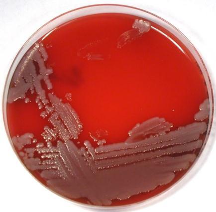
7 Gram Positive Bacteria 1.2 Streptococcus A. Electron Microscope. B. Light Microscope (Gram Stain). C. Light Microscope (Methylene Blue). D. Colonies of Streptococcus on Nutrient Agar. E. Colonies of Streptococcus on Blood Agar. F. Colonies of Streptococcus on Blood Agar showing different types of hemolysis. G+ve, cocci, non-spore former, appear in chains. Small colonies, smooth, cocci, show hemolysis on Blood Agar. 6


8 Gram Positive Bacteria 1.3 Bacillus B. megaterium Bacillus cereus. A. Electron Microscope. B. Light Microscope. C. Light Microscope. D. Colonies of Bacillus cereus on Nutrient Agar. E. Colonies of Bacillus megaterium on Nutrient Agar. F. Colonies of Bacillus subtilis on Nutrient Agar. G+ve, bacilli, endospore former. Rough, opaque, large, branched end, white colonies. 7


9 Gram Positive Bacteria 1.4 Lactobacillus A. Electron Microscope. B. Light Microscope. C. Light Microscope. D. Light Microscope E. Light Microscope F. Colonies of Lactobacillus on Nutrient Agar. G+ve, long bacilli, non-spore former. Smooth, opaque, small, white colonies. 8


10 Gram Positive Bacteria 1.5 Pediococcus A. Electron Microscope. B. Light Microscope. C. Light Microscope. D. Colonies of Pediococcus on Nutrient Agar. G+ve, coccbacilli, appear in pairs or tetrads. Smooth, opaque, small, white colonies. 9


11 Gram Positive Bacteria 1.6 Leuconostoc A. Electron Microscope. B. Light Microscope. C. Colonies on Nutrient Agar. G+ve, ovoid-cocci, appear in pairs. Smooth, opaque, slime colonies. 1.7 Clostridium A. Electron Microscope. B. Light Microscope. C. Pink Colonies on D.R.C.M. G+ve, rod-shaped, spore forming anaerobic bacteria, drum stick like cell. Pink round colonies on D.R.C.M. media after adding NaOH for 20-30sec. 10


12 Other Gram Positive Bacteria 1.Actinomycetes A. Electron Microscope. B. Light Microscope. C. Light Microscope. D. Different morphologies of Actinomycetes. G+ve, non-spore former, appear like branched network hyphae. Colonies small, fragile, compressed, chalky, may be white, yellow or red. 11


13 Other Gram Positive Bacteria 2.Micrococcus A. Electron Microscope. B. Light Microscope. C. Colony on Nutrient Agar. 3.Bacillus anthracis D. Electron Microscope. E. Light Microscope. F. Colony on Nutrient Agar. 12


14 4.Listeria monocytogenes A. Electron Microscope. B. Light Microscope. C. Colony on M.M.A. media. Acid Fast Stain Gram Positive like Bacteria Mycobacterium tuberculosis A. Electron Microscope. B. Light Microscope. C. Colony on Agar media. 13


15 2- Gram Negative Bacteria 2.1 Escherichia coli A. Electron Microscope. B. Light Microscope. C. Light Microscope. D. Colonies on MaCconkey Agar. E. Colonies on Endo Agar. F. Colonies on E.M.B. Agar. G-ve, rod-shaped, non- spore former. Lactose fermenter, Pink colonies on Endo Agar & MaCconkey Agar, Green metallic sheen on EMB Agar. 14


16 Gram Negative Bacteria 2.2 Pseudomonas A. Electron Microscope. B. Light Microscope. C. Colonies on MaCconkey Agar. D. Colonies on Milk Agar. E. Colonies on Nutrient Agar. F. Colonies on Olive Oil Agar. G-ve, short bacilli, non- spore former. Lactose non fermenter, Pale colonies on MaCconkey Agar, mucoid colonies, secrete Pyocyanin pigment on Nutrient Agar & Milk Agar, Protease producer cause clear zones on Milk Agar, Lipase producer cause clear zones on Olive Oil media Agar. 15


17 Gram Negative Bacteria 2.3 Salmonella A. Electron Microscope. B. Light Microscope. C. Colonies on MaCconkey Agar. D. Colonies on S-S Agar. E. Colonies on XLD. F. Transparent with Black center. G-ve, short bacilli, non- spore former. Lactose non fermenter, Pale colonies on MaCconkey Agar, H 2 S producer, appear as Black colonies on S-S Agar while appear Transparent colony with black center on XLD medium. 16


18 Gram Negative Bacteria 2.4 Shigella A. Electron Microscope. B. Light Microscope. C. Colonies on MaCconkey Agar. D. Colonies on S-S Agar. E. Colonies on XLD. F. Transparent Colonies on XLD. G-ve, short bacilli, non- spore former. Lactose non fermenter, Pale colonies on MaCconkey Agar, H 2 S producer, appear as Black colonies on S-S Agar while appear Transparent colony on XLD medium. 17


19 Gram Negative Bacteria 2.5 Serratia A. Electron Microscope. B. Light Microscope. C. Colonies on MaCconkey Agar. D. Colonies on Nutrient Agar E. Colonies on Blood Agar. F. Serratia colonies. G-ve, rods, non- spore former. Small, smooth red colonies, Lactose non fermenter, Pale colonies on MaCconkey Agar, produce red pigments. 18


20 Other Gram Negative Bacteria 1.Brucella melitensis A. Electron Microscope. B. Light Microscope. C. Colony on Blood Agar. 2.Vibrio Cholerae A. Electron Microscope. B. Light Microscope. C. Colony on T.C.B.S. 3.Achromobacter A. Electron Microscope. B. Light Microscope. C. Colony on Agar media. 19


21 Other Gram Negative Bacteria 4.Flavobacterium A. Electron Microscope. B. Light Microscope. C. Colony on Agar media. 5.Proteus A. Electron Microscope. B. Light Microscope. C. Colony on Blood Agar. 6.Alcaligenes A. Electron Microscope. B. Light Microscope. C. Colony on Blood Agar. 20

22 21


23 1. Molds 1.1 Penicillium A. Electron Microscope. B. Light Microscope. C. Colony on Agar media. Brush-like conidiophore carries conidia. Green or Green-greyish color colonies grows over fruits especially citrus. 1.2 Cladosporium A. Electron Microscope. B. Light Microscope. C. Colony on Agar media. Conidiophores are more or less distinct from the vegetative hyphae, are erect, straight or flexuous, unbranched or branched only in the apical region, with geniculate sympodial elongation in some species. Colonies are rather slow growing, mostly olivaceous-brown to blackish brown but also sometimes grey, buff or brown, suede-like to floccose, often becoming powdery due to the production of abundant conidia. 22


24 Molds 1.3 Asperigillus niger A. Electron Microscope. B. Light Microscope. C. Colony on Agar media. Non-Branched conidiophore with bulb end carries conidia like sun rays. Pin like black growth. 1.4 Asperigillus flavus A. Electron Microscope. B. Light Microscope. C. Colony on Agar media. Non-Branched conidiophore with bulb end carries conidia. Pin like green growth. 23


25 Molds 1.5 Mucor A. Electron Microscope. B. Light Microscope. C. Colony on Agar media. Sporangia contain spores, do not have rhizoids. Cotton like white growth spotted with black color. 1.6 Rhizopus A.Electron Microscope. C.Colony on Agar media. B.Light Microscope. Sporangia contain spores, have rhizoids. Cotton like white growth spotted with black color. 24


26 Molds 1.7 Alternaria A. Electron Microscope. B. Light Microscope. C. Colony on Agar media. Pineapple like conidia multi-cellular, spetated horizontally & vertically, arrange in chains. Dark green deeply grown colonies, oil-drop like colony when seen upside down the Petridish. 1.8 Curvularia A. Electron Microscope. B. Light Microscope. C. Colony on Agar media. Swollen conidia, spetated horizontally only, arrange in triple or pentagonal arrangements. Green or black deeply grown colonies. 25


27 Molds 1.9 Fusarium A. Electron Microscope. B. Light Microscope. C. Colony on Agar media. Spindle-like conidia, multi-cellular. Colonies appear brown or pink in center & with white edges. 1.10Geotrichum A. Electron Microscope. B. Light Microscope. C. Colony on Agar media. Chains of hyaline, smooth, one-celled, subglobose to cylindrical, slimy arthroconidia (ameroconidia) by the holoarthric fragmentation of undifferentiated hyphae. Colonies are fast growing, flat, white to cream, dry and finely suede-like with no reverse pigment. 26


28 Molds 1.11 Botrytis A. Electron Microscope. B. Light Microscope. C. Colony on Agar media. Grape-like conidia on the conidiophore. Colonies appear white while the conidia are grey (Grey Mold Rot) Sporotrichum A. Electron Microscope. B. Light Microscope. C. Colony on Agar media. Septate hyphae, conidiophores, aleuriconidia, arthroconidia, and chlamydospores are observed. Hyphae contain clamp connections (bridges) at the septa. Conidiophores may be short or long, simple or branched. The texture is velvety to powdery. From the front, the color is initially white and then becomes rosy beige, pinkish, yellow or orange. From the reverse, it is tannish. 27
29 28


30 2. Yeast 2.1 Saccharomyces A. Electron Microscope. B. Light Microscope. C. Colony on Agar media. Unicellular cocci or ovoid shape, larger than bacterial cells. Flat, smooth, moist, glistening or dull, and cream to tannish cream in color. 2.2 Rhodotorula A. Electron Microscope. B. Light Microscope. C. Colony on Agar media. Unicellular cocci or ovoid shape, larger than bacterial cells. Colony color can vary from being cream colored to orange/red/pink or yellow. 29


31 Yeast 2.3 Endomyces D. Electron Microscope. E. Light Microscope. F. Colony on Agar media. A species of yeast-like fungus that is the perfect state of Geotrichum, it forms a real mycelium, cocci or ovoid ascospore, the mycelium divided into cylindrical arthrospores with round edges. Large white colonies. 2.4 Candida D. Electron Microscope. E. Light Microscope. F. Colony on Agar media. Unicellular cocci or ovoid shape, larger than bacterial cells. Flat, smooth, large colonies. 30
32 References Dennis Kunkel Microscopy, Inc. Science Stock Photography cited by Free Encyclopedia online cited by Mycology online reference cited by The University of Adelaide, Australia, Mycology online cited by Tony Hart, Paul Shears (2004).Color Atlas of Medical Microbiology: Elsevier s Health Sciences Department. US National library of Medicine cited by 31
Overview of the major bacterial pathogens The major bacterial pathogens are presented in this table:
 Practical Microbiology 30/11/2018 University of Sulaimani college of Pharmacy Year2 Lab. 5: Overview of the major bacterial pathogens The major bacterial pathogens are presented in this table: Major Bacterial
Laboratory Training and Procedures Bacteriological Techniques Sputum smear Antoine Pierson (BiolTrop)
") Laboratory Training and Procedures Bacteriological Techniques Sputum smear Antoine Pierson (BiolTrop) Cocci gram positif Bacterial species Macroscopique Culture media Microscopic appearance Reference characteristics
Laboratory Training and Procedures Bacteriological Techniques Sputum smear Antoine Pierson (BiolTrop) Cocci gram positif Bacterial species Macroscopique Culture media Microscopic appearance Reference characteristics
NUT-TTC/EMB Code 5541
 NUT-TTC/EMB Code 5541 COMING SOON! BioPaddles Colony Identification App Nutrient-TTC Agar (NUT-TTC) Eosin Methylene Blue Agar (EMB) USE: Isolation and differentiation of Gram (-) enteric bacilli. Coliform
colony size color morphology haemolysis S. aureus S. epidermidis
 practical 2.: STAPHYLOCOCCUS 1. Prepare a heat fixed smear of the culture of S.aureus. (Gram staining, microscopy). 2. Prepare a heat fixed smear of the culture of S.aureus. and S.epidermidis (mixed smear),
Microbiology. Definition of a Microorganism. Microorganisms in the Lab. The Study of Microorganisms
 Microbiology The Study of Microorganisms Definition of a Microorganism Derived from the Greek: Mikros, «small» and Organismos, organism Microscopic organism which is single celled (unicellular) or a mass
Shape, Arrangement, and Size. Cocci (s., coccus) bacillus (pl., bacilli) 9/21/2013
 bacillus (pl., bacilli) 9/21/2013") Shape, Arrangement, and Size Cocci (s., coccus) are roughly spherical cells. The other common shape is that of a rod, sometimes called a bacillus (pl., bacilli). Spiral-shaped procaryotes can be either
Shape, Arrangement, and Size Cocci (s., coccus) are roughly spherical cells. The other common shape is that of a rod, sometimes called a bacillus (pl., bacilli). Spiral-shaped procaryotes can be either
BIOL 3702L: MICROBIOLOGY LABORATORY SCHEDULE, SUMMER 2015
 BIOL 3702L: MICROBIOLOGY LABORATORY SCHEDULE, SUMMER 2015 Week of May 18 th Introduction to the Microbiology Laboratory: Become familiar with the laboratory and its safety features Review safety rules
Figure Page 117 Microbiology: An Introduction, 10e (Tortora/ Funke/ Case)
") Chapter 11 The Prokaryotes: Domains Bacteria and Archaea Objective Questions 1) Which of the following are found primarily in the intestines of humans? A) Gram-negative aerobic rods and cocci B) Aerobic,
Chapter 11 The Prokaryotes: Domains Bacteria and Archaea Objective Questions 1) Which of the following are found primarily in the intestines of humans? A) Gram-negative aerobic rods and cocci B) Aerobic,
Microbial Taxonomy. Classification of living organisms into groups. A group or level of classification
 Lec 2 Oral Microbiology Dr. Chatin Purpose Microbial Taxonomy Classification Systems provide an easy way grouping of diverse and huge numbers of microbes To provide an overview of how physicians think
Obligate anaerobes - cannot grow in the presence of oxygen Facultative anaerobes - can grow with or without oxygen Aerobic - require oxygen
 PROKARYOTES *include bacteria and archaea *singular: bacterium / plural: bacteria PROPERTIES 1. Bacteria are classified into two kingdoms: Eubacteria (true bacteria) and Archaebacteria (Ancient Bacteria).
GUJARAT UNIVERSITY Syllabus for First Year Microbiology Semester I and II Effective from June 2017
 GUJARAT UNIVERSITY Syllabus for First Year Microbiology Semester I and II Effective from June 2017 1. A student offering Microbiology programme will be offered two theory papers of core course MI 101 and
DEPARTMENT OF ANIMAL HEALTH TECHNOLOGY COURSE OUTLINE - FALL 2014 LAB PROCEDURES AND MICROBIOLOGY AH 174 E- MAIL:
 DEPARTMENT OF ANIMAL HEALTH TECHNOLOGY COURSE OUTLINE - FALL 2014 LAB PROCEDURES AND MICROBIOLOGY AH 174 INSTRUCTOR: Dr. Chris Mizzi Kristy Mergeart, RAHT PHONE: 780-835-6617 780-835-6779 OFFICE: AS 133
Laboratory Exercise # 7: Aseptic Technique
 Laboratory Exercise # 7: Aseptic Technique Purpose: The purpose of this laboratory exercise is to acquaint the student with the procedures of aseptic transfer of microbiological cultures. ntroduction:
THE IDENTIFICATION OF TWO UNKNOWN BACTERIA AFUA WILLIAMS BIO 3302 TEST TUBE 3 PROF. N. HAQUE 5/14/18
 THE IDENTIFICATION OF TWO UNKNOWN BACTERIA AFUA WILLIAMS BIO 3302 TEST TUBE 3 PROF. N. HAQUE Introduction: The identification of bacteria is important in order for us to differentiate one microorganism
Introduction to microbiology
 Sulaimani University College of Pharmacy Microbiology Introduction to microbiology Dr. Abdullah Ahmed Hama PhD. Molecular Medical Parasitology abdullah.hama@spu.edu.iq 1 Definition Microbiology: is the
INTRODUCTION budding, binary fission hyphae mycelium Figure 1.
 INTRODUCTION Although most of our work in this lab is done on bacteria, fungi are nonetheless an important aspect in microbiology. Besides being important food providers, fungi play central roles in recycling
Microstructure of Colonies of Rod-Shaped Bacteria
 JOURNAL OF BACTERIOLOGY, Oct. 1971, p. 515-525 Copyright 0 1971 American Society for Microbiology Vol. 108, No. I Printed in U.S.A. Microstructure of Colonies of Rod-Shaped Bacteria D. B. DRUCKER AND D.
D-10 VALUES OF MICRO-ORGANISMS
 INFORMATION SHEET SYNERGY HEALTH D-10 VALUES OF MICRO-ORGANISMS Synergy Health Applied Sterilisation Technologies (AST) operates as a global supplier of sterilisation, specialist applications and contamination
_ + Discriminates aerobic organisms that produce catalase to degrade hydrogen peroxide into water and oxygen
 Lab 11 Goals and Objectives: Catalase Test Exercise 39: Oxidation and Fermentation Tests (Catalase) Exercise 67: Staphylococci Identification (MSA & Coagulase) Exercise 68: Streptococci & Enterococci Identification
ENTEROBACTER AEROGENES UNKNOWN BACTERIA FLOW CHART UNKNOWN LAB REPORT, MICROBIOLOGY ENTEROBACTER AEROGENES
 ENTEROBACTER AEROGENES UNKNOWN BACTERIA PDF UNKNOWN LAB REPORT, MICROBIOLOGY ENTEROBACTER AEROGENES IDENTIFICATION OF AN UNKNOWN BACTERIAL SPECIES OF 1 / 5 2 / 5 3 / 5 enterobacter aerogenes unknown bacteria
Practical examination
 Practical examination I. Sterile media 1. Bouillon, 2. Slant agar, tube agar 4. Enrichment media: meat bouillon 3., 5., 6.: Agar, blood agar and chocolate agar plates 7. Selective and differentiating media
INTERPRETATION OF THE GRAM STAIN
 INTERPRETATION OF THE GRAM STAIN DISCLOSURE Relevant relationships with commercial entities none Potential for conflicts of interest within this presentation none Steps taken to review and mitigate potential
Introduction To Microbiology CLS 311
 Introduction To Microbiology CLS 311 What is microbiology? It is a branch of biology that studies microorganisms and their effects on humans Microorganisms a collection of organisms that share the characteristic
ID Membranes for Microbial Rapid Identification
 ID Membranes for Microbial Rapid Identification Chromogenic Reaction by Specific Substrates on Membranes Classical plates are still used today as first steps in modern microbiology. Smart, inexpensive
Gram negative bacilli
 Gram negative bacilli 1-Enterobacteriaceae Gram negative bacilli-rods Enterobacteriaceae Are everywhere Part of normal flora of humans and most animals They are cause of -30-35% septisemia -more than 70%
Microbiology. Microbiology derived by Greek mikros (small) bios (life) logos (science)
 bios (life) logos (science)") MBIO140 Lecture-1 Microbiology derived by Greek mikros (small) bios (life) logos (science) Microbiology The study of organisms too small to be seen individually with the naked eye during part or all of
MBIO140 Lecture-1 Microbiology derived by Greek mikros (small) bios (life) logos (science) Microbiology The study of organisms too small to be seen individually with the naked eye during part or all of
Required Materials: immersion oil microscopes Kim-wipes prepared microscope slides
 Microbiology CA/IA Lab Microscopic Examination of Microbes September 10 Objectives: 1. learn how to use a microscope to examine microbes 2. learn to recognize the characteristics of different microbes
Game plan Lecture Lab Prelabs
 Game plan Lecture Binary fission Growth curves Physical requirements for growth Chemical requirements for growth Lab Lab Exam Prelabs Growth Curve Bring books and APO-3 for next class Microbial growth
Considerations with Antibiotic Therapy PART
 Considerations with Antibiotic Therapy PART 1 The Wonderful World of Microbiology 1 Despite the promises of the household-products industry, almost every surface is covered in microorganisms almost all
Biology I: Macaw Book Unit IV: Microbiology
 Biology I: Macaw Book Unit IV: Microbiology Chapter 21 Fungus Fungus Lab Name: Date: Hour: Fungus Lab Pre-Lab Discussion Fungi are heterotrophic eukaryotes with cell walls that contain chitin. Most people
MICROBIAL BIOCHEMISTRY BIOT 309. Dr. Leslye Johnson Sept. 30, 2012
 MICROBIAL BIOCHEMISTRY BIOT 309 Dr. Leslye Johnson Sept. 30, 2012 Phylogeny study of evoluhonary relatedness among groups of organisms (e.g. species, populahons), which is discovered through molecular
CLASSIFICATION OF MICROORGANISMS
 CLASSIFICATION OF MICROORGANISMS DISCLOSURE Relevant relationships with commercial entities none Potential for conflicts of interest within this presentation none Steps taken to review and mitigate potential
Worksheet for Morgan/Carter Laboratory #13 Bacteriology
 Worksheet for Morgan/Carter Laboratory #13 Bacteriology Ex. 13-1: INVESTIGATING CHARACTERISTICS OF BACTERIA Lab Study A: Colony Morphology Table 13.1 Characteristics of Bacterial Colonies Name of Bacteria
Kharkov National Medical University. Head of Microbiology, Virology and Immunology Department Minukhin Valeriy Vladimirivich
 Kharkov National Medical University Head of Microbiology, Virology and Immunology Department Minukhin Valeriy Vladimirivich Tkachenko Victoria 1, 5, 11, 14, 19, 21, 30 Kovalenko Natalia 2, 12, 25, 29 Siritsa
Proteus & Pseudomonas
 Proteus & Pseudomonas Ahmad Ausama Al-Kazzaz Anas Huthaifa AL-Dewachi Ameer Saadallah Zacko Al-Ta i Supervised by: Dr. Khalid Ahmad Ausama Al-Kazzaz Proteus Proteus is a genus of Gram-negative Proteobacteria
Domain: Eukarya Kingdom: FUNGI
 Domain: Eukarya Kingdom: FUNGI Fungi are eukaryotic heterotrophs that have cell walls. They are part of the nature s recycling system. They break down organic compounds. Fungi are used in wine, beer, cheese,
CH 5 Mostly Microorganisms. Microorganisms covered in this chapter:
 Biology 2201 Name: CH 5 Mostly Microorganisms Microorganisms covered in this chapter: Kingdom Bacteria or Monera: Pg. 132-137 Bacteria are simple, prokaryotic organisms. They can be classified according
ISOLATION AND IDENTIFICATION OF BACTERIA FROM SOME SPOILED FRUITS
 Plant Archives Vol. 16 No. 2, 2016 pp. 834-838 ISSN 0972-5210 ISOLATION AND IDENTIFICATION OF BACTERIA FROM SOME SPOILED FRUITS Lalita Chaudhary* and T. S. Dhaka Department of Botany, D.A.V. (PG) College,
Kingdom Fungi. 1. Student will be able to describe the characteristic features in the kingdom Fungi.
 Kingdom Fungi Molds, Sac Fungi, Mushrooms, and Lichens Essential Question(s): What makes fungi have their own kingdom? Objectives: 1. Student will be able to describe the characteristic features in the
Objects of the Medical Microbiology revision a) Pathogenic microbes (causing diseases of human beings or animals) b) Normal microflora (microbes commo
 Pathogenic microbes (causing diseases of human beings or animals) b) Normal microflora (microbes commo") Institute for Microbiology, Medical Faculty of Masaryk University and St. Anna Faculty Hospital in Brno Miroslav Votava MORPHOLOGY AND STRUCTURE OF BACTERIAL CELL The 2nd lecture for 2nd-year students
Institute for Microbiology, Medical Faculty of Masaryk University and St. Anna Faculty Hospital in Brno Miroslav Votava MORPHOLOGY AND STRUCTURE OF BACTERIAL CELL The 2nd lecture for 2nd-year students
Day 2 - Viewing a prepared slide of mixed bacteria on high power.
 Purpose Bacteria Lab To compare the quantity and the different types of bacteria from four different locations within the school. To identify 3 different bacterial colonies on a prepared slide. Materials
Bacteria are very small
 BACTERIA BACTERIA Bacteria are very small Bacteria are very small compared to cells with nuclei (Eukaryotic cells) This is a pore in human skin and the yellow spheres are bacteria CLASSIFICATION OF BACTERIA
THE GRAM STAIN OBJECTIVE/RATIONALE KEY POINTS
 THE GRAM STAIN OBJECTIVE/RATIONALE One of the first procedures preformed by the medical microbiologist for the identification of bacteria is the Gram Stain. The student will learn the procedure for performing
Know Your Microbes Introduction to food microbiology Factors affecting microbial growth Temperature Time
 Know Your Microbes Know Your Microbes Introduction to food microbiology Factors affecting microbial growth Temperature Time ph Water activity (Aw) Nutrient availability Atmosphere Hurdle technology Foodborne
Bacteria are very small
 BACTERIA BACTERIA Bacteria are very small Bacteria are very small compared to cells with nuclei This is a pore in human skin and the yellow spheres are bacteria BACTERIA LIVE ALMOST EVERYWHERE Hot springs
ISSN: IJBPAS, December, 2014, 3(12):
:") : 2753-2762 ISSN: 2277 4998 MICROBIAL DIVERSITY OF FOREST ECOSYSTEM OF ACHANAKMAR TIGER RESERVE IN CHHATTISGARH STATE OF INDIA BARIK K*, SAO S AND PARIHAR DK 2 Department of Microbiology, Dr. C.V. Raman
: 2753-2762 ISSN: 2277 4998 MICROBIAL DIVERSITY OF FOREST ECOSYSTEM OF ACHANAKMAR TIGER RESERVE IN CHHATTISGARH STATE OF INDIA BARIK K*, SAO S AND PARIHAR DK 2 Department of Microbiology, Dr. C.V. Raman
The Scope of Food Microbiology p. 1 Micro-organisms and Food p. 2 Food Spoilage/Preservation p. 2 Food Safety p. 4 Fermentation p.
 The Scope of Food Microbiology p. 1 Micro-organisms and Food p. 2 Food Spoilage/Preservation p. 2 Food Safety p. 4 Fermentation p. 4 Microbiological Quality Assurance p. 4 Micro-organisms and Food Materials
Domain Bacteria. BIO 220 Microbiology Jackson Community College
 Domain Bacteria BIO 220 Microbiology Jackson Community College John Ireland, Ph.D. 2006 Scientific Nomenclature Domain - Bacteria Phylum Important for gross characteristics Class Intermediate characteristics
PROFILE OF BACTERIA AND FUNGI ON MONEY COINS
 151 East African Medical Journal Vol. 86 No. 4 April 2009 PROFILE OF BACTERIA AND FUNGI ON MONEY COINS J.K.N. Kuria, BVM, MSc, PhD, Senior Lecturer, Department of Veterinary Pathology and Microbiology,
CATALOGUE OF TEST, CONTROL OR BIOASSAY STRAINS FROM BCCM CULTURE COLLECTIONS
 CATALOGUE OF TEST, CONTROL OR BIOASSAY STRAINS FROM BCCM CULTURE COLLECTIONS LIST OF STRAINS INVOLVED IN NORMS AND STANDARDISED MICROBIOLOGICAL METHODS The Belgian Co-ordinated Collections of Micro-organisms
B. Correct! Bacillus anthraces produces spores that can cause anthrax. D. Incorrect! Diphtheria is caused by Corynebacterium diphtheriae.
 Microbiology - Problem Drill 09 - The Prokaryotes No. 1 of 10 1. Bacillus anthraces is most closely associated with which of the following? (A) Botulism poisoning (B) Anthrax (C) Gangrene (D) Diphtheria
Characteristics of Predominant Microorganisms in Food
 CHAPTER 2 Characteristics of Predominant Microorganisms in Food CONTENTS I. Introduction...14 II. Classification of Microorganisms...15 III. Nomenclature...16 IV. Morphology and Structure of Microorganisms
Bacteria. The Three Types of Important Heterotrophic Bacteria
 Bacteria Kingdom Monera Prokaryote (their genetic material is not bound with a membrane) Classified according to shape - Spherical (cocci) - Spiral - Rod Shaped -TWO TYPES: Heterotrophic (organism that
Have you had a cold, flu, or other infectious disease recently? Do you
 45 The World of Microbes r e a d i n g Have you had a cold, flu, or other infectious disease recently? Do you know what caused your illness? Microbes cause most infectious diseases. Microbes include the
Microscopy, Staining, and Classification
 PowerPoint Lecture Presentations prepared by Mindy Miller-Kittrell, North Carolina State University C H A P T E R 4 Microscopy, Staining, and Classification Figure 4.3 The limits of resolution (and some
Microbiological Testing Summary
 BACTERICIDAL ACTIVITY EN 1276 - Chemical disinfectants and antiseptics - Quantitative suspension test for the evaluation of bactericidal activity of chemical disinfectants and antiseptics used in food,
INTRODUCTION. Gram Stain
 INTRODUCTION In microbiology, organisms are so small that additional techniques are often required for proper viewing under the microscope. Cytological stains, or dyes that stain cells or cellular features,
Kingdom Fungi. Learning Objectives. Introduction. Activity1: Zygomycota. Revised Fall 2017
 Kingdom Fungi Revised Fall 2017 ** You will require your text book Biological Science during this lab ** Learning Objectives Building on the learning objectives from your lab syllabus, you will be expected
PREDICTIVE MICROBIOLOGICAL MODELS: WHAT ARE THEY AND HOW CAN THEY BE USED IN THE FOOD INDUSTRY?
 PREDICTIVE MICROBIOLOGICAL MODELS: WHAT ARE THEY AND HOW CAN THEY BE USED IN THE FOOD INDUSTRY? WHY USE MODELS? Predictive microbiological models are tools that can be used to assess product shelf-life
Food Testing- Bacillus species. Dr Roy Betts Head of Microbiology Campden BRI, Chipping Campden. UK
 Food Testing- Bacillus species. Dr Roy Betts Head of Microbiology Campden BRI, Chipping Campden. UK Who are Campden BRI? Independent Food Research Organisation Membership based with over 2400 members International
Lab Exercise: Diversity of Eukaryotic Microbes
 Lab Exercise: Diversity of Eukaryotic Microbes OBJECTIVES 1. To observe representatives of major types of microbes. 2. To cultivate select representatives of major types of microbes. 3. Understand key
Major Events in the History of Earth
 Major Events in the History of Earth Cenozoic Humans Land plants Animals Origin of solar system and Earth Multicellular eukaryotes 1 Proterozoic eon 2 Archaean eon 3 4 Single-celled eukaryotes Atmospheric
SSI ENTERIC PRODUCT INFORMATION. Detects all Enterobacteria. Direct identification. Rapid diagnosis. Cost saving
 SSI ENTERIC M E D I U M Detects all Enterobacteria Direct identification Rapid diagnosis Cost saving SSI Diagnostica 2 Herredsvejen 3400 Hillerød Denmark PRODUCT INFORMATION Tel: +45 4829 9100 Fax: +45
Ladue Microbe Mission Test SCORE: / 90 Name: Date:
 Ladue Microbe Mission Test SCORE: / 90 Name: Date: You may not return to previous stations. However, you can move to another station early if you want to do so. I won t judge you for your grammar/writing
Evaluation of the efficiency of Mxxxx as a barrier against microrganisms crossing
 Evaluation of the efficiency of as a barrier against microrganisms crossing A) composition of filter The filter of has the following characteristics: 1. An outer layer, which is composed by a medical,
LABORATORY 7 ENDOSPORE STAIN AND BACTERIAL MOTILITY
 LABORATORY 7 ENDOSPORE STAIN AND BACTERIAL MOTILITY A. Endospore Stain B. Bacterial Motility A. ENDOSPORE STAIN DISCUSSION A few genera of bacteria, such as Bacillus and Clostridium have the ability to
EZ-COMP EZ-COMP For Training and Proficiency Testing Product Details
 EZ-COMP For Training and Proficiency Testing Mixed microorganism populations Identified by codes rather than descriptions Refrigerated storage Traceable to reference culture Product warranty Product Details
Pharmaceutical Microbiology Forum Newsletter Vol. 12 (4) Page 3 of 14 (NCIMB 8545, CIP NBRC. Salmonella enterica ssp typhimurium
 Page 3 of 14 (NCIMB 8545, CIP NBRC. Salmonella enterica ssp typhimurium") Page 3 of 14 Continued from page 2 Table 2. Absence of Specified Details Media Growth Promotion Organisms for Trypticase Soy Staphylococcus aureus Escherichia coli Pseudomonas aeruginosa Salmonella Staphylococcus
Page 3 of 14 Continued from page 2 Table 2. Absence of Specified Details Media Growth Promotion Organisms for Trypticase Soy Staphylococcus aureus Escherichia coli Pseudomonas aeruginosa Salmonella Staphylococcus
Vocabulary- Bacteria (34 words)
") Biology II BACTERIA Vocabulary- Bacteria (34 words) 1. Prokaryote 21. phototroph 2. Peptidoglycan 22. chemotroph 3. Methanogen 23. obligate anaerobe 4. Halophile 24. facultative anaerobe 5. Thermoacidophile
Biology II BACTERIA Vocabulary- Bacteria (34 words) 1. Prokaryote 21. phototroph 2. Peptidoglycan 22. chemotroph 3. Methanogen 23. obligate anaerobe 4. Halophile 24. facultative anaerobe 5. Thermoacidophile
INTERNATIONAL RESEARCH JOURNAL OF PHARMACY
 INTERNATIONAL RESEARCH JOURNAL OF PHARMACY www.irjponline.com ISSN 2230 8407 Research Article ANTIMICROBIAL ACTIVITY OF LACTIC ACID BACTERIAL ISOLATES Utkarsha S. Shivsharan 1 *, Milind J. Bhitre 2 1 DSTSM
MICROBIOLOGY CHAPTER 1 INTRODUCTION TO MICROORGANISMS
 MICROBIOLOGY CHAPTER 1 INTRODUCTION TO MICROORGANISMS 1:1 What is Microbiology? MICROBIOLOGY: the study of living organisms that are individually too small to be seen with the unaided eye e.g. bacteria,
If you are looking for the book by R. C. W. Berkeley The Aerobic Endospore-Forming Bacteria: Classification and Identification (Special publications
 The Aerobic Endospore-Forming Bacteria: Classification And Identification (Special Publications Of The Society For General Microbiology) By R. C. W. Berkeley READ ONLINE If you are looking for the book
Fungi Coloring Worksheet
 Fungi Coloring Worksheet The basic structural features of fungi are not cells but hyphae. Hyphae are microscopic branching filaments filled with cytoplasm and nuclei. Each thread consists of a tube formed
MORPHOLOGY: the study of form and structure
 MICROBIOLOGY CHAPTER 3 Bacteria Morphology 3:1 Bacteria Structure and Function MORPHOLOGY: the study of form and structure Structure of Bacteria 1. PROKARYOTIC no membrane bound nucleus nor other organelles
Labquality External Quality Assessment Programmes General Bacteriology 1 2/2014
 Labquality External Quality Assessment Programmes General Bacteriology 1 2/2014 Photos and text: Markku Koskela, M.D., Ph.D. Clinical microbiology specialist NordLab Oulu, Finland Specimen 21/2014 Pus
Principles of Biotechnology Lectures of week 4 MICROBIOLOGY AND BIOTECHNOLOGY
 Principles of Biotechnology Lectures of week 4 MICROBIOLOGY AND BIOTECHNOLOGY INTRODUCTION TO MICROBIOLOGY What are microbes? Germs, microbe s s microorganisms are minute living things that individually
Agronomy 485/585 Test #1 October 2, 2014
 Agronomy 485/585 Test #1 October 2, 2014 Name Part I. Circle the one best answer (2 points each). 1. The most important microbial group in promoting soil structure likely is the. a) actinomycetes b) algae
CYTOLOGICAL CHANGES IN AGING BACTERIAL CULTURES
 CYTOLOGICAL CHANGES IN AGING BACTERIAL CULTURES B. R. CHATTERJEE AND ROBERT P. WILLIAMS Department of Microbiology, Baylor University College of Medicine, Houston, Texas Received for publication March
Introduction to Microbiology. CLS 212: Medical Microbiology Miss Zeina Alkudmani
 Introduction to Microbiology CLS 212: Medical Microbiology Miss Zeina Alkudmani Microbiology Micro- means very small (that needs a microscope to see). Microbiology is the study of very small living organisms.
General Fungus Anatomy: Yeast: single cell fungi that reproduces by fission or budding
 Make-Up Assignment: Using the notes below as a guide, look up the organisms you are required to draw on the internet or in a book. Draw the organism in the circles provided and write a description of the
Nadia Langha Biology 106 Honors Project
 Nadia Langha Biology 106 Honors Project Cyanobacteria Domain Bacteria Division Cyanophyta Cyanobacteria also known as BlueGreen Algae -Cyano=blue Bacteria are more closely related to prokaryotic bacteria
MEDICAL MYCOLOGY. Medical Mycology Reference Laboratory
 MEDICAL MYCOLOGY Medical Mycology Reference Laboratory Laboratory of Parasitology and Mycology Institute of Microbiology and Immunology Medical Faculty in Belgrade, 2010 The goal of teaching: 1. The morphology
علم األحياء الدقيقة Microbiology Introduction to Bacteriology تركي محمد الداود مكتب 2 ب 45
 علم األحياء الدقيقة Microbiology Introduction to Bacteriology د. تركي محمد الداود مكتب 2 ب 45 Occurrence & distribution of bacteria - They live everywhere. They occur in water (fresh and salty), in soil
CLASSIFICATION OF BACTERIA
 CLASSIFICATION OF BACTERIA DISCLOSURE Relevant relationships with commercial entities none Potential for conflicts of interest within this presentation none Steps taken to review and mitigate potential
MICROBIOLOGY LAB #1 SAFETY RULES & GRAM STAIN METHOD
 MICROBIOLOGY LAB #1 SAFETY RULES & GRAM STAIN METHOD Precaution processes are extremely important when working with cultures in the lab for the safety of the microbiologist from getting diseases from bacteria
CELL DIFFERENTIATION & GROWTH OF ORGANISMS BIOLOGY TEAM
 CELL DIFFERENTIATION & GROWTH OF ORGANISMS BIOLOGY TEAM Agricultural Technology Faculty Brawijaya University 2013 OVERVIEW Growth Definition & Terminology Differentiation & Growth of Unicellular Organisms
Microchem Silliker Private Limited, A-513, Microchem House, TTC Industrial Area, Mahape, Navi Mumbai, Maharashtra
 Last Amended on 28.03.2014 Page 1 of 8 I. FOOD & AGRICULTURAL PRODUCTS 1. All type of Food and Agri. products Aerobic Plate Count IS: 5402 2012 ISO: 4833 Part 1 2013 Yeast & Mould Count IS: 5403 1999 ISO-21527-2:2008
THE OHIO JOURNAL OF SCIENCE
 THE OHIO JOURNAL OF SCIENCE VOL. XXXVIII SEPTEMBER, 1938 No. 5 STUDIES IN ANTIBIOSIS BETWEEN BACTERIA AND FUNGI 1 CONST. J. ALEXOPOULOS, R. ARNETT, and A. V. McINTOSH Department of Biology, Kent State
EASTERN ARIZONA COLLEGE Microbiology
 EASTERN ARIZONA COLLEGE Microbiology Course Design 2015-2016 Course Information Division Science Course Number BIO 205 (SUN# BIO 2205) Title Microbiology Credits 4 Developed by Ed Butler/Revised by Willis
Characterization and Cytotoxicity Assay of Pigment Producing Microbes
 International Journal of Current Microbiology and Applied Sciences ISSN: 2319-7706 Volume 5 Number 6 (2016) pp. 370-376 Journal homepage: http://www.ijcmas.com Original Research Article http://dx.doi.org/10.20546/ijcmas.2016.506.042
Electric polarization properties of single bacteria measured with electrostatic force microscopy
 Electric polarization properties of single bacteria measured with electrostatic force microscopy Theoretical and practical studies of Dielectric constant of single bacteria and smaller elements Daniel
NAME: Microbiology BI234 MUST be written and will not be accepted as a typed document. 1.
 Chapter 3 Study Guide Explain the 3 main characteristics that help differentiate prokaryotes from eukaryotes. What are the 7 structures/substances found in all bacterial cells? What are 8 specific structures
Policy # MI_BYID Department of Microbiology. Page Quality Manual TABLE OF CONTENTS. Vitek MS Guide to Bacteria and Yeast Identification:...
 Department of Microbiology Version: 1.1 CURRENT 1 of 18 Prepared by QA Committee Issued by: Laboratory Manager Revision Date: 4/20/2018 Approved by Laboratory Director: Annual Review Date: 5/1/2019 Microbiologist-in-Chief
Microbiology Helmut Pospiech
 Microbiology http://researchmagazine.uga.edu/summer2002/bacteria.htm 05.04.2018 Helmut Pospiech The Species Concept in Microbiology No universally accepted concept of species for prokaryotes Current definition
Adding the class. Prerequisites. Welcome to Bio 139 General Microbiology. Syllabus. Amy Rogers, M.D., Ph.D.
 Adding the class Welcome to Bio 139 General Microbiology Amy Rogers, M.D., Ph.D. Lectures MW 12:00-1:15 PM (section 8) Labs: MW 1:30-2:45 PM or MW 3:00 PM-4:15 PM I anticipate a large number of students
Sample Date: March 30, 2018 Date Received: March 31, 2018 Date of Report: April 9, 2018 (877) Fax: (877)
 Fax: (877)") U.S. Micro-Solutions, Inc. * 075 South Main Street, Suite 04 * Greensburg, PA 560 Phone: (877) 876-4276 Fax: (724) 853-4049 AIHA-LAP, LLC EMLAP #03009 075 South Main Street, Suite 04 Greensburg, PA 560
U.S. Micro-Solutions, Inc. * 075 South Main Street, Suite 04 * Greensburg, PA 560 Phone: (877) 876-4276 Fax: (724) 853-4049 AIHA-LAP, LLC EMLAP #03009 075 South Main Street, Suite 04 Greensburg, PA 560
A word of caution about a little knowing Lab organisms limit the view of the world of microbiology
 Diversity The world of living things (Figure from Madigan et al. 2002) Microbes in all three domains Two of the domains are exclusively prokaryotic and microbial The third contains both unicellular and
Biology Test Pack WALCH PUBLISHING
 Biology Test Pack WALCH PUBLISHING Table of Contents To the Teacher........................................................... v Testing Students Who Do Not Test Well.....................................
Acta Cryst. (2014). D70, doi: /s
. D70, doi: /s") Acta Cryst. (2014). D70, doi:10.1107/s1399004714021166 Supporting information Volume 70 (2014) Supporting information for article: Elucidation of the bicarbonate binding site and insights into the carboxylation
Acta Cryst. (2014). D70, doi:10.1107/s1399004714021166 Supporting information Volume 70 (2014) Supporting information for article: Elucidation of the bicarbonate binding site and insights into the carboxylation
21-2 Classification of Fungi Slide 2 of 44
 2 of 44 Fungi are classified according to their structure and method of reproduction. The four main groups of fungi are: Common molds (Zygomycota) Sac fungi (Ascomycota) Club fungi (Basidiomycota) Imperfect