POSITIONAL DISCRIMINATION AND RE-DEVELOPMENT OF SYNAPSES IN THE LEECH WHITMANIA PIGRA
|
|
|
- Todd Curtis
- 5 years ago
- Views:
Transcription
 47 'rimed in Great Britain The Company of Biologists Limited 1990 POSITIONAL DISCRIMINATION AND RE-DEVELOPMENT OF SYNAPSES IN THE LEECH WHITMANIA PIGRA BY REN-JI ZHANG, LIXIA ZHU,")
1 . exp. Biol. 153, (1990) 47 'rimed in Great Britain The Company of Biologists Limited 1990 POSITIONAL DISCRIMINATION AND RE-DEVELOPMENT OF SYNAPSES IN THE LEECH WHITMANIA PIGRA BY REN-JI ZHANG, LIXIA ZHU, DAN-BING WANG, FAN ZHANG AND DONG-JING ZOU* Department of Biology, Peking University, Beijing , The People's Republic of China Summary Identified neurones in the leech Whitmania pigra have a stable morphology with bilaterally symmetrical branching arborizations, and with axons on both sides arranged symmetrically in the connectives. Each anterior pagoda cell (AP) receives electrical and/or chemical synaptic input from mechanoreceptive cells on both sides of the body. 1 The position in the body can be discriminated by the postsynaptic responses of the APs: as a rule, the responses to input from contralateral receptive neurones are stronger than those to input from ipsilateral ones, and the neurone with its receptive field on the dorsal side produces a stronger response than the neurone with a ventrally sited receptive field. APs integrate postsynaptic potentials and spikes. There are no connections between the two AP cells and so it is possible that positional discrimination depends upon a circuit comparing the inputs. After the body wall has been cut round and rotated by 180, the mechanoreceptive cells and annular erector motoneurones reinnervate the body wall strictly according to their original orientation, and repair is bilaterally synchronous. This eliminates a role for target cell guidance, particularly in the adult leech. When an extra Retzius cell is implanted into cultured ganglia, synapses develop between the host and the implanted neurone. Such synapses generally show lower coupling ratios or PSP fluctuations. However, the specific electrical connection between the Retzius cells shows a normal coupling ratio. Introduction The leech preparation used in this investigation has a number of advantages: (1) the sensory and motor neurones are identifiable and innervate defined receptive fields (Nicholls and Baylor, 1968) or muscle groups (Bowling et al. 1978); (2) in Whitmania pigra the axons of sensory and motor neurones have a stable structural pattern and show symmetrical arborizations in the ganglia and connectives; (3) a pair of identified neurones, the anterior pagoda (AP) cells, can discriminate and integrate the PSPs evoked by mechanoreceptive signals from both sides of the body. These neurones express their ability for spatial discrimination both centrally Present address: Department of Pharmacology, Biocenter, Basel, Switzerland. Key words: leech, synapse, discrimination, integration, re-development.

2 48 R.-J. ZHANG AND OTHERS and peripherally. Centrally, discrimination occurs at the level of the synapse, where the strength of excitation varies depending upon the position of the receptive field. The second site is located at the growth cone of the peripheral terminal which differentiates the position to be innervated. The two problems, that of forming connections and that of signalling, involve very different mechanisms and different experimental approaches. Expression of neurone morphology The stability of neuronal architecture The morphology of leech neurones was reviewed by Muller etal. (1981). Recently, the stability of axon architecture in embryonic leech has been recognized (Gao and Macagno, 1987). In order to assess the stability of axon architecture in Whitmania pigra, individual neurones were visualized by injection with horseradish peroxidase and the number of cells showing morphological variation was expressed as a percentage of the total number of neurones examined. In some cells no variation was found; 100% of T cells (N=52), P cells (7V=31) and N cells (N=177) had a stable morphology. Of the Retzius (R) cells, 88 % (71 of a sample of 81) showed a consistent axon pattern. The main variations of Retzius cell architecture occurred on the collateral branches towards the anterior connectives (Wang and Zhang, 1990). Of the 260 AP cells examined, 96 % had a stable morphology; from the second to the nineteenth segmental ganglion, the branching pattern was the same. Symmetrical arborization of axons In the leech the T, P and N mechanoreceptive cells supply receptive fields in the periphery. Similarly, the innervation of the annuli erector muscles on both sides of the body by the annuli erector neurones (AE) is symmetrical. Several neurones also show symmetrical axonal arborization within the mature central nervous system. In Whitmania pigra, T, P, N, AE and Retzius cells all show symmetrical axonal branching (Wang and Zhang, 1990) and AP neurones show symmetrical axonal branching throughout the chain of segmental ganglia (Fig. 1). Symmetrical loci of axons in connectives The central nervous system (CNS) of the leech consists of a series of ganglia linked by connectives. Neurones send out their axons to the adjacent anterior and posterior ganglia. Retzius cells send out a collateral axon from each of these branches towards the anterior and posterior roots separately, and also to the connectives. These branches are always located in a particular part of the connectives. Retzius and N cell axons are located in the ventral-lateral area of Fig. 1. Symmetrical branching pattern of the AP neurones throughout the chain of segmental ganglia in Whitmania pigra. The order of the ganglia is indicated by the number. Scale bar,

3 Synapse discrimination and re-development in leech 49 f
4 50 R.-J. ZHANG AND OTHERS Ventral Dorsal Fig. 2. Symmetrical loci of identified axons in connectives. Upper axon, loci of Retzius cells; lower axon, loci of N cells. connectives (Fig. 2). There is no obvious difference in the position of these two neurones. Positional discrimination and integration of receptive signals on AP neurones Synaptic connections between receptive neurones and the AP cell The AP neurone contains both acetylchonine (ACh) (Wallace, 1981) and FMRFamide (Kuhlman et al. 1988); its function is unknown, and its connections with other cells have not been ascertained. In Whitmania pigra, rectifying electrical synapses occur between the receptive cells (T, P, medial N) and the AP neurones. When ganglia were bathed in normal Ringer's solution no facilitation of excitatory postsynaptic potentials (EPSPs) could be found when paired electrical stimuli were delivered to medial P cells. When a depolarizing current was passed into a medial P cell bathed in a Ringer's solution containing lommoll" 1 Ca 2+ or 15 mmol P 1 Mg 2 " 1 ", EPSPs recorded in the contralateral AP neurone were the same as those recorded in normal Ringer. When a train of depolarizing impulses was

5 Synapse discrimination and re-development in leech 51 Table 1. Types of synoptic connection between T, P and N sensory cells and the AP neurone T P N Ipsilateral medial Contralateral medial Contralateral lateral Electrical synapse Rectifying Rectifying Rectifying Rectifying Chemical synapse IPSP EPSP passed to the medial P cell, synaptic facilitation and suppression were not seen on AP neurones; the coupling ratio recorded from a medial P cell when the hyperpolarizing current was passed into an AP neurone was only This indicates that there is a rectifying connection between medial P and AP cells. The inhibitory chemical synapse from a medial N cell to an AP cell seems to be a polysynaptic connection. The connections between other cells and the AP neurone are shown in Table 1. In culture, the synapses formed between medial N and AP cells were either rectifying or non-rectifying electrical connections, but only rectifying connections formed between lateral N and AP cells (Vyklicky and Nicholls, 1988). Positional discrimination of receptive fields by the AP neurone To maintain a constant level of stimulus intensity from different sensory cells, depolarizing currents were passed into the somata of homologous dorsal, lateral and ventral T cells so that three spikes were produced. These spikes elicited EPSPs on the AP neurone showing obvious differences in amplitude. Thus, we can place in order of rank the strength of the excitation evoked by T cells with different receptive fields. In the three modalities of the mechanoreceptive cell, the rules are: (1) that the EPSPs evoked by the contralateral receptive cell are stronger than those evoked by the ipsilateral receptive cell; and (2) that the EPSPs elicited by the neurone with a dorsally situated receptive field (dorsal T cell) are stronger than those from the neurone with a ventrally situated receptive field (ventral T cell), while the neurone with a lateral receptive field (lateral T cell) produces EPSPs in the middle of the range. Integration of signals from different receptor cells by the AP neurone The AP neurone can sum the EPSPs elicited by the bilateral medial P cells from both sides (Fig. 3). The contralateral medial N cell elicits either an electrically induced EPSP or a chemically induced IPSP on the AP neurone, but the ipsilateral medial N cell produces only an electrically induced EPSP. The passage of a train of ^mpulses simultaneously into the bilateral medial N cells resulted in the sup- Ppression of the spontaneous potentials of the AP neurone. The contralateral lateral N cell excited the AP cell to discharge action potentials. When a train of
6 52 R.-J. ZHANG AND OTHERS Ipsilateral Pm C Ipsilateral Pm Contralateral Pm 25 ms J5mV Contralateral Pm 50 ms 50mV AP 50mV 50mV B Ipsilateral Pm Contralateral Pm Ipsilateral Pm 2mV J 50 mv 50mV Contralateral Pm 25 ms 50 ms,w*^^ 5mV 2mV AP 50mV 2mV Fig. 3. Summation of EPSPs elicited by the bilateral medial P cells (Pm) on the AP neurone. The EPSP elicited by the contralateral Pm cell (A), and the ipsilateral Pm cell (B) acting independently on the same AP neurone; summation of EPSPs elicited by successive excitation of the Pm cells on both sides (C,D). Arrowhead, starting point of EPSP produced by each Pm cell. current impulses was applied simultaneously to the contralateral lateral and medial N cells, the frequency of impulse discharge from the lateral N cell was decreased because of the suppression exerted by the medial N cell. An increase in the frequency of the current passing through the medial N cell enhanced the suppression effect upon the lateral N cell (Fig. 4). The peripheral innervation of the AP neurone is not fully known, so we can only comment on its central function. The arborization of the AP neurone is morphologically similar to that of the L and the AE motor neurones, with Fig. 4. Integration of EPSPs on an AP neurone. Passing current at 5.3 Hz into the contralateral Nl cell excites the AP cell; this is suppressed by passing a current at 4.5 Hz into the medial N cell on the same side (A); increasing the duration of the 4.5 Hz current passing into the medial N cell enhanced the suppression (B), as did increasing the frequency of the current to 10 Hz (C).
7 Synapse discrimination and re-development in leech - 1UX) ms Contralateral AP '
8 54 R.-J. ZHANG AND OTHERS contralateral innervation and with few synaptic varicosities. The axon branches of the AP cell can be seen in the anterior root, in the posterior root and in its dorsal branch, indicating that it is probably a motor neurone. The function of the AP neurone is similar to that of the L and AE neurone, since stimulating the T, P and N cells can elicit excitatory monosynaptic potentials in the ipsilateral and contralateral AP, L and AE neurones (Nicholls and Purves, 1970). However, for the AP cell, T and P cells elicit synergistic potentiation, while the medial N cell produces an antagonistic depression. In addition, the AP neurone can discriminate and integrate the afferent signals from the T, P and N receptive cells. There are no electrical or chemical connections linking the two AP cells bilaterally (Sunderland, 1980), but the L and AE cells do have such a connection. Reinnervation of peripheral terminals In a number of different animals changing or rotating the position of the target area has been used to study neural regeneration (Sperry, 1943; Miner, 1956; Jacobson and Backer, 1968; Bloom and Tompkins, 1976; Jacobson, 1978; Murphey, 1985; Koopowitz and Holman, 1988; Edwards, 1988). The results obtained from such experiments were variable. Nerves in the cricket or in Xenopus reinnervated their original target, but in the leech neurones kept their original spatial orientation after the body wall had been rotated in any one of three dimensions, even when the nerve roots had been crushed after rotation. This means that, in the course of reconstruction, regrowing nerves take the same pathway as they do in development and are uninfluenced by the position of the original target. Fig. 5 shows diagrammatically that the T cells reinnervate the original position of their receptive fields. This is also true of all the other Anterior 180 rotation, 91 days Posterior Fig. 5. Reconstruction of the receptive fields of the three T cells after the body wall had been rotated by 180 for 91 days. Td, dorsal T cell; Tl, lateral T cell; Tv, ventral T cell; ***, cut line; D, dorsal; V, ventral.
9 Synapse discrimination and re-development in leech 55 Table 2. Reinnervated field after body wall rotation in Whitmania pigra Idumber of annuli innervated Receptive field Contractive T P N 9 AE 9 Normal leech Body wall rotation 360, 150 days 180, days 90, 64 days Body wall rotated and root crushed 90, days ! AE, annular erector neurone. mechanoreceptive cells and the AE motor neurone (Table 2). After the direction of the annulus erector (AE) muscles has been changed by 90, the reinnervated muscles contract at right angles to the rest of the annuli. A more interesting phenomenon is that the three types of mechanoreceptive cells, T, P and N, are repaired synchronously. Similarly, the innervated areas of receptive and motor neurones on each side and the receptive and/or motor functions on each side are also repaired synchronously. Fig. 6 shows the bilateral reinnervation of a lateral P cell. Formation of novel synapses in ganglia The specificity of synaptic connections between identified neurones in the leech has been convincingly demonstrated in situ and also in culture (Nicholls, 1987). Recently, a further report has shown that when a single neurone is implanted into a cultured ganglion, novel synapses are formed and are characterized by features such as the fluctuation of postsynaptic potentials and a lower electrical coupling ratio (Zhang, 1989). In dissociated cell culture it is possible to induce the formation of electrical synapses; the electrical coupling ratio starts at a weak level, becoming stronger within 2 days (Liu and Nicholls, 1989). In contrast, the coupling ratio of synapses formed in situ is much higher. For example, in the ganglia, the electrical coupling ratio is not less than 0.5 when depolarizing current is passed into R-R cell pairs, and ranges from 0.5 to 0.9 for both original and novel connections formed by the third day. However, in dissociated cell culture, the coupling ratio ranges from 0.03 on the fourth day to 0.07 by day 21 (Table 3). An obvious difference between the in situ and the dissociated cell culture situation is the presence of glial cells in situ. The coupling ratio of glial cells is about 0.3 under Ihyperpolarizing current (Kuffler and Potter, 1964). After glial cells have been killed, the reformed S-S cell connection has a coupling ratio of 0.04 with
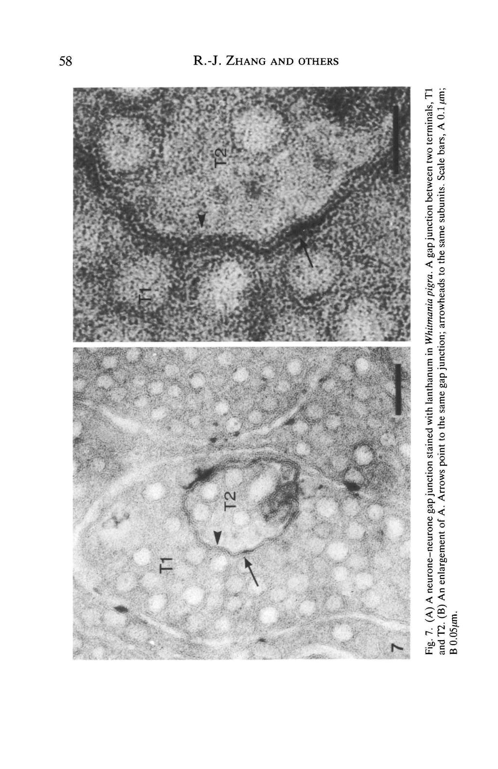
10 56 R.-J. ZHANG AND OTHERS 180 rotation, 91 days V V D V Anterior Dmm D V Fig. 6. Synchronous repair of bilateral PI cells in the isolated ganglion XII 91 days after the body wall was rotated. Abbreviations as in Fig. 5. depolarizing current and 0.06 with hyperpolarizing current (Elliott and Muller, 1983). Electron microscope observations on gap junctions Gap junctions between the terminals of homologous neurones Electrical synaptic connections have been reported between many neurones in the leech; for example, between pairs of Retzius, T, L, AE and S cells (Muller etal. 1981). However, until now there have been few electron micrographs of these areas in the leech. The gap junction between nerve terminals in the leech displays a gap of approximately 2nm in width. Fig. 7 shows an electron micrograph of a putative gap junction, filled with lanthanum, between two terminals. Neurone glial gap junctions The entire central nervous system is composed of two kinds of cell, neurones
11 Synapse discrimination and re-development in leech 57 Table 3. Coupling ratios of electrical synapses between Retzius cells Coupling ratio Paired cells In normal ganglion R-R In culturec1 ganglion R-R Implanted Retzius cell in cultured ganglion R'-R' 9 days R'-R 3 days R-R' 3 days Nl-R' 3 days L-R' 3 days R'-L 3 days Dissociated cell culture R-R 21 days 4 days R-L 9 days R-Nm 7 days Nm-R 7 days DP HP Eckert (1963) Zhang (1987) References Miyazaki et at. (1976) Zhang (1989) Zhang and Nicholls (1983) Zhang (1989) Zhang (1989) Zhang (1989) Zhang (1989) Zhang (1989) Fuchs et at. (1981) Liu and Nicholls (1989) Fuchs et al. (1981) Vyklicky and Nicholls (1988) Vyklicky and Nicholls (1988) R, Retzius cell in situ; R', implanted extra Retzius cell; Nl, lateral N cell; Nm, medial N cell; L, longitudinal motor neurone. DP, depolarizing current; HP, hyperpolarizing current. and glial cells, but 'what specialized structures or junctions (if any) occur between neurones and glia?' (Nicholls, 1981). Recently a few articles have appeared that suggest arthropod-type gap junctions occur between neurones and glia, e.g. in houseflies (Saint Marie and Carlson, 1985) and in crayfish (Cuadras and Marti- Subirana, 1987). The leech CNS is a classical model for research on the functional relationship between neurones and glia (Kuffler and Potter, 1964; Kuffler and Nicholls, 1966). Its outstanding advantage is that there are only 10 glial cells in each ganglion and these have defined positions. The six capsules forming each ganglion are distributed symmetrically, and there is one glial cell in each capsule, two glial cells envelop the neuropile lying longitudinally along the midline and two gliocytes occur on each side of the ganglion, one in the anterior and one in the posterior connective. Thus, the anatomical distribution of glial cells is also symmetrical. Conclusion The central nervous system of the leech contains identified neurones that have symmetrical axonal arborizations. The anatomipal distribution of glial cells is also symmetrical. This symmetry has been exploited by researchers for studying both
12

13 Synapse discrimination and re-development in leech 59 the integrative mechanisms for localization of sensory stimuli and the regeneration of synaptic connections in the ganglion. Afferent signals from the receptor neurones result in synaptic inputs that are discriminated and integrated by the AP neurone. Our results give rise to a number of questions including those of whether position perception depends upon a neuronal circuit in the CNS comparing input from both sides of the body, and how the bilateral synchrony of neuronal repair is achieved. Is regeneration dependent upon the rate of axoplasmic transport and are glial cells involved in the process, for example by the production of extracellular attachment or other molecules? The central nervous system of leeches should provide an ideal preparation for answering such questions. This work was supported by grants from the Chinese Academy of Science, the Chinese Natural Science Foundation and the Chinese Education Commission. References BLOOM, E. M. AND TOMPKINS, R. (1976). Selective re-innervation in skin rotation grafts in Rana pipien. J. exp. Biol. 195, BOWLING, D. J., NICHOLLS, J. G. AND PARNAS, I. (1978). Destruction of a single cell in the C.N.S. of leech as a means of analysing its connections and functional role. J. Physiol., Lond. 282, CUADRAS, J. AND MARTI-SUBIRANA, A. (1987). Glial cells of the crayfish and their relationship with neurons. An ultrastructural study. /. Physiol., Paris 82, ECKERT, R. (1963). Electrical interaction of paired ganglion cells in leech. J. gen. Physiol. 46, EDWARDS, J. S. (1988). Sensory regeneration in arthropods: implications of homeostasis and of ectopic sensilla. Am. Zool. 28, ELLIOTT, E. J. AND MULLER, K. J. (1983). Accurate regeneration of an electrical synapse between two leech neurons after destruction of the ensheathing glia cell. J. Physiol., Lond. 344, FUCHS, P. A., NICHOLLS, J. G. AND READY, D. F. (1981). Membrane properties and selective connexions of identified leech neurones in culture. /. Physiol., Lond. 316, GAO, W.-Q. AND MACAGNO, E. R. (1987). Extension and retraction of axonal projections by some developing neurons in the leech depend upon the existence of neighboring homologous. II. The AP and AE neurons. J. Neurobiol. 18, JACOBSON, M. (1978). Developmental Neurobiology. 2nd edn. New York: Plenum Press. JACOBSON, M. AND BACKER, R. E. (1968). Neuronal specification of cutaneous nerves through connections with skin grafts in the frog. Science 160, Koopowrrz, H. AND HOLMAN, M. (1988). Neuronal repair and recovery of function in the polyclad flatworm, Notoplana acticola. Am. Zool. 28, KUFFLER, S. W. AND NICHOLLS, J. G. (1966). The physiology of neuroglia cells. Ergeb. Physiol. 57, KUFFLER, S. W. AND POTTER, D. D. (1964). Glia in the leech central nervous system: physiological properties and neuron-glia relationship. /. Neurophysiol. 27, KUHLMAN, J. R., Li, C. AND CALABRESE, L. (1988). FMRFamide-like substance in the leech. I. Immunocytochemical localization. /. Neurosci. 5, Liu, Y. AND NICHOLLS, J. G. (1989). Steps in the development of chemical and electrical synapses by pairs of identified leech neurons in culture. Proc. R. Soc. B 236, MINER, N. (1956). Integumental specification of sensory fibers in the development of cutaneous local sign. J. comp. Neurol. 105, MIYAZAKI, S., NICHOLLS, J. G. AND WALLACE, B. G. (1976). Modification and regeneration of
. Competition and chemoaffinity in insect sensory system. Trends Neurosci. 8, 120-125. NICHOLLS, J.")
14 60 R.-J. ZHANG AND OTHERS synaptic connections in culture leech ganglia. Cold Spring Harbor Symp. quant. Biol. 40, MULLER, K. J., NICHOLLS, J. G. AND STENT, G. S. (1981). Neurobiology of the Leech. New York: Cold Spring Harbor Laboratory, pp MURPHEY, R. K. (1985). Competition and chemoaffinity in insect sensory system. Trends Neurosci. 8, NICHOLLS, J. G. (1981). Introduction. Perspectives on the cell biology of glia. In Glial-Neuron Interaction (ed. J. E. Treherne), pp Cambridge, London: Cambridge University Press. NICHOLLS, J. G. (1987). The search for connection: Studies of Regeneration in the Nervous System of the Leech. Sunderland, MA: Sinaeur Associates Inc. NICHOLLS, J. G. AND BAYLOR, D. A. (1968). Specific modalities and receptive fields of sensory neurons in the CNS of leech. J. Neurophysiol. 31, NICHOLLS, J. G. AND PURVES, D. (1970). Monosynaptic chemical and electrical connexions between sensory and motor cells in the central nervous system of the leech. J. Physiol, Lond. 209, SAINT MARIE, R. L. AND CARLSON, S. D. (1985). Interneuronal and glial-neuronal gap junctions in the lamina ganglionaris of the compound eye of the housefly, Musca domestica. Cell Tissue Res. 241, SPERRY, R. W. (1943). Effect of 180 degree rotation of the retinal field on visuomotor coordination. J. exp. Zool. 92, SUNDERLAND, A. J. (1980). A hitherto undocumented pair of neurons in the segmental ganglion of the leech which receive synaptic input from mechanosensory cells. Comp. Biochem. Physiol. 67A, VYKLICKY, L. AND NICHOLLS, J. G. (1988). Specificity of connections formed by nociceptive cells of the leech in tissue culture. J. exp. Biol. 134, WALLACE, B. G. (1981). Neurotransmitter chemistry. In Neurobiology of the Leech (ed. K. J. Muller, J. G. Nicholls and G. S. Stent), pp New York: Cold Spring Harbor Laboratory. WANG, D.-B. AND ZHANG, R.-J. (1990). The morphological similarity and symmetry of neurons in the leech, Whitmania pigra. Sci. in China (Series B) 33, (In Chinese). ZHANG, R.-J. (1987). Chinese leech Whitmania pigra as a suitable specimen for neurobiological research. Chinese J. physiol. Sci. 3, ZHANG, R.-J. (1989). Re-development of synaptic connections after implanted single 5-HT containing neuron in isolated leech ganglia. Sci. in China (Series B) 32, ZHANG, R.-J. AND NICHOLLS, J. G. (1983). Identified neurons implanted into leech ganglia sprout and form synaptic connection. Devi Brain Res. 8,
ELECTRICAL PROPERTIES AND ANION PERMEABILITY OF DOUBLY RECTIFYING JUNCTIONS IN THE LEECH CENTRAL NERVOUS SYSTEM
 exp. Biol. 137, 1-11 (1988) rinted in Great Britain The Company of Biologists Limited 1988 ELECTRICAL PROPERTIES AND ANION PERMEABILITY OF DOUBLY RECTIFYING JUNCTIONS IN THE LEECH CENTRAL NERVOUS SYSTEM
NEW GROWTH ELICITED IN ADULT LEECH MECHANOSENSORY NEURONES BY PERIPHERAL AXON DAMAGE
 exp. Biol. 143, 419-434 (1989) 419 rinted in Great Britain The Company of Biologists Limited 1989 NEW GROWTH ELICITED IN ADULT LEECH MECHANOSENSORY NEURONES BY PERIPHERAL AXON DAMAGE BY B. A. BANNATYNE,
Nervous Tissue. Neurons Electrochemical Gradient Propagation & Transduction Neurotransmitters Temporal & Spatial Summation
 Nervous Tissue Neurons Electrochemical Gradient Propagation & Transduction Neurotransmitters Temporal & Spatial Summation What is the function of nervous tissue? Maintain homeostasis & respond to stimuli
Chapter 9. Nerve Signals and Homeostasis
 Chapter 9 Nerve Signals and Homeostasis A neuron is a specialized nerve cell that is the functional unit of the nervous system. Neural signaling communication by neurons is the process by which an animal
T, P and N neurones were identified in the head ganglion than would be. of evidence suggested that they were primary sensory neurones.
 J. Phyiiol. (1976), 263, pp. 489-512 489 With 2 plate and 13 text-figurea Printed in Great Britain PHYSIOLOGICAL PROPERTIES AND RECEPTIVE FIELDS OF MECHANOSENSORY NEURONES IN THE HEAD GANGLION OF THE LEECH:
Nervous Tissue. Neurons Neural communication Nervous Systems
 Nervous Tissue Neurons Neural communication Nervous Systems What is the function of nervous tissue? Maintain homeostasis & respond to stimuli Sense & transmit information rapidly, to specific cells and
Neurons, Synapses, and Signaling
 Chapter 48 Neurons, Synapses, and Signaling PowerPoint Lecture Presentations for Biology Eighth Edition Neil Campbell and Jane Reece Lectures by Chris Romero, updated by Erin Barley with contributions
Lecture 6: Non-Cortical Visual Pathways MCP 9.013/7.68, 03
 Lecture 6: Non-Cortical Visual Pathways MCP 9.013/7.68, 03 Roger W. Sperry The problem of central nervous reorganization after nerve regeneration and muscle transposition. R.W. Sperry. Quart. Rev. Biol.
Nervous System Organization
 The Nervous System Nervous System Organization Receptors respond to stimuli Sensory receptors detect the stimulus Motor effectors respond to stimulus Nervous system divisions Central nervous system Command
Dendrites - receives information from other neuron cells - input receivers.
 The Nerve Tissue Neuron - the nerve cell Dendrites - receives information from other neuron cells - input receivers. Cell body - includes usual parts of the organelles of a cell (nucleus, mitochondria)
Neurons and Nervous Systems
 34 Neurons and Nervous Systems Concept 34.1 Nervous Systems Consist of Neurons and Glia Nervous systems have two categories of cells: Neurons, or nerve cells, are excitable they generate and transmit electrical
Chapter 48 Neurons, Synapses, and Signaling
 Chapter 48 Neurons, Synapses, and Signaling Concept 48.1 Neuron organization and structure reflect function in information transfer Neurons are nerve cells that transfer information within the body Neurons
MEMBRANE POTENTIALS AND ACTION POTENTIALS:
 University of Jordan Faculty of Medicine Department of Physiology & Biochemistry Medical students, 2017/2018 +++++++++++++++++++++++++++++++++++++++++++++++++++++++++++++++++++++++++ Review: Membrane physiology
BIOLOGY 11/10/2016. Neurons, Synapses, and Signaling. Concept 48.1: Neuron organization and structure reflect function in information transfer
 48 Neurons, Synapses, and Signaling CAMPBELL BIOLOGY TENTH EDITION Reece Urry Cain Wasserman Minorsky Jackson Lecture Presentation by Nicole Tunbridge and Kathleen Fitzpatrick Concept 48.1: Neuron organization
Chapter 37 Active Reading Guide Neurons, Synapses, and Signaling
 Name: AP Biology Mr. Croft Section 1 1. What is a neuron? Chapter 37 Active Reading Guide Neurons, Synapses, and Signaling 2. Neurons can be placed into three groups, based on their location and function.
NOTES: CH 48 Neurons, Synapses, and Signaling
 NOTES: CH 48 Neurons, Synapses, and Signaling A nervous system has three overlapping functions: 1) SENSORY INPUT: signals from sensory receptors to integration centers 2) INTEGRATION: information from
Neurons, Synapses, and Signaling
 LECTURE PRESENTATIONS For CAMPBELL BIOLOGY, NINTH EDITION Jane B. Reece, Lisa A. Urry, Michael L. Cain, Steven A. Wasserman, Peter V. Minorsky, Robert B. Jackson Chapter 48 Neurons, Synapses, and Signaling
Control and Integration. Nervous System Organization: Bilateral Symmetric Animals. Nervous System Organization: Radial Symmetric Animals
 Control and Integration Neurophysiology Chapters 10-12 Nervous system composed of nervous tissue cells designed to conduct electrical impulses rapid communication to specific cells or groups of cells Endocrine
Introduction Principles of Signaling and Organization p. 3 Signaling in Simple Neuronal Circuits p. 4 Organization of the Retina p.
 Introduction Principles of Signaling and Organization p. 3 Signaling in Simple Neuronal Circuits p. 4 Organization of the Retina p. 5 Signaling in Nerve Cells p. 9 Cellular and Molecular Biology of Neurons
Nervous System Organization
 The Nervous System Chapter 44 Nervous System Organization All animals must be able to respond to environmental stimuli -Sensory receptors = Detect stimulus -Motor effectors = Respond to it -The nervous
Information processing. Divisions of nervous system. Neuron structure and function Synapse. Neurons, synapses, and signaling 11/3/2017
 Neurons, synapses, and signaling Chapter 48 Information processing Divisions of nervous system Central nervous system (CNS) Brain and a nerve cord Integration center Peripheral nervous system (PNS) Nerves
Neurons, Synapses, and Signaling
 Chapter 48 Neurons, Synapses, and Signaling PowerPoint Lecture Presentations for Biology Eighth Edition Neil Campbell and Jane Reece Lectures by Chris Romero, updated by Erin Barley with contributions
Pioneering and pathfinding by an identified neuron in the embryonic leech
 J. Embryol. exp. Morph. 86, 155-167, (1985) 155 Printed in Great Britain The Company of Biologists Limited 1985 Pioneering and pathfinding by an identified neuron in the embryonic leech JOHN Y. KUWADA
Neurons, Synapses, and Signaling
 Chapter 48 Neurons, Synapses, and Signaling PowerPoint Lectures for Biology, Eighth Edition Lectures by Chris Romero, updated by Erin Barley with contributions from Joan Sharp and Janette Lewis Copyright
Purpose: Perception, Movement, Learning, Memory, Thinking, Communication Functions:
 Nervous System Purpose: Perception, Movement, Learning, Memory, Thinking, Communication Functions: Sensory Input: Obtaining stimulation from the environment (light, heat, pressure, vibration, chemical,
Overview Organization: Central Nervous System (CNS) Peripheral Nervous System (PNS) innervate Divisions: a. Afferent
 Peripheral Nervous System (PNS) innervate Divisions: a. Afferent") Overview Organization: Central Nervous System (CNS) Brain and spinal cord receives and processes information. Peripheral Nervous System (PNS) Nerve cells that link CNS with organs throughout the body.
Overview Organization: Central Nervous System (CNS) Brain and spinal cord receives and processes information. Peripheral Nervous System (PNS) Nerve cells that link CNS with organs throughout the body.
BIOLOGY. 1. Overview of Neurons 11/3/2014. Neurons, Synapses, and Signaling. Communication in Neurons
 CAMPBELL BIOLOGY TENTH EDITION 48 Reece Urry Cain Wasserman Minorsky Jackson Neurons, Synapses, and Signaling Lecture Presentation by Nicole Tunbridge and Kathleen Fitzpatrick 1. Overview of Neurons Communication
INTERSEGMENTAL TO INTRASEGMENTAL CONVERSION BY GANGLIONIC FUSION IN LATERAL GIANT INTERNEURONES OF CRAYFISH
 J. exp. Biol. 107, 515-519 (1983) 5 \ 5 Printed in Great Britain The Company of Biologists Limited 1983 INTERSEGMENTAL TO INTRASEGMENTAL CONVERSION BY GANGLIONIC FUSION IN LATERAL GIANT INTERNEURONES OF
SHORT COMMUNICATION MULTIMODALITY OF OCELLAR INTERNEURONES OF THE AMERICAN COCKROACH BY TAKAHIRO OHYAMA AND YOSHIHIRO TOH
 J. exp. Biol. 125, 405-409 (1986) 405 Printed in Great Britain The Company of Biologists Limited 1986 SHORT COMMUNICATION MULTIMODALITY OF OCELLAR INTERNEURONES OF THE AMERICAN COCKROACH BY TAKAHIRO OHYAMA
Neurons, Synapses, and Signaling
 CAMPBELL BIOLOGY IN FOCUS URRY CAIN WASSERMAN MINORSKY REECE 37 Neurons, Synapses, and Signaling Lecture Presentations by Kathleen Fitzpatrick and Nicole Tunbridge, Simon Fraser University SECOND EDITION
Neurophysiology. Danil Hammoudi.MD
 Neurophysiology Danil Hammoudi.MD ACTION POTENTIAL An action potential is a wave of electrical discharge that travels along the membrane of a cell. Action potentials are an essential feature of animal
Quantitative Electrophysiology
 ECE 795: Quantitative Electrophysiology Notes for Lecture #4 Wednesday, October 4, 2006 7. CHEMICAL SYNAPSES AND GAP JUNCTIONS We will look at: Chemical synapses in the nervous system Gap junctions in
Housekeeping, 26 January 2009
 5 th & 6 th Lectures Mon 26 & Wed 28 Jan 2009 Vertebrate Physiology ECOL 437 (MCB/VetSci 437) Univ. of Arizona, spring 2009 Neurons Chapter 11 Kevin Bonine & Kevin Oh 1. Finish Solutes + Water 2. Neurons
Neurons. 5 th & 6 th Lectures Mon 26 & Wed 28 Jan Finish Solutes + Water. 2. Neurons. Chapter 11
 5 th & 6 th Lectures Mon 26 & Wed 28 Jan 2009 Vertebrate Physiology ECOL 437 (MCB/VetSci 437) Univ. of Arizona, spring 2009 Neurons Chapter 11 Kevin Bonine & Kevin Oh 1. Finish Solutes + Water 2. Neurons
37 Neurons, Synapses, and Signaling
 CAMPBELL BIOLOGY IN FOCUS Urry Cain Wasserman Minorsky Jackson Reece 37 Neurons, Synapses, and Signaling Lecture Presentations by Kathleen Fitzpatrick and Nicole Tunbridge Overview: Lines of Communication
Nervous system. 3 Basic functions of the nervous system !!!! !!! 1-Sensory. 2-Integration. 3-Motor
 Nervous system 3 Basic functions of the nervous system 1-Sensory 2-Integration 3-Motor I. Central Nervous System (CNS) Brain Spinal Cord I. Peripheral Nervous System (PNS) 2) Afferent towards afferent
1. Neurons & Action Potentials
 Lecture 6, 30 Jan 2008 Vertebrate Physiology ECOL 437 (MCB/VetSci 437) Univ. of Arizona, spring 2008 Kevin Bonine & Kevin Oh 1. Intro Nervous System Fxn (slides 32-60 from Mon 28 Jan; Ch10) 2. Neurons
Physiology 2 nd year. Neuroscience Optional Lecture
 Academic year 2018/2019 Physiology 2 nd year Semester 1 Curricula Nervous system physiology Blood physiology Acid-base equilibrium Bibliography: Boron & Boulpaep Medical Physiology, 3 rd edition Physiology
لجنة الطب البشري رؤية تنير دروب تميزكم
 1) Hyperpolarization phase of the action potential: a. is due to the opening of voltage-gated Cl channels. b. is due to prolonged opening of voltage-gated K + channels. c. is due to closure of the Na +
Nervous Systems: Neuron Structure and Function
 Nervous Systems: Neuron Structure and Function Integration An animal needs to function like a coherent organism, not like a loose collection of cells. Integration = refers to processes such as summation
Reliability and Effectiveness of Transmission from Exteroceptive Sensory Neurons to Spiking Local Interneurons in the Locust
 The Journal of Neuroscience, April 1992, 12(4): 1477-l 499 Reliability and Effectiveness of Transmission from Exteroceptive Sensory Neurons to Spiking Local Interneurons in the Locust Malcolm Burrows Department
Using Optical Flow to Characterize Sensory-Motor Interactions in a Segment of the Medicinal Leech
 The Journal of Neuroscience, March 15, 2002, 22(6):2283 2298 Using Optical Flow to Characterize Sensory-Motor Interactions in a Segment of the Medicinal Leech Davide Zoccolan and Vincent Torre Scuola Internazionale
recognized unambiguously. Their input resistances were approximately 4 times
 J. Phyriol. (1981), 316, pp. 23-223 23 With 4 plate and 13 text-figurem Printed in Great Britain MEMBRANE PROPERTIES AND SELECTIVE CONNEXIONS OF IDENTIFIED LEECH NEURONES IN CULTURE BY PAUL A. FUCHS, JOHN
The Nervous System. Nervous System Organization. Nerve Tissue. Two parts to the nervous system 11/27/2016
 The Nervous System Nervous System Organization Animals must be able to respond to environmental stimuli. Three functions of the nervous system: Sensory input conduction of signals from sensory receptors.
What are neurons for?
 5 th & 6 th Lectures Mon 26 & Wed 28 Jan 2009 Vertebrate Physiology ECOL 437 (MCB/VetSci 437) Univ. of Arizona, spring 2009 Kevin Bonine & Kevin Oh 1. Finish Solutes Water 2. Neurons Neurons Chapter 11
Membrane Potentials, Action Potentials, and Synaptic Transmission. Membrane Potential
 Cl Cl - - + K + K+ K + K Cl - 2/2/15 Membrane Potentials, Action Potentials, and Synaptic Transmission Core Curriculum II Spring 2015 Membrane Potential Example 1: K +, Cl - equally permeant no charge
Neurochemistry 1. Nervous system is made of neurons & glia, as well as other cells. Santiago Ramon y Cajal Nobel Prize 1906
 Neurochemistry 1 Nervous system is made of neurons & glia, as well as other cells. Santiago Ramon y Cajal Nobel Prize 1906 How Many Neurons Do We Have? The human brain contains ~86 billion neurons and
PROPERTY OF ELSEVIER SAMPLE CONTENT - NOT FINAL. The Nervous System and Muscle
 The Nervous System and Muscle SECTION 2 2-1 Nernst Potential 2-2 Resting Membrane Potential 2-3 Axonal Action Potential 2-4 Neurons 2-5 Axonal Conduction 2-6 Morphology of Synapses 2-7 Chemical Synaptic
Biomedical Instrumentation
 Biomedical Instrumentation Winter 1393 Bonab University The Origin of BioPotentials Bioelectric Signals Bioelectrical potential is a result of electrochemical activity across the membrane of the cell.
SHORT COMMUNICATION ACETYLCHOLINE DEPOLARIZES BARNACLE PHOTORECEPTORS
 J. exp. Biol. 117, 481-485 (1985) 43 \ Printed in Great Britain The Company of Biologists Limited 1985 SHORT COMMUNICATION ACETYLCHOLINE DEPOLARIZES BARNACLE PHOTORECEPTORS BY LESLIE C. TIMPE* Department
BIOLOGY. Neurons, Synapses, and Signaling CAMPBELL. Reece Urry Cain Wasserman Minorsky Jackson
 CAMPBELL BIOLOGY TENTH EDITION Reece Urry Cain Wasserman Minorsky Jackson 48 Neurons, Synapses, and Signaling Lecture Presentation by Nicole Tunbridge and Kathleen Fitzpatrick Lines of Communication The
SENSORY FIELD MAPS IN THE SKIN OF A LEECH FOR TOUCH, PRESUURE AND NOICEPTIVE NEURONS. Italy
 SENSORY FIELD MAPS IN THE SKIN OF A LEECH FOR TOUCH, PRESUURE AND NOICEPTIVE NEURONS By Zana R. Majeed 1,2, Josh Titlow 1, John G. Nicholls 3 and Robin L. Cooper 1 1 Department of Biology, University of
Axon guidance I. Paul Garrity March 15, /9.013
 Axon guidance I Paul Garrity March 15, 2004 7.68/9.013 Neuronal Wiring: Functional Framework of the Nervous System Stretch reflex circuit Early theories of axonogenesis Schwann: many neurons link to form
Nerve Signal Conduction. Resting Potential Action Potential Conduction of Action Potentials
 Nerve Signal Conduction Resting Potential Action Potential Conduction of Action Potentials Resting Potential Resting neurons are always prepared to send a nerve signal. Neuron possesses potential energy
Neurophysiology. + = Na + - = Cl - Proteins HOW? HOW?
 All animal cells have electric potential differences (voltages) across plasma s only electrically excitable cells can respond with APs Luigi Galvani (1791) Animal electricity Electrical fluid passed through
Modulation of central pattern generator output by peripheral sensory cells in Drosophila larvae. BioNB4910 Cornell University.
 Modulation of central pattern generator output by peripheral sensory cells in Drosophila larvae BioNB4910 Cornell University Goals 1) Observe the behavioral effects of remotely activating different populations
UNIT I INTRODUCTION TO ARTIFICIAL NEURAL NETWORK IT 0469 NEURAL NETWORKS
 UNIT I INTRODUCTION TO ARTIFICIAL NEURAL NETWORK IT 0469 NEURAL NETWORKS Elementary Neuro Physiology Neuron: A neuron nerve cell is an electricallyexcitable cell that processes and transmits information
A. Visceral and somatic divisions. B. Sympathetic and parasympathetic divisions. C. Central and peripheral divisions
 Ch 8: Neurons: Cellular and Network Properties, Part 1 Review of the Nervous System Objectives: Describe the Cells of the NS Explain the creation and propagation of an electrical signal in a nerve cell
Math in systems neuroscience. Quan Wen
 Math in systems neuroscience Quan Wen Human brain is perhaps the most complex subject in the universe 1 kg brain 10 11 neurons 180,000 km nerve fiber 10 15 synapses 10 18 synaptic proteins Multiscale
Interneurons in the Flight System of the Locust: Distribution, Connections, and Resetting Properties
 THE JOURNAL OF COMPARATIVE NEUROLOGY 215:33-50 (1983) Interneurons in the Flight System of the Locust: Distribution, Connections, and Resetting Properties R.M. ROBERTSON AND K.G. PEARSON Department of
Biosciences in the 21st century
 Biosciences in the 21st century Lecture 1: Neurons, Synapses, and Signaling Dr. Michael Burger Outline: 1. Why neuroscience? 2. The neuron 3. Action potentials 4. Synapses 5. Organization of the nervous
MCDB 4777/5777 Molecular Neurobiology Lecture 29 Neural Development- In the beginning
 MCDB 4777/5777 Molecular Neurobiology Lecture 29 Neural Development- In the beginning Learning Goals for Lecture 29 4.1 Describe the contributions of early developmental events in the embryo to the formation
MULTIPLE CHOICE. Choose the one alternative that best completes the statement or answers the question.
 Exam Name MULTIPLE CHOICE. Choose the one alternative that best completes the statement or answers the question. 1) Which body fluid compartment contains high levels of K +, large anions, and proteins?
The growth rate of sensory nerve fibres in the mammalian embryo
 Development 00, 307-3 (987) Printed in Great Britain The Company of Biologists Limited 987 307 The growth rate of sensory nerve fibres in the mammalian embryo ALUN M. DAVIES Department of Anatomy, Si George's
Propagation& Integration: Passive electrical properties
 Fundamentals of Neuroscience (NSCS 730, Spring 2010) Instructor: Art Riegel; email: Riegel@musc.edu; Room EL 113; time: 9 11 am Office: 416C BSB (792.5444) Propagation& Integration: Passive electrical
Neural Tissue. PowerPoint Lecture Presentations prepared by Jason LaPres. Lone Star College North Harris Pearson Education, Inc.
 12 Neural Tissue PowerPoint Lecture Presentations prepared by Jason LaPres Lone Star College North Harris An Introduction to the Nervous System The Nervous System Includes all neural tissue in the body
Physiology Unit 2. MEMBRANE POTENTIALS and SYNAPSES
 Physiology Unit 2 MEMBRANE POTENTIALS and SYNAPSES In Physiology Today Ohm s Law I = V/R Ohm s law: the current through a conductor between two points is directly proportional to the voltage across the
Ch 8: Neurons: Cellular and Network Properties, Part 1
 Developed by John Gallagher, MS, DVM Ch 8: Neurons: Cellular and Network Properties, Part 1 Objectives: Describe the Cells of the NS Explain the creation and propagation of an electrical signal in a nerve
Squid. Announcements. L03. ROOTS of NEUROETHOLOGY IN CELLULAR NEUROBIOLOGY. Outline. The Discovery of the Giant Squid Axon KROGH S PRINCIPLE
 L03. ROOTS of NEUROETHOLOGY IN CELLULAR NEUROBIOLOGY Announcements 1) Course website: http://courses.cit.cornell.edu/bionb4240/index.htm Google Chrome? 2) Writing Assignments W1 3) Discussion section Wednesday
Ch 33. The nervous system
 Ch 33 The nervous system AP bio schedule Tuesday Wed Thursday Friday Plant test Animal behavior lab Nervous system 25 Review Day (bring computer) 27 Review Day (bring computer) 28 Practice AP bio test
Mechanisms of integration in the nervous system
 Journal of Experimental Biology Mechanisms of integration in the nervous system A discussion meeting organised by Malcolm Burrows at Wakulla Springs 26-29 March 1984 Company of Biologists Ltd Mechanisms
NEURONS, SENSE ORGANS, AND NERVOUS SYSTEMS CHAPTER 34
 NEURONS, SENSE ORGANS, AND NERVOUS SYSTEMS CHAPTER 34 KEY CONCEPTS 34.1 Nervous Systems Are Composed of Neurons and Glial Cells 34.2 Neurons Generate Electric Signals by Controlling Ion Distributions 34.3
Effects of Betaxolol on Hodgkin-Huxley Model of Tiger Salamander Retinal Ganglion Cell
 Effects of Betaxolol on Hodgkin-Huxley Model of Tiger Salamander Retinal Ganglion Cell 1. Abstract Matthew Dunlevie Clement Lee Indrani Mikkilineni mdunlevi@ucsd.edu cll008@ucsd.edu imikkili@ucsd.edu Isolated
Lecture 04, 04 Sept 2003 Chapters 4 and 5. Vertebrate Physiology ECOL 437 University of Arizona Fall instr: Kevin Bonine t.a.
 Lecture 04, 04 Sept 2003 Chapters 4 and 5 Vertebrate Physiology ECOL 437 University of Arizona Fall 2003 instr: Kevin Bonine t.a.: Bret Pasch Vertebrate Physiology 437 1. Membranes (CH4) 2. Nervous System
Neurophysiology. Membrane Potential. body is electrically neutral. = resting membrane potential
 Membrane Potential body is electrically neutral Neurophysiology there are small differences in electrical charge between inside and outside of cell membranes à due to differences in + and ions on inside
Development of neuronal circuits and behaviors in the medicinal leech
 Brain Research Bulletin, Vol. 53, No. 5, pp. 561 570, 2000 Copyright 2001 Elsevier Science Inc. Printed in the USA. All rights reserved 0361-9230/00/$ see front matter PII S0361-9230(00)00390-7 Development
THE ANATOMY OF A LOCUST VISUAL INTERNEURONE; THE DESCENDING CONTRALATERAL MOVEMENT DETECTOR
 J. Exp. Bid. (1974), 6o, 1-12 I jvith 3 plates and 5 text-figures tainted in Great Britain THE ANATOMY OF A LOCUST VISUAL INTERNEURONE; THE DESCENDING CONTRALATERAL MOVEMENT DETECTOR BY M. O'SHEA, C. H.
Peripheral Nerve II. Amelyn Ramos Rafael, MD. Anatomical considerations
 Peripheral Nerve II Amelyn Ramos Rafael, MD Anatomical considerations 1 Physiologic properties of the nerve Irritability of the nerve A stimulus applied on the nerve causes the production of a nerve impulse,
ESSENTIAL LEARNING OUTCOMES:
 ESSENTIAL LEARNING OUTCOMES: Upon satisfactory completion of BIO 2331 - Anatomy and Physiology I, the student should be able to perform the following outcomes and supporting objectives: Outcome: A. Critical/Creative
tre of Mark Louie D. Lop
 NERVE PHYSIOLOGY Mark Louie D. Lopez College of Science Polytechnic University of the Philippines FUNCTIONS OF NERVOUS SYSTEM Sensory input or detection Integration processing transmission of information
Limulus. The Neural Code. Response of Visual Neurons 9/21/2011
 Crab cam (Barlow et al., 2001) self inhibition recurrent inhibition lateral inhibition - L16. Neural processing in Linear Systems: Temporal and Spatial Filtering C. D. Hopkins Sept. 21, 2011 The Neural
FRTF01 L8 Electrophysiology
 FRTF01 L8 Electrophysiology Lecture Electrophysiology in general Recap: Linear Time Invariant systems (LTI) Examples of 1 and 2-dimensional systems Stability analysis The need for non-linear descriptions
Adaptation in the Neural Code of the Retina
 Adaptation in the Neural Code of the Retina Lens Retina Fovea Optic Nerve Optic Nerve Bottleneck Neurons Information Receptors: 108 95% Optic Nerve 106 5% After Polyak 1941 Visual Cortex ~1010 Mean Intensity
Introduction to CNS neurobiology: focus on retina
 Introduction to CNS neurobiology: focus on retina September 27, 2017 The retina is part of the CNS Calloway et al., 2009) 1 Retinal circuits: neurons and synapses Rods and Cones Bipolar cells Horizontal
How to read a burst duration code
 Neurocomputing 58 60 (2004) 1 6 www.elsevier.com/locate/neucom How to read a burst duration code Adam Kepecs a;, John Lisman b a Cold Spring Harbor Laboratory, Marks Building, 1 Bungtown Road, Cold Spring
THE LOCALIZATION OF FUNCTION IN THE ROOT OF AN INSECT SEGMENTAL NERVE
 Exp. Biol. (1963), 40, SS3-s6i 553 A 2 plates and 2 text-figures Printed in Great Britain THE LOCALIZATION OF FUNCTION IN THE ROOT OF AN INSECT SEGMENTAL NERVE BY ANN FIELDEN* Department of Zoology, University
ELECTRICAL CHARACTERISTICS OF THE MEMBRANE OF AN IDENTIFIED INSECT MOTOR NEURONE
 J. exp. Biol. (1980), 86, 49-61 With 7 figures Printed in Great Britain ELECTRICAL CHARACTERISTICS OF THE MEMBRANE OF AN IDENTIFIED INSECT MOTOR NEURONE BY G. F. GWILLIAM* AND M. BURROWS Department of
Intersegmental Coordination of Rhythmic Motor Patterns
 review J Neurophysiol 90: 531 538, 2003; 10.1152/jn.00338.2003. Intersegmental Coordination of Rhythmic Motor Patterns Andrew A.V. Hill, Mark A. Masino, and Ronald L. Calabrese Biology Department, Emory
Physiology Unit 2. MEMBRANE POTENTIALS and SYNAPSES
 Physiology Unit 2 MEMBRANE POTENTIALS and SYNAPSES Neuron Communication Neurons are stimulated by receptors on dendrites and cell bodies (soma) Ligand gated ion channels GPCR s Neurons stimulate cells
Cells. Steven McLoon Department of Neuroscience University of Minnesota
 Cells Steven McLoon Department of Neuroscience University of Minnesota 1 Microscopy Methods of histology: Treat the tissue with a preservative (e.g. formaldehyde). Dissect the region of interest. Embed
Organization of the nervous system. Tortora & Grabowski Principles of Anatomy & Physiology; Page 388, Figure 12.2
 Nervous system Organization of the nervous system Tortora & Grabowski Principles of Anatomy & Physiology; Page 388, Figure 12.2 Autonomic and somatic efferent pathways Reflex arc - a neural pathway that
Introduction and summary of the chapters
 Introduction and summary of the chapters 1. Electroreception Electroreception is the ability of animal species to detect weak electric fields. It is mediated by a sensory system that occurs in some aquatic
SHORT COMMUNICATION NON-SPIKING INTERNEURONES IN THE PEDAL GANGLIA OF A SWIMMING MOLLUSC
 J. exp. Biol. 134, 443-450 (19S8) 443 Printed in Greol Britain The Company of Biologists Limited I9SS SHORT COMMUNICATION NON-SPIKING INTERNEURONES IN THE PEDAL GANGLIA OF A SWIMMING MOLLUSC BY ANDREW
Coordination of Cellular Pattern-Generating Circuits that Control Limb Movements: The Sources of Stable Differences in Intersegmental Phases
 The Journal of Neuroscience, April 15, 2003 23(8):3457 3468 3457 Coordination of Cellular Pattern-Generating Circuits that Control Limb Movements: The Sources of Stable Differences in Intersegmental Phases
Nervous & Endocrine System
 3/19 HW Day 1 Read pages 897-900 Complete Vocab. on pg 897 Aim: What is Regulation? Do Now: What 2 organ systems are involved in regulation? Nervous & Endocrine System Regulation: The control and coordination
Membrane Potentials and Bioelectricity
 Membrane Potentials and Bioelectricity Hugh Purdy Honors University Physics II November 29, 2010 Most, if not all, cells in the human body have a net electric charge to some degree on either side of their
Hair Cells: The Sensory Transducers of the Inner Ear
 Chapter 1 Hair Cells: The Sensory Transducers of the Inner Ear Hair cells are specialized cells that transform a mechanical motion into changes in membrane potential. Such changes, whereby one form of
The development of the retinotectal projection in Xenopus with one compound eye
 /. Embryol. exp. Morph. Vol 33, 3, pp. 775-787, 1975 775 Printed in Great Britain The development of the retinotectal projection in Xenopus with one compound eye By JOAN D. FELDMAN 2 AND R. M. GAZE 1 From
According to the diagram, which of the following is NOT true?
 Instructions: Review Chapter 44 on muscular-skeletal systems and locomotion, and then complete the following Blackboard activity. This activity will introduce topics that will be covered in the next few
THE MECHANISM OF THE PUPAL GIN TRAP
 J. Exp. Biol. (1973), 59, 95-i 7 95 ^ith 1 plate and 10 text-figures inttd in Great Britain EPri: THE MECHANISM OF THE PUPAL GIN TRAP I. SEGMENTAL GRADIENTS AND THE CONNEXIONS OF THE TRIGGERING SENSILLA