Receptor Functions of the Receptor-Type Protein Tyrosine Phosphatase PTPRO
|
|
|
- Patrick Watson
- 5 years ago
- Views:
Transcription
1 University of Miami Scholarly Repository Open Access Dissertations Electronic Theses and Dissertations Receptor Functions of the Receptor-Type Protein Tyrosine Phosphatase PTPRO Amy Elizabeth Hower University of Miami, Follow this and additional works at: Recommended Citation Hower, Amy Elizabeth, "Receptor Functions of the Receptor-Type Protein Tyrosine Phosphatase PTPRO" (2008). Open Access Dissertations This Open access is brought to you for free and open access by the Electronic Theses and Dissertations at Scholarly Repository. It has been accepted for inclusion in Open Access Dissertations by an authorized administrator of Scholarly Repository. For more information, please contact
2
3 UNIVERSITY OF MIAMI RECEPTOR FUNCTIONS OF THE RECEPTOR-TYPE PROTEIN TYROSINE PHOSPHATASE PTPRO By Amy E. Hower A DISSERTATION Submitted to the Faculty of the University of Miami in partial fulfillment of the requirements for the degree of Doctor of Philosophy Coral Gables, Florida December 2008
4 2008 Amy E. Hower All Rights Reserved
5 UNIVERSITY OF MIAMI A dissertation submitted in partial fulfillment of the requirements for the degree of Doctor of Philosophy RECEPTOR FUNCTIONS OF THE RECEPTOR-TYPE PROTEIN TYROSINE PHOSPHATASE PTPRO Amy E. Hower Approved: John L. Bixby, Ph.D. Professor of Molecular and Cellular Pharmacology and Neurological Surgery Terri A. Scandura, Ph.D. Dean of the Graduate School Ellen Barrett, Ph.D. Professor of Physiology and Biophysics Vance Lemmon, Ph.D. Professor of Neurological Surgery Carlos T. Moraes, Ph.D. Professor of Neurology and Cell Biology and Anatomy Vladlen Slepak, Ph.D. Professor of Molecular and Cellular Pharmacology
6 HOWER, AMY E. (Ph.D., Neuroscience) Receptor Functions of the Receptor-type Protein (December 2008) Tyrosine Phosphatase PTPRO. Abstract of a dissertation at the University of Miami. Dissertation supervised by Professor John L. Bixby. No. of pages in text. (122) Protein tyrosine phosphorylation regulates many aspects of cell growth and differentiation. Since cellular tyrosine phosphorylation levels are controlled by the antagonizing actions of the protein tyrosine kinases (PTKs) and the protein tyrosine phosphatases (PTPs), these enzymes play a direct role in regulating processes as diverse as oncogenesis and neuronal development. In particular, the transmembrane group of PTPs, known as the receptor-type protein tyrosine phosphatases (RPTPs), has been linked to regulation of axon growth and guidance during development and regeneration. The regulation of activity of these RPTPs is of clear importance, yet the fundamental mechanisms underlying this regulation are poorly understood. While extracellular ligands are well known to dimerize and activate the receptor protein tyrosine kinases, the extent to which RPTP regulation parallels this scenario is largely unknown. We have examined the dimerization state and the relationship this state has with the phosphatase activity of the neuronal RPTP, PTPRO. We have found that PTPRO, a Type III RPTP, can exist in a dimerized state, likely regulated by disulfide linkages in the intracellular domain. Ligand addition to a chimeric PTPRO increases dimerization of the transmembrane and intracellular domains. Ligand addition to the chimeric PTPRO also decreases its phosphatase activity towards artificial peptides and a putative substrate,
7 TrkC, a protein also known to be important in neuronal development. PTPRO s regulation of TrkC may be physiologically relevant as the proteins can be co-precipitated from transfected cells and PTPRO s dephosphorylation of TrkC is efficient compared to that of other RPTPs. The decrease in PTPRO s activity upon ligand-induced dimerization was unexpected as dimerization of a structurally-similar RPTP family member suggested the opposite functional outcome. This work suggests a complex relationship between dimerization and activity for the Type III RPTPs, which include PTPRO. The results presented in this dissertation will extend the current knowledge on RPTP functions and the cellular processes they regulate.
8 I would like to dedicate this dissertation to my grandparents, Ruth, Marion, Ivan, and Robert (for my roots), my parents, Gail and Robert (for my foundation), and my husband, Tomas (for every single day). iii
9 Acknowledgments I would like to thank many people whose help, in a variety of ways, enabled me to complete this body of work and achieve this goal. First, I would like to thank my advisor, Dr. John Bixby, for his great scientific insight and support. I am certain that the experiences I have learned from him will enable me to be a better scientist. I would also like to thank my committee chair, Dr. Ellen Barrett, and the other committee members, Drs. Vance Lemmon, Carlos Moraes, and Vlad Slepak. Their time and thoughts have helped guide this project as well as myself through graduate school. I would like to thank many members of the lab who either helped guide me experimentally, or helped make life in the lab easier or better in any way. I would especially like to thank many of my fellow graduate students, who understand best what endeavor you are undertaking, and can therefore provide encouragement and support through their friendships. In particular I would like to thank Laurie, Cindy, Shannon, Alma, Florence, and Andrea for being great friends throughout graduate school. I would like to thank all the graduate school and program coordinators as well as all the departmental staff members who do a lot of coordinating, management, and clerical work which eases life as a graduate student tremendously. Most importantly I would like to thank my family, in particular my parents, Gail and Rob, for their lifelong commitment to support and encourage any endeavor I have chosen. Any achievements I reach are built upon the foundation provided by their love and support. Lastly, but most importantly, I would like to thank my husband Tomas. More than I can express, his love, support, encouragement, generosity, and understanding made everyday of this endeavor better, and on many days, made them flat-out possible. iv
10 Table of Contents 1. CHAPTER 1 General Introduction...1 Relationship of PTPRO and neuronal development...1 Mechanisms and molecular interactions controlling axon growth and guidance...2 Ligands regulating axon growth and guidance...4 Receptors regulating axon growth and guidance...8 Receptor-type protein tyrosine phosphatases...11 Ligand versus Receptor Function...22 Receptor function...22 RPTP dimerization-induced receptor functions CHAPTER 2 Ligand addition dimerizes PTPRO and decreases its phosphatase activity and ability to dephosphorylate TrkC...33 Overview...33 Background...34 Results...37 Discussion...58 Materials and Methods...64 v
11 3. CHAPTER 3 General discussion...69 Summary...69 Relevance of receptor functions...70 Significance of dimerization regulating the receptor functions...76 Purpose of receptor functions on axon growth and guidance...80 Significance of TrkC as a substrate...85 Significance of the PTPRO receptor functions...89 Future directions...90 TABLE OF ABBREVIATIONS REFERENCES vi
12 CHAPTER 1 General Introduction Relationship of PTPRO and neuronal development The investigations presented in this dissertation focus on a protein known to control neuronal development. This protein, PTPRO, is a receptor-type protein tyrosine phosphatase (RPTP). While this and other RPTPs are understood to be critical to axon growth and guidance, how PTPRO is regulated to achieve these functions is poorly understood. The work presented in this dissertation explores the mechanisms that regulate PTPRO functionality. Since these functions likely pertain to PTPRO s role in development of the nervous system, this chapter will first introduce the major protein interactions known to govern neuronal development. This discussion will be followed by an introduction to the most well known cell surface receptors and their ligands responsible for influencing neuronal growth and guidance. PTPRO is one such receptor, therefore a detailed introduction to the protein family to which PTPRO belongs will be given after the more general overviews. As the focus of this dissertation is PTPRO s receptor functions and how these functions pertain to PTPRO s role in development, attention will also be given to the mechanisms of regulation and influences on axon growth and guidance for other RPTPs. A more thorough understanding of these mechanisms in RPTPs, including PTPRO, will provide insight into the molecular regulation, cellular physiology and neuronal phenotypes governed by these specific proteins, and should contribute to the field of neuronal development in general. 1
13 2 Mechanisms and molecular interactions controlling axon growth and guidance Many molecular components involved in the complex behaviors of axon growth and guidance have been identified and are understood to varying degrees. Guidance cues provided to growing axons enable target recognition. These cues can be diffusible, bound to another cell, or found in the extracellular matrix. A diffusible factor can instruct growing axons over long distances, whereas a contact-mediated cue provides short-range information. Both soluble and contact-mediated signals can be either positive (attractive) or negative (repulsive). Thus interactions between axons and guidance cues can be categorized into four major classes; namely chemoattraction, chemorepulsion, contact-mediated attraction and contact-mediated repulsion. Chemoattraction and repulsion guide axons along soluble concentration gradients. Contact-mediated signals either attract or repel axons that contact the cell or the extracellular space bearing the cue. A more detailed discussion of these mechanisms, including examples of the proteins modulating them, will be presented later in this chapter. Extracellular cues encountered by growth cones encounter serve as ligands. Ligands are recognized by the growth cone when bound by proteins known as receptors. Expressed within the plasma membrane of a growth cone, receptors are positioned to communicate the information relayed from extracellular cues to the intracellular growth machinery. Receptors that are instructive for axon growth and guidance often achieve this function by altering signaling events within the growth cone that tell an axon whether to grow, collapse, or turn. Many of these signaling cascades ultimately regulate the cytoskeleton of a growth cone, allowing for an increase in actin polymerization or a breakdown of filaments, producing the end result of outgrowth or steering. Therefore,
14 3 axon growth and guidance is dependent upon the intricate roles played by these receptors and their ligands. The signal transduction pathways altered by ligand-receptor interactions are often regulated by tyrosine phosphorylation. Protein phosphorylation commonly activates or inactivates the targeted protein. A change in protein activity can permit or prevent downstream signaling events, either directly or indirectly. For this reason, protein phosphorylation can serve as a pivotal switch for regulating cellular events. Cellular tyrosine phosphorylation levels are regulated by the opposing actions of the protein tyrosine kinases (PTK) and the protein tyrosine phosphatases (PTPs). Transmembrane members of these families of enzymes are known as the receptor protein tyrosine kinases (RPTKs) and the receptor-type protein tyrosine phosphatases (RPTPs) respectively. Since phosphorylation levels are controlled in concert by the antagonizing activities of the kinases and the phosphatases, both classes of enzymes are crucial for regulating neural developmental events. Many well-studied receptors expressed in the growth cone that are responsible for neuronal development are RPTKs. Examples include the Trk and Eph receptors, which will be discussed in more detail in this chapter. These RPTKs are capable of phosphorylating substrates that are part of signaling cascades, many of which ultimately regulate the cytoskeleton of the growth cone. Several members of the RPTP receptor group have also been found to be important for axon growth and guidance. PTPRO, which is the focus of the following chapters, is an example of a developmentally important RPTP. What remains poorly understood are the molecular dynamics that allow this and other RPTPs to govern neuronal development.
15 4 As stated, the influence many RPTKs have on neuronal development depends on changes in the receptor s kinase activity. Thus, it seems reasonable to assume that alterations in phosphatase activity of the RPTPs are crucial for their role as regulators of axon growth and guidance. The work presented in this dissertation investigates the molecular regulation of the receptor functions of PTPRO and discusses to what extent these functions may influence neuronal behaviors. To better understand the RPTPs within the context of axon growth and guidance, an introduction to these proteins as well as other key receptors and ligands regulating neuronal development will be discussed next. Ligands regulating axon growth and guidance Netrin Netrin was one of the first ligands identified with the capability to guide axons. Initially classified as a chemoattractant, netrin is a diffusible protein able to lead axons toward the source of a secreted gradient (Tessier-Lavigne and Goodman, 1996). In the developing spinal cord, netrin secreted from floor plate cells at the midline attracted spinal cord commissural axons toward the floor plate (Hedgecock et al., 1990; Ishii et al., 1992; Kennedy et al., 1994; Serafini et al., 1996; Serafini et al., 1994; Tessier-Lavigne and Goodman, 1996). Later investigations identified netrin as an attractive guidance cue for other neuronal populations. Retinal ganglion axons were led out of the optic disk by netrin secreted from neuroepithelial glial cells (Deiner et al., 1997). Loss of netrin by genetic mutation led to misguidance and/or positional errors of retinal axons and neuroendocrine axons near the hypothalamic ventral midline, suggesting that netrin plays
16 5 a role in hypothalamic axon guidance as well (Deiner and Sretavan, 1999). Netrin promoted axon outgrowth of mouse spiral ganglion cells and acted as an attractant for chick acoustic ganglion cells in culture, suggesting that netrin is involved in development of the auditory system (Lee and Warchol, 2008). While netrin is attractive for the neuron populations mentioned above, netrin can also behave as a chemorepellent. Ventrally originating trochlear motor axons were repelled from the ventrally originating source of netrin in the floor plate (Colamarino and Tessier-Lavigne, 1995). This repulsion guides the motor axons dorsally along a circumferential path to the dorsal midline of the neural tube before they grow out to the periphery (Colamarino and Tessier-Lavigne, 1995). Semaphorin Although netrin was among the first identified chemoattractants, it was not the first chemorepellent discovered. The first belonged to the protein family collectively known as the semaphorins (Culotti and Kolodkin, 1996). The semaphorins consist of soluble and membrane-bound family members. A secreted semaphorin exerted longrange repulsion on chick sensory neurons in the initial experiment that classified semaphorins as chemorepulsive to vertebrate axons (Luo et al., 1993). Since then, semaphorins have been shown to be repulsive to a number of neuronal populations and, like netrin, semaphorins have also been characterized as bifunctional, capable of not only repulsion, but also attraction in a variety of scenarios (Bagnard et al., 1998; de Castro et al., 1999; Falk et al., 2005; Tran et al., 2007). Other chemoattractants and chemorepellents In addition to netrin and semaphorins, several other chemoattractive and chemorepulsive guidance molecules have been described. Among the better known is a
17 6 protein family called the Slits. Slits were identified as chemorepellents responsible for preventing ipsilateral Drosophila commissural axons from crossing the midline and contralateral axons from re-crossing the midline (Andrews et al., 2007; Kidd et al., 1999; Rajagopalan et al., 2000; Simpson et al., 2000). Slit s are also involved in vertebrate commissural axon guidance at the midline and along longitudinal pathways (Dickson and Gilestro, 2006). Sonic hedgehog, initially characterized as a morphogen, was found to be another chemoattractant for vertebrate commissural neurons (Charron et al., 2003; Charron and Tessier-Lavigne, 2005; Okada et al., 2006; Sanchez-Camacho et al., 2005). Neurotrophins in axon outgrowth Although the majority of the molecules mentioned thus far regulate axon guidance, axon outgrowth is a distinct and equally necessary process for proper neuronal development. Axon outgrowth or extension can be regulated by the family of neurotrophins. The secreted polypeptide neurotrophin family consists of the nerve growth factor (NGF), neurotrophin-3 (NT-3), brain-derived neurotrophic factor (BDNF), and neurotrophin-4 (NT-4). NGF s capacity to induce axon outgrowth led to its initial discovery. This trophic effect was evidenced by the extension of neurites from sensory and sympathetic ganglia in the presence of a secreted growth factor (e.g., tumors, explants, etc.) (Levi-Montalcini, 1952; Levi-Montalcini, 1987; Levi-Montalcini et al., 1954). This response was documented for years before the definitive identification of NGF as the first growth factor protein responsible (Cohen, 1960; Levi-Montalcini, 1952; Levi-Montalcini, 1987; Levi-Montalcini et al., 1954). Today, NGF and other neurotrophins are among some of the best-characterized growth factors shown to be necessary for the survival and outgrowth of multiple neuronal
18 7 populations. Both NGF and NT-3 promote axon outgrowth of sympathetic and sensory neurons in vitro and in vivo (Reichardt, 2006; Zhou and Snider, 2006). BDNF induces neurite extension from CNS neurons, including retinal ganglion cells (RGCs) and hippocampal neurons (Reichardt, 2006; Zhou and Snider, 2006). In addition to the neurotrophin family, other secreted growth factors have been suggested recently to contribute to axon outgrowth, but the specific roles played by these factors within this context have been less studied (Markus et al., 2002a). Neurotrophins in axon guidance In addition to promoting neurite outgrowth and cell survival, NGF was the first identified soluble factor capable of inducing a chemotropic axon guidance effect. NGF was found to be a chemoattractant for chick dorsal root ganglion axons (Gundersen and Barrett, 1979). These axons not only grew towards an NGF-secreting source, but continuously reoriented their directed growth to steer towards a source of soluble NGF delivered through a moving pipette (Gundersen and Barrett, 1979). BDNF and NT-3 have also been shown to behave as chemoattractants for Xenopus spinal neurons and chick forebrain neurons in vitro (Ming et al., 1997; Song et al., 1998; Song et al., 1997; Sun et al., 2000b). Furthermore, explant studies have supported a chemoattractive role for the neurotrophins; multiple neuronal populations were found to grow towards a source of ectopically-expressed neurotrophin (Genc et al., 2004; Tucker et al., 2001). Mutant mice overexpressing or misexpressing neurotrophins were shown to exhibit similar phenotypes in vivo as well (Ringstedt et al., 1997; Tessarollo et al., 2004). For example, DRG and vestibular sensory axons were rerouted toward regions of artificiallyhigh neurotrophin expression in these mice (Ringstedt et al., 1997; Tessarollo et al.,
19 8 2004). These studies are consistent with the idea that neurotrophins can induce axon outgrowth, and further suggest that neurotrophins may be involved in axon guidance as well. However, unlike the neurotrophin s role in neuronal survival and outgrowth, the biological significance of neurotrophin chemoattraction is still unclear. Receptors regulating axon growth and guidance The extracellular soluble factors discussed in the previous sections achieve their influence on axon growth and guidance by acting as ligands for specialized receptors located on the plasma membrane of the growth cone. For those receptors involved in axon growth and guidance, ligand binding to these receptors instructs the axon to grow, retract, or turn. Many axon guidance receptors were identified by genetic knockouts that produced the same phenotype as a knockout of their respective ligands (or vice-versa). Examples of receptors for ligands mentioned previously include the Robos, the receptors for Slit, and the plexins and neuropilins that comprise receptors for the semaphorins (Pellet-Many et al., 2008). The bifunctionality that some ligands possess is often achieved by a single ligand binding to multiple receptors, each with a different signaling outcome. Such is the case for netrin. Netrin is chemoattractive when acting through its receptor deleted in colorectal cancer (DCC), but is repulsive when binding to the UNC-5 receptor family, either in the presence or absence of DCC (Hong et al., 1999; Round and Stein, 2007; Stein et al., 2001). Unlike the bifunctionality of netrin, the dual functions of survival and outgrowth regulated by neurotrophins appear to be controlled by a single receptor. This is to say
20 9 that a single neurotrophin ligand binds the same receptor to affect both survival and axon extension. A single neurotrophin receptor is thought to regulate multiple downstream signaling cascades (Markus et al., 2002b; Zhou and Snider, 2006). It is, in part, the activation of one of these pathways over the other that may contribute to an individual receptor s ability to produce multiple functionalities within a cell (Markus et al., 2002b; Zhou and Snider, 2006). The high-affinity receptors for the neurotrophins NGF and NT- 3 are TrkA and TrkC respectively. The neurotrophins BDNF and NT-4 can both interact with the TrkB receptor. The Trk receptors are single transmembrane-domain RPTKs. Ligand binding to a Trk receptor dimerizes the receptor and increases its kinase activity, resulting in downstream signaling pathway activation (Reichardt, 2006; Zhou and Snider, 2006). Knockouts of the neurotrophin receptors are phenotypically similar to loss-offunction mutants of the respective high-affinity neurotrophin ligands. Contact-mediated Receptors and Ligands Ligands capable of binding receptors and inducing intracellular growth and guidance changes can be soluble, as discussed previously, or contact-mediated. Contactmediated ligands include membrane-bound ligands expressed on the surfaces of neighboring cells contacted by neurons or ligands existing in the extracellular matrix. Receptors found on neuronal growth cones bind to the contact-mediated ligands, which alters their ability to signal. Receptor-mediated changes in signal transduction pathways, in turn, result in growth and guidance changes in the axon. While soluble cues are longrange, contact-mediated cues are short-range. Contact-mediated ligand-receptor interactions, like those of soluble ligands, can result in either repulsion or adhesion respectively.
21 10 Eph receptors (EphRs) and their ephrin ligands (ephs) are among the most wellstudied proteins involved in contact-mediated repulsion. The EphRs are the largest family of vertebrate RPTKs. Like all other RPTKs, EphRs are expressed in cell membranes. Unlike many of the ligands discussed previously, ephs are also membranebound. The ephs are either tethered to cell membranes by GPI linkage (ephrin-as) or are transmembrane proteins themselves (ephrin-bs). Since both ephs and eph receptors can be transmembrane proteins, eph-ephr binding has the capacity to alter signaling both within the receptor-expressing cells and within the ligand-expressing cells. The signaling cascade triggered by the receptor is termed forward signaling, and the cascade originating from the ligand is termed reverse signaling. Both forward and reverse signaling from eph-ephr interactions have been shown to regulate guidance of commissural axons at the midline and, perhaps most notably, retinal ganglion cell projections to the tectum (Egea and Klein, 2007; Huot, 2004; Klein, 2005). Under certain conditions, including variable ligand/receptor expression or differential expression of a downstream signaling mediator, the ephrin and/or EphR signals have been shown to yield an attractive phenotype (Egea and Klein, 2007; McLaughlin and O'Leary, 2005). However, the majority of eph-ephr interactions studied have suggested this interaction to be repulsive (Egea and Klein, 2007; McLaughlin and O'Leary, 2005). Both positive and negative eph-ephr interactions have been demonstrated to occur simultaneously. For example, when ephs and EphRs were expressed in unique microdomains within the cell membrane of a chick motor axon growth cone, the ephrins interacting with other ephrins in cis caused growth cone spreading when they encountered EphRs in trans (Klein, 2005; Marquardt et al., 2005).
22 11 Simultaneously, the co-expressed EphRs signaled growth cone collapse when they contacted ephrin ligands in trans (Klein, 2005; Marquardt et al., 2005). The ephrin/ephr interaction is not the only contact-mediated repulsion known. Some members of the semaphorin family are membrane bound. Similarly to the soluble semaphorins, the membrane bound semaphorins were shown to mediate repulsion (Tran et al., 2007). Interestingly, repulsion from the secreted semaphorin could force axons to fasciculate, whereas repulsion from the transmembrane semaphorin could induce defasciculation in the axons on which it was expressed. Contact-mediated ligand and receptor interactions are not all negative however. Many of the proteins involved in contact-mediated attraction are cell adhesion molecules (CAMs), including immunoglobulin (Ig) superfamily CAMs like L1, as well as the cadherins and integrins. Many CAMs can interact homophilically and heterophilically, both in cis and in trans. As such, many of these membrane-bound proteins are able to serve as ligands, receptors or both. Homophilic CAMs serve simultaneously as receptors and ligands. Receptor-type protein tyrosine phosphatases Another family of receptors important for axon growth and guidance is the receptor-type protein tyrosine phosphatase (RPTP) family. There are 107 known protein tyrosine phosphatases (PTPs) in the human genome and the RPTPs comprise 21 of these (Alonso et al., 2004). Based on the sequence homology of the catalytic domains, all of the known PTPs can be grouped into four classes (Alonso et al., 2004). The RPTPs belong to the largest group of Class I cysteine-based PTPs (Fig. 1.1 A). The Class I PTPs
23 12 can be further subdivided by substrate specificity into the classical PTPs and the dualspecificity PTPs. While the dual-specificity PTPs exhibit a range of substrate specificities, the classical PTPs, which include both receptors (RPTPs) and non-receptors (nr-ptps), are tyrosine specific. The remaining PTP groups are the low-molecular weight PTP (Fig. 1.1 B), the dual-specificity CDC25 group (Fig. 1.1 C), and a group whose activity is derived from a reactive aspartic acid residue (Fig. 1.1 D) (Alonso et al., 2004). The reactive aspartic acid residue is unique to this fourth PTP group (Fig. 1.1 D) (Alonso et al., 2004). The other three PTP groups utilize a cysteine-based catalytic mechanism (Alonso et al., 2004). Phosphatases are enzymes responsible for removing phosphate from substrates that have been phosphorylated by the kinases. While all groups of phosphatases previously described can dephosphorylate substrates, each group possesses unique structural properties. The differences in protein structure among the PTPs can determine both the location within a cell where the PTP is expressed as well as which substrates the PTPs target. Figure 1.1 illustrates the domain structures and substrate specificities of the different PTP classes and subclasses.


24 Figure
25 Figure 1.1. Classification, substrate specificity, and structural subunit components of all PTPs in the human genome. The cartoon illustration depicts the structural domains of the four major classes and subfamilies of PTPs. The four PTP classes are divided by letter (A-D) and colored background, with the name of each class listed at the top, followed in parentheses by the number of PTPs represented in each category. In brackets, following the group names and numbers, are the substrate specificities. (A) Class I PTPs are further subdivided into groups based on sequence homology and structural similarities. These subfamilies are designated by name and number of members in parentheses appearing the top of boxed areas within the green background. The 21 classical RPTPs are a subfamily of the Class I PTPs. The RPTPs can be further subdivided into eight structural types. The Type of each RPTP is listed below the line under each Type s representative family member(s) listing. The classical PTPs, which include the RPTPs and the non-receptor PTPs (NRPTPs), all target phospho-tyrosine on protein substrates specifically. The remaining subgroups within the Class I PTPs are VH1-like enzymes which have a variety of substrates. The MKPs are specific for the tyrosine and threonine residues of the mitogenactivated protein (MAP) kinases. While most of the atypical dual-specificity phosphatases appear to dephosphorylate various protein substrates, PIR dephosphorylates mrna. The groups PRLs, CDC14s, and Slingshots all dephosphorylate protein substrates, whereas the last two groups, PTENs and Myotubularins, dephosphorylate inositol phospholipids. (B) The low-molecular weight PTP represents Class II of the cysteine-based PTPs. It has been shown to dephosphorylate several protein substrates, including some protein kinases. (C) The dual-specificity CDC25 group members make up the third cysteine-based class, Class III. These proteins dephosphorylate the dual inhibitory residues on cyclin-dependent kinases (Cdks), activating them and allowing progression through the cell cycle. (D) The PTPs that use an aspartic acid instead of a cysteine for catalysis make up the last class of PTPs. This group was suggested to dephosphorylate protein substrates such as itself and also the RNA polymerase II enzyme. Figure was adapted from (Alonso et al., 2004). 14
26 15 As mentioned, the RPTPs are members of the classical phosphotyrosine-specific PTPs (Alonso et al., 2004). Although all classical PTPs share a common chemical mechanism for dephosphorylating tyrosine residues, differences in structural components and regulatory subunits allow for diversity of substrates and functionality. The RPTPs are a family consisting of 21 known transmembrane proteins. The family is further subdivided into eight structural types based on similarities in the extracellular domain (ECD) motifs. The ECDs vary in size and composition, but many contain motifs similar to those found in CAMs. The intracellular domains (ICDs) of RPTPs contain either one (D1) or two (D1, D2) conserved catalytic domains. Due to the structural similarities the RPTPs share with the CAMs and the transmembrane kinases (RPTKs), it has been hypothesized that RPTP function could mirror that of the CAMs and RPTKs. Many RPTKs and CAMs have been established to play important roles in axon growth and guidance. The involvement of RPTPs in neuronal development has also been investigated. Invertebrate RPTPs While less is known about RPTPs compared to RPTKs, several RPTPs have been demonstrated to play crucial roles in axon growth and guidance. The first evidence implicating these proteins in axon growth and guidance came from knockouts of the Type IIa and III RPTPs in Drosophila, which display aberrant pathfinding of CNS and motor axons during development (Desai et al., 1996; Desai et al., 1997; Schindelholz et al., 2001; Sun et al., 2000a; Sun et al., 2001). Further investigations using genetic manipulations in Drosophila have shown RPTP-mediated axon outgrowth and guidance phenotypes in other parts of the nervous system such as the mushroom bodies, antennal
27 16 lobes, photoreceptors, and optic lobes (Clandinin et al., 2001; Garrity et al., 1999; Kurusu and Zinn, 2008; Maurel-Zaffran et al., 2001; Newsome et al., 2000). In fact, all six RPTPs expressed in the Drosophila nervous system have been linked to axon outgrowth or guidance phenotypes (Desai et al., 1996; Jeon et al., 2008; Johnson and Van Vactor, 2003; Krueger et al., 1996; Schindelholz et al., 2001; Sun et al., 2000a; Sun et al., 2001). Some RPTPs appear to mediate a positive signal for axon outgrowth. For example, loss of Ptp69D function stalled outgrowth from axons out of the mushroom body peduncles into the antennal lobe (Kurusu and Zinn, 2008). This result suggested that Ptp69D s activity was necessary for normal axon extension of that cell population. Other RPTPs appear to mediate a negative signal. Loss of a negative RPTP signal could permit axons that no longer express the RPTP to grow beyond their targets, presumably due to loss of a stop signal. For example, loss of DLAR allows medial antennal lobe axons to cross the midline (Kurusu and Zinn, 2008). In the presence of DLAR these axons would normally have stopped and remained ipsilateral (Kurusu and Zinn, 2008). Vertebrate RPTPs Type II and III subfamilies of RPTPs have also been shown to play roles in axon growth and guidance in vertebrates. Support for this function of vertebrate Type II and Type III RPTPs has been demonstrated both in vivo and in vitro. Type IIa RPTPs All three Type IIa RPTPs, PTPRS (Elchebly et al., 1999; Meathrel et al., 2002; Rashid-Doubell et al., 2002; Wallace et al., 1999), LAR/PTPRF (Yeo et al., 1997), and PTPRD (Uetani et al., 2000) have been shown to be important for proper vertebrate nervous system development in vivo. Knockout of the PTPRS gene in mice led to
28 17 several motor and proprioceptive defects (Wallace et al., 1999). These defects may have been caused by loss of the PTPRS protein from neuronal populations responsible for the skills lost. Such populations include the DRGs, spinal motor neurons, and cerebellum, all of which normally express PTPRS (Wallace et al., 1999). Altered long-term potentiation (LTP) and spatial learning deficits were seen in mice lacking the catalytically-active domain of PTPRD. These deficits may have been due to loss of a functional protein from relevant neuronal populations where PTPRD would have been normally expressed (Uetani et al., 2000; Wallace et al., 1999). Double knockout mice of both PTPRD and PTPRS showed a decrease in motorneuron number and stalled outgrowth of motor axons into their targets (Uetani et al., 2006). LAR/PTPRF knockout mice exhibited spatial learning deficits which may be linked to the anatomical defects found in cholinergic neuron size in the forebrain and decreased innervation from cholinergic neurons into their hippocampal target (Kolkman et al., 2004; Yeo et al., 1997). In vitro studies first suggested the importance of the Type IIa RPTPs in axon growth and guidance. The ECD of PTPRD has been shown to mediate neuronal adhesion, induce neurite outgrowth and promote positive (attractive) axon guidance in vitro (Sun et al., 2000b; Wang and Bixby, 1999). A plated substrate of the ECD of PTPRD was adhesive to several chick neuronal populations, including those that expressed endogenous PTPRD, such as the forebrain and retina (Johnson and Holt, 2000; Wang and Bixby, 1999). This PTPRD-ECD substrate promoted neurite outgrowth in forebrain neurons (Johnson and Holt, 2000; Wang and Bixby, 1999). A soluble form of the ECD of PTPRD applied as a gradient to chick forebrain neurons not only promoted growth cone extension, but was also chemoattractive (Sun et al., 2000b). The outgrowth
29 18 and attraction were mechanistically distinct functions since each was regulated via distinct intracellular pathways (Sun et al., 2000b). The adhesive, outgrowth and attractive behaviors that resulted from PTPRD-ECD application were all attributable to PTPRD as a ligand (Sun et al., 2000b; Wang and Bixby, 1999). However, they may have involved the receptor functions of endogenous PTPRD also (Sun et al., 2000b; Wang and Bixby, 1999). The endogenous receptor s involvement is possible since homophilic binding of the ECDs of PTPRD in trans has been demonstrated (Wang and Bixby, 1999). Perturbation of endogenous PTPRS or its native ligand by application of functionblocking antibodies or soluble PTPRS-ECD was shown to decrease outgrowth and alter growth cone morphology in retinal explants (Ledig et al., 1999a; Ledig et al., 1999b). It was suggested that the application of these soluble proteins did not disrupt homophilic interactions, but rather prevented endogenous heterologous ligand from binding to the native PTPRS receptor (Haj et al., 1999; Ledig et al., 1999a). Disruption of the ligand/receptor interaction also caused defects in retinotectal projections in ovo (Rashid- Doubell et al., 2002). These results suggest the normal function of the PTPRS interaction with its heterologous ligand is that of enhancing neurite outgrowth. However, it is unclear whether the phenotype produced was due to regulation conferred by the ligand or the receptor. In another study, overexpression of a truncated mutant construct consisting of a catalytically-inactive form of the PTPRS cytoplasmic domain increased outgrowth by Xenopus RGCs (Johnson et al., 2001). This catalytically-dead construct likely functions as a dominant negative, and therefore suggests that PTPRS signaling normally inhibits neurite outgrowth. Collective interpretation of the above data for PTPRS supports a
30 19 scenario in which PTPRS interacting with its native heterophilic ligand causes a decrease in PTPRS phosphatase activity, which is mimicked by the dominant negative effect of the catalytically-dead ICD, either of which leads to an increase in retinal axon outgrowth. These data suggest a role for the receptor function of PTPRS in regulating axon outgrowth. A role for the Type IIa RPTPs during regeneration of the nervous system has also been demonstrated. Interestingly, while mice lacking PTPRS exhibit more rapid PNS regeneration following sciatic nerve crush (McLean et al., 2002), LAR knockout mice undergo slower PNS regeneration following the same injury (Van der Zee et al., 2003; Xie et al., 2001). Similar effects are seen in other neuronal populations. PTPRSdeficient mice exhibit a faster rate of growth following facial motor neuron crush and LAR activity-deficient mice exhibit delayed CNS collateral sprouting (Thompson et al., 2003; Van der Zee et al., 2003). The data for PTPRS suggest that loss of PTPRS leads to an increase in axon regrowth. These findings for PTPRS following injury are consistent with the results described earlier which suggested that a reduction in PTPRS signaling had a positive effect on neurite outgrowth. However, it is still unclear as to how LAR might be influencing regeneration. One study investigating regeneration of the mixed motor and sensory sciatic nerve in LAR-deficient mice found that while motor functions recover normally, recovery of sensory functions is delayed (Van der Zee et al., 2003). This result suggests that the influence LAR has on regeneration affects only the dorsal root ganglion (DRG) neurons, which express LAR. Support for this idea comes from an earlier study showing that mrna levels of LAR decrease in DRG neurons following sciatic nerve injury, while
31 20 levels of PTPRS increase concomitantly (Haworth et al., 1998). Interestingly, protein expression of LAR increases in DRGs following injury (Xie et al., 2001). A detailed examination showed that while total expression of LAR increases, the ratio of LAR isoform expression is altered, favoring the isoform that can interact with the lamininnidogen complex. Therefore the discrepancy between the report examining protein expression compared to the study measuring mrna levels could be explained by the specificity of the probes used (Xie et al., 2001). Changes in the protein expression profile of LAR to favor an isoform that can interact with heterophilic ligands may suggest that the receptor function is important for regeneration. The delay in sensory, but not motor, sciatic nerve regeneration of LAR-deficient mice is also seen in mice that lack only the phosphatase domains of LAR (Schaapveld et al., 1997; Van der Zee et al., 2003). This similarity also suggests that the receptor functions may contribute to LAR s role in regeneration. Despite several indications that the activities of PTPRS and LAR are important for nerve regeneration, further investigation is needed to clarify the exact role of these RPTPs. Type IIb RPTPs The Type IIb RPTPs have also been implicated in axon growth and guidance. PTPRM is expressed in retinal ganglion cells (RGCs) at the growth cone and is developmentally regulated (Ledig et al., 1999b; Stoker et al., 1995a; Stoker et al., 1995b). In vitro studies demonstrate that PTPRM can regulate RGC outgrowth (Burden-Gulley and Brady-Kalnay, 1999; Oblander et al., 2007). An early study examining the gross morphology of PTPRK-deficient mice concluded that the mice did not suffer neuronal defects (Skarnes et al., 1995). However, the same study drew a similar conclusion for
32 21 LAR-deficient mice, which have now been shown, following a more detailed analysis, to indeed exhibit neuronal defects (Skarnes et al., 1995; Yeo et al., 1997). In vitro studies suggest that PTPRK homophilic interactions can promote neurite outgrowth (Drosopoulos et al., 1999). Type III RPTPs Less is understood about the Type III RPTPs in vertebrate neural development. PTPRO, a Type III RPTP, has expression patterns in chick and mouse systems to make it a likely candidate for regulating development - a role supported by in vitro studies to be described later (Bodden and Bixby, 1996; Ledig et al., 1999b; Tagawa et al., 1997). Studies from our lab and others also implicate PTPRO in the guidance of motor axons (Stepanek et al., 2005) and retinal axons (Shintani et al., 2006; Stepanek et al., 2001). Other RPTPs More recent data have indicated the involvement of other types of RPTPs in neuronal development. PTPRA, a Type IV RPTP, has been shown to be expressed in the neocortex and hippocampus of the mouse (Petrone et al., 2003; Ye et al., 2008). PTPRA knockout mice display neuronal radial migration defects and misdirected or inverted apical dendritic projection abnormalities (Petrone et al., 2003; Ye et al., 2008). Functionally, the knockout mice have deficits in LTP and short-term (working) memory (Petrone et al., 2003). PTPRA is also expressed on radial and Muller glia in the retina and optic tectum of the chick (Ledig et al., 1999b). This expression profile suggests PTPRA may serve a migrational or polarization function during development of the visual system as well (Ledig et al., 1999b). Although this study did not find evidence for
33 22 PTPRA expression in the retinal or tectal neurons themselves, further analysis would probably be needed to exclude this possibility (Ledig et al., 1999b). Ligand versus Receptor Function Even though the evidence demonstrates a role for RPTPs in nervous system development, it is still unclear exactly how they perform these functions. Evidence exists suggesting that RPTPs have homophilic and heterophilic ligands. But perhaps more important than whether ligands for RPTPs are homophilic or heterophilic, is the fact that evidence is now mounting that these proteins may have important regulatory roles both as ligands and as receptors. Investigations are underway to determine whether it is the RPTP s role as a ligand or a receptor that mediates particular nervous system developmental processes. The investigations performed in this study focus on the receptor functions of the RPTP PTPRO. Receptor Function As discussed, RPTPs are important to the process of neuronal development. The question now becomes whether these functions reside in the phosphatase activity. If so, how is this activity regulated? There have been comparisons made between the regulation of the cytoplasmic RTKs and the cytoplasmic classical PTPs. Both of these enzymes are known to regulate their respective activities by intramolecular interactions. For example, regulatory domains present in Src kinases bind their catalytic domain or phosphorylated tail, causing conformational changes that prevent kinase activity (Weiss and Schlessinger, 1998). Similarly, intramolecular interactions between one of the SHP-
34 23 2 phosphatase regulatory domains and its catalytic domain can block the SHP-2 active site (Weiss and Schlessinger, 1998). When a substrate is presented, the intramolecular suppression of both the kinase and the phosphatase can be overcome to render the cytoplasmic proteins active. Thus it seems reasonable to question whether there are any similarities between the receptor versions of these enzymes in terms of regulation as there is between the cytoplasmic proteins. Oligomerization The kinase activity of RPTKs is regulated by oligomerization of the transmembrane monomers, often induced by ligand binding. In those cases studied, oligomerization of the RPTK leads to transphosphorylation and activation of the kinase. RPTKs for which this receptor behavior has been demonstrated include the Trk receptors and EphRs, both of which have been mentioned previously (Egea and Klein, 2007; Himanen and Nikolov, 2003; Reichardt, 2006). Oligomerization of transmembrane receptors that are not themselves kinases can also regulate phosphorylation events that lead to axon growth and steering instructions. An example is the netrin chemoattractive receptor DCC. This ligand-receptor interaction leads to attraction only when homodimerization of DCC is induced by netrin binding, whereas netrin-induced heterodimerization of DCC with UNC-5 can lead to repulsion (Hong et al., 1999; Stein and Tessier-Lavigne, 2001; Stein et al., 2001). In this instance, oligomerization brings together cytoplasmic proteins that are associated with the ICDs of the receptor monomers (Round and Stein, 2007). These associated proteins include cytoplasmic kinases and phosphatases as well as other adaptor or binding proteins (Round and Stein, 2007). When receptor dimerization clusters the associated
35 24 cytoplasmic proteins, they can initiate signaling cascades resulting in cytoskeletal changes, the details of which are still under investigation (Round and Stein, 2007). Depending on the structure of a given protein, dimerization between monomeric proteins can take place via covalent or non-covalent interactions. Covalent interactions include disulfide-linked bonds which form between free cysteine residues on each monomer. Non-covalent associations, which can also hold monomers together, may include hydrophobic or van der Waals interactions. RPTP dimerization-induced receptor functions The receptor functions of some RPTPs have been investigated. To date, a complete mechanism involving each step including ligand binding, dimerization, activity changes, substrate regulation, and finally, cellular phenotype produced has not yet been compiled for a single RPTP. However, there have been data suggesting that dimerization can occur in the RPTPs PTPRA, PTPRC, PTPRS, PTPRR and PTPRH. Also similar to the RPTKs, dimerization of RPTPs may also induce their receptor functions, producing a change in phosphatase activity. However, only two of the above RPTPs have been investigated thoroughly enough to correlate dimerization state with activity, namely, PTPRA and PTPRC. Interestingly, unlike the result of RPTK dimerization, the activities of each of these RPTPs has been shown to decrease upon dimerization. Dimerization and receptor functions of PTPRA PTPRA dimerization was evidenced on Western blots from lysates of cells overexpressing full-length or truncated versions of PTPRA that had been exposed to a chemical cross-linking agent, Bis(Sulfosuccinimidyl)suberate (BS 3 ) (Jiang et al., 2000).
36 25 The dimerization was determined to be homodimerization by Western blot analysis of BS 3 mediated cross-linked cells co-transfected with a mixture of full-length and truncated PTPRA (Jiang et al., 2000; Tertoolen et al., 2001). In these experiments, three different molecular-weight bands were observed, corresponding respectively to homodimers of the full-length proteins, homodimers of the truncated proteins, and heterodimers composed of full-length and truncated PTPRA (Jiang et al., 2000; Tertoolen et al., 2001). This result suggested that the observed PTPRA dimerization was a homophilic interaction and was not occurring between PTPRA and some other similarlysized protein. Homodimerization was also evidenced by fluorescence resonance energy transfer (FRET) analysis in cells overexpressing tagged full-length or truncated mutants (Tertoolen et al., 2001). Western blot analysis of cell lysates expressing multiple variants of PTPRA with various truncated or mutated protein domains were used to determine the domains essential for PTPRA dimerization. BS 3 mediated cross-linking efficiency between PTPRA variants demonstrated that the ECD, TMD, and ICD were all capable of dimerization (Jiang et al., 2000). However, a following study using FRET confirmed that the TMD, but not the ECD, was involved in dimerization (Tertoolen et al., 2001). It was also proposed that the juxtamembrane region and D1 catalytic domain of the ICD contributed to dimerization, but the supporting data were not shown (Tertoolen et al., 2001). The dimerization of PTPRA does not appear to be regulated by disulfide linkages. Evidence supporting the lack of cysteine dimers was visualized by Western blot analysis of cell lysates expressing PTPRA before and after exposure to the chemical cross-linker,
37 26 BS 3 (Jiang et al., 2000; Jiang et al., 1999; Tertoolen et al., 2001). No PTPRA dimers were seen on non-reduced Western blots unless the lysates had prior exposure to the cross-linking agent (Jiang et al., 2000; Jiang et al., 1999; Tertoolen et al., 2001). Dimerization of PTPRA was predicted to decrease PTPRA s activity based on crystallographic data illustrating a helix-loop-helix structure which could sterically impede access of a substrate to the catalytically-active site (Fig. 1.2) (Bilwes et al., 1996). Due to the presumed functional blockade, the helix-loop-helix structure was termed an inhibitory wedge (Fig. 1.2) (Bilwes et al., 1996). To examine the activity of PTPRA upon dimerization, disulfide-linked dimers were created by introduction of a cysteine residue in a specific location of the PTPRA monomer (Jiang et al., 1999). Indeed, the forced dimerization decreased PTPRA s activity, as measured by the level of dephosphorylation of a negative regulatory tyrosine on c-src (Jiang et al., 1999). Mutations of important structural residues in the inhibitory wedge abrogated the inhibitory effect of dimerization, lending support to the hypothesized functional role of the wedge domain (Jiang et al., 1999).

38 Figure 1.2. Model of inactivation by the inhibitory wedge caused by ligand-induced dimerization. The helix-loop-helix region N-terminal to the catalytically-active D1 domain forms a wedge-shaped projection. When brought into close proximity of a like-monomer, the wedge can sterically occlude the catalytically-active cleft, rendering the RPTP inactive. The rotational coupling between monomers in a dimer allows the inhibitory wedge to occlude the active site on the paired monomer. Incorrect rotational coupling would result in a twisted orientation, such that the inhibitory wedge of one monomer faced away from its paired monomer. In the twisted orientation, the wedge would be unable to suppress activity since the wedge of one monomer would not align with the paired monomer s active site. Figure modified from (Weiss and Schlessinger, 1998). 27
39 28 Another important mechanism regulating dimerization-induced activity changes of PTPRA was also elucidated by forced disulfide-linked dimerization of PTPRA by the addition of a cysteine residue into multiple locations in the ECD (Jiang et al., 1999). Each cysteine was positioned such that it faced a different direction around one complete turn of an alpha-helix. Although addition of a cysteine into any position around the helix induced disulfide-linked dimers, only one residue position influenced the activity upon dimerization (Jiang et al., 1999). This result suggested that mere dimerization was not enough to regulate PTPRA s activity. Instead, regulation of dimer-mediated activity required alignment of the interface between two monomers so that any physical regulatory domains, such as the inhibitory wedge, could participate. This level of regulation is termed rotational coupling (Jiang et al., 1999). Dimerization and receptor functions of PTPRC PTPRC/CD45 is another RPTP for which dimerization and receptor functions have been investigated. Western blot analysis has shown that PTPRC can exist as a dimer in cell lysates previously exposed to a chemical cross-linking agent (Takeda et al., 1992; Xu and Weiss, 2002). Effective chemical cross-linkers included Dithiobis(succinimidyl)propionate (DSP), Dimethyl 3,3 -dithiobispropionimidate (DTBP) or Ethylene glycol bis[sulfosuccinimidylsuccinate] (Sulfo-EGS) (Takeda et al., 1992; Xu and Weiss, 2002). Under these conditions, dimers were seen in endogenouslyexpressed PTPRC from primary human donor blood cells and cultured cell lines, as well as in stably-expressed PTPRC from transfected cell lines (Takeda et al., 1992; Xu and Weiss, 2002). This dimerization was suggested to be homodimerization in a 2D-gel analysis that revealed the PTPRC dimer-complex was composed of equally-sized proteins
40 29 (Takeda et al., 1992). Furthermore, cells expressing a short-ecd PTPRC-isoform displayed FRET when labeled with a mixture of fluorophore-conjugated antibodies to PTPRC (Dornan et al., 2002). Interestingly, dimers of the larger heavily-glycosolated PTPRC isoform were not detected by FRET or Western blotting, suggesting that dimerization-induced regulation could be restricted to certain isoforms (Dornan et al., 2002; Xu and Weiss, 2002). Isoform-specific dimerization could confer an additional level of regulation on RPTP functionality. PTPRC homodimerization levels may be influenced by competition from other protein binding partners for available PTPRC. Originally, both monomeric and dimeric PTPRC/CD45 were found bound to a small 30 kd accessory protein, CD45-AP (Takeda et al., 1994; Takeda et al., 1992). However, a later study found that the ratio of dimeric to monomeric PTPRC decreased in cells expressing CD45-AP compared to cells that lacked the accessory protein (Takeda et al., 2004). This finding suggests that binding to CD45-AP prevents PTPRC dimerization (Takeda et al., 2004). Similar to PTPRA, PTPRC dimers do not appear to be the result of disulfidelinkages since no dimers were visualized on non-reducing gels without the prior addition of a crosslinking agent (Takeda et al., 1992; Xu and Weiss, 2002). Though the precise protein domains responsible for PTPRC dimerization have not been thoroughly examined, preliminary analysis suggests the ECD and/or TMD may be involved (Xu and Weiss, 2002). Multiple investigations have attempted to correlate activity with PTPRC dimerization. An early study showed the activity of cross-linked PTPRC was 5-10 times higher when assayed in the presence of DTT, breaking apart the cross-linked dimers into
41 30 monomers (Takeda et al., 1992). Though these results suggested that monomeric PTPRC was more active than dimeric, the conclusion could not be resolved in this study due to possible interference from accessory proteins in the assay (Takeda et al., 1992). However, three more recent studies have utilized different strategies to address this question. A study utilizing a chimeric EGFR-PTPRC expressed in T cells showed that the addition of EGF, which was assumed, but not shown, to dimerize the chimera, decreased T cell antigen receptor (TCR) signaling (Desai et al., 1993). The reduction in TCR signaling resulted in decreased total cellular phospho-protein and calcium levels during T cell antigen receptor activation (Desai et al., 1993). This result suggested that dimerization of the chimeric PTPRC led to decreased phosphatase activity since the data demonstrated that ligand addition led to an inability of the chimeric PTPRC to dephosphorylate the TCR-induced kinases, abrogating the propagation of the kinase activity (Desai et al., 1993). Furthermore, the decrease in PTPRC activity was dependent on the inhibitory wedge as mutations in that region of the chimera eliminated the ability of EGF to decrease the chimera s activity (Majeti et al., 1998). Likewise, forced disulfide-linked dimerization of the shortest PTPRC-isoform, by addition of a cysteine residue into the ECD, decreased TCR signaling (Xu and Weiss, 2002). Mutations in the inhibitory wedge of the disulfide-linked mutants reversed the inhibition (Xu and Weiss, 2002). These results are consistent with the previous data suggesting dimerization decreases PTPRC activity, a suppression regulated by the inhibitory wedge. Additionally, using a direct measure of phosphatase activity (a phospho-peptide substrate), cells lacking the accessory protein CD45-AP had less phosphatase activity than cells containing the accessory protein (Takeda et al., 2004). The cells lacking
42 31 CD45-AP also displayed a higher ratio of dimeric to monomeric PTPRC compared to cells containing the accessory protein (Takeda et al., 2004). These results suggest that dimeric PTPRC has less activity than monomeric. Expression of the PTPRC full-length protein, the smallest PTPRC-isoform, and the EGFR-PTPRC chimera all have a high intrinsic phosphatase activity (Desai et al., 1993). Taken together, the above data suggest that dimerization of PTPRC decreases its activity. Dimerization and receptor functions of PTPRO Evidence exists that suggesting the receptor functions of the RPTP, PTPRO, are likely involved in the protein s influence over neuronal development. The ability of PTPRO to control proper retinotectal projections is dependent on the phosphatase activity of PTPRO, and specifically on its ability to dephosphorylate the EphR (Shintani et al., 2006). This activity-dependent relationship was suggested by the misrouted retinotectal projections produced by expression of a catalytically-dead mutant of PTPRO (Shintani et al., 2006). The mutant phenotype mimicked the defect attributed to knockdown of wildtype PTPRO, and behaved opposite to the phenotype produced by overexpression of functional PTPRO (Shintani et al., 2006). Therefore, both too much and too little PTPRO activity is detrimental to the proper guidance of the retinotectal projections. While these results suggest that the regulation of PTPRO s phosphatase activity influences axon growth and guidance, precise details of what these receptor functions are or how they are regulated remain unknown. A better understanding of how PTPRO and other RPTPs are regulated could be beneficial in the design of targeted therapeutic strategies which aim to modulate RPTP functions during cellular processes where they are pivotal, such as cancer or neuronal disease or injury.
43 32 The goal of the ensuing chapters is to investigate the dimerization and receptor functions of an RPTP previously established as influential in axon growth and guidance: specifically, PTPRO. Chapter 2 discusses dimerization and regulation of functional activity in the Type III RPTP, PTPRO. Chapter 3 discusses how the findings reported in this dissertation relate to the literature pertaining to RPTP function, activity, and neuronal developmental roles.
44 CHAPTER 2 Ligand addition dimerizes a chimeric PTPRO and decreases its phosphatase activity and ability to dephosphorylate TrkC * OVERVIEW Receptor protein tyrosine phosphatases (RPTPs), like receptor tyrosine kinases (RTKs), regulate neuronal differentiation. While RTKs are dimerized and activated by extracellular ligands, the extent to which RPTPs dimerize, and the effects of dimerization on phosphatase (PTPase) activity, are poorly understood. We have examined a neuronal type III RPTP, PTPRO; we find that PTPRO can form dimers, and that disulfide linkages in PTPRO s intracellular domain likely regulate dimerization. Dimerization of PTPRO s transmembrane and intracellular domains can be achieved by ligand addition to cells expressing a chimeric fusion protein. Ligand addition to cells expressing the chimeric fusion protein decreases the chimera s PTPase activity towards artificial peptides and towards a putative substrate, TrkC. Thus it is hypothesized that dimerization decreases activity of PTPRO based on the correlation drawn between these measurements and ligand addition. Dephosphorylation of TrkC by PTPRO may be physiologically relevant, as it is efficient, and TrkC and PTPRO can be co-precipitated from transfected cells. Inhibition of PTPRO s PTPase activity by ligand addition was unexpected, as dimerization of a related RPTP, PTPRJ, increases PTPase activity. Thus, our results suggest a complex relationship between dimerization and activity in type III RPTPs. * Work described in this chapter has been submitted for publication: Hower, A.E., Beltran, P.J., and Bixby, J.L., Dimerization of PTPRO decreases its phosphatase activity and ability to inactivate TrkC submitted to Molecular and Cellular Neuroscience. 33
45 34 BACKGROUND The control of tyrosine phosphorylation by protein tyrosine kinases and protein tyrosine phosphatases (PTPs) regulates many aspects of cell growth and differentiation, including axon guidance (Bixby, 2000; Chisholm and Tessier-Lavigne, 1999; Johnson and Van Vactor, 2003; Stoker, 2001). The transmembrane receptor protein tyrosine kinases (RPTKs) are controlled by extracellular ligands that dimerize them, activating their enzymatic activity (Heldin, 1995; Ullrich and Schlessinger, 1990; Weiss and Schlessinger, 1998). However, extracellular ligand regulation of transmembrane PTPs, known as receptor PTPs (RPTPs), is poorly understood. There are 21 known RPTPs, which are divided into five structural classes (type I- type V), based primarily on motifs present in their extracellular domains (ECDs) (Alonso et al., 2004). In most cases, the nature of ligands for these RPTPs, the regulation of their dimerization state, and the effects of dimerization on phosphatase activity are not known. We have focused on the type III RPTP, PTPRO, which is most strongly expressed in the developing nervous system (Beltran et al., 2003; Bodden and Bixby, 1996; Tagawa et al., 1997; Thomas et al., 1994) and regulates axon guidance during development (Shintani et al., 2006; Stepanek et al., 2005). Dimerization is an important mechanism for regulating many Type I transmembrane proteins (Heldin, 1995; Ullrich and Schlessinger, 1990; Weiss and Schlessinger, 1998). Several studies have examined the dimerization state of other RPTPs. Dimerization in vivo has been demonstrated for CD45/PTPRC, and homodimerization has been shown to occur with several RPTPs (PTPRA, PTPRC, PTPRH, PTPRS) when these are overexpressed in tissue culture cells (Jiang et al., 2000;
46 35 Lee et al., 2007; Takeda et al., 1992; Walchli et al., 2005; Xu and Weiss, 2002). For PTPRA, dimerization is mediated, at least in part, by the transmembrane domain (TMD) (Tertoolen et al., 2001); this mechanism of dimerization has also been proposed for PTPRS (Lee et al., 2007). Dimerization through TMD interactions has been suggested as a general mechanism for RPTPs (Chin et al., 2005), but has not been tested for most native RPTPs. In fact, dimerization of the type III RPTP, PTPRH, appears to be mediated through its ECD (Walchli et al., 2005). The dimerization of RPTPs has physiological significance, since it appears that dimerization state can be regulated both by extracellular ligands (den Hertog et al., 1999) and by changes in oxidation state (den Hertog et al., 2005). Dimerization of PTPRA or PTPRC results in a decrease in PTPase activity (Desai et al., 1993; Jiang et al., 1999; Takeda et al., 2004; Xu and Weiss, 2002). The loss of activity upon dimerization is thought to be caused by steric constraints mediated by a helix-loop-helix region on the intracellular domain (ICD) that has been called an inhibitory wedge (Bilwes et al., 1996; Majeti et al., 1998). However this inhibitory wedge region is not highly conserved among RPTPs, and is unlikely to be a general mechanism through which dimerization regulates activity (Hoffmann et al., 1997; Nam et al., 1999; Nam et al., 2005). Interestingly, it appears that dimerization of the type III RPTP, PTPRJ, results in an increase in PTPase activity (Takahashi et al., 2006). The structural basis of this activation is unknown. The mechanisms of dimerization and its consequences for PTPase activity are poorly understood for type III RPTPs. There are 5 mammalian type III RPTPs, characterized by variable numbers of FN type III repeats in their ECDs, and a single
47 36 intracellular catalytic motif. Of this group, only PTPRH has been shown to dimerize in cells, whereas dimerization of another type III RPTP (PTPRB) was not seen using the same experimental methodology (Walchli et al., 2005). The relationship between dimerization and PTPase activity is also not clear for this group of RPTPs. Dimerization of PTPRH has been proposed to decrease its activity (Walchli et al., 2005). However, application of a bivalent antibody directed against the PTPRJ ECD increased PTPase activity toward cellular substrates (Takahashi et al., 2006). Furthermore, application of a complex mixture of extracellular matrix proteins to cells expressing PTPRJ caused an upregulation of PTPase activity, which could be due to ligand-mediated dimerization (Sorby et al., 2001). Whether other type III RPTPs dimerize, and how such dimerization influences activity, are not known. In this paper we examine the dimerization of the type III RPTP, PTPRO, and the relationship between dimerization and the PTPase activity. We find that PTPRO can exist in a dimerized state in cells that is likely regulated, at least partially, by disulfide linkages in the ICD. Using a chimeric protein strategy to induce dimerization of PTPRO s TMD and ICD, we found that dimerization of PTPRO is correlated with a decrease in PTPase activity. The chimeric protein comprises the ECD of the high-affinity NGF receptor, TrkA, fused to the TMD and ICD of PTPRO. Thus the dimerization and activity changes are caused by application of NGF onto cells expressing the chimera. The activity changes resulting from NGF application on the PTPRO chimera are capable of regulating the phosphorylation of the NT-3 receptor TrkC, which is likely to be a relevant substrate for PTPRO.
48 37 RESULTS: PTPRO forms dimers in living cells To examine whether PTPRO forms dimers, COS-7 cells were transfected with a full-length flag-tagged version of PTPRO (flroflag). On Western blots run under reducing conditions, immunoreactive bands were seen near 150 kd and 190 kd, with a smear near 230 kd (Fig. 2.1A). A similar pattern has previously been seen for PTPRO in both native tissue and transfected cells (Beltran et al., 2003). Under non-reducing conditions, an immunoreactive band appeared around 350 kd, which likely represents the dimeric form of full-length PTPRO (Fig. 2.1B, arrow). This dimeric form was fully reduced by β-mercaptoethanol, suggesting that at least some dimers are maintained in the presence of SDS by disulfide linkages (Fig. 2.1A).
 COS-7 cells and cells transfected with increasing amounts of flag-tagged full-length PTPRO (flroflag), were analyzed on anti-flag Western blots in both reducing (A)")
49 Figure 2.1. Full-length PTPRO forms disulfide-linked dimers. Lysates from untransfected (un) COS-7 cells and cells transfected with increasing amounts of flag-tagged full-length PTPRO (flroflag), were analyzed on anti-flag Western blots in both reducing (A) and non-reducing (B) conditions. Both monomeric (150 kd, 190 kd, and 235 kd) and dimeric (ca 350 kd; arrow) forms of PTPRO can be seen in B. The oligomerized form is fully reduced by beta-mercaptoethanol (βme) (A). This dual western blot analysis performed under reducing and non-reducing conditions for a single full-length PTPRO cell lysate was repeated in 10 independent experiments, with similar results. 38
50 39 To examine dimerization of PTPRO without requiring disulfide bonding, we coexpressed flroflag with a second construct consisting of full length PTPRO fused to a monomeric Venus (mvenus) tag (flrovenus). The mvenus GFP derivative is designed to prevent artifactual dimerization of fusion proteins (Nagai et al., 2002; Zacharias et al., 2002). Precipitation of flroflag led to co-precipitation of flrovenus, indicating the presence of PTPRO-PTPRO dimers (Fig. 2.2). Co-precipitation was specific, as it did not occur in the presence of excess flag peptide, or if GFP was co-expressed instead of the tagged PTPRO construct. A TrkA-PTPRO fusion protein is expressed on the surface of transfected cells The ability of PTPRO to form dimers in cells raises the question of the effect of dimerization on enzymatic activity. Because native ligands for the PTPRO ECD have not been identified, we produced a chimeric cdna encoding the ECD of the high affinity nerve growth factor (NGF) receptor, TrkA, fused to the TMD and ICD of PTPRO (Trk- RO) (Fig. 2.3A). Application of NGF to cells expressing this chimeric protein should dimerize it, leading to dimerization of the intracellular catalytic domains. The Trk-RO chimera was tagged with mvenus (Trk-ROv), allowing visualization in live cells. Western blot analysis of COS-7 cells transfected with Trk-ROv revealed an immunoreactive doublet at around 135 kd, similar to the predicted size of the fusion protein (Fig. 2.3B). The relationship between the two bands in the doublet is not clear, though native PTPRO can also appear as a doublet. Confocal microscopy demonstrated cell surface localization, as assessed by co-staining with a fluorescent wheat germ agglutinin conjugate (Fig. 2.3C). Trk-ROv was also seen in intracellular puncta, likely
, transfected with flrovenus alone (c), or co-transfected with flroflag and cytoplasmic GFP (cyto. GFP) (d).")
51 Figure 2.2. Full-length PTPRO self-associates in cells. COS-7 cells were cotransfected with flroflag and an mvenus tagged version of PTPRO (flrovenus) (a,b). Control COS-7 cells were left untransfected (e), transfected with flrovenus alone (c), or co-transfected with flroflag and cytoplasmic GFP (cyto. GFP) (d). Lysates were precipitated with anti-flag beads and Western blots were probed for both the flag and the Venus (GFP) epitopes. Precipitation of flroflag co-precipitated flrovenus (b), indicating the presence of oligomers; specific precipitation was blocked with excess flag peptide (a). flrovenus was not precipitated directly in the absence of flroflag (c), and no bands corresponding to flrovenus were seen if GFP was co-transfected instead (d). Results are representative of four independent experiments. 40


52 Figure
53 Figure 2.3. A fluorescent Trk-RO chimera is expressed on cell membranes and the cell surface. (A) A Trk-RO chimera was produced by fusing the ECD of the high affinity NGF receptor, TrkA, to the TMD and ICD of PTPRO. (B) Western blot analysis of cell lysates transfected with either Trk-ROv (a) or cytoplasmic GFP (b) were run under reducing conditions and probed with anti-gfp (upper panels), and anti-gapdh (lower panel) as a loading control. The chimera is recognized by anti-gfp and seen as a band near the expected molecular size of 135 kd. A 26 kd band present in the Trk-ROv lysate is likely a fragment containing the mvenus tag. A 27 kd band is present in the GFPtransfected cell lysate at the expected molecular weight for the control construct. Results are representative of 11 independent experiments. (C) The Trk-RO chimera was tagged on the intracellular C-terminus with mvenus (Trk-ROv). Confocal imaging of COS-7 cells transfected with Trk-ROv (green), stained live with an impermeant fluorescent wheat germ agglutinin (WGA; red) to mark the cell surface. A single Z-slice through two cells (a) shows that both the Trk-ROv and WGA can be visualized at the cell periphery. Both are absent from the cell center (likely the nucleus), and Trk-ROv can be seen in an epi-nuclear location, apparently in cell trafficking intermediates. The entire Z-stack is shown in b; the top and side windows are orthogonal views at the locations corresponding to the x (green) and y (red) lines that cross the center image, and show that Trk-ROv is localized together with WGA on the cell surface. Confocal imaging of WGA-labeled transfected cells was performed in a single experiment. Images are representative of multiple cells within the experiment and results of Trk-ROv surface localization differ from the intracellular localization of a WGA-labeled GFP-transfected condition performed as a control within the experiment. 42
54 43 representing trafficking intermediates. Surface expression in transfected cells was also demonstrated by staining of live cells with antibodies to the extracellular domain of TrkA and by imaging the fluorescent protein using TIRF microscopy (data not shown). To determine what percentage of Trk-ROv was expressed on the plasma membrane relative to the total amount of Trk-ROv expression within the cell, biotinylation experiments were performed. Quantification of these experiments revealed that a range of % of the total Trk-ROv that was expressed was present on the plasma membrane at steady state (Fig. 2.4). Precipitation of Trk-ROv by the biotinavidin complex was specific as none was detected in GFP transfected control cells or in cells expressing Trk-ROv but not exposed to biotin prior to precipitation with avidin (Fig. 2.4). These data suggest that while it is a minority of the total Trk-ROv that is expressed on the plasma membrane, there is some available at steady state for interactions with NGF applied to the culture media. The Trk-RO chimera is dimerized by NGF To determine whether Trk-ROv can dimerize in transfected cells, we examined the chimeric protein on Western blots under non-reducing conditions. Similar to full length PTPRO, both monomeric and dimeric Trk-ROv were present under non-reducing conditions (Fig. 2.5A), and the oligomerized form could be completely reduced by β- mercaptoethanol (Fig. 2.5B). To test whether NGF can increase dimerization of the chimeric phosphatase, we incubated Trk-ROv-transfected cells for 30 minutes in the presence and absence of 100 ng/ml NGF. Indeed, the relative amount of dimeric Trk-


55 Figure 2.4. A minority of Trk-ROv chimera is expressed on the plasma membrane. (A) Live cells expressing Trk-ROv or cytoplasmic GFP were exposed or not to biotin at 4 C for 30 min. Following removal of biotin, cells were subsequently lysed and precipitated with avidin. Bound and unbound proteins were examined on anti-gfp Western blots. Less Trk-ROv was precipitated by the biotin-avidin complex than that which was not precipitated (compare band intensities between first lane and forth lane). Precipitation was specific as it did not occur in the absence of biotin or in GFP control cells. (B) Quantification revealed an average of 16% of the total Trk-ROv expressed was precipitated by the biotin-avidin complex (16.06 ± 5.16; mean ± SEM; N=3). 44
 were analyzed on anti-gfp Western blots under non-reducing (A) and reducing (B) conditions.")
56 Figure 2.5. NGF increases disulfide linkage of the Trk-ROv chimera. Lysates of cytoplasmic GFP expressing control cells and cells expressing Trk-ROv (± NGF) were analyzed on anti-gfp Western blots under non-reducing (A) and reducing (B) conditions. Both a 135 kda monomeric form (doublet) and a dimerized form (arrows) of the Trk-ROv chimera can be seen in non-reducing conditions. NGF addition increases the relative amount of the dimer approximately two-fold (1.83 ± 0.3 fold; mean ± SEM; p < 0.02; N=11). Dimers are fully reduced by βme. No bands are visualized within the molecular weight range displayed in control cells expressing GFP, as that construct is 28 kda. Statistics were performed using a t test. 45
57 46 ROv increased upon treatment with NGF (Fig. 2.5A). Quantification revealed nearly a two-fold increase in the relative amount of dimerized Trk-ROv upon NGF addition. We also measured self-association of Trk-ROv using a co-precipitation strategy similar to that used for full-length PTPRO. Cells were co-transfected with Trk-ROv and a flag-tagged version of the Trk-RO chimera (Trk-ROf). Precipitation of Trk-ROf coprecipitated Trk-ROv, and this association was increased at least two-fold when cells were incubated with NGF. Co-precipitation was specific, as it did not occur in the presence of excess flag peptide, or if GFP was co-expressed instead of the tagged Trk- ROv construct (Fig. 2.6). To demonstrate homodimerization between differently tagged Trk-RO chimeras, a similar experiment was performed with a different mixture of tagged chimeras. Instead of the venus-tagged construct, cells were co-transfected with Trk-ROf and a version tagged with mcherry (Shaner et al., 2004) (Trk-ROc). Precipitation of Trk-ROf coprecipitated Trk-ROc. Co-precipitation was specific, as it did not occur in the absence of Trk-ROf, or if a cytoplasmic mcherry was co-expressed instead of the tagged Trk-ROc construct (Fig. 2.7).
58 Figure 2.6. NGF increases self-association of the Trk-RO chimera. Cells were co-transfected with a flag-tagged (Trk-ROf) and Venus-tagged (Trk-ROv) chimera (a-c). Control cells were transfected with Trk-ROv alone (d), or co-transfected with Trk-ROf and cytoplasmic GFP (GFP) (e). Cell lysates were incubated in the presence (b,c) or absence (a,d,e) of 100 ng/ml NGF before precipitation of lysates with anti-flag. In lane c precipitation was done in the presence of excess flag peptide. Bound proteins were run on Western blots and probed for flag and Venus (GFP). Precipitation of Trk-ROf led to specific co-precipitation of Trk-ROv, and the amount of association was increased at least two-fold after incubation with NGF (compare a, b) (2.39 ± 0.39; mean ± SEM; p < 0.02; N =4). Statistics were performed using a t test. 47
59 Figure 2.7. Differently tagged Trk-RO chimeric constructs homodimerize in cells. Cells were co-transfected with a flag-tagged (Trk-ROf) and mcherry-tagged (Trk-ROc) chimera (first lane). Control cells were transfected with Trk-ROc alone (middle lane), or co-transfected with Trk-ROf and cytoplasmic mcherry (cyto. Cherry; last lane). Cell lysates were precipitated with anti-flag. Bound proteins were run on Western blots and probed for flag and Cherry (RFP). Precipitation of Trk-ROf led to specific coprecipitation of Trk-ROc which was not seen in control conditions (compare top panel of the first lane to all others). 48
60 49 Ligand application decreases Trk-RO PTPase activity The Trk-ROv chimera was designed to offer the opportunity to explore the relationship between dimerization of PTPRO s catalytic domain and PTPase activity. To examine this question, we measured PTPase activity in lysates of COS cells transfected with Trk-ROv. Basal PTPase levels in lysates from untransfected COS cells were 5-15 nmol/min/mg protein, which is consistent with levels found in most tissues in vivo (Maher, 1991). In COS cells transfected with Trk-ROv, PTPase levels increased nearly 4-fold in cells compared at similar levels of confluence (Fig. 2.8A,B). Both basal PTPase activity and that conferred by Trk-ROv were inhibited by the non-selective PTPase inhibitor, vanadate, in a single experiment. Further, the increase in PTPase activity conferred by expression of Trk-ROv was not seen if an inactive mutant version of the chimera (Trk-ROv-CS) was expressed (Fig. 2.8B). Thus most of the PTPase activity measured in Trk-ROv transfected cells was attributable to the activity of the PTPRO catalytic domain. To test whether conditions that alter the dimerization of the Trk-ROv chimera also alter its PTPase activity, we treated Trk-ROv transfected cells with NGF (100 ng/ml). NGF treatment of cells expressing Trk-ROv decreased their PTPase activity by 33 % (Fig. 2.8B). In control experiments, NGF treatment did not affect PTPase activity in cells transfected with GFP, or with the inactive Trk-ROv-CS mutant. Since basal PTPase activity is not responsive to NGF, the 33 % decrease in total activity suggests a 44 % decrease in the activity conferred by Trk-ROv. Our data thus indicate that dimerization of the PTPRO catalytic domain correlates with a decrease in intrinsic PTPase activity. This
, and cells transfected with Trk-ROv, a catalytically-inactive version of Trk-ROv (Trk-ROv- CS), or GFP as a control.")
61 Figure 2.8. NGF addition to the Trk-ROv chimera decreases PTPase activity. Phosphatase activity was measured in lysates from untransfected COS-7 cells (Un), and cells transfected with Trk-ROv, a catalytically-inactive version of Trk-ROv (Trk-ROv- CS), or GFP as a control. (A) Representative experiment plotting phosphatase activity against protein levels in transfected and untransfected lysates. (B) Mean PTPase activity (+/- SEM) of transfected and untransfected cells, incubated with or without 100 ng/ml NGF prior to lysis, normalized to the unstimulated GFP control. Expression of the Trk- ROv chimera increased total cellular phosphatase activity almost 4-fold. This increase can be attributed to the activity of the Trk-ROv chimera since it was not seen with expression of a catalytically-dead mutant, in which the catalytic cysteine was mutated to a serine (Trk-ROv-CS). Nearly all activity was abolished in the presence of the tyrosine PTPase inhibitor vanadate in a single experiment. **significant difference from GFP; p<0.001; N=15 independent experiments. NGF treatment of cells expressing Trk-ROv led to a significant decrease in PTPase activity, while it did not change PTPase levels in cells expressing GFP or the inactive C-S mutant. *significant difference from Trk-ROv no NGF; p<0.05; N=15 independent experiments. N=3 and N=9 for the Trk-ROv-CS and GFP experiments with NGF, respectively. N=9 for the untransfected control condition. Statistics were performed using a one-way analysis of variance test (ANOVA) for all conditions graphed except Trk-ROv with vanadate which only has an N=1. The ANOVA analysis was followed by a Student-Newman-Keuls multiple comparisons test. 50
62 51 result is opposite to that reported for another type III RPTP, PTPRJ (Takahashi et al., 2006). PTPRO binds to and dephosphorylates TrkC The previous assay measured the ability of Trk-ROv to dephosphorylate a synthetic peptide in cell lysates. To obtain a more physiologically relevant measure of PTPase activity, we examined the ability of the chimeric protein to dephosphorylate a native protein in an intact cell. One potential PTPRO substrate is the receptor for NT-3, TrkC. PTPRO is co-expressed with TrkC in neurons in several locations, including sensory ganglia, cranial ganglia, spinal cord, and cortex (Beltran et al., 2003). To test whether PTPRO could interact with TrkC in cells, we used a substrate trapping strategy. Catalytically-inactive mutants of various PTPs have been shown to exhibit increased affinity for substrates, and therefore to be useful as substrate traps, for substrate identification using immunoprecipitation (Flint et al., 1997; Herbst et al., 1996; Xie et al., 2002). In particular, a double mutant of PTP1B in which two conserved residues (aspartate and glutamine) are mutated to alanine exhibits very high affinity for substrates (Xie et al., 2002). We therefore co-expressed TrkC with either flroflag or the substrate-trapping mutant version of this construct (flro-daqa). flro-daqa has no detectable PTPase activity (data not shown), and would be expected to undergo prolonged interaction with substrates. Precipitation of flro-daqa co-precipitated a phosphotyrosine-containing band at around 140 kd, the predicted size for TrkC; this band was also recognized by a pan-trk antibody (Fig. 2.9A). The 140 kd band was not co-precipitated with the wild type version of PTPRO, suggesting that interaction of TrkC


63 Figure
64 Figure 2.9. TrkC interacts with and is dephosphorylated efficiently by PTPRO. (A) COS cells were transfected with TrkC and with flag-tagged PTPRO (flroflag) or the substrate-trapping mutant (flro-daqa). The cell lysate and the anti-flag immunoprecipitates were subjected to Western blotting with anti-py (upper panel) or anti-pan-trk (lower panel) antibodies. The lysate (lys) from cells co-transfected with flro-daqa showed numerous strong PY bands. Tyrosine-phosphorylated proteins at 185 kd, 140 kd, and 125 kd were immunoprecipitated with flro-daqa; the band at 125 kd was not consistently seen. The precipitated 140 kd band was recognized by the pan-trk antibody. (B) COS cells were untransfected (Un), transfected with TrkC alone (Trk), or co-transfected with TrkC plus full-length PTPRO (Trk/RO), SHP-2 (Trk/SHP), PTPRM (Trk/RM), or PTPRD (Trk/RD). Lysates were subjected to SDS-PAGE and Western blotting with antibodies to phosphotyrosine (py) and to TrkC (Trk) (2 different gels with the same lysates). 140 kd marker (arrowheads) points to TrkC band. Numbers in parentheses are ratios of phospho-trk to total Trk, with the Trk alone lane arbitrarily set to 10. Untransfected COS cells show a tyrosine phosphorylated protein at 140 kd that is not immunoreactive for Trk. Expression of TrkC led to an autophosphorylated Trk band that was substantially dephosphorylated by full-length PTPRO. SHP-2 also efficiently dephosphorylated TrkC. PTPRM was about half as effective as PTPRO, and PTPRD was almost completely ineffective. Expression of PTPRO, PTPRM and PTPRD was independently confirmed by Western blots with specific antibodies (data not shown). (C) A TrkC-expressing cell line was mock transfected (no PTP) or transiently transfected with two different amounts of PTPRO plasmid (RO) or PTPRD (RD). Cells were then unstimulated (Co), or stimulated with NT-3 for 20 minutes prior to cell lysis. Lysates were separated on SDS-PAGE for Western blotting with anti-phosphotyrosine antibodies (py). Numbers in parentheses are relative band intensities of the phosphorylated band corresponding to phospho-trk, with the NGF-stimulated Trk alone lane arbitrarily set to 10. NT-3 led to a robust phosphorylation of TrkC (compare first 2 lanes), and this phosphorylation was almost completely suppressed by PTPRO expression (2 different examples are shown). In contrast, PTPRD expression had no effect on TrkC phosphorylation (last lane). Experiments for this figure were performed by PJB. 53
65 54 with the catalytic domain of PTPRO is involved. This TrkC-PTPRO association was selective as numerous tyrosine phosphorylated proteins in the lysate failed to coprecipitate with flro-daqa (Fig. 2.9A). To determine if PTPRO can effectively dephosphorylate TrkC, we co-expressed TrkC with several PTPs, including the non-receptor PTP, SHP-2, as well as 3 RPTPs PTPRO, PTPRD, and PTPRM. Expression of PTPRO substantially reduced tyrosine phosphorylation of TrkC, while both PTPRM and PTPRD were less effective (Fig. 2.9B). SHP-2, a cytoplasmic PTP which is known to modulate Trk responses (Araki et al., 2000; Chen et al., 2002; Okada et al., 1996) was very efficient at dephosphorylation of TrkC (Fig. 2.9B). Thus PTPRO is relatively effective at dephosphorylating TrkC. The previous experiments were done with TrkC that was autophosphorylated through overexpression, which may not involve biologically relevant tyrosine residues. To examine this issue, we produced COS cell lines stably expressing TrkC, transiently transfected these cells with PTPRO, and lysed them after incubation in the presence and absence of the TrkC ligand, NT-3 (75 ng/ml; 20 minutes). Western blot analysis showed that expression of PTPRO, but not PTPRD, strongly reduced or abolished NT-3 induced TrkC phosphorylation (Fig. 2.9C). These results indicate that PTPRO can diminish activation of TrkC induced by its native ligand. Together with the results described above, they suggest that TrkC may be a relevant substrate for PTPRO. Ligand addition to Trk-RO decreases its ability to dephosphorylate TrkC. The finding that PTPRO can efficiently dephosphorylate TrkC provides an opportunity to test whether dimerization of Trk-RO correlates with its ability to
66 55 dephosphorylate relevant substrates. Fluorescent microscopy of cells co-transfected with TrkC tagged with mvenus (TrkCv) and Trk-RO tagged with mcherry (Shaner et al., 2004) (Trk-ROc) demonstrated partially overlapping expression in intracellular puncta and on cell surfaces (Fig. 2.10A and data not shown). To ensure that the chimeric phosphatase could effectively dephosphorylate TrkC, we probed Western blots of lysates from co-transfected cells for phosphotyrosine and for TrkCv expression. As expected, expression of increasing amounts of Trk-ROc led to increasing dephosphorylation of autophosphorylated TrkCv (Fig. 2.10B). To determine if ligand-induced phosphorylation of TrkC could be reversed by the chimeric phosphatase, we co-expressed TrkCv and either Trk-ROc or a catalytically-dead mutant version (Trk-ROc-CS). We treated these cells with NT-3 (50 ng/ml) to phosphorylate TrkC, and examined TrkC phosphorylation in the presence or absence of NGF (100 ng/ml). As predicted, co-expression of Trk-ROc led to a decrease in NT-3 induced TrkC phosphorylation compared to that seen with expression of the inactive mutant (Fig. 2.11A). Treatment with NGF increased the relative phosphorylation of TrkC 2-fold, while NGF did not increase TrkC phosphorylation in the presence of the inactive mutant (Fig. 2.11B). Thus NGF application onto Trk-ROc decreases its activity toward a putative substrate in intact cells.
 and the Trk-RO chimera tagged with mcherry (Trk-ROc) (A) Fluorescent microscopy showed that the two proteins were extensively co-localized in")
, or 6 μg (d) of Trk-ROc plasmid were analyzed on Western blots probed with anti- GFP (GFP) to show expression levels of TrkCv and with anti-phosphotyrosine (py).")
67 Figure The Trk-RO chimera dephosphorylates TrkC in intact cells. Cells were co-transfected with mvenus-tagged TrkC (TrkCv) and the Trk-RO chimera tagged with mcherry (Trk-ROc) (A) Fluorescent microscopy showed that the two proteins were extensively co-localized in membrane compartments. Images are representative of 20 independent experiments. (B) Lysates from untransfected cells (lane a) or cells transfected with the TrkCv plasmid alone (b) or together with μg (c), or 6 μg (d) of Trk-ROc plasmid were analyzed on Western blots probed with anti- GFP (GFP) to show expression levels of TrkCv and with anti-phosphotyrosine (py). Phosphorylation of both TrkCv (arrows) and an unidentified band at 115kD (arrowhead) decreased with increasing expression of Trk-ROc. Increasing expression of Trk-ROc decreases phospho-trkcv relative to total TrkCv by 73% (lane c) and 93% (lane d) respectively, if TrkCv alone (lane b) is arbitrarily set to 100% in this experiment. Results demonstrating efficient dephosphorylation of TrkCv by Trk-ROc are representative of 9 independent experiments analyzing the transfected amounts of DNA shown in lanes b and d, and assessed under similar experimental procedures. 56
, and incubated in the presence of NT-3 alone or both NT-3 and NGF.")
68 Figure NGF addition to the Trk-ROc chimera decreases its ability to dephosphorylate TrkC. Cells were co-transfected with TrkCv and either Trk-ROc or a catalytically-dead mutant (Trk-ROc-CS), and incubated in the presence of NT-3 alone or both NT-3 and NGF. Cell lysates were examined on Western blots probed with anti-py (top panel) and anti-gfp (bottom panel). (A) NGF treatment led to an increase in TrkCv phosphotyrosine levels when Trk-ROc was co-expressed, while NGF did not affect levels with co-expression of the inactive Trk-ROc-CS mutant. Image is representative of 5 independent experiments. (B) Quantification of relative intensities of phospho-trkcv normalized to TrkCv expression. NGF treatment increased relative TrkCv phosphorylation almost 2-fold when Trk-ROc was present. *significantly different from the Trk-ROc-CS mutant (p<0.01); and from TrkCv alone (p<0.05); N=5 independent experiments. Statistics were performed using a one-way analysis of variance test (ANOVA) followed by a Tukey-Kramer multiple comparisons test. 57
69 58 DISCUSSION: We have shown that the type III RPTP, PTPRO, can dimerize in cells (Fig ), and that this dimerization does not require the ECD (Fig ), unlike the situation reported for the related type III RPTP, PTPRH. Dimerization is evidenced by the appearance of an appropriately-sized band on non-reducing Western blots and by coprecipitation of differently-tagged full-length PTPRO constructs (Fig ). Using the same two methods of detection, dimers are also detected in the chimeric PTPRO, suggesting that the dimerization interaction likely occurs between the TMDs and/or ICDs (Fig ). Further, dimerization of the PTPRO catalytic domain (Fig ) correlates with decreases in its activity (Fig. 2.8, 2.11), which is unlike what occurs for PTPRJ, another closely related RPTP. Enhancement of dimerization was visualized following NGF addition onto chimeric PTPRO as an increase in intensity of a dimersized band on non-reducing Western blots and as an increase in band intensity of coprecipitated chimeric PTPRO (Fig ). Reduced phosphatase activity upon NGF addition to the chimera was evidenced by decreases in peptide catalysis and increases in TrkC phosphorylation; visualized by a colorimetric assay and anti-phosphotyrosine Western blots, respectively (Fig. 2.8, 2.11). Finally, we have provided evidence that the high-affinity receptor for NT-3, TrkC, is a likely relevant substrate for PTPRO (Fig ). These data are discussed in more detail in a following section. These results not only further our understanding of the regulation and function of PTPRO, but also have general implications for RPTP regulation.
70 59 Disulfide-linked dimerization of the TMD and ICD of PTPRO PTPRO can exist in a dimerized state when expressed in cells, and this dimerization can be regulated by disulfide bonds. Disappearance of dimer-sized bands from non-reducing Western blots by the addition of a reducing agent supports the involvement of disulfide linkages (Fig. 2.1, 2.5). Because the chimeric form of PTPRO (Trk-ROv) can form disulfide-linked dimers, at least one of the cysteine residues involved is likely to be located in PTPRO s ICD (there are no cysteines in the TMD). Of the 8 cysteines in PTPRO s ICD, the catalytic cysteine (C1187) is unlikely to be involved, since a similar percentage of dimerization was seen for wt Trk-RO and for the mutant form lacking this cysteine (Trk-RO-CS; data not shown). In general, it appears that catalytic cysteines in the active (D1) domain are not involved in dimerization (den Hertog et al., 2005). Disulfide bonding is thought to be a common modifier of dimerization for PTPs, and can be regulated by oxidation state (Lee et al., 2007; van der Wijk et al., 2004; Walchli et al., 2005). It is difficult to develop a general scheme of RPTP dimerization from available data. The ECD of PTPRH is required for dimerization (Walchli et al., 2005), and the ECDs of several other RPTPs are likely involved in dimerization (Cismasiu et al., 2004; Jiang et al., 2000; Xu and Weiss, 2002). However, the TMD has been implicated in the dimerization of PTPRS (Lee et al., 2007), and may also play a role in the dimerization of PTPRA (Jiang et al., 2000; Tertoolen et al., 2001). Although we did not test the role of the TMD in PTPRO homodimerization, there is evidence that interactions of the PTPRO TMD may contribute to dimer formation (Chin et al., 2005).
71 60 Activity and dimerization relationship of PTPRO In two distinct assays we found that NGF application led to dimerization of PTPRO s TMD and ICD and a decrease in enzymatic activity. These data lead to the hypothesis that dimerization of the chimeric proteins causes the decrease in activity. This hypothesis is likely to be correct for several reasons. First, the ECD of TrkA is sufficient to form dimers in the presence of NGF, and the structure of the TrkA ECD suggests that NGF-induced conformational changes of TrkA monomers are unlikely (Wehrman et al., 2007). Second, chimeric receptors using the ECD of TrkA have been shown to transduce signals from distinct receptors that depend on multimerization but have no other features in common with PTPRO (Basile et al., 2004; Stein and Tessier-Lavigne, 2001; Stein et al., 2001). Finally, it is unlikely that conformational changes induced by NGF in the TrkA ECD could be transmitted to the catalytic domain of PTPRO through a single transmembrane helix (Lemmon and Schlessinger, 1998). Our data on the relationship between PTPRO dimerization and activity stem from studies of the Trk-RO chimera; it is therefore important to consider the extent to which these data provide insight into the behavior of PTPRO itself. We would argue that our chimeric protein is indeed relevant to the native RPTP. First, it is clear that a tremendous amount has been learned about RTKs from the study of similar chimeras (Hong et al., 1999; Kelly-Spratt et al., 2002; Marron et al., 2000; Riedel et al., 1986; Stein and Tessier- Lavigne, 2001; Weidner et al., 1993; Zhu et al., 1994), as well as a chimera produced from CD45/PTPRC (Desai et al., 1993). Second, we produced a chimera that contains not only the ICD, but also the TMD of PTPRO; this seems even more likely than the chimera
72 61 produced by Desai et al. to model the behavior of the full-length protein. Third, the Trk- RO chimera and the full-length protein dimerized to a similar extent when expressed in cells (data not shown). Finally, the fact that we obtained a functional effect with NGF treatment suggests that rotational coupling between induced dimerization and productive protein-protein interactions (Blanchetot et al., 2002; Jiang et al., 1999) was occurring with our chimera. Activity and dimerization relationship of PTPRO compared to other RPTPs The correlation between dimerization and decreased activity shown in this study for PTPRO is consistent with a scheme of RPTP regulation first proposed for CD45/PTPRC and PTPRA (Bilwes et al., 1996; Desai et al., 1993; Jiang et al., 1999; Takeda et al., 2004; Xu and Weiss, 2002), and later for PTPRZ (Fukada et al., 2006). However, each of the RPTPs cited above have tandemly repeated catalytic domains (D1 and D2), while type III RPTPs like PTPRO have only one catalytic domain. Antibodymediated dimerization of a second type III RPTP, CD148/DEP-1/PTPRJ, has been shown to increase catalytic activity (Takahashi et al., 2006), and our preliminary studies with PTPRJ are consistent with these results (data not shown). The results for PTPRJ and PTPRO appear contradictory, but it may be that the exact mode and context of dimerization are important in determining the functional consequences (Jiang et al., 1999; Jiang and Hunter, 1999; van der Wijk et al., 2003). Oligomerization of PTPRJ, for example, may decrease activity under some circumstances (Tangye et al., 1998). In our view, it is premature to draw general conclusions about the relationship between RPTP dimerization and enzymatic activity.
73 62 The decrease in activity caused by dimerization of PTPRA appears to depend on interactions between a helix-loop-helix region near the D1 catalytic domain (termed the inhibitory wedge ) and the catalytic core of its dimer-related neighbor (Bilwes et al., 1996; Jiang et al., 1999). There are data consistent with the involvement of this inhibitory wedge in dimerization-mediated decreases in activity for CD45/PTPRC (Majeti et al., 1998; Xu and Weiss, 2002) and for LAR/PTPRF (Xie et al., 2006), and the wedge model has been proposed to hold for the type III RPTP, PTPRJ, as well (Matozo et al., 2007). However, the evidence for PTPRH is quite indirect, and the original wedge model appears incompatible with structural information developed for PTPRK, PTPRM, or even for PTPRF and PTPRC themselves (Eswaran et al., 2006; Hoffmann et al., 1997; Nam et al., 1999; Nam et al., 2005). A low-resolution structural study of the PTPRO catalytic domain suggests that the inhibitory wedge model is also unlikely to explain the possible dimerization-mediated inhibition for this type III RPTP (Girish and Gopal, 2007). Thus, it is unclear which mechanisms account for dimerization-induced decreases in activity for different RPTPs, and there is no structure-based proposal for the increase in activity observed with PTPRJ dimerization. Additional structural studies will be required to provide insight into the diversity of these RPTP regulatory mechanisms. Activity and dimerization relationship in response to native ligand binding in RPTPs Although much can be learned from studies using antibodies and chimeric approaches to dimerization, a full appreciation of the regulatory events requires the study of natural ligands of RPTPs. The best-studied example of an RPTP ligand is for PTPRZ. The secreted growth factor pleiotrophin binds to PTPRZ, leading to a decrease in enzymatic activity and increases in phosphotyrosine levels on relevant substrates (Fukada
74 63 et al., 2006; Meng et al., 2000; Perez-Pinera et al., 2007). A putative ligand for PTPRH, found in the extracellular matrix mixture Matrigel, may lead to a decrease in PTPRH activity, though the relationship to dimerization is unclear (Sorby et al., 2001). Interestingly, CD45/PTPRC has an interacting protein called CD45-AP, which appears to act by inhibiting dimerization of PTPRC and thereby increasing activity (Takeda et al., 2004). This identification of an RPTP ligand acting in cis (within the plane of the membrane in which the RPTP is expressed) may provide a useful paradigm for other RPTPs. In the developing retina, binding activity for the ECD of PTPRO can be found in the same cells as PTPRO itself (Stepanek et al., 2001). These observations suggest that PTPRO may also have cis ligands for its ECD, though these remain to be identified. It will clearly be important to identify and characterize the native ligands for PTPRO and other RPTPs. Regulation of PTPRO substrates Elucidation of the signaling mechanisms employed by PTPRO requires identification of its physiological substrates. Previous work from our lab identified NPCD (neuronal pentraxin with chromo domain) as a PTPRO substrate (Chen and Bixby, 2005a; Chen and Bixby, 2005b), and more recent work implicates Eph receptors as substrates of PTPRO (Shintani et al., 2006). In this study we provide evidence that TrkC is a substrate for PTPRO. First, TrkC and PTPRO can be shown to interact (Fig. 2.9 and data not shown). Further, co-expression of TrkC and PTPRO leads to efficient dephosphorylation of TrkC, and at least some of the residues involved are those phosphorylated in response to TrkC s natural ligand (Fig. 2.9B-C). Our results also show that TrkC phosphorylation levels can be regulated by ligand addition to the chimeric
75 64 PTPRO, which correlates with an increased dimerization state of PTPRO. Since TrkC and PTPRO are co-expressed in both central and peripheral neurons (Beltran et al., 2003), TrkC is a plausible candidate for a PTPRO substrate. The role played by PTPRO in the regulation of TrkC function in neural development is unknown, but clearly important to test. In summary, our results show that the TMD and ICD of PTPRO are involved in a functional dimerization that correlates with a decrease in enzymatic activity toward both an artificial peptide and a putative natural substrate. These results contrast with those for relatives of PTPRO, including PTPRH, in which the ECD appears to be required for dimerization, and PTPRJ, in which dimerization correlates with an increase in enzymatic activity. It will be important to identify the structural basis for the inhibition of PTPRO activity during dimerization, since the inhibitory wedge structure described for PTPRA does not appear generalizable. Understanding how these receptors function will pave the way for strategies to regulate their activity, both experimentally and therapeutically. MATERIALS AND METHODS: Expression Constructs The Trk-RO chimera fused the ECD of human TrkA (nt of M23102) with the TMD and ICD of chick PTPRO (nt of U65891). This and all other constructs, unless otherwise noted, were placed into the pcdna3.0 expression vector (Invitrogen). Trk-RO was fused at its C-terminal either to mvenus (a monomeric version of YFP; (Nagai et al., 2002; Zacharias et al., 2002) or to mcherry (monomeric version of DsRed; (Shaner et al., 2004) after a short flexible linker peptide (2x GGGS). Full length
76 65 PTPRO was fused to mvenus in the same way. Flag-tagged proteins Trk-ROf and flroflag were created by adding restriction sites for insertion of Trk-RO or flro respectively into the C-terminal p3xflag-cmv expression vector (Sigma). The catalytically inactive Trk-RO mutant was made by mutating the catalytic C to S using a site directed mutagenesis kit (Strategene). The substrate trap mutant of flro (flro- DAQA) used PCR to mutate the highly conserved D1153 and Q1231 to alanines. The TrkA, TrkC, and TrkCvenus construct, in which the mvenus tag was placed C-terminal to human TrkC in pcdna3, were gifts from Dr. Pantelis Tsoulfas (University of Miami). The PTPRM construct was a gift from Dr. Martijn Gebbink (University of Utrecht). The mvenus and mcherry DNA were gifts from Dr. Atsushi Miyawaki (RIKEN, Japan) and Dr. Roger Tsien (University of California, San Diego) respectively. DNA constructs were transfected into the COS-7 cell line using Nucleofector and the appropriate electroporation kit (Amaxa), or with standard transfection reagents (Fugene, Roche; Lipofectamine, Invitrogen). Transfected cells were analyzed 2 days post transfection for all experiments. Transfection efficiency for constructs averaged approximately 40 %. Live immunostaining with WGA-594 (Molecular Probes/Invitrogen) was performed according to the manufacturer s instructions. Briefly WGA-594 was incubated with live cells for 10 min at 37 C without permeabilization. Cells were then rinsed 2x in HBSS, fixed in 4% buffered paraformaldehyde for 10 min, rinsed twice, and coverslipped. Trk-expressing cell lines COS cells were transfected using Lipofectamine (Invitrogen). Following transfection cells were selected with 600 μg /ml G418 (Gentamacin), which was replaced
77 66 along with growth medium every 3 days. Ten colonies were lifted by aspiration to establish independent clones; the rest of the plate was pooled for Western blotting and immunohistochemistry for TrkC. Stable TrkC-expressing cells were transiently transfected with PTPRO, PTPRD, or a control vector, and cultured for 18 hrs prior to 24 hrs of serum starvation. Cells were treated with 75 ng/ml NT-3 for 20 mins prior to lysis and examination by phosphotyrosine Western blotting. Western blot membranes were stripped and re-probed to analyze total TrkC levels. Stimulation with neurotrophins or antibody NGF and NT-3 were purchased from Preprotech. Neurotrophins or vehicle (PBS % BSA for NGF or H 2 O % BSA for NT-3) were diluted into pre-warmed (37 C) serum-free media, and incubated with cells for 30 min at 37 C. Cells were rinsed twice with cold PBS and lysed on ice by scraping in 0.02 M Imidazole / M EDTA / M EGTA / 0.5 % CHAPS / protease inhibitors (1x Roche Complete Tablets). Crude cell lysates were spun in a benchtop centrifuge for 10 min at 13,000 rpm to pellet nuclei and debris. Cleared supernatants were transferred into a clean Eppendorf tube for analysis. Immunoprecipitation and Western Blot Analysis For immunoprecipitation experiments, anti-flag agarose beads (E-Z view, Sigma) or GammaBind Sepharose beads (GE Healthcare Bio-Sciences/Amersham Biosciences) were used according to the manufacturer s protocols. Briefly, lysates were added to rinsed beads and incubated overnight at 4 C with rotation. Beads were then pelleted by
78 67 centrifugation and supernatant removed, followed by 3 successive washes in TBS (50 mm Tris HCl, 150 mm NaCl, ph 7.4) for 5 minutes at 4 C with rotation. Bound proteins were eluted with sample buffer and loaded onto gels for SDS-PAGE and blotting onto nitrocellulose. Western blotting was done essentially as described (Stepanek et al., 2005). Normalization was performed using internal controls in the same lanes. Primary antibodies included a mouse monoclonal antibody to the ECD of Trk (Zymed Laboratories), a polyclonal anti-gfp (Invitrogen), monoclonal anti-flag (M2) (Sigma), polyclonal anti-rfp (#62341; Abcam), anti-phosphotyrosine (4G10) (Upstate), antiphosphotyrosine (PY-20) (BD Bioscience), an HRP-conjugated anti-phosphotyrosine (RC20) (BD Bioscience), anti-pan-trk (c-14) (Santa Cruz), and a goat anti-trkc (Huang et al., 1999) which was a generous gift from Dr. Louis Reichardt (UCSF). Secondary antibodies included Alexa Fluor 680 goat anti-rabbit IgG (Molecular Probes) and multiple IRDye 800-conjugated IgG antibodies (Rockland). Cell Surface Biotinylation Assay Biotinylation experiments were performed using the Cell Surface Biotinylation Kit (Pierce), mostly according to the manufacturer s protocol. Briefly, transfected cells were rinsed twice with cold PBS prior to addition of Sulfo-NHS-SS-Biotin dissolved in cold PBS or plain PBS for no biotin control cells. Cells were incubated for 30 min at 4 C on a slowly moving orbitial shaker, prior to quenching. Cells were rinsed again with PBS, followed by addition of fresh PBS + quenching solution. Cells were scraped and pelleted and the PBS + quenching solution removed. Cells were rinsed with cold TBS and resuspended in lysis buffer + protease inhibitors. Cell lysates were sonicated on ice
79 68 for 5x 1 sec bursts on low power. Lysates were incubated on ice for 30 min, repeating sonication every 10 min and occasional brief vortexing. Lysates were cleared and, following removal of an aliquot for Western analysis, supernatants were transferred to columns containing rinsed Immobilized NeutrAvidin Gel. Lysates + avidin mixtures were incubated for 1 hour with end-over-end rotation at room temperature. Columns were spun in a 4 C bench top centrifuge and flow-through was collected and reserved for Western blot analysis. Columns were washed four times with wash buffer + protease inhibitors. Bound proteins were eluted from column by incubation with end-over-end rotation for 1 hr at room temperature with Sample Buffer + 50 mm fresh DTT. Columns were spun and eluates were analyzed on Western blots with the pre-avidin and flowthrough samples. PTPase Assay Cell lysates were quantified for total protein content using an Amido Black protein assay (Schaffner and Weissmann, 1973). PTPase activity was measured by dephosphorylation of a synthetic peptide using the PTP Assay Kit 2 (Upstate), according to the manufacturer s instructions. Standards provided in the kit and controls were run on each plate with every experiment. Background release was defined by samples with buffer replacing the lysate. Background-subtracted measurements were fitted to a standard curve to yield data in the form of nmol/min of phosphate released.
80 CHAPTER 3 General discussion Summary The structure of RPTPs suggests they can function as both ligands and receptors. The focus of this dissertation has been to investigate the receptor functions of the Type III RPTP, PTPRO. The receptor functions of many single TMD-containing proteins involve receptor dimerization and intracellular signaling changes. Accordingly, the dimerization and phosphatase activity of PTPRO were investigated in this study. The evidence presented demonstrates that full-length and chimeric PTPRO can dimerize in cells, likely regulated by disulfide linkages in the ICD. Ligand addition to the chimeric PTPRO was shown to increase dimerization and decrease phosphatase activity. Although not investigated directly by this study, it is likely that PTPRO dimerization decreases its activity, as discussed in Chapter 2. Also presented in this study is evidence suggesting that TrkC is a substrate of PTPRO. Furthermore, the ligand-induced decrease in PTPRO activity resulted in increased TrkC phosphorylation. The directionality of the receptor function found for PTPRO was unexpected, as a structurally-similar RPTP, PTPRJ, increases its activity upon dimerization. Nevertheless, the results described for PTPRO can be fit to existing data and provide insight into RPTP functions and roles in axon growth and guidance. 69
81 70 Relevance of receptor functions The structural components comprising the ECDs of many RPTPs are reminiscent of CAMs, and the ICDs are enzymes. As such, the RPTPs have been speculated to hold possibly distinct functions as ligands and receptors or both. Data in the literature suggest that ligand and receptor functions may be separable roles played by the RPTPs. However, both functions may be involved in RPTP regulation of axon growth and guidance. RPTPs as heterophilic or homophilic ligands or receptors RPTP receptor and ligand functions may be mediated by heterophilic or homophilic interactions, and there is evidence for both of these. Examples of each of these interactions have been found to affect neural development. Homophilic ligand functions Homophilic ligand functions of several RPTPs have been shown to influence cellular behaviors. Ligand functions are thought to be separate from receptor functions when they do not involve the proposed phosphatase activity of the receptor. The Type II RPTPs PTPRM, PTPRK, and PTPRD are all capable of trans homodimerization (Brady- Kalnay et al., 1993; Gebbink et al., 1993; Sap et al., 1994; Wang and Bixby, 1999; Zondag et al., 1995). PTPRM s trans homophilic-binding interaction and its capacity to mediate cell aggregation are independent of PTPRM phosphatase activity. This result was demonstrated by equally effective self-aggregation of full-length PTPRM-expressing cells compared to cells expressing a catalytically-dead mutant or a truncated version of the PTPRM protein lacking its phosphatase domains (Brady-Kalnay et al., 1993; Gebbink et al., 1993). Additionally, cell aggregation mediated by trans interactions of full-length
82 71 PTPRM did not alter the general cellular phosphotyrosine levels (Brady-Kalnay et al., 1993; Gebbink et al., 1993). The data suggest that the capacity of PTPRM to aggregate cells is dependent on ligand functions and not receptor functions. This suggestion may represent a generalizable theme for homophilically-binding RPTPs in cell adhesion. Homophilic receptor functions Although the cell-aggregation described above does not involve PTPRM receptor functions, some cellular behaviors are dependent upon the phosphatase activity of this RPTP. The capacity of PTPRM to regulate RGC outgrowth relies on its PTPase activity. PTPRM expressed in RGCs has been shown to interact with N- and E-cadherin as well as the PTPRM ECD (Burden-Gulley and Brady-Kalnay, 1999; Ensslen-Craig and Brady- Kalnay, 2005; Oblander et al., 2007). Overexpression of a catalytically-dead mutant PTPRM in RGCs or exposure of wildtype RGCs to function-blocking peptides was shown to decrease neurite outgrowth of RGCs grown on N-, E-cadherin, or PTPRM substrates, but not laminin substrates (Burden-Gulley and Brady-Kalnay, 1999; Ensslen- Craig and Brady-Kalnay, 2005; Oblander et al., 2007). The data suggest that the receptor functions of PTPRM are important for its ability to regulate axon outgrowth. Since PTPRM phosphatase activity is not required for simple adhesion, it is possible that the ligand-function mediating homophilic cell-aggregation is separable from PTPRM s function as a receptor, mediating heterophilic or homophilic retinal axon outgrowth. This suggestion is consistent with the idea that RPTPs hold dual-functionality as ligands and/or receptors. PTPRD homophilic interactions may also be capable of influencing axon outgrowth and guidance in vitro (Sun et al., 2000b; Wang and Bixby, 1999). These
83 72 neuronal behaviors appear to involve both receptor and ligand functions of the PTPRD interaction. The homophilic ligand interaction was evidenced by self-aggregation of PTPRD-expressing cells and beads coated with a fusion protein containing the PTPRD ECD (Wang and Bixby, 1999). Purified PTPRD-ECD fusion protein was shown to immunoprecipitate only a PTPRD-sized protein from lysates of brain membranes (Wang and Bixby, 1999). The PTPRD-ECD fusion protein was also shown to be an adhesive substrate, neurite growth promoting, and an attractive guidance cue for PTPRDexpressing chick forebrain neurons in vitro (Wang and Bixby, 1999). Both axon extension and axon turning behaviors involved intracellular signaling cascades, albeit likely distinct pathways (Sun et al., 2000b). Intracellular signaling suggests PTPRD receptor functions may be involved in these behaviors (Sun et al., 2000b). Furthermore, neither the outgrowth nor steering behaviors were likely to be explained simply by the adhesive properties of the PTPRD homophilic interaction (Sun et al., 2000b). Growth promotion was determined to be separable from the adhesive function since PTPRD-ECD was not capable of inducing outgrowth from all neuronal populations which adhered to PTPRD (Wang and Bixby, 1999). Furthermore, forebrain neurons were shown to adhere but not grow neurites on a PDL substrate unless soluble PTPRD-ECD was added, which increased outgrowth in a dose-dependent manner (Wang and Bixby, 1999). The distinction between the adhesive and outgrowth properties of PTPRD-ECD was further characterized in another study which demonstrated that loss of five of the most C- terminal FN-type III repeats in the ECD of a PTPRD-ECD/Fc fusion protein decreased adhesion but enhanced the potency to promote neurite outgrowth of chick forbrain neurons grown on this substrate compared to a full length PTPRD-ECD/Fc substrate
84 73 (Gonzalez-Brito and Bixby, 2006). The data suggest adhesive and growth promoting ligand functions for PTPRD. While the data support receptor signaling involvement in neurite outgrowth, whether the receptor functions belong to PTPRD remains unclear. Chick forebrain neuron chemoattraction toward soluble PTPRD-ECD was shown to be regulated differently from axon outgrowth. While both behaviors involved downstream intracellular signaling, it appeared that the chemoattraction was more likely to involve the PTPRD receptor functions. Support for this suggestion was demonstrated by the abolishment of growth cone steering in the presence of the tyrosine phosphatase inhibitor, vanadate (Sun et al., 2000b). However, the axon growth rate was largely unaffected by the presence of vanadate (Sun et al., 2000b). Conversely, competitive inhibition of the cyclic nucleotides camp or cgmp resulted in reduced growth rate without affecting growth cone steering (Sun et al., 2000b). These data suggest that PTPRD can function as a ligand and as a receptor. PTPRO ligand function Evidence has been reported that supports a role for PTPRO as both a ligand and receptor in axon growth and guidance. PTPRO has been suggested to behave as a heterophilic ligand. PTPRO s ECD was shown to be inhibitory and chemorepulsive to RGC axon outgrowth in vitro (Stepanek et al., 2001). This interaction did not appear to occur through homophilic binding, and thus suggests a role for PTPRO as a ligand for a heterophilic receptor (Stepanek et al., 2001). Support for a heterophilic interaction was obtained by showing that the retinal layers recognized by an anti-ptpro antibody were not identical with the layers that bound the PTPRO-ECD (Stepanek et al., 2001). This finding suggests the presence of a heterophilic binding partner (Stepanek et al., 2001).
85 74 Evidence against PTPRO homophilic interactions includes the failure of the PTPRO- ECD to immunoprecipitate endogenous PTPRO from brain lysates containing high levels of PTPRO, and the failure of PTPRO-expressing neurons to adhere to the PTPRO ECD (Bodden and Bixby, 1996; Ledig et al., 1999b; Stepanek et al., 2001). Additionally, we are not aware of any repulsive cues that act homophilically (Stepanek et al., 2001). The data suggest PTPRO may act in trans as a ligand for an unidentified heterophilic receptor capable of mediating repulsive axon guidance. This ligand function of PTPRO may be separable from its receptor function. PTPRO receptor functions A role for the receptor function of PTPRO has also been shown in retinotectal development of the chick both in vivo and in vitro (Shintani et al., 2006). Overexpression of a catalytically-inactive PTPRO produced a guidance defect that was similar to the defect resulting from knockdown of PTPRO. Both PTPRO knockdown and mutant overexpression led to an aberrant increase in EphR phosphorylation (Shintani et al., 2006). Additionally, the retinal neurons exhibiting the guidance defect produced by overexpression of catalytically-inactive PTPRO were on the opposite side of the retina (which exhibit different levels of native PTPRO expression) than the neuronal population which exhibited a guidance defect produced by overexpression of wildtype PTPRO (Shintani et al., 2006). These results suggest that the phosphatase activity of PTPRO is important for regulating retinotectal projections. Loss of PTPRO has been shown to be detrimental to the development of other axonal populations in the chick. Knockdown of endogenous PTPRO in the developing chick limb resulted in abnormal motor axon outgrowth (Stepanek et al., 2005). Similarly,
86 75 a PTPRO knockout mouse exhibits abnormal guidance of nociceptive and proprioceptive projections in the spinal cord (M. Gonzalez-Britto and J.L. Bixby, unpublished data). All three populations of axons express endogenous PTPRO, suggesting that receptor functions of this RPTP are involved. In summary, the receptor functions of PTPRO examined in this dissertation appear likely to be relevant for the role PTPRO performs in axon guidance. Other RPTP receptor functions relevant to axon growth and guidance Receptor functions of other RPTPs are also relevant for axon growth and guidance. PTPase activity of the Drosophila RPTP, PTP69D, is required for axons to extend out of the mushroom body into the antennal lobes and for proper motor axon guidance (Kurusu and Zinn, 2008). This finding was evidenced by a similar phenotype produced in flies with a catalytically-dead (D to A) PTP69D mutation and in knockout flies (Desai and Purdy, 2003; Desai et al., 1996; Desai et al., 1997; Kurusu and Zinn, 2008). The abnormal phenotype displayed by the D-A mutant was also similar to the phenotypes generated by null- and incomplete-knockdown of PTP69D, as well as a weak missense mutation (Kurusu and Zinn, 2008). These results are consistent with the view that the defects arising from the catalytically-inactive PTP69D were attributable to the lack of phosphatase activity. This finding suggests that the receptor function of PTP69D is responsible for proper axon guidance (Kurusu and Zinn, 2008). The results described for PTP69D add to the accumulating evidence suggesting that RPTP PTPase activity is important for maintaining proper axonal development. This suggestion underscores the necessity of RPTPs as receptors and not just ligands for regulating axon outgrowth. Furthering this idea, it was shown that the kinase activity of
87 76 one substrate downstream of PTP69D, Abelson tyrosine kinase (Abl), was also necessary for proper formation of the motor nerve in which both PTP69D and Abl are expressed and interact (Wills et al., 1999a; Wills et al., 1999b). Interestingly, the kinase activity of Abl is not relevant for Abl s functions in other contexts (Henkemeyer et al., 1990). These findings lend indirect support to the idea that the phosphatase activity of PTP69D is critical within the context of axon guidance, since the PTPase activity may be necessary to regulate the required kinase activity of substrates such as Abl. Collectively, the results suggest a biologically-relevant role for the receptor functions of RPTPs, which may be separate from the RPTP ligand roles, and may have physiological influences in nervous system development. Significance of dimerization regulating the receptor functions The results presented in this dissertation suggest that the receptor functions controlling the phosphatase activity of PTPRO may be regulated by dimerization. The physiological relevance of RPTP dimerization is still undetermined in the context of neuronal development. Ligand-induced dimerization has been shown to produce a change in activity for certain RPTPs, and the phosphatase activity in a different set of RPTPs has been shown to affect neuronal development. However, unlike the RPTKs, a direct link between RPTP dimerization state and its influence over axon growth and guidance has not yet been established. Only one RPTP has been identified to exist as a dimer in vivo. PTPRC appeared at a size indicative of a dimer by Western blot analysis after a crosslinking agent was added to human donor blood cells (Xu and Weiss, 2002).
88 77 Recently, however, in experiments described below, indirect correlations were found to link the dimerization state, receptor activity, and neuronal development for the RPTP, PTPRA. PTPRA was found to be able to bind both Close Homolog of L1 (CHL1) and NB-3, a contactin family member, in co-expressing cultured cells (Ye et al., 2008). Clustering in vitro of either CHL1 or NB-3, which are themselves binding partners, induced PTPRA to relocate from a diffuse expression across the cell to co-localize with these clusters (Ye et al., 2008). Clustering of CHL1 or NB-3 also resulted in increased PTPRA phosphatase activity, detected indirectly by increased dephosphorylation of p59fyn, the active form of this known substrate of PTPRA (Ye et al., 2008). Thus, the association of PTPRA with NB-3 or CHL1 appeared to have increased PTPRA activity (Ye et al., 2008). Although there is no direct evidence, these results may suggest that association of the binding partners with PTPRA may prevent PTPRA cishomodimerization, an interaction known to decrease PTPRA s activity (Jiang et al., 1999; Ye et al., 2008). The receptor function of PTPRA, possibly controlled by its dimerization state, has relevance for this protein s involvement in CNS development. Knockout mice lacking either PTPRA or its substrate, p59fyn, exhibited similar inverted apical dendritic outgrowth in cortical pyramidal cells, suggesting that regulation of PTPRA PTPase activity was likely to play a role (Sasaki et al., 2002; Ye et al., 2008). These results are consistent with the idea that PTPRA s receptor functions are likely to contribute to pyramidal cell development. Further, knockouts of each of the proteins PTPRA, CHL1, and NB-3, displayed reduced p59fyn activity. These findings suggest that PTPRA s regulation of p59fyn is likely influenced by association with the transmembrane protein
89 78 binding partners, CHL1 and NB-3, via cis clustering. Additionally, knockout mice of these binding partners, as well as heterozygous mice for null mutations in either NB-3, CHL1, or both, displayed misoriented dendritic outgrowth, particularly in layer V of the visual cortex, similar to the PTPRA knockout (Ye et al., 2008). The neuronal development data together with the previously mentioned biochemical studies may support the hypothesis that clustering of PTPRA monomers with CHL1 and NB-3 prevents PTPRA homodimerization and renders it active, allowing PTPRA to dephosphorylate substrates important for cortex development. Monomerization of PTPRA by cis binding partners is reminiscent of PTPRC s interaction with its binding partner, CD45-AP (Takeda et al., 2004). This interaction was also suggested to decrease the ratio of dimeric to monomeric PTPRC (Takeda et al., 2004). Furthermore, this association also correlated with increased PTPRC phosphatase activity (Takeda et al., 2004). The data suggest that mechanisms exist in vivo to reverse or prevent the signaling outcome of RPTP homodimerization. Together, the PTPRC and PTPRA data support the model that RPTP dimerization state regulates RPTP receptor functions which, in turn, may affect proper neural development under the appropriate context. Indirect support for this idea comes from studies using synthetic peptides derived from the helix-loop-helix "wedge" domain. Peptides were developed consisting of 24 amino acids corresponding to the helix-loop-helix wedge domain of either PTPRF/LAR or PTPRM (Xie et al., 2006). This domain in other RPTPs was shown to sterically hinder the active site, thus decreasing activity upon homodimerization. If the helix-loop-helix region of LAR and PTPRM could interact homophilically with their respective active
90 79 sites in the same way as these other RPTPs, the peptides might function as specific RPTP inhibitors, enabling targeted RPTP-manipulation for therapeutic treatment. The study found that beads coated with each peptide self-aggregated, and that the LAR peptide bound to wild-type LAR from PC12 cells (Xie et al., 2006). This finding suggests the peptides bind homophilically with the cognate regions of their respective full-length RPTP. This peptide-rptp interaction may mimic the dimerization-induced behavior of this putative regulatory domain. To investigate whether the interaction between the peptide and the full-length RPTP influenced neurite outgrowth, peptides were applied to LAR-expressing PC12 cells and PTPRM-expressing retinal ganglion cells (Oblander et al., 2007; Xie et al., 2006). The peptides were shown to interfere with their respective full-length RPTP counterpart s mediation of neurite outgrowth (Oblander et al., 2007; Xie et al., 2006). The LAR peptide augmented neurite-like outgrowth from PC12 cells, similar to the effect conferred by knockdown of endogenous LAR protein (Xie et al., 2006). The PTPRM peptide inhibited axon outgrowth of RGCs on multiple substrates (PTPRM, E-cadherin, and N-cadherin) but not on a laminin substrate (Oblander et al., 2007; Xie et al., 2006). These results were similar to the phenotype produced by knockdown of endogenously-expressed PTPRM protein (Ensslen-Craig and Brady- Kalnay, 2005; Oblander et al., 2007; Xie et al., 2006). Previous studies have demonstrated that PTPRM-mediated axon outgrowth in these neurons relies on PTPRM activity. The peptides used in the study currently under discussion were designed to mimic the activity inhibition proposed for the wedge domains of PTPRA and PTPRC. Therefore these findings suggest indirectly that the wedge domains of other RPTPs, such as PTPRM and LAR, may be capable of regulating their RPTP receptor functions as well
91 80 (Burden-Gulley and Brady-Kalnay, 1999; Ensslen-Craig and Brady-Kalnay, 2005; Oblander et al., 2007). Crystallographic studies of several RPTPs, including LAR and PTPRM, do not support the view that the helix-loop-helix wedge is capable of sterically hindering the catalytic site (Hoffmann et al., 1997; Nam et al., 1999). However, the results using the function-blocking peptides suggest that the wedge domain may still be able to regulate RPTP activity in a manner not obvious from the crystal structure. This finding may relate to PTPRO as well, which also has a wedge domain structure that does not appear to be inhibitory (Girish and Gopal, 2007). Purpose of receptor functions on axon growth and guidance RPTP substrates are critical for axon growth and guidance Substrates of RPTPs are likely involved in signaling cascades that lead to changes in cytoskeletal dynamics and axon growth. For example, the Drosophila RPTPs, DLAR and PTP69D, appear to antagonize the function of Abl and Enabled (Ena) which in turn are known to regulate actin dynamics (Song et al., 2008; Wills et al., 1999a). Abl and Ena were shown to bind the intracellular domains of DLAR and PTP69D in vitro, and DLAR was also shown to dephosphorylate both Abl and Ena (Wills et al., 1999a). Further, both DLAR and PTP69D mutants show phenotypic similarity to Ena mutants (Wills et al., 1999a), and genetic interaction studies support a functional relationship among PTP69D, Abl, and Ena (Song et al., 2008). The Abl and Ena substrates have been reported to play important roles in axon development. Genetic mutation of Abl or Ena was shown to cause motor axon defects in Drosophila (Wills et al., 1999a; Wills et al., 1999b). In other species, Abl was shown to
92 81 be involved in a signaling pathway which included the proteins Ena/VASP (homologous to mammalian Mena) and Chickadee (homologous to mammalian Profilin), both of which are known to regulate actin polymerization (Lanier and Gertler, 2000). The data collectively suggest RPTP receptors are upstream regulators of Abl and Ena, capable of controlling the substrate s actin polymerization activities (Song et al., 2008). Other RPTP substrates are also important for axon growth and guidance. LAR was shown to bind Trio, a Rho/Rac exchange factor important for cytoskeletal regulation (Debant et al., 1996). The cytoplasmic tyrosine kinases Src and Fyn are also substrates of various RPTPs. In the mouse, LAR was shown to associate with Src and dephosphorylate the Src negative regulatory tyrosine residue, leading to Src activation (Tsujikawa et al., 2002; Yang et al., 2006). PTPRA was also capable of dephosphorylating this residue, activating Src (Zheng and Shalloway, 2001; Zheng et al., 1992). This is relevant to neurite outgrowth as Src and Fyn kinases were shown to play a role in L1- and NCAM-mediated axon extension respectively (Beggs et al., 1994; Ignelzi et al., 1994). These kinases were also shown to be downstream of Trk receptors in neurotrophin responses (Altun-Gultekin and Wagner, 1996; Tsuruda et al., 2004; Yang et al., 2006). RPTPs and cytoplasmic PTPs may differentially dephosphorylate a shared substrate Interestingly, the cytoplasmic PTP PEP (a proline-enriched PTP) was also shown to dephosphorylate Src kinase, but on a different site, the autophosphorylation residue (Cloutier and Veillette, 1999). Dephosphorylation of the autophosphorylation residue inactivates Src rather than activating it (Cooper and Howell, 1993; Thomas and Brugge, 1997). This finding suggests that while the RPTPs discussed previously activate Src, this
93 82 cytoplasmic PTP inactivates it. This suggestion offers an interesting level of regulation for protein phosphorylation. It is generally supposed that kinases and phosphatases antagonize one another, resulting in activation or inhibition of a single protein substrate. However, the differential regulation of Src by the RPTPs and the cytoplasmic PTP PEP suggests that phosphatases may themselves oppose each other by dephosphorylating distinct residues in a common substrate. RPTP regulation of opposing signaling cascades Adding complexity to the signaling capacities of the RPTPs, a recent study in Drosophila demonstrated that a single RPTP was capable of interacting with two different cytoplasmic kinases that regulate opposing signaling cascades. The RPTP PTP69D, previously established to interact with and antagonize the Abl/Ena pathway, also interacts with Src64B, a src-family kinase member (Song et al., 2008; Wills et al., 1999a). Defects in axon guidance caused by loss of PTP69D were reversed in flies by a mutation in Abl, and exacerbated by a mutation in Src64B (Desai et al., 1997; Song et al., 2008). This finding is consistent with the idea that Src64B functions synergistically with PTP69D, while Abl antagonizes it. This suggestion presents another possible mechanism for RPTP receptor function to govern axon development. If the downstream kinases, Abl and Src64B, can mutually antagonize axon guidance instructions delivered by each other, then a signal delivered by a single RPTP may tell an axon to stop or grow, turn or collapse, or even turn towards or away from a signal. Thus RPTPs may possess the capacity to act as gatekeepers for opposing downstream signaling pathways. It seems reasonable and efficient that both positive and negative growth signals may be transduced via the same receptor.
94 83 RPTP phosphorylation may regulate RPTP function There are a few RPTPs that can themselves be phosphorylated, which may or may not affect RPTP signaling cascades. PTP69D can be phosphorylated in cultured cells (Fashena and Zinn, 1997). The D2 domains of DLAR and PTP69D can be phosphorylated by the kinase Abl, which itself is a substrate for these RPTPs (Wills et al., 1999a). A phosphorylated residue on an RPTP may provide a docking-site for other regulatory proteins. Proteins using this residue for binding to the RPTP may act in concert or in opposition to the signaling pathway of the kinase which phosphorylated the residue initially. As such, docking proteins may augment or antagonize the RPTP s own activity. RPTP phosphorylation may confer preferential specificity towards a subset of substrates, either by allowing docking of certain substrates over others, or by altering RPTP ICD-conformation such that only certain substrates may access the active site. Phosphorylation-mediated substrate specificity was recently suggested for PTPRA. A Y to F mutant of PTPRA which cannot be phosphorylated, is less able to dephosphorylate the Src family kinase Fyn than wildtype PTPRA (Maksumova et al., 2007). The mutant PTPRA was also less efficient at dephosphorylating Cbp kinase (Maksumova et al., 2007). However, Lck, another Src-family kinase substrate was equally dephosphorylated by either wildtype or mutant PTPRA, and Pyk2 was more efficiently dephosphorylated by mutant PTPRA than by wildtype PTPRA (Maksumova et al., 2007). This result suggests that PTPRA phosphorylation differentially regulates substrate specificity. In this way RPTP phosphorylation may regulate the behavioral outcome resulting from RPTP activity without changing the activity of the RPTP itself.
95 84 Alternatively, another way RPTP phosphorylation may regulate cellular behavior is by turning the RPTP into a competitive substrate for itself or other RPTPs. Active DLAR is not phosphorylated on its D1 domain but a catalytically-inactive DLAR is phosphorylated weakly on its D1 domain (Wills et al., 1999a). This result suggests that DLAR D1 is a substrate for itself (Wills et al., 1999a). A recent study reported PTPRC was able to directly dephosphorylate PTPRA s ICD (Maksumova et al., 2007). Phosphorylation of precipitated PTPRA decreased upon incubation with purified PTPRC, but not with a catalytically-inactive (C-S) PTPRC mutant in vitro (Maksumova et al., 2007). These studies demonstrate that RPTPs can serve as substrates for other RPTPs or themselves. It may be possible that a phosphorylated RPTP may preferentially dephosphorylate itself or another non-homologous RPTP before another substrate. This scenario may allow augmentation of an antagonizing kinase s signal by occupying the RPTP's availability for dephosphorylation of the kinase, which allows the kinase to remain active. RPTP dimerization may play a part in this level of RPTP regulation. Phosphorylation of the ICDs of RPTPs may provide a binding site for RPTPs to dimerize in the absence of ligand. Thus, the intracellular domains may be brought together as with ligand-induced dimerization and the activity regulation imposed by this dimerization may mimic that of an extracellular cue. Also similar to ligand-induced dimerization, the ICD associations may sterically hinder another substrate's access to the RPTPs, thus preventing the propagation of a signaling cascade that may have ensued upon interaction with the RPTPs.
96 85 Whether PTPRO can be phosphorylated is unknown. Western blot analysis of cell lysates expressing PTPRO or the Trk-RO chimera revealed that these proteins are doublets (Chapter 2), which might reflect a differential phosphorylation state. Consistent with this idea, only one band in the TrkC doublet was recognized by a phospho-tyrosine antibody (Chapter 2). Significance of TrkC as a substrate Data presented in this dissertation suggest that TrkC is a substrate for PTPRO. Recently, other RPTPs have also been shown to regulate the neurotrophin receptors. In particular, LAR can modulate phosphorylation of TrkB and TrkA, and PTPRS can interact with all three Trk receptors. PTPRO efficiently decreased NT-3-induced phosphorylation of TrkC (Fig.7 and 9; Chapter 2), which would be predicted to decrease NT-3 signaling. Interestingly, LAR s interaction with the neurotrophin receptor TrkB augmented BDNF-induced TrkB phosphorylation and subsequent signaling (Yang et al., 2006). A phosphatase that can increase substrate phosphorylation is counterintuitive. However, further investigation revealed that TrkB was not a direct substrate of LAR (Yang et al., 2006). Instead, the Src negative regulatory residue was more likely the direct LAR substrate, which in turn led to changes in the BDNF/TrkB response (Yang et al., 2006). LAR had previously been shown to associate with and dephosphorylate the negative regulatory phosphorylation site of Src, leading to Src activation (Tsujikawa et al., 2002). Furthermore, hippocampal neurons in LAR knockout mice have decreased BDNF-induced Src activation compared
97 86 to wildtype mice (Yang et al., 2006). Taken together, these results suggest a mechanism for LAR to regulate neurotrophin signaling indirectly. Unlike the situation described for LAR and TrkB, the observation made in this dissertation was that PTPRO could complex with and dephosphorylate TrkC (Fig. 7, Chapter 2). Therefore, the PTPRO-TrkC interaction appears more likely to be caused by direct interaction of the proteins since the correlation between phosphorylation and dephosphorylation is more straightforward than the LAR-TrkB situation described above. Other known interactions of an RPTP with a Trk receptor share regulatory similarities with our findings on PTPRO and TrkC. In cultured cells, overexpressed PTPRS was able to bind stably to both overexpressed TrkA and TrkC, and to dephosphorylate all three Trk receptors (Faux et al., 2007). Knockdown of LAR in PC12 cells increased NGF-induced phosphorylation of TrkA (Xie et al., 2006). This result implicates a possible simplistic relationship, whereby loss of the phosphatase results in increased phosphorylation of the kinase. Similarly, addition of a LAR helix-loop-helix peptide, which binds the helix-loop-helix region of LAR near its catalytic site, prevented TrkA s association with LAR (Xie et al., 2006). Application of the peptide also enhanced TrkA phosphorylation in cells, similar to knockdown of LAR (Xie et al., 2006). Although the peptide s mechanism of action is still unknown, the peptide was designed to mimic the regulation that may be imposed on LAR when it undergoes homophilic dimerization (Xie et al., 2006). One possible mode of operation of the peptide may be to prevent TrkA s direct association with LAR, which in turn prevents dephosphorylation of TrkA. Alternatively, the peptide may inhibit LAR phosphatase activity, thus preventing TrkA dephosphorylation. A number of other explanations are possible, however the end
98 87 result is similar to that which occurs when LAR expression is knocked down (Xie et al., 2006). These observations are reminiscent of the interaction between PTPRO and TrkC described in Chapter 2. These results demonstrated that ligand-induced dimerization of PTPRO correlated with enhanced TrkC phosphorylation. The effect on TrkC phosphorylation could be a result of PTPRO dimerization sterically hindering or competitively binding the TrkC binding-site. Alternatively, enhanced TrkC phosphorylation may have resulted from PTPRO homodimerization sterically hindering the active site, rendering PTPRO inactive for all substrates. However, since ligand addition to PTPRO also decreased the activity toward a soluble phospho-peptide, the latter explanation seems more probable. In any case, further investigation, including identification of the TrkC binding region on PTPRO, is needed to resolve this question. RPTP substrate specificity Although phosphatases were initially believed to be promiscuous, it is now clear that substrate specificity exists. This applies to the cytoplasmic phosphatases such as PTP1B, SHP1 and SHP2 as well as the Type III RPTPs PTPRH and PTPRB (Walchli et al., 2004). Using a phage library of randomly-generated phospho-peptides and substratetrapping mutants of each of these PTPs, the PTPs were shown to have strong preferences for certain substrate sequences (Walchli et al., 2004). Positive results were investigated by examining peptide dephosphorylation by a full-length PTP (Walchli et al., 2004). Our results suggest that PTPRO s ability to dephosphorylate TrkC is efficient compared to several other RPTPs including PTPRD (Fig. 7, Chapter 2). One explanation for this may be that PTPRO preferentially binds to or dephosphorylates TrkC.
99 88 PTPRS has been shown to bind the neurotrophin receptor TrkA whereas PTPRA did not (Faux et al., 2007). This result was demonstrated by co-immunoprecipitation of PTPRS, but not PTPRA, with TrkA from co-expressing cultured cell lysates (Faux et al., 2007). Since PTPRA and PTPRS are both expressed in rat sensory neurons, this substrate specificity may have physiological relevance (Faux et al., 2007; Haworth et al., 1998). There is also evidence of substrates that other RPTPs can dephosphorylate more efficiently than PTPRO. For example, while the type III RPTPs PTPRH and PTPRB dephosphorylated the growth hormone receptor, PTPRO did not (Pasquali et al., 2003). It was also suggested that PTPRH could bind the autophosphorylated insulin receptor more efficiently than PTPRO (Walchli et al., 2000). These results support the idea of substrate specificity, and suggest that PTPRO s regulation of TrkC is likely to be specific and relevant. Physiological relevance of PTPRO and TrkC Although our studies of the PTPRO-TrkC interaction were performed in cultured cells overexpressing the proteins, there are reasons to think that the interaction may hold physiological relevance. As stated in Chapter 2, PTPRO and TrkC mrna have overlapping expression in multiple neuronal populations (Beltran et al., 2003). In the DRG, PTPRO immunoreactivity was found in 80% of the TrkC immunopositive neurons (Beltran et al., 2003). Further, PTPRO co-expression decreases TrkC phosphorylation induced by NT-3, its activating ligand, and this dephosphorylation appears to be regulated by PTPRO dimerization. Finally, proprioceptive (TrkC positive) DRG neurons
100 89 exhibit axon guidance phenotypes in mice lacking full-length PTPRO (M. Gonzalez- Britto and J.L. Bixby, unpublished data). Regulation of neurotrophin pathways has been demonstrated for other RPTPs. Addition of a putative LAR function-blocking peptide to PC12 cells led to both increased neurite outgrowth and reduced cell death (Xie et al., 2006). This was associated with increased TrkA activation. However, overexpression of PTPRS in NGF-stimulated DRG neuron cultures led to a reduction in neurite outgrowth, but did not affect cell survival (Faux et al., 2007). Interestingly, knockdown of a different intracellular substrate of PTPRO, NPCD, was shown to inhibit NGF-induced neurite extension from PC12 cells without affecting neuronal survival (Chen and Bixby, 2005b). Perhaps the NPCD protein is involved in the same downstream signaling pathway that TrkA utilizes to control neurite extension. Alternatively, since NPCD is a phosphoprotein itself, NPCD may directly influence TrkA s activity by acting as an intermediary between PTPRO and TrkA, similar to the proposed role of Src with LAR and TrkB (Yang et al., 2006). Significance of the PTPRO receptor functions It is of interest to consider how our data fit with the known roles of PTPRO in axon growth and guidance. Only one study addresses PTPRO s receptor function within this biological context. This study suggested that PTPRO activity was essential for proper retinal axon projections into the chick optic tectum (Shintani et al., 2006). This was demonstrated by comparing retinotectal projections after overexpression of PTPRO, or of a catalytically-inactive PTPRO (D-A) mutant, or knockdown of the endogenous PTPRO. The mutant PTPRO produced aberrant tectal projections similar to those seen
101 90 with knockdown of PTPRO. Overexpression of wildtype PTPRO produced a distinct phenotype. Thus loss or gain of PTPRO activity led to abnormal regulation of retinal axon guidance. This study also demonstrated that EphRs are substrates of PTPRO (Shintani et al., 2006). Thus the guidance defects may have developed due to alteration in PTPRO s ability to regulate the EphR activation. Our study suggests that PTPRO s activity can be regulated through dimerization. This kind of regulation would be important, given that either too much or too little activity has functional consequences for axon guidance. While the Shintani et al. study demonstrated a need for regulation of PTPRO catalytic activity, it does not offer insight into whether PTPRO activity may be differentially regulated across the retina. Differential regulation of PTPRO across the retina by a ligand could result in altered PTPRO activity. Identification of PTPRO ligands in the retina and elsewhere would be of great interest to the study of PTPRO receptor functions. Future directions PTPRO dimerization decreases its activity This dissertation presented evidence consistent with the idea that dimerization of PTPRO decreases its catalytic activity. Further experiments are required to test this hypothesis thoroughly. To determine if dimerization causes a decrease in PTPRO activity, dimerization needs to be prevented in the presence of ligand. One way to prevent a change in PTPRO dimerization upon ligand addition would be to cross-link all of the protein prior to ligand
102 91 addition. Exposure of cells to a chemical cross-linker, such as BS 3 or DTBP, would be predicted to prevent mobility of chimeric PTPRO, disabling further alignment of monomers upon NGF addition. The idea behind this experiment is that addition of NGF may bind Trk-RO, but the immobilized proteins would not be able to change between monomeric or dimeric states. Analysis of phosphatase activity in cross-linked cells, exposed or not exposed to NGF, may reveal whether the previously observed NGFinduced increase in activity was due to a change in monomeric PTPRO instead of in dimeric. Another experiment to test this idea would be analysis of phosphatase activity in ligand-stimulated cells transfected with Trk-RO alone or co-transfected with Trk-RO and a truncated version of this protein lacking the PTPRO-ICD. NGF stimulation of these cells followed by cross-linking and NGF immunoprecipitation would allow phosphatase activity analysis on isolated NGF-bound proteins. In principle, if dimerization causes inhibition, the immunoprecipitated NGF-bound Trk-RO would be predicted to exhibit no activity. However, precipitates of co-transfected cells would have a mixture of Trk-RO homodimers, truncated Trk-RO homodimers, and heterodimers of full-length and truncated Trk-RO. Thus, this condition would be predicted to maintain some PTPRO activity since the heterodimers would still have a monomeric PTPRO-ICD available for substrate dephosphorylation. Following phosphatase activity analysis, dimerization levels of the immunoprecipitated NGF-bound proteins can be verified on non-reducing Western blots.
103 92 Receptor functions of other RPTPs important for neuronal development The receptor functions of other RPTPs known to play important roles in neuronal behaviors would also be of interest to investigate. As discussed in Chapter 1, PTPRD has been shown to play a role in the proper development of the nervous system in vivo and to control neuronal adhesion and regulate axon growth and guidance in vitro (Sun et al., 2000b; Uetani et al., 2000; Wallace et al., 1999; Wang and Bixby, 1999). How PTPRD regulates these behaviors is unknown. There are two pieces of evidence that support the possibility that PTPRD receptor function is involved. First, the chemoattractive effect seen by application of soluble PTPRD-ECD was eliminated in the presence of vanadate (Sun et al., 2000b). This result suggests that PTPRD-ECD binding to full-length PTPRD may change (increase) the receptor s phosphatase activity which signals the growth cone to turn, a behavior lost when RPTP receptor function is silenced by vanadate. Support for PTPRD receptor function involvement also exists in vivo. Mice lacking only the catalytic domain of PTPRD displayed enhanced LTP and impaired spatial learning (Uetani et al., 2000). This result supports a possible physiological role for the receptor function of PTPRD. Interestingly, indirect evidence suggests that PTPRD receptor functions may be regulated opposite to those of its structurally-similar relative, PTPRS, and those which agree with the generally accepted hypothesis that ligand binding dimerizes and decreases RPTP activity. The first piece of evidence has already been mentioned (loss of chemoattraction by vanadate). Secondly, another study found that overexpression of a catalytically-dead PTPRD-ICD decreased outgrowth in Xenopus RGCs in culture (Johnson et al., 2001). The inactive PTPRD-ICD was thought to act as a dominant-
104 93 negative toward the endogenously-expressed PTPRD (Johnson and Holt, 2000; Johnson et al., 2001). This result suggests that decreased PTPRD activity results in decreased RGC outgrowth. Interestingly, outgrowth was no longer affected by the inactive PTPRD- ICD if a cellular substrate containing a putative PTPRD ligand was not present in the cell cultures (Johnson et al., 2001). Therefore, the data suggest that the putative PTPRD ligand increases PTPRD activity which positively regulates RGC outgrowth. Furthermore, if the unidentified ligand were to dimerize PTPRD, then these results would suggest that ligand-induced dimerization of PTPRD increases its activity. For this reason, PTPRD receptor functions would be an interesting neuronal RPTP to investigate. A chimeric PTPRD (Trk-PTPRD) was made following the design of chimeric PTPRO described in Chapter 2 (Fig. 3.1A). A venus-tagged construct (Trk-PTPRDv) has been shown to express in cell membranes of cultured cells and molecular weights have been verified by Western blots (Fig. 3.1). Analysis of ligand-induced dimerization and activity changes can be performed as done in Chapter 2 for the chimeric PTPRO. Preliminary investigations have been performed, however, further experiments will be needed to interpret these results.


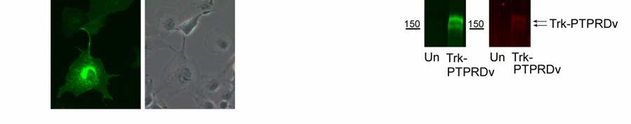

105 Figure
106 Figure 3.1. A fluorescent Trk-PTPRDv chimera is expressed on cell membranes. (A) A Trk-PTPRDv chimera was produced by fusing the ECD of the high affinity NGF receptor, TrkA, to the TMD and ICD of PTPRD. (B) The Trk-PTPRD chimera was tagged on the intracellular C-terminus with mvenus (Trk-PTPRDv). Fluorescent imaging of COS-7 cells transfected with Trk-PTPRDv (green, left panels), and their corresponding phase contrast light microscopy image (right panels). Visualization of the fluorescent Trk-PTPRDv appears punctate and concentrated in various subcellular locations compared to the diffuse expression of cytoplasmic GFP in control cells (C). (D) Western blot analysis of cell lysates transfected with Trk-PTPRDv or cytoplasmic GFP probed with anti-gfp (red), and anti-gapdh (green) as a loading control. Trk-PTPRDv appears as doublet band near the expected molecular size of 170 kd (thin double arrows). 90 kd and 26 kd bands are likely proteolytic or degraded fragments containing the venus tag (asterisks). A single thick arrow points to GAPDH and arrowhead to cytoplasmic GFP. (E) Western blot analysis of COS-7 cell lysates from untransfected (Un) or Trk- PTPRDv transfected cells run under reducing conditions and probed with both anti- PTPRD (green, left blot) and anti-gfp antibodies (red, right blot). The doublet band corresponding to Trk-PTPRDv is recognized by both antibodies (double thin arrows) in transfected cell lysates but not untransfected. (F) Anti-GFP western blot analysis of Trk- PTPRDv expressing COS cell lysates before (lys) and after cell fractionation demonstrate Trk-PTPRDv is expressed in cell membranes. Almost all Trk-PTPRDv expression (arrow) is seen in the membrane fraction (memb) with very little seen in cytoplasmic (cyto) or nuclear fractions (nucl). The cleaved or degraded fragment containing the mvenus tag is also located in cell membranes (arrowhead). 95
107 96 Influence of PTPRO dimerization and activity changes in neuronal development The role RPTPs play in nervous system development is of primary interest to our laboratory. The influence that PTPRO receptor functions have in the nervous system would therefore be of great interest. The chimeric construct we designed can serve as an excellent tool to regulate PTPRO receptor functions when expressed in axons. Ligand addition to the chimeric PTPRO would allow the control to turn on and off the phosphatase activity of PTPRO on command. This level of modulation is currently not possible for PTPRO, as no ligands have been identified for this protein to date. Preliminary investigations of this nature have been conducted, although it is too soon to report on the phenotypes produced by such investigations. However, characterization of chimeric PTPRO expression in neurons suggests that experimental manipulation of the chimera within this system may be possible. The venus-tagged Trk- ROv construct can be expressed in E7 chick forebrain neurons in culture. The protein is expressed throughout the cell including the axon and growth cone (Fig. 3.2A). Trk-ROv is expressed on cell surfaces as it can be visualized by TIRF microscopy (Fig. 3.2B panel a). TIRF illuminates only a narrow optical depth within the cell closest to the coverslip, the majority of which is cell membrane. Thus, visualizing Trk-ROv using this technique suggests that it is expressed on the cell surface (Fig. 3.2B panel a). Openfield fluorescence at the same magnification also supports membrane localization, demonstrated by the bright perimeter around the cell as well as punctate staining within the cell (Fig. 3.2B panel b). The punctate fluorescence is likely due to Trk-ROv localization in membranous organelles or vesicles involved in protein trafficking (Fig. 3.2B panel b). Short-term time lapse analysis demonstrates that Trk-ROv is rapidly
108 97 transported anterogradely and retrogradely between the growth cone and cell body, likely in vesicles moving along axonal microtubules (Fig. 3.2C). Long-term time lapse analysis demonstrates that neurons expressing Trk-ROv are able to extend primary neurites and display secondary branching (Fig. 3.3). However, cells expressing Trk-ROv can also be seen to retract axons, lose expression or possibly even die (data not shown). Thus a thorough investigation will be needed to quantify how expression of this chimera affects neurons, both in the presence and absence of ligand.


. B.")


 between the cell body and neurite tip.")
109 Figure 3.2. A fluorescent Trk-RO is expressed on cell surface of chick forebrain neurons. A. The venus-tagged Trk-ROv expresses in transfected dissociated E7 chick forebrain neurons, including expression in the growth cones. DIC image is overlaid with fluorescent (green). B. TIRF microscopy (a) and openfield fluorescence microscopy (b) suggest Trk-ROv expression is on the cell surface membrane and other membranous locations within the cell. C. Short time series of a single chick forebrain neuron expressing Trk-ROv shows the fluorescent protein is trafficked both anterogradely (1, 4) and retrogradely (2, 3) between the cell body and neurite tip. Top panel is an overlay of DIC and fluorescent images, followed by four panels of only the fluorescent images taken during a time series that spans 14 s. Numbered arrows track positions of certain puncta. 98
.")
110 Figure 3.3. Trk-ROv expression in chick forebrain culture. Time series of transfected E7 chick forebrain neurons expressing a fluorescent Trk-ROv chimera (green). Fluorescent image is overlaid with DIC image. 99
111 100 The findings presented in this dissertation demonstrating that PTPRO can modify TrkC signaling suggest that DRG neurons would be another ideal cell population in which to begin investigations into the effect of PTPRO receptor functions. Since some of these neurons express PTPRO and TrkC endogenously, comparing TrkC signaling between wild-type and PTPRO-knockout mice would be an excellent study to begin these investigations. Comparing axon outgrowth between NT-3-sensitive DRG neurons from wild-type mice and PTPRO-knockout mice would also be a logical starting point to examine any behavioral regulation PTPRO may exert on TrkC-mediated functions. PTPRO and PTPRD receptor functions in cancer Recent studies have implicated PTPRO and PTPRD to play a role in oncogenesis. PTPRD is a frequently deleted gene in small cell and non-small cell lung cancer cell lines (Sato et al., 2005). The detected PTPRD genomic deletions commonly led to reduced mrna transcripts of some PTPRD isoforms assessed by QRT-PCR, despite frequent localization of the deletions to non-coding exons (Sato et al., 2005). Therefore, the biological significance of these deletions is still unknown (Sato et al., 2005). Similar correlations were found in primary squamous cell carcinomas taken from patients exhibiting PTPRD deletions (Purdie et al., 2007). This study speculated that while the deletions may lead to N-terminal truncation, a second start codon may allow transcription of a truncated PTPRD that may act as a dominant negative (Purdie et al., 2007). The data suggest that while the mechanisms involved are far from being understood, perhaps PTPRD can act as a tumor suppressor, in which loss of the suppression leads to tumorigenesis.
112 101 A role as a tumor suppressor has also been suggested for PTPRO (Motiwala et al., 2004). PTPRO mrna expression was found to be reduced in cell lines and primary lung cancer tumors, which was suggested to be due to methylation-imposed suppression of transcription (Motiwala et al., 2004). Accordingly, removal of the methyl-block increased PTPRO expression and decreased tumorigenicity. Similarly, overexpression of PTPRO in a non-small cell lung cancer cell line reduced tumorigenesis and slowed cell cycle progression (Motiwala et al., 2004). Since PTPRO and PTPRD may behave as tumor suppressors, it would be interesting to investigate if their phosphatase activity has any responsibility in this function. Overexpression of the chimeric constructs into cancer cell lines could be examined for changes in growth rate or tumorigenesis. NGF addition to these cells to induce dimerization of chimeric PTPRO and decrease its activity could help determine the extent to which catalytic activity is involved. Preliminary experiments have been conducted overexpressing Trk-ROv in the non-small cell lung cancer cell line, A549. Expression of this construct can be visualized by the fluorescent venus tag (Fig. 3.4A). Expression of Trk-ROv had little effect on the growth rate of these cells after the first day of expression, but decreased cell growth on the following two days (Fig. 3B). While these results are preliminary, they offer support for the idea that the chimera may be a useful tool to investigate how RPTP receptor functions contribute to tumor suppression. An understanding of the role that RPTPs play in cancer may lead to the design of intelligent and specific therapeutic strategies.

113 Figure 3.4. Expression of Trk-ROv in non-small cell lung cancer decreases growth rate. A. The fluorescent Trk-ROv can be visualized after transfection into the non-small cell lung cancer cell line A549. Trk-ROv is green in the fluorescent image in right panel and the same view is shown using phase contrast in left panel. B. Growth rates were assessed in Trk-ROv-transfected and GFP-transfected control A549 cells each day for 3 days posttransfection. Growth rates were measured by automated counting of viable cells using the Vi-CELL system on triplicate samples in each condition at each time point. Preliminary results indicate a decrease in growth rate with expression of Trk-ROv compared to GFP controls over time. 102
Axon Guidance. Multiple decision points along a growing axon s trajectory Different types of axon guidance cues:
 Axon Guidance Multiple decision points along a growing axon s trajectory Different types of axon guidance cues: Contact mediated - requires direct contact by growth cone Long range - growth cone responds
Axon guidance I. Paul Garrity March 15, /9.013
 Axon guidance I Paul Garrity March 15, 2004 7.68/9.013 Neuronal Wiring: Functional Framework of the Nervous System Stretch reflex circuit Early theories of axonogenesis Schwann: many neurons link to form
Cell Death & Trophic Factors II. Steven McLoon Department of Neuroscience University of Minnesota
 Cell Death & Trophic Factors II Steven McLoon Department of Neuroscience University of Minnesota 1 Remember? Neurotrophins are cell survival factors that neurons get from their target cells! There is a
Conclusions. The experimental studies presented in this thesis provide the first molecular insights
 C h a p t e r 5 Conclusions 5.1 Summary The experimental studies presented in this thesis provide the first molecular insights into the cellular processes of assembly, and aggregation of neural crest and
The Molecular Biology of Axon Guidance
 Thursday, January 25, 2001 Science -- Tessier-Lavigne and Goodman 274 (5290): 1123 Page: 1 Institution: HARVARD UNIVERSITY Sign In as Individual FAQ The Molecular Biology of Axon Guidance Marc Tessier-Lavigne
Cellular Neurobiology BIPN 140 Fall 2016 Problem Set #8
 Cellular Neurobiology BIPN 140 Fall 2016 Problem Set #8 1. Inductive signaling is a hallmark of vertebrate and mammalian development. In early neural development, there are multiple signaling pathways
5- Semaphorin-Plexin-Neuropilin
 5- Semaphorin-Plexin-Neuropilin 1 SEMAPHORINS-PLEXINS-NEUROPILINS ligands receptors co-receptors semaphorins and their receptors are known signals for: -axon guidance -cell migration -morphogenesis -immune
THE PROBLEMS OF DEVELOPMENT. Cell differentiation. Cell determination
 We emphasize these points from Kandel in Bi/CNS 150 Bi/CNS/NB 150: Neuroscience Read Lecture Lecture Friday, October 2, 2015 Development 1: pp 5-10 Introduction Brains evolved All higher animals have brains
Reading. Lecture VI. Making Connections 9/17/12. Bio 3411 Lecture VI. Making Connections. Bio 3411 Monday September 17, 2012
 Lecture VI. Making Connections Bio 3411 Monday September 17, 2012!! 1! Reading NEUROSCIENCE: 5 th ed, pp!507?536! 4 th ed, pp 577-609 Bentley, D., & Caudy, M. (1983). Nature, 304(5921), 62-65. Dickson,
Trophic Factors. Trophic Factors. History 2. History Growth Factors. Giles Plant
 217 - Growth Factors Giles Plant Role in: Growth and Trophic Factors Soluble/diffusible factors - polypeptides Proliferation Differentiation (ie Cancer) Survival (degenerative diseases) Innervation Maintenance
Computational Model Of Axon Guidance
 Mestrado em Engenharia Informática Dissertação Relatório Final arxiv:1508.01537v1 [q-bio.nc] 6 Aug 2015 Computational Model Of Axon Guidance Rui André Ponte Costa racosta@student.dei.uc.pt Orientadores:
Bio 3411, Fall 2006, Lecture 19-Cell Death.
 Types of Cell Death Questions : Apoptosis (Programmed Cell Death) : Cell-Autonomous Stereotypic Rapid Clean (dead cells eaten) Necrosis : Not Self-Initiated Not Stereotypic Can Be Slow Messy (injury can
MIT 9.14 Class The growth of the long extensions of neurons and related topics
 9.14 - Brain Structure and its Origins Spring 2005 Massachusetts Institute of Technology Instructor: Professor Gerald Schneider A sketch of the central nervous system and its origins G. E. Schneider 2005
Hierarchical Organization of Guidance Receptors: Silencing of Netrin Attraction by Slit Through a Robo/DCC Receptor Complex
 Hierarchical Organization of Guidance Receptors: Silencing of Netrin Attraction by Slit Through a Robo/DCC Receptor Complex Elke Stein and Marc Tessier-Lavigne* Axonal growth cones that cross the nervous
Introduction Principles of Signaling and Organization p. 3 Signaling in Simple Neuronal Circuits p. 4 Organization of the Retina p.
 Introduction Principles of Signaling and Organization p. 3 Signaling in Simple Neuronal Circuits p. 4 Organization of the Retina p. 5 Signaling in Nerve Cells p. 9 Cellular and Molecular Biology of Neurons
10/2/2015. Chapter 4. Determination and Differentiation. Neuroanatomical Diversity
 Chapter 4 Determination and Differentiation Neuroanatomical Diversity 1 Neurochemical diversity: another important aspect of neuronal fate Neurotransmitters and their receptors Excitatory Glutamate Acetylcholine
A Theoretical Model of Axon Guidance by the Robo Code
 NOTE Communicated by Arjen van Ooyen A Theoretical Model of Axon Guidance by the Robo Code Geoffrey J. Goodhill geoff@georgetown.edu Department of Neuroscience, Georgetown University Medical Center, Washington,
Axon Guidance at the Midline: From Mutants to Mechanisms
 Critical Reviews in Biochemistry and Molecular Biology, 39:319 341, 2004 Copyright c Taylor & Francis Inc. ISSN: 1040-9238print / 1549-7798online DOI: 10.1080/10409230490906797 Axon Guidance at the Midline:
Molecules making waves in axon guidance
 REVIEW Molecules making waves in axon guidance Alfredo Varela-Echavarria and Sarah Guthrie 1 Department of Developmental Neurobiology, United Medical London SE1 9RT UK The exquisite complexity of neural
Signal Transduction. Dr. Chaidir, Apt
 Signal Transduction Dr. Chaidir, Apt Background Complex unicellular organisms existed on Earth for approximately 2.5 billion years before the first multicellular organisms appeared.this long period for
Neurite initiation. Neurite formation begins with a bud that sprouts from the cell body. One or several neurites can sprout at a time.
 Neurite initiation. Neuronal maturation initiation f-actin polarization and maturation tubulin stage 1: "spherical" neuron stage 2: neurons extend several neurites stage 3: one neurite accelerates its
Massive loss of neurons in embryos occurs during normal development (!)
") Types of Cell Death Apoptosis (Programmed Cell Death) : Cell-Autonomous Stereotypic Rapid Clean (dead cells eaten) Necrosis : Not Self-Initiated Not Stereotypic Can Be Slow Messy (injury can spread) Apoptosis
Types of Cell Death Apoptosis (Programmed Cell Death) : Cell-Autonomous Stereotypic Rapid Clean (dead cells eaten) Necrosis : Not Self-Initiated Not Stereotypic Can Be Slow Messy (injury can spread) Apoptosis
Neurite formation & neuronal polarization
 Neurite formation & neuronal polarization Paul Letourneau letou001@umn.edu Chapter 16; The Cytoskeleton; Molecular Biology of the Cell, Alberts et al. 1 An immature neuron in cell culture first sprouts
Formation of the Cortex
 Formation of the Cortex Neuronal Birthdating with 3 H-thymidine 3H-thymidine is incorporated into the DNA during the S-phase (replication of DNA). It marks all mitotic cells Quantitative technique. (you
C. elegans L1 cell adhesion molecule functions in axon guidance
 C. elegans L1 cell adhesion molecule functions in axon guidance Biorad Lihsia Chen Dept. of Genetics, Cell Biology & Development Developmental Biology Center C. elegans embryogenesis Goldstein lab, UNC-Chapel
Reference: Forscher, P., Kaczmarek, L.K., Buchanan, J. and Smith, S.J. (1987) Cyclic AMP induces changes in distribution and transport of organelles
 Cyclic AMP induces changes in distribution and transport of organelles") Reference: Forscher, P., Kaczmarek, L.K., Buchanan, J. and Smith, S.J. (1987) Cyclic AMP induces changes in distribution and transport of organelles within growth cones of Aplysia bag cell neurons. J.
Reference: Forscher, P., Kaczmarek, L.K., Buchanan, J. and Smith, S.J. (1987) Cyclic AMP induces changes in distribution and transport of organelles within growth cones of Aplysia bag cell neurons. J.
Neural development its all connected
 Neural development its all connected How do you build a complex nervous system? How do you build a complex nervous system? 1. Learn how tissue is instructed to become nervous system. Neural induction 2.
Zool 3200: Cell Biology Exam 5 4/27/15
 Name: Trask Zool 3200: Cell Biology Exam 5 4/27/15 Answer each of the following short answer questions in the space provided, giving explanations when asked to do so. Circle the correct answer or answers
Regulation and signaling. Overview. Control of gene expression. Cells need to regulate the amounts of different proteins they express, depending on
 Regulation and signaling Overview Cells need to regulate the amounts of different proteins they express, depending on cell development (skin vs liver cell) cell stage environmental conditions (food, temperature,
REQUIREMENTS FOR THE BIOCHEMISTRY MAJOR
 REQUIREMENTS FOR THE BIOCHEMISTRY MAJOR Grade Requirement: All courses required for the Biochemistry major (CH, MATH, PHYS, BI courses) must be graded and passed with a grade of C- or better. Core Chemistry
Report. Functional Diversity of Robo Receptor Immunoglobulin Domains Promotes Distinct Axon Guidance Decisions
 Current Biology 20, 567 572, March 23, 2010 ª2010 Elsevier Ltd All rights reserved DOI 10.1016/j.cub.2010.02.021 Functional Diversity of Robo Receptor Immunoglobulin Domains Promotes Distinct Axon Guidance
COMPUTER SIMULATION OF DIFFERENTIAL KINETICS OF MAPK ACTIVATION UPON EGF RECEPTOR OVEREXPRESSION
 COMPUTER SIMULATION OF DIFFERENTIAL KINETICS OF MAPK ACTIVATION UPON EGF RECEPTOR OVEREXPRESSION I. Aksan 1, M. Sen 2, M. K. Araz 3, and M. L. Kurnaz 3 1 School of Biological Sciences, University of Manchester,
Neurite formation & neuronal polarization. The cytoskeletal components of neurons have characteristic distributions and associations
 Mechanisms of neuronal migration & Neurite formation & neuronal polarization Paul Letourneau letou001@umn.edu Chapter 16; The Cytoskeleton; Molecular Biology of the Cell, Alberts et al. 1 The cytoskeletal
Massachusetts Institute of Technology Harvard Medical School Brigham and Women s Hospital VA Boston Healthcare System 2.79J/3.96J/BE.
 Massachusetts Institute of Technology Harvard Medical School Brigham and Women s Hospital VA Boston Healthcare System 2.79J/3.96J/BE.441/HST522J INTEGRINS I.V. Yannas, Ph.D. and M. Spector, Ph.D. Regulator
The majority of cells in the nervous system arise during the embryonic and early post
 Introduction Introduction The majority of cells in the nervous system arise during the embryonic and early post natal period. These cells are derived from population of neural stem cells first shown by
Advanced Higher Biology. Unit 1- Cells and Proteins 2c) Membrane Proteins
 Membrane Proteins") Advanced Higher Biology Unit 1- Cells and Proteins 2c) Membrane Proteins Membrane Structure Phospholipid bilayer Transmembrane protein Integral protein Movement of Molecules Across Membranes Phospholipid
Advanced Higher Biology Unit 1- Cells and Proteins 2c) Membrane Proteins Membrane Structure Phospholipid bilayer Transmembrane protein Integral protein Movement of Molecules Across Membranes Phospholipid
Cell Migration I: Neural Crest Cell Migration. Steven McLoon Department of Neuroscience University of Minnesota
 Cell Migration I: Neural Crest Cell Migration Steven McLoon Department of Neuroscience University of Minnesota 1 Types of Cell Movement passive: active: cell sheets flow cilia or flagella ameboid adhesion
Reception The target cell s detection of a signal coming from outside the cell May Occur by: Direct connect Through signal molecules
 Why Do Cells Communicate? Regulation Cells need to control cellular processes In multicellular organism, cells signaling pathways coordinate the activities within individual cells that support the function
Signal Transduction Phosphorylation Protein kinases. Misfolding diseases. Protein Engineering Lysozyme variants
 Signal Transduction Phosphorylation Protein kinases Misfolding diseases Protein Engineering Lysozyme variants Cells and Signals Regulation The cell must be able to respond to stimuli Cellular activities
REQUIREMENTS FOR THE BIOCHEMISTRY MAJOR
 REQUIREMENTS FOR THE BIOCHEMISTRY MAJOR Grade Requirement: All courses required for the Biochemistry major (CH, MATH, PHYS, BI courses) must be graded and passed with a grade of C- or better. Core Chemistry
CELL-CELL COMMUNICATION
 CELL-CELL COMMUNICATION paracrine & juxtacrine signalling autocrine & intracrine signalling methods to study cell-cell communication: attraction & repulsion chemotaxis & chemokinesis substrate preference
Multiple B-Class Ephrins and EPH Receptors Regulate Midline Axon Guidance in the Developing Mouse Forebrain
 University of Miami Scholarly Repository Open Access Dissertations Electronic Theses and Dissertations 2006-05-16 Multiple B-Class Ephrins and EPH Receptors Regulate Midline Axon Guidance in the Developing
6 Mechanotransduction
 6.1 Motivation The process of converting physical forces into biochemical signals and integrating these signals into the cellular response is referred to as mechnotransduction [11, 20]. To fully understand
The neuron as a secretory cell
 The neuron as a secretory cell EXOCYTOSIS ENDOCYTOSIS The secretory pathway. Transport and sorting of proteins in the secretory pathway occur as they pass through the Golgi complex before reaching the
NGF - twenty years a-growing
 NGF - twenty years a-growing A molecule vital to brain growth It is twenty years since the structure of nerve growth factor (NGF) was determined [ref. 1]. This molecule is more than 'quite interesting'
Migration of Neurons During Embryonic Development. Christine Simmons Saint Louis University April 15 th, 2008
 Migration of Neurons During Embryonic Development Christine Simmons Saint Louis University April 15 th, 2008 Embryonic Development of the CNS Central Nervous System (CNS) arises from Surface ectoderm Skin,
MCDB 4777/5777 Molecular Neurobiology Lecture 29 Neural Development- In the beginning
 MCDB 4777/5777 Molecular Neurobiology Lecture 29 Neural Development- In the beginning Learning Goals for Lecture 29 4.1 Describe the contributions of early developmental events in the embryo to the formation
Signal Transduction Mechanisms in Commissural Axon Guidance: The Role of Intracellular Tyrosine Kinases in Netrin-Dcc/Frazzled Axon Attraction
 University of Pennsylvania ScholarlyCommons Publicly Accessible Penn Dissertations 1-1-2012 Signal Transduction Mechanisms in Commissural Axon Guidance: The Role of Intracellular Tyrosine Kinases in Netrin-Dcc/Frazzled
Targeting Cell Surface Receptors for Axon Regeneration in the CNS
 INVITED SUBMISSION FOR PERSPECTIVE (Revised) TITLE Targeting Cell Surface Receptors for Axon Regeneration in the CNS AUTHORS Menghon Cheah 1, Melissa R Andrews 2, * AFFILIATIONS 1 John van Geest Centre
Cells to Tissues. Peter Takizawa Department of Cell Biology
 Cells to Tissues Peter Takizawa Department of Cell Biology From one cell to ensembles of cells. Multicellular organisms require individual cells to work together in functional groups. This means cells
Molecular Cell Biology 5068 In Class Exam 2 November 8, 2016
 Molecular Cell Biology 5068 In Class Exam 2 November 8, 2016 Exam Number: Please print your name: Instructions: Please write only on these pages, in the spaces allotted and not on the back. Write your
16 The Cell Cycle. Chapter Outline The Eukaryotic Cell Cycle Regulators of Cell Cycle Progression The Events of M Phase Meiosis and Fertilization
 The Cell Cycle 16 The Cell Cycle Chapter Outline The Eukaryotic Cell Cycle Regulators of Cell Cycle Progression The Events of M Phase Meiosis and Fertilization Introduction Self-reproduction is perhaps
Role of Organizer Chages in Late Frog Embryos
 Ectoderm Germ Layer Frog Fate Map Frog Fate Map Role of Organizer Chages in Late Frog Embryos Organizer forms three distinct regions Notochord formation in chick Beta-catenin localization How does beta-catenin
AXON GUIDANCE MECHANISMS AND MOLECULES: LESSONS FROM INVERTEBRATES
 AXON GUIDANCE MECHANISMS AND MOLECULES: LESSONS FROM INVERTEBRATES Sofia J. Araújo and Guy Tear Vertebrates and invertebrates share the formidable task of accurately establishing the elaborate connections
The Transmembrane Receptor UNC-40 Directs Muscle Arm Extension in Caenorhabditis elegans
 The Transmembrane Receptor UNC-40 Directs Muscle Arm Extension in Caenorhabditis elegans by Kevin Ka Ming Chan A thesis submitted in conformity with the requirements for the degree of Master of Science
MCN. Complex Genetic Interactions among Four Receptor Tyrosine Phosphatases Regulate Axon Guidance in Drosophila
 MCN Molecular and Cellular Neuroscience 17, 274 291 (2001) doi:10.1006/mcne.2000.0939, available online at http://www.idealibrary.com on Complex Genetic Interactions among Four Receptor Tyrosine Phosphatases
Geert Geeven. April 14, 2010
 iction of Gene Regulatory Interactions NDNS+ Workshop April 14, 2010 Today s talk - Outline Outline Biological Background Construction of Predictors The main aim of my project is to better understand the
Pioneer midbrain longitudinal axons navigate using a balance of Netrin attraction and Slit repulsion
 Pioneer midbrain longitudinal axons navigate using a balance of Netrin attraction and Slit repulsion Kim et al. Kim et al. Neural Development 2014, 9:17 Kim et al. Neural Development 2014, 9:17 RESEARCH
THE ROLE OF NCK IN DCC-SENSITIVE SPINAL CORD CIRCUITS. Ciaran Lane
 THE ROLE OF NCK IN DCC-SENSITIVE SPINAL CORD CIRCUITS by Ciaran Lane Submitted in partial fulfilment of the requirements for the degree of Master of Science at Dalhousie University Halifax, Nova Scotia
Visual pigments. Neuroscience, Biochemistry Dr. Mamoun Ahram Third year, 2019
 Visual pigments Neuroscience, Biochemistry Dr. Mamoun Ahram Third year, 2019 References Webvision: The Organization of the Retina and Visual System (http://www.ncbi.nlm.nih.gov/books/nbk11522/#a 127) The
Chem Lecture 10 Signal Transduction
 Chem 452 - Lecture 10 Signal Transduction 111202 Here we look at the movement of a signal from the outside of a cell to its inside, where it elicits changes within the cell. These changes are usually mediated
Chimeric Axon Guidance Receptors: The Cytoplasmic Domains of Slit and Netrin Receptors Specify Attraction versus Repulsion
 Cell, Vol. 97, 917 926, June 25, 1999, Copyright 1999 by Cell Press Chimeric Axon Guidance Receptors: The Cytoplasmic Domains of Slit and Netrin Receptors Specify Attraction versus Repulsion Greg J. Bashaw
[Frontiers in Bioscience 13, , January 1, 2008] How axons see their way axonal guidance in the visual system. Corinna Haupt, Andrea B.
![[Frontiers in Bioscience 13, , January 1, 2008] How axons see their way axonal guidance in the visual system. Corinna Haupt, Andrea B.](/thumbs/80/82512425.jpg "[Frontiers in Bioscience 13, , January 1, 2008] How axons see their way axonal guidance in the visual system. Corinna Haupt, Andrea B.") [Frontiers in Bioscience 13, 3136-3149, January 1, 2008] How axons see their way axonal guidance in the visual system Corinna Haupt, Andrea B. Huber Institute of Developmental Genetics, GSF - National
[Frontiers in Bioscience 13, 3136-3149, January 1, 2008] How axons see their way axonal guidance in the visual system Corinna Haupt, Andrea B. Huber Institute of Developmental Genetics, GSF - National
The Control of Semaphorin-1a-Mediated Reverse Signaling by Opposing Pebble and RhoGAPp190 Functions in Drosophila
 Article The Control of Semaphorin-1a-Mediated Reverse Signaling by Opposing Pebble and RhoGAPp190 Functions in Drosophila Sangyun Jeong, 1 Katarina Juhaszova, 1 and Alex L. Kolodkin 1, 1 Solomon H. Snyder
Leading the Blind: Guidance of Neuronal and Vascular Growth
 Eukaryon, Vol. 5, March 2009, Lake Forest College Review Article Leading the Blind: Guidance of Neuronal and Vascular Growth Brian Kinsman* Department of Biology Lake Forest College Lake Forest, Illinois
REVIEWS. Branch management: mechanisms of axon branching in the developing vertebrate CNS
 Branch management: mechanisms of axon branching in the developing vertebrate CNS Katherine Kalil and Erik W. Dent Abstract The remarkable ability of a single axon to extend multiple branches and form terminal
During evolution, organisms have come to perform more
 Vol 436 14 July 2005 doi:10.1038/nature03875 Common mechanisms of nerve and blood vessel wiring Peter Carmeliet 1 & Marc Tessier-Lavigne 2 REVIEWS Blood vessels and nerve fibres course throughout the body
Slit proteins: molecular guidance cues for cells ranging from neurons to leukocytes Kit Wong, Hwan Tae Park*, Jane Y Wu* and Yi Rao
 583 Slit proteins: molecular guidance cues for cells ranging from neurons to leukocytes Kit Wong, Hwan Tae Park*, Jane Y Wu* and Yi Rao Recent studies of molecular guidance cues including the Slit family
Developmental genetics: finding the genes that regulate development
 Developmental Biology BY1101 P. Murphy Lecture 9 Developmental genetics: finding the genes that regulate development Introduction The application of genetic analysis and DNA technology to the study of
Cell Cell Communication in Development
 Biology 4361 Developmental Biology Cell Cell Communication in Development June 25, 2008 Cell Cell Communication Concepts Cells in developing organisms develop in the context of their environment, including
Phosphorylation of DCC by Fyn mediates Netrin-1 signaling in growth cone guidance
 Published Online: 22 November, 2004 Supp Info: http://doi.org/10.1083/jcb.200405053 Downloaded from jcb.rupress.org on April 8, 2018 JCB: ARTICLE Phosphorylation of DCC by Fyn mediates Netrin-1 signaling
The N-terminal Leucine-Rich Regions in Slit Are Sufficient To Repel Olfactory Bulb Axons and Subventricular Zone Neurons
 The Journal of Neuroscience, March 1, 2001, 21(5):1548 1556 The N-terminal Leucine-Rich Regions in Slit Are Sufficient To Repel Olfactory Bulb Axons and Subventricular Zone Neurons Jin-hui Chen, 1 Leng
RabGDI controls axonal midline crossing by regulating Robo1 surface expression
 Philipp et al. Neural Development 2012, 7:36 RESEARCH ARTICLE RabGDI controls axonal midline crossing by regulating Robo1 surface expression Open Access Melanie Philipp 1, Vera Niederkofler 2, Marc Debrunner
targets. clustering show that different complex pathway
 Supplementary Figure 1. CLICR allows clustering and activation of cytoplasmic protein targets. (a, b) Upon light activation, the Cry2 (red) and LRP6c (green) components co-cluster due to the heterodimeric
Biol403 - Receptor Serine/Threonine Kinases
 Biol403 - Receptor Serine/Threonine Kinases The TGFβ (transforming growth factorβ) family of growth factors TGFβ1 was first identified as a transforming factor; however, it is a member of a family of structurally
Turning of Retinal Growth Cones in a Netrin-1 Gradient Mediated by the Netrin Receptor DCC
 Neuron, Vol. 19, 1211 1224, December, 1997, Copyright 1997 by Cell Press Turning of Retinal Growth Cones in a Netrin-1 Gradient Mediated by the Netrin Receptor DCC José R. de la Torre,* Veit H. Höpker,
Matrix interactions modulate neurotrophin-mediated neurite outgrowth and pathfinding
 NEURAL REGENERATION RESEARCH April 215,Volume 1,Issue www.nrronline.org INVITED review Matrix interactions modulate neurotrophin-mediated neurite outgrowth and pathfinding Christopher M. Madl 1, Sarah
Activation of a receptor. Assembly of the complex
 Activation of a receptor ligand inactive, monomeric active, dimeric When activated by growth factor binding, the growth factor receptor tyrosine kinase phosphorylates the neighboring receptor. Assembly
1- Eph- ephrin. Eph receptors and ephrin ligands. *glycosylphosphatidylinositol. anchored
 1- Eph- ephrin Eph receptors and ephrin ligands * *glycosylphosphatidylinositol membrane anchored 1 Midline guidance in the visual system In animals with binocular vision, most retinal axons (red) cross
Crk-associated substrate (Cas) signaling protein functions with integrins to specify axon guidance during development
 signaling protein functions with integrins to specify axon guidance during development") RESEARCH ARTICLE 2337 Development 134, 2337-2347 (2007) doi:10.1242/dev.004242 Crk-associated substrate (Cas) signaling protein functions with integrins to specify axon guidance during development Zhiyu
RESEARCH ARTICLE 2337 Development 134, 2337-2347 (2007) doi:10.1242/dev.004242 Crk-associated substrate (Cas) signaling protein functions with integrins to specify axon guidance during development Zhiyu
Neurophysiology. Danil Hammoudi.MD
 Neurophysiology Danil Hammoudi.MD ACTION POTENTIAL An action potential is a wave of electrical discharge that travels along the membrane of a cell. Action potentials are an essential feature of animal
Lecture 6: Non-Cortical Visual Pathways MCP 9.013/7.68, 03
 Lecture 6: Non-Cortical Visual Pathways MCP 9.013/7.68, 03 Roger W. Sperry The problem of central nervous reorganization after nerve regeneration and muscle transposition. R.W. Sperry. Quart. Rev. Biol.
Signal transduction by Cell-Cell and Cell-Matrix contacts
 Signal transduction by Cell-Cell and Cell-Matrix contacts - Integrins as central adhesion receptors - - Focal Adhesion Kinase (FAK) as a central signal molecule - Structure of Cell/Cell and Cell/Matrix
Cell-Cell Communication in Development
 Biology 4361 - Developmental Biology Cell-Cell Communication in Development October 2, 2007 Cell-Cell Communication - Topics Induction and competence Paracrine factors inducer molecules Signal transduction
Developmental Biology 3230 Midterm Exam 1 March 2006
 Name Developmental Biology 3230 Midterm Exam 1 March 2006 1. (20pts) Regeneration occurs to some degree to most metazoans. When you remove the head of a hydra a new one regenerates. Graph the inhibitor
Introduction. Gene expression is the combined process of :
 1 To know and explain: Regulation of Bacterial Gene Expression Constitutive ( house keeping) vs. Controllable genes OPERON structure and its role in gene regulation Regulation of Eukaryotic Gene Expression
Cell Adhesion and Signaling
 Cell Adhesion and Signaling mchuang@ntu.edu.tw Institute of Anatomy and Cell Biology 1 Transactivation NATURE REVIEWS CANCER VOLUME 7 FEBRUARY 2007 85 2 Functions of Cell Adhesion cell cycle proliferation
Supplemental table S7.
 Supplemental table S7. GO terms significantly enriched in significantly up-regulated genes of the microarray. K: number of genes from the input cluster in the given category. F: number of total genes in
Lecture 10: Cyclins, cyclin kinases and cell division
 Chem*3560 Lecture 10: Cyclins, cyclin kinases and cell division The eukaryotic cell cycle Actively growing mammalian cells divide roughly every 24 hours, and follow a precise sequence of events know as
Molecular Cell Biology 5068 In Class Exam 1 September 30, Please print your name:
 Molecular Cell Biology 5068 In Class Exam 1 September 30, 2014 Exam Number: Please print your name: Instructions: Please write only on these pages, in the spaces allotted and not on the back. Write your
Morphogens as conserved axon guidance cues Yimin Zou 1 and Anna I Lyuksyutova 2
 Morphogens as conserved axon guidance cues Yimin Zou 1 and Anna I Lyuksyutova 2 Morphogen family proteins are now widely appreciated as axon guidance cues. Because their roles as morphogens are highly
ADAM FAMILY. ephrin A INTERAZIONE. Eph ADESIONE? PROTEOLISI ENDOCITOSI B A RISULTATO REPULSIONE. reverse. forward
 ADAM FAMILY - a family of membrane-anchored metalloproteases that are known as A Disintegrin And Metalloprotease proteins and are key components in protein ectodomain shedding Eph A INTERAZIONE B ephrin
RANK. Alternative names. Discovery. Structure. William J. Boyle* SUMMARY BACKGROUND
 RANK William J. Boyle* Department of Cell Biology, Amgen, Inc., One Amgen Center Drive, Thousand Oaks, CA 91320-1799, USA * corresponding author tel: 805-447-4304, fax: 805-447-1982, e-mail: bboyle@amgen.com
The EGF Signaling Pathway! Introduction! Introduction! Chem Lecture 10 Signal Transduction & Sensory Systems Part 3. EGF promotes cell growth
 Chem 452 - Lecture 10 Signal Transduction & Sensory Systems Part 3 Question of the Day: Who is the son of Sevenless? Introduction! Signal transduction involves the changing of a cell s metabolism or gene
Cellular Neuroanatomy I The Prototypical Neuron: Soma. Reading: BCP Chapter 2
 Cellular Neuroanatomy I The Prototypical Neuron: Soma Reading: BCP Chapter 2 Functional Unit of the Nervous System The functional unit of the nervous system is the neuron. Neurons are cells specialized
7.06 Problem Set #4, Spring 2005
 7.06 Problem Set #4, Spring 2005 1. You re doing a mutant hunt in S. cerevisiae (budding yeast), looking for temperaturesensitive mutants that are defective in the cell cycle. You discover a mutant strain
Physiology 2 nd year. Neuroscience Optional Lecture
 Academic year 2018/2019 Physiology 2 nd year Semester 1 Curricula Nervous system physiology Blood physiology Acid-base equilibrium Bibliography: Boron & Boulpaep Medical Physiology, 3 rd edition Physiology
Sarah Bashiruddin Georgina Lopez Jillian Merica Sarah Wardlaw
 Sarah Bashiruddin Georgina Lopez Jillian Merica Sarah Wardlaw Introduction: Dr. Carol Erickson and her lab study the cellular and molecular mechanisms by which neural crest cells differentiate and migrate
Analysis and Simulation of Biological Systems
 Analysis and Simulation of Biological Systems Dr. Carlo Cosentino School of Computer and Biomedical Engineering Department of Experimental and Clinical Medicine Università degli Studi Magna Graecia Catanzaro,
Reading Assignments. A. Genes and the Synthesis of Polypeptides. Lecture Series 7 From DNA to Protein: Genotype to Phenotype
 Lecture Series 7 From DNA to Protein: Genotype to Phenotype Reading Assignments Read Chapter 7 From DNA to Protein A. Genes and the Synthesis of Polypeptides Genes are made up of DNA and are expressed