HISTO-ANATOMICAL OBSERVATIONS REGARDING VIOLA L. SPECIES IN THE GÂRBOAVELE RESERVE (COUNTY OF GALAŢI) Introduction
|
|
|
- Beryl Lawson
- 6 years ago
- Views:
Transcription
1 Analele Ştiinţifice ale Universităţii Al. I. Cuza Iaşi s. II a. Biologie vegetală, 2014, 60, 1: ISSN: , E-ISSN: HISTO-ANATOMICAL OBSERVATIONS REGARDING VIOLA L. SPECIES IN THE GÂRBOAVELE RESERVE (COUNTY OF GALAŢI) Anca MEREACRE 1 *, Angela TONIUC 1, Constantin TOMA 1 Abstract: The authors research the structure of vegetative organs of five different vernal species of Viola L. which grow in the Gârboavele - Galaţi nature reserve. The histological features, most of them quantitative ones, which differentiate the five species of Viola L., are pointed out: the thickness of the root and rhizomes peridermis, the number of palisadic layers of the leaf lamina, the presence or absence of tector hairs and of oxaliferous cells, their frequency and position. Key words: Viola, histo-anatomy, vegetative organs, Gârboavele-Galaţi reserve. Introduction There are 28 species of Viola L. in the Romanian flora (Grințescu et al., 1955; Sârbu et al., 2013), four of which known to grow in the Gârboavele - Galaţi nature reserve: Viola arvensis Murray, V. elatior Fr., V. hirta L. and V. odorata L. (Mititelu et al., 1968); to the above mentioned ones we bring a 5-th one, recently found by us: V. kitaibeliana Schult. Two of the species are annual ones (V. arvensis and V. kitaibeliana), while three of them are perennial plants (V. elatior, V. hirta and V. odorata), and have a thick rhizome, with or without aerial runners (V. hirta). The anatomic structure of Viola species has been little investigated until now, as can be seen from the two synthesis treaties, the one about the anatomy of dicotyledonous plants (Metcalfe and Chalk, 1972) and the one about the leaves of angiosperms in general (Napp- Zinn, 1973, 1974). When presentations of tissue structures of various vegetative organs are made, the authors mention, among others the Viola genre, too; Viola odorata being most frequently quoted. Burduja and Moţiu (1982) achieve a detailed histo-anatomical study of the aerial parts of Viola hymettia Boiss., a species closely related (often mistaken for ) to V. arvensis, V. tricolor and V. kitaibeliana. The aerial parts of V. arvensis are used (besides V. tricolor) in traditional medicine for their anti-inflammatory, expectorant and diuretic and other properties. The need to know more about the morpho-anatomic features of both species resulted in research work dedicated to these aspects (Meyer, 1916; Toiu et al., 2009, 2010). Reference to structure features of Viola species can be found in works of comparative (Petit, 1887; Howard, 1962; Uphof and Hummel, 1962) or ecological anatomy (Constantin, 1885), but also in some with a special focus on vernal species (Keller, 1934; Ubaidulaev, 1959; Ivanskaja, 1962, 1963; Goryşina, 1965). 1 Faculty of Biology, Al. I. Cuza University, Bd. Carol I, no.11, Iaşi Romania; anca_mereacre@yahoo.com (corresponding author*)
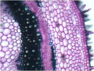
2 Mention must be made that three of the species studied by us (Viola arvensis, V. kitaibeliana and V. odorata) have been analyzed from an anatomical point of view besides other plants growing in Northern Iran (Yousefi et al., 2012). From the above we can conclude that there are few histo-anatomical data referring to Viola L. species, reason why, in our work, we are attempting a comparative analysis of the vegetative organs of the five vernal Viola species mentioned above, which grow in the Gârboavele - Galaţi nature reserve. Materials and methods The material under study was collected in the Gârboavele - Galaţi nature reserve in May 2012, at the anthesis stage. This forest-park is at 90 m altitude, and are predominantly: Quercus pubescens, Q. pedunculiflora, Ulmus foliacea, Tilia tomentosa, to which are added Acer campestre, A. tataricum, Pyrus pyraster, Cotinus coggygria and another species (Mititelu et al., 1968). The plants were fixed and preserved in 70% ethyl alcohol. The vegetative organs (the root, rhizome, aerial runner, aerial stem, and leaf) were sectioned and coloured (in iodine green and ruthenium red) according to the usual histo-anatomical technique in plant research. Cross-sections and the tangential/longitudinal ones have been analyzed under the Olympus CX31 optic microscope and photographed with an Olympus C 5060 camera (Andrei and Predan, 2003). Results and discussions THE ROOT (Plate I, Figs. 1-3) The rhizodermis of V. odorata and V. hirta occasionally presents long absorbent hairs; the rhizodermis, as well as the external layer of the bark of the other three species is exfoliates early. The bark of V. odorata and V. hirta is differentiated in the exodermis, cortical parenchyma and endodermis; the other species keep only the internal layers of the cortical parenchyma (at V. hirta with aerial gaps) and the casparian type of endodermis; some of the parenchymatous cells of V. hirta and V. odorata contain calcium oxalate ursines. The central cylinder is the anatomical area with a secondary structure, due to the activity of the vascular cambium which has produced a thin external phloem ring and a thick central xylem body. The phloem ring consists of sieve-tubes, companiong cells and numerous cells of amiliferous parenchyma (collenchymatous in V. arvensis, arranged in strictly radial rows in V. elatior). Only Viola odorata has an incipient secondary structure, the phloem has few parenchyma cells, and the xylem has very few libriform elements. The central xylem body comprises vessels of varied diameters, irregularly dispersed in the fundamental lignified mass. The axial part of the stem of V. arvensis, V. hirta and V. kitaibeliana is less lignited, wih fewer and smaller vessels which are separated only by cellulosic parenchyma. The species were determined by Ion Sârbu, Ph.D, whom we warmly thank in this paper, too. 14
3 Thus, the early passage from the primary to the secondary structure can be noticed both in perennial and in annual species, but only at the level of the central cylinder, on account of the vascular cambium. THE RHIZOME (Plate I, Fig. 5) Only Viola hirta and V. odorata present rhizomes, both are perennial species, but only one of them has aerial runners with adventitious roots from the nodes (V. odorata). The contour of its cross section is very irregular, with ribs of various size in V. hirta and a symmetrical structure in V. odorata. The structure of both species is typically secondary, resulting from the activity of both lateral meristems, but especially of the vascular cambium. The epidermis and the largest part of the cortical parenchyma have exfoliated. In the deep persistent internal parenchymatic layers of V. odorata some cells contain, like the ones in the pith, calcium oxalate ursines. The phellogen (or cork cambium), differentiated at the level of the internal cortical parenchyma or of the pericycle (V. hirta), has produced one or two areas of suber and the phellem. The vascular cambium has produced the secondary conducting tissues, of a fascicular type in V. odorata and ring type in V. hirta. The secondary phloem ring is relatively thick and contains conducting elements (sieve-tubes, companion cells) in the close vicinity of the cambium and numerous cells of amiliferous parenchyma towards the exterior, disposed in radial rows, some containing calcium oxalate ursines. The secondary xylem ring is much thicker, strongly sclerified and intensely lignified, made up of irregularly dispersed vessels of varied diameters, many libriform fibers and few parenchyma cells. The xylem ring is crossed by some parenchymaticcellulosic medullar rays In the thickness of the V. hirta ring we can distinguish several (4-5) rings, each of them having more numerous early vessels and libriform fibers with thinner walls, and fewer later vessels and libriform fibers with thicker walls. In the early xylem areas we can see some islands of cellulosic parenchyma. The pith is parenchymatic-cellulosic, and many cells contain calcium oxalate ursines. THE AERIAL RUNNER (Plate I, Fig. 4) The aerial runner, which we find only in V. odorata has a flat-elliptical contour in cross-section, with a slightly concave face (the inferior one), which corresponds to the place where the endogenously formed adventitious roots will come out; here the phloem is reduced and interrupted by the cord which forms the adventitious root. When dealing with thinner and younger material, the contour of the cross-section is circular, the cortical parenchyma is exfoliating, the suber zone has 3-4 layers of suberified cells, the phellodermis area is visibly collenchymatized, and the phloem ring has a different thickness on the circumference of the organ. The structure is typically secondary and asymmetrical for thicker and older material. The aerial runner surface is covered by a relatively thick peridermis, with 4-6 layers of suber (exfoliating) and an equal number of phellodermis layers, which are tangentially elongated and are disposed in radial series, some of them containing calcium oxalate 15
4 ursines. From place to place there are remains of the primary cortical parenchyma, adhering to the suber. The central cylinder is very thick, but irregular thickness along the circumference of the aerial runner, so that it gets the shape of a cordiform contour in cross-section, the xylem getting into direct contact with the suber along one of the sides. We see that the cambium produces an incomplete, but thick, phloem ring (sieve-tubes and companion cells) towards the interior and numerous parenchyma cells, disposed in radial rows and with moderately thickened walls, towards the exterior. Cambium also produces a thick, completely lignified, xylem ring (vessels, libriform fibers, parenchyma cells), pierced by 1-2 parenchymatic-cellulosic medullar rays. Many of medullar rays cells, as well as those of the liberian parenchyma, contain calcium oxalate ursines. At a closer analysis one can notice, inside the secondary xylem ring, 6-7 incomplete, concentric rings which can be recognized by the early vessels of a larger diameter and by the terminal libriform areas. THE AERIAL STEM (Plate II, Figs. 6-11) The structure of the two annual species (V. arvensis and V. kitaibeliana) is and remains only a primary one. The structure of the three perennial species (V. elatior, V. hirta and V. odorata) is mostly of secondary origin in the lower third of the stem, but only at the level of the central cylinder, due to the activity of the vascular cambium. The contour of the cross-section varies from circular to elliptically-circular (modified by two lateral wings (V. elatior), to trapezoid (V. arvensis) or square shaped at the tip and triangle shaped at the basis of the stem (V. kitaibeliana). The epidermis has isodiametric cells, with thicker internal and external walls than the other ones; the external wall is covered by a thin, lightly striped cuticle. From place to place, at least in the upper third of the stem, we can notice stomata and tector hairs. There are numerous unicellular relatively short hairs on the lateral wings in the lower third of the stem in V. arvensis, V. elatior and V. kitaibeliana. The bark is parenchymatic, of meatic type, but it also has aerial gaps (V. elatior, V. odorata), and it ends with an endodemoid (V. arvensis) or with a casparian endodermis (V. hirta, V. odorata). The hypodermic layer is collenchymatized in the ribs most frequently of an angular or tangential type (V. kitaibeliana).cells with calcium oxalate ursines have been notice in V. elatior, V. hirta and V. odorata. The central cylinder of annual species has conducting tissue of fascicular type (with a primary structure) while the perennial species have ring type tissue (towards the basis of the stem, with a secondary structure). The number of vascular bundles varies from the tip (4) towards the basis of the stem (8-12) in V. kitaibeliana, it is constant (4) in V. arvensis or it is higher (8-10) at the tip of the stem. In perennial species the vascular bundles unite giving birth to two concentric rings at the stem basis. In the primary structure the phloem has sieve-tubes and companion cells, while the xylem has vessels and cellulosic parenchyma type of cells. The secondary phloem ring has, in addition to this, parenchyma cells, and the secondary xylem ring has also libriform fibers. The medullar rays, sclerified and lignified, form, together with the secondary xylem, 16
5 a thick and compact ring. We can observe thin sclerenchymatic fiber cords on the external face of the phloemic ring (V. elatior). The pith is parenchymatic-cellulosic and many of its cells contain calcium oxalate ursines (V. odorata, V. elatior, and V. hirta). Many pith cells of the annual species become disorganized thus creating a smaller or larger central aerial cavity, along the whole stem. Such small aerial cavities in the pith have been noticed in V. elatior, too. THE LEAF (Plates III, IV, V) The petiole (Plate III, Figs ) The contour of the cross-section varies from triangular (V. kitaibeliana) to semicircular modified by two lateral-adaxial wings (three in V. odorata). The epidermis has isodiametric cells, whose exterior walls are slight thicker than the others and they are covered by a very thin cuticle. From place to place one can notice stomata and rare unicellular tector hairs (more frequent on the lateral-adaxial wings), shorter (V. elatior) or longer ones (V. hirta). The fundamental parenchyma is slightly collenchymatised under the epidermis (especially in the lateral-adaxial wings), with aerial cavities on adaxial face (V. arvensis, and V. odorata); some of the cells contain calcium oxalate ursines (V. elatior and V. odorata). The conducting tissues form 3 vascular bundles: a large median one and two small lateral-adaxial ones, all of them with a primary structure. The lamina (Plate IV, Figs ; Plate V, Figs ) The epidermis, in front side view, presents cells with irregular contours, with slight waving lateral walls on the upper face of the lamina and strongly waving ones on its lower face; only V. kitaibeliana presents lateral walls with high amplitude waves on both faces of the limb. Anisocytic type stomata (cruciferous ones) can be noticed in the two types of epidermis, with a higher frequency on the surface unit of the lower epidermis, this indicating an amphystomatic leaf. In V. hirta the stomata are not present or are extremely rare in the upper epidermis, indicating a hypostomatic leaf. The mesophyll comprises two types of assimilating tissues: palisadic and spongy ones, that the limb has a bifacial heterofacial structure (dorsiventral leaf) in all five Viola species we have investigated. The differences between species refer to the number of palisadic layers (one in V. hirta and two in the other species), to the length of the composing cells (longer in V. kitaibeliana and shorter in V. elatior and V. hirta ), to the presence or absence (V. arvensis ) of unicellular tector hairs, to the presence (V. hirta and V. odorata) or absence of oxaliferous cells. Conclusions The general structure plan of all investigated vegetative organs is similar in all five Viola species. The passage from the primary to the secondary structure of the root takes place very early both in perennial and annual species, but only at the level of the central cylinder, that is on account of the vascular cambium. 17
6 The rhizome, present only in perennial species (V. hirta and V. odorata) has a secondary structure, as a result of the activity of both lateral meristems; the vascular cambium and the cork cambium (phellogen). In the very thick secondary xylem ring we can distinguish several areas, each with early and late wooden vessels (V. hirta). Both species present many oxaliferous cells in the primary and medullar cortical parenchyma. The aerial runner, present only in V. odorata, has, like the rhizome, a secondary structure, with a thick peridermis, very thick xylem ring, oxaliferous cells in the medullar rays, phelloderm and liberian parenchyma. The five species we have investigated are differentiated only by the presence or absence of tector hairs, of oxaliferous cells and central aerial cavity. The leaf petiole has a different cross-section contour, with two lateral-adaxial wings always visible. The species under study differ by the presence or absence of tector hairs, oxaliferous cells and aerial cavities in the fundamental axial parenchyma. All five species present 3 vascular bundles. The foliar lamina has a homogenous, bifacial, heterofacial (dorsiventral) structure, the difference between the species being mostly quantitative ones (the number of palisadic layers, the length of their cells), more rarely qualitative ones: the presence or absence of tector hairs and oxaliferous cells. REFERENCES Andrei, M., Predan, G.M.I., Practicum de morfología şi anatomía plantelor. Edit. Ştiinţelor Agricole, Bucureşti. Burduja, C., Moţiu, T., Cercetări morfologice şi histo-anatomice ale organelor vegetative de la specia Viola hymettia Boiss. et Heldr. Culeg. de studii şi artic. de Biol., Grăd. Bot. Iaşi. 2: Costantin, J., Recherches sur l influence qu exerce le milieu sur la structure des racines. Ann. des Sci. Nat., Bot., ser. 7. 1: Goryşina, T.K., Anatomiceskoe stroenie list ev rannevesennyh efemeroidov dubovogo lesa. Vestnik Leningradskogo Universiteta, Ser. Biol., 20, vyp 1(3): Grinţescu, G., Guşuleac, M., Nyárády, E., Viola L. In Flora R.P.R., 3. Edit. Acad. Rom., Bucureşti: Howard, R.A., The vascular structure of the petiole as a taxonomic character. Proc. 15-th Internat. Hort. Congress, Nice, 1958: Ivanskaja, E.N., Sur quelques particularités de la structure anatomique de la feuille de plantes en rosettes et en coussinet, d altitude. Dokl. AN SSSR. 146, 2: Ivanskaja E.N., O stroenie mezofila list ev nekotoryh vysokogornyh ratenij ţentral nogo Kavkaza. Zapiski Ţentral. Kavkazkogo otd. Vsesoiuz-go botanici-go obscestva, Ordjonikidze. 1: Keller E.F., Osobennosti anatomiceskogo stroenia list ev u vesennih efemero-odnoletnikov. Sovremennaia botanika. 2, 4: Metcalfe, C.R., Chalk, L., Anatomy of the Dicotyledons, 1. Clarendon Press, Oxford: Meyer, F.J., Bau und Ontogenie des Wasserleitungssystems der vegetativen Organe von Viola tricolor var. arvensis. Thesis, Marburg (cf. Bot. Zbl, 141, 1919: ). Mititelu, D., Gociu, Z., Patraşcu, A., Gheorghiu, V., Flora şi vegetaţia pădurii-parc Gîrboavele-Galaţi. Analele Științ. Univ. Al. I. Cuza Iaşi, s. II a. Biol. 14, 1: Napp-Zinn, Kl., 1973, Anatomie des Blattes. II. Blattanatomie der Angiospermen. Handbuch der Pflanzenanatomie, Bd. VIII, 2 A 1-2, Gebrüder Borntraeger, Berlin. Petit, L., Le petiol des Dicotyledones au point de vue de l anatomie comparée et de la taxinomie. These, Bordeaux. Sârbu, I., Ştefan, N., Oprea, A., Plante vasculare din România. Edit. VictorBVictor, Bucureşti: Toiu, A., Oniga, I., Tămaş, M., 2009 Cercetări morfologice şi anatomice asupra speciei Viola tricolor L. (Violaceae). Rev. Med. Chir. Soc. Med. Nat. Iaşi. 113 (2, suppl. 4):
7 Toiu, A., Oniga, I., Tămaş, M., Morphological and anatomical researches on Viola arvensis Murray (Violaceae). Farmacia. 58, 5: Ubaidulaev, U., Anatomo-ekologhiceskoe issledovania list ev nekotoryh efemeroidov i efemerov zapadnogo Tian-Şan. Avtoreferat, Taşkent. Uphof,T.C.J., Hummel, K., Plant Hairs. Encyclopedia of Plant Anatomy, 4. Gebrüder Borntraeger, Berlin. Yousefi, N., Mehrvarz, S.S., Marcussen, T., Anatomical studies on selected species of Viola (Violaceae) in Iran. Nordic Journal of Botany. 30, 4: EXPLANATION OF THE PLATES PLATE I Fig. 1.Viola arvensis - cross-section through the root (Oc.10 x Ob.4) Fig. 2. Viola kitaibeliana - cross-section through the root (Oc.10 x Ob.4) Fig. 3. Viola hirta - cross-section through the root details (Oc.10 x Ob.20) Fig. 4. Viola odorata - cross-section through the aerial runner details (Oc.10 x Ob.20) Fig. 5. Viola hirta - cross-section through the rhizome (Oc.10 x Ob.4) PLATE II Fig. 6. Viola elatior - cross-section through the upper level of the stem (Oc.10 x Ob.10) Fig. 7.Viola elatior - cross-section through the upper level of the stem details (Oc.10 x Ob.20) Fig. 8. Viola kitaibeliana - cross-section through the middle level of the stem (Oc.10 x Ob.10) Fig. 9. Viola hirta - cross-section through the middle level of the stem (Oc.10 x Ob.10) Fig. 10. Viola arvensis - cross-section through the basal level of the stem (Oc.10 x Ob.40) Fig. 11. Viola elatior - cross-section through the basal level of the stem (Oc.10 x Ob.20) PLATE III Fig. 12. Viola.kitaibeliana - cross-section through the petiole (Oc.10 x Ob.10) Fig. 13. Viola elatior - cross-section through the petiole (Oc.10 x Ob.10) Fig. 14. Viola arvensis - cross-section through the petiole (Oc.10 x Ob.10) Fig. 15. Viola odorata - cross-section through the petiole (Oc.10 x Ob.10) Fig. 16. Viola elatior - cross-section through the petiole details (Oc.10 x Ob.20) Fig. 17. Viola hirta - cross-section through the petiole details (Oc.10 x Ob.20) PLATE IV Fig. 18. Viola arvensis the lower and upper lamina epidermis, in front view (Oc.10 x Ob.40) Fig. 19. Viola elatior - the lower and upper lamina epidermis, in front view (Oc.10 x Ob.40) Fig. 20. Viola hirta - the lower and upper lamina epidermis, in front view (Oc.10 x Ob.40) Fig. 21. Viola odorata - the lower and upper lamina epidermis, in front view (Oc.10 x Ob.40) PLATE V Fig. 22. Viola arvensis - cross-section through the lamina in the midrib part (Oc.10 x Ob.20) Fig. 23. Viola odorata - cross-section through the lamina in the midrib part (Oc.10 x Ob.20) Fig. 24. Viola elatior - cross-section through the lamina in the midrib part (Oc.10 x Ob.20) Fig. 25. Viola kitaibeliana - cross-section through the lamina in the midrib part (Oc.10 x Ob.10) Fig. 26. Viola.hirta - cross-section through the lamina between the veins - details (Oc.10 x Ob.40) 19


8 PLATE I Figure 1. Viola arvensis Figure 2. Viola kitaibeliana Figure 3. Viola hirta Figure 4. Viola odorata Figure 5. Viola hirta 20


9 PLATE II Figure 6. Viola elatior Figure 7. Viola elatior Figure 8. Viola kitaibeliana Figure 9. Viola hirta Figure 10. Viola arvensis 21 Figure 11. Viola elatior


10 PLATE III Figure 12. Viola kitaibeliana Figure 13. Viola elatior Figure 14. Viola arvensis Figure 15. Viola odorata Figure 16. Viola elatior 22 Figure 17. Viola hirta


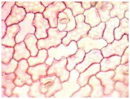
11 Lower epidermis Upper epidermis PLATE IV Figure 18. Viola arvensis Figure 19. Viola elatior Figure 20. Viola hirta Figure 21. Viola odorata 23


12 PLATE V Figure 22. Viola arvensis Figure 23. Viola odorata Figure 24. Viola elatior Figure 25. Viola kitaibeliana Figure 26. Viola hirta 24
Irina Berciu *, Constantin Toma Department of Biology, Al. I. Cuza University, Iasi
 HISTO-ANATOMICAL ASPECTS OF VEGETATIVE ORGANS OF THYMUS DACICUS BORB. AND THYMUS GLABBRESCENS WILLD. Irina Berciu *, Constantin Toma Department of Biology, Al. I. Cuza University, Iasi * Correspondence:
RODICA RUGINĂ, C. TOMA
 Analele ştiinţifice ale Universităţii Al. I. Cuza Iaşi Tomul LIII, s. II a. Biologie vegetală, 2007 HISTO-ANATOMICAL ASPECTS OF SOME LONICERA L. SPECIES RODICA RUGINĂ, C. TOMA Abstract. The authors investigate
HISTO-ANATOMICAL LESS KNOW ASPECTS UPON SOME LAMIACEAE TAXA CAMELIA IFRIM *, IRINA TOMA ** Introduction. Material and method
 Analele ştiinţifice ale Universităţii Al. I. Cuza Iaşi Tomul L, s. II a. Biologie vegetală, 2004 HISTO-ANATOMICAL LESS KNOW ASPECTS UPON SOME LAMIACEAE TAXA CAMELIA IFRIM *, IRINA TOMA ** Abstract: The
 Question 1: State the location and function of different types of meristem. Meristems are specialised regions of plant growth. The meristems mark the regions where active cell division and rapid division
ANATOMY OF PLANTS Introduction: The study of gross internal structure of plant organs by the technique of section cutting is called plant anatomy.
 ANATOMY OF PLANTS Introduction: The study of gross internal structure of plant organs by the technique of section cutting is called plant anatomy. (Pandey, 2002). Various plant organ viz. root, stem, leaves,
A COMPARATIVE STUDY REGARDING THE MORPHOLOGY AND ANATOMY OF THE VEGETATIVE APPARATUS IN TWO OCIMUM BASILICUM L. BREEDS.
 Analele ştiinţifice ale Universităţii Al. I. Cuza Iaşi Tomul LIV, fasc. 2, s.ii a. Biologie vegetală, 2008 A COMPARATIVE STUDY REGARDING THE MORPHOLOGY AND ANATOMY OF THE VEGETATIVE APPARATUS IN TWO OCIMUM
Anatomy of Flowering Plants. K C Meena PGT Biology
 Anatomy of Flowering Plants K C Meena PGT Biology Tissues A group of similar cells performing same function. Types of plant tissues - Meristematic tissues and permanent tissues. Meristematic tissues Have
Class XI Chapter 6 Anatomy of Flowering Plants Biology
 Class XI Chapter 6 Anatomy of Flowering Plants Biology Question 1: State the location and function of different types of meristem. Meristems are specialised regions of plant growth. The meristems mark
HISTO-ANATOMICAL OBSERVATIONS REFERRING TO SOME MELAMPYRUM SPECIES
 Analele ştiinţifice ale Universităţii Al. I. Cuza Iaşi Tomul LV, fasc. 2, s.ii a. Biologie vegetală, 2009 HISTO-ANATOMICAL OBSERVATIONS REFERRING TO SOME MELAMPYRUM SPECIES ASPAZIA BĂEŞU *, C. TOMA **,
NOTES ON THE MORPHO-ANATOMY OF ACONITUM DEGENII GAYER. Introduction
 Analele ştiinţifice ale Universităţii Al. I. Cuza Iaşi Tomul LV, fasc. 2, s.ii a. Biologie vegetală, 2009 NOTES ON THE MORPHO-ANATOMY OF ACONITUM DEGENII GAYER IRINA STĂNESCU *, C. MARDARI *, C. TĂNASE
CONTRIBUTIONS REGARDING THE LEAF HISTO-ANATOMY OF SOME PELARGONIUM SPECIES
 Rev. Med. Chir. Soc. Med. Nat., Iaşi 2013 vol. 117, no. 3 PHARMACY ORIGINAL PAPERS CONTRIBUTIONS REGARDING THE LEAF HISTO-ANATOMY OF SOME PELARGONIUM SPECIES Cristina Elena Iancu, Oana Cioanca, Cornelia
Visit For All NCERT solutions, CBSE sample papers, Question papers, Notes for Class 6 to 12. Chapter-6 ANATOMY OF FLOWERING PLANTS
 Chapter-6 ANATOMY OF FLOWERING PLANTS POINTS TO REMEMBER Anatomy : Anatomy is the study of internal structure of organisms. Plant anatomy includes organisation and structure of tissues. Tissue : A group
Downloaded from
 POINTS TO REMEMBER : 6. Anatomy of Flowering Plants Study of internal structure of plant is called anatomy. In plants cells are the basic unit. Cells organized into tissues and tissues organized into organs.
COMPARATIVE HISTO-ANATOMICAL ANALYSIS OF THE VEGETATIVE ORGANS OF SEDUM TELEPHIUM L. SSP. MAXIMUM (L.) KROCK. IN VITRO AND FROM NATURE
 KROCK. IN VITRO AND FROM NATURE") ARDELEAN MIRELA, STĂNESCU IRINA, CACHIŢĂ-COSMA DORINA J. Plant Develop. 16 (2009): 3 8 COMPARATIVE HISTO-ANATOMICAL ANALYSIS OF THE VEGETATIVE ORGANS OF SEDUM TELEPHIUM L. SSP. MAXIMUM (L.) KROCK. IN VITRO
ARDELEAN MIRELA, STĂNESCU IRINA, CACHIŢĂ-COSMA DORINA J. Plant Develop. 16 (2009): 3 8 COMPARATIVE HISTO-ANATOMICAL ANALYSIS OF THE VEGETATIVE ORGANS OF SEDUM TELEPHIUM L. SSP. MAXIMUM (L.) KROCK. IN VITRO
Chapter 35~ Plant Structure and Growth
 Chapter 35~ Plant Structure and Growth Plant Organization Plant morphology is based on plant s evolutionary history Need to draw in nutrients from the ground and the air Plant Organs Root system = roots
Secondary growth in stems
 Secondary growth in stems Secondary growth Some of the meristematic cells in plants with secondary growth keep their meristematic state and become cells of the cambium. The addition of secondary vascular
Plant Anatomy and Tissue Structures
 Plant Anatomy and Tissue Structures The Two Major Plant Systems Reproductive shoot (flower) Terminal bud Node Internode Angiosperm plants have threse major organs: Roots Stems Leaves & Flowers Terminal
Chapter 29: Plant Tissues
 Chapter 29: Plant Tissues Shoots and Roots Shoots (Leaves and Stem) Produce food by photosynthesis Carry out reproductive functions Roots Anchor the plant Penetrate the soil and absorb water and dissolved
THE SPECIE PARIETARIA LUSITANICA L., CAULINE AND FOLIAR HISTOANATOMICAL STUDY Mariana Arcuş, Gabriela Lilios, Emanuela Gheorma, Corina Moromete
 THE SPECIE PARIETARIA LUSITANICA L., CAULINE AND FOLIAR HISTOANATOMICAL STUDY Mariana Arcuş, Gabriela Lilios, Emanuela Gheorma, Corina Moromete OVIDIUS CONSTANŢA UNIVERSITY, FACULTY OF PHARMACY Summary
VEGETATIVE ANATOMY OF TWO GALIUM L. SPECIES (RUBIACEAE) Introduction
 Introduction") Analele ştiinţifice ale Universităţii Al. I. Cuza Iaşi Tomul LIV, fasc. 2, s.ii a. Biologie vegetală, 2008 VEGETATIVE ANATOMY OF TWO GALIUM L. SPECIES (RUBIACEAE) ANCA HEMCINSCHI *, RAMONA GALEŞ **, C.
Analele ştiinţifice ale Universităţii Al. I. Cuza Iaşi Tomul LIV, fasc. 2, s.ii a. Biologie vegetală, 2008 VEGETATIVE ANATOMY OF TWO GALIUM L. SPECIES (RUBIACEAE) ANCA HEMCINSCHI *, RAMONA GALEŞ **, C.
TARGET STUDY MATERIAL
 TARGET STUDY MATERIAL Plus-1 Botany VOL I TARGET EDUCATIONAL INSTITUTION Target Educational institution is the one and only Entrance coaching and CBSE 10 th coaching centre at Mukkam with advanced technologies
THE LEAF STRUCTURE OF SOME NEPENTHES DANSER SPECIES IRINA STĂNESCU, C. TOMA. Introduction
 Analele ştiinţifice ale Universităţii Al. I. Cuza Iaşi Tomul LIV, fasc. 1, s. II a. Biologie vegetală, 2008 THE LEAF STRUCTURE OF SOME NEPENTHES DANSER SPECIES IRINA STĂNESCU, C. TOMA Abstract: The authors
Topic 2: Plant Structure & Growth Ch. 35 Angiosperms are the most complex plants. They are composed of cells, tissues, organs and organ systems.
 Topic 2: Plant Structure & Growth Ch. 35 Angiosperms are the most complex plants. They are composed of cells, tissues, organs and organ systems. Fig. 35.8 Plant Cells pp.798-802 Types of plant cells Include:
Chapter #35~ Plant Structure and Growth
 Chapter #35~ Plant Structure and Growth What part of a plant is represented by each of these: Carrot Celery Red Pepper Tomato Lettuce Garbanzo Bean Angiosperm structure Three basic organs: Roots (root
A group of cells with common origin is called a tissue. The cells of a tissue usually perform a common function.
 Anatomy of Flowering Plants Tissues A group of cells with common origin is called a tissue. The cells of a tissue usually perform a common function. Types of Tissue: There are two main types of plant tissues,
2/25/2013. o Plants take up water and minerals from below ground o Plants take up CO2 and light from above ground THREE BASIC PLANT ORGANS ROOTS
 o Plants take up water and minerals from below ground o Plants take up CO2 and light from above ground THREE BASIC PLANT ORGANS o Roots o Stems o Leaves ROOTS o Anchor plant o Absorb water and minerals
Lecture 4 Root Put line under your answer! There is only one correct answer in the multiple choice questions
 Lecture 4 Root Put line under your answer! There is only one correct answer in the multiple choice questions 1. The perception of gravity by a root is thought to take place in a) root hairs b) the region
Plants. Tissues, Organs, and Systems
 Plants Tissues, Organs, and Systems Meristematic cells Specialized cells that are responsible for producing specialized cells, they produce three types of tissue in the body of a plant. Meristematic Cells
Plant Structure And Growth
 Plant Structure And Growth The Plant Body is Composed of Cells and Tissues Tissue systems (Like Organs) made up of tissues Made up of cells Plant Tissue Systems Ground Tissue System Ø photosynthesis Ø
ANATOMY OF FLOWERING PLANTS
 ANATOMY OF FLOWERING PLANTS Finish Line & Beyond The Tissues The Tissue System Anatomy of Dicotyledonous and Monocotyledonous Plants Secondary Growth THE TISSUES A tissue is a group of cells having a common
Plant Structure. Objectives At the end of this sub section students should be able to:
 Name: 3.2 Organisation and the Vascular Structures 3.2.1 Flowering plant structure and root structure Objectives At the end of this sub section students should be able to: 1. Label a diagram of the external
CHAPTER 6 ANATOMY OF FLOWERING PLANTS
 84 BIOLOGY CHAPTER 6 ANATOMY OF FLOWERING PLANTS 6.1 The Tissues 6.2 The Tissue System 6.3 Anatomy of Dicotyledonous and Monocotyledonous Plants 6.4 Secondary Growth You can very easily see the structural
CHAPTER 6 ANATOMY OF FLOWERING PLANTS
 84 BIOLOGY CHAPTER 6 ANATOMY OF FLOWERING PLANTS 6.1 The Tissues 6.2 The Tissue System 6.3 Anatomy of Dicotyledonous and Monocotyledonous Plants 6.4 Secondary Growth You can very easily see the structural
CONSIDERATIONS UPON THE ANATOMICAL FEATURES OF SOME TAXA OF TRADESCANTIA GENERA
 Buletinul Grădinii Botanice Iaşi Tomul 14, 2007 CONSIDERATIONS UPON THE ANATOMICAL FEATURES OF SOME TAXA OF TRADESCANTIA GENERA IFRIM CAMELIA Abstract: The structure of two taxa of the Tradescantia genre
Plant Tissues and Organs. Topic 13 Plant Science Subtopics , ,
 Plant Tissues and Organs Topic 13 Plant Science Subtopics 13.1.2, 13.1.3, 13.1.4 Objectives: List and describe the major plant organs their structure and function List and describe the major types of plant
THE TISSUES A tissue is a group of cells having a common origin and usually performing a common function. Tissues. Parenchyma
 1 CHAPTER 6 ANATOMY OF FLOWERING PLANTS Study of internal structure of plants is called anatomy. Plants have cells as the basic unit, cells are organised into tissues and in turn the tissues are organised
The Shoot System: Primary Stem Structure - 1
 The Shoot System: Primary Stem Structure - 1 Shoot System The shoot system comprises the leaves and stems of plants. Leaves are located at nodes on the stem; the distance along the stem between nodes is
Chapter 28 Active Reading Guide Plant Structure and Growth
 Name: AP Biology Mr. Croft Chapter 28 Active Reading Guide Plant Structure and Growth In this unit on plants, the challenge for students will be to learn the new vocabulary. As we work through this unit,
UNIT 6 - STRUCTURES OF FLOWERING PLANTS & THEIR FUNCTIONS
 6.1 Plant Tissues A tissue is a group of cells with common function, structures or both. In plants we can find 2 types of tissues: Meristem Permanent tissues Meristem is found in regions with continuous
Plant Structure. Lab Exercise 24. Objectives. Introduction
 Lab Exercise Plant Structure Objectives - Be able to identify plant organs and give their functions. - Learn distinguishing characteristics between monocot and dicot plants. - Understand the anatomy of
NOTES: CH 35 - Plant Structure & Growth
 NOTES: CH 35 - Plant Structure & Growth In their evolutionary journey, plants adapted to the problems of a terrestrial existence as they moved from water to land ANGIOSPERMS (flowering plants) -most diverse
HISTO-ANATOMICAL ASPECTS OF THE AJUGA GENEVENSIS L. AND AJUGA REPTANS L. VEGETATIVE ORGANS. Introduction
 Analele Ştiinţifice ale Universităţii Al. I. Cuza Iaşi s. II a. Biologie vegetală, 2012, 58, 1: 11-18 http://www.bio.uaic.ro/publicatii/anale_vegetala/anale_veg_index.html ISSN: 1223-6578, E-ISSN: 2247-2711
Histology and Anatomy of Flowering Plants
 Histology and Anatomy of Flowering Plants Very Short Answer Type Questions 1. The transverse section of a plant material shows the following anatomical features: a) The vascular bundles are conjoint, scattered
Name: Plant stems and leaves (p. 1 of )
") Name: Plant stems and leaves (p. 1 of ) Introduction: Plants have a variety of configurations but the same basic structures. The three main parts of a plant are the roots, stems, and leaves. The tracheids
Name: Plant stems and leaves (p. 1 of ) Introduction: Plants have a variety of configurations but the same basic structures. The three main parts of a plant are the roots, stems, and leaves. The tracheids
Today: Plant Structure Exam II is on F March 31
 Next few lectures are on plant form and function Today: Plant Structure Exam II is on F March 31 Outline Plant structure I. Plant Cells structure & different types II. Types of meristems Apical meristems:
Plants. Plant Form and Function. Tissue Systems 6/4/2012. Chapter 17. Herbaceous (nonwoody) Woody. Flowering plants can be divided into two groups:
 Woody. Flowering plants can be divided into two groups:") Monocots Dicots 6/4/2012 Plants Plant Form and Function Chapter 17 Herbaceous (nonwoody) In temperate climates, aerial parts die back Woody In temperate climates, aerial parts persist The Plant Body Functions
Monocots Dicots 6/4/2012 Plants Plant Form and Function Chapter 17 Herbaceous (nonwoody) In temperate climates, aerial parts die back Woody In temperate climates, aerial parts persist The Plant Body Functions
tree of life phylogeny morphology gram stain chapter 28-29, other groups of organisms Bacteria
 tree of life chapter 28-29, other groups of organisms phylogeny key lineages of prokaryotes Domain Archaea (sister to eukarya) 3 clades defined by genetic characters Domain Bacteria Firmicutes Spirochaetes
tree of life phylogeny gram stain morphology chapter 28-29, other groups of organisms Bacteria
 tree of life chapter 28-29, other groups of organisms phylogeny key lineages of prokaryotes Domain Archaea (sister to eukarya) 3 clades defined by genetic characters Domain Bacteria Firmicutes Spirochaetes
Exercise 12. Procedure. Aim: To study anatomy of stem and root of monocots and dicots.
 Aim: To study anatomy of stem and root of monocots and dicots. Principle: The study of internal morphology, i.e., cells of various tissues in an organ of a living body is called Anatomy. Tissue, which
2.1 PLANT TISSUE HALIMAHTUN SAEDIAH BT ABU BAKAR KOLEJ TEKNOLOGI TIMUR
 2.1 PLANT TISSUE HALIMAHTUN SAEDIAH BT ABU BAKAR KOLEJ TEKNOLOGI TIMUR GENERAL Plant cell are differentiated possessing structural adaptations that make specific functions possible. Modifications of cell
The three principal organs of seed plants are roots, stems, and leaves.
 23 1 Specialized Tissues in Plants Seed Plant Structure The three principal organs of seed plants are roots, stems, and leaves. 1 of 34 23 1 Specialized Tissues in Plants Seed Plant Structure Roots: absorb
Plant Anatomy: roots, stems and leaves
 Plant Anatomy: roots, stems and leaves The plant body has a hierarchy of organs, tissues and cells Plants, like animals, have organs composed of different tissues, which are composed of cells. Tissue is
Angiosperms: Dicotyledons
 Angiosperms: Dicotyledons This section contains anatomical descriptions of stem and twig xylem, as well as the bark and pith regions of 244 dicotyledonous species belonging to 61 families. Angiosperms:
MORPHO-ANATOMICAL CONSIDERATIONS UPON THE SHOOT OF SOME ROSA L. CULTIVARS FROM THE BOTANIC GARDEN OF IASI (1 ST NOTE)
") DELINSCHI VIOLETA, STĂNESCU IRINA, MIHALACHE MIHAELA, ADUMITRESEI LIDIA J. Plant Develop. 16 (2009):9 16 MORPHO-ANATOMICAL CONSIDERATIONS UPON THE SHOOT OF SOME ROSA L. CULTIVARS FROM THE BOTANIC GARDEN
DELINSCHI VIOLETA, STĂNESCU IRINA, MIHALACHE MIHAELA, ADUMITRESEI LIDIA J. Plant Develop. 16 (2009):9 16 MORPHO-ANATOMICAL CONSIDERATIONS UPON THE SHOOT OF SOME ROSA L. CULTIVARS FROM THE BOTANIC GARDEN
Chapter 23 Notes Roots Stems Leaves
 Chapter 23 Notes Roots Stems Leaves I. Specialized tissue in plants - effective way to ensure the plant s survival A. Seed plant structure 1. Roots - a. Absorbs water and dissolves nutrients b. anchors
ROOTS. Syllabus Theme A Plant Structure and Function. Root systems. Primary Growth of Roots. Taproot system. Fibrous root system.
 Syllabus Theme A lant Structure and Function A2: Structure and function of the basic plant organs ampbell & Reece hap. 35 Selected page numbers ROOTS Functions Anchors the vascular plant Absorbs minerals
STEMS Anytime you use something made of wood, you re using something made from the stem of a plant. Stems are linear structures with attached leaves
 STEMS OUTLINE External Form of a Woody Twig Stem Origin and Development Stem Tissue Patterns Herbaceous Dicotyledonous Stems Woody Dicotyledonous Stems Monocotyledonous Stems Specialized Stems Wood and
Honors Biology I Ch 29 Plant Structure & Function
 3 Basic types of plant cells Honors Biology I Ch 29 Plant Structure & Function 1) Parenchyma cells- loosely packed or cells with a and thin, Involved in metabolic functions 2) Collenchyma cells- thicker
OCR (A) Biology A-level
 Biology A-level") OCR (A) Biology A-level Topic 3.3: Transport in plants Notes Plants require a transport system to ensure that all the cells of a plant receive a sufficient amount of nutrients. This is achieved through
OCR (A) Biology A-level Topic 3.3: Transport in plants Notes Plants require a transport system to ensure that all the cells of a plant receive a sufficient amount of nutrients. This is achieved through
Plant Organization. Learning Objectives. Angiosperm Tissues. Angiosperm Body Plan
 Plant Organization Learning Objectives 1. List and give the major function of the three main types of plant tissues 2. Identify a monocot verses a eudicot plant by observing either root, stem, leaf, or
PLANT TISSUES 12 MARCH 2014
 PLANT TISSUES 12 MARCH 2014 Lesson Description In this lesson we: Identify the different types of plant tissue Be able to relate the different structures with the different functions Plant Tissue Summary
Plant Anatomy: roots, stems and leaves
 Plant Anatomy: roots, stems and leaves The plant body has a hierarchy of organs, tissues and cells Plants, like animals, have organs composed of different tissues, which are composed of cells. Tissue is
Effects of Sun-Blotch on the Anatomy of the Avocado Stem
 California Avocado Association 1935 Yearbook 20: 125-129 Effects of Sun-Blotch on the Anatomy of the Avocado Stem Charles A. Schroeder Because of the comparatively recent discovery of the avocado disease
II. SIMPLE TISSUES Bot 404--Fall A. Introduction to Tissues (DIAGRAM allow a full page)
") II. SIMPLE TISSUES Bot 404--Fall 2004 A. Introduction to Tissues (DIAGRAM allow a full page) B. Definitions Adaxial = facing the axil; upper surface of leaf Abaxial = facing away from the axil; lower surface
II. SIMPLE TISSUES Bot 404--Fall 2004 A. Introduction to Tissues (DIAGRAM allow a full page) B. Definitions Adaxial = facing the axil; upper surface of leaf Abaxial = facing away from the axil; lower surface
CONTRIBUTIONS TO THE HISTO-ANATOMICAL STUDY OF THE CALENDULA OFFICINALIS L. LEAVES TREATED WITH THIOPHANATE METHYL (TOPSIN M) Introduction
 Introduction") Analele ştiinţifice ale Universităţii Al. I. Cuza Iaşi Tomul LIV, fasc. 1, s. II a. Biologie vegetală, 2008 CONTRIBUTIONS TO THE HISTO-ANATOMICAL STUDY OF THE CALENDULA OFFICINALIS L. LEAVES TREATED WITH
Analele ştiinţifice ale Universităţii Al. I. Cuza Iaşi Tomul LIV, fasc. 1, s. II a. Biologie vegetală, 2008 CONTRIBUTIONS TO THE HISTO-ANATOMICAL STUDY OF THE CALENDULA OFFICINALIS L. LEAVES TREATED WITH
COMPARATIVE STEM AND LEAF ANATOMY OF THE GENUS ODONTITES (SCROPHULARIACEAE) IN IRAN
 IN IRAN") COMPARATIVE STEM AND LEAF ANATOMY OF THE GENUS ODONTITES (SCROPHULARIACEAE) IN IRAN SH. Saeidi-Mehrvaz Saeidi-Mehrvarz, SH. 2004. 10 10: Comparative stem and leaf anatomy of the genus Odontites (Scrophulariaceae)
COMPARATIVE STEM AND LEAF ANATOMY OF THE GENUS ODONTITES (SCROPHULARIACEAE) IN IRAN SH. Saeidi-Mehrvaz Saeidi-Mehrvarz, SH. 2004. 10 10: Comparative stem and leaf anatomy of the genus Odontites (Scrophulariaceae)
Roots anchor plants and absorb water and minerals in solution. A germinating seed radicle becomes the first root. Four zones, or regions, of young
 Roots anchor plants and absorb water and minerals in solution. A germinating seed radicle becomes the first root. Four zones, or regions, of young roots are recognized: (1) A protective root cap that also
Chapter 29. Table of Contents. Section 1 Plant Cells and Tissues. Section 2 Roots. Section 3 Stems. Section 4 Leaves. Plant Structure and Function
 Plant Structure and Function Table of Contents Section 1 Plant Cells and Tissues Section 2 Roots Section 3 Stems Section 4 Leaves Section 1 Plant Cells and Tissues Objectives Describe the three basic types
MORPHOLOGICAL AND HISTO-ANATOMICAL ASPECTS AT SOME DICOTILEDONATE SEEDLINGS RELATED TO THE VASCULAR TRANSITION
 Analele ştiinţifice ale Universităţii Al. I. Cuza Iaşi Tomul LII, s. II a. Biologie vegetală, 2006 MORPHOLOGICAL AND HISTO-ANATOMICAL ASPECTS AT SOME DICOTILEDONATE SEEDLINGS RELATED TO THE VASCULAR TRANSITION
ANATOMICAL STUDY OF THE VEGETATIVE ORGANS OF GARDENIA JASMINOIDES ELLIS (RUBIACEAE) Rodica Bercu
 Rodica Bercu") Summary ANATOMICAL STUDY OF THE VEGETATIVE ORGANS OF GARDENIA JASMINOIDES ELLIS (RUBIACEAE) Rodica Bercu FACULTY OF NATURAL AND AGRICULTURAL SCIENCES, OVIDIUS UNIVERSITY, CONSTANTZA rodicabercu@yahoo.com
Summary ANATOMICAL STUDY OF THE VEGETATIVE ORGANS OF GARDENIA JASMINOIDES ELLIS (RUBIACEAE) Rodica Bercu FACULTY OF NATURAL AND AGRICULTURAL SCIENCES, OVIDIUS UNIVERSITY, CONSTANTZA rodicabercu@yahoo.com
SESSION 6: SUPPORT AND TRANSPORT SYSTEMS IN PLANTS PART 1
 SESSION 6: SUPPORT AND TRANSPORT SYSTEMS IN PLANTS PART 1 KEY CONCEPTS In this session we will focus on summarising what you need to know about: - Anatomy of dicotyledonous plants Root and stem: distribution
Plant Structure and Function
 Plant Structure and Function A Meridian Biology AP Study Guide by John Ho and Tim Qi Plant Terms Growth: Growth Types Type Location Description Primary Primary Vertical growth (up-down), dominant direction
Bring Your Text to Lab!!!
 Bring Your Text to Lab!!! Vascular Plant Anatomy: Flowering Plants Objectives: 1. To observe what the basic structure of vascular plants is, and how and where this form originates. 2. To begin to understand
THE OHIO JOURNAL OF SCIENCE
 THE OHIO JOURNAL OF SCIENCE VOL. XXIV JULY, 1924 No.. 4 THE VASCULAR ANATOMY OF CALAMOVILFA LONGIFOLIA.* ERNEST LINCOLN STOVER Eastern Illinois State Teachers' College The present study of the anatomy
MAGNOLIA botany. evergreen ; spicy odor of blooms; chambered pith; hairy leaves(lower epidermis) & petioles
 & petioles") MAGNOLIA botany Angiosperm: primitive, ancestral (Not Eudicot, Not Monocot): order Ranales: family Magnoliaceae: Magnolia grandiflora (southern magnolia) evergreen ; spicy odor of blooms; chambered pith;
MAGNOLIA botany Angiosperm: primitive, ancestral (Not Eudicot, Not Monocot): order Ranales: family Magnoliaceae: Magnolia grandiflora (southern magnolia) evergreen ; spicy odor of blooms; chambered pith;
Plant Structure and Function. Roots, Stems, and Leaves
 Plant Structure and Function Roots, Stems, and Leaves What is a Plant? Plants are living things that have: roots, stems, and leaves (some have flowers) Plants are made of cells that have cell walls, a
Phytochemical resources on Thymus stojanovii degen. Species (labiatae)
") Phytochemical resources on Thymus stojanovii degen. Species (labiatae) M. ARCUŞ, V. SCHRÖDER Faculty of Pharmacy, Ovidius University, Romania, Constanta *Corresponding author Arcus Mariana Ovidius University
Phytochemical resources on Thymus stojanovii degen. Species (labiatae) M. ARCUŞ, V. SCHRÖDER Faculty of Pharmacy, Ovidius University, Romania, Constanta *Corresponding author Arcus Mariana Ovidius University
ANATOMICAL ASPECTS OF THE ORNAMENTAL PLANT SPATHIPHYLLUM WALLISII REGEL
 Studii şi Cercetări Martie 2010 Biologie 18 13-17 Universitatea Vasile Alecsandri din Bacău ANATOMICAL ASPECTS OF THE ORNAMENTAL PLANT SPATHIPHYLLUM WALLISII REGEL Rodica Bercu, Marius Făgăraş Key words:
ANATOMICAL STRUCTURE OF SOME EUPHORBIA SPECIES FROM THE ROMANIAN FLORA
 Muzeul Olteniei Craiova. Oltenia. Studii i comunic ri. tiin ele Naturii. Vol. XXII/2006 ISSN 1454-6914 ANATOMICAL STRUCTURE OF SOME EUPHORBIA SPECIES FROM THE ROMANIAN FLORA STRUCTURA ANATOMIC A UNOR SPECII
BIOL 305L Laboratory One
 Please print Full name clearly: BIOL 305L Laboratory One General plant anatomy a great place to start! Introduction Botany is the science of plant life. Traditionally, the science included the study of
The Plant body has a hierarch of organs, tissues, and cells. [2]
![The Plant body has a hierarch of organs, tissues, and cells. [2]](/thumbs/77/75254697.jpg "The Plant body has a hierarch of organs, tissues, and cells. [2]") GUIDED READING - Ch. 35 PLANT STRUCTURE NAME: Please print out these pages and HANDWRITE the answers directly on the printouts. Typed work or answers on separate sheets of paper will not be accepted. Importantly,
GUIDED READING - Ch. 35 PLANT STRUCTURE NAME: Please print out these pages and HANDWRITE the answers directly on the printouts. Typed work or answers on separate sheets of paper will not be accepted. Importantly,
Plant Structure and Function Extension
 Plant Structure and Function Extension NGSSS: SC.912.L.14.7 Relate the structure of each of the major plant organs and tissues to physiological processes. (AA) Part 1A: Leaves The leaf of a plant serves
CHAPTER 6 ANATOMY OF FLOWERING PLANTS MULTIPLE CHOICE QUESTIONS
 ANATOMY OF FLOWERING PLANTS 27 27 CHAPTER 6 ANATOMY OF FLOWERING PLANTS MULTIPLE CHOICE QUESTIONS 1. A transverse section of stem is stained first with safranin and then with fast green following the usual
Plant Structure, Growth, and Development
 Chapter 35 Plant Structure, Growth, and Development PowerPoint Lecture Presentations for Biology Eighth Edition Neil Campbell and Jane Reece Lectures by Chris Romero, updated by Erin Barley with contributions
Lecture 19. A Sieve Plate with large Sieve Pores. Secondary Phloem. Secondary phloem (cont d)
") Lecture 19 Secondary phloem (cont d) Secondary Phloem in Tilia americana (American Basswood) Secondary Phloem of Tilia Stained with Toluidine Blue & viewed with Crossed Polarizers. Secondary Phloem A Sieve
Lecture 19 Secondary phloem (cont d) Secondary Phloem in Tilia americana (American Basswood) Secondary Phloem of Tilia Stained with Toluidine Blue & viewed with Crossed Polarizers. Secondary Phloem A Sieve
Non Permanent Tissues - Meristematic Tissue
 PLANT TISSUES Non Permanent Tissues - Meristematic Tissue Undifferentiated plant cells that are continually dividing by mitosis Large thin walled cells No vacuole Dense cytoplasm Large nucleus Found at
Stems and Transport in Vascular Plants. Herbaceous Stems. Herbaceous Dicot Stem 3/12/2012. Chapter 34. Basic Tissues in Herbaceous Stems.
 Bud scale Terminal bud Stems and Transport in Plants One year's growth Terminal bud scale scars Axillary bud Leaf scar Node Internode Node Chapter 34 Lenticels Terminal bud scale scars Bundle scars A Woody
Plant Organs. Roots & Stems
 Plant Organs Roots & Stems I. Roots A. F(x)s = grow underground 1. Absorb water & nutrients from soil 2. Anchor plant in the soil 3. Make hormones important for growth & development I. Roots B. Structure
Forms strands that conduct water, minerals, and organic compounds. Much of the inside of nonwoody parts of plants. Includes roots, stems, and leaves
 Biology II Vascular plants have 3 tissue systems: Dermal Protective outer layer of plant Vascular Forms strands that conduct water, minerals, and organic compounds Ground Much of the inside of nonwoody
This exam is the property of CR Hardy. It may not be sold or distributed in any manner.
 Relevant Sample Questions for Exam 1 from an old exam. Bio 221 Concepts of Botany Dr. Hardy Relevant bits from Exam 1 (Spring 2013) Name: Instructions: -Please do not turn this page over until Prof. Hardy
Overview of Plant Tissues
 Plant Tissue Growth Key Concepts Overview of Plant Tissues Seed-bearing vascular plants have a shoot system with stems, leaves, and reproductive parts Most also have a root system These systems consist
Leaf. It is composed of:
 LEAF It is composed of: Leaf a leaf stalk called petiole; if it lacks leaf is sessile; the expanded part called lamina or blade; a strand of vascular tissue (veins) in the blade; a pair of leafy outgrowth
23 Structure of Flowering Plants
 23 Structure of Flowering Plants Flowering plants first evolved around 125 million years ago. www.mrcbiology.com 1 23 Structure of Flowering Plants www.mrcbiology.com 2 24 Structure of Flowering Plants
Chapter 35: Plant Structure, Growth and Development - No two Plants Are Alike Plant structure
 Chapter 35: Plant Structure, Growth and Development - No two Plants Are Alike Plant structure Systems Root and Shoot system Organs Roots, Stems, Leaves Tissues Dermal, Vascular, Ground Cells parencyma,
Fun with Botany 2009
 Fun with Botany 2009 Fun with Botany April, 2002 Plant Uses and Types Gymnosperms Angiosperms Monocots Dicots Gymnosperms Keep leaves which are either needles or flat scales Seeds are not enclosed Give
-Each asexual organs. -Anchors the plant -Absorbs water and minerals -Stores sugars and starches
 Plants are made up of: -organs, tissues, and cells The three major plant organs are: -Roots, stems, and leaves -Each asexual organs Plants have a Root System beneath the ground that us a multicellular
(A) Buds (B) Lateral meristem (C) Apical meristem (D) Stem (E) Trichomes
 Buds (B) Lateral meristem (C) Apical meristem (D) Stem (E) Trichomes") AP Biology - Problem Drill 17: Plant Structure Question No. 1 of 10 1. What are hair-like outgrowths that protect and absorb nutrients? Question #01 (A) Buds (B) Lateral meristem (C) Apical meristem (D)
AP Biology - Problem Drill 17: Plant Structure Question No. 1 of 10 1. What are hair-like outgrowths that protect and absorb nutrients? Question #01 (A) Buds (B) Lateral meristem (C) Apical meristem (D)
! Xylem - Chief conducting tissue for water and minerals absorbed by the roots.
 + Complex Tissues! Complex tissues are made up of two or more cell types.! Xylem - Chief conducting tissue for water and minerals absorbed by the roots.! Vessels - Made of vessel elements.! Long tubes
Plant Structure, Growth, and Development
 Chapter 35 Plant Structure, Growth, and Development PowerPoint Lecture Presentations for Biology Eighth Edition Neil Campbell and Jane Reece Lectures by Chris Romero, updated by Erin Barley with contributions
Anatomy of dicotyledonous plants
 Anatomy of dicotyledonous plants Differences between Monocotyledons and Dicotyledons All plants are classified as producing seeds or not producing seeds. Those that produce seeds are divided into flowering