SAXS/SANS data processing and overall parameters
|
|
|
- Natalie Burns
- 5 years ago
- Views:
Transcription


1 EMBO Global Exchange Lecture Course 30 November 2012 Hyderabad India SAXS/SANS data processing and overall parameters Petr V. Konarev European Molecular Biology Laboratory, Hamburg Outstation BioSAXS group
2 Small-angle scattering: experiment Detector Sample Log (Intensity) Monochromatic beam Wave vector k, k=2π/λ 2θ s=4π sinθ/λ, nm -1 Radiation sources: k 1 X-ray generator (λ = nm) Synchrotron (λ = nm) Thermal neutrons (λ = nm) Scattering vector s=k 1 -k, s=4π sinθ/λ

")


3 BIOSAXS beamline P12 (Petra-3) Pilatus 2M 2D Raw Data Iron nanoparticles (June 2011)
 Active area 250*290 mm 2,")

4 PILATUS Pixel X-ray Detector at P12 PILATUS 2M (24*100K modules) Active area 250*290 mm 2, pixel size: 172μm Readout time: 3.6ms, framing rate: 50Hz Silver behenate Axis calibration standard
5 Raw data reduction steps Radial integration of 2D image into 1D curve Exact coordinates of the beam center are required for integration (determined from AgBeh data) Mask file is used to eliminate beamstop and inactive detector area Associated errors in the data points are computed from the numbers of counts using Poisson statistics Data are normalized to the pindiode value (intensity of the transmitted beam) and exposure time Data are transferred into ASCII format containing 3 columns: s I(s) Er(s)


, a.u.")
6 Small Angle Scattering Radial averaging Normalization against: data collection time, transmitted sample intensity. Log I(s), a.u. s = 4π sinθ/λ s, nm -1
7 Scattering by matter X-rays are scattered mostly by electrons Thermal neutrons are scattered mostly by nuclei Scattering amplitude from an ensemble of atoms A(s) is the Fourier transform of the scattering length density distribution in the sample ρ(r) Experimentally, scattering intensity I(s) =[A(s)] 2 is measured.
8 Small-angle scattering: contrast I sample (s) I matrix (s) I particle (s) To obtain scattering from the particles, matrix scattering must be subtracted, which also permits to significantly reduce contribution from parasitic background (slits, sample holder etc) Contrast Δρ = <ρ(r) - ρ s >, where ρ s is the scattering density of the matrix, may be very small for biological samples


9 X-rays neutrons X-rays: scattering factor increases with atomic number, no difference between H and D Neutrons: scattering factor is irregular, may be negative, huge difference between H and D Element H D C N O P S Au At. Weight N electrons bx,10-12 cm bn,10-12 cm
10 Sample and buffer scattering Looking for protein signals less than 5% above background level
11 Sample and buffer scattering
12 Sample and buffer scattering I(s) = [ T I (s) T I (s) ( T T ) I (s)] m s s ctt s m m m Det( s) s e Here, subscripts s, m and e denote the scattering from sample, matrix (e.g. solvent) and empty cell (camera background), T stands for transmission, c for sample concentration and Det(n) is the detector response function. For solution scattering studies T s usually equals to T m and the third term vanishes.
13 Analysis of biological SAS data lg I, relative 3 2 Scattering curve I(s) Overall Parameters R g D max MM exp 1 Excluded Volume s, nm -1
14 Overall parameters Radius of gyration R g (Guinier, 1939) I(s) 1 I( 0 ) exp( R 3 2 g s 2 ) Maximum size max : p(r)=0 for r> Dmax Maximum size D max : p(r)=0 for r> Dmax Excluded particle volume (Porod, 1952) V = 2 2π I(0)/Q; Q = 0 s 2 I( s) ds
15 Program PRIMUS- graphical package for data manipulations and analysis data manipulations (averaging, background subtraction, merging of data in different angular ranges, extrapolation to infinite dilution ) evaluation of radius of gyration and forward intensity (Guinier plot, module AUTORG), estimation of Porod volume calculation of distance/size distribution function p(r)/v(r) (module GNOM) data fitting using the parameters of simple geometrical bodies (ellipsoid, elliptic/hollow cylinder, rectangular prism) (module BODIES) data analysis for polydisperse and interacting systems, mixtures and partially ordered systems (modules OLIGOMER, SVDPLOT, MIXTURE and PEAK) P.V. Konarev, V.V. Volkov, A.V. Sokolova, M.H.J. Koch, D.I. Svergun J.Appl. Cryst. (2003) 36,

16 PRIMUS: graphical user interface
17 Data quality Radiation damage Log I(s), a.u. sample s, nm -1
, a.u.")

18 Data quality Radiation damage Log I(s), a.u. sample same sample again RADIATION DAMAGE! s, nm -1
19 Merging data Log I(s) Low and High Concentration 1 mg/ml 10 mg/ml s, nm -1
20 Merging data Log I(s) Low and High Concentration s, nm -1
21 Merging data Log I(s) Low and High Concentration s, nm -1
22 Merging data Log I(s) Low and High Concentration s, nm -1
23 Merging data Log I(s) Low and High Concentration s, nm -1
24 Extrapolation to zero concentration Infinite dilution Log I(s) 10 mg/ml 1 mg/ml 0 mg/ml? s, nm -1
 a.u.")

25 Shape and size Log I(s) a.u. lysozyme apoferritin s, nm -1

, relative -1-1")


26 The scattering is related to the shape Solid sphere 0 lg I(s), relative Hollow sphere s, s, nm nm -1-1 Dumbbell Flat disc Long rod
27 Guinier law I 0 sin( sr ) ( s ) = 4 π p ( r ) ds sr For small values of x, sinx/x can be expressed as : sin( sr sr ) = 1 ( sr ) 3! 2 + ( sr ) 5! 4.. Hence, close to the origin: I(s) = I(0)[1-ks 2 + ] I(0)exp(-ks 2 ) The scattering curve of a particle can be approximated by a Gaussian curve in the vicinity of the origin I ( s ) I ( 0 ) exp( 3 ) This is a classical formula derived by Andre Guinier in 1938, in his first SAXS application (to defects in metals) s 2 R g 2 /
28 Radius of gyration Radius of gyration : R 2 g = V r V Δρ() r r 2 rdvr Δρ() r dv R g is the quadratic mean of distances to the center of mass weighted by the contrast of electron density. R g is an index of non sphericity. For a given volume the smallest R g is that of a sphere : Ellipsoïd of revolution (a, b) Cylinder (D, H) a + b Rg = D H R g = ideal monodisperse r Rg = 3 R 5
29 Guinier plot example The law is generally used under its log form : ln[ I( s)] ln[ I(0)] s 2 2 R g /3 A linear regression yields two parameters : I(0) (y-intercept) R g from the slope Validity range : 0 < sr g <1.3

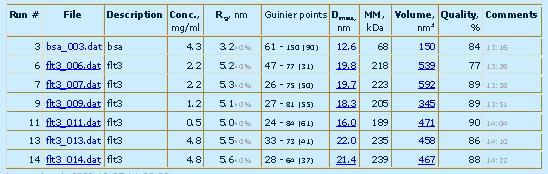
30 PRIMUS: Guinier plot I( s) = I(0) exp( s 2 Rg 2 3) Rg radius of gyration Rg = e-2 I0 = M MBSA*(I(0)/IBSA(0)) Guinier


 J. Appl. Cryst.")
31 PRIMUS: AutoRG module AutoRg Petoukhov, M.V., Konarev, P.V., Kikhney, A.G. & Svergun, D.I. (2007) J. Appl. Cryst., 40, s223-s228.
32 Rods and platelets In the case of very elongated particles, the radius of gyration of the cross-section can be derived using a similar representation, plotting this time si(s) vs s 2 si ( s) exp( s 2 2 R c Finally, in the case of a platelet, a thickness parameter is derived from a plot of s 2 I(s) vs s 2 : s 2 I( s) exp( s 2 2 R t / ) 2) with R t = T / 12 T : thickness
33 Porod law and excluded particle Volume I(s) ~ s -4 Intensity decay is proportional to s -4 at higher angles (for globular particles of uniform density) 2 K is a constant determined to ensure the 2π I(0) V P = 0 [ I( s) K] s 2 ds asymptotical intensity decay proportional to s -4 at higher angles following the Porod's law for homogeneous particles V p is excluded volume of the hydrated partcile, for globular macromolecultes its value in nm 3 is approximately twice (1.7 times) of the molecular mass in kda V p =120 nm 3 MM exp =(70±5) kda
34 V PRIMUS: Porod plot = 2 π I ( 0 ) Q = 2 π I ( 0 ) s ( I ( s ) K ) ds 0 V = V excluded volume of particle Porod
=γ(r)/γ(0) Probability to find a point at distance r from a given point inside the particle")
35 Real/reciprocal space transformation p(r)=r 2 γ(r) distance distribution function γ 0 (r)=γ(r)/γ(0) Probability to find a point at distance r from a given point inside the particle i r ij j

36 Distance distribution function from simple shapes

37 Distance distribution function of helix

38 PRIMUS: GNOM menu Gnom Indirect Fourier Transform Run
39 PRIMUS: P(R) function Gnom Indirect Fourier Transform
40 Estimation of overall parameters in GNOM J ( s) = D max D min K( s, r) p( r) dr The operator K(s,r) includes the Fourier transform and smearing effects This is a typical ill-posed problem, i.e. small errors in J(s) may lead to large errors in p(r). Tikhonov s regularization method is used in GNOM to solve this problem 2 T [ p] = J Kp + α α Ω( p) J Ω(p) a stabilizer that take into account the smoothness, nonnegativity of p(r) and the systematic deviations between experimental J(s) and the restored function J(α,s)=Kp (α) D.I. Svergun (1992) JAC, 25,
41 SANS data from bacteriophage T7 in D 2 O buffer (importance of smearing effects) [ I( Q) ] = W ( u) W ( t) W ( λ) I ( Q u) 0 {[ ] } t λ J ( Q) = W dλdtdu w l λ Bacteriophage T7 is a large bacterial virus with MM of 56 MDa consisting of an icosahedral protein capsid (diameter of about 600A ) that contains a doublestranded DNA molecule. The skewed shape of p(r) function is typical for hollow particles which is in agreement with a core-shell like structure of the virus. DNA molecule (having lower contrast in D2O than the protein) is located inside the protein capsid of the phage.
42 AUTOGNOM automated version of GNOM for monodisperse systems In the original version of GNOM the maximum particle size D max is a user-defined parameter and successive calculations with different D max are required to select its optimum value. This optimum D max should provide a smooth real space distance distribution function p(r) such that p(d max ) and its first derivative p'(d max ) are approaching zero, and the back-transformed intensity from the p(r) fits the experimental data. Petoukhov, M.V., Konarev, P.V., Kikhney, A.G. & Svergun, D.I. (2007) J. Appl. Cryst., 40, s223-s228.
43 Estimation of D max with GNOM (under-estimation) 6.0 Poor fit to experimental data Distance distribution function p(r) goes to zero too abruptly
44 Estimation of D max with GNOM (over-estimation) 12.0 Good fit to experimental data BUT: Distance distribution function p(r) becomes negative
45 Estimation of D max with GNOM (correct case) 8.0 Good fit to experimental data Distance distribution function p(r) goes smoothly to zero
46 AUTOGNOM automated version of GNOM for monodisperse systems The maximum size is determined from automated comparison of the p(r) functions calculated at different D max values ranging from 2R g to 4R g, where R g is the radius of gyration provided by AUTORG. The calculated p(r) functions and corresponding fits to the experimental curves are compared using the perceptual criteria of GNOM (Svergun, 1992) together with the analysis of the behavior of p(r) function near D max and the best p(r) function is chosen for the final output. Petoukhov, M.V., Konarev, P.V., Kikhney, A.G. & Svergun, D.I. (2007) J. Appl. Cryst., 40, s223-s228.


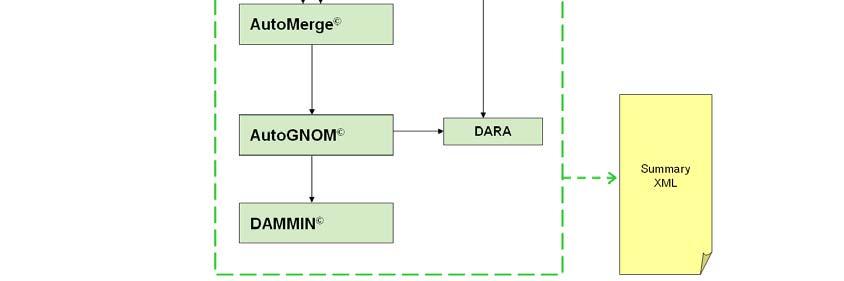


47 An automated SAXS pipeline at P12 Hardwareindependent analysis block Data normalization 2D-1D reduction Data processing Check for radiation damage Computation of overall parameters Database search Ab initio modelling XML-summary file generation
48 Kratky plot This provides a sensitive means of monitoring the degree of compactness of a protein as a function of a given parameter. This is most conveniently represented using the so-called Kratky plot of s 2 I(s) vs s. Globular particle : bell-shaped curve Gaussian chain : plateau at large s-values but beware: a plateau does not imply a Gaussian chain
49 SAXS patterns of globular and flexible proteins Natively unfolded Globular Multidomain with flexible linkers
50 Summary of model-independent information I(0)/c, i.e. molecular mass (from Guinier plot or p(r) function) Radius of gyration R g (from Guinier plot or p(r) function) Radii of gyration of thickness or cross-section (anisometrc particles) Maximum particle size D max (from p(r) function) Particle volume V (from I(0) and Porod invariant) Globular or unfoded (From Kratky plot)
51 Thank you!
Data reduction and processing tutorial
 Data reduction and processing tutorial Petr V. Konarev European Molecular Biology Laboratory, Hamburg Outstation BioSAXS group EMBL BioSAXS beamline X33, 2012 Optics Vacuum cell Completely redesigned 2005-2012
ID14-EH3. Adam Round
 Bio-SAXS @ ID14-EH3 Adam Round Contents What can be obtained from Bio-SAXS Measurable parameters Modelling strategies How to collect data at Bio-SAXS Procedure Data collection tests Data Verification and
How to judge data quality
 SSRL Workshop: Small-Angle X-ray Scattering and Diffraction Studies, March 28-30, 2016 How to judge data quality Tsutomu Matsui SSRL Lab / Dept. of Chemistry Stanford University Subject of this session
Introduction to Biological Small Angle Scattering
 Introduction to Biological Small Angle Scattering Tom Grant, Ph.D. Staff Scientist BioXFEL Science and Technology Center Hauptman-Woodward Institute Buffalo, New York, USA tgrant@hwi.buffalo.edu SAXS Literature
Small Angle X-Ray Solution Scattering of Biological Macromolecules
 Small Angle X-Ray Solution Scattering of Biological Macromolecules Emre Brookes UltraScan Workshop 15 June 2014 Overview Experimental method Sample preparation Experimental data analysis Experimental method
Biological Small Angle X-ray Scattering (SAXS) Dec 2, 2013
 Dec 2, 2013") Biological Small Angle X-ray Scattering (SAXS) Dec 2, 2013 Structural Biology Shape Dynamic Light Scattering Electron Microscopy Small Angle X-ray Scattering Cryo-Electron Microscopy Wide Angle X- ray
Biological Small Angle X-ray Scattering (SAXS) Dec 2, 2013 Structural Biology Shape Dynamic Light Scattering Electron Microscopy Small Angle X-ray Scattering Cryo-Electron Microscopy Wide Angle X- ray
Small-Angle Scattering from Biomolecular Solutions
 T H E U N I V E R S I T Y of T E X A S S C H O O L O F H E A L T H I N F O R M A T I O N S C I E N C E S A T H O U S T O N Small-Angle Scattering from Biomolecular Solutions For students of HI 6001-125
Characterizing Biological Macromolecules by SAXS Detlef Beckers, Jörg Bolze, Bram Schierbeek, PANalytical B.V., Almelo, The Netherlands
 Characterizing Biological Macromolecules by SAXS Detlef Beckers, Jörg Bolze, Bram Schierbeek, PANalytical B.V., Almelo, The Netherlands This document was presented at PPXRD - Pharmaceutical Powder X-ray
Structure Analysis by Small-Angle X-Ray and Neutron Scattering
 Structure Analysis by Small-Angle X-Ray and Neutron Scattering L. A. Feigin and D. I. Svergun Institute of Crystallography Academy of Sciences of the USSR Moscow, USSR Edited by George W. Taylor Princeton
Small-Angle Scattering Atomic Structure Based Modeling
 Small-Angle Scattering Atomic Structure Based Modeling Alejandro Panjkovich EMBL Hamburg 07.12.2017 A. Panjkovich (EMBL) BioSAS atomic modeling 07.12.2017 1 / 49 From the forest to the particle accelerator
Scattering of X-rays. EMBO Practical Course on Solution Scattering from Biological Macromolecules Hamburg 4 11 September 2001
 Scattering of X-rays Books on SAS - " The origins" (no recent edition) : Small Angle Scattering of X-rays A. Guinier and A. Fournet, (1955), in English, ed. Wiley, NY - Small-Angle X-ray Scattering O.
Introduction to biological small angle scattering
 Introduction to biological small angle scattering Frank Gabel (IBS/ILL) EMBO Practical Course (May 6th 013) F. Gabel (May 6th 013) EMBO Practical Course Length-scales and tools in structural biology small
Small-Angle X-ray Scattering (SAXS) SPring-8/JASRI Naoto Yagi
 SPring-8/JASRI Naoto Yagi") Small-Angle X-ray Scattering (SAXS) SPring-8/JASRI Naoto Yagi 1 Wikipedia Small-angle X-ray scattering (SAXS) is a small-angle scattering (SAS) technique where the elastic scattering of X-rays (wavelength
Small-Angle X-ray Scattering (SAXS) SPring-8/JASRI Naoto Yagi 1 Wikipedia Small-angle X-ray scattering (SAXS) is a small-angle scattering (SAS) technique where the elastic scattering of X-rays (wavelength
Part 8. Special Topic: Light Scattering
 Part 8. Special Topic: Light Scattering Light scattering occurs when polarizable particles in a sample are placed in the oscillating electric field of a beam of light. The varying field induces oscillating
BM29 biosaxs data processing tutorial
 HERCULES 2014 BM29 biosaxs data processing tutorial Page 2 OUTLINE Sample Changer Primary Data Processing Model Validation HPLC-SAXS Primary Data Processing Model Validation Ab Initio Model Software in
Small Angle X-ray Scattering (SAXS)
") Small Angle X-ray Scattering (SAXS) We have considered that Bragg's Law, d = λ/(2 sinθ), supports a minimum size of measurement of λ/2 in a diffraction experiment (limiting sphere of inverse space) but
Small Angle X-ray Scattering (SAXS) We have considered that Bragg's Law, d = λ/(2 sinθ), supports a minimum size of measurement of λ/2 in a diffraction experiment (limiting sphere of inverse space) but
Methoden Moderner Röntgenphysik II - Vorlesung im Haupt-/Masterstudiengang, Universität Hamburg, SoSe 2016, S. Roth
 > 31.05. : Small-Angle X-ray Scattering (SAXS) > 0.06. : Applications & A short excursion into Polymeric materials > 04.06. : Grazing incidence SAXS (GISAXS) Methoden Moderner Röntgenphysik II - Vorlesung
P. Vachette IBBMC (CNRS-Université Paris-Sud), Orsay, France
, Orsay, France") Scattering of X-rays P. Vachette IBBMC (CNRS-Université Paris-Sud), Orsay, France SAXS measurement Sample SAXS measuring cell SAXS measurement Scattering experiment X-ray beam?? Detector SAXS measurement
Scattering of X-rays P. Vachette IBBMC (CNRS-Université Paris-Sud), Orsay, France SAXS measurement Sample SAXS measuring cell SAXS measurement Scattering experiment X-ray beam?? Detector SAXS measurement
SAXS Basics for BioSAXS. Robert P. Rambo Diamond Light Source B21
 SAXS Basics for BioSAXS Robert P. Rambo Diamond Light Source B21 Scattering and Size SAXS 0.11 nm (1.1 Å) smaller than C-C bond 6000x range 690 nm (6900 Å) 3.5x larger than E.coli 10x larger than a ribosome
Modelling against small angle scattering data. Al Kikhney EMBL Hamburg, Germany
 Modelling against small angle scattering data Al Kikhney EMBL Hamburg, Germany Validation of atomic models CRYSOL Rigid body modelling SASREF BUNCH CORAL Oligomeric mixtures OLIGOMER Flexible systems EOM
SAXS and SANS facilities and experimental practice. Clement Blanchet
 SAXS and SANS facilities and experimental practice Clement Blanchet SAS experiment Detector X-ray or neutron Beam Sample 2 s Buffer X-rays Roengten, 1895 Electromagnetic wave The electromagnetic spectrum
Small Angle X-ray Scattering: Going Beyond the Bragg Peaks
 Small Angle X-ray Scattering: Going Beyond the Bragg Peaks V A Raghunathan This article gives an introduction to the principles of small angle scattering. Some applications of this technique are also briefly
Methoden moderner Röntgenphysik II Streuung und Abbildung
 Methoden moderner Röntgenphysik II Streuung und Abbildung Stephan V. Roth DESY 1.5.15 Outline > 1.5. : Small-Angle X-ray Scattering (SAXS) > 19.5. : Applications & A short excursion into Polymeric materials
SAS Data Analysis Colloids. Dr Karen Edler
 SAS Data Analysis Colloids Dr Karen Edler Size Range Comparisons 10 1 0.1 0.01 0.001 proteins viruses nanoparticles micelles polymers Q = 2π/d (Å -1 ) bacteria molecules nanotubes precipitates grain boundaries
A program for SAXS data processing and analysis *
 A program for SAXS data processing and analysis * LI Zhi-Hong( ) 1) Beijing Synchrotron Radiation Facility, Institute of High Energy Physics, Chinese Academy of Sciences, Beijing 149, China Abstract: A
Small-angle scattering studies of biological macromolecules in solution
 INSTITUTE OF PHYSICS PUBLISHING Rep. Prog. Phys. 66 (2003) 1735 1782 REPORTS ON PROGRESS IN PHYSICS PII: S0034-4885(03)12688-7 Small-angle scattering studies of biological macromolecules in solution Dmitri
Small-angle X-ray scattering a (mostly) theoretical introduction to the basics
 theoretical introduction to the basics") Small-angle X-ray scattering a (mostly) theoretical introduction to the basics András Wacha Research Centre for Natural Sciences, Hungarian Academy of Sciences Contents Introduction A bit of history The
Small-angle X-ray scattering a (mostly) theoretical introduction to the basics András Wacha Research Centre for Natural Sciences, Hungarian Academy of Sciences Contents Introduction A bit of history The
Application of Bayesian analysis to indirect Fourier transformation in small-angle scattering
 Journal of Applied Crystallography ISSN 0021-8898 Editor: Gernot Kostorz Application of Bayesian analysis to indirect Fourier transformation in small-angle scattering Bente Vestergaard and Steen Hansen
Robust, high-throughput solution structural analyses by small angle X-ray scattering (SAXS)
") nature methods Robust, high-throughput solution structural analyses by small angle X-ray scattering (SAXS) Greg L Hura, Angeli L Menon, Michal Hammel, Robert P Rambo, Farris L Poole II, Susan E Tsutakawa,
nature methods Robust, high-throughput solution structural analyses by small angle X-ray scattering (SAXS) Greg L Hura, Angeli L Menon, Michal Hammel, Robert P Rambo, Farris L Poole II, Susan E Tsutakawa,
The Small Angle X-ray Scattering Technique: An Overview
 The Small Angle X-ray Scattering Technique: An Overview Dr. Gianluca Croce, Ph.D DISTA - Univ. Piemonte Orientale Via T. Michel 11,15121 Alessandria (Italy) gianluca.croce@mfn.unipmn.it Dr. Gianluca Croce
Development of Novel Small- Angle X-ray Scattering Data Analysis Methods for Study of Flexible Proteins. Michael Kachala EMBL-Hamburg, Germany
 Development of Novel Small- Angle X-ray Scattering Data Analysis Methods for Study of Flexible Proteins Michael Kachala EMBL-Hamburg, Germany 60 mkl >1 mg/ml Monocromatic X-ray beam Sample Mono- or polydisperse
Supplemental Information for:
 Supplemental Information for: New Insight into the Structure of RNA in Red clover necrotic mosaic virus and the Role of Divalent Cations Revealed by Small-Angle Neutron Scattering Stanton L. Martin a,
2) Measure sample and empties. 3) Measure transmissions of sample and empties. 4) Normalize to exposure time and transmission. 5) Subtract the empties
 Measure sample and empties. 3) Measure transmissions of sample and empties. 4) Normalize to exposure time and transmission. 5) Subtract the empties") 1) Measure calibrants and direct beam to do Angular Integrations (transforms 2D into 1D) Need the distance SD Need center of beam Need to calculate q-vector 2) Measure sample and empties 3) Measure transmissions
1) Measure calibrants and direct beam to do Angular Integrations (transforms 2D into 1D) Need the distance SD Need center of beam Need to calculate q-vector 2) Measure sample and empties 3) Measure transmissions
Introduction to SAXS at SSRL
 Everything You Ever Wanted to Know About Introduction to SAXS at SSRL SAXS But Were Afraid to Ask John A Pople Stanford Synchrotron Radiation Laboratory, Stanford Linear Accelerator Center, Stanford CA
introduction to SAXS for polymers -a user view-
 introduction to SAXS for polymers -a user view- Luigi Balzano DSM Ahead/Material Science Center Geleen, The Netherlands luigi.balzano@dsm.com Synchrotron and Neutron Workshop (SyNeW) 2015 Utrecht, June
Introduction to X-ray and neutron scattering
 UNESCO/IUPAC Postgraduate Course in Polymer Science Lecture: Introduction to X-ray and neutron scattering Zhigunov Alexander Institute of Macromolecular Chemistry ASCR, Heyrovsky sq., Prague -16 06 http://www.imc.cas.cz/unesco/index.html
Light scattering Small and large particles
 Scattering by macromolecules E B Incident light Scattered Light particle Oscillating E field from light makes electronic cloud oscillate surrounding the particle Intensity: I E Accelerating charges means
Acta Cryst. (2017). D73, , doi: /s
. D73, , doi: /s") Supporting information Volume 73 (2017) Supporting information for article: 2017 publication guidelines for structural modelling of small-angle scattering data from biomolecules in solution: an update
Supporting information Volume 73 (2017) Supporting information for article: 2017 publication guidelines for structural modelling of small-angle scattering data from biomolecules in solution: an update
Methoden moderner Röntgenphysik II: Streuung und Abbildung
 . Methoden moderner Röntgenphysik II: Streuung und Abbildung Lecture 7 Vorlesung zum Haupt/Masterstudiengang Physik SS 2014 G. Grübel, M. Martins, E. Weckert Location: Hörs AP, Physik, Jungiusstrasse Tuesdays
Methoden moderner Röntgenphysik II Streuung und Abbildung
 Methoden moderner Röntgenphysik II Streuung und Abbildung Stephan V. Roth DESY 5.6.14 Two phase Model single particle approximation > Amplitude: Δ 3 > Intensity: = > Closer look at Iq for dilute systems:
Static and dynamic light scattering. Cy Jeffries EMBL Hamburg
 Static and dynamic light scattering. Cy Jeffries EMBL Hamburg Introduction. The electromagnetic spectrum. visible 10-16 10-10 10-8 10-4 10-2 10 4 (l m) g-rays X-rays UV IR micro wave Long radio waves 400
SI Text S1 Solution Scattering Data Collection and Analysis. SI references
 SI Text S1 Solution Scattering Data Collection and Analysis. The X-ray photon energy was set to 8 kev. The PILATUS hybrid pixel array detector (RIGAKU) was positioned at a distance of 606 mm from the sample.
Small Angle X-Ray Scattering What information can you get from this technique?
 Small Angle X-Ray Scattering 1 What information can you get from this technique? Small Angle X-Ray Scattering 2 A wide range of fields: Medicine Biology Chemistry Physics Archaeology Environmental and
PySaxs A Python module and GUI for SAXS data treatment
 DIRECTION DES SCIENCES DE LA MATIERE IRAMIS Laboratoire Interdisciplinaire sur l Organisation Nanométrique et Supramoléculaire PySaxs A Python module and GUI for SAXS data treatment Olivier Taché Collaborative
Structural characterization. Part 2
 Structural characterization Part Determining partial pair distribution functions X-ray absorption spectroscopy (XAS). Atoms of different elements have absorption edges at different energies. Structure
Surfactant adsorption and aggregate structure at silica nanoparticles: Effect of particle size and surface modification. Supplementary Information
 Surfactant adsorption and aggregate structure at silica nanoparticles: Effect of particle size and surface modification Bhuvnesh Bharti, Jens Meissner, Urs Gasser and Gerhard H. Findenegg* * e-mail: findenegg@chem.tu-berlin.de
Internal structure of 15 nm 3-helix micelle revealed by small-angle neutron scattering and coarse-grained MD simulation
 Internal structure of 15 nm 3-helix micelle revealed by small-angle neutron scattering and coarse-grained MD simulation JooChuan Ang 1, Dan Ma, Reidar Lund 3, Sinan Keten, and Ting Xu *1,4,5 1 Department
Estimation of chord length distributions from small-angle scattering using indirect Fourier transformation
 Journal of Applied Crystallography ISSN 0021-8898 Estimation of chord length distributions from small-angle scattering using indirect Fourier transformation Steen Hansen Copyright International Union of
Small-Angle X-ray Scattering (SAXS)/X-ray Absorption Near Edge Spectroscopy (XANES).
/X-ray Absorption Near Edge Spectroscopy (XANES).") S1 Small-Angle X-ray Scattering (SAXS)/X-ray Absorption Near Edge Spectroscopy (XANES). The combined SAXS/XANES measurements were carried out at the µspot beamline at BESSY II (Berlin, Germany). The beamline
S1 Small-Angle X-ray Scattering (SAXS)/X-ray Absorption Near Edge Spectroscopy (XANES). The combined SAXS/XANES measurements were carried out at the µspot beamline at BESSY II (Berlin, Germany). The beamline
Measuring the size and shape of macromolecules. Hydrodynamics: study of the objects in water How do the move? Translation Rotation
 Measuring the size and shape of macromolecules Hydrodynamics: study of the objects in water How do the move? Translation Rotation 1) Movement with no external forcefree diffusion 2) Movement under the
Interaction of Gold Nanoparticle with Proteins
 Chapter 7 Interaction of Gold Nanoparticle with Proteins 7.1. Introduction The interfacing of nanoparticle with biomolecules such as protein is useful for applications ranging from nano-biotechnology (molecular
Supplemental Information. Structural and Mechanistic Paradigm. of Leptin Receptor Activation Revealed
 Structure, Volume 22 Supplemental Information Structural and Mechanistic Paradigm of Leptin Receptor Activation Revealed by Complexes with Wild-Type and Antagonist Leptins Kedar Moharana, Lennart Zabeau,
Studying conformational dynamics and molecular recognition using integrated structural biology in solution Michael Sattler
 Studying conformational dynamics and molecular recognition using integrated structural biology in solution Michael Sattler http://www.nmr.ch.tum.de http://www.helmholtz-muenchen.de/stb/ Outline Dynamics
SMALL-ANGLE NEUTRON SCATTERING (SANS) FOR CHARACTERIZATION OF MULTI-COMPONENT SYSTEMS
 FOR CHARACTERIZATION OF MULTI-COMPONENT SYSTEMS") Chapter SMALL-ANGLE NEUTRON SCATTERING (SANS) FOR CHARACTERIZATION OF MULTI-COMPONENT SYSTEMS.1. Introduction The nanoparticles and macromolecules are known to be two important constituents of colloids.
Chapter SMALL-ANGLE NEUTRON SCATTERING (SANS) FOR CHARACTERIZATION OF MULTI-COMPONENT SYSTEMS.1. Introduction The nanoparticles and macromolecules are known to be two important constituents of colloids.
Introduction to biological small angle scattering
 Introduction to biological small angle scattering Frank Gabel (IBS/ILL) HERCULES Specialized Course 16 (September 15 th 014) Length-scales and tools in structural biology small angle scattering in solution
A Brief Review of Two Theoretical Models Used to Interpret the SAXS Intensities Measurements in Heterogeneous Thin Films.
 A Brief Review of Two Theoretical Models Used to Interpret the SAXS Intensities Measurements in Heterogeneous Thin Films. M. Cattani*, M. C. Salvadori and F. S. Teixeira Institute of Physics, University
BCM Protein crystallography - I. Crystal symmetry X-ray diffraction Protein crystallization X-ray sources SAXS
 BCM 6200 - Protein crystallography - I Crystal symmetry X-ray diffraction Protein crystallization X-ray sources SAXS What SAXS can do Small-angle X-ray scattering (SAXS) yields information on biological
arxiv:physics/ v2 [physics.chem-ph] 8 Dec 2004
![arxiv:physics/ v2 [physics.chem-ph] 8 Dec 2004](/thumbs/96/126569728.jpg "arxiv:physics/ v2 [physics.chem-ph] 8 Dec 2004") arxiv:physics/0407001v2 [physics.chem-ph] 8 Dec 2004 Size Information Obtained Using Static Light Scattering Technique Yong Sun February 2, 2008 Abstract Detailed investigation of static light scattering
arxiv:physics/0407001v2 [physics.chem-ph] 8 Dec 2004 Size Information Obtained Using Static Light Scattering Technique Yong Sun February 2, 2008 Abstract Detailed investigation of static light scattering
Scattering experiments
 Scattering experiments Menu 1. Basics: basics, contrast, q and q-range. Static scattering: Light, x-rays and neutrons 3. Dynamics: DLS 4. Key examples Polymers The Colloidal Domain The Magic Triangle Length-
Complementary use of SAXS and SANS. Jill Trewhella University of Sydney
 Complementary use of SAXS and SANS Jill Trewhella University of Sydney Conceptual diagram of the small-angle scattering experiment The conceptual experiment and theory is the same for X-rays and neutrons,
Approximation of the structure factor for nonspherical hard bodies using polydisperse spheres
 Journal of Applied Crystallography ISSN 21-8898 Approximation of the structure factor for nonspherical hard bodies using polydisperse spheres Steen Hansen J. Appl. Cryst. (213). 46, 18 116 Copyright c
Small Angle X-Ray Scattering
 SAXS Small Angle X-Ray Scattering Röntgenkleinwinkelstreuung Intensions Determination of the particle size and the morphology of solid materials: Intensions Determination of the particle size and the morphology
Moment of inertia. Contents. 1 Introduction and simple cases. January 15, Introduction. 1.2 Examples
 Moment of inertia January 15, 016 A systematic account is given of the concept and the properties of the moment of inertia. Contents 1 Introduction and simple cases 1 1.1 Introduction.............. 1 1.
Supporting Information for: Complexation of β-lactoglobulin Fibrils and Sulfated Polysaccharides
 Supporting Information for: Complexation of β-lactoglobulin Fibrils and Sulfated Polysaccharides Owen G Jones 1, Stephaandschin 1, Jozef Adamcik 1, Ludger Harnau 2, Sreenath Bolisetty 1, and Raffaele Mezzenga
Polymer solutions and melts
 Course M6 Lecture 9//004 (JAE) Course M6 Lecture 9//004 Polymer solutions and melts Scattering methods Effects of excluded volume and solvents Dr James Elliott Online teaching material reminder Overheads
Data Collection. Overview. Methods. Counter Methods. Crystal Quality with -Scans
 Data Collection Overview with a unit cell, possible space group and computer reference frame (orientation matrix); the location of diffracted x-rays can be calculated (h k l) and intercepted by something
Electronic Supplementary Information (ESI) Synthesis of gold nanoparticles in a biocompatible fluid from sputtering deposition onto castor oil
 Synthesis of gold nanoparticles in a biocompatible fluid from sputtering deposition onto castor oil") Electronic Supplementary Information (ESI) Synthesis of gold nanoparticles in a biocompatible fluid from sputtering deposition onto castor oil Heberton Wender, a Luciane F. de Oliveira, b Adriano F. Feil,
Electronic Supplementary Information (ESI) Synthesis of gold nanoparticles in a biocompatible fluid from sputtering deposition onto castor oil Heberton Wender, a Luciane F. de Oliveira, b Adriano F. Feil,
Gold-poly(N-isopropylacrylamide) core-shell colloids with homogeneous density profiles: A small angle scattering study
 core-shell colloids with homogeneous density profiles: A small angle scattering study") Electronic Supplementary Material (ESI) for Physical Chemistry Chemical Physics. This journal is the Owner Societies 2014 Supporting Information Gold-poly(N-isopropylacrylamide) core-shell colloids with
Electronic Supplementary Material (ESI) for Physical Chemistry Chemical Physics. This journal is the Owner Societies 2014 Supporting Information Gold-poly(N-isopropylacrylamide) core-shell colloids with
Solution structure and dynamics of biopolymers
 Solution structure and dynamics of biopolymers Atomic-detail vs. low resolution structure Information available at different scales Mobility of macromolecules in solution Brownian motion, random walk,
SAXS COURSE Richard Koschuch. HECUS M.Braun Graz X-Ray Systems. and. Institute of Biophysics and X-Ray Structure Research,
 SAXS COURSE 2003 Richard Koschuch HECUS M.Braun Graz X-Ray Systems and Institute of Biophysics and X-Ray Structure Research, Austrian Academy of Sciences, Graz, Austria The SWAXS Instrument Scheme of the
Lecture 26: More on Gel Filtration Chromatography and the Trypsin Resurrection Experiment
 Biological Chemistry Laboratory Biology 3515/Chemistry 3515 Spring 2018 Lecture 26: More on Gel Filtration Chromatography and the Trypsin Resurrection Experiment 12 April 2018 c David P. Goldenberg University
How DLS Works: Interference of Light
 Static light scattering vs. Dynamic light scattering Static light scattering measures time-average intensities (mean square fluctuations) molecular weight radius of gyration second virial coefficient Dynamic
Sem /2007. Fisika Polimer Ariadne L. Juwono
 Chapter 8. Measurement of molecular weight and size 8.. End-group analysis 8.. Colligative property measurement 8.3. Osmometry 8.4. Gel-permeation chromatography 8.5. Ultracentrifugation 8.6. Light-scattering
Structural characterization. Part 2
 Structural characterization Part Scattering angle Crystalline materials Bragg s law: Scattering vector Q ~ d -1, where d is interplanar distance Q has dimension [m -1 ], hence large Q (large scattering
Sample preparation and characterization around SAXS
 Sample preparation and characterization around SAXS Experimental verification and validation? Rob Meijers EMBL Hamburg Garbage in? The right stuff Molecular weight Oligomerization state Monodispersity
Appendix C - Persistence length 183. Consider an ideal chain with N segments each of length a, such that the contour length L c is
 Appendix C - Persistence length 183 APPENDIX C - PERSISTENCE LENGTH Consider an ideal chain with N segments each of length a, such that the contour length L c is L c = Na. (C.1) If the orientation of each
The Effect of Linker DNA on the Structure and Interaction of Nucleosome Core Particles
 Electronic Supplementary Material (ESI) for Soft Matter. This journal is The Royal Society of Chemistry 2018 SUPPLEMENTARY INFORMATION The Effect of Linker DNA on the Structure and Interaction of Nucleosome
Bayesian estimation of hyperparameters for indirect Fourier transformation in small-angle scattering
 Journal of Applied Crystallography ISSN 0021-8898 Bayesian estimation of hyperparameters for indirect Fourier transformation in small-angle scattering Steen Hansen Copyright International Union of Crystallography
Anomalous Small-Angle X-Ray Scattering of Core-shell Nanoparticles
 Anomalous Small-Angle X-Ray Scattering of Core-shell Nanoparticles Larissa Veiga University of Campinas Supervisor: Ulla Vainio DESY Summer Students Program 2008 September, 12 th 2008 1 1 Introduction
Measuring S using an analytical ultracentrifuge. Moving boundary
 Measuring S using an analytical ultracentrifuge Moving boundary [C] t = 0 t 1 t 2 0 top r bottom 1 dr b r b (t) r b ω 2 = S ln = ω 2 S (t-t dt r b (t o ) o ) r b = boundary position velocity = dr b dt
The Use of the Ultra Small Angle X-ray Scattering Technique to study the Solid Structure of Edible Fat Systems
 The Use of the Ultra Small Angle X-ray Scattering Technique to study the Solid Structure of Edible Fat Systems Fernanda Peyronel Alejandro Marangoni & David Pink Session: Analytical and Quality Control
Neutron Scattering (Basics).
.") Neutron Scattering (Basics). Cy M. Jeffries Introduction One of the new frontiers of structural biology is the study of the interactome: There are approximately 30 000 structural genes in the human genome.
Neutron Scattering (Basics). Cy M. Jeffries Introduction One of the new frontiers of structural biology is the study of the interactome: There are approximately 30 000 structural genes in the human genome.
Dilute-solution properties of biomacromolecules as indicators of macromolecular structure and interactions
 Dilute-solution properties of biomacromolecules as indicators of macromolecular structure and interactions José García de la Torre, Departament of Physical Chemistry University of Murcia, Spain jgt@um.es
Scattering of X-rays
 Scattering of X-rays P. Vachette I2BC (CNRS-Université Paris-Sud-CEA), Orsay, France SAXS measurement Sample SAXS measuring cell SAXS measurement Scattering experiment SAXS pattern? X-ray beam? I(q) 1000
Study of the Phase Composition of Fe 2 O 3 Nanoparticles
 WDS'9 Proceedings of Contributed Papers, Part III, 28 212, 29. ISBN 978-8-7378-13-3 MATFYZPRESS Study of the Phase Composition of Fe 2 O 3 Nanoparticles V. Valeš, J. Poltierová-Vejpravová, A. Mantlíková,
SUPPLEMENTARY INFORMATION
 Supplementary Information DNA-Programmable Nanoparticle Crystallization Sung Yong Park,* 1 Abigail K. R. Lytton-Jean,* 1 Byeongdu Lee 2, Steven Weigand 3, George C. Schatz 1 and Chad A. Mirkin 1 1 Department
Main Notation Used in This Book
 Main Notation Used in This Book z Direction normal to the surface x,y Directions in the plane of the surface Used to describe a component parallel to the interface plane xoz Plane of incidence j Label
Robert Botet FRACTAL DUST PARTICLES: LIGHT SCATTERING AND ADSORPTION ANOMALIES. Laboratoire de Physique des Solides - Université Paris-Sud (France)
") FRACTAL DUST PARTICLES: LIGHT SCATTERING AND ADSORPTION ANOMALIES (Kandinski, 1926) Robert Botet Laboratoire de Physique des Solides - Université Paris-Sud (France) ALMOST-KNOWN KNOWNS ABOUT FRACTAL DUST
FRACTAL DUST PARTICLES: LIGHT SCATTERING AND ADSORPTION ANOMALIES (Kandinski, 1926) Robert Botet Laboratoire de Physique des Solides - Université Paris-Sud (France) ALMOST-KNOWN KNOWNS ABOUT FRACTAL DUST
Non-particulate 2-phase systems.
 (See Roe Sects 5.3, 1.6) Remember: I(q) = Γ ρ (r) exp(-iqr) dr In this form, integral does not converge. So deviation from the mean = η(r) = ρ(r) - (See Roe Sects 5.3, 1.6) Remember: I(q) = Γ ρ
Nucleation rate (m -3 s -1 ) Radius of water nano droplet (Å) 1e+00 1e-64 1e-128 1e-192 1e-256
 Radius of water nano droplet (Å) 1e+00 1e-64 1e-128 1e-192 1e-256") Supplementary Figures Nucleation rate (m -3 s -1 ) 1e+00 1e-64 1e-128 1e-192 1e-256 Calculated R in bulk water Calculated R in droplet Modified CNT 20 30 40 50 60 70 Radius of water nano droplet (Å) Supplementary
Supplementary Figures Nucleation rate (m -3 s -1 ) 1e+00 1e-64 1e-128 1e-192 1e-256 Calculated R in bulk water Calculated R in droplet Modified CNT 20 30 40 50 60 70 Radius of water nano droplet (Å) Supplementary
Polymer dynamics. Course M6 Lecture 5 26/1/2004 (JAE) 5.1 Introduction. Diffusion of polymers in melts and dilute solution.
 5.1 Introduction. Diffusion of polymers in melts and dilute solution.") Course M6 Lecture 5 6//004 Polymer dynamics Diffusion of polymers in melts and dilute solution Dr James Elliott 5. Introduction So far, we have considered the static configurations and morphologies of
Course M6 Lecture 5 6//004 Polymer dynamics Diffusion of polymers in melts and dilute solution Dr James Elliott 5. Introduction So far, we have considered the static configurations and morphologies of
Handout 7 Reciprocal Space
 Handout 7 Reciprocal Space Useful concepts for the analysis of diffraction data http://homepages.utoledo.edu/clind/ Concepts versus reality Reflection from lattice planes is just a concept that helps us
University of Illinois at Chicago Department of Physics. Electricity & Magnetism Qualifying Examination
 University of Illinois at Chicago Department of Physics Electricity & Magnetism Qualifying Examination January 7, 28 9. am 12: pm Full credit can be achieved from completely correct answers to 4 questions.
Synchrotron SAXS Studies of Nanostructured Materials and Colloidal Solutions. A Review
 Materials Research, Vol. 5, No. 1, 1-11, 22. 22 Review Article Synchrotron SAXS Studies of Nanostructured Materials and Colloidal Solutions. A Review A.F. Craievich Institute of Physics, University of
Combined SANS and SAXS in studies of nanoparticles with core-shell structure
 Indian Journal of Pure & Applied Physics Vol. 44, October 006, pp. 74-78 Combined SANS and SAXS in studies of nanoparticles with core-shell structure P S Goyal & V K Aswal* UGC-DAE CSR, Mumbai Centre (*Solid
Electromagnetic Field Theory (EMT)
") Electromagnetic Field Theory (EMT) Lecture # 9 1) Coulomb s Law and Field Intensity 2) Electric Fields Due to Continuous Charge Distributions Line Charge Surface Charge Volume Charge Coulomb's Law Coulomb's
Electromagnetic Field Theory (EMT) Lecture # 9 1) Coulomb s Law and Field Intensity 2) Electric Fields Due to Continuous Charge Distributions Line Charge Surface Charge Volume Charge Coulomb's Law Coulomb's
High Brilliance SAXS on Synchrotrons
 High Brilliance SAXS on Synchrotrons Manfred Roessle Luebeck University of Applied Science Lab of X-ray Engineering X-ray generation Synchrotron Sources Shanghai Synchrotron Radiation Facility China Deutsches
Supporting information for: Norovirus capsid proteins self-assemble through. biphasic kinetics via long-lived stave-like.
 Supporting information for: Norovirus capsid proteins self-assemble through biphasic kinetics via long-lived stave-like intermediates Guillaume Tresset,, Clémence Le Cœur, Jean-François Bryche, Mouna Tatou,
Part 1: What is XAFS? What does it tell us? The EXAFS equation. Part 2: Basic steps in the analysis Quick overview of typical analysis
 Introduction to XAFS Part 1: What is XAFS? What does it tell us? The EXAFS equation Part 2: Basic steps in the analysis Quick overview of typical analysis Tomorrow Measurement methods and examples The
THE SOLUTION CONFORMATION OF NOVEL ANTIBODY FRAGMENTS STUDIED USING THE PROTEOMELAB XL-A ANALYTICAL ULTRACENTRIFUGE
 APPLICATION INFORMATION Peter J. Morgan, Olwyn D. Byron, Stephen E. Harding Department of Applied Biochemistry and Food Science University of Nottingham Sutton Bonington, U. K. Introduction One of the