The Small Angle X-ray Scattering Technique: An Overview
|
|
|
- Allen Daniel
- 6 years ago
- Views:
Transcription
1 The Small Angle X-ray Scattering Technique: An Overview Dr. Gianluca Croce, Ph.D DISTA - Univ. Piemonte Orientale Via T. Michel 11,15121 Alessandria (Italy) gianluca.croce@mfn.unipmn.it Dr. Gianluca Croce 1
2 An Overview X-ray scattering techniques are a family of non-destructive analytical techniques which reveal information about the crystallographic structure, chemical composition, and physical properties of materials and thin films. These techniques are based on observing the scattered intensity of an X-ray beam hitting a sample as a function of incident and scattered angle, polarization, and wavelength or energy. Materials that do not have long range order may also be studied by scattering methods that rely on elastic scattering of monochromatic X-rays: Small angle X-ray scattering (SAXS) probes structure in the nanometer to micrometer range by measuring scattering intensity at scattering angles 2θ close to 0 Dr. Gianluca Croce 2
3 An Overview Small-angle X-ray scattering (SAXS) is a small-angle scattering (SAS) technique where the elastic scattering of X-rays (wavelength nm) by a sample which has inhomogeneities in the nm-range, is recorded at very low angles (typically ). This angular range contains information about the shape and size of macromolecules, characteristic distances of partially ordered materials, pore sizes, and other data. SAXS is capable of delivering structural information of macromolecules between 5 and 25 nm, of repeat distances in partially ordered systems of up to 150 nm. (Glatter, O.; O. Kratky (1982). Small Angle X-ray Scattering. Academic Press.


4 A SAXS Instrument Conceptually, a SAXS experiment is simple: a sample is illuminated by X-rays and the scattered radiation is registered by a detector. Until the 1970s, SAXS experiments were done on instruments equipped by laboratory X-ray tubes. Nowadays the X-ray source can be also a synchrotron light which provides a higher X-ray flux. Also neutrons can be employed. Dr. Gianluca Croce 4
5 A SAXS Instrument The major problem that must be overcome in SAXS instrumentation is the separation of the weak scattered intensity from the strong main beam. Dr. Gianluca Croce 5

6 A SAXS Instrumentation Laboratory SAXS instruments can be divided into two main groups: 1. Point-collimation instruments have pinholes that shape the X-ray beam to a small circular or elliptical spot that illuminates the sample. The scattered intensity is small and therefore the measurement time is in the order of hours or days in case of very weak scatterers. 2. Line-collimation instruments confine the beam only in one dimension so that the beam profile is a long but narrow line. The illuminated sample volume is much larger compared to point-collimation and the scattered intensity at the same flux density is proportionally larger. Thus measuring times with line-collimation SAXS instruments are much shorter compared to point-collimation and are in the range of minutes to hours. Dr. Gianluca Croce 6


7 A SAXS Instrumentation Synchrotron SAXS: Data can be collected in the minute/(milli)second time regime. The camera length is variable between 1 and 10 m

8 SAXS Beamline SAXIER: Small-Angle X-Ray Scattering Initiative for Europe The X33 beamline for biological small-angle scattering. EMBL/DESY, Hamburg, Germany The ID13 beamline ESRF, Grenoble, France The small- and wide-angle scattering beamline SWING. SOLEIL, France The Austrian small-angle scattering beamline BL 5.2 L. ELETTRA, Italy The non-crystalline diffraction beamline 2.1 in long geometry.srs Daresbury, UK


9 A SAXS Instrumentation The detector and data acquisition system allows measurements in one- and two-dimensional manner. 1D Dr. Gianluca Croce 2D 9
10 The scattering pattern contains the information on the structure of the sample.?????? Dr. Gianluca Croce 10
11 Why Small Angle? 1) When an X-ray passes through an object it causes electrons to oscillate to the same frequency as that of the incident wave 2) These oscillating charges give rise coherent secondary electromagnetic waves with the same wavelenght as that of the incident beam 3) Any scattering process is characterized by a reciprocity law which gives an inverse relationship between object size and scattering angle Dr. Gianluca Croce 11
12 Why Small Angle? object " 1 angle 1 2 Colloidal dimensions (between tens and several thousand Å) are enormously large compared to the X-ray wavelength (i.e. CuKa =1.54A) Dr. Gianluca Croce 12
13 Why Small Angle? λ[nm] Probed dimension nm 44nm 6.8nm 68nm 11µm 110µm The SAXS technique is a tool which permits us to measure the scattered intensity at angle smaller than one degree Diffraction < SAXS < Optical Microscope Dr. Gianluca Croce 13
14 SAXS vs. TEM Trasmission Electron Microscopy (TEM) cover the same dimensional range investigated by SAXS The two techniques are complementary. WHY????? Dr. Gianluca Croce 14
 cover the same dimensional range investigated by SAXS The two techniques are complementary TEM good for: Direct and detailed image")
15 SAXS vs. TEM Trasmission Electron Microscopy (TEM) cover the same dimensional range investigated by SAXS The two techniques are complementary TEM good for: Direct and detailed image Local details Local surface Faithfully represents local complexities TEM not good for: Tiny image SAXS Statistically significant average information Dr. Gianluca Croce 15
16 SAXS vs. TEM Trasmission Electron Microscopy (TEM) cover the same dimensional range investigated by SAXS The two techniques are complementary SAXS good for: Global parameters and distribution Different sample states(solid, wet or solutions samples ) No destructive/artifacts in sample preparation In situ transition study Dr. Gianluca Croce 16
17 SAXS vs. TEM Transmission Electron Microscopy (TEM) cover the same dimensional range investigated by SAXS The two techniques are complementary: - TEM needs SAXS to obtain significant sampling and make quantitative statements - SAXS needs TEM for clues about the shape of particles for use in data analysis Dr. Gianluca Croce 17
18 What is an object? What SAXS sees as objects are the spatial variations in the electron density inside the irradiated portion of matter Electronic density difference is also called contrast An example: A homogenous particle (i.e. a macromolecule) with a constant electron density dispersed in a matrix (i.e. a solvent) with a different electron density particle matrix Dr. Gianluca Croce 18

19 What is an object? On an atomic scale, the electron density continuously varies from higher values in the area surrounding an atomic position to lower values in the space between atoms Dr. Gianluca Croce 19
20 What is an object? On an atomic scale, the electron density continuously varies from higher values in the area surrounding an atomic position to lower values in the space between atoms The SAXS explores dimensions of few nanometers. This implies that SAXS is not sensitive to electron density fluctuation on an atomic scale. Consequently a portion of matter of homogenous composition is considered as a homogenous particle (a particle with constant electron density) particle matrix Dr. Gianluca Croce 20


21 Adman, E. T., Godden, J. W., Turley, J Biol Chem 270 pp (1995) Hao Q, et al., Acta Crystallographica D55, (1), (1999) Dr. Gianluca Croce 21
22 What is an object? The scattering is a good probe for matter with higher electron density contrast The SAXS technique is good for: - amorphous particle - crystalline particle - homogeneous material with even voids(pores, blisters, cracks and crazes) particle matrix Dr. Gianluca Croce 22
23 SAXS is used for the determination of the microscale or nanoscale structure of particle systems in terms of such parameters as averaged particle sizes, shapes, distribution, and surface-to-volume ratio. The materials can be solid or liquid and they can contain solid, liquid or gaseous domains (so-called particles) of the same or another material in any combination. Not only particles, but also the structure of ordered systems like lamellae, and fractal-like materials can be studied. The method is accurate, non-destructive and usually requires only a minimum of sample preparation. Applications are very broad and include colloids of all types, metals, cement, oil, polymers, plastics, proteins, foods and pharmaceuticals and can be found in research as well as in quality control. Dr. Gianluca Croce 23


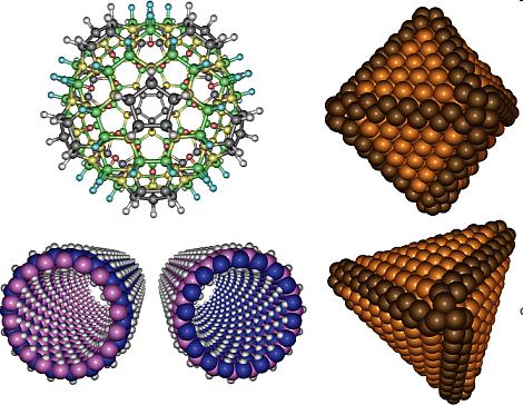
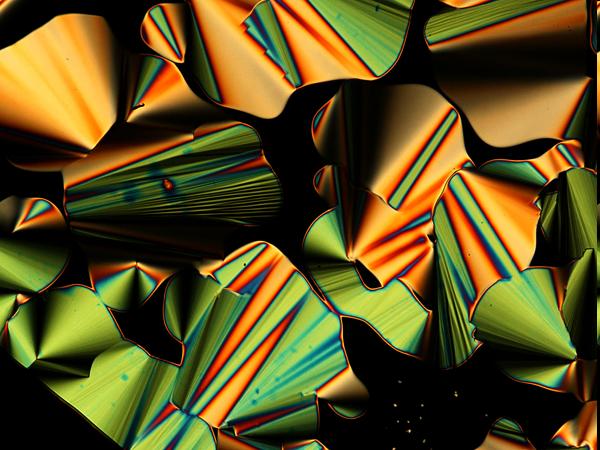


24 LIQUID Protein; Pharmaceuticals. SOLID Powder; Liquid Crystal; Nano Materials; Polymer; Fiber. Dr. Gianluca Croce 24

25 Single Particle Scattering The scattering of a single particle in a matrix is simply its Fourier transform. FT FT Dr. Gianluca Croce 25
26 Single Particle Scattering The Fourier trasform can be calculated analytically for a large number of shape: a b Ellipsoidal particle with constant orientation The scattering intensity I 0 (h): Where: % 4 ( I 0 (h,a,b) = "# 2 ' & 3 $ab2 * ) 2 % ' & 3sin x + x cos x x 3 ( * ) 2 x = a 2 h b 2 h 2 2 % h ' h = & 4" sin# ( $ ) * Scattering vector with h1 and h2 component on the dector plane Dr. Gianluca Croce 26
27 Single Particle Scattering a b % 4 ( 2 % 3sin x + x cos x I 0 (h,a,b) = "# 2 ' & 3 $ab2 * ' ) & x 3 ( * ) 2 Electron Density Difference (particle/matrix) Volume of the ellipsoid Bivariate function (surface) whose contour lines have an ellipsoidal shape oriented perpendicularly to the particle Dr. Gianluca Croce 27
28 Single Particle Scattering # % $ 3sin x " x cos x x 3 & ( ' 2 Bivariate function (surface) whose contour lines have an ellipsoidal shape oriented perpendicularly to the particle a b h1 h2 x = a 2 h b 2 h 2 2 % h ' h = & 4" sin# ( * $ ) Scattering vector with h1 and h2 component on the detector plane Dr. Gianluca Croce 28
29 Single Particle Scattering This last term is a consequence of the reciprocal law between the scattering particle and its scattering pattern OR Between a function and its Fourier transform a b h1 h2 Dr. Gianluca Croce 29
30 Single Particle Scattering Line sections of the scattering patterns (=linear detector) of an ellipsoid of different shapes The curves scattering decrease inversely to the vector h1 Remember: object " 1 angle Dr. Gianluca Croce 30
31 Single Particle Scattering A 1D scattering curve theoretically is composed by a central peak with a series of rapidly damped peaks of much smaller intensity For simplicity is useful to represent the data in a logarithmic scale to better appreciate the small intensity peaks Log(I 0 ) Dr. Gianluca Croce 31
32 Single Particle Scattering Scattering equation of particles of different shape: Dr. Gianluca Croce 32
33 Single Particle Scattering 1D scattering curve of particles of different shape and equal dimensions Dr. Gianluca Croce 33
34 Group of Particle Scattering In a real case we normally study system composed by many particles dispersed in a matrix (i.e. a solvent) A dilute system of ellipsoidal particle with the same orientation: Same dimension: a simple sum of the scattering of each single particle Different dimension: multiply the function for the scattering of a single particle by the distribution of sizes Dr. Gianluca Croce 34
35 Group of Particle Scattering What does it mean Multiply the function for the scattering of a single particle by the distribution of sizes? I 0 (h,a,b) = "" D(a,b)I 0 (h,a,b)dadb Function of the semi-axes of the ellipsoid; a and b parameters control the dimensions of the particles Equation with described the scattering for a single ellipsoid: % 4 ( 2 % 3sin x + x cos x ( I 0 (h,a,b) = "# 2 ' & 3 $ab2 * ' * ) & ) x 3 2 Dr. Gianluca Croce 35
36 Group of Particle Scattering If the orientation of the particles vary or if their orientation changes over time (random orientation): The scattering is averaged in all directions and the pattern will be isotropic Dr. Gianluca Croce 36
37 Group of Particle Scattering In this real situation, a distribution of directions should be introduced for mimic the scattering pattern I(h,a,b) Obviously the information on particle shapes and dimensions is mixed and it s necessary to make assumptions about their shape or size distribution Dr. Gianluca Croce 37
38 Group of Particle Scattering Dr. Gianluca Croce 38
39 Group of Particle Scattering(Densely Packed) When particles are very close one to another They cause a constructive inference similar to the Bragg peaks (diffraction). If these distance have some degree of regularity Dr. Gianluca Croce 39
40 Data Analysis A SAXS signal contain information about shape and size particles (also distances) These information are confused in a SAXS pattern These contributions are impossible to separate without external information (i.e. particle shape, a certain distribution, etc ) Data Analysis Manipulation Retrieve particles information processing experimental data by means of mathematical transformations to a assumed shape From an assumed particle distribution, calculate the scattering intensity in order to try to fit the experimental data Dr. Gianluca Croce 40
41 Guinier Approximation At smallest angle, the scattering curve of a dilute system (similar size particles) can be approximated: I(h) "e #ar 2 h 2 g Gaussian Function Where: Rg is called Gyration Radius a is a factor depending on particle orientation(i.e. random orientation a=1/3) Dr. Gianluca Croce 41
42 Guinier Approximation The Gyration Radius is defined in analogy to the radius of inertia in mechanics. R is defined as the mean square distance from the centre of gravity (R= 2 ) where the role of the mass is played by the electrons. The equation of randomly oriented particle (a=1/3) is: "R 2 h 2 3 I(h) = I(0)e lni(h) Rg can be calculated from the slope of the strain region of the plot h 2 Dr. Gianluca Croce 42
43 Guinier Approximation Dr. Gianluca Croce 43
44 Guinier Approximation The Rg is a parameter doesn t provide any geometrical information of particles. It s always necessary to consider an a priori geometry (shape) of the particles. If the particle shape is defined, the Guinier approximation provides a good indication of average size particles. Guinier Region LnI(h) h^2 The Guinier approximation works well when, in a dilute system, the particles preserve the same shape with also different dimension (in a limited range) If the particle dimensions are drastically different may occur to experimentally observe two definite Guinier region (determinable separately) Dr. Gianluca Croce 44
 Dr.")
45 Porod Law Examining the queues of a SAXS signal, Porod observed that in most case the data follow an asymptotic behavior with a fourth powerlaw decay This behavior is expected for so-called two-phase system (when the el. density ρ can assume only two value: ρ1 and zero) Dr. Gianluca Croce 45
46 Porod Law Mathematically for large value of h: 2" I( h) = S! h 2 (! 4 1 # 1) Where S represents the surface area of the particles and may be determined from experimental data. In an ideal case (spherical particle with ρ=ρ 1 ;0) the Porod Plot trends to a plateau. This constant value is proportional to the surface area at the interface. Porod Plot h^4i(h) h Dr. Gianluca Croce 46
47 Integral Quantities Porod also demonstrated that normalizing the intensities (Absolute Intensity), the following relation for a two-phase system is possible: 1 V $ % h 2 I(h)dh = v(1" v)(# 2 "# 1 ) 2 V = scattering volume 0 ρ1;ρ2 = electronic density of two phases v;1-v = volumetric fraction of two phase Scattering Power This quantity is calculable from the experimental data if it s possible to extrapolate them to zero (Guinier) and to infinity (Porod) This equation permit to estimate the volumetric fraction knowing the electronic density, and viceversa, of a two-phase system. Dr. Gianluca Croce 47
48 Bibliography Glatter, O. & Kratky, O., eds. Small Angle X-ray Scattering. Academic Press, ISBN Free available on-line (Short link: L.A. Feigin & D.I. Svergun: Structure Analysis by Small-Angle X-Ray and Neutron Scattering. New York: Plenum Press, ISBN Free available on-line (Short link: Lipfert J, Doniach S. Small-angle X-ray scattering from RNA, proteins, and protein complexes. Annu Rev Biophys Biomol Struct. 2007;36: SAXIER: Small-Angle X-Ray Scattering Initiative for Europe ( Dr. Gianluca Croce 48
Small Angle X-ray Scattering (SAXS)
") Small Angle X-ray Scattering (SAXS) We have considered that Bragg's Law, d = λ/(2 sinθ), supports a minimum size of measurement of λ/2 in a diffraction experiment (limiting sphere of inverse space) but
Small Angle X-ray Scattering (SAXS) We have considered that Bragg's Law, d = λ/(2 sinθ), supports a minimum size of measurement of λ/2 in a diffraction experiment (limiting sphere of inverse space) but
Structure Analysis by Small-Angle X-Ray and Neutron Scattering
 Structure Analysis by Small-Angle X-Ray and Neutron Scattering L. A. Feigin and D. I. Svergun Institute of Crystallography Academy of Sciences of the USSR Moscow, USSR Edited by George W. Taylor Princeton
Introduction to X-ray and neutron scattering
 UNESCO/IUPAC Postgraduate Course in Polymer Science Lecture: Introduction to X-ray and neutron scattering Zhigunov Alexander Institute of Macromolecular Chemistry ASCR, Heyrovsky sq., Prague -16 06 http://www.imc.cas.cz/unesco/index.html
Structural characterization. Part 2
 Structural characterization Part Determining partial pair distribution functions X-ray absorption spectroscopy (XAS). Atoms of different elements have absorption edges at different energies. Structure
Undulator Radiation Inside a Dielectric Waveguide
 Undulator Radiation Inside a Dielectric Waveguide A.S. Kotanjyan Department of Physics, Yerevan State University Yerevan, Armenia Content Motivation On features of the radiation from an electron moving
Biological Small Angle X-ray Scattering (SAXS) Dec 2, 2013
 Dec 2, 2013") Biological Small Angle X-ray Scattering (SAXS) Dec 2, 2013 Structural Biology Shape Dynamic Light Scattering Electron Microscopy Small Angle X-ray Scattering Cryo-Electron Microscopy Wide Angle X- ray
Biological Small Angle X-ray Scattering (SAXS) Dec 2, 2013 Structural Biology Shape Dynamic Light Scattering Electron Microscopy Small Angle X-ray Scattering Cryo-Electron Microscopy Wide Angle X- ray
introduction to SAXS for polymers -a user view-
 introduction to SAXS for polymers -a user view- Luigi Balzano DSM Ahead/Material Science Center Geleen, The Netherlands luigi.balzano@dsm.com Synchrotron and Neutron Workshop (SyNeW) 2015 Utrecht, June
The SAXS Guide. Getting acquainted with the principles. New Edition with special contributions
 The SAXS Guide Getting acquainted with the principles New Edition with special contributions The SAXS Guide Getting acquainted with the principles 4 th edition by Heimo Schnablegger Yashveer Singh Special
Introduction to SAXS at SSRL
 Everything You Ever Wanted to Know About Introduction to SAXS at SSRL SAXS But Were Afraid to Ask John A Pople Stanford Synchrotron Radiation Laboratory, Stanford Linear Accelerator Center, Stanford CA
Small-Angle X-ray Scattering (SAXS) SPring-8/JASRI Naoto Yagi
 SPring-8/JASRI Naoto Yagi") Small-Angle X-ray Scattering (SAXS) SPring-8/JASRI Naoto Yagi 1 Wikipedia Small-angle X-ray scattering (SAXS) is a small-angle scattering (SAS) technique where the elastic scattering of X-rays (wavelength
Small-Angle X-ray Scattering (SAXS) SPring-8/JASRI Naoto Yagi 1 Wikipedia Small-angle X-ray scattering (SAXS) is a small-angle scattering (SAS) technique where the elastic scattering of X-rays (wavelength
Main Notation Used in This Book
 Main Notation Used in This Book z Direction normal to the surface x,y Directions in the plane of the surface Used to describe a component parallel to the interface plane xoz Plane of incidence j Label
Characterizing Biological Macromolecules by SAXS Detlef Beckers, Jörg Bolze, Bram Schierbeek, PANalytical B.V., Almelo, The Netherlands
 Characterizing Biological Macromolecules by SAXS Detlef Beckers, Jörg Bolze, Bram Schierbeek, PANalytical B.V., Almelo, The Netherlands This document was presented at PPXRD - Pharmaceutical Powder X-ray
SAXS and SANS facilities and experimental practice. Clement Blanchet
 SAXS and SANS facilities and experimental practice Clement Blanchet SAS experiment Detector X-ray or neutron Beam Sample 2 s Buffer X-rays Roengten, 1895 Electromagnetic wave The electromagnetic spectrum
Small Angle X-ray Scattering: Going Beyond the Bragg Peaks
 Small Angle X-ray Scattering: Going Beyond the Bragg Peaks V A Raghunathan This article gives an introduction to the principles of small angle scattering. Some applications of this technique are also briefly
Synchrotron Methods in Nanomaterials Research
 Synchrotron Methods in Nanomaterials Research Marcel MiGLiERiNi Slovak University of Technology in Bratislava and Centre for Nanomaterials Research, Olomouc marcel.miglierini@stuba.sk www.nuc.elf.stuba.sk/bruno
Small Angle X-Ray Scattering
 SAXS Small Angle X-Ray Scattering Röntgenkleinwinkelstreuung Intensions Determination of the particle size and the morphology of solid materials: Intensions Determination of the particle size and the morphology
Structural characterization. Part 2
 Structural characterization Part Scattering angle Crystalline materials Bragg s law: Scattering vector Q ~ d -1, where d is interplanar distance Q has dimension [m -1 ], hence large Q (large scattering
X-Ray Diffraction as a key to the Structure of Materials Interpretation of scattering patterns in real and reciprocal space
 X-Ray Diffraction as a key to the Structure of Materials Interpretation of scattering patterns in real and reciprocal space Tobias U. Schülli, X-ray nanoprobe group ESRF OUTLINE 1 Internal structure of
Introduction to biological small angle scattering
 Introduction to biological small angle scattering Frank Gabel (IBS/ILL) EMBO Practical Course (May 6th 013) F. Gabel (May 6th 013) EMBO Practical Course Length-scales and tools in structural biology small
Light scattering Small and large particles
 Scattering by macromolecules E B Incident light Scattered Light particle Oscillating E field from light makes electronic cloud oscillate surrounding the particle Intensity: I E Accelerating charges means
Chapter 12. Nanometrology. Oxford University Press All rights reserved.
 Chapter 12 Nanometrology Introduction Nanometrology is the science of measurement at the nanoscale level. Figure illustrates where nanoscale stands in relation to a meter and sub divisions of meter. Nanometrology
ID14-EH3. Adam Round
 Bio-SAXS @ ID14-EH3 Adam Round Contents What can be obtained from Bio-SAXS Measurable parameters Modelling strategies How to collect data at Bio-SAXS Procedure Data collection tests Data Verification and
General theory of diffraction
 General theory of diffraction X-rays scatter off the charge density (r), neutrons scatter off the spin density. Coherent scattering (diffraction) creates the Fourier transform of (r) from real to reciprocal
Scattering Lecture. February 24, 2014
 Scattering Lecture February 24, 2014 Structure Determination by Scattering Waves of radiation scattered by different objects interfere to give rise to an observable pattern! The wavelength needs to close
Electronic Supplementary Information (ESI) Synthesis of gold nanoparticles in a biocompatible fluid from sputtering deposition onto castor oil
 Synthesis of gold nanoparticles in a biocompatible fluid from sputtering deposition onto castor oil") Electronic Supplementary Information (ESI) Synthesis of gold nanoparticles in a biocompatible fluid from sputtering deposition onto castor oil Heberton Wender, a Luciane F. de Oliveira, b Adriano F. Feil,
Electronic Supplementary Information (ESI) Synthesis of gold nanoparticles in a biocompatible fluid from sputtering deposition onto castor oil Heberton Wender, a Luciane F. de Oliveira, b Adriano F. Feil,
Quiz 1 XRD ) Explain the error in the following statement: "a laser beam is a focused beam of monochromatic light".
 Explain the error in the following statement: a laser beam is a focused beam of monochromatic light.") Quiz 1 XRD 092706 Diffraction involves constructive interference between waves that emanate from structurally organized matter such as from atoms in a crystal. X-ray diffraction uses a relationship of
Quiz 1 XRD 092706 Diffraction involves constructive interference between waves that emanate from structurally organized matter such as from atoms in a crystal. X-ray diffraction uses a relationship of
CHARACTERIZATION of NANOMATERIALS KHP
 CHARACTERIZATION of NANOMATERIALS Overview of the most common nanocharacterization techniques MAIN CHARACTERIZATION TECHNIQUES: 1.Transmission Electron Microscope (TEM) 2. Scanning Electron Microscope
How to judge data quality
 SSRL Workshop: Small-Angle X-ray Scattering and Diffraction Studies, March 28-30, 2016 How to judge data quality Tsutomu Matsui SSRL Lab / Dept. of Chemistry Stanford University Subject of this session
arxiv:physics/ v2 [physics.chem-ph] 8 Dec 2004
![arxiv:physics/ v2 [physics.chem-ph] 8 Dec 2004](/thumbs/96/126569728.jpg "arxiv:physics/ v2 [physics.chem-ph] 8 Dec 2004") arxiv:physics/0407001v2 [physics.chem-ph] 8 Dec 2004 Size Information Obtained Using Static Light Scattering Technique Yong Sun February 2, 2008 Abstract Detailed investigation of static light scattering
arxiv:physics/0407001v2 [physics.chem-ph] 8 Dec 2004 Size Information Obtained Using Static Light Scattering Technique Yong Sun February 2, 2008 Abstract Detailed investigation of static light scattering
On the use of Kumakhov Polycapillaries to improve laboratory
 ICXOM Frascati (INFN - LNF) 25-30 September 2005 On the use of Kumakhov Polycapillaries to improve laboratory Energy Dispersive X-ray X Diffractometry and Reflectometry B. Paci 1, V. Rossi Albertini 1,
Introduction to Biological Small Angle Scattering
 Introduction to Biological Small Angle Scattering Tom Grant, Ph.D. Staff Scientist BioXFEL Science and Technology Center Hauptman-Woodward Institute Buffalo, New York, USA tgrant@hwi.buffalo.edu SAXS Literature
Part 8. Special Topic: Light Scattering
 Part 8. Special Topic: Light Scattering Light scattering occurs when polarizable particles in a sample are placed in the oscillating electric field of a beam of light. The varying field induces oscillating
AP5301/ Name the major parts of an optical microscope and state their functions.
 Review Problems on Optical Microscopy AP5301/8301-2015 1. Name the major parts of an optical microscope and state their functions. 2. Compare the focal lengths of two glass converging lenses, one with
XRD RAPID SCREENING SYSTEM FOR COMBINATORIAL CHEMISTRY
 Copyright(c)JCPDS-International Centre for Diffraction Data 2001,Advances in X-ray Analysis,Vol.44 1 XRD RAPID SCREENING SYSTEM FOR COMBINATORIAL CHEMISTRY Bob B. He, John Anzelmo, Peter LaPuma, Uwe Preckwinkel,
Transmission Electron Microscopy
 L. Reimer H. Kohl Transmission Electron Microscopy Physics of Image Formation Fifth Edition el Springer Contents 1 Introduction... 1 1.1 Transmission Electron Microscopy... 1 1.1.1 Conventional Transmission
Roger Johnson Structure and Dynamics: X-ray Diffraction Lecture 6
 6.1. Summary In this Lecture we cover the theory of x-ray diffraction, which gives direct information about the atomic structure of crystals. In these experiments, the wavelength of the incident beam must
Small Angle X-Ray Solution Scattering of Biological Macromolecules
 Small Angle X-Ray Solution Scattering of Biological Macromolecules Emre Brookes UltraScan Workshop 15 June 2014 Overview Experimental method Sample preparation Experimental data analysis Experimental method
Transmission Electron Microscopy and Diffractometry of Materials
 Brent Fultz James Howe Transmission Electron Microscopy and Diffractometry of Materials Fourth Edition ~Springer 1 1 Diffraction and the X-Ray Powder Diffractometer 1 1.1 Diffraction... 1 1.1.1 Introduction
SOLID STATE 18. Reciprocal Space
 SOLID STATE 8 Reciprocal Space Wave vectors and the concept of K-space can simplify the explanation of several properties of the solid state. They will be introduced to provide more information on diffraction
GISAXS, GID and X-Ray Reflectivity in Materials Science
 united nations educational, scientific and cultural organization the abdus salam international centre for theoretical physics international atomic energy agency SCHOOL ON SYNCHROTRON RADIATION AND APPLICATIONS
A program for SAXS data processing and analysis *
 A program for SAXS data processing and analysis * LI Zhi-Hong( ) 1) Beijing Synchrotron Radiation Facility, Institute of High Energy Physics, Chinese Academy of Sciences, Beijing 149, China Abstract: A
Beyond the Geometric toward the Wave Optical Approach in the Design of Curved Crystal and Multilayer Optics for EDXAS
 Beyond the Geometric toward the Wave Optical Approach in the Design of Curved Crystal and Multilayer Optics for EDXAS Vito Mocella CNR IMM Napoli Units, Italy In collaboration with C. Ferrero, C. Morawe,
Structure analysis: Electron diffraction LEED TEM RHEED
 Structure analysis: Electron diffraction LEED: Low Energy Electron Diffraction SPA-LEED: Spot Profile Analysis Low Energy Electron diffraction RHEED: Reflection High Energy Electron Diffraction TEM: Transmission
Crystals, X-rays and Proteins
 Crystals, X-rays and Proteins Comprehensive Protein Crystallography Dennis Sherwood MA (Hons), MPhil, PhD Jon Cooper BA (Hons), PhD OXFORD UNIVERSITY PRESS Contents List of symbols xiv PART I FUNDAMENTALS
Methoden moderner Röntgenphysik II Streuung und Abbildung
 Methoden moderner Röntgenphysik II Streuung und Abbildung Stephan V. Roth DESY 1.5.15 Outline > 1.5. : Small-Angle X-ray Scattering (SAXS) > 19.5. : Applications & A short excursion into Polymeric materials
Lecture 19 Optical MEMS (1)
") EEL6935 Advanced MEMS (Spring 5) Instructor: Dr. Huikai Xie Lecture 19 Optical MEMS (1) Agenda: Optics Review EEL6935 Advanced MEMS 5 H. Xie 3/8/5 1 Optics Review Nature of Light Reflection and Refraction
EEL6935 Advanced MEMS (Spring 5) Instructor: Dr. Huikai Xie Lecture 19 Optical MEMS (1) Agenda: Optics Review EEL6935 Advanced MEMS 5 H. Xie 3/8/5 1 Optics Review Nature of Light Reflection and Refraction
The Compact Solution for Nanostructure Analysis. SAXSpace
 The Compact Solution for Nanostructure Analysis SAXSpace New to SAXS? Here are some essentials. What SAXS is SAXS, Small-Angle X-ray Scattering, is a non-destructive method for investigating nanostructures
 Name : Roll No. :.. Invigilator s Signature :.. CS/B.Tech/SEM-2/PH-201/2010 2010 ENGINEERING PHYSICS Time Allotted : 3 Hours Full Marks : 70 The figures in the margin indicate full marks. Candidates are
SAS Data Analysis Colloids. Dr Karen Edler
 SAS Data Analysis Colloids Dr Karen Edler Size Range Comparisons 10 1 0.1 0.01 0.001 proteins viruses nanoparticles micelles polymers Q = 2π/d (Å -1 ) bacteria molecules nanotubes precipitates grain boundaries
SAXS Basics for BioSAXS. Robert P. Rambo Diamond Light Source B21
 SAXS Basics for BioSAXS Robert P. Rambo Diamond Light Source B21 Scattering and Size SAXS 0.11 nm (1.1 Å) smaller than C-C bond 6000x range 690 nm (6900 Å) 3.5x larger than E.coli 10x larger than a ribosome
SAXS/SANS data processing and overall parameters
 EMBO Global Exchange Lecture Course 30 November 2012 Hyderabad India SAXS/SANS data processing and overall parameters Petr V. Konarev European Molecular Biology Laboratory, Hamburg Outstation BioSAXS group
Chapter 10. Nanometrology. Oxford University Press All rights reserved.
 Chapter 10 Nanometrology Oxford University Press 2013. All rights reserved. 1 Introduction Nanometrology is the science of measurement at the nanoscale level. Figure illustrates where nanoscale stands
Strain, Stress and Cracks Klaus Attenkofer PV Reliability Workshop (Orlando) April 7-8, 2015
 April 7-8, 2015") Strain, Stress and Cracks Klaus Attenkofer PV Reliability Workshop (Orlando) April 7-8, 2015 1 BROOKHAVEN SCIENCE ASSOCIATES Overview Material s response to applied forces or what to measure Definitions
Strain, Stress and Cracks Klaus Attenkofer PV Reliability Workshop (Orlando) April 7-8, 2015 1 BROOKHAVEN SCIENCE ASSOCIATES Overview Material s response to applied forces or what to measure Definitions
Methoden Moderner Röntgenphysik II - Vorlesung im Haupt-/Masterstudiengang, Universität Hamburg, SoSe 2016, S. Roth
 > 31.05. : Small-Angle X-ray Scattering (SAXS) > 0.06. : Applications & A short excursion into Polymeric materials > 04.06. : Grazing incidence SAXS (GISAXS) Methoden Moderner Röntgenphysik II - Vorlesung
Macroscopic Polymer Analogues
 Macroscopic Polymer Analogues G. BEAUCAGE, S. SUKUMARAN, S. RANE, D. J. KOHLS Polymer Research Center and Department of Materials Science and Engineering, P.O. Box 210012, University of Cincinnati, Cincinnati,
Chapter 2. X-ray X. Diffraction and Reciprocal Lattice. Scattering from Lattices
 Chapter. X-ray X Diffraction and Reciprocal Lattice Diffraction of waves by crystals Reciprocal Lattice Diffraction of X-rays Powder diffraction Single crystal X-ray diffraction Scattering from Lattices
SUPPLEMENTARY INFORMATION
 SI - N. Poccia et al. Evolution and Control of Oxygen Order in a Cuprate Superconductor SUPPLEMENTARY INFORMATION 1. X-ray diffraction experiments The diffraction data were collected on the x-ray diffraction
Small-angle X-ray scattering a (mostly) theoretical introduction to the basics
 theoretical introduction to the basics") Small-angle X-ray scattering a (mostly) theoretical introduction to the basics András Wacha Research Centre for Natural Sciences, Hungarian Academy of Sciences Contents Introduction A bit of history The
Small-angle X-ray scattering a (mostly) theoretical introduction to the basics András Wacha Research Centre for Natural Sciences, Hungarian Academy of Sciences Contents Introduction A bit of history The
Experiment Report Form
 EUROPEAN SYNCHROTRON RADIATION FACILITY INSTALLATION EUROPEENNE DE RAYONNEMENT SYNCHROTRON Experiment Report Form The double page inside this form is to be filled in by all users or groups of users who
Lecture 9: Introduction to Diffraction of Light
 Lecture 9: Introduction to Diffraction of Light Lecture aims to explain: 1. Diffraction of waves in everyday life and applications 2. Interference of two one dimensional electromagnetic waves 3. Typical
Overview of scattering, diffraction & imaging in the TEM
 Overview of scattering, diffraction & imaging in the TEM Eric A. Stach Purdue University Scattering Electrons, photons, neutrons Radiation Elastic Mean Free Path (Å)( Absorption Length (Å)( Minimum Probe
Methoden moderner Röntgenphysik II: Streuung und Abbildung
 . Methoden moderner Röntgenphysik II: Streuung und Abbildung Lecture 7 Vorlesung zum Haupt/Masterstudiengang Physik SS 2014 G. Grübel, M. Martins, E. Weckert Location: Hörs AP, Physik, Jungiusstrasse Tuesdays
Chap. 3. Elementary Quantum Physics
 Chap. 3. Elementary Quantum Physics 3.1 Photons - Light: e.m "waves" - interference, diffraction, refraction, reflection with y E y Velocity = c Direction of Propagation z B z Fig. 3.1: The classical view
Temperature ( o C)
") Viscosity (Pa sec) Supplementary Information 10 8 10 6 10 4 10 2 150 200 250 300 Temperature ( o C) Supplementary Figure 1 Viscosity of fibre components (PC cladding blue; As 2 Se 5 red; CPE black) as
Viscosity (Pa sec) Supplementary Information 10 8 10 6 10 4 10 2 150 200 250 300 Temperature ( o C) Supplementary Figure 1 Viscosity of fibre components (PC cladding blue; As 2 Se 5 red; CPE black) as
Light Source I. Takashi TANAKA (RIKEN SPring-8 Center) Cheiron 2012: Light Source I
 Cheiron 2012: Light Source I") Light Source I Takashi TANAKA (RIKEN SPring-8 Center) Light Source I Light Source II CONTENTS Introduction Fundamentals of Light and SR Overview of SR Light Source Characteristics of SR (1) Characteristics
Light Source I Takashi TANAKA (RIKEN SPring-8 Center) Light Source I Light Source II CONTENTS Introduction Fundamentals of Light and SR Overview of SR Light Source Characteristics of SR (1) Characteristics
Basic Crystallography Part 1. Theory and Practice of X-ray Crystal Structure Determination
 Basic Crystallography Part 1 Theory and Practice of X-ray Crystal Structure Determination We have a crystal How do we get there? we want a structure! The Unit Cell Concept Ralph Krätzner Unit Cell Description
SAXSpace. The modular solution for nanostructure analysis. ::: Innovation in Materials Science
 SAXSpace The modular solution for nanostructure analysis ::: Innovation in Materials Science New to SAXS? Here are some essentials. What SAXS is Internal Structure SAXS, Small-Angle X-ray Scattering, is
Waves Part III Electromagnetic waves
 Waves Part III Electromagnetic waves Electromagnetic (light) waves Transverse waves Transport energy (and momentum) Can travel through vacuum (!) and certain solids, liquids and gases Do not transport
Experiment title: SAXS measurement of waterborne polymer/clay nanocomposites.
 Beamline: BM16 Shifts: 6 Experiment title: SAXS measurement of waterborne polymer/clay nanocomposites. Date of experiment: from: 02/05/07 to: 31/07/08 Local contact(s): Dr. Francois FAUTH Experiment number:
Structure determination at high pressures: X-ray diffraction
 Structure determination at high pressures: X-ray diffraction Julio Pellicer Porres MALTA Consolider Team Dpto. de Física Aplicada ICMUV, Univ. València, Spain Julio.Pellicer@uv.es 1 Outline XRD at high
Robert Botet FRACTAL DUST PARTICLES: LIGHT SCATTERING AND ADSORPTION ANOMALIES. Laboratoire de Physique des Solides - Université Paris-Sud (France)
") FRACTAL DUST PARTICLES: LIGHT SCATTERING AND ADSORPTION ANOMALIES (Kandinski, 1926) Robert Botet Laboratoire de Physique des Solides - Université Paris-Sud (France) ALMOST-KNOWN KNOWNS ABOUT FRACTAL DUST
FRACTAL DUST PARTICLES: LIGHT SCATTERING AND ADSORPTION ANOMALIES (Kandinski, 1926) Robert Botet Laboratoire de Physique des Solides - Université Paris-Sud (France) ALMOST-KNOWN KNOWNS ABOUT FRACTAL DUST
4. Other diffraction techniques
 4. Other diffraction techniques 4.1 Reflection High Energy Electron Diffraction (RHEED) Setup: - Grazing-incidence high energy electron beam (3-5 kev: MEED,
Electromagnetic fields and waves
 Electromagnetic fields and waves Maxwell s rainbow Outline Maxwell s equations Plane waves Pulses and group velocity Polarization of light Transmission and reflection at an interface Macroscopic Maxwell
Chapter 5. Effects of Photonic Crystal Band Gap on Rotation and Deformation of Hollow Te Rods in Triangular Lattice
 Chapter 5 Effects of Photonic Crystal Band Gap on Rotation and Deformation of Hollow Te Rods in Triangular Lattice In chapter 3 and 4, we have demonstrated that the deformed rods, rotational rods and perturbation
Characteristics and Properties of Synchrotron Radiation
 Characteristics and Properties of Synchrotron Radiation Giorgio Margaritondo Vice-président pour les affaires académiques Ecole Polytechnique Fédérale de Lausanne (EPFL) Outline: How to build an excellent
EE 527 MICROFABRICATION. Lecture 5 Tai-Chang Chen University of Washington
 EE 527 MICROFABRICATION Lecture 5 Tai-Chang Chen University of Washington MICROSCOPY AND VISUALIZATION Electron microscope, transmission electron microscope Resolution: atomic imaging Use: lattice spacing.
SMALL-ANGLE NEUTRON SCATTERING (SANS) FOR CHARACTERIZATION OF MULTI-COMPONENT SYSTEMS
 FOR CHARACTERIZATION OF MULTI-COMPONENT SYSTEMS") Chapter SMALL-ANGLE NEUTRON SCATTERING (SANS) FOR CHARACTERIZATION OF MULTI-COMPONENT SYSTEMS.1. Introduction The nanoparticles and macromolecules are known to be two important constituents of colloids.
Chapter SMALL-ANGLE NEUTRON SCATTERING (SANS) FOR CHARACTERIZATION OF MULTI-COMPONENT SYSTEMS.1. Introduction The nanoparticles and macromolecules are known to be two important constituents of colloids.
The ideal fiber pattern exhibits 4-quadrant symmetry. In the ideal pattern the fiber axis is called the meridian, the perpendicular direction is
 Fiber diffraction is a method used to determine the structural information of a molecule by using scattering data from X-rays. Rosalind Franklin used this technique in discovering structural information
Atomic Motion via Inelastic X-Ray Scattering
 Atomic Motion via Inelastic X-Ray Scattering Cheiron School Beamline Practical - Tuesday ONLY at BL43LXU Alfred Q.R. Baron with H. Uchiyama We will introduce students to the use of inelastic x-ray scattering,
Neutron Instruments I & II. Ken Andersen ESS Instruments Division
 Neutron Instruments I & II ESS Instruments Division Neutron Instruments I & II Overview of source characteristics Bragg s Law Elastic scattering: diffractometers Continuous sources Pulsed sources Inelastic
Scattering Techniques and Geometries How to choose a beamline. Christopher J. Tassone
 Scattering Techniques and Geometries How to choose a beamline Christopher J. Tassone Why Care About Geometries? How do you decide which beamline you want to use? Questions you should be asking Do I want
Lecture 11: Introduction to diffraction of light
 Lecture 11: Introduction to diffraction of light Diffraction of waves in everyday life and applications Diffraction in everyday life Diffraction in applications Spectroscopy: physics, chemistry, medicine,
How DLS Works: Interference of Light
 Static light scattering vs. Dynamic light scattering Static light scattering measures time-average intensities (mean square fluctuations) molecular weight radius of gyration second virial coefficient Dynamic
APPLICATION INFORMATION
 A-1995A APPLICATION INFORMATION Particle Characterization THE SIZING OF NON-SPHERICAL,SUB-MICRON PARTICLES USING POLARIZATION INTENSITY DIFFERENTIAL SCATTERING (PIDS ) Introduction Nearly all laser-based
Part 3 - Image Formation
 Part 3 - Image Formation Three classes of scattering outcomes Types of electron microscopes Example SEM image: fly nose Example TEM image: muscle Skeletal muscle. Cell and Tissue Ultrastructure Mercer
Vibrational Spectroscopies. C-874 University of Delaware
 Vibrational Spectroscopies C-874 University of Delaware Vibrational Spectroscopies..everything that living things do can be understood in terms of the jigglings and wigglings of atoms.. R. P. Feymann Vibrational
arxiv: v1 [cond-mat.soft] 25 Jan 2019
![arxiv: v1 [cond-mat.soft] 25 Jan 2019](/thumbs/88/115743811.jpg "arxiv: v1 [cond-mat.soft] 25 Jan 2019") Shape anisotropy of magnetic nanoparticles in (Co 86 Nb 12 Ta 2 ) x (SiO 2 ) 1 x composite films revealed by grazing-incidence small-angle X-ray scattering arxiv:1901.08825v1 [cond-mat.soft] 25 Jan 2019
Shape anisotropy of magnetic nanoparticles in (Co 86 Nb 12 Ta 2 ) x (SiO 2 ) 1 x composite films revealed by grazing-incidence small-angle X-ray scattering arxiv:1901.08825v1 [cond-mat.soft] 25 Jan 2019
Model Answer (Paper code: AR-7112) M. Sc. (Physics) IV Semester Paper I: Laser Physics and Spectroscopy
 M. Sc. (Physics) IV Semester Paper I: Laser Physics and Spectroscopy") Model Answer (Paper code: AR-7112) M. Sc. (Physics) IV Semester Paper I: Laser Physics and Spectroscopy Section I Q1. Answer (i) (b) (ii) (d) (iii) (c) (iv) (c) (v) (a) (vi) (b) (vii) (b) (viii) (a) (ix)
Model Answer (Paper code: AR-7112) M. Sc. (Physics) IV Semester Paper I: Laser Physics and Spectroscopy Section I Q1. Answer (i) (b) (ii) (d) (iii) (c) (iv) (c) (v) (a) (vi) (b) (vii) (b) (viii) (a) (ix)
DIFFRACTION PHYSICS THIRD REVISED EDITION JOHN M. COWLEY. Regents' Professor enzeritus Arizona State University
 DIFFRACTION PHYSICS THIRD REVISED EDITION JOHN M. COWLEY Regents' Professor enzeritus Arizona State University 1995 ELSEVIER Amsterdam Lausanne New York Oxford Shannon Tokyo CONTENTS Preface to the first
Light as a Transverse Wave.
 Waves and Superposition (Keating Chapter 21) The ray model for light (i.e. light travels in straight lines) can be used to explain a lot of phenomena (like basic object and image formation and even aberrations)
Laboratory Manual 1.0.6
 Laboratory Manual 1.0.6 Background What is X-ray Diffraction? X-rays scatter off of electrons, in a process of absorption and re-admission. Diffraction is the accumulative result of the x-ray scattering
 Name : Roll No. :.... Invigilator s Signature :.. CS/B.Tech (NEW)/SEM-2/PH-201/2013 2013 PHYSICS - I Time Allotted : 3 Hours Full Marks : 70 The figures in the margin indicate full marks. Candidates are
High-Resolution. Transmission. Electron Microscopy
 Part 4 High-Resolution Transmission Electron Microscopy 186 Significance high-resolution transmission electron microscopy (HRTEM): resolve object details smaller than 1nm (10 9 m) image the interior of
Lecture notes 5: Diffraction
 Lecture notes 5: Diffraction Let us now consider how light reacts to being confined to a given aperture. The resolution of an aperture is restricted due to the wave nature of light: as light passes through
Setting The motor that rotates the sample about an axis normal to the diffraction plane is called (or ).
.") X-Ray Diffraction X-ray diffraction geometry A simple X-ray diffraction (XRD) experiment might be set up as shown below. We need a parallel X-ray source, which is usually an X-ray tube in a fixed position
X-Ray Diffraction X-ray diffraction geometry A simple X-ray diffraction (XRD) experiment might be set up as shown below. We need a parallel X-ray source, which is usually an X-ray tube in a fixed position
An Introduction to Diffraction and Scattering. School of Chemistry The University of Sydney
 An Introduction to Diffraction and Scattering Brendan J. Kennedy School of Chemistry The University of Sydney 1) Strong forces 2) Weak forces Types of Forces 3) Electromagnetic forces 4) Gravity Types
Introduction to Dynamic Light Scattering with Applications. Onofrio Annunziata Department of Chemistry Texas Christian University Fort Worth, TX, USA
 Introduction to Dynamic Light Scattering with Applications Onofrio Annunziata Department of Chemistry Texas Christian University Fort Worth, TX, USA Outline Introduction to dynamic light scattering Particle
Elastic and Inelastic Scattering in Electron Diffraction and Imaging
 Elastic and Inelastic Scattering in Electron Diffraction and Imaging Contents Introduction Symbols and definitions Part A Diffraction and imaging of elastically scattered electrons Chapter 1. Basic kinematical
X-Rays From Laser Plasmas
 X-Rays From Laser Plasmas Generation and Applications I. C. E. TURCU CLRC Rutherford Appleton Laboratory, UK and J. B. DANCE JOHN WILEY & SONS Chichester New York Weinheim Brisbane Singapore Toronto Contents
Neutron and x-ray spectroscopy
 Neutron and x-ray spectroscopy B. Keimer Max-Planck-Institute for Solid State Research outline 1. self-contained introduction neutron scattering and spectroscopy x-ray scattering and spectroscopy 2. application