Arabidopsis thaliana. A Thesis NAN-YEN CHOU
|
|
|
- Prosper Powell
- 5 years ago
- Views:
Transcription
1 REGULATION OF BRANCHING BY PHYTOCHROME B AND PPFD IN Arabidopsis thaliana A Thesis by NAN-YEN CHOU Submitted to the Office of Graduate Studies of Texas A&M University in partial fulfillment of the requirements for the degree of MASTER OF SCIENCE August 2008 Major Subject: Agronomy
2 REGULATION OF BRANCHING BY PHYTOCHROME B AND PPFD IN Arabidopsis thaliana A Thesis by NAN-YEN CHOU Submitted to the Office of Graduate Studies of Texas A&M University in partial fulfillment of the requirements for the degree of MASTER OF SCIENCE Approved by: Chair of Committee Committee Members Head of Department Scott Finlayson Dirk Hays Hisashi Koiwa David Baltensperger August 2008 Major Subject: Agronomy
3 iii ABSTRACT Regulation of Branching by Phytochrome B and PPFD in Arabidopsis thaliana. (August 2008) Nan-Yen Chou, B.A., National Taiwan University Chair of Advisory Committee: Dr. Scott Finlayson The branching or tillering of crops is an important agronomic trait with a major impact on yield. Maintaining an appropriate number of branches allows the plant to use limited light resources and to produce biomass or yield more effectively. The branching process includes the initiation of the axillary meristem leading to bud formation and the further outgrowth of the axillary buds. Phytohormones, including cytokinins and auxin, are known to play major roles in regulating axillary bud outgrowth. Light signals, including light quantity and light quality, are among the most important factors regulating plant growth and are perceived by the action of specialized photoreceptors, including phytochromes. Phytochromes sense red (R) and far-red (FR) light and allow some plants to perceive and respond to competing neighbors by evoking the shade avoidance syndrome (SAS). One component of the SAS is inhibition of branching. Phytochrome B (phyb) is especially important in sensing shade signals and loss of phyb function results in a constitutive shade avoidance phenotype, including reduced branching. While it has been anecdotally reported that phyb-deficient Arabidopsis branches less than wild type, a detailed study of the defects in the process is lacking. In this research, the interactions between light signals, phytochromes and
4 iv phytohormones in the regulation of branching were assessed using an integrated physiological, molecular and genetic approach.
5 v ACKNOWLEDGEMENTS First, I want to dedicate this thesis to my Lord Jesus, who has kept and sustained me through my life. Furthermore, it is my great pleasure to thank the many people who made this thesis possible. It is difficult to overstate my gratitude to my advisor, Dr. Scott Finlayson. With his enthusiasm, his inspiration, his patience, and his great efforts, I had the strongest support and guidance from him throughout my graduate study. I have learned so much from his wide knowledge and logical way of thinking. I would have been lost without him. I also owe special thanks to all my fellow lab members for their advice and support. I am extremely grateful to my entire family, including my grandparents, my brother, my uncles, and my aunts for their constant unconditional love and encouragement, now and always. I would also like to express my thanks to Chen-yu Peng, Ling-hui Chu, and all the others who have been praying for me ever since we came to know one another. I am indebted to many of my friends, and I am especially grateful to Hsiao-ling Lu, Wei-lun Hsu, Wan-chi Yang, and Ta-chun Wang for standing beside me all the time, helping me get through the difficult times, and for all the emotional support and caring they provided. Most importantly, I thank my parents, Chu-ching Chou and Jo-chao Wang. They bore me, raised me, supported me, loved me, and taught me how to love. They have always given me the confidence I have needed to endure all the difficulties throughout my life.
6 vi TABLE OF CONTENTS Page ABSTRACT...iii ACKNOWLEDGEMENTS...v TABLE OF CONTENTS vi LIST OF FIGURES vii 1. INTRODUCTION LITERATURE REVIEW Arabidopsis thaliana Axillary meristem development Shoot branching Hormones involved in branching regulation Environmental factors influencing shoot branching Red: Far red ratio Light quantity Phytochromes Phytochrome A Phytochrome B Branching-related genes MATERIALS AND METHODS Plant materials and growth conditions Branching analysis Branching rate analysis Analysis of the mrna abundance of branching-related genes Histological analysis Statistics RESULTS AND DISCUSSION SUMMARY AND CONCLUSIONS LITERATURE CITED VITA...86
7 vii LIST OF FIGURES FIGURE 1 Phenotypes of WT, phya, phyb, and phyaphyb grown under low light... 2 Numbers of rosette leaves, rosette branches, rosette branches/axil, and the value of (rosette branches + axillary buds)/axil of WT, phya, phyb, and phyaphyb under low light. 3 Number of primary cauline branches of WT, phya, phyb, and phyaphyb under low light Phenotypes of WT, phya, phyb, and phyaphyb grown under high light... 5 Number of rosette leaves, rosette branches, rosette branches/axil, and number of rosette axillary buds of WT, phya, phyb, and phyaphyb under high light Number of primary cauline branches of WT, phya, phyb, and phyaphyb under high light Relative ratios of secondary cauline branches/axil of WT, phya, phyb, phyaphyb under low light Relative ratios of secondary cauline branches/axil of WT, phya, phyb, phyaphyb under high light Median longitudinal sections of 14-d-old WT plant and 13-d-old phyb plant grown under low light Interval between anthesis and the onset of elongation of the topmost three rosette buds of WT, phya, phyb, and phyaphyb under low light The lengths of the main inflorescence (M), the topmost rosette branch R(n), the next topmost rosette branch R(n-1), and the third topmost rosette branch R(n-2) of WT, phya, phyb, and phyaphyb from the day of the onset of elongation of bud R(n) to the 10 th day after anthesis under low light..... Page
8 viii FIGURE 12 The elongation rates of the main inflorescence (M), the topmost rosette branch R(n), the next topmost rosette branch R(n-1), and the third topmost rosette branch R(n-2) of WT, phya, phyb, and phyaphyb from the day of the onset of elongation to the 10 th day after anthesis, from the day of the onset of elongation to the 3 rd day, and from the 4 th day after the onset of elongation to the 10 th day after anthesis under low light Interval between anthesis and the onset of elongation of the topmost three rosette buds of WT, phya, phyb, and phyaphyb under high light The lengths of the main inflorescence (M), the topmost rosette branch R(n), the next topmost rosette branch R(n-1), and the third topmost rosette branch R(n-2) of WT, phya, phyb, and phyaphyb from the day of the onset of elongation of bud R(n) to the 10 th day after anthesis under high light The elongation rates of the main inflorescence (M), the topmost rosette branch R(n), the next topmost rosette branch R(n-1), and the third topmost rosette branch R(n-2) of WT, phya, phyb, and phyaphyb from the day of the onset of elongation to the 10 th day after anthesis, from the day of the onset of elongation to the 3 rd day, and from the 4 th day after the onset of elongation to the 10 th day after anthesis under high light TBL1 mrna abundance in the topmost three rosette axillary buds from WT, phya, phyb, and phyaphyb grown under low light BRC2 mrna abundance in the topmost three rosette axillary buds from WT, phya, phyb, and phyaphyb grown under low light TBL1 mrna abundance in the topmost three rosette axillary buds of WT, phya, phyb, and phyaphyb grown under high light BRC2 mrna abundance in the topmost three rosette axillary buds from WT, phya, phyb, and phyaphyb grown under high light DRM1 mrna abundance in the topmost three rosette axillary buds from WT, phya, phyb, and phyaphyb grown under low light... Page
9 ix FIGURE Page 21 DRM1 mrna abundance in the topmost three rosette axillary buds from WT, phya, phyb, and phyaphyb grown under high light ARR5 mrna abundance in the topmost three rosette axillary buds from WT, phya, phyb, and phyaphyb grown under low light ARR5 mrna abundance in the topmost three rosette axillary buds from WT, phya, phyb, and phyaphyb grown under high light The proposed model of axillary bud outgrowth regulation by light quality and quantity perceived by photoreceptors mediating the expression level of TBL
10 1 1. INTRODUCTION The branching or tillering of crops is an important agronomic trait with a major impact on yield. Maintaining an appropriate number of branches allows the plant to use limited light resources and to produce biomass or yield more effectively. A more thorough knowledge of branching mechanisms may help in further research to modify plants for higher biomass or yield. The branching process includes the initiation of the axillary meristem leading to bud formation and the further outgrowth of the axillary buds. Once formed, an axillary bud can either grow out to give rise to an individual branch or remain dormant. Both axillary meristem initiation and bud outgrowth can potentially determine the final number of the branches on a plant. Although two axillary meristems usually form in each axil, it is typical for only one meristem of these to give rise to a bud with the potential to form a branch (Grbic and Bleecker, 2000). Therefore, the number of rosette leaves can be considered as the primary rosette branching potential of the plant. Generally, only the upper 4-5 rosette buds of WT give rise to branches by the 10 th day after anthesis under long days with moderate PPFD and R:FR. Under long day conditions bud outgrowth and elongation usually occurs after anthesis, starting from the topmost bud, and progresses sequentially to the lower buds (Grbic and Bleecker, 2000). The first axillary meristem forms in the youngest leaf axil, which is in the topmost position. A network of environmental signals, complex hormone actions, and genetic mechanism allows plants to determine the final branching pattern. It was mentioned by Kull and Tulva (2002) that there are many environmental factors that can influence the branching ability of plants, such as temperature, nutrient availability, amount of water, light quality, and light intensity. Light signals are among the most important factors regulating plant growth and are perceived by the action of several specialized photoreceptors, which include phytochromes, cryptochromes, and phototropins (Quail 2002a). Phytochromes can This thesis follows the style of Plant Physiology.
11 2 absorb the entire visible spectrum, however they are best known as red (R) and far-red (FR) light receptors. There are five species in the phytochrome family in Arabidopsis, phytochrome A-E, playing different, but some what overlapping roles in the regulation of plant development. Phytochrome A (phya) and B (phyb) are generally considered the two main phytochromes among the various species. Light is often a limiting factor for plant growth; therefore plants may compete with the adjacent vegetation for more light. When plants are growing in a crowded community, they sense the neighboring competitors through lowered R:FR (as low as 0.05 in closed canopies) perceived by phytochromes (Vandenbussche et al., 2005). To overcome the shading stress, they have to either tolerate shade or avoid shade. Plants having the shade avoidance responses exhibit a suite of syndromes, such as elongated stems, early flowering, and increased apical dominance resulting in decreased branching in dicot plants and tillering reduction in monocot plants. These morphological changes in response to shade are collectively termed the shade avoidance syndrome (SAS) which serves as a strategy for plants to avoid shade, enhance light capture, and increase the chances of reproductive success. It has been well documented that the phyb mutants of various species possess constitutive shade avoidance responses (Yanovsky et al., 1995; Franklin et al., 2003). The Arabidopsis hy3 mutant was found to possess a constitutive shade avoidance response in the aspects of cotyledon expansion, anthocyanin synthesis, hypocotyl elongation, petiole elongation, and stem elongation that was due to loss of phytochrome B function (Somers et al., 1991). Considered together with the overexpression of phytochrome B, which results in an extremely short hypocotyl phenotype (Wagner et al., 1991), it was suggested that phytochrome B plays the major role in transducing R:FR signals. Mutants deficient in phyb have now been reported in several other species, including cucumber (López-Juez et al., 1992), Brassica rapa (Devlin et al., 1992), Arabidopsis (Reed et al., 1993), tomato (Van et al., 1995b) sorghum (Childs et al., 1997), Nicotiana plumbaginifolia (Hudson et al., 1997), rice (Takano et al., 2005), and maize (Sheehan et al., 2007). In addition to reduced R:FR, a reduction in blue light and total light intensity (PPFD: photosynthetic photon flux density) can also contribute to a shade avoidance phenotype (Vandenbussche et al., 2003b). It has been suggested that Arabidopsis responses to
12 3 quantity shading may share a similar auxin-dependent mechanism with those of quality shading (Vandenbussche et al., 2003b; Vandenbussche et al., 2005). The phyb deficient sorghum mutant, phyb-1, was used to study the role of phyb-perceived light signals in the regulation of branching and its relationship with other branching-related genes. Several branching-related genes have been identified to be upor down-regulated during branching. The TEOSINTE BRANCHED1 (TB1) gene was first identified in maize and its expression was negatively correlated with the bud outgrowth (Doebley et al., 1995). The expression level of the TB1 ortholog in sorghum was also found to be negatively correlated with the growth of axillary buds repressed by light signals (Kebrom et al., 2006). The Arabidopsis TBL1 (BRC1) gene (TEOSINTE BRANCHED1-LIKE1, BRANCHED1) was found to be a homolog of the TB1 gene, and has been identified as a branching-inhibiting gene in Arabidopsis (Aguilar-Martinez et al., 2007, Finlayson, 2007). Another branching-related gene, Dormancy-associated gene (DRM1), was proposed to be an early marker of bud dormancy since its expression was found to be very responsive to decapitation, auxin treatment, and other phenomena that regulate bud outgrowth (Stafstrom et al., 1998; Tatematsu et al., 2005; Kebrom et al., 2006; Aguilar-Martinez et al., 2007, Finlayson et al., 2007). The expression of DRM1 was also found to be regulated by light signals and phyb in sorghum, and its expression correlated with that of TB1 (Kebrom et al., 2006). A thorough understanding of branching processes in Arabidopsis is still unclear. Moreover, the roles of phytochromes and PPFD have not been investigated in detail. Additionally, the relationship and the dominance of phytochromes and other branching related genes are also unknown. The main objective of this research is to determine the roles of phytochromes A and B under high and low PPFD in the regulation of branching in Arabidopsis thaliana. Wild type Columbia (Col-0), the phyb null mutant, the phya null mutant, and the phyaphyb double mutant will be used to quantify branching responses under varying light intensities (PPFD). The main hypothesis is that the phyb mutant will produce fewer branches compared to WT; however the effect is predicted to be suppressed by high PPFD as a consequence of higher energy input. The branching patterns of the various genotypes under high and low light are proposed to coincide with up- or down-regulation of several branching-related genes regulated by phytochromes.
13 4 To thoroughly understand the mechanism of branching through the interaction of light, hormones and several branching-related genes, the expression of the auxin responsive gene DRM1, CK responsive gene ARR5, and the branching genes TBL1, and BRC2 in response to light signals will also be investigated.
14 5 2. LITERATURE REVIEW 2.1 Arabidopsis thaliana Arabidopsis thaliana is a member in Brassicaceae or mustard family. Arabidopsis itself is not an economically important plant. However, it is closely related to other Brassicaceae or mustard members, such as canola, cabbage, broccoli, cauliflower, and turnip. Moreover, Arabidopsis has become a common tool in physiological, biochemical, genetic, and biological research over the past 40 years due to several advantages. Arabidopsis develops, reproduces, and responds to stimuli in a manner similar to many crop plants. It is a rapidly growing plant with a 28-day life cycle and it produces many seeds that are easy and cheap to grow. It is relatively small in size and requires little space, which allows researchers to grow them in greenhouses or in growth chambers instead of the field. Moreover, Arabidopsis has a small genome of 125 Mbp that has only 5 chromosomes in a haploid genome. It is also the first plant for which the genome was completely sequenced, which makes it an excellent model system for basic research. The genetic information from Arabidopsis helps us to understand genetic and physiological processes in other crop species. Leaves of Arabidopsis can be divided into two groups: cauline and rosette leaves. Both cauline and rosette leaves can have two meristems formed in the axils, though in WT Ler this occurs rarely (Grbic and Bleeker, 2000). A previous study of branch development in the Landsberg erecta (Ler) ecotype indicated that cotyledonary axils do not give rise to axillary buds (Grbic and Bleeker, 2000). Cauline leaves are generally smaller than rosette leaves and are associated with longer internodes. Arabidopsis can complete its entire life cycle in six weeks. Under long days, it typically transitions from vegetative growth to reproductive growth and start producing flowers about three weeks after planting. Once it has gone through the floral transition, it stops producing more vegetative leaves. The numbers of cauline and rosette leaves are fixed at this stage. 2.2 Axillary meristem development An axillary meristem is a small mound of cells in the axil of a leaf primordium that is isolated from the shoot apical meristem (SAM). Cell division is essential for the development of axillary meristems. Initially the cells produced by cell division in the
15 6 meristem continue to be meristem cells, so the meristem itself becomes larger. Once established, the axillary meristem will begin producing its own leaf primordia, and following leaf and stem formation, the axillary meristem becomes an axillary bud. In some species the leaf primordia of axillary buds develop into bud scales that envelop and protect the axillary bud. In Arabidopsis grown under long days axillary meristem formation initiates in a basipetal wave following the floral transition (Hempel and Feldman, 1994, Grbic and Bleecker, 2000). The buds that subsequently form may then remain arrested, or grow out to form branches, again beginning with the topmost position and progressing downwards through the rosette. In plants grown under conditions promting extended vegetative growth, or in late flowering mutants, axillary meristem initiation can occur in an acropetal wave at lower positions during the vegetative phase, and then in the typical basipetal wave in the upper positions following the floral transition (Grbic and Bleecker, 2000). There are three stages of axillary meristem development that have been proposed: dormancy, transition, and sustained growth (Stafstrom and Sussex, 1992; Devitt and Stafstrom, 1995; Cline, 1997; Napoli et al., 1999; Shimizu-Sato and Mori, 2001; Morris et al., 2005; Beveridge, 2006). Bud dormancy describes a stage of a metabolically active axillary bud that grows with an extremely low or negligible rate (Dun et al., 2006). Each axillary bud has the potential to break dormancy and then give rise to an individual shoot. The determination of the axillary bud to stay dormant or start elongation is a key step in regulating plant architecture, which involves interactions among genotypes, environmental cues, and endogenous hormones. Once the identity of the axillary bud is determined, short and long range signals control whether or not the axillary bud can reach its potential to form a mature branch or whether it will stay dormant (Beveridge et al., 2003). In many plants, the SAM plays a role in inhibiting the outgrowth of axillary buds after bud initiation. This phenomenon, whereby the leading shoot dominates the growth of the axillary buds below it, is referred to as apical dominance. Decapitation often leads to the release of apical dominance and further rapid outgrowth of axillary meristems, as in peas and Arabidopsis (Beveridge et al., 2000; Cline, 1996). This allows plants to maintain vigorous growth under competitive growing conditions (Beveridge et al., 2003),
16 7 however, this mechanism needs to be tightly regulated to prevent self-shading or diversion of resources away from reproductive organs. The removal of the shoot tip, which is the source of auxin, induces the axillary buds to enter a transition state between dormancy and growth. It has been proposed that the fate of the axillary buds in the transition stage (whether to revert back to dormancy or grow out) is partly regulated by communication among axillary buds and between the shoot top and axillary buds on the plant (Stafstrom et al., 1998; Shimizu-Sato and Mori, 2001). Auxin is the most well-known branching regulator and is transported from the shoot top and young leaves to influence bud outgrowth at more basal positions. Auxin may indirectly regulate the outgrowth of axillary buds by influencing the supply of CK to the axillary buds (Cline, 1994). It has been suggested that axillary buds at different locations exhibit varying responses to CK/auxin treatments. King and Van Staden (1988) found buds in pea plants at nodes 1 and 2 elongate in response to CK application, whereas those at nodes 3 and 4 cannot respond to CK unless the plant was decapitated. Exogenous auxin can repress the outgrowth of axillary buds (Thimann and Skoog, 1933). It was further found that exogenous auxin applied to decapitated peas can significantly restore the apical dominance. However, decapitated Arabidopsis show relatively weak responses to exogenous auxin (Cline, 1996; Beveridge et al., 2000; Cline et al., 2001). These results strongly imply that auxin deficiency alone does not trigger initial bud outgrowth in Arabidopsis. The discovery of genes expressed in the meristem and in early organ primordia is a useful method to study the regulation of meristem development. The SHOOT MERISTEMLESS (STM) gene is required for meristem initiation and maintenance (Barton and Poethig, 1993; Clark et al., 2006; Felix et al., 1996; Endrizzi et al., 1996). The STM gene encodes a homeodomain-containing protein of the KNOTTED class and is expressed in the meristem founder cells in the embryo (Long et al., 1996; Long and Barton, 1998). The STM transcript remains exclusively expressed in the meristem throughout the life span of the plant and is found in all types of meristems including primary, axillary, and floral. Thus, STM can be used as a marker of meristem fate determination, even early in the development of the meristems.
17 8 2.3 Shoot branching In dicot plants, the elongation of the axillary bud is known as branching, whereas in monocot plants, branches are known as tillers. In the monocot system, the axillary meristem in the first few nodes can give rise to tillers. A major contributor to the architecture of plants is the degree of shoot branching. The SAM of Arabidopsis remains active throughout the life span of plants and leads to life long apical dominance which serves as one of the major determinants of the degree of shoot branching. The pattern of shoot branching depends not only on the initiation of the axillary meristems, but also the formation and outgrowth of the axillary buds (Schmitz and Theres, 2005). The process of branch development has been divided into five stages according to different molecular events and axillary meristem or bud sizes (Schmitz and Theres, 2005). The first stage is the establishment of axil identity (Aida et al., 1999; Takada et al., 2001; Vroeman et al., 2003; Shuai et al., 2002; Greb et al., 2003). The second stage is axillary meristem initiation, and the third is the organization of the meristem. The fourth stage is the formation of the axillary bud and the last (fifth) stage is the outgrowth of the bud. Auxin, the uncharacterized MAX-related hormone (discussed below) and TB1 (or homologs of TB1) are major regulators of branching that affect this last stage (Lincoln et al., 1990; Arumingtyas et al., 1992; Rameau et al., 2002; Stirnberg et al., 2002; Snowden et al., 2005; Takeda et al., 2003). The axillary bud often remains dormant after formation. One or more cues are required for breaking bud dormancy, which depend on environmental, developmental, and genetic conditions. The environmental stimuli, hormone networks, and branching-related genes will be discussed later in this thesis. 2.4 Hormones involved in branching regulation Apical dominance contributes abundantly to the activity of the axillary bud, which is regulated by a network of interacting hormones. Among them, auxin is the most well-established hormone in the regulation of branching. Basipetally transported auxin may prevent branching by reducing the synthesis and/or import of cytokinins (CK) into the bud (Sachs and Thimann, 1967; Li et al., 1995; Chatfield et al., 2000; Leyser, 2003;
18 9 Nordstrom et al., 2004; Tanaka et al., 2006). It has been proposed that the ratio of CK to auxin strongly determines branch development (Sachs and Thimann 1967; Bangerth, 1994; Li et al., 1995; Chatfield et al., 2000). This is supported by phenotypic observations in many Arabidopsis mutants impaired in different aspects of auxin and/or cytokinin physiology (Hobbie and Estelle, 1994; Catterou et al., 2002). The AUXIN-RESISTANT1 (AXR1) protein of Arabidopsis mediates many auxin responses by inducing destabilization of the Aux/IAA transcriptional repressor proteins in response to auxin (Gray et al., 2001). The axr1-12 mutant in Arabidopsis possesses impaired auxin signaling and thus loses many auxin-related responses, such as apical dominance, which leads to a hyperbranching phenotype (Lincoln et al., 1990; Stirnberg et al., 1999). This genetic evidence, along with classical physiological experiments, has been taken as support for a role of auxin in the regulation of shoot branching. CK is an essential plant hormone that is involved in the regulation of many aspects of plant development, such as seed germination, meristem formation, apical dominance, and stem growth and differentiation (Heyl and Schulling, 2003; Mok and Mok, 2001). The roots are traditionally considered to be the major site for CK synthesis in plant (Chen et al. 1985). However, it was later found that the synthesis of CK can also occur in the aerial part of plants, especially in the young developing leaves with active cell division (Nordstrom et al., 2004). Biosynthesis is not the only process regulating CK responses in meristems. CK signaling is also a target of the regulatory network controlling meristem activity. CK affects the expression of a variety of genes, among which are a family of Arabidopsis Response Regulators (ARR), which serve as transcriptional regulators in the phosphorelay-mediated CK signal transduction network in Arabidopsis. These genes can be divided into two types: type A and type B. Type A ARR proteins includes ARR3-9,15-17, and Type B includes ARR1-2,10-14, Expression of the genes in the type A family can be induced exclusively in response to exogenous CK, while type B ARRs do not respond transcriptionally to CK treatment (Brandstatter and Kieber, 1998; Taniguchi et al., 1998). Among the type A ARRs, ARR4 and ARR5 were found to be able to respond transcriptionally to exogenous CK within 10 min (Brandstatter and Kieber, 1998; D Agostino et al., 2000).
19 10 Several CK related mutants have been reported with altered branching phenotypes. Arabidopsis hoc (high organogenic capacity) was identified as a CK over-producing mutant, which is capable of generating shoots without exogenous growth regulators (Catterou et al., 2002). hoc possess a bushy phenotype with 2 fold higher CK level in the shoots compared to WT. It was found that hoc displays a de-etiolation phenotype in darkness, which can be mimicked by exogenous application of CK to WT in darkness (Catterou et al., 2002). A relationship between CK and auxin was first proposed by Sachs and Thimann (1967), who suggested that endogenous auxin inhibits cytokinin production in the buds. It was recently found that auxin negatively regulates the level of CK in pea, both at nodes and in the roots, through the repression of the CK biosynthesis enzyme, adenosine-phosphate-isopentenyl transferase (IPT) (Tanaka et al., 2006). Previous research found that decapitation correlated with the export of CK from the roots and accumulation in the buds of chickpeas (Mader et al., 2003). It is also the similar case in pea (Balla et al., 2002). The influence of a novel branching-related, carotenoid-derived hormone has recently been studied. Genes involved in this carotenoid-derived hormone pathway have been identified and described in Arabidopsis (MORE AXILLARY GROWTH1-4 [MAX1-MAX4]), pea (RAMOSUS1-5 [RMS1-RMS5]), petunia (DECREASED APICAL DOMINANCE1-3 [DAD1-DAD3]), and rice (DWARF3, DWARF10 and HIGH TILLERING DWARF1). Arabidopsis with loss of function mutations in these genes show multi-branching phenotypes with axillary buds resistant to the inhibitory effects of apically applied auxin (Stirnberg et al., 2002; Bainbridge et al., 2005, McSteen and Leyser, 2005, and Bennett et al., 2006). This suggests a branching-repressor role for these genes and the existence of an interaction with auxin to inhibit branching. The branching phenotype of max1, max3, and max4 in Arabidopsis; rms1, rms2, and rms5 in pea; dad1 in petunia can be restored by grafting WT rootstock to mutant scion, suggesting that these mutants are deficient in a long range upwardly graft transmissible signal that inhibits branching (Turnbull et al., 2002; Sorefan et al., 2003; Beveridge et al., 1994; Foo et al., 2001; Morris et al., 2001; Napoli, 1996).
20 11 MAX3 and MAX4 in Arabidopsis have been shown to encode divergent members of the carotenoid cleavage dioxygenase (CCD) family that can act on multiple linear and cyclic carotenoid substrates and generate a mobile signal (Booker et al., 2004; Sorefan et al., 2003). MAX3 encodes for CCD7 (Booker et al., 2004; Schwartz et al., 2004), and MAX4 encodes for CCD8 (Sorefan et al., 2003). Individual MAX3 and MAX4 over-expression transgenic plant have been developed to examine their effects on branching, however, no significant phenotypic differences were observed from WT (Booker et al., 2004; Bainbridge et al., 2005). This suggests that co-expression of MAX3 and MAX4 may be required for the production of the signal (Bainbridge et al., 2005). The MAX-dependent signal generated by MAX3 and MAX4 requires further modification by MAX1, which is a cytochrome p450 family member, to synthesize the MAX-dependent hormone (Booker et al., 2005). MAX2 has been identified as an F-box LRR containing member of the SCF family of ubiquitin ligases that functions in protein degradation (Stirnberg et al., 2002). The MAX-dependent hormone requires MAX2 in the shoot for its perception and/or transduction (Stirnberg et al., 2002; Booker et al., 2005). It was suggested that the SCF MAX2 complex might act in the degradation of proteins that activate axillary bud outgrowth (Stirnberg et al., 2002). The role of MAX2 as a component of an SCF ubiquitin ligase was later confirmed by Stirnberg et al. (2007). It was found that over-expression of MAX2 is able to rescue the phenotype of max2 mutants with a functional F-box domain, while over-expression of MAX2 is not effective in the max2 mutant without the F-box domain. They were able to confirm that axillary bud outgrowth is controlled by the SCF MAX2 complex (Stirnberg et al., 2007). The active site of MAX2 was recently found to be locally at the nodes or in the center of the axillary buds in response to the MAX-dependent hormone, and it is required at each node to suppress the associated axillary bud outgrowth (Stirnberg et al., 2007). The interactions between auxin and the putative Arabidopsis MAX-dependent hormone have been studied. max mutant buds are resistant to the inhibitory effects of apically supplied auxin (Sorefan et la., 2003; Bennett et al., 2006). However, the AXR1-mediated auxin signaling pathway was not found to be directly involved in the MAX-dependent regulation of branching, while PIN1, an auxin efflux facilitator, was
21 12 (Bennett et al., 2006). Upon perceiving the MAX-dependent hormone signal, MAX2 appears to facilitate a decrease the accumulation of PIN1 and thus reduces auxin transport capacity in the main stem. max mutants were found to have increased auxin transport capacity resulting from increased abundance of PIN1 (Bennett et al., 2006). The accumulation of PIN1 in the stem may allow increased export of auxin from the axillary buds into the stem and consequently decrease the negative effects of auxin on cytokinin accumulation in the buds by increasing the synthesis of CK or the import of CK from the synthesis sites to the buds (Tanaka et al., 2006 Mader et al., 2003). This may cause a reduction in the apical dominance and lead to the hyperbranching phenotype of max mutants (Bennett et al., 2006). Other studies have provided evidence that auxin application can enhance the expression of MAX4. The up-regulation of MAX4 was detected in the root, especially the root tip (Sorefan et al., 2003), suggesting that MAX4 is required for the negative action of auxin on bud outgrowth. In summary, this MAX-dependent hormone can be considered as a second messenger of auxin in negatively regulating bud outgrowth into branches through the PIN1-dependent pathway. Studies in a variety of species have identified genes orthologous to the MAX genes of Arabidopsis (Stirnberg et al., 2002; Bainbridge et al., 2005; McSteen and Leyser, 2005; Bennett et al., 2006). The gene products appear to possess properties similar to their Arabidopsis counterparts, with some variations. The abundance of pea RMS1 and petunia DAD1 are modulated by feedback regulation, while this effect on MAX4 is less apparent (Bainbridge et al., 2005; Snowden et al., 2005; Simons et al., 2007). Additionally, auxin is able to increase the expression of RMS1 in pea to a greater extent than observed with the Arabidopsis ortholog MAX4. The up-regulation can be detected in the nodes and has been proposed to be sufficiently rapid to inhibit bud growth in response to auxin application (Sorefan et al., 2003). Additionally, the relationship between CK and the SMS has been studied more extensively in pea. The RMS pathway was suggested to be able to regulate CK levels and RMS1 and RMS5 transcript abundances through a feedback signal that can move from the shoot to the roots (Foo et al., 2007). The expression of RMS1 and RMS5 are elevated in rms mutants, except for
22 13 rms2, where levels of RMS1 and RMS5 are reduced. This suggests a key role of RMS2 in the feedback regulation of the RMS pathway (Foo et al., 2005; Johnson et al., 2006). Various studies have shown that there is a correlation between the inhibition of bud outgrowth and the Abscisic acid (ABA) content of the bud. The amount of ABA is somewhat higher in the dormant buds compared to growing buds of Phaseolus and Elytrigia (Gocal et al., 1991; Pearce et al., 1995), which suggests a role for ABA in inhibiting bud growth. This negative role of ABA in the regulation of bud outgrowth was later supported by the finding that the ABA content of the axillary buds is under the control of auxin moving down from the shoot apex (Grossmann and Hansen, 2001). Several lines of evidence have inferred an interaction between auxin and ABA in regulating axillary bud outgrowth. In Arabidopsis, ABA was found to enhance apical dominance caused by auxin when applied basally, while apically applied ABA was found to reduce the inhibition by auxin (Chatfield et al., 2000). Decapitation of many plants, which releases the apical dominance, is also accompanied by a reduction of ABA content in the axillary buds (Tamas, 1995; Geuns et al., 2001). Moreover, dormant buds are observed to have higher ABA content than non-dormant buds, while application of ABA to the shoot apex can release axillary bud inhibition (Chatfield et al., 2000). These data suggest a secondary role for ABA downstream of auxin in the regulation of bud outgrowth. However, using ABA-insensitive Arabidopsis mutants, abi1-1 and abi2-1, evidence has been produced that suggests that auxin can inhibit axillary bud outgrowth independently of ABA activity. These results together suggest that ABA is not required for auxin to suppress the axillary bud outgrowth, but may be a contributing factor. There may be multiple bud outgrowth regulation pathways mediated by auxin. Early research has found that application of GA can increase apical dominance by increasing auxin content (Holmes et al., 1970). Several molecular lines of evidence, using mutants deficient in GIBBERELIC ACID INSENSITIVE (GAI) and REPRESSOR OF GAI-3 (RGA), suggest that GA may influence the formation of axillary meristems (Schmitz and Theres, 1999). GAI and RGA encode negative regulators of GA responses that appear to have partially redundant or overlapping functions (Dill and Sun, 2001). gai and rga loss of function mutants exhibit reduced GA responsiveness, but do not exhibit a mutant phenotype unless GA levels are abnormally low. The hyperbranching
23 14 phenotype of ga1-3, a GA deficient mutant, is partially rescued by loss of RGA function (Dill and Sun, 2001), and while gai alone does not have significant effects on axillary branching, double mutations in gai and rga can restore the ga1-3 hyperbranching phenotype to WT levels (Dill and Sun, 2001). These findings also indicate that GAI and RGA regulate axillary branching through bud outgrowth instead of meristem formation. The interaction between GA and auxin in the regulation of branching was also studied in tobacco and pea (Wolbang and Ross, 2001; Ross et al., 2001). Both GA 20 and GA 1 levels were decreased in a decapitated tobacco plant due to the reduced conversion of GA 19 to GA 20 and GA 20 to GA 1 and the promotion of GA deactivation pathways (Wolbang and Ross, 2001). Exogenous IAA applied to the decapitated plant counteracted these effects and allowed the enhancement of GA 20 and GA 1. These results suggest GA plays a negative role in regulating branching and that auxin is required for GA biosynthesis in tobacco (Wolbang and Ross, 2001). It was proposed that auxin may serve in the role of messenger linking apical dominance with the biosynthesis of bioactive GA in tobacco. 2.5 Environmental factors influencing shoot branching Shoot branching is genetically controlled, yet environmental variations, including parameters such as light intensity and R:FR also contribute to the branching pattern. Bud dormancy induced by environmental factors is termed ecodormancy (Horvath et al., 2003). In addition to environmental signals, stresses like cold or drought may lead to the prevention of bud outgrowth by inducing ABA synthesis in the plant (Gilmour et al., 1991) Red: Far red ratio The red: far red ratio (R:FR) is typically given as the photon irradiance between 655 and 665 nm divided by the photon irradiance between 725 and 735 nm. An optimized R:FR ratio is required for plants to time germination and for subsequent growth and development (Franklin et al., 2005). Phytochromes are the pigments known to be responsible for sensing R:FR. Among the various family members (phya-phye in
24 15 Arabidopsis), phytochrome B (phyb) plays the major role in sensing R:FR (Franklin et al. 2003; Chen et al. 2004). The photosynthetic pigment in leaves, chlorophyll, absorbs light over most of the visible spectrum. FR is not absorbed by chlorophyll and is thus transmitted through the leaf, or reflected by the leaf, resulting in a reduction in the R:FR ratio near the plant. Therefore, the R:FR ratio decreases as the plant density increases and increases as the plant density decreases. Light is often a limiting factor for plant growth which leads to competition between adjacent vegetation for light. Plants use the R:FR as an indicator of potential nearby competitors and while some species tolerate shade, many others alter their development to try to outcompete their neighbors. To respond to, and avoid shade, many plants increase the elongation rate of stems and petioles with a corresponding reduction of leaf area (leaf shape), thickness (leaf structure) and chlorophyll content. Plants sensing low R:FR also elevate the leaf angle, increase the allocation of energy to shoot growth at the expense of root growth, and flower earlier. In addition to these modifications, apical dominance is increased resulting in reduced branching in dicot plants and tillering reduction in monocot. These morphological changes in response to shade are collectively termed the shade avoidance syndromes (SAS) which serves as a strategy for plants to avoid shade and enhance light capture in rapidly growing populations (Smith and Whitelam, 1997; Ballare, 1999). Deregibus et al. (1983) showed that high R:FR could stimulate Lolium multiflorum to develop more tillers. Casal et al. (1986) showed that the tillering activity of Lolium multiflorum was regulated by the R:FR. Irradiation with R was shown to reverse the reduced tillering of plants grown under low R:FR. Moreover, irradiation of the base of Lolium with intermediate-low R:FR reduced tillering (Casal et al., 1987). It was concluded that phytochrome was involved in the process. This conclusion was further supported by work by Wan and Sosebee (1998) demonstrating that high R:FR was able to stimulate both basal and aerial tiller production in Eragrostis curvula Light quantity Low R:FR is a well-characterized factor that contributes to the shade avoidance syndrome (Smith, 1982; Ballare, 1999). However, studies have shown that
25 16 approximately half of the total shade-avoiding responses can also be observed under neutral shading with constant R:FR and thus likely involve transduction pathways that are not phyb dependent (Stuefer and Huber, 1998). Induction of the shade avoidance syndrome as a result of low light intensity was reported for hypocotyl elongation in cucumber (Ballare et al., 1991) and stem elongation in tobacco (Casal and Sanchez, 1994). Two well characterized shade avoidance phenotypes in Arabidopsis, elongated petioles and unexpanded leaf blades, were also reported to occur under low light intensity (Tsukaya et al., 2002). The shading responses caused by changes in light quality were examined using an ethylene and auxin insensitive Arabidopsis mutant, ACC-related long hypocotyl 1 (alh1; Vandenbussche et al., 2003a). alh1 has reduced responses to low PPFD and displays a phenotype similar to phyb (Vandenbussche et al., 2003b). From these data, it is logical to propose that light quantity may have effects similar to light quality on branching and may act through the regulation of auxin-dependent mechanisms. Presumably, these auxin-dependent mechanisms could influence R:FR responsive branching as well. Arabidopsis grown under low PPFD (15 μmol m -2 s -1 ) exhibits increased production of ethylene and expression of several auxin-induced genes (Vandenbussche et al., 2003b). One of the auxin-induced genes that is induced by low PPFD is IAA3/SHY2, which is required for the elongation growth of the phyb mutant (Tian et al., 1999). Therefore, it was concluded that phenotypic adaptations upon quantity shading are mediated by auxin. However, some of these findings are not consistent with the conclusion drawn by Finlayson et al. (2007) that only low R:FR can increase ethylene production in WT sorghum, while PPFD has no significant effects. The differences in ethylene production may be due to the depletion of CO 2 in the Arabidopsis system that caused inhibition of ethylene production rather than light signals per se. Ethylene production has been demonstrated to be negatively regulated by light quantity through photosynthetic depletion of CO 2 (Bassi and Spencer, 1982), an activator of the ACC oxidase enzyme (Smith and John, 1993). Therefore, light quantity may have similar effects on branching as light quality, acting via the regulation of auxin rather than ethylene. It is possible that enhanced auxin levels, transport or sensitivity may lead to typical shading responses, including increased apical dominance.
26 17 Blue light (B) acting through cryptochromes has been proposed to play a substantial role in regulating plant development including the shade avoidance syndrome, (Ballare et al., 1991; Kozuka et al., 2005). It was suggested by Pierik et al., (2004) that some of the shade avoidance responses, such as hyponasty and stem elongation in Arabidopsis can be caused by the reduced blue light intensity via an ethylene-dependent pathway. Sucrose availability was also reported to have different effects on the growth promotion of leaf blades and petioles in Arabidopsis (Kozuka et al., 2005). It was found that the sugar-insensitive mutants are unable to expand the leaf blades under blue light and expand normally under R, suggesting the blue light perceived by cryptochromes can regulate leaf blade expansion through a sugar-dependent pathway (Kozuka et al., 2005). 2.6 Phytochromes Phytochromes are a family of plant photoreceptors that mediate many aspects of plant development in response to R: FR changes, such as seed germination, de-etiolation, chlorophyll accumulation, leaf development, stem elongation, floral induction, light regulated gene expression, modulation of the circadian clock, and anthocyanin accumulation. Phytochrome was first purified by Butler et al. in 1959 using etiolated oat seedlings which have relatively abundant phytochrome. Phytochrome is a soluble chromoprotein that consists of a chromophore (phytochromobilin in higher plants) and an apoprotein of about 125kDa. The functional holoprotein is a dimer with each of the polypeptides folding into two main domains, the N-terminal and C-terminal domains. The N-terminal domain is covalently attached to the phytochromobilin chromophore through a cystein residue while the C-terminal domain mediates dimerization. In Arabidopsis, phytochromes are encoded by five discrete genes, PHYA, PHYB, PHYC, PHYD, and PHYE (Sharrock and Quail, 1989; Clack et al., 1994). Phytochromes are synthesized in the inactive R absorbing form (P r, ~ maximum absorbance 660 nm), and will turn into the FR absorbing form (P fr,~ maximum absorbance 730 nm) upon exposure to R. The P r form possesses the Cis isomer form of phytochromobilin, while the P fr form possesses the Trans isomer form of phytochromobilin. The transformation between these two forms is reversible. Hence P fr will undergo a conformational change into P r upon absorbing FR. The P fr is the
27 18 biologically active state, which can spontaneously transform into P r in the dark, a process termed dark reversion. The ratio of the P fr to P r depends on the relative proportion of R and FR in the ambient light, the forward and reverse rates of photo-conversions between two forms, and the rates of thermal inter-conversion (Rockwell et al., 2006). Generally, the R:FR of the ambient light plays the major role in determining the ratio of P fr to P r, and thus the amount of active phytochrome After photoactivation, phya and phyb P fr move from the cytosol to the nucleus and induce target gene expression by interacting with specific transcription factors. PIF3 (PHYTOCHROME INTERACTING FACTOR 3) has been demonstrated to be a transcription factor mediating transcription signaled by phytochromes (Monte et al., 2004; Kim et al., 2003; Martinez-Garcia et al., 2000). PIF3 is a member of the basic helix-loop-helix (bhlh) transcription factor family, is nuclear localized, and has surfaces to interact with both P fr A and P fr B in a light-dependent manner (Ni et al., 1998; Khanna et al., 2004; Al-sady et al., 2006). Arabidopsis PIF3-deficient mutants showed reduced sensitivity to both R and FR, but with a stronger reduction in sensitivity to R. This suggests that PIF3 is involved in mediating phya and phyb signaling responses, but with a major role in the phyb signaling transduction pathway (Ni et al., 1998). Moreover, it was later found that the affinity of PIF3 to phyb was 10-fold higher than it is to phya, which is consistent with a major role of PIF3 in mediating phyb signaling and a minor role in phya signaling pathways (Zhu et al., 2000). PIF3 is involved in negatively regulating phyb-mediated hypocotyl elongation, which is independent of phya. PIF3 was also found to negatively regulate phya- and phyb-mediated cotyledon expansion and opening (Kim et al., 2003). PIF3 acts early in regulating the rapid (within 1 h) phytochrome-induced changes in gene expression triggered by the initial exposure of dark-grown seedlings to light (Monte et al., 2004). A number of bhlh proteins with similar functionality to PIF3 have been found to be involved in phytochrome signaling pathways and are known as PHYTOCHROME INTERACTING FACTORs (PIFs) or PHYTOCHROME INTERACTING FACTOR-LIKE (PILs) (Huq and Quail, 2002; Salter et al., 2003; Oh et al., 2004; Leivar et al., 2008). Early studies (Mohr, 1962; Blaauw et al., 1968; Mandoli et al., 1981) have divided phytochrome-mediated responses into three classes according to their energy
28 19 requirements: low-fluence responses (LFRs), very-low-fluence responses (VLFRs), and high irradiance responses (HIRs). LFRs such as Arabidopsis seed germination, some de-etiolation responses and orientation of the chloroplasts require irradiances in the range of 10-1 to 10 2 µmol m -2 of R, with a relatively short exposure time. VLFRs require much lower photon-fluence in the range of 10-6 to 10-3 µmol m -2 of R for a short time period. Because of the overlapping absorption spectra of the P r and P fr, even small a amount of FR is sufficient to generate P fr A (Shinomura et al., 1996). HIRs, which are both fluence and exposure time dependent, require high, continuous irradiation of FR or blue light. LFRs and VLFRs are usually R and FR photoreversible, but some of the VLFRs require that less than 1% of the phytochrome needs to be converted to P fr to saturate the response, and therefore are not reversible. Phytochromes have been classified into two groups, type I and type II, according to light stability (Furuya et al., 1989; Vince-Prue, 1991). Phytochrome type I is the gene product of PHYA, which is unstable in light and accumulates in dark-grown seedlings. The gene products of PHYB-PHYE are type II phytochromes (Vierstra et al., 1993; Chory et al., 1997) and are light stable. Type I phytochrome (phya) is responsible for VLFRs and HIRs. Type II phytochromes are responsible for LFRs. Phytochromes are also classified into three groups according to their amino acid sequences and the similarities of the encoded proteins, phya and phyc; phyb and phyd; and phye (Clack et al., 1994; Mathews et al., 1997). phyd and phyb share 80% sequence similarity in the apoproteins, while phya, B, C, and E share only 46% to 53% homology. Studies with distinct phytochrome mutants alone, and various combinations, have indicated that each of them have distinct but somewhat overlapping roles. Among them, phyb is believed to be the major R:FR receptor mediating plant development. So far, mutants deficient in phyb have been reported in several species, including cucumber (López-Juez et al., 1992), Brassica rapa (Devlin et al., 1992), Arabidopsis (Reed et al., 1993), tomato (Van et al., 1995a) sorghum (Childs et al., 1997), Nicotiana plumbaginifolia (Hudson et al., 1997), and rice (Takano et al., 2005). Mutants deficient in phya have been characterized in Arabidopsis (Dehesh et al., 1993; Nagatani et al., 1993; Parks and Quail, 1993., Whitelam et al., 1993), potato (Yanovsky et al., 2000), and rice (Takano et al., 2005). The hy1 and hy2 mutants in Arabidopsis (Parks and Quail,
29 ), pew mutant in Nicotiana plumbaginifolia (Kraepiel et al., 1994), and elongated mesocotyl1 (em1) mutant in maize (Sawers et al., 2002) were characterized as defective in phytochrome chromophore biosynthesis. Since all phytochromes share the same chromophore, these mutants are deficient in all phytochromes. The Arabidopsis phyb null mutant was found to be able to detect and respond to end-of-day (EOD) FR, which suggests the involvement of additional phytochromes in mediating development in response to R:FR (Whitelam and Smith, 1991; Robson et al., 1993). Evidence suggests the involvement of phyd and phye (Devlin et al., 1996; Devlin et al., 1998; Devlin et al., 1999). Further investigations have revealed that phyd and phye have redundant roles similar to phyb in response to R:FR, mediating leaf morphology and flowering time in Arabidopsis (Farnklin et al., 2003). phyd is the most closely related member to phyb. It appears to have similar photosensory and regulatory activity to phyb, but is a relatively minor contributor (Aukerman et al., 1997). phyc is involved in photomorphogenesis throughout the plant life cycle, and is believed to play a significant role in blue light sensing (Franklin et al., 2003). However, the phenotype of the phyc mutant is dependent on phyb (Monte et al., 2003). Overexpression of phyc in Arabidopsis confers a phenotype with moderately enhanced sensitivity to R as regards hypocotyl growth inhibition with no detectable effects on sensitivity to FR (Qin et al., 1997). This suggests that phyc acts similar to phyb in photosensory responses, but distinct from phya. It has been suggested that phyc, in response to R:FR changes, may act as the transcriptional suppressor of a ATHB-2 (HAT4) homeobox gene (Franklin et al., 2003), encoding for a homeodomain-leu zipper protein involved in cotyledon expansion and lateral root formation (Steindler et al., 1999). After emergence of seedlings, phya initially dominates the de-etiolation processes in the FR-enriched environment (Yanovsky et al., 1995). However, phya rapidly declines to very low levels due to its light-lability characteristic. phyb then dominates in the fully-de-etiolated plants under a R-enriched environment (Smith and Whitlam, 1997; Whitelam and Devlin, 1997).
30 Phytochrome A phya is the only type I phytochrome, and has distinct functions compared with all the other phytochrome species. The active P fr form was found to inhibit the transcription of its own gene by feedback regulation (Lissemore and Quail., 1988). The phya protein is degraded rapidly in light, and accumulates only in the dark-grown seedlings (Quail, 1991). There is evidence inferring that phya protein may be tagged and degraded by the ubiquitin system and 26S proteasome (Vierstra, 1994). The P r A from has been demonstrated to have a half life of about 1 week, whereas the half life of P fr A is about 1-2 h (Clough and Vierstra, 1997). The abundance of phya transcripts is dependent largely upon light signals perceived and transduced by the phya signal transduction pathway that leads to a rapid decrease of phya transcription. In addition, phya mrna is unstable in the light, leading to a significant reduction of phya mrna levels in Arabidopsis and tomato under continuous white light (Sharock and Quail, 1989; Somers et al., 1991; Quail, 1994). phya is involved in the regulation of development under continuous FR (Whitelam et al., 1993). The relative abundance of phya can affect FR-mediated responses, such as hypocotyl elongation, seed germination (along with phye), and de-etiolation. These responses together are called the FR high-irradiance response (FR-HIR) (Hennig et al. 2002; Nagatani et al., 1993; Parks and Quail, 1993; Whitelam et al., 1993). Arabidopsis phya mutants do not exhibit de-etiolation under FR (Whitelam et al., 1993; Dehesh et al., 1993). The Arabidopsis phya deficient mutation at locus fhy2 or hy8 possesses elongated hypocotyls under FR, but not under white light. Arabidopsis phya mutants grown under white light were initially considered to have a phenotype indistinguishable from WT (Whitelam et al., 1993). However, more recent investigations have inferred that phya plays the major role, along with phyb and phye playing minor roles, in inhibiting leaf and internode elongation growth in Arabidopsis under white light (Franklin et al., 2003) Phytochrome B PhyB, along with other type II phytochrome members, is expressed at low levels in both dark and light conditions. The products of type II phytochrome genes are
31 22 light-stable. PhyB transcripts in potato, rice and Arabidopsis are stable regardless of light treatments (Sharock and Quail, 1989; Somers et al., 1991; Dehesh et al., 1991; Heyer and Gatz., 1992; Clack et al., 1994). Mutants deficient in phyb have been identified and investigated in many species. The Arabidopsis phyb mutant (hy3) displays a constitutive shade avoidance phenotype, which is pale, spindly, with long petioles, early flowering, and increased apical dominance (Reed et al., 1993). phyb deficient mutants of Brassica rapa (ein) also showed shade avoidance syndromes in the aspects of cotyledon expansion, anthocyanin synthesis, hypocotyl elongation, petiole elongation, and stem elongation. The long hypocotyl (lh) mutant of cucumber also exhibited a phenotype similar to hy3 in Arabidopsis, displaying several shade avoidance phenotypes (López-Juez et al., 1992). Mutants deficient in phyb were also characterized in sorghum (Childs et al., 1997), rice (Takano et al., 2005), and maize (Sheehan et al., 2007). These mutants display many characteristics of a constitutive SAS. Several studies investigating the interactions between phya and phyb have been conducted. Red light pulses, perceived mainly by phyb, have an impact on hypocotyl elongation in Arabidopsis phya mutants but not in the WT. This indicates a suppression of phyb function by the presence of phya (Hennig et al., 1999). A study of the mutual signaling regulation of phya and phyb in Arabidopsis found that phya has a negative effect on phyb-mediated responses, such as LFRs (Cerdan et al., 1999). phya does not affect the levels of phyb. It was also determined that phya acts antagonistically to phyb signaling in VLFRs, whereas phya acts synergistically with phyb signaling in HIRs (Cerdan et al., 1999). The signaling pathways of phya and phyb have further been studied and both appear to have shared and independent early signaling pathways, with the downstream pathways possibly integrating to regulate further responses (Quail, 2002a; Quail, 2002b). Therefore, while some interactions between phyb and phya have been documented, the full extent of these interactions remains unknown. phyb plays a major role in sensing the R:FR, and mediating shade avoidance responses (Smith, 2000; Quail, 2002a; Quail, 2002b). Furthermore, the role of phyb in the regulation of branching has been studied in sorghum (Kebrom et al., 2006), and at
32 23 least reported anecdotally in Arabidopsis (Reed et al., 1993). However, little research has been conducted to study the role of any of the other phytochromes in branching. 2.7 Branching-related genes An investigation into the genes whose expression is associated with dormancy or subsequent growth could provide insights into growth regulation of axillary buds. Genes involved in branching regulation can be classified into two categories according to the phenotypes of the mutants; those that regulate axillary meristem initiation, and those that regulate bud outgrowth. The REVOLUTA (REV), LATER SUPPRESOR (LAS), PINHEAD, and REGULATORS OF AXILLARY MERISTEMS (RAX) genes in Arabidopsis (Otsuga et al., 2001; Greb et al., 2003; McConnell and Barton, 1995; Talbert et al., 1995; Muller et al., 2006); the BLIND (Bl) and LATERAL SUPPRESSOR (Ls) in tomato (Mapelli and Kinet, 1992; Schmitz et al., 2002; Schumacher et al., 1999); the BARREN STALK1 gene in maize (Gallavotti et al., 2004); the MONOCULM1 and LAX PANICLE genes in rice (Li et al., 2003; Komatsu et al., 2003) have been shown to be involved in the initiation of the axillary bud formation, which is the early step of the lateral branching process. These mutants possess impaired axillary bud formation. RAX in Arabidopsis has been identified to be a homolog of BLIND (Bl) in tomato (Muller et al., 2006). Arabidopsis LAS and rice MONOCULM1 have been shown to be homologs of Ls in tomato (Greb et al., 2003). REV homologous genes have been further studies in other species including maize (Juarez et al., 2004) and bamboo (Peng et al., 2007). Genes affecting the outgrowth of axillary buds, the latter step of the lateral branching process, include MAX1-4 in Arabidopsis (RMS1-6 in pea; DAD1-3 in petunia) and TB1 (or homologs of TB1) and are the major regulators at this stage (Arumingtyas et al., 1992; Rameau et al., 2002; Stirnberg et al., 2002; Snowden et al., 2005; Takeda et al., 2003). The discovery of the TB1 gene resulted from QTL analysis of morphological differences between maize and teosinte (Doebley et al., 1995). tb1 mutants in teosinte and maize were later analyzed to determine the function of TB1 in regulating branching (Hubbard et al., 2002). It was demonstrated that TB1 is involved not only in regulating the number of branches, but the length as well. Branches from the basal nodes were
33 24 elongated the most in maize tb1 mutants. It was also demonstrated that the hyper-branching phenotype of the tb1 mutant was due to the presence of more than one axillary bud per leaf axil and the outgrowth of normally dormant buds, rather than an increase in the node number. The expression of TB1 in maize was also found to occur mainly in axillary meristems, rather than in the shoot apical meristem or in ground tissue. It was determined that TB1 expression is negatively correlated with axillary bud outgrowth in both maize and teosinte (Doebley et al., 1995). The TB1 protein is a putative transcription factor that locally suppresses bud outgrowth s in monocots. Orthologs of TB1 have been characterized in some other species. The TB1 ortholog in rice (OsTB1) was identified based on its position and sequence similarity to the TB1 gene in maize (Takeda et al., 2003). The function of OsTB1 in rice was determined using over-expression transgenic plants and loss-of-function mutants. The OsTB1 over-expression transgenic plants possessed a significantly reduced tillering phenotype. The fine culm1 (fc1) mutant in rice contains the loss-of-function mutation of OsTB1 gene (fc1) exhibiting a thin seedling, and hyper-tillering phenotype. The number of axillary buds did not show any significant differences. The function of OsTB1 was thus determined to be that of a negative lateral branching regulator acting locally in the axillary buds after the buds were initiated. It was proposed that the OsTB1 eliminates the promoting effects of CK or enhances the inhibiting action of auxin on the meristematic activity of axillary buds (Takeda et al., 2003). The regulatory circuit that modulates tillering associated with planting density was also studied using fc1. Mutant fc1 planted under high density were found to have several dormant buds compared to fc1 planted in normal conditions. This suggests the presence of other factors in rice that may control tillering along with OsTB1 (Takeda et al., 2003). TB1 is a member of the TCP domain family, named after its first characterized members, TB1 in maize (Zea maize), CYCLOIDEA (CYC) in snapdragon (Anthirrinum majus), and the PCFs in rice (Cubas et al., 1999), which appear to be transcription factors involved in regulating the cell cycle at the G 1 /S transition phase (Boer and Murray, 2000). Members of the TCP domain family share a highly conserved domain, a non-canonical basic-helix-loop-helix (bhlh) structure in the N-terminal region that is involved in DNA-binding and dimerization. Some members of the TCP domain family
34 25 are known to act as transcription factors involved in various developmental control pathways and have been identified only in angiosperms so far. TCP domain family members are divided into two subfamilies, TCP-C and TCP-P, according to their primary sequence similarities of the DNA binding domain and length (Cubas et al., 1999; Cubas et al., 2002). The ones that are more closely related to CYC and TB1 belong to TCP-C subfamily, such as CYC, TB1, and TCP1-TCP5. The ones that are more closely related to PCFs fall into the TCP-P subfamily, such as PCF1-2, and TCP6-9. The TCP-C subfamily has been shown to be involved in development of flower, leaf shapes, and shoot branching (Cubas, 2004; Doebley et al., 1995). The TCP-C subfamily contains up to three phosphorylation sites within the bhlh domain sequences that code for nuclear localized signal proteins, while TCP-P contains only a portion of it (Hunter et al., 1992). The bhlh structure is required for both DNA binding and dimerization (Kosugi and Ohashi, 1997). Phosphorylation has been shown to affect nuclear localization, DNA binding, and transcriptional activation of regulatory proteins (Hunter and Karin, 1992). Members of the TCP-P subfamily are known to be involved in determining the organ border and the influencing cell growth and proliferation (Weir et al., 2004). However, the functions of members of the TCP-P subfamily are less well studied. The similarity of the phosphorylation sites within the bhlh domain between TCP-C and TCP-P subfamilies suggests that a similar regulatory mechanism may act on the activity of the different TCP proteins (Cubas et al., 1999). In Arabidopsis, the TCP domain family constitutes a small gene family of 24 members located on all five chromosomes (Cubas, 2002). Other members of the TCP domain family also play important roles in plant development. CYC is involved in the floral meristem and primordia development. It inhibits stamen development at dorsal positions through the repression of the expression of the CYCLIN D3 gene, one of the key factors of the G 1 phase of the cell division cycle (Gaudin et al., 2000). PCF1 and PCF2 are DNA-binding proteins that bind specifically to the cis-element of the promoter of PCNA (proliferating cell nuclear antigen) gene in rice. PCNA is expressed only in the meristem tissues and is involved in cell cycle control at the G1/S phase boundary and DNA replication (Kosugi and Ohashi, 1997; Kosugi, 1995).
35 26 PCFs are transcriptional activators, which are possibly involved in axillary bud outgrowth regulation of Arabidopsis (Tatematsu et al.,2005). A direct ortholog of TB1 in Arabidopsis or in other dicot plant genomes is absent (Lukens et al., 1999; Ward and Leysor, 2004). However, three homologs of TB1 have been identified in Arabidopsis and named BRANCHED1, BRANCHED2 and TCP1 (BRC1 and BRC2, Aguilar-Martinez et al., 2007) or TEOSINTE BRANCHED1-LIKE1 and TEOSINTE BRANCHED-LIKE2 (TBL1 and TBL2, Finlayson, 2007). Based on the phylogenies, it was hypothesized that two of the TCP family members, TCP18 (BRC1, TBL1) and TCP12 (BRC2, TBL2), might have functions similar to that of TB1 in maize. The tbl1 mutant was found to exhibit a non-pleiotropic hyperbranching phenotype in which about 80 percent of primary buds grew into branches. The enhancement of branching ability was almost doubled compared to the branching of WT, which branched from about 47 percent of potential sites. The increased branching appears to be due mainly to the release of buds in lower positions that are normally repressed in WT. Moreover, the cotyledon axils never give rise to individual branches in WT, but those of tbl1 sometimes do (Finlayson, 2007). The function of TCP1 remains obscure. TBL1 expression was found to be abundant in unelongated axillary buds in the last rosette axil (Finlayson, 2007). In contrast, the expression of TBL1 was not detectable or extremely low in buds that had already elongated and any other organs such as leaves, roots, or stems. The level of TBL1 expression in the lower rosette buds of WT was about 4-fold higher than in the upper rosette buds, which is consistent with the observation that the upper buds usually successfully give rise to branches, while the lower ones usually do not. Together, it suggests that TBL1 is required for plants to arrest the outgrowth of axillary buds (Finlayson, 2007). The correlation of the expression of BRC1 to environmental stimuli that regulate bud dormancy, such as growing density, was also studied. BRC1 expression was doubled in the high density plants compared to the low density plants to arrest the bud outgrowth. It was concluded that BRC1 is required for bud dormancy in responses to environmental signals (Aguilar-Martínez et al., 2007).
36 27 TBL1 appears to act downstream of auxin and the MAX-dependent hormone, and the abundance of TBL1 in buds is negatively correlated with the outgrowth of the primary and secondary axillary buds (Finlayson, 2007). This was further supported by the expression of BRC1 that was found to be largely reduced in the buds of max mutants, in accordance with their hyperbranching phenotypes (Aguilar-Martínez et al., 2007). However, the involvement of the AXR1-dependent pathway in the regulation of TBL1 (BRC1) through auxin is not determined yet. BRC1 was suggested to be independent of the AXR1-dependent pathway as the expression was not significantly altered in ycc1, axr1, and amp1 mutants. In contrast, other data indicated that the expression of TBL1 appeared to be at least partly regulated by auxin through an AXR1-dependent pathway (Finlayson, 2007). It has been proposed that the MAX-dependent hormone acts to limit auxin transport in the main stem, remote from the axillary bud and to affect bud outgrowth, at least partially, by an AXR1-independent pathway (Bennett et al., 2006). If this is the case, then there should be at least one other compound connecting the MAX-dependent hormone pathway and the expression of BRC1 (Aguilar-Martínez et al., 2007). The role of the TB1 gene (SbTB1) and its relationship to phyb in sorghum has been studied. The expression level of SbTB1 mrna was found to be higher in the buds of phyb-1 mutants compared to WT (Kebrom et al., 2006). It was suggested that phyb mediates axillary bud outgrowth in response to light signals by suppressing the expression of the SbTB1 gene. This result is consistent with the phenotype of phyb-1, which branches less than WT. WT was grown under low R:FR to reduce the proportion of the active P fr B and thereby enhance the apical dominance that leads to a reduced branching phenotype (Kebrom et al., 2006). The expression of SbTB1 was found to be higher in the buds of low R:FR treated WT than those maintained under high R:FR. This reflects the increase apical dominance induced by the decrease of R:FR. PsDRM1 and PsDRM2 were first isolated from a dormant bud cdna library of pea (Stafstrom et al., 1998). Both PsDRM1 and PsDRM2 mrna abundances are relatively high in dormant buds. However, PsDRM1 is a better marker of dormancy than PsDRM2. The expression level of PsDRM1 decreases about 20-fold within 6 h of decapitation, and becomes undetectable after 12 h. In addition, PsDRM1 expression is relatively high in
37 28 other non-growing organs, matured state roots and shoots, and low in the growing organs. The function of pea DRM1 and its orthologs is not clear yet. However, its expression was found to be very responsive to auxin treatment or decapitation. The DRM1 gene and its orthologs have been characterized as markers of bud dormancy (Stafstrom et al., 1998; Tatematsu et al., 2005; Kebrom et al., 2006). The expression of DRM1 in buds of Arabidopsis max2 and axr1-12 mutants has been found to be lower than in WT, which is consistent with the hyper-branching phenotypes (Finlayson, 2007). However, it was suggested that the decreased expression of DRM1 is not required for bud outgrowth since there was almost equivalent expression of DRM1 in the axillary buds of hyperbranching tbl1 and WT (Finlayson, 2007). Additonally, WT and 35S:YUCCA buds also showed similar levels of DRM1 expression, suggesting that auxin may not be directly involved in its regulation. The correlative relationship between DRM1 expression and dormancy and the changes in the expression of DRM1 in response to auxin, though imperfect, may provide an indication of the role of auxin in the regulation of branching mediated by phytochrome-perceived light signals. Type A Arabidopsis Response Regulators (ARRs) are CK signal transduction components that are transcriptionally responsive to CK signals. On perception of the CK, a signaling cascade through a phosphorelay process activates the type B ARRs (ARR1, 2, 10, etc.) which act as transcriptional activators essential for cell proliferation and shoot formation (Hwang and Sheen, 2001). The activation of type-b ARRs results in transcription of repressor-type ARRs (type-a ARRs: ARR4, 5, 6, 7, etc.), which play a negative feedback role in the CK signaling cascade (Hwang and Sheen, 2001). The complete functions of most of these proteins are still unclear, however changes in the expression of ARRs (ARR5) in response to CK provides a means to assess the involvement of CK in the regulation of branching mediated by phytochrome-perceived light signals.
38 29 3. MATERIALS AND METHODS 3.1 Plant materials and growth conditions Arabidopsis thaliana was used as plant material. Wild type (Col-60000), phyb, phya, and phyaphyb mutants were in the Columbia ecotype background. Seeds were stratified in distilled water for 3 days at 4 C and then planted on a commercial soilless growing medium (Metromix 200). Plants were grown in trays (30 x 60 cm) with 36 cells and 1 plant per cell in a growth chamber with 18 h photoperiod (long days) at 24 C during the day and 18 C during the night. Each cell was fertilized with 7 ml of 1X Hoagland s solution once a week until harvest. Plants were maintained under low light (160 µmol m -2 s -1 ) or high light (310 µmol m -2 s -1 ) with a constant R:FR of 12. Light was measured with a Licor Li-1800 spectroradiometer and the R:FR was calculated as the photon flux from 650 to 670 divided by the photon flux from 720 to Branching analysis First, the roles of phya and phyb in regulating branching in Arabidopsis were determined by growing WT, phyb, phya, phyaphyb under both low (160 µmol m -2 s -1 ) and high light (310 µmol m -2 s -1 ) and then conducting an architectural analysis on the 10 th day after anthesis. Thirty six individual plants of each genotype were examined. The architectural analysis included measurements of the time to flowering, the length of the main inflorescence, the number of primary and secondary rosette and cauline leaves, numbers and lengths of primary rosette and cauline branches (shoots > 3mm), numbers of secondary branches (shoots > 3mm), and numbers of primary and secondary rosette and cauline buds or meristems (shoots < 3mm). The number of rosette leaves was taken as the branching potential of the plant. The value of primary branches/ rosette leaves was used to compare the differences in bud outgrowth taking variations in rosette leaf number into account. Similarly, the value of cauline branches/cauline leaves was calculated to compare the differences in inflorescence branching. The architectural analyses described above were applied to the various genotypes to assess the potential roles of phya and phyb in the regulation of branching. A reduction in the number of branches with the same number of rosette leaves and axillary buds as WT would suggest a positive role of a phytochrome in regulating the outgrowth
39 30 and elongation of the axillary buds rather than the initiation of axillary meristems. A reduction in the number of axillary buds with the same number of rosette leaves compared to WT would suggest a positive role in promoting the initiation of axillary buds. A reduction in the number of rosette leaves in phyb with the same value of ([branches + axillary buds]/rosette leaves) as WT would suggest a role in regulating branching by modifying the number of rosette leaves. 3.3 Branching rate analysis The elongation rate of the main inflorescence and the three topmost primary rosette branches was investigated by measuring the branch lengths from the day of the start of elongation (buds > 3mm) of the topmost bud until the 10 th day after anthesis. This measurement would determine the effects of elongation rate and the effects of the timing of outgrowth on the final length of the topmost three branches. The rate of elongation over a specified time period was derived using the slopes of the trend lines of the length values plotted against time. 3.4 Analysis of the mrna abundance of branching-related genes The roles of phytochromes and light in branching were further assessed by measuring the expression of various branching-related genes (including TBL1, BRC2, ARR5, and DRM1) in the three topmost rosette axillary buds from WT, phyb, phya, and phyaphyb grown under both low and high light by quantitative real-time PCR (QPCR). Axillary buds of various genotypes from the three topmost rosette leaf axils were collected by position before the onset of the outgrowth. Three biological replicates were collected, with approximately 12 buds in each replicate. Harvested buds were kept in 25 μl of lysis/biding solution (Ambion) and immediately frozen at -20 C. Total RNA was extracted with TRIzol (Invitrogen). RNA concentration and purity were estimated by spectrophotometry. Gel electrophoresis was used to verify RNA quality and ensure similar concentrations among samples. Three and a half units of RQ1 DNAse was added to 5 μg of RNA from each sample to digest DNA according to the manufacture s protocol (Promega) followed by re-extraction of the RNA with TRIzol
40 31 (Invitrogen). The concentration of each RNA sample after re-extraction was measured by spectrophotometery and each RNA sample was suspended to the same concentration. cdna was synthesized from the RNA using the Superscript III kit according to the manufacture s protocol (Invitrogen). Controls (minus RT) were also prepared by substituting water for the reverse transcriptase. RNAseH was added afterwards to remove the remaining RNA. The cdna was then diluted 1:5 for further use. QPCR was performed using 2 replicates of plus RT sample and 1 of minus RT sample (used to verify that genomic DNA contamination did not substantially affect results). 10 μl QPCR reactions were run using the SYBR Green Jumpstart kit (Sigma) on an ABI 7900 HT SDS instrument (ABI), following the manufacturer s recommendations. TBL1, BRC2, ARR5, and DRM1 primers were used at 50 nm each forward and reverse. A standard curve for each primer set was generated from a dilution series of known concentrations of amplicons. Cycle threshold values of the target genes were determined and converted to the actual transcript number per reaction using the 18S ribosomal RNA as a control. as a control. The dissociation curve of each reaction was checked to verify primer specificity. For TBL1, the primer combination TCTAGAAGCTTATGAACAACAACATTTTCAG (forward) and CCCGGGAATTCGACTAAAATGACGAAAAAGCC (reverse) was used. For BRC2, the primer combination TCTAGAAGCTTATGTTTCCTTCTTTCATTAC (forward) and CCCGGGAATTCTCAATTAGGGTTTTTAGTTA (reverse) was used. For DRM1, the primer combination TTGGAGTTCCAGGGCTCACT (forward) and ATGTTGTGGCTGGACCTCA (reverse) was used. For ARR5, the primer combination TTGCGTCCCGAGATGTTAGAT (forward) and TGAGTAACCGCTCGATGAACTTC (reverse) was used. For 18S rrna, the primer combination AAACGGCTACCACATCCAAG (forward), ACTCGAAAGAGCCCGGTATT (reversed) was used. The expression levels of the various genes were compared in buds from different positions of individual genotypes, as well as between buds from the same position of various genotypes.
41 Histological analysis Histological analysis was used to assess the early timing of the formation of the topmost rosette axillary meristems/buds. WT and phyb were harvested at various developmental stages and the rosette parts were collected with the rosette leaves eliminated. The specimens were fixed in FAA (formaldehyde: alcohol: acetic acid: water = 10%: 50%: 5%: 35%) for 24 hrs immediately after harvest, and then stored in 70% ethanol. The specimens were then dehydrated according to a TBA (tertiary-butanol) dehydration schedule and stored in 100% TBA. Twenty five mm Hg of vacuum was applied for 20 minutes to remove the air in the specimens. The TBA was replaced with liquid paraffin and kept in the oven at 60 C for 3 days with daily paraffin changes. Twenty mm Hg of vacuum was applied to allow the paraffin to fill in the specimens. Specimens were then embedded in paraffin blocks, and stored at 4 C. The specimens were sectioned to 15 μm with a micotome. Sections were put onto slides and kept on the slide warmer at 50 C for at least 24 h, and then stained in a safranin-o and fast-green series using an HMS programmable slide stainer (Carl Zeiss). Stained slides were covered with a cover glass using Permount mounting medium (Fisher Scientific). Slides were then observed with a brightfield Zeiss microscope. 3.6 Statistics Eighteen biological replicates of each genotype/light treatment were used for architectural analysis, and each experiment was performed twice. The data from the two experiments were combined giving a total of 36 observations per genotype/light treatment. Three biological replicates of each genotype/light treatment were collected for the gene expression analysis, with approximately 12 buds in each replicate. Each experiment was performed twice, and the most representative data was presented. All statistics analyses were run using SPSS software and Analysis of Variance (ANOVA). A Duncan s test was used for post hoc comparisons with significance at α = 0.05.
, phya (B), phyb (C), and phyaphyb (D) grown under low light. Figure 1 shows that phenotypes of various genotypes grown under low light (PPFD: 160 µmol m -2 s -1 ).")

42 33 4. RESULTS AND DISCUSSION Objective 1. Quantification of the architecture of WT, phya, phyb, and phyaphyb under high and low PPFD. Figure 1. Phenotypes of WT (A), phya (B), phyb (C), and phyaphyb (D) grown under low light. Figure 1 shows that phenotypes of various genotypes grown under low light (PPFD: 160 µmol m -2 s -1 ). Architectural analysis revealed that WT and phya had similar numbers of rosette branches under low light. This result is consistent with earlier reports that indicated that the phenotypes of WT and phya were similar when grown under white light (Whitelam et al., 1993 and Dehesh et al., 1993) and suggest that phya does not have a significant role in regulating rosette branching. However, the number of rosette leaves in phya was significantly higher than WT (Fig2. A), which contrasts with the previous reports of equivalent WT and phya phenotypes (Whitelam et al., 1993 and Dehesh et al., 1993). The differences in the number of rosette leaves of phya and WT further leads to a significantly higher number of axillary buds and a significantly lower value of branches/axil in phya (Fig2. C, D). The branch numbers of
43 34 phyb and phyaphyb were similar to each other, but less than that observed in WT and phya (Fig2. B). The numbers of rosette leaves, rosette branches, rosette axillary buds, and the value of rosette branches/axil of phyb and phyaphyb were significantly lower than WT and phya (Fig2 A, B, C, D). Taken together, these results suggest that under low light phya has a negative role in determining the number of rosette leaves (branching potential), while phyb has a positive role in determining the number of rosette leaves. phyb has a greater effect on regulating the number of rosette leaves than phya, resulting in a reduced number of rosette leaves in phyaphyb compared to WT, but not compared to phyb (Fig. 2B). The increased number of rosette leaves in phya may be an indirect consequence of the negative influence of phya on phyb-mediated responses (Cerdan et al., 1999). Loss of phya function has been shown to result in the accumulation of P fr B, which appears to play a positive role in the production of rosette leaves during the vegetative stage. Loss of phya function did not have effects on phyb-mediated responses in phyb deficient mutants (Mazzella et al., 2001; Casal, 2002). These interactions may lead to the significantly higher number of rosette leaves in phya compared to WT, whereas phyb and phyaphyb leaf numbers were reduced compared to WT, but similar to each other (Fig. 2A). The significantly reduced branching potential of phyb and phyaphyb was one factor that contributed to their reduced branching phenotypes (Fig.2C). The value of rosette branches/axil was calculated to minimize the effects of branching potential and focus on the activity of the axillary buds. The significantly lower values of rosette branches/axil of phya, phyb, and phyaphyb compared to WT suggest that both phya and phyb play a positive role in determining the activity of the axillary buds and the subsequent regulation of bud outgrowth and elongation, with phyb having a greater effect than phya (Fig. 2C). All four of the genotypes had almost 100% bud initiation (rosette branches+axillary buds)/axil (Fig. 2D). It was previously suggested that he pattern of shoot branching depends on both of the initiation of the axillary meristems and the formation and outgrowth regulation of the axillary buds (Schmitz and Theres, 2005). Here in this research, we found that phya and phyb do not have major effects on rosette axillary

, rosette branches (B), rosette branches/axil (C), and the value of (rosette branches + axillary buds)/axil (D) of WT, phya, phyb, and phyaphyb under low light.")
44 35 meristem initiation. It is the outgrowth regulation of the axillary buds that determines the variations in the final architecture. In summary, the similar branching phenotypes of WT and phya and reduced branching phenotypes of phyb and phyaphyb under low light are due to both variations in number of rosette leaves that leads to variations in axillary meristem numbers (but not initiation frequency per se) and variations in axillary bud outgrowth and elongation. Figure 2. Numbers of rosette leaves (A), rosette branches (B), rosette branches/axil (C), and the value of (rosette branches + axillary buds)/axil (D) of WT, phya, phyb, and phyaphyb under low light. Numbers within bars indicate the actual value. Bars with different letters are significantly different at α = The numbers of primary cauline branches of the various genotypes were analyzed to determine the roles of phya and phyb in the regulation of cauline branching (Fig. 3). phya had significantly increased numbers of cauline branches, while phyb and phyaphyb did not show any significant differences in the number of cauline branches compared to WT (Fig. 3). All four of the genotypes had almost 100% bud initiation (cauline branches+axillary buds)/ axil (data not shown) and elongation. Therefore, phya and phyb do not have effects on cauline axillary meristem initiation or outgrowth. The

, while phyb has no effect in determining the number of cauline leaves under low light.")

45 36 greater numbers of cauline branches of phya is due to an increase in the number of cauline leaves on the main inflorescence. This suggests that phya has a negative role in determining the number of cauline leaves (branching potential), while phyb has no effect in determining the number of cauline leaves under low light. Figure 3. Number of primary cauline branches of WT, phya, phyb, and phyaphyb under low light. Numbers within bars indicate the actual value. Bars with different letters are significantly different at α = Figure 4. Phenotypes of WT (A), phya (B), phyb (C), and phyaphyb (D) grown under high light.
46 37 Figure 4 shows the phenotypes of various genotypes grown under high light (PPFD: 310 µmol m -2 s -1 ). Under high light, phya did not show a significant difference in the number of rosette branches compared to WT (Fig. 5B). phyb and phyaphyb had significantly lower numbers of rosette branches than phya, while no significant differences were observed between phyb, phyaphyb and WT (Fig. 5B). It should be noted that even the differences that were significant were relatively minor. Higher light intensity (PPFD) provides higher energy input to the plants, which appears to allow the plants to increase branching regardless of the genotype. However, by comparing the differences in the number of rosette branches under low and high light it was found that high light caused a 104% increase of rosette branches in phyb, 120% in phyaphyb; but only increased branching by 13% in WT, and 32.7% in phya (Fig. 2B, 5B). This indicates that high light is able to overcome some shade avoidance phenotypes of phyb deficient mutants. Therefore, the effect of the lesion in phyb on rosette branch production was greatly reduced by high light intensity, causing a decrease in the differences of the rosette branch numbers between WT and phyb, phyaphyb. phyb and phyaphyb had significantly fewer rosette leaves, with similar numbers of rosette branches compared to WT under high light (Fig. 5A, B). Higher energy input contributes more to the activity of the rosette axillary buds in phyb deficient mutants than other genotypes leading to similar rosette branches/axil in phyb deficient mutants and phyb sufficient genotypes (Fig. 5C). The equalization of rosette branches/axil results from increases in the branching potential and the activities of rosette axillary buds which are proportionally greater in the phyb-deficient genotypes. The effects of higher energy input on the regulation of the activity of the rosette axillary buds in WT and phya are less obvious than in phyb deficient mutants. phya had a significantly higher number of rosette leaves (branching potential) than WT under high light (Fig. 5A) as well as under low light (Fig. 2A), indicating that phya has a consistent negative role in determining the number of rosette leaves regardless of the light intensity. Under high light all four genotypes initiated buds (rosette branches+buds) in every leaf axil (Fig. 5D). High light intensity did not have significant effects on the (rosette
, rosette branches (B), rosette branches/axil (C), and number of rosette axillary buds (D) of WT, phya, phyb, and phyaphyb under high light.")
.")
47 38 branches+buds)/axil compared to low light (Fig. 2D, 5D). This suggests low light intensity provided sufficient energy for rosette meristems to form buds. Figure 5. Number of rosette leaves (A), rosette branches (B), rosette branches/axil (C), and number of rosette axillary buds (D) of WT, phya, phyb, and phyaphyb under high light. Numbers within bars indicate the actual value. Bars with different letters are significantly different at α = Compared to WT, phya had greater numbers of cauline branches, while phyb and phyaphyb numbers were the same as WT (Fig. 6). All four genotypes had almost 100% cauline bud initiation (cauline branches + axillary buds)/axil (data not shown) and outgrowth. Therefore, phya and phyb do not have effects on cauline axillary meristem initiation or outgrowth. The elevated number of cauline branches in phya was due to an increase in the number of cauline leaves on the main inflorescence. This again suggests that phya has a negative role in determining the number of cauline leaves (branching potential) regardless of light intensity (Fig. 3, Fig. 6). phyb did not affect cauline leaf number under either high or low light.
48 39 The number of cauline branches in the various genotypes was elevated by higher light input. However, the effect was smaller compared to the effect of higher light input on the regulation of rosette branch number in the various genotypes (Figs. 2, 5). This suggests that the number of cauline branches is highly programmed and that the environmental influence is minor, whereas the environment can have greater effects on the rosette branching pattern. Figure 6. Number of primary cauline branches of WT, phya, phyb, and phyaphyb under high light. Numbers within bars indicate the actual value. Bars with different letters are significantly different at α = Figure 7 shows the relative ratios of secondary cauline branches/axil of the various genotypes grown under low light. It is clear that WT and phya had similar secondary cauline branching patterns, suggesting that phya does not influence this process (Fig. 7), while phyb has a pattern similar to phyaphyb. Therefore, phya does not play a major role in regulating secondary cauline branching under these conditions, while phyb has a greater effect on the regulation of secondary cauline branches/axil. The effects of the phyb lesion were greater in the more basal branches compared to WT (Fig. 7). These results demonstrate a positive role for phyb in regulating secondary cauline branching, with pronounced effects at nodes C(n) and C(n+1) (Fig. 7), but no effect at the upper nodes (Fig. 7).

.")
49 40 Figure 7. Relative ratios of secondary cauline branches/axil of WT, phya, phyb, phyaphyb under low light. Bars with different letters are significantly different at α = Figure 8 shows the relative ratios of secondary cauline branches/axil of the various genotypes grown under high light. Differences in secondary cauline branching were minor and somewhat inconsistent between genotypes, precluding a role for phyb in this process under high light (Fig. 8). Compared to low light, high light promoted secondary cauline branching in the two lower nodes in phyb and phyaphyb, but had little effect in WT and phya (Fig. 8). This suggests that high light intensity is able to overcome the negative effects of loss of phyb function in the secondary cauline branching pattern. The effects of phyb deficiency are greatly reduced by high light, compared to low light, which results in a more similar secondary cauline branching pattern among the various genotypes (Figs. 7, 8).

, and presumably extra energy could promote branch growth.")
50 41 Figure 8. Relative ratios of secondary cauline branches/axil of WT, phya, phyb, phyaphyb under high light. Bars with different letters are significantly different at α = High light intensity was found to be able to restore the rosette branching pattern of phyb deficient mutants by an unknown mechanism. Sucrose, the product of photosynthesis, was found to be able to enhance the growth of leaf blades and promote leaf blade expansion in shaded conditions (Kozuka et al., 2005), and presumably extra energy could promote branch growth. On the other hand, high light intensity may not only play a role in offering more photosynthetic resources to the plants, but might also provide a role redundant to high light quality (R:FR) as a developmental signal. This hypothesis is consistent with the findings of Vandenbussche et al. (2003b) that suggest that Arabidopsis responses to quantity shading may share a similar mechanism with those of quality shading. Blue light was proposed to play the major role in contributing to the phenotypic changes of the shade-avoiding plants under different light intensity environments (Vandenbussche et al., 2005), and may be participating in the branching responses noted here. A growing body of evidence suggests that plants adjust growth to light intensity at a variety of specific wavelengths, including that of blue light
51 42 (Vandenbussche et al., 2003b; Millenaar et al., 2005; Pierik et al., 2004). Blue light has been suggested to play a role as a signal for the shade avoidance syndrome through the action of cryptochromes (Ballare et al., 1991; Kozuka et al., 2005). Taken together, it is possible that light intensity may have effects on plant development through producing different amount of photosynthate and/or by triggering a blue light dependent signaling pathway. The relatively greater changes in the branching patterns of phyb and phyaphyb under high light may indicate the functioning of two semi-independent signaling pathways. One pathway operates via sensing of R:FR by phyb and perhaps other phytochromes. The other pathway operates via light intensity effects and is dominant to the R:FR pathway. Therefore, if light quantity is low, R:FR signals become important, but when light quantity is high, R:FR plays a minor role in determining branching. It should be noted that Arabidopsis phyb mutants can respond to both EOD FR and reduced R:FR and thus display some shade avoidance responsiveness (Whitelam and Smith, 1991; Robson et al., 1993; Devlin et al., 1996). These observations indicate that phyb is not the only mediator of the shade avoidance syndrome in Arabidopsis. There appear be additional phytochromes that participate in transducing shade signals in Arabidopsis (Whitelam and Smith, 1997), and cry1 and cry2 have been implicated as well (Kim et al. 2005). Moreover, it has been suggested that some photoreceptors serve not only as light quality detectors but also as photon counters (Ballare et al., 1999). PhyC was also found to have a significant role in blue light sensing (Franklin et al., 2003) and thus is a possible candidate for a blue light responsive light quantity sensor. In summary, high light quantity may play a role similar to high light quality and rescue the phyb deficient phenotype, possibly through the mediation of other phytochromes and/or cryptochromes. It is known that every primary rosette leaf possesses two axillary buds, and at least one of them has a potential to grow out (Grbic and Bleeker, 2000). However, the data presented here indicate that the actual rosette branching is less than 50% of the branching potential under high light regardless of genotype. Our anecdotal observations suggest that phyb sufficient genotypes suffer stress when grown under PPFD greater than 310 µmol m -2 s -1 (data not shown), and show reduced numbers of rosette branches and rosette
52 43 branches/axil under these conditions. These findings indicate that light intensity of approximately 310 µmol m -2 s -1 is able to promote WT Arabidopsis to reach its maximum branching ability, which is about 50% of the actual branching potential. The inability of WT to branch beyond this level may be due to constitutive shading of the lower buds from the upper rosette leaves. To assess the branching ability of the lower buds in the future, light supplied directly to every individual axillary bud could be used to overcome the intrinsic shading factor. Objective 2. To determine if the branching patterns of WT, phya, phyb, and phyaphyb are regulated through the formation of the buds, the timing of the onset of bud elongation, and/or the elongation rates of the branches. The timing of meristem initiation was investigated by examining thin sections of the leaf axils of WT and phyb. Under low light, axillary meristem formation in WT was found to occur at least as early as 14 days after planting (Fig 9A), and at least as early as 13 days after planting in phyb under low light (Fig 9B). Therefore, meristem formation occurred with similar timing regardless of phyb status.
 of the three topmost rosette buds of WT, phya, phyb, and phyaphyb under low")
53 44 Figure 9. Median longitudinal sections of 14-d-old WT plant (A) and 13-d-old phyb plant (B) grown under low light. White arrows indicate the first initiated axillary meristems. Figure 10 shows the interval in days between anthesis of the main inflorescence and the onset of elongation (buds > 3mm) of the three topmost rosette buds of WT, phya, phyb, and phyaphyb under low light. The patterns of the delay in elongation were very similar in WT and phya, but differed from phyb and phyaphyb which were also very similar to each other. The delay until the onset of elongation of the three topmost rosette buds in phyb and phyaphyb was consistently longer than in WT and phya with the greatest difference at node R(n-2) and the smallest at node R(n) (Fig. 10). This suggests that the lesion in phyb is responsible for the delay of elongation in the phyb deficient mutants. Taken together, the data suggest that phyb plays a more dominant role in determining outgrowth of the axillary buds than meristem initiation timing. In summary, phya did not have a significant effect on the timing of elongation of the three topmost rosette buds, while phyb did. Thus it is apparent that phyb normally promotes rapid outgrowth of rosette axillary buds. The timing of the onset of elongation of the three topmost rosette buds contributes, in part, to the final architecture of the plants.

, and R(n-2) had slower elongation rates than branch R(n) throughout the measurement period (Fig. 11B, C, D).")
54 45 Figure 10. Interval between anthesis and the onset of elongation of the topmost three rosette buds of WT, phya, phyb, and phyaphyb under low light. Bars with different letters within genotypes are significantly different at α = Figure 11 shows the lengths of the main inflorescence and the three topmost rosette axillary buds each day from the onset of elongation of bud R(n) to the 10 th day after anthesis under low light. phyb showed elongation patterns similar to those of phyaphyb. Branches R(n-1), and R(n-2) had slower elongation rates than branch R(n) throughout the measurement period (Fig. 11B, C, D). The finals lengths of branches R(n), R(n-1), and R(n-2) of phyb deficient mutants are shorter than those of phyb sufficient genotypes with significance. No significant differences were observed from the main inflorescence from various genotypes. This suggests that, under low light, phyb plays a positive role in the elongation of the branches, but not the main inflorescence. The elongation patterns of phya were similar to WT under low light (Fig. 11A-D). Branches R(n-1) and R(n-2) of WT and phya elongated at a lower rate than R(n) during the first six days, but after day six, the elongation rates of all three branches were similar (Fig. 11A-C). The similar phenotypes of phya and WT seem to suggest that phya does not play a role in regulating branch elongation.
 (A), the topmost rosette branch R(n) (B), the next topmost rosette branch R(n-1) (C), and the third topmost rosette branch R(n-2) (D) of WT, phya, phyb, and")

55 46 Figure 11. The lengths of the main inflorescence (M) (A), the topmost rosette branch R(n) (B), the next topmost rosette branch R(n-1) (C), and the third topmost rosette branch R(n-2) (D) of WT, phya, phyb, and phyaphyb from the day of the onset of elongation of bud R(n) to the 10 th day after anthesis under low light. Linear regressions were fitted to the elongation data of the various genotypes, and the slope of each regression was taken as the overall elongation rate. Under low light, the elongation rate of the main inflorescence of the various genotypes was significantly higher than that of branch R(n), which was greater than branch R(n-1), which in turn was significantly higher than branch R(n-2) (Fig. 12A). Corresponding branches R(n), R(n-1), and R(n-2) of WT and phya had similar elongation rates, while those of phyb and phyaphyb were dissimilar from WT and phya, but similar to each other (Fig. 12A). Thus, the branching phenotypes of the various genotypes are due in part to the elongation rates of the branches after outgrowth is initiated. The ranking of branch elongation is consistent with the proposed activity of the buds: R(n) > R(n-1) > R(n-2) (Fig. 12A).
56 47 The plots of branch length vs. time (Fig. 11B-D) indicated that elongation occurred in two phases- a slower early phase and a faster late phase, therefore, the calculation of elongation rates was separated into two intervals: from the 1 st day - 3 rd day (Fig. 12B), and from the 4 th day - 10 th day after anthesis for each branch position (Fig. 12C). The elongation rates from the 1 st day - 3 rd day of elongation period were consistent with the trends observed from the 1 st day of elongation to the 10 th day after anthesis, while the elongation rates from the 4 th day - 10 th day after anthesis did not show significant differences, with one exception in phyaphyb and phyb. (Fig. 12). The data also indicate that the final architecture of WT and phya are mainly determined by early differences in branch elongation, while both early and late branch elongation contributes to the final architecture of phyb and phyaphyb under low light. Overall, phyb plays a positive role in the onset of bud elongation timing and in determining the elongation rate, while phya has no significant effects. This is perhaps not surprising since phya is known to act only on the regulation of limited aspects of plant development after de-etiolation, such as hypocotyl growth and internode and leaf morphology (Whitelam et al., 1993; Franklin et al., 2003). The previously reported synergistic relationship of phya and phyb (Cerdan et al., 1999) is not observed in this research. If phya acts synergistically with phyb signaling, we should expect a significantly lower elongation rate of phyaphyb compared to phyb. However, no significant differences were observed throughout the elongation measurement period. This suggests the synergistic relationship of phya and phyb is limited and light dependent, which is consistent with the previous knowledge that phya acts synergistically to phyb signaling in HIRs (Cerdan et al., 1999).
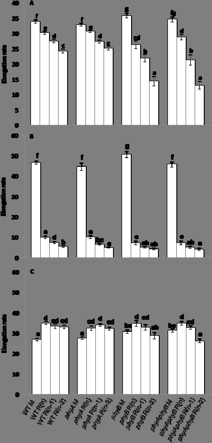
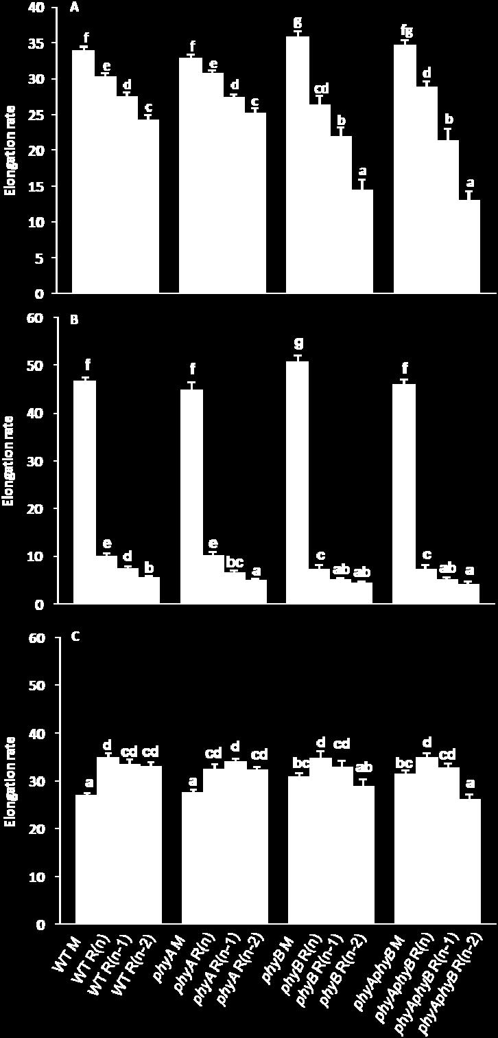
57 48
.")

58 49 Figure 13 shows the intervals between anthesis of the main inflorescence and the elongation of the topmost rosette buds of WT, phya, phyb, and phyaphyb under high light. The differences in the delay until elongation of buds between the various genotypes at all positions were minor, and did not exceed 0.7 days (Fig. 13). The effect of phyb deficiency on elongation timing of the buds observed under low light was not apparent under high light. Higher light input enables the upper buds from the various genotypes to start elongating earlier than under low light, and this promotion was relatively greater in the phyb deficient mutants than in WT and phya (Figs 10, 13). Therefore, high light eliminated the inhibitory effects of the phyb lesion on the timing of the onset of elongation. Figure 13. Interval between anthesis and the onset of elongation of the topmost three rosette buds of WT, phya, phyb, and phyaphyb under high light. Bars with different letters within genotypes are significantly different at α = In contrast to the elongation patterns of the various genotypes grown under low light, the lengths of the main inflorescence and the three topmost branches of phyb and phyaphyb were consistently greater than those of WT and phya under high light (Fig. 14). This suggests a negative role for phyb in regulating elongation of both the main


59 50 inflorescence and the three topmost branches under high light (Fig. 14). The negative role of phyb in elongation of the main inflorescence is well established; however, phyb loss of function is typically believed to inhibit branching, as was observed under low light (Fig. 2, Fig. 11). Under high light this pattern was unexpectedly reversed. The branching phenotypes of phyb deficient mutants in many aspects were recued to almost the same level as phyb sufficient genotypes. This may be due to the compensation of high light intensity on the constitutive low light quality phenotypes of phyb deficient mutants, perhaps due to increased photoassimilation, and/or light quantity signaling described above. Figure 14. The lengths of the main inflorescence (M) (A), the topmost rosette branch R(n) (B), the next topmost rosette branch R(n-1) (C), and the third topmost rosette branch R(n-2) (D) of WT, phya, phyb, and phyaphyb from the day of the onset of elongation of bud R(n) to the 10 th day after anthesis under high light.
60 51 The ranking of the bud activity with respect to the timing of elongation onset was consistent among genotypes: R(n) > R(n-1) > R(n-2). However, the differences between the activities of the three topmost rosette axillary buds under high light were not as great as those under low light (Figs 10, 13). Linear regressions were again fitted to the elongation data of the various genotypes, and the slope of each regression was taken as the overall elongation rate (Fig. 15A). Under high light, the main inflorescences of WT, phya, phyb, and phyaphyb elongated at a significantly greater rate than branch R(n), which had an elongation rate significantly greater than R(n-1). No significant differences were observed between the elongation rates of R(n-1) and R(n-2) (Fig. 15A). Rosette axillary branches from phyb and phyaphyb elongated at a significantly higher rate than those from phyb sufficient genotypes (Fig 15A). While the trends of branch elongation rates within genotypes were still consistent- R(n) > R(n-1) > R(n-2), the differences in the branch elongation rates of phyb deficient and phyb sufficient genotypes were reversed in high compared to low light (Figs. 12A, 15A). This again suggests a role of light quantity in inducing shade avoidance responses in a manner similar to light quality as discussed above. The calculation of elongation rates was again separated into two intervals: from the 1 st day - 3 rd day (Fig. 15B), and from the 4 th day - 10 th day after anthesis (Fig. 15C). The main inflorescence of the various genotypes under high light elongated significantly faster than R(n) from the 1st day to 3 rd day of elongation. R(n) elongated significantly faster than R(n-1) and R(n-2). The elongation rates of R(n-1) were not significantly different from R(n-2) (Fig. 15B). The elongation rates of the three branches within genotypes were similar (Fig. 15C). Loss of phyb function resulted in an increase in the elongation rate of the top branches under high light. The elongation rates of the main inflorescence and the three topmost buds during first three days of elongation contributed more to the final architecture of the various genotypes. However, the reversal of the trends in elongation rates of the branches of phyb deficient and phyb sufficient genotypes under high light compared to those under low light was determined by both early and late phase elongation.
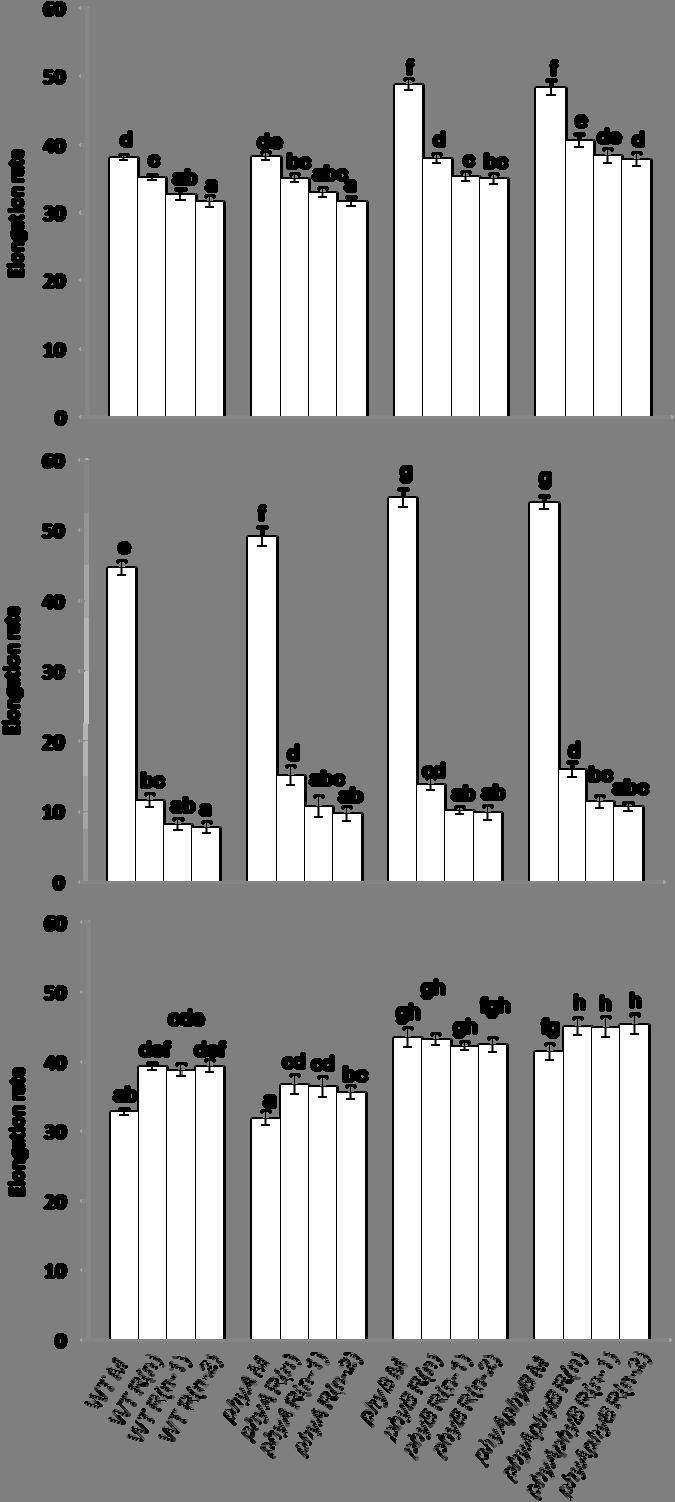
61 52
62 53 Objective 3. Assessment of the changes in the expression of branching-related genes in buds of WT, phya, phyb, and phyaphyb under high and low PPFD. The expression of the branching-regulator TBL1 has previously been demonstrated to be negatively correlated with the developmental stage of the axillary buds (Aguilar-Martinez et al., 2007, Finlayson, 2007). TBL1 is expressed at modest levels in unelongated buds and decreases to very low levels once buds begin to grow out. Here, the changes in the expression of branching-related genes in buds of WT, phya, phyb, and phyaphyb were assessed to determine how phya and phyb regulate the molecular physiology of the bud, and also to confirm the proposed activity ranking of the axillary buds from the various genotypes. The mrna abundance of TBL1 in the topmost three rosette axillary buds of the various genotypes under low light (Fig. 16) was generally in accordance with the hypothesized activity of the axillary buds [R(n) > R(n-1) > R(n-2)]. Loss of phyb function resulted in elevated expression of TBL1. This is consistent with the previous report that TBL1 (BRC1) is required for plants to arrest the outgrowth of axillary buds (Finlayson, 2007; Aguilar-Martínez et al., 2007), and indicates that TBL1 expression is regulated by phyb in a manner similar to tb1 in sorghum (Kebrom et al., 2006). TBL1 expression in the lower buds of phya was reduced compared to WT, though no differences in branching were observed (Figs. 2B, 2C, 2D, 7, 10, 11). It is difficult to explain these results without considering the possibility of sampling errors. Additionally, the lack of statistically significant differences in some instances are likely due to the variation in the status of harvested buds. TBL1 expression varies with the physiological status of the buds, which is difficult to determine visually. Errors in the timing of bud collection could have contributed noise to the analysis.

.")
63 54 Figure 16. TBL1 mrna abundance in the topmost three rosette axillary buds from WT, phya, phyb, and phyaphyb grown under low light. Results are the means of QPCR analysis of three biological replicates ± SEM. Bars with different letters are significantly different at α = It is known that mutations in TBL1 and BRC2 result in hyper-branching phenotypes (Aguilar-Martı nez et al., 2007; Finlayson, 2007). BRC2 expression may not be as closely related to bud outgrowth as TBL1 since loss of BRC2 function results in only a moderate hyper-branching phenotype (Aguilar-Martı nez et al., 2007). Moreover, tbl1/brc2 double mutants possess the phenotype of tbl1 (Aguilar-Martı nez et al., 2007; Finlayson, 2007). BRC2 has been suggested to function in a manner similar to TBL1 as an inhibitor of branching that is expressed in the axillary buds. The pattern of BRC2 mrna abundance was hypothesized to be similar to the pattern of TBL1 mrna abundance in the buds of the various genotypes. Therefore, the expression of BRC2 in the topmost three rosette axillary buds was also assessed. In general, bud R(n) had the least BRC2 abundance, R(n-1) had an intermediate amount, and R(n-2) had the greatest. This trend was observed in WT, phyb and phyaphyb, though it was not always statistically significant in every case. phya showed a slightly different pattern with lowest BRC2 expression in bud R(n-1), however, this level was not statistically different from that of bud R(n) (Fig. 17). Differences in BRC2

.")
64 55 expression between genotypes were minor. Figure 17. BRC2 mrna abundance in the topmost three rosette axillary buds from WT, phya, phyb, and phyaphyb grown under low light. Results are the means of QPCR analysis of three biological replicates ± SEM. Bars with different letters are significantly different at α = The ranking of the TBL1 RNA abundance in the three topmost rosette axillary buds of WT, phya, phyb and phyaphyb under high light was also generally in accordance with the outgrowth potential of the respective buds (Fig 18). In some cases the differences between buds at different positions within a genotype were not statistically significant, but the numerical trends consistently ranked the TBL1 content lowest in bud R(n), intermediate in R(n-1) and highest in R(n-2). This pattern was similar to that observed under low light, but the TBL1 mrna abundance in the three topmost axillary buds of WT were reduced with the greatest reduction in R(n-1) (32%) under high compared to low light. The TBL1 mrna abundance in the three topmost axillary buds of phya were reduced with the greatest reduction in R(n-2) (58%) under high compared to low light. The TBL1 mrna abundance in the three topmost axillary buds of phyb were reduced with the greatest reduction in R(n) (7%) under high compared to low light. The TBL1
65 56 mrna abundance in the three topmost axillary buds of phyaphyb were reduced with the greatest reduction in R(n-2) (11%) under high compared to low light (Figs 16, 18). The differences in the expression of BRC2 caused by high light are not as substantial as TBL1. However, it was found that most of the axillary buds of the various genotypes possess reduced expression of BRC2 under high light compared to low light (Figs 17, 19). The expression pattern of TBL1 was in accordance with the hypothesis that higher light can increase the activity of rosette axillary buds. This suggests that the enhancement of elongation rate and the earlier onset of elongation that together contribute to the activities of axillary buds under high light was regulated by decreased expression of TBL1 locally in the buds. The reduction of TBL1 expression caused by high light is greater in the phyb deficient mutants than that in the phyb sufficient genotypes. This result is also in agreement with the branching analysis of the three topmost buds (branches) under low and high light. High light has more effect on the branching of phyb deficient mutants than phyb sufficient genotypes. Taken together, it suggests that phyb is not the only mediator of TBL1 expression in Arabidopsis, since TBL1 expression is still responsive to light quantity in the phyb deficient mutants. This is in accordance with our hypothesis that in addition to phyb, other photosensors and/or the plant s sugar status could regulate TBL1 expression.

.")
66 57 Figure 18. TBL1 mrna abundance in the topmost three rosette axillary buds of WT, phya, phyb, and phyaphyb grown under high light. Results are the means of QPCR analysis of three biological replicates ± SEM. Bars with different letters are significantly different at α = The BRC2 mrna abundance patterns within genotypes from plants grown under high light (Fig. 19) were dissimilar from those observed under low light (Fig. 17), with the exception of phyaphyb. BRC2 levels were higher in WT than in phyb, which is consistent with the enhanced branching of phyb under high light (Figs 2, 5). BRC2 expression levels in phyb and phyaphyb were reduced in high light compared to low light, with one exception in bud R(n) of phyb, suggesting a role for PPFD in regulating BRC2 expression in addition to TBL1 expression. It is possible that the somewhat anomalous BRC2 expression patterns observed may be due in part to the very low expression level of this gene, which results in elevated noise.

, negatively regulate axillary buds outgrowth (Aguilar-Martinez et al.")
67 58 Figure 19. BRC2 mrna abundance in the topmost three rosette axillary buds from WT, phya, phyb, and phyaphyb grown under high light. Results are the means of QPCR analysis of three biological replicates ± SEM. Bars with different letters are significantly different at α = In summary, it may be concluded that the expression of TBL1 in Arabidopsis is regulated by the light signals perceived by phyb, which is negatively correlated to bud outgrowth but not bud initiation. These are in accordance with the previous report that the expression TBL1 (BRC1), negatively regulate axillary buds outgrowth (Aguilar-Martinez et al., 2007; Finlayson, 2007), and was proposed to be negatively regulated by phyb (Finlayson, 2007). These findings also agree with the previous research in other species, such as rice, which suggests that OsTB1 expression suppress the axillary bud outgrowth instead of formation (Takeda et al., 2003). It was also consistent with the findings of Kebrom et al. (2006) that phyb regulates bud outgrowth through mediating the expression level of SbTB1 in response to various light signals. Objective 4. Assessment the association of hormones with the activity of buds of WT, phya, phyb, and phyaphyb under high and low PPFD. Previous research has related the DRM1 expression level to the dormancy status of buds (Stafstrom et al., 1998; Tatematsu et al., 2005; Kebrom et al., 2006). The
68 59 expression of DRM1 was also found to be regulated by light signals and phyb in sorghum, and its expression correlated with that of TB1 (Kebrom et al., 2006). The DRM1 expression level in the three topmost axillary buds of various genotypes was assessed as an indicator of the status of the buds and to probe the possible role of auxin in their development. Under low light, the expression patterns of DRM1 in the three topmost buds from WT, phya and phyb and phyaphyb generally correlate with those of the TBL1 expression levels (Fig 16, 20), with lowest levels in R(n) and highest levels in R(n-2). Between genotypes, the DRM1 expression levels in the buds R(n) and R(n-1) from phyb and phyaphyb are significantly higher than the respective buds in WT, which is in accordance with the physiological analysis indicating that phyb has a positive role in determining bud activity. DRM1 expression may provide an indication that phyb plays greater role in regulating the topmost two buds, as opposed to those at more basal positions since DRM1 levels are nearly equivalent in bud R(n-2) of WT and phyb. The results could also suggest a possible role for auxin in triggering the outgrowth of axillary buds. However, DRM1 expression is less representative of proposed bud activity, [R(n) > R(n-1) > R(n-2)] compared to TBL1, indicating that auxin is not the only regulator of bud outgrowth or that auxin is acting through mechanisms unrelated to DRM1 expression. This may be consistent with the previous report that auxin deficiency alone does not trigger initial bud outgrowth in Arabidopsis (Cline, 1996; Beveridge et al., 2000; Cline et al., 2001).

69 60 Figure 20. DRM1 mrna abundance in the topmost three rosette axillary buds from WT, phya, phyb, and phyaphyb grown under low light. Results are the means of QPCR analysis of three biological replicates ± SEM. Bars with different letters are significantly different at α = Under high light, phyb and phyaphyb show similar DRM1 expression patterns as that of TBL1 under high light. No differences in DRM1 expression in the three topmost buds of WT were observed, and the pattern in phya was opposite that predicted, though the differences were not large (Fig 21). However, the DRM1 levels of phyb and phyaphyb were higher than WT and phya, which is inconsistent with its proposed utility as an indicator of dormancy, an issue previously raised by Finlayson (2007). TBL1 expression appears to be a better indicator of the developmental stages of axillary buds than DRM1 expression.


70 61 Figure 21. DRM1 mrna abundance in the topmost three rosette axillary buds from WT, phya, phyb, and phyaphyb grown under high light. Results are the means of QPCR analysis of three biological replicates ± SEM. Bars with different letters are significantly different at α = Previous research has indicated that ARR5, a type A ARR, responds exclusively to exogenous CK transcriptionally within 10 min (Brandstatter and Kieber, 1998; D Agostino et al., 2000). ARR5 expression was monitored to determine if there were differences in CK activity within the different buds. Under low light, the expression patterns of ARR5 of the three topmost buds from WT, phya and phyb and phyaphyb generally correlated inversely with those of TBL1 and DRM1 expression levels (Fig 16, 22), with the lowest levels in R(n-2) and highest levels in R(n). The results could suggest a possible role for CK in triggering the outgrowth of axillary buds. This is also consistent with the role of auxin acting negatively on the amount of CK in axillary buds (Cline, 1994). However, ARR5 expression is less representative of proposed bud activity, [R(n) > R(n-1) > R(n-2)] compared to TBL1.

71 62 Figure 22. ARR5 mrna abundance in the topmost three rosette axillary buds from WT, phya, phyb, and phyaphyb grown under low light. Results are the means of QPCR analysis of three biological replicates ± SEM. Bars with different letters are significantly different at α = phyb and phyaphyb show ARR5 expression patterns that are the inverse of TBL1 under high light. The pattern in WT was as predicted highest in R(n), lowest in R(n-2), though no statistical differences between R(n-1) and R(n-2) were observed (Fig 23). These results are consistent with the role of CK, which has been previously suggested to be highly correlated with bud activity and negatively regulated by auxin (Sachs and Thimann, 1967; Li et al., 1995; Chatfield et al., 2000; Leyser, 2003; Nordstrom et al., 2004; Tanaka et al., 2006). No differences in ARR5 expression in the three topmost buds of phya were observed. However, the ARR5 levels of phyb and phyaphyb were higher than WT and phya, which is consistent with its proposed utility as an indicator of bud activity since these buds elongated earlier and at a higher rate than WT (Figs 13, 14, 15, 23).


72 63 Figure 23. ARR5 mrna abundance in the topmost three rosette axillary buds from WT, phya, phyb, and phyaphyb grown under high light. Results are the means of QPCR analysis of three biological replicates ± SEM. Bars with different letters within are significantly different at α = 0.05.
73 64 5. SUMMARY AND CONCLUSIONS Objective 1. Quantification of the architecture of WT, phya, phyb and phyaphyb under high and low PPFD. Branching is regulated by light signals perceived by phytochromes. While it has been anecdotally reported that Arabidopsis phyb mutants branch less than WT, no data defining the lesion in branching caused by loss of phyb function has been available. The hypothesis proposed herein was that phyb would have a large effect on the regulation of axillary bud outgrowth, while phya would have little or no effect on branching. Therefore, it was predicted that phya and WT would not have significant differences in branching patterns under low light intensity, while phyb and phyaphyb would have significantly reduced numbers of primary and secondary rosette branches, primary and secondary cauline branches, rosette leaves, and axillary buds. High light intensity (PPFD) was hypothesized to be able to rescue the reduction of branching and rosette leaves in phyb and phyaphyb. The blue wavelengths in white light may act as a signal of environmental quality and play a role similar to R:FR in determining shade avoidance. Such blue light responses could be evoked in phyb deficient mutants as well as plants with functional phyb, possibly through the action of cryptochromes. Light quantity might also affect branching by determining the amount of photosynthates available for branch growth. WT and phya were found to have similar branching patterns in the number of rosette branches, ratio of secondary cauline branches / axil. However, phya was found to have more rosette leaves than WT under both low and high light conditions. phyb and phyaphyb had similar branching patterns in the number of rosette branches and the ratio of secondary cauline branches/axil. Loss of phyb function led to reduced numbers of rosette leaves and branches, lowered rosette branches/axil and secondary cauline branches/axil, and delayed and slower elongation of the topmost three rosette branches. The high light environment may have provided more photosynthate to allow WT, phyb, phya, and phyaphyb to increase the number of rosette leaves and branches, the ratio of rosette branches/axil and secondary cauline branches/axil. The differences resulting from high light were greater in mutants deficient in phyb (phyb and phyaphyb)
74 65 than in WT and phya. In fact, high light quantity partially rescued the phenotypes of phyb and phyaphyb resulting in branching patterns similar to WT and phya. Along with increased photoassimilation, it is also possible that high light could provide a signal, perceived by other phytochromes and/or cryptochromes, that overrides the R:FR signal normally perceived by phyb. Objective 2. To determine if the branching patterns of WT, phya, phyb, and phyaphyb are regulated through the formation of the buds, the timing of the onset of bud elongation, and/or the elongation rates of the branches. It was hypothesized that the branching patterns of WT, phya, phyb, and phyaphyb are determined by both the formation of the buds and through elongation of the buds. Histological analysis indicated that phyb does not influence meristem initiation, but does play a positive role in promoting the onset of bud elongation and also the bud elongation rate. phya was hypothesized to possess a similar pattern of branching as WT. It was found that phya does not play a role in regulating the onset of bud outgrowth or elongation. The data also indicate that the final architecture of WT and phya are mainly determined by early differences in branch elongation, while both early and late branch elongation contributes to the final architecture of phyb and phyaphyb under low light. Loss of phyb function resulted in a delay in the onset of bud outgrowth under low light, but promoted it under high light. Obviously, the role of phyb in this, and other processes, is conditional upon the light quantity. The early (day 1 to day 3) elongation of the topmost three rosette buds of WT and phya contributed more to the final differences in length of the branches than the later (day 4 to day 10) elongation regardless of the variations in light quantity. While early elongation also contributed greater to the differences in final branch length in phyb and phyaphyb, later elongation was also a factor. The ranking of the elongation rate of the top most three branches was: R(n) > R(n-1) > R(n-2) under both low and high light intensity, in all genotypes assessed.
75 66 Objective 3. Assessment of the changes in the expression of branching-related genes in buds of WT, phya, phyb, and phyaphyb under high and low PPFD. Branching-related gene expression was investigated in the buds to determine if it was linked to light signals perceived by phytochromes. The expression levels of TBL1 and BRC2 in the topmost three axillary buds and their elongation rates in the various genotypes were proposed to be in accordance with the activity of the axillary buds. The ranking of TBL1 and BRC2 expression level in the topmost three axillary buds was therefore predicted to be: R(n) < R(n-1) < R(n-2) under both low and high light intensity. This hypothesis was based on the known roles of TBL1 and BRC2 as negative regulators of bud outgrowth. It was found that the trend of TBL1 mrna abundance in the topmost three buds was consistent within genotypes regardless of variations in the light. In accordance with the hypothesis, bud R(n) of various genotypes had the lowest expression levels, and buds R(n-2) had the highest expression levels. The differences in TBL1 RNA abundance between buds were occasionally non-significant. Increasing the size of the sampling population and/or increasing the number of replicated measurements would likely help resolve this issue. The trend of BRC2 RNA abundance in the topmost three buds was similar to, but not as clear as, TBL1 expression. This suggests that BRC2 is a weaker negative regulator of axillary bud activity compared to TBL1. This is consistent with the more similar phenotypes of brc2 and WT. The phenotype of the tbl1/brc2 double mutant was indistinguishable from the tbl1 single mutant (Aguilar-Martinez et al., 2007; Finlayson, 2007). The axillary bud outgrowth was negatively regulated by TBL1 (BRC1) expression (Aguilar-Martinez et al., 2007; Finlayson, 2007), which was proposed to be negatively regulated by phyb (Finlayson, 2007). The proposed hypothesis was that the level of TBL1 mrna in the axillary buds of phyb and phyaphyb would be higher than that in WT and phya, which would suggest that it is through the expression of TBL1 that phyb inhibits the growth of the axillary buds. The branching and TBL1 expression patterns of phya were predicted to not be significantly different from WT. Our results show that the TBL1 expressions of the three top most buds from phyb deficient mutants are consistently higher than those in the respective buds from phyb sufficient genotypes,
76 67 which is in accordance with the hypothesized relationship of phyb and TBL1 in branching regulation. It was found that the expression of TBL1 can be reduced in response to high light through a phyb-independent pathway. The reduction of TBL1 expression in the axillary buds of various genotypes grown under high light is in accordance with the increased axillary bud activity caused by high light, with a higher reduction in the phyb deficient mutants. The data suggest that multiple pathways regulate TBL1 expression in response to the light environment. Objective 4. Assessment of the association of hormones with the activity of buds of WT, phya, phyb, and phyaphyb under high and low PPFD. The expression level of DRM1, an auxin responsive gene, was proposed to be negatively associated with the activity of axillary buds and positively associated with TBL1 expression. It was hypothesized that the ranking of the DRM1 expression level of the topmost three axillary buds would be: R(n) < R(n-1) < R(n-2) under both low and high light intensity. Our results show that the expression level of DRM1 in the axillary buds of Arabidopsis was not as highly correlated with the developmental status of the buds as TBL1 expression was. The expression level of ARR5, a CK responsive gene, was proposed to be positively associated with the activity of axillary buds and negatively associated with TBL1 expression. It was hypothesized that the ranking of the ARR5 expression level of the topmost three axillary buds would be: R(n) > R(n-1) > R(n-2) under both low and high light intensity. ARR5 expression generally followed this trend, but was less well correlated with bud activity than TBL1 expression was. Figure 24 shows the proposed model of regulation of branching by light quality and quantity. The two pathways appear to converge to regulate TBL1 expression in the buds. This also suggests that phyb is not the only regulator of TBL1 expression in Arabidopsis.


77 68 Figure 24. The proposed model of axillary bud outgrowth regulation by light quality and quantity perceived by photoreceptors mediating the expression level of TBL1.
Electromagenetic spectrum
 Light Controls of Plant Development 1 Electromagenetic spectrum 2 Light It is vital for photosynthesis and is also necessary to direct plant growth and development. It acts as a signal to initiate and
Analysis of regulatory function of circadian clock. on photoreceptor gene expression
 Thesis of Ph.D. dissertation Analysis of regulatory function of circadian clock on photoreceptor gene expression Tóth Réka Supervisor: Dr. Ferenc Nagy Biological Research Center of the Hungarian Academy
CONTROL OF PLANT GROWTH AND DEVELOPMENT BI-2232 RIZKITA R E
 CONTROL OF PLANT GROWTH AND DEVELOPMENT BI-2232 RIZKITA R E The development of a plant the series of progressive changes that take place throughout its life is regulated in complex ways. Factors take part
Cytokinin. Fig Cytokinin needed for growth of shoot apical meristem. F Cytokinin stimulates chloroplast development in the dark
 Cytokinin Abundant in young, dividing cells Shoot apical meristem Root apical meristem Synthesized in root tip, developing embryos, young leaves, fruits Transported passively via xylem into shoots from
Plant Growth and Development
 Plant Growth and Development Concept 26.1 Plants Develop in Response to the Environment Factors involved in regulating plant growth and development: 1. Environmental cues (e.g., day length) 2. Receptors
Phytochrome Regulation of Branching in Arabidopsis 1[W][OA]
![Phytochrome Regulation of Branching in Arabidopsis 1[W][OA]](/thumbs/76/73891908.jpg "Phytochrome Regulation of Branching in Arabidopsis 1[W][OA]") Phytochrome Regulation of Branching in Arabidopsis 1[W][OA] Scott A. Finlayson*, Srirama R. Krishnareddy, Tesfamichael H. Kebrom, and Jorge J. Casal Department of Soil and Crop Sciences, Texas A&M University,
Phytochrome Regulation of Branching in Arabidopsis 1[W][OA] Scott A. Finlayson*, Srirama R. Krishnareddy, Tesfamichael H. Kebrom, and Jorge J. Casal Department of Soil and Crop Sciences, Texas A&M University,
Apical dominance models can generate basipetal patterns of bud activation
 Apical dominance models can generate basipetal patterns of bud activation Przemyslaw Prusinkiewicz 1, Richard S. Smith 1 and Ottoline Leyser 2 1 Department of Computer Science, University of Calgary 2
23-. Shoot and root development depend on ratio of IAA/CK
 Balance of Hormones regulate growth and development Environmental factors regulate hormone levels light- e.g. phototropism gravity- e.g. gravitropism temperature Mode of action of each hormone 1. Signal
LECTURE 4: PHOTOTROPISM
 http://smtom.lecture.ub.ac.id/ Password: https://syukur16tom.wordpress.com/ LECTURE 4: PHOTOTROPISM LECTURE FLOW 1. 2. 3. 4. 5. INTRODUCTION DEFINITION INITIAL STUDY PHOTROPISM MECHANISM PHOTORECEPTORS
Figure 18.1 Blue-light stimulated phototropism Blue light Inhibits seedling hypocotyl elongation
 Blue Light and Photomorphogenesis Q: Figure 18.3 Blue light responses - phototropsim of growing Corn Coleoptile 1. How do we know plants respond to blue light? 2. What are the functions of multiple BL
Useful Propagation Terms. Propagation The application of specific biological principles and concepts in the multiplication of plants.
 Useful Propagation Terms Propagation The application of specific biological principles and concepts in the multiplication of plants. Adventitious Typically describes new organs such as roots that develop
10/4/2017. Chapter 39
 Chapter 39 1 Reception 1 Reception 2 Transduction CYTOPLASM CYTOPLASM Cell wall Plasma membrane Phytochrome activated by light Cell wall Plasma membrane Phytochrome activated by light cgmp Second messenger
CBMG688R. ADVANCED PLANT DEVELOPMENT AND PHYSIOLOGY II G. Deitzer Spring 2006 LECTURE
 1 CBMG688R. ADVANCED PLANT DEVELOPMENT AND PHYSIOLOGY II G. Deitzer Spring 2006 LECTURE Photomorphogenesis and Light Signaling Photoregulation 1. Light Quantity 2. Light Quality 3. Light Duration 4. Light
Flower Development Pathways
 Developmental Leading to Flowering Flower Development s meristem Inflorescence meristem meristems organ identity genes Flower development s to Flowering Multiple pathways ensures flowering will take place
THE ROLE OF THE PHYTOCHROME B PHOTORECEPTOR IN THE REGULATION OF PHOTOPERIODIC FLOWERING. AnitaHajdu. Thesis of the Ph.D.
 THE ROLE OF THE PHYTOCHROME B PHOTORECEPTOR IN THE REGULATION OF PHOTOPERIODIC FLOWERING AnitaHajdu Thesis of the Ph.D. dissertation Supervisor: Dr. LászlóKozma-Bognár - senior research associate Doctoral
Plants are sessile. 10d-17/giraffe-grazing.jpg
 Plants are sessile www.mccullagh.org/db9/ 10d-17/giraffe-grazing.jpg Plants have distinct requirements because of their sessile nature Organism-level requirements Must adjust to environment at given location
Outline. Leaf Development. Leaf Structure - Morphology. Leaf Structure - Morphology
 Outline 1. Leaf Structure: Morphology & Anatomy 2. Leaf Development A. Anatomy B. Sector analysis C. Leaf Development Leaf Structure - Morphology Leaf Structure - Morphology 1 Leaf Structure - Morphology
Major Plant Hormones 1.Auxins 2.Cytokinins 3.Gibberelins 4.Ethylene 5.Abscisic acid
 Plant Hormones Lecture 9: Control Systems in Plants What is a Plant Hormone? Compound produced by one part of an organism that is translocated to other parts where it triggers a response in target cells
Chapter 39. Plant Response. AP Biology
 Chapter 39. Plant Response 1 Plant Reactions Stimuli & a Stationary Life u animals respond to stimuli by changing behavior move toward positive stimuli move away from negative stimuli u plants respond
Photoreceptor Regulation of Constans Protein in Photoperiodic Flowering
 Photoreceptor Regulation of Constans Protein in Photoperiodic Flowering by Valverde et. Al Published in Science 2004 Presented by Boyana Grigorova CBMG 688R Feb. 12, 2007 Circadian Rhythms: The Clock Within
Chapter 39. Plant Reactions. Plant Hormones 2/25/2013. Plants Response. What mechanisms causes this response? Signal Transduction Pathway model
 Chapter 39 Plants Response Plant Reactions Stimuli & a Stationary life Animals respond to stimuli by changing behavior Move toward positive stimuli Move away from negative stimuli Plants respond to stimuli
Engineering light response pathways in crop plants for improved performance under high planting density
 Engineering light response pathways in crop plants for improved performance under high planting density Tom Brutnell Boyce Thompson Institute for Plant Research Cornell University, Ithaca NY 6000 years
Figure 1. Identification of UGT74E2 as an IBA glycosyltransferase. (A) Relative conversion rates of different plant hormones to their glucosylated
 Relative conversion rates of different plant hormones to their glucosylated") Figure 1. Identification of UGT74E2 as an IBA glycosyltransferase. (A) Relative conversion rates of different plant hormones to their glucosylated form by recombinant UGT74E2. The naturally occurring auxin
Figure 1. Identification of UGT74E2 as an IBA glycosyltransferase. (A) Relative conversion rates of different plant hormones to their glucosylated form by recombinant UGT74E2. The naturally occurring auxin
Phytochromes and Shade-avoidance Responses in Plants
 Annals of Botany 96: 169 175, 2005 doi:10.1093/aob/mci165, available online at www.aob.oupjournals.org BOTANICAL BRIEFING Phytochromes and Shade-avoidance Responses in Plants KEARA A. FRANKLIN and GARRY
CONTROL SYSTEMS IN PLANTS
 AP BIOLOGY PLANTS FORM & FUNCTION ACTIVITY #5 NAME DATE HOUR CONTROL SYSTEMS IN PLANTS HORMONES MECHANISM FOR HORMONE ACTION Plant Form and Function Activity #5 page 1 CONTROL OF CELL ELONGATION Plant
Pea Has Its Tendrils in Branching Discoveries Spanning a Century from Auxin to Strigolactones 1
 Update on Pea Shoot Branching Pea Has Its Tendrils in Branching Discoveries Spanning a Century from Auxin to Strigolactones 1 Christine A. Beveridge*, Elizabeth A. Dun, and Catherine Rameau School of Biological
15. PHOTOPERIODISM. 1. Short day plants
 15. PHOTOPERIODISM Photoperiodism is the phenomenon of physiological changes that occur in plants in response to relative length of day and night (i.e. photoperiod). The response of the plants to the photoperiod,
Reproduction, Seeds and Propagation
 Reproduction, Seeds and Propagation Diploid (2n) somatic cell Two diploid (2n) somatic cells Telophase Anaphase Metaphase Prophase I One pair of homologous chromosomes (homologues) II Homologues condense
The shade avoidance syndrome: multiple responses mediated by multiple phytochromes
 Plant, Cell and Environment (1997) 20, 840 844 TECHNICAL REPORT (white this line if not required) The shade avoidance syndrome: multiple responses mediated by multiple phytochromes H. SMITH & G. C. WHITELAM
Plant Development. Chapter 31 Part 1
 Plant Development Chapter 31 Part 1 Impacts, Issues Foolish Seedlings, Gorgeous Grapes Gibberellin and other plant hormones control the growth and development of plants environmental cues influence hormone
CBSE Quick Revision Notes (Class-11 Biology) CHAPTER-15 PLANT GROWTH AND DEVELOPMENT
 CHAPTER-15 PLANT GROWTH AND DEVELOPMENT") CBSE Quick Revision Notes (Class-11 Biology) CHAPTER-15 PLANT GROWTH AND DEVELOPMENT Root, stem leaves, flower, fruits and seeds arise in orderly manner in plants. The sequence of growth is as follows-
CBSE Quick Revision Notes (Class-11 Biology) CHAPTER-15 PLANT GROWTH AND DEVELOPMENT Root, stem leaves, flower, fruits and seeds arise in orderly manner in plants. The sequence of growth is as follows-
PLANT HORMONES-Introduction
 PLANT HORMONES-Introduction By convention hormone are said to be a substances whose site of synthesis and site of action are different; the two events are separated by space and time. Hormones are known
Supplemental Data. Wang et al. (2014). Plant Cell /tpc
. Plant Cell /tpc") Supplemental Figure1: Mock and NPA-treated tomato plants. (A) NPA treated tomato (cv. Moneymaker) developed a pin-like inflorescence (arrowhead). (B) Comparison of first and second leaves from mock and
Supplemental Figure1: Mock and NPA-treated tomato plants. (A) NPA treated tomato (cv. Moneymaker) developed a pin-like inflorescence (arrowhead). (B) Comparison of first and second leaves from mock and
GENETIC ANALYSES OF ROOT SYSTEM DEVELOPMENT IN THE TOMATO CROP MODEL
 GENETIC ANALYSES OF ROOT SYSTEM DEVELOPMENT IN THE TOMATO CROP MODEL Kelsey Hoth 1 Dr. Maria Ivanchenko 2 Bioresourse Research 1, Department of Botany and Plant Physiology 2, Oregon State University, Corvallis,
The signal transducing photoreceptors of plants
 Int. J. Dev. Biol. 49: 653-664 (2005) doi: 10.1387/ijdb.051989kf The signal transducing photoreceptors of plants KEARA A. FRANKLIN*, VICTORIA S. LARNER and GARRY C. WHITELAM Department of Biology, University
COMPETITIVE CANALIZATION OF AUXIN IN PEA CAN BE INVOLVED IN INITIATION OF AXILLARY BUD OUTGROWTH
 COMPETITIVE CANALIZATION OF AUXIN IN PEA CAN BE INVOLVED IN INITIATION OF AXILLARY BUD OUTGROWTH Medveďová Z. 1, Balla J. 1, 2, Procházka S. 1 1 CEITEC - Central European Institute of Technology, Mendel
Leucine-rich repeat receptor-like kinases (LRR-RLKs), HAESA, ERECTA-family
, HAESA, ERECTA-family") Leucine-rich repeat receptor-like kinases (LRR-RLKs), HAESA, ERECTA-family GENES & DEVELOPMENT (2000) 14: 108 117 INTRODUCTION Flower Diagram INTRODUCTION Abscission In plant, the process by which a plant
Leucine-rich repeat receptor-like kinases (LRR-RLKs), HAESA, ERECTA-family GENES & DEVELOPMENT (2000) 14: 108 117 INTRODUCTION Flower Diagram INTRODUCTION Abscission In plant, the process by which a plant
CONTROL OF GROWTH BY HORMONES
 CONTROL OF GROWTH BY HORMONES Growth and organogenesis are controlled......by genes (independent of environment): e.g., number of primary vascular bundles, general shape of a leaf or flower...by genes
BIO1PS 2012 Plant Science Lecture 4 Hormones Pt. I
 BIO1PS 2012 Plant Science Lecture 4 Hormones Pt. I Dr. Michael Emmerling Department of Botany Room 410 m.emmerling@latrobe.edu.au Hormones and Ghost gum Eucalyptus papuana Coordination ~3 Lectures Leaves
Class XI Chapter 15 Plant Growth and Development Biology
 Question 1: Define growth, differentiation, development, dedifferentiation, redifferentiation, determinate growth, meristem and growth rate. (a) Growth It is an irreversible and permanent process, accomplished
Class XI Chapter 15 Plant Growth and Development Biology
 Question 1: Define growth, differentiation, development, dedifferentiation, redifferentiation, determinate growth, meristem and growth rate. (a) Growth It is an irreversible and permanent process, accomplished
Molecular Genetics of. Plant Development STEPHEN H. HOWELL CAMBRIDGE UNIVERSITY PRESS
 Molecular Genetics of Plant Development STEPHEN H. HOWELL CAMBRIDGE UNIVERSITY PRESS Contents Preface A Word on Genetic Nomenclature page xiii xvii 1 Approaches to the Study of Plant Development 1 Pattern
Plant Responses to Internal and External Signals
 LECTURE PRESENTATIONS For CAMPBELL BIOLOGY, NINTH EDITION Jane B. Reece, Lisa A. Urry, Michael L. Cain, Steven A. Wasserman, Peter V. Minorsky, Robert B. Jackson Chapter 39 Plant Responses to Internal
Chapter 31 Active Reading Guide Plant Responses to Internal and External Signals
 Name: AP Biology Mr. Croft Chapter 31 Active Reading Guide Plant Responses to Internal and External Signals This concept brings together the general ideas on cell communication from Chapter 5.6 with specific
can affect division, elongation, & differentiation of cells to another region of plant where they have an effect
 Note that the following is a rudimentary outline of the class lecture; it does not contain everything discussed in class. Plant Hormones Plant Hormones compounds regulators growth or can affect division,
Plant Stimuli pp Topic 3: Plant Behaviour Ch. 39. Plant Behavioural Responses. Plant Hormones. Plant Hormones pp
 Topic 3: Plant Behaviour Ch. 39 Plants exist in environments that are constantly changing. Like animals, plants must be able to detect and react to stimuli in the environment. Unlike animals, plants can
Responses to Light. Responses to Light
 Sensory Systems in Plants Chapter 41 Pigments other than those used in photosynthesis can detect light and mediate the plant s response to it Photomorphogenesis refers to nondirectional, light-triggered
Ch Plant Hormones
 Ch. 39 Plant Hormones I. Plant Hormones Chemical signals that coordinate the parts of an organism. Only minute amounts are needed to get the desired response. Control plant growth and development by affecting
Chapter 4. Biology of Flowering Plants. Regulation of Plant Growth by Plant Hormones
 BOT 3015L (Sherdan/Outlaw/Aghoram); Page 1 of 8 Chapter 4 Biology of Flowering Plants Regulation of Plant Growth by Plant Hormones Objectives Plant Growth Regulators. Know the names of the plant growth
A. Stimulus Response:
 Plant Hormones A. Stimulus Response: A house plant on a windowsill grows light. If you rotate the plant, it reorients its growth until its leaves face the window again. The growth of a shoot towards light
Is that artificial turf or real grass? Its thicker than Bermuda!
 Is that artificial turf or real grass? Its thicker than Bermuda! 1 Using Plant Growth Regulators Growth regulators DO NOT interfere with plant respiration, photosynthesis, or other internal plant functions
Actions of auxin. Hormones: communicating with chemicals History: Discovery of a growth substance (hormone- auxin)
") Hormones: communicating with chemicals History- discovery of plant hormone. Auxin Concepts of hormones Auxin levels are regulated by synthesis/degradation, transport, compartmentation, conjugation. Polar
Hormones: communicating with chemicals History- discovery of plant hormone. Auxin Concepts of hormones Auxin levels are regulated by synthesis/degradation, transport, compartmentation, conjugation. Polar
Arabidopsis thaliana. Lucia Strader. Assistant Professor, Biology
 Arabidopsis thaliana Lucia Strader Assistant Professor, Biology Arabidopsis as a genetic model Easy to grow Small genome Short life cycle Self fertile Produces many progeny Easily transformed HIV E. coli
Light Regulation of Flowering Time in Arabidopsis
 Chapter 38 Light Regulation of Flowering Time in Arabidopsis Xuhong Yu and Chentao Lin Introduction Plant development is dependent on not only endogenous conditions but also environmental factors. One
AP Plants II Practice test
 AP Plants II Practice test Multiple Choice Identify the letter of the choice that best completes the statement or answers the question. The figure below shows the results of a study to determine the effect
STOLLER ENTERPRISES, INC. World leader in crop nutrition
 A new paradigm for crop production - Page 1 of 6 A NEW PARADIGM FOR CROP PRODUCTION Most agronomists are taught about the chemical process of manufacturing photosynthates (PS). The plants breathe in carbon
Plant Structure and Organization - 1
 Plant Structure and Organization - 1 In our first unit of Biology 203 we will focus on the structure and function of the higher plants, in particular the angiosperms, or flowering plants. We will look
Name Class Date. In the space provided, write the letter of the description that best matches the term or phrase.
 Assessment Chapter Test B Plant Responses In the space provided, write the letter of the description that best matches the term or phrase. 1. thigmonasty 2. auxin 3. ethylene 4. phytochrome 5. abscisic
The Branching Gene RAMOSUS1 Mediates Interactions among Two Novel Signals and Auxin in Pea
 The Plant Cell, Vol. 17, 464 474, February 2005, www.plantcell.org ª 2005 American Society of Plant Biologists The Branching Gene RAMOSUS1 Mediates Interactions among Two Novel Signals and Auxin in Pea
Plant Structure, Growth, and Development
 Plant Structure, Growth, and Development Plant hierarchy: Cells Tissue: group of similar cells with similar function: Dermal, Ground, Vascular Organs: multiple kinds of tissue, very diverse function Organ
The basal-branching pea mutant rms7-1
 Morris, S.E., Beveridge, C.A., Murfet, I.C, Prioul, S. and Rameau, C. The basal-branching pea mutant rms7-1 Australian Res. Council Cent, of Excellence for Integrative Legume Res., and School of Life Sci.
Interactions between Axillary Branches of Arabidopsis
 Molecular Plant Volume 1 Number 2 Pages 388 400 March 2008 RESEARCH ARTICLE Interactions between Axillary Branches of Arabidopsis Veronica Ongaro a, Katherine Bainbridge a,b, Lisa Williamson a and Ottoline
Simon Scofield, Walter Dewitte, and James AH Murray* School of Biosciences; Cardiff University; Cardiff, UK
 Short Communication Plant Signaling & Behavior 9, e28934; April; 2014 Landes Bioscience Short Communication STM sustains stem cell function in the Arabidopsis shoot apical meristem and controls KNOX gene
Chapter 33 Control Systems in Plants
 Chapter 33 Control Systems in Plants PowerPoint Lectures for Biology: Concepts & Connections, Sixth Edition Campbell, Reece, Taylor, Simon, and Dickey Copyright 2009 Pearson Education, Inc. Lecture by
Questions for Biology IIB (SS 2006) Wilhelm Gruissem
 Wilhelm Gruissem") Questions for Biology IIB (SS 2006) Plant biology Wilhelm Gruissem The questions for my part of Biology IIB, Plant Biology, are provided for self-study and as material for the exam. Please note that the
Questions for Biology IIB (SS 2006) Plant biology Wilhelm Gruissem The questions for my part of Biology IIB, Plant Biology, are provided for self-study and as material for the exam. Please note that the
Epigenetics and Flowering Any potentially stable and heritable change in gene expression that occurs without a change in DNA sequence
 Epigenetics and Flowering Any potentially stable and heritable change in gene expression that occurs without a change in DNA sequence www.plantcell.org/cgi/doi/10.1105/tpc.110.tt0110 Epigenetics Usually
Bio 100 Guide 27.
 Bio 100 Guide 27 http://www.offthemarkcartoons.com/cartoons/1994-11-09.gif http://www.cneccc.edu.hk/subjects/bio/album/chapter20/images/plant_growth.jpg http://pgjennielove.files.wordpress.com/2008/06/apical_meristem.png
GENETIC CONTROL OF FLOWERING TIME IN ARABIDOPSIS
 Annu. Rev. Plant Physiol. Plant Mol. Biol. 1998. 49:345 70 Copyright c 1998 by Annual Reviews. All rights reserved GENETIC CONTROL OF FLOWERING TIME IN ARABIDOPSIS Maarten Koornneef, Carlos Alonso-Blanco,
Chapter 39 Plant Responses to Internal and External Signals
 Chapter 39 Plant Responses to Internal and External Signals Overview: Stimuli and a Stationary Life Plants, being rooted to the ground, must respond to whatever environmental change comes their way For
Biological Roles of Cytokinins
 Direct Control of Shoot Meristem Activity by a Cytokinin-Activating Enzyme By Kurakawa et. Al. Published in Nature Presented by Boyana Grigorova Biological Roles of Cytokinins Cytokinins are positive regulators
Light perception. phytochromes, cryptochromes, phototropins.
 Light perception phytochromes, cryptochromes, phototropins. all photoreceptors consist of proteins bound to light absorbing pigments i.e. chromophores. the spectral sensitivity of each photoreceptor depends
Environmental Plant Physiology Photosynthesis - Aging. Department of Plant and Soil Sciences
 Environmental Plant Physiology Photosynthesis - Aging krreddy@ra.msstate.edu Department of Plant and Soil Sciences Photosynthesis and Environment Leaf and Canopy Aging Goals and Learning Objectives: To
TREES. Functions, structure, physiology
 TREES Functions, structure, physiology Trees in Agroecosystems - 1 Microclimate effects lower soil temperature alter soil moisture reduce temperature fluctuations Maintain or increase soil fertility biological
Photosynthesis - Aging Leaf Level. Environmental Plant Physiology Photosynthesis - Aging. Department of Plant and Soil Sciences
 Environmental Plant Physiology Photosynthesis and Environment Leaf and Canopy Aging krreddy@ra.msstate.edu Department of Plant and Soil Sciences Goals and Learning Objectives: To understand the effects
What is Growth? Increment in biomass Increase in volume Increase in length or area Cell division, expansion and differentiation. Fig. 35.
 What is Growth? Increment in biomass Increase in volume Increase in length or area Cell division, expansion and differentiation Fig. 35.18 Copyright 2002 Pearson Education, Inc., publishing as Benjamin
Arabidopsis BRANCHED1 Acts as an Integrator of Branching Signals within Axillary Buds W
 The Plant Cell, Vol. 19: 458 472, February 2007, www.plantcell.org ª 2007 American Society of Plant Biologists Arabidopsis BRANCHED1 Acts as an Integrator of Branching Signals within Axillary Buds W José
LECTURE 04: PHYTOCHROME
 http://smtom.lecture.ub.ac.id/ Password: https://syukur16tom.wordpress.com/ Password: LECTURE 04: PHYTOCHROME Photoreversibility is the most distinctive property of phytochrome 9/19/2017 1 LECTURE OUTCOMES
Plant hormones: a. produced in many parts of the plant b. have many functions
 Plant hormones: a. produced in many parts of the plant b. have many functions Illustrated with 4 plant hormones: Gibberellins Auxin Cytokinins Ethylene Gibberellins Gibberellins illustrate how plant hormones
Phytochrome E Influences Internode Elongation and Flowering Time in Arabidopsis
 The Plant Cell, Vol. 10, 1479 1487, September 1998, www.plantcell.org 1998 American Society of Plant Physiologists Phytochrome E Influences Internode Elongation and Flowering Time in Arabidopsis Paul F.
Suppression of Tiller Bud Activity in Tillering Dwarf Mutants of Rice
 Plant Cell Physiol. 46(1): 79 86 (2005) doi:10.1093/pcp/pci022, available online at www.pcp.oupjournals.org JSPP 2005 Rapid Paper Suppression of Tiller Bud Activity in Tillering Dwarf Mutants of Rice Shinji
The role of the N-terminal NTE domain of PHYTOCHROMEs in red and far red light perception
 The role of the N-terminal NTE domain of PHYTOCHROMEs in red and far red light perception Theses of the Ph.D. dissertation János Bindics Supervisor: Dr. Ferenc Nagy Hungarian Academy of Sciences Biological
Life Science Journal 2014;11(9) Cryptochrome 2 negatively regulates ABA-dependent seed germination in Arabidopsis
 Cryptochrome 2 negatively regulates ABA-dependent seed germination in Arabidopsis") Cryptochrome 2 negatively regulates ABA-dependent seed germination in Arabidopsis Sung-Il Kim 1, Sang Ik Song 3, Hak Soo Seo 1, 2, 4 * 1 Department of Plant Science and Research Institute of Agriculture
Cryptochrome 2 negatively regulates ABA-dependent seed germination in Arabidopsis Sung-Il Kim 1, Sang Ik Song 3, Hak Soo Seo 1, 2, 4 * 1 Department of Plant Science and Research Institute of Agriculture
Seeing without eyes-how plants learn from light
 Seeing without eyes-how plants learn from light by STEPHEN DAY 1. INTRODUCTION Plants detect the intensity, direction, colour, and duration of light and use this information to regulate their growth and
Plant Molecular and Cellular Biology Lecture 10: Plant Cell Cycle Gary Peter
 Plant Molecular and Cellular Biology Lecture 10: Plant Cell Cycle Gary Peter 9/10/2008 1 Learning Objectives Explain similarities and differences between fungal, mammalian and plant cell cycles Explain
Part III. Characterization and mapping of a new locus involved in the regulation of branching in Arabidopsis
 Part III Characterization and mapping of a new locus involved in the regulation of branching in Arabidopsis 73 Abstract The final plant architecture depends on the number and the position of lateral inflorescences,
Trees are: woody complex, large, long-lived self-feeding shedding generating systems compartmented, self optimizing
 BASIC TREE BIOLOGY Trees are: woody complex, large, long-lived self-feeding shedding generating systems compartmented, self optimizing Roots: absorb water and minerals store energy support and anchor
Embryo Development. Embryo Development. Embryo Development. Embryo Development (Cont.) Vegetative Plant Development
 Vegetative Plant Development") Vegetative Plant Development Chapter 37 Embryo Development Begins once the egg cell is fertilized -The growing pollen tube enters angiosperm embryo sac and releases two sperm cells -One sperm fertilizes
Vegetative Plant Development Chapter 37 Embryo Development Begins once the egg cell is fertilized -The growing pollen tube enters angiosperm embryo sac and releases two sperm cells -One sperm fertilizes
Mutation of the cytosolic ribosomal protein-encoding RPS10B gene affects shoot meristematic function in Arabidopsis
 Stirnberg et al. BMC Plant Biology 2012, 12:160 RESEARCH ARTICLE Mutation of the cytosolic ribosomal protein-encoding RPS10B gene affects shoot meristematic function in Arabidopsis Petra Stirnberg 1, Jin-Ping
Topic 14. The Root System. II. Anatomy of an Actively Growing Root Tip
 Topic 14. The Root System Introduction. This is the first of two lab topics that focus on the three plant organs (root, stem, leaf). In these labs we want you to recognize how tissues are organized in
Plant Growth Regulators(NCERT)
") Plant Growth Regulators(NCERT) Promoters: 1. Auxins: -first isolated from urine, contains Zinc. -Natural: Indole Acetic Acid (IAA) Indole Butyric Acid (IBA) -Synthetic: Naphthalene Acetic Acid (NAA) 2-4
Plant Growth Regulators(NCERT) Promoters: 1. Auxins: -first isolated from urine, contains Zinc. -Natural: Indole Acetic Acid (IAA) Indole Butyric Acid (IBA) -Synthetic: Naphthalene Acetic Acid (NAA) 2-4
16. TRANSMISSION OF STIMULUS - THEORIES OF FLOWERING.
 16. TRANSMISSION OF STIMULUS - THEORIES OF FLOWERING. Photoperiodic Induction The influence of the length of day and night on the initiation of flowering is called photoperiodic induction or photo induction.
Ch 25 - Plant Hormones and Plant Growth
 Ch 25 - Plant Hormones and Plant Growth I. Patterns of plant growth A. Plant continue to grow, even in old age. i.e. new leaves, needles, new wood, new cones, new flowers, etc. B. Meristem continues to
Auxin Acts in Xylem-Associated or Medullary Cells to Mediate Apical Dominance
 The Plant Cell, Vol. 15, 495 507, February 2003, www.plantcell.org 2003 American Society of Plant Biologists Auxin Acts in Xylem-Associated or Medullary Cells to Mediate Apical Dominance Jonathan Booker,
Reflexions, le site de vulgarisation de l'université de Liège
 When tomatoes flower 3/13/12 Understanding the mechanisms responsible for tomato plant flowering will enable new selection procedures to be developed in order to obtain even more productive varieties.
From basic research to crop improvement. Dirk Inze VIB-UGent Center for Plant Systems Biology
 From basic research to crop improvement Dirk Inze VIB-UGent Center for Plant Systems Biology Oct 2017 The Great Challenge By 2050 70% more food on the same land area Growing world population Climate change
The mode of development in animals and plants is different
 The mode of development in animals and plants is different Outcome of animal embryogenesis is a mini edition of the adult Outcome of plant embryogenesis is a simple structure with -root apical meristem
POTASSIUM IN PLANT GROWTH AND YIELD. by Ismail Cakmak Sabanci University Istanbul, Turkey
 POTASSIUM IN PLANT GROWTH AND YIELD by Ismail Cakmak Sabanci University Istanbul, Turkey Low K High K High K Low K Low K High K Low K High K Control K Deficiency Cakmak et al., 1994, J. Experimental Bot.
Newly made RNA is called primary transcript and is modified in three ways before leaving the nucleus:
 m Eukaryotic mrna processing Newly made RNA is called primary transcript and is modified in three ways before leaving the nucleus: Cap structure a modified guanine base is added to the 5 end. Poly-A tail
AP Biology Plant Control and Coordination
 AP Biology Plant Control and Coordination 1. What is the effect of the plant hormone ethylene on fruit ripening? 2. How does fruit change as it ripens? 3. What is the mechanism behind ripening? 4. Why
Plant. Responses and Adaptations. Plant Hormones. Plant Hormones. Auxins. Auxins. Hormones tell plants:
 Plant Responses and Adaptations Plant Hormones Hormone - a substance that is produced in 1 part of an organism & affects another part of the same individual (a chemical messenger) Plant hormones are chemical