PAS Signaling Mechanisms in Aer and Aer2
|
|
|
- Oswin Justin Jennings
- 6 years ago
- Views:
Transcription

1 Loma Linda University Digital Archive of Research, Scholarship & Creative Works Loma Linda University Electronic Theses, Dissertations & Projects PAS Signaling Mechanisms in Aer and Aer2 Darysbel Garcia Follow this and additional works at: Part of the Bacteriology Commons, Genetic Processes Commons, Medical Microbiology Commons, and the Pathogenic Microbiology Commons Recommended Citation Garcia, Darysbel, "PAS Signaling Mechanisms in Aer and Aer2" (2017). Loma Linda University Electronic Theses, Dissertations & Projects This Dissertation is brought to you for free and open access by Digital Archive of Research, Scholarship & Creative Works. It has been accepted for inclusion in Loma Linda University Electronic Theses, Dissertations & Projects by an authorized administrator of Digital Archive of Research, Scholarship & Creative Works. For more information, please contact
2 LOMA LINDA UNIVERSITY School of Medicine in conjunction with the Faculty of Graduate Studies PAS Signaling Mechanisms in Aer and Aer2 by Darysbel Garcia A Dissertation submitted in partial satisfaction of the requirements for the degree Doctor of Philosophy in Microbiology and Molecular Genetics June, 2017
3 2017 Darysbel Garcia All Rights Reserved
4 Each person whose signature appears below certifies that this dissertation in his/her opinion is adequate, in scope and quality, as a dissertation for the degree Doctor of Philosophy. Barry L. Taylor, Emeritus Professor of Basic Sciences, Co-Chairperson Kylie J. Watts, Assistant Professor of Basic Sciences, Co-Chairperson Penelope Duerksen-Hughes, Professor of Basic Sciences Paul Herrmann, Professor of Pathology and Human Anatomy and Associate Professor for Clinical Laboratory Science Mark S. Johnson, Associate Professor of Basic Sciences Subburaman Mohan, Research Professor Medicine iii
5 ACKNOWLEDGEMENTS Undertaking this PhD could not have been possible without the help of so many people that have contributed or supported me throughout this journey. Thank you Lord for your unconditional love and providing me with everything I needed to succeed throughout my PhD experience. Thank you for your encouraging Word and for allowing me to see how marvelous you are via studying the smallest living organisms on this planet. To Dr. Marino de Leon and Dr. Carlos A. Casiano: thank you for introducing me to the world of research via the Undergraduate Training Program in the Center for Health Disparities and Molecular Medicine at Loma Linda University. I am so grateful for being allowed to participate in the Initiative for Maximizing Student Development program. I have learned so much from all the resources provided to me via this program. Without the Initiative for Maximizing Student Development program I would have never thought I was capable of achieving a PhD. Thank you for seeing the potential in me that I didn t know existed. To my advisors Dr. Barry L. Taylor and Dr. Kylie J. Watts: thank you for your guidance and for preparing me for a successful research career. Your financial support that allowed me to attend various scientific conferences is gratefully acknowledged. I am honored to have worked with you. I thank my committee members Drs. Paul H. Herrmann, Penelope Deurksen-Hughes, Subburaman Mohan, and Mark S. Johnson: your advice and suggestions have been valuable in shaping my dissertation. To the faculty and iv
6 staff that have helped me throughout my PhD: thank you for your support and words of wisdom. To my lab mates Lauren A. Abraham, Dr. Daniel Salcedo, Dr. Suzanne Phillips, Dr. Asharie J. Campbell, and Dr. Emilie Orillard: thank you for taking time to explain concepts and strategies for finishing experiments. I would also like to thank the rotating high school and university students (Jennifer Ngo, Vinicius Cabido, and Virginia Henry) who assisted my research. The opportunity to mentor these students was a valued part of my training. I offer special thanks to my friends for their hospitality, advice, and support in times when I needed it. I am so grateful that you found time to encourage and help me even when you were busy with your postdoctoral fellowships and families. I pray that our friendship will continue on in Heaven. Last but not least, I would like to thank my family because without their support and prayers I would not have made it this far. To my husband s family: thank you for supporting everything my husband and I do. To my family in the United States and Puerto Rico: thank you for keeping my studies and me in your prayers. To my parents: thank you for all the sacrifices you have made so that I could get the best Christian education and for always keeping me in your prayers. To my husband: thank you for loving me and being such a great husband. I know with both of us being in school and working hasn't been easy, but I am just so happy and blessed to go through all this by your side. To my son: you are such a blessing and joy. You are a wonderful gift from God and I love you very much. v
7 CONTENTS Approval Page... iii Acknowledgements... iv List of Figures... x List of Tables... xii List of Abbreviations... xiii Abstract... xvi Chapter 1. Introduction... 1 Bacterial Chemosensory Systems... 1 Chemoreceptors... 1 Chemosensory Arrays... 3 Chemotaxis... 4 Chemotaxis Proteins... 5 Adaptation... 9 E. coli Chemotaxis System E. coli Aerotaxis P. aeruginosa Chemosensory Systems PAS Domains: Signal Input Domains PAS Domains have a Conserved Structure PAS Domains Bind Diverse Cofactors and Ligands PAS Domain Localization PAS Domain Signaling Mechanism Aer PAS Domain Aer Binds FAD and Indirectly Senses O Aer PAS Structure Different States of FAD and the Aer Output Response Aer PAS Signaling Mechanism Aer2 PAS Domain Aer PAS Cofactor and Ligand Aer2 PAS Structure Aer2 Heme Coordination vi
8 Aer2 PAS O2 Stabilization and Signaling HAMP Domains: Signal Transducer Domains HAMP Structure HAMP Signaling Mechanism Aer and Aer2 HAMP Domains Aer and Aer2 HAMP Structure Aer and Aer2 PAS-HAMP Signaling Mechanisms Aer Signaling Mechanism Aer2 signaling Mechanism Purpose and Approach of this Dissertation Delineating PAS-HAMP interaction surfaces and signalling-associated changes in the aerotaxis receptor Aer Summary Introduction Results Mapping the in vivo Accessibility of Residues in Aer Accessibility of HAMP and Proximal Signaling Domain Residues in Aer Comparison of HAMP Accessibility in the Presence and Absence of the PAS Domain Mapping Inaccessible Surfaces of the PAS Domain PAS-PAS Crosslinking PAS-HAMP Interactions Defined by Disulfide Crosslinking Comparison of the Kinase-on and Kinase-off States Discussion The Aer PAS-HAMP Interaction Surface Changes in the PAS N-cap Orientation During Signaling Changes in the HAMP Conformation and PAS-HAMP Interactions During Signaling Aer Signaling Model Supplementary Information Experimental Procedures Bacterial Strains and Plasmids vii
9 Mutant Construction Expression and Aerotaxis Assays In vivo Accessibility Assays using PEG-mal In vivo Disulfide Crosslinking In silico Modeling Acknowledgements References Gas Sensing and Signaling in the PAS-Heme Domain of the Pseudomonas aeruginosa Aer2 Receptor Abstract Importance Introduction Results Aer2 PAS Coordinates Heme with a Uniquely Positioned Histidine Residue PAS Structures Suggest a Possible Signaling Mechanism The I Trp is Important for Gas Binding and Signal Initiation Substitutions at the Hβ Leu Alter Gas Binding and Signaling Aer2 Signaling is Disrupted by Alanine Replacements at Conserved Residues Signal-on Behavior is Independent of Aer2 Methylation Discussion The E Histidine Coordinates Heme in the Aer2 PAS Domain The Hydrophobic Heme Cleft is Critical for Stabilizing Heme Binding in Aer Oxygen is the Native Ligand of the Aer2 PAS Domain The Role of Aer2 PAS Residues in Ligand Binding and Signal Transduction Materials and Methods Bacterial Plasmids and Strains Mutagenesis and Cloning Steady-State Cellular Aer2 Levels Behavioral Assays Protein Purification Heme Binding Gas Binding Affinities viii
10 Met-Heme Absorption Spectra Acknowledgements References Additional Findings Investigating Aer PAS-HAMP and PAS-Proximal Signaling Domain Interactions in Kinase-off and Kinase-on Signaling States General Discussion Aer PAS Domain Study Conclusion Aer2 PAS Domain Study Conclusion Future Directions Aer PAS Domain Aer2 PAS Domain Impact of this Work References ix
11 FIGURES Figures Page 1. Comparison of transmembrane and cytoplasmic chemoreceptors Subregions of the kinase control module Chemotaxis sensing and protein phosphorylation cascade E. coli chemoreceptors and Che proteins Comparison of the cellular location and structures of the Aer and Aer2 chemoreceptors The Aer2 Receptor interacting with Che Proteins Aer PAS domain model PAS domains accommodate diverse cofactors The different possible states of FAD in the Aer PAS domain Structure of the Aer2 PAS domain Overlay of unliganded and liganded Aer2 PAS structures Model of the Aer HAMP domain based on the structure of Af1503 HAMP Graphical abstract Models of the aerotaxis receptor, Aer, and the Aer PAS and HAMP domains Western blots of Aer-Cys proteins showing examples of low, intermediate, and high PEGylation under native conditions Accessibility of residues in the HAMP and proximal signaling domains as inferred from reactivity with PEG-mal Probing the PAS domain for solvent accessibility and PAS-PAS proximity using PEGylation and disulfide crosslinking Disulfide crosslinking between the Aer PAS and HAMP domains x
12 19. Influence of the PAS kinase-on lesion, N85S, on the accessibility of residues in the HAMP and proximal signaling domains to PEG-mal PAS space-filled model overlaying a region of previously determined kinase-on lesions with residues shown in the current study to be sequestered or that preferentially crosslinked with the HAMP domain Working model of Aer showing the relationship between PAS and HAMP in the kinase-off and kinase-on states, based on current and previous data P. aeruginosa Aer2 and the structure of its PAS domain Heme coordination in the Aer2 PAS domain Steady-state cellular levels of full-length Aer2 proteins and PAS peptides in E. coli Aer2 mutant phenotypes in temporal assays PAS peptide heme content and gas binding affinities S1. Influence of PAS-Cys substitutions on Aer-mediated behavior in E. coli BT3312 (aer tsr) S2. WebLogo sequence alignment of 100 Aer2 PAS domain-like sequences S3. Examples of gas titrations using 10 M purified Aer2 [ ] PAS Peptides xi
13 TABLES Tables Page 1. PAS domains with b-type heme xii
14 ABBREVIATIONS Aer AS ATP β-me CAM CCW CuPhe Cys-less CW dntp ETS FAD FADH FADH2 FMN HAMP aerotaxis and energy taxis receptor amphipathic sequence adenosine triphosphate β-mercaptoethanol chloramphenicol counterclockwise copper phenanthroline cysteine-less clockwise deoxyribonucleotide triphosphates electron transport system flavin adenine dinucleotide semiquinone form of FAD hydroquinone form of FAD flavin mononucleotide histidine kinases, adenylate cyclases, methyl accepting proteins, phosphatases Hb HbN herg HIF IPTG hemoglobin homodimeric hemoglobin human ether-a-go-go related gene hypoxia inducible factor isopropyl β-d-thiogalacto-pyranoside xiii
15 LB LOV Mb MCP MmoS N-cap NifL Luria-Bertani medium light, oxygen, voltage myoglobin protein methyl-accepting chemoreceptor methane monooxydase S N-terminal cap nitrogen fixation protein L Nik1 nuclear shuttle protein-interacting kinase 1 PAS period clock protein, aryl hydrocarbon receptor, single minded protein PEG-mal methoxypolyethylene glycol-maleimide 5000 PIR PMF PYP SAM SDS-PAGE protein interaction region proton motive force photoactive yellow protein S-adenosyl methionine sodium dodecyl sulfate-polyacrylamide gel electrophoresis SMART Tap Tar TB simple, modular, architecture, research, tool transmembrane peptide receptor transmembrane aspartate receptor tryptone broth TBST tris buffered saline tween 20 TEMED N,N,N,N -tetramethylethylenediamine xiv
16 TM Trg Tsr transmemebrane transmembrane ribose and galactose receptor transmembrane serine receptor xv
17 ABSTRACT OF THE DISSERTATION PAS Signaling Mechanisms in Aer and Aer2 by Darysbel Garcia Doctor of Philosophy, Graduate Program in Microbiology and Molecular Genetics Loma Linda University, California USA, June 2017 Dr. Barry L Taylor and Dr. Kylie J. Watts, Co-Chairpersons PAS domains are widespread signal sensors that share a conserved three-dimensional αβ fold that consists of a central β-sheet flanked by several α- helices. The aerotaxis receptor Aer from Escherichia coli and the Aer2 chemoreceptor from Pseudomonas aeruginosa both contain PAS domains. Aer senses oxygen (O2) indirectly via an FAD cofactor bound to its PAS domain, while Aer2 directly binds O2 to its PAS b-type heme cofactor. The Aer and Aer2 PAS domains both interact with a signal transduction domain known as a HAMP domain. The PAS-HAMP arrangement differs between Aer and Aer2, with Aer- PAS residing adjacent to its HAMP domain, and Aer2-PAS being sandwiched linearly between three N-terminal and two C-terminal HAMP domains. The differences between these PAS-HAMP architectures raise the possibility of two different PAS-HAMP signaling mechanisms: a lateral PAS-HAMP signaling mechanism for Aer, and a linear PAS-HAMP signaling mechanism for Aer2. This dissertation focuses on uncovering the PAS-HAMP transduction mechanisms and clarifying the signaling of conserved residues in Aer and Aer2 PAS. In Aer, I determined that a region on the PAS β-scaffold was sequestered by direct interaction with the HAMP domain. These data support a novel lateral PAS- xvi
18 HAMP arrangement that is crucial for Aer signaling. In Aer2, I demonstrated that unique PAS domain residues are involved in heme-binding, oxygen-binding and PAS signal initiation. My data provide the first functional corroboration of the Aer2 PAS signaling mechanism previously proposed from structure. The work presented in this dissertation demonstrates two variations of PAS-HAMP signaling mechanisms, both involving a global conformational change of the PAS domain that is transmitted from the PAS β-scaffold to the HAMP domain. My Aer and Aer2 studies provide the first direct evidence that HAMP domains can be activated by either linear or lateral interaction with a sensor module. Studying PAS-HAMP signaling mechanisms will help in understanding how sensing domains activate chemosensory systems that are involved in the survival of both commensal and pathogenic bacteria. xvii
19 CHAPTER ONE INTRODUCTION Bacterial Chemosensory Systems Bacterial chemosensory systems are found in 58% of prokaryotes (Wuichet et al., 2010) and grant bacteria the ability to sense and respond to both external and internal signals. The best understood chemosensory system is the chemotaxis system of Escherichia coli whose main purpose is to navigate cells towards an optimal nutritional environment. Contrary to the simplicity of the E. coli chemotaxis system, Pseudomonas aeruginosa contains four chemosensory systems, and each system has a different function (Kato et al., 2008, Wuichet et al., 2010). Chemosensory systems consist of chemoreceptors and effector proteins. Chemoreceptors are capable of detecting chemoeffectors (e.g., environmental ph and temperature) or ligands (e.g., sugars, amino acids, and O2), while effector proteins help translate sensory signals into cellular behaviors such as biofilm formation, directed motility, and gene modification (Bi et al., 2015). The research described in this dissertation investigates the signaling mechanisms of both Aer-directed aerotaxis in E. coli and Aer2-directed excitation signaling in P. aeruginosa. These comparatively simple behavioral systems occur in a single cell and can be dissected by biochemical and genetic techniques. Chemoreceptors There are two main classes of chemoreceptors: membrane-bound and soluble cytoplasmic chemoreceptors (Collins et al., 2014) (Fig. 1). In Gram- 1
20 negative bacteria like E. coli and P. aeruginosa, membrane-bound chemoreceptors are anchored to the inner membrane of cells by a transmembrane (TM) domain. Membrane-bound chemoreceptors are the most abundant and most studied type of chemoreceptor, accounting for 86% of bacterial chemoreceptors (Collins et al., 2014). Membrane-bound chemoreceptors usually bind their ligands in the periplasmic space, allowing them to transmit information from the environment into the cell interior (Fig. 1). However, some membrane-bound receptors like Aer have an intracellular sensing domain that monitors the internal state. The recent discovery of soluble cytoplasmic chemoreceptors has expanded the field of signal recognition and transduction. Sequenced genomes in the SMART database ( revealed that out of 8,384 chemoreceptors, 14% (1,129 chemoreceptors) were cytoplasmic chemoreceptors (Collins et al., 2014). Little is known about the function of cytoplasmic chemoreceptors, but the cytoplasmic chemoreceptors in Rhodobacter sphaeroides and Sinorhizobium meliloti have been speculated to monitor internal stimuli and modulate chemotactic responses (Alexandre et al., 2001, Porter et al., 2008, Armitage et al., 1997). Although the functions of most soluble cytoplasmic chemoreceptors remain to be elucidated, cytoplasmic and membrane-bound chemoreceptors share similarities in their structural composition, making it easier to study their signal transduction mechanisms. Both cytoplasmic and membrane-bound chemoreceptors consist of a signal sensor domain, a signal transduction domain, and an output domain, which 2
21 allows the receptor to elicit a response by interacting with cytoplasmic proteins (Fig. 1). Figure 1. Comparison of transmembrane and cytoplasmic chemoreceptors. Transmembrane and cytoplasmic chemoreceptors are composed of a signal input domain such as a PAS domain, a signal transducer domain such as a HAMP domain, and a signal output domain or kinase control module. Abbreviation: IM, inner membrane. Chemosensory Arrays Higher-order structures of chemoreceptors, called chemosensory arrays, are critical in receptor function (Ames et al., 2002). Both membrane-bound and cytoplasmic receptor chemosensory arrays consist of chemoreceptor 3
22 homodimers, clustered into trimers-of-dimers (Ames et al., 2002, Studdert et al., 2005) and stabilized by the chemotaxis proteins CheA and CheW (Studdert et al., 2005). Transmembrane and cytoplasmic chemoreceptors both form 12-nm hexagonal arrays (Briegel et al., 2009). However, some cytoplasmic receptors have a sandwiched architecture consisting of two CheA and CheW baseplates sandwiched between two opposing receptor arrays (Briegel et al., 2014). In addition, chemosensory arrays are found at cell poles and at sites of future cell division known as lateral patches (Maddock et al., 1993, Kentner et al., 2006). Polar patches move along the curvature of the cell pole while lateral patches remained fixed (Kentner et al., 2006). Chemotaxis Bacteria respond to changes in chemical gradients in their surrounding environment (Adler, 1966). For both E. coli and P. aeruginosa, chemotaxis involves modulation of their swimming patterns. E. coli has a peritrichous flagellar arrangement, whereas P. aeruginosa has a single polar flagellum. Bacterial swimming patterns result from the direction in which the individual flagella rotate, which itself is dependent on the stimuli the bacteria sense. In an isotropic environment, E. coli s swimming pattern is known as a random walk in which cells swim smoothly and then tumble for ~0.1 sec (Berg et al., 1972). If the concentration of an attractant increases, bacteria swim smoothly by suppressing tumbling. When a repellent stimulus is encountered, bacteria tumble frequently to change swimming direction in search of a more favorable environment (Berg, 4
23 2003). In monotrichous bacteria such as P. aeruginosa, there is a brief reversal in direction instead of tumbling (Taylor et al., 1974). The ability of E. coli and P. aeruginosa to change swimming direction is due to the interaction of chemotaxis proteins with the flagellar motor/s of the cell. Chemotaxis Proteins The output domain in all chemoreceptors is known as the kinase control module (Fig. 2) (Alexander et al., 2007). The kinase control module is highly conserved in sequence and protein structure, consisting of two monomers with antiparallel helices and a hairpin tip or U-turn that forms a supercoiled, four helix bundle (Fig. 2). The kinase control module can be divided into three regions: i) the adaptation region, which contains four to six glutamine or glutamic acid residues that are methylation sites for adaptational modification in methylaccepting chemoreceptors (Terwilliger et al., 1983, Terwilliger et al., 1984), ii) the flexible region that contains a glycine hinge (Alexander et al., 2007), and iii) the protein interaction region where the kinase control module interacts with downstream chemotaxis proteins (Fig. 2). Chemotaxis proteins CheA (histidine kinase), CheW (docking protein), CheB and CheD (methylesterase and deamidase), and CheR (methyltransferase) all interact with the kinase control module (Hazelbauer et al., 2008). Structurally, CheA is a homodimer with each dimer consisting of five structural subunits (P1, P2, P3, P4, and P5) that have different functions (Jahreis et al., 2004, Morrison et al., 1994, Swanson et al., 1993). 5
24 Figure 2. Subregions of the kinase control module. The kinase control module consists of i) an adaptation region containing methylation sites, ii) a flexible region, and iii) a protein interaction region (PIR). The circles in the adaptation region represent glutamine (black circles) and glutamic acid residues (white circles). Glutamine residues must be deaminated before they can be methylated. The black lines in the flexible region represent the glycine hinge. The U-turn allows the kinase control module to form a supercoiled four-helix bundle. Refer to Fig. 1 for the location of the kinase control region in the context of a complete chemoreceptor. 6
25 The P1 subunit has autokinase activity. The P2 subunit binds either CheB or the response regulator CheY and transfers phosphoryl groups from the P1 subunit to specific aspartate residues in CheB and CheY (McEvoy et al., 1996, Stewart et al., 2000, Jahreis et al., 2004). Dimerization of CheA occurs at the P3 subunit (Park et al., 2004), and the P4 subunit catalyzes the transfer of phosphate from adenosine triphosphate (ATP) to a histidine residue on the P1 subunit (His48 in E. coli CheA) (Garzon et al., 1996, Bilwes et al., 2001). Together, the P3 and P4 subunits provide sites for contact with chemoreceptors (Miller et al., 2006). The fifth subunit, P5, binds to both CheW and chemoreceptors, and is necessary for CheA activation by receptors (Bourret et al., 1993, Zhao et al., 2006b, Zhao et al., 2006a). Although the exact docking mechanism of CheW remains to be elucidated, it is crucial for CheA activation (Fig. 3). Chemoreceptor signaling activates CheA autophosphorylation, with subsequent transfer of the phosphate to CheY (Fig. 3). The phosphorylation site on E. coli CheY is Asp57, and this triggers a conformational change in CheY that promotes binding to the flagellar motor protein FliM (Formaneck et al., 2006, Stock et al., 2006). When phosphorylated CheY (CheY-P) binds to FliM, this changes the direction of flagellar rotation from counterclockwise (CCW; the default direction) to clockwise (CW), causing the cell to tumble (Fig. 3) (Formaneck et al., 2006, Stock et al., 2006). In order to stop the cell tumbling, the CheZ phosphatase rapidly moves from the membrane to the cytoplasm to dephosphorylate CheY (Zhao et al., 2002) (Fig. 3). 7
26 Figure 3. Chemotaxis sensing and protein phosphorylation cascade as observed in E. coli and P. aeruginosa chemotaxis. When a receptor encounters a repellent, it activates CheA (magenta), which autophosphorylates by catalyzing phosphate transfer from ATP. Phosphorylated CheA transfers a phosphate group to CheY (yellow). Phosphorylated CheY binds to the flagellar motor (brown), changing the direction of flagellar rotation from counterclockwise (CCW) to clockwise (CW), resulting in cell tumbling. Phosphorylated CheA also transfers a phosphate group to CheB (green), activating it to demethylate the receptor, countering the repellent signal and reseting the receptor to the prestimulus state. In the presence of an attractant, CheA, CheY and CheB are not phosphorylated and flagellar motor rotation remains CCW, producing smooth swimming. S-adenosyl-methionine (SAM) provides methyl groups for CheR (red) to methylate glutamic acid residues in the adaptation region of the kinase control module, countering the attractant signal and reseting the receptor to the prestimulus state. The lifetime of CheY-P is inversely proportional to the activity of the CheZ (white) phosphatase. The circles in the receptors represent glutamine (black circles) and glutamic acid residues (white circles). Abbreviations: IM, inner membrane; SAM, S-adenosyl methionine; CCW, counterclockwise; CW, clockwise. 8
27 P. aeruginosa has a chemotaxis system containing homologous protein components to that of the E. coli chemotaxis system. Adaptation Chemoreceptors detect minuscule changes in stimulus concentration by comparing a stimulus change against a constant background (Borroni et al., 1988). The adaptation state of E. coli chemoreceptors are controlled by CheB and CheR, whereas P. aeruginosa has the additional controller, CheD. In E. coli, CheB and CheR bind to a C-terminal pentapeptide sequence that enhances the catalytic reactions of CheR and CheB in the adaptation region of the kinase control module (Fig. 2) (Barnakov et al., 1999, Wu et al., 1996). In the presence of an attractant, S-adenosyl-methionine (SAM) provides methyl groups for CheR to methylate the glutamic acid residues in the adaptation region, forming glutamyl methyl esters (Fig. 3) (Boyd et al., 1980, Terwilliger et al., 1983). The addition of methyl groups to the adaptation region renders the chemoreceptor more signalon (CW) biased (Starrett et al., 2005), countering the smooth (signal-off) signal sent by the attractant, and reseting cellular behavior to that of a random-walk (Hazelbauer et al., 2008, Macnab et al., 1972). In the presence of a repellent, activated CheA phosphorylates CheB (CheB-P) and CheB-P catalyzes the hydrolysis of the methyl ester bond on the glutamyl methyl esters (Fig. 3) (Boyd et al., 1980). Demethylation of the receptor triggers a conformational change in the receptor that inactivates bound CheA and makes the receptor more signal-off (CCW) biased; this counters the tumbling (signal-on) signal sent by the repellent, 9
28 and resets cellular behavior to that of a random-walk. Thus, receptor methylation establishes a bacterial short-term memory (of a few seconds) that keeps a record of the stimuli concentration (Berg et al., 1975). By continually resetting the behavior to a random walk, small changes in stimuli can be monitored over a large range of concentrations (Macnab et al., 1972, Hazelbauer et al., 2008). Notably, P. aeruginosa has an alternative adaptation system that also incorporates CheD. Although the role of CheD in P. aeruginosa has not been demonstrated, studies on Bacillus subtilis CheD indicate that CheD is involved in the deamination of chemoreceptors (Glekas et al., 2012). In B. subtilis this increases receptor-mediated kinase activity, but in P. aeruginosa, this decreases Aer2-mediated kinase activity. E. coli Chemotaxis System The chemotaxis system of E. coli is a well-studied chemosensory system that serves as a model for signal transduction. The E. coli chemotaxis system consists of five transmembrane chemoreceptors (Tsr, Tar, Trg, Tap, and Aer) that guide the cells toward optimal concentrations of life-sustaining nutrients and energy-generating environments (Fig. 4). The Tar and Tsr receptors are high abundance chemoreceptors making up 90% of chemoreceptors in the cell, while Trg, Tap, and Aer are low abundance chemoreceptors (Li et al., 2004, Springer et al., 1977). E. coli chemoreceptors are homodimers with similar structures. Tsr, Tar, Trg, and Tap all contain a periplasmic sensing domain, a TM region that anchors the receptor to the inner membrane, a cytoplasmic signal transduction 10
29 domain known as a HAMP domain, and a kinase control module (see Fig. 1). Tsr senses the attractants serine and proton motive force (Clarke et al., 1979, Edwards et al., 2006), Tar senses the attractants aspartate and maltose (Clarke et al., 1979), Trg senses the attractants ribose, glucose, and galactose (Kondoh et al., 1979), whereas Tap senses attractant peptides (Grebe et al., 1998) (Fig. 4). Aer is different from the other chemoreceptors in that it has a cytoplasmic sensor that is an FAD-containing PAS domain (Figs. 4 and 5). Aer is anchored to the inner membrane by a TM domain, and is preceded by an F1 domain that links the TM domain with the N-terminal PAS domain (Bibikov et al., 1997a, Bibikov et al., 2000, Repik et al., 2000b). The HAMP domain is C-terminal to the TM domain and is connected to the kinase control module (Fig. 5). Aer is a redox detector that indirectly senses O2 by the reduction or oxidation of FAD bound to the PAS domain (Rebbapragada et al., 1997b, Bibikov et al., 2000, Edwards et al., 2006). 11

30 Figure 4. The five E. coli chemoreceptors. Serine and aspartate are the only E. coli attractants that do not require a binding protein to bind chemoreceptor ligand binding sites. Aer is the only E. coli chemoreceptor that infers ligand concentraction indirectly and lacks methylation sites. Reversible methylation of specific glutamic acid residues on the chemoreceptors is represented by black and white circles. Abbreviations: PMF, proton motive force. Because the Tsr, Tar, Trg, and Tap receptors are methylated and demethylated by the adaptation enzymes CheB and CheR, they are also called methyl-accepting chemoreceptors, or MCPs (Grebe et al., 1998). The high abundance receptors Tar and Tsr are the only E. coli receptors that have a C- terminal pentapeptide (NWETF) for binding CheR and CheB (Grebe et al., 1998). Thus, E. coli s low-abundance chemoreceptors are unable to directly bind CheR/B (Weerasuriya et al., 1998, Feng et al., 1997). Aer does not have a C- terminal pentapeptide or an adaptation region with methylatable residues. To compensate for this, Aer has a methylation-independent adaptation mechanism that has not been convincingly explained (Bibikov et al., 2004, Niwano et al., 1982). Aer signals may be dampened by the biasing influence of the MCPs, as 12
31 well as by unknown cellular compensatory changes in cellular redox (Bibikov et al., 2004). In order for Trg and Tap to detect minuscule changes in attractants or repellents, they need adaptational assistance. Tar and Tsr provide such assistance by forming assistance neighborhoods, (Hazelbauer et al., 2008). Assistance neighborhoods are achieved through trimers of receptor dimers. The close proximity of the receptors in the trimer-of-dimers enables CheR and CheB to interact with the adaptation region of low-abundance chemoreceptors (Li et al., 2005). Tar and Tsr contributes to the mobility of CheR within receptor clusters by allowing receptors lacking a pentapeptide to be methylated. The methylation sites are found within the adaptation region of the kinase control module with a sequence of Glu-Glu-X-X-Ala-Ser/Thr, with the second G residue being able to be methylated by CheR (Terwilliger et al., 1986). CheR moves through receptor clusters like a gibbon swinging through the branches of a tree which is known as molecular brachiation (Levin et al., 2002). Instead of moving through receptor clusters like CheR, the close proximity of the receptor units allows CheB to dock onto the pentapeptide of a high abundance receptor, resulting in an increased local CheB concentration that enables CheB to demethylate low abundance receptors. (Barnakov et al., 1999). E. coli Aerotaxis Aerotaxis is the movement of microorganisms towards an optimal concentration of O2 (Taylor, 1983). Aerotaxis in E. coli is mediated by both the Aer and Tsr chemoreceptors (Bibikov et al., 1997a, Rebbapragada et al., 1997a). 13
32 Aer and Tsr both require the electron transport system (ETS), and detect external O2 gradients either via changes in electron transport and redox (Aer) or by changes in proton motive force (PMF) (Tsr) (Edwards et al., 2006). In E. coli, a substrate specific dehydrogenase accepts electrons from organic matter and transfers them onto quinones. The quinones shuttle electrons through the membrane, which are then passed to an electron acceptor by a terminal reductase (Gennis et al., 1996). As electrons move across the membrane via the ETS, protons are translocated to the periplasmic space, which generates an electrochemical gradient of protons (PMF) across the membrane (Krulwich et al., 2011). 14
33 Figure 5. Comparison of the cellular location and structures of the Aer and Aer2 chemoreceptors. Aer consists of i) a transmembrane region (TM) that tethers the receptor to the inner membrane of the cell, ii) an F1 linker that connects the PAS domain to the transmembrane region, iii) a PAS domain with an associated FAD cofactor, iv) a HAMP domain, v) proximal signaling domain, and vi) a kinase control module. Aer2 consists of: i) three N-terminal and two C-terminal HAMP domains, ii) a PAS domain with a b-type heme cofactor, and iii) a kinase control module with methylation sites (QEEE) and a C-terminal pentapeptide (GWEEF) for binding adaptation enzymes. Abbreviations: TM, transmembrane; FAD, flavin adenine dinucleotide. 15
34 The terminal acceptor of electrons in aerobic respiration is O2. Tsr mediates aerotaxis by monitoring changes in PMF while Aer mediates aerotaxis by responding to changes in redox (Edwards et al., 2006). Notably, strong Aer responses have been linked to redox changes in NADH dehydrogenase I, although there is no absolute requirement (Edwards et al., 2006). P. aeruginosa Chemosensory Systems Unlike E. coli, which has one chemosensory system and five chemoreceptors, P. aeruginosa contains four chemosensory systems (gene Clusters I-V) and 26 chemoreceptors. The four chemosensory systems include: i) the Che system (gene Clusters I and V), which is involved in flagellar-mediated chemotaxis (Kato et al., 1999, Masduki et al., 1995), ii) the Che2 system (Cluster II), who's function remains to be elucidated (Guvener et al., 2006, Hong et al., 2004a, Ferrandez et al., 2002), iii) the Wsp system (Cluster III), which regulates biofilm formation (Kato et al., 2008, Sampedro et al., 2015), and the Pil-Chp system (gene Cluster IV), which regulates pilus-mediated twitching motility (Darzins, 1994, Kearns et al., 2001). Although the function of the Che2 system remains unknown, it contains a set of genes (chey2, A2, W2, R2, B2, D) whose products are expressed in stationary phase and are held together by the Che2 chemoreceptor, Aer2 (also known as McpB), at the cell pole (Guvener et al., 2006, Hong et al., 2005, Schuster et al., 2004). With the exception of CheD, the Che2 system proteins CheY2, CheA2, CheW2, CheR2, and CheB2 are homologs of the E. coli chemotaxis proteins (Fig. 6). 16
35 The kinase control module of P. aeruginosa Aer2 is predicted to have four methylation sites (QEEE) and a C-terminal pentapeptide (GWEEF) (Fig. 5) (Watts et al., 2011b). The Aer2 C-terminal pentapeptide sequence is speculated to bind the Che2 adaptation enzymes CheD, CheB2 and CheR2, which catalyze Aer2 deamidation, demethylation and methylation, respectively. The methyltransferase CheR2 has been shown to specifically methylate Aer2 (Garcia-Fontana et al., 2014).The Che2 proteins do not interact with Che (chemotaxis) proteins, suggesting that the Che2 proteins form different signal transduction complexes (Guvener et al., 2006). Aer2 is a cytoplasmic receptor (Fig. 5) that was initially reported to be an aerotaxis receptor, but no aerotaxis response has been confirmed (Guvener et al., 2006, Watts et al., 2011b, Ferrandez et al., 2002). The PAS sensing domain of Aer2 binds b-type heme, and the receptor can both bind and respond to oxygases like O 2, carbon monoxide (CO) and nitroc oxide (NO) (Watts et al., 2011b). Interestingly, CheB2, which demethylates Aer2, is crucial to the virulence of P. aeruginosa in both a Caenorhabditis elegans infection model and a mouse lung infection model (Garvis et al., 2009). Specifically, a deletion in CheB2 lowered the pathogenesis of P. aeruginosa. Thus, Aer2 may alter the virulence and in vivo survival of P. aeruginosa. 17
 for additional modification of specific glutamine and glutamic acid residues (QEEE).")
36 Figure 6. The Aer2 receptor and its associated Che2 proteins. The Che2 proteins CheR2, CheD, and CheB2 bind to the C-terminal pentapeptide sequence of Aer2 (GWEEF) for additional modification of specific glutamine and glutamic acid residues (QEEE). CheW2 docks CheA2 to the receptor. CheA2 transfers ATPderived phosphate to CheY2. Phosphorylated CheY2 leads to a cellular response that remains to be elucidated. Abbreviations: SAM, S-adenosyl methionine. 18
37 PAS Domains: Signal Input Domains Chemoreceptors sense chemoeffectors via their signal input domain. One signal input domain in chemoreceptors is the PAS (Per-Arnt-Sim) domain. The PAS acronym was created for the first three proteins in which PAS domains were discovered: a sensory protein in the fly clock protein Period (PER, involved in circadian rhythms) (Crews et al., 1988), the mammalian transcription factor Arylhydrocarbon receptor nuclear translocator protein (ARNT, which participates in the activation of the xenobiotic response) (Hoffman et al., 1991), and the Singleminded protein in insects (SIM, involved in cell fate determination) (Crews et al., 1988). Currently, ~99,300 PAS domain containing proteins have been discovered in archaea, bacteria, eukaryotes, and viruses ( PAS domains function as initiators of cellular signaling responses by monitoring changes in light, redox potential, gas molecules, small ligands, or the overall energy of the cell. PAS domains have been found in proteins such as transcriptional activators, histidine kinase sensor proteins, photoreceptors, clock proteins, and ion channels (Taylor et al., 1999). PAS Domains have a Conserved Structure The prototype for the three-dimensional fold of the PAS domain superfamily is based on the crystal structures of five proteins: i) the full length photoactive yellow protein (PYP) of Halorhodospira halophila (Brudler et al., 2000), ii) the heme domain of the FixL proteins from Bradyrhizobium japonicum (BjFixL) (Gong et al., 2000, Gong et al., 1998, Hao et al., 2002) and Rhizobium meliloti (RmFixL) 19
, and iv) the flavin mononucleotide (FMN) containing LOV2 domain from the plant blue-light receptor phy3 (Crosson et al., 2001). Structural Figure 7.")
38 (Miyatake et al., 2000), iii) the N-terminal domain of the human ether-a-go-go related gene (herg) voltage-dependent potassium channel (Morais Cabral et al., 1998), and iv) the flavin mononucleotide (FMN) containing LOV2 domain from the plant blue-light receptor phy3 (Crosson et al., 2001). Structural Figure 7. Aer PAS domain model based on the structure of Azotobacter vinelandii NifL [pdb 2GJ3, (Key et al., 2007)]. -helices in purple, β-strands in green, loops in grey, and FAD in yellow. 20
39 comparisons of resolved PAS domains reveal a conserved three-dimensional PAS fold that consists of: i) a β-scaffold that is comprised of five antiparallel β- strands, denoted Aβ, Bβ, Gβ, Hβ, and Iβ, and are in the topological order B-A-I- H-G or , ii) α-helices, denoted Cα, Dα, Eα, and Fα, that flank the β- sheet, iii) and a helical linker that connects the PAS core to the β-scaffold (Moglich et al., 2009b, Taylor et al., 1999) (Fig. 7). The PAS core creates a hydrophobic pocket on the β-sheet within which ligand or cofactor binding can occur. The PAS domain structure can be visualized as a left-handed glove. PAS domains can also have an N-terminal cap (N-cap) that is a helical lariat (helixturn-helix) that is necessary for the stability and/or signaling of the PAS domain (Watts et al., 2006b, Ke et al., 2014). PAS Domains Bind Diverse Cofactors and Ligands Sequence alignments of PAS domains reveal variations in the structure and length of the PAS core region (Zhulin et al., 1997, Zhulin & Taylor, 1998). These variations give PAS domains the ability to bind a wide range of ligands or cofactors (Moglich et al., 2009b) (Fig. 8). PAS domains can have cofactors that act as sensors through cofactor modifications; for example, (FAD) in the Aer PAS domain is oxidized and reduced (Rebbapragada et al., 1997a, Bibikov et al., 1997a). PAS domains can also act as sensors through cofactors that bind ligands; for example, heme in the Aer2 PAS domain senses the binding of oxygases (Sawai et al., 2012, Watts et al., 2011b). Some PAS domains sense via the direct binding of ligands; for example, the PAS domain of the citrate sensor, CitA, 21
. Figure 8.")
40 senses the binding of its ligand citrate (Reinelt et al., 2003). However, many PAS domains do not bind ligands or cofactors and are instead involved in signal transduction and protein-protein interactions (Lindebro et al., 1995). Figure 8. PAS domains accommodate diverse cofactors. The cofactors shown in this figure are FMN (green) from Adiantum Phy3 LOV2 PAS, heme (red) from Sinorhizobium FixL PAS, 4-hydroxycinnamic acid (yellow) from Halorhodospira PYP PAS, citrate (purple) from Klebsiella CitA PAS, and FAD (orange) from Azotobacter NifL PAS. Figure kindly provided by Dr. Sean Crosson, University of Chicago. PAS Domain Localization In bacterial proteins, PAS domains can be found in the cytoplasm, periplasm, or extracytoplasmic locations (Henry et al., 2011). However, classifying extracytoplasmic PAS domains remains controversial due to slight 22
41 differences in their structure. Being localized in diverse places allows PAS proteins to sense changes in intracellular or extracellular environments. Approximately one third of PAS proteins contain multiple PAS domains (Henry et al., 2011). Multiple PAS domains in a chemoreceptor can function as signal sensors or serve as linkers to an effector region, another PAS domain, or to a signaling domain (Little et al., 2012). An example of a protein with multiple PAS domains is plant phytochrome B. This photochrome contains one PAS domain in the N-terminus and one in the C-terminus of the protein. The N-terminal PAS domain is involved in light sensing (Oka et al., 2008), whereas the C-terminal PAS domain provides a nuclear localization signal where it is involved in photoregulation of gene expression (Ni et al., 1999). PAS Domain Signaling Mechanisms The signaling mechanisms used by PAS domains are thought to be conserved among PAS domains. In general, PAS sensing domains sense a stimulus and undergo global conformational changes in order to accommodate and stabilize the ligand or cofactor modification (Rajagopal et al., 2003, Key et al., 2005). The conformational changes usually propagate to the -sheet, and are ultimately transmitted to other modules or domains (Moglich et al., 2009b). The transmission of the conformational changes acts as a signal to activate the receptor and elicit a response to the stimulus. 23
42 Aer PAS Domain Aer Binds FAD and Indirectly Senses O2 The acronym for the aerotaxis receptor, Aer, stands for air, energy, and redox. Aer guides E. coli cells to O2 and energy rich niches. The Aer PAS domain senses redox changes via its non-covalently bound FAD cofactor (Bibikov et al., 1997a, Rebbapragada et al., 1997b, Edwards et al., 2006). In PAS domains that contain FAD as a cofactor, the FAD is involved in sensing cellular redox and energy. The Azotobacter vinelandii NifL (AvNifL) PAS domain was the first FADbound PAS structure to be resolved (Key et al., 2007). More recently, the crystal structure of the PAS-FAD domain from Methylococcus capsulatus MmoS was likewise solved (Ukaegbu et al., 2009a). Despite the fact that the PAS structure of Aer has not yet been resolved, a series of conserved residues between Aer, NifL, and MmoS provide insight into possible Aer PAS residue interaction with FAD and signal transduction from FAD to the -scaffold of the PAS domain. Aer PAS Structure The crystal structure of NifL and sequence similarities between the PAS domains of NifL and Aer has allowed a homology model to be created for the Aer PAS domain (Fig. 7). Based on this structural model, and sequence similarities between the Aer, NifL, and MmoS PAS domains, critical residues for FAD binding and signaling have been proposed. For Aer, this includes residues that bind FAD such as Asn85 (equivalent to NifL-N102 and MmoS-N164), which hydrogen bonds to the N3 and O4 atoms of the isoalloxazine ring of FAD (Zoltowski et al., 24
43 2007, Key et al., 2007, Ukaegbu et al., 2009a), and Trp70 (equivalent to NifL- W87 and MmoS-W149), which forms stacking interactions with FAD and is used to predict whether a PAS domain binds FAD (Xie et al., 2010, Key et al., 2007, Ukaegbu et al., 2009b). The residues that contact the isoalloxazine ring of FAD such as Arg57, His58, Asp60, and Asp68 are proposed to not only bind FAD, but participate in converting the redox state of FAD into conformational changes within the PAS domain (Repik et al., 2000b). Different States of FAD and the Aer Output Response It is unknown how the FAD bound to Aer is reduced, but there are two possible scenarios in which a redox change could occur. The first possibility is that the Aer PAS domain is reduced by a cytoplasmic electron donor such as NADH. The second possibility is that the Aer PAS domain is reduced via direct interactions with the ETS (Edwards et al., 2006). It is theoretically possible that the Aer FAD cofactor has three different redox states: an oxidized or quinone (FAD) state that occurs during starvation or high O2 concentrations, ii) a semiquinone (FADH ) state which occurs when there is an electron donor and an electron acceptor such as O2, and iii) a hydroquinone (FADH2) state that occurs in anaerobic conditions (Fig. 9) (Repik et al., 2000b). In one proposed model, the oxidized and fully reduced state of FAD renders the receptor kinase-on, which activates CheA; this results in CW flagellar rotation, causing cells to tumble (Fig. 3). The semiquinone state of FAD favors a kinase-off receptor leading to the inactivation of CheA, which results in CCW flagellar rotation and smooth 25
44 swimming. The three state model was used to explain why E. coli mutants with wild-type behavior were signal-off in response to increased O2 levels, whereas an Aer-Y111C mutant had an inverted signal-on response (Repik et al., 2000a). The Tyr111 side chain projects into the FAD pocket and might alter the redox potential of the FAD cofactor so that it is fully oxidized during maximal electron transport. Recently, the existence of the quinone and semiquinone states of the Aer FAD cofactor were biochemically confirmed in vitro (Fig. 9) (Samanta et al., 2016); they were kinase-on and kinase-off respectively. However, the authors could not fully reduce Aer-FAD to the hydroquinone state, even though it must be formed in vivo. This conclusion is based on the fact that the in vivo receptor cycles between the kinase-off state aerobically (consistent with the semiquinone, not the quinone state), and the kinase-on state anaerobically (further reduction from the semiquinone state). Thus, other in vivo factors are most likely necessary to stabilize the fully reduced state of FAD. Figure 9. The different possible states of FAD in the Aer PAS domain. The quinone (FAD) and semiquinone (FAD / FADH ) state of FAD have been biochemically confirmed to exist in Aer in vitro. The hydroquinone state (FADH /FADH2) has not been confirmed (Samanta et al., 2016). 26
45 Aer PAS Signaling Mechanism In the Aer PAS domain, changes between the different redox states of FAD cause conformational changes. It is proposed that in the absence of O2, FAD is in the hydroquinone state and the hydrogen bond network surrounding the FAD pocket is reorganized to allow a conformational signal to be propagated to the Aβ, Iβ, and Hβ strands that are located directly behind the FAD pocket (Key et al., 2007, Zoltowski et al., 2007). Genetic, biochemical, and behavioral studies have supported a role for the N-terminal (N-cap) helix in signaling (Watts et al., 2006b). A similar N-cap displacement that is proposed for Aer is seen in PYP (Pellequer et al., 1998), and in the LOV2 protein (Harper et al., 2003), where a C-terminal helix displacement is involved in PAS signal transmission. In Aer, signal-on lesions have been discovered on the PAS Hβ and Iβ strands (Campbell et al., 2010). Residues within the signal-on cluster could be crosslinked to the downstream HAMP domain, which transduces signals from the PAS domain to the kinase control module (Campbell et al., 2010). This suggests that the PAS β-scaffold is the site that communicates with the downstream regions of Aer. Aer2 PAS Domain Aer2 PAS Cofactor and Ligand PAS domains that bind b-type heme are most often O2 sensors. Two wellknown b-type heme binding PAS domains are found in the FixL protein from Bradyrhizobium japonicum (BjFixL) and the E. coli direct O2 sensor DOS 27
, resulting in the repression of genes")
46 (EcDos). FixL is a signal-transducing protein that shuts down nitrogen fixation in response to the presence of O2. DOS senses O2 availability within biofilms and catalyzes the conversion of cyclic-di-gmp to linear di-gmp when there is an increase in O2 (Shimizu, 2013), resulting in the repression of genes involved in biofilm formation (Tuckerman et al., 2009). The PAS domain of Aer2 binds b-type heme (Fig. 10) and detects O2, CO and NO (Watts et al., 2011b). Figure 10. Structure of the Aer2 PAS domain [with cyanomet heme; pdb 3VOL, (Sawai et al., 2012)]. -helices in purple, β-strands in green, loops in grey, and heme in red. Aer2 PAS Structure Two crystal structures have been solved for the Aer2 PAS domain. One of 28
47 the structures is ligand-bound [cyanomet (Fe 3+ - CN), Fig. 10] and the other is unliganded [ferric (Fe 3+ )] (Fig. 11) (Sawai et al., 2012, Airola et al., 2013a). The composition of the Aer2 PAS domain is somewhat unique compared with other PAS-heme domains. Unlike FixL or DOS, structures of the Aer2 PAS domain revealed an extended Cα/Dα helix and a short 310 helix called Eƞ in place of the E helix (Fig. 10). The heme-binding pocket of Aer2 is a hydrophobic cavity surrounded by several helices. Although the Aer2 PAS domain has unique features, it also shares similar structural features with the Aer PAS domain, e.g., a PAS β-scaffold that is predicted to relay signals from PAS to the downstream domains of the receptor. Aer2 PAS Heme Coordination Heme coordination in PAS domains can be penta-coordinate (five coordinate bonds to the heme iron) or hexa-coordinate (six coordinate bonds to the heme iron) (Table 1). Heme coordination usually involves endogenous axial ligands such as a proximal coordinating residue for penta-coordinated heme or proximal and distal coordinating residues for hexa-coordinated heme. Heme coordination usually involves a conserved histidine or cysteine residue (Rao et al., 2011). In the reduced state of Aer2, the heme is penta-coordinate (Watts et al., 2011b). In DOS and FixL, a histidine residue on the PAS Fα helix coordinates heme-binding, and the same histidine is conserved in Aer2 (His239). However, in 29
48 Table 1. PAS domains with b-type heme. Protein Coordinating Residue Proximal Distal Gas Sensed Coordination of Reduced Heme O2- Stabilizing Residue Aer2 Eη His N/A O 2 Penta-coordinate Iβ Trp FixL Fα His N/A O 2 Penta-coordinate Gβ Arg PDEA-1 DOS RcoM Fα His Fα His Fα His N/A O 2 Penta-coordinate Gβ Arg FG loop Met O 2 Hexa-coordinate Gβ Arg FG loop Met CO Hexa-coordinate N/A NPAS2-A Cα His loop Gβ His/Cys b CO Hexa-coordinate N/A YybT N/A N/A NO N/A N/A BdlA PAS-A N/A N/A NO N/A N/A a Data collected from (Gilles-Gonzalez et al., 1994, Delgado-Nixon et al., 2000b, Chang et al., 2001, Gonzalez et al., 2002, Kerby et al., 2008, He et al., 2009, Airola et al., 2010a, Rao et al., 2011, Watts et al., 2011b, Petrova & Sauer, 2012, Sawai et al., 2012, Uchida et al., 2012, Airola et al., 2013a). b The distal coordinating residue for NPAS2-A is His in the ferrous state and Cys in the ferric state. contrast to DOS and FixL, Aer2 PAS structures revealed a possible hemecoordinating residue on the short Eƞ helix (His234). There are several PAS domains that coordinate b-type heme differently to that of DOS and FixL (Table 1). The heme binding PAS domain of the YybT family of proteins lack a potential 30
49 proximal ligand for heme coordination and are proposed to coordinate b-type heme via bulky hydrophobic residues in the heme pocket (Rao et al., 2011). Thus, an uncommon heme-binding mode exists for PAS domains besides the canonical heme-binding PAS domains of DOS and FixL. Aer2 PAS O2 Stabilization and Signaling Overlaying the two Aer2-PAS structures revealed two highly conserved residues, Leu264 and Trp283, that appear to change orientations between liganded and unliganded PAS (Fig. 11). These residues are proposed to be involved in both ligand binding and PAS signaling (Airola et al., 2013a, Sawai et al., 2012). In the ligand bound state, Leu264 on the Hβ-strand is suspended over the heme, occupying the position where ligand will bind. Upon ligand binding, Leu264 shifts away from the heme, and Trp283 on Iβ rotates 90 to stabilize O2 by forming a hydrogen bond. The suggestion that Trp283 stabilizes O2 is completely novel to heme-binding PAS domains, as well as to non-pas containing heme proteins. Other amino acids have been shown to stabilize O2 in heme-binding PAS domains. For example, DOS and FixL use an arginine residue on F to stabilize O2 (Table 1). Some non-pas containing heme proteins including both the truncated hemoglobin (HbN) and DosT of Mycobacterium tuberculosis, as well as hemoglobin (Hb) of Ascaris suum, stabilize O2 with a distal tyrosine residue (Huang et al., 1996, Yeh et al., 2000). In the vertebrate Hbs and myoglobin (Mb), a distal histidine residue stabilizes O2 binding (Martinkova et al., 2013). 31
, which were the first")
50 HAMP domains: Signal Transducer Domains In chemoreceptors, PAS domains often interact with a signal transducer known as a HAMP domain (Aravind et al., 1999, Williams et al., 1999). The HAMP acronym stands for Histidine kinases, Adenylate cyclases, Methyl accepting proteins of chemotaxis, and Phosphatases (Williams & Stewart, 1999, Aravind & Ponting, 1999), which were the first protein types discovered to contain HAMP domains. HAMP domains have been identified in ~93,500 proteins from bacteria, archaea and lower eukaryotes ( where they mediate signal transduction between signal input and output domains. Figure 11. Overlay of unliganded (ferric heme, yellow) and liganded (cyanomet, grey) Aer2 PAS structures demonstrating a global conformational change with Trp283 rotating 90 for ligand stabilization and Leu264 moving away from the heme iron (Sawai et al., 2012, Airola et al., 2013a). 32
51 HAMP Structure The first HAMP structure was solved for the Archaeoglobus fulgidus Af1503 protein (Hulko et al., 2006). HAMP monomers contain two amphipathic α- helices (AS1 and AS2) that dimerize to form a parallel four-helix bundle (Fig. 12). HAMP domains can be classified into canonical or divergent groups according to their structure (Dunin-Horkawicz et al., 2010). The canonical HAMP group includes Aer and consists of a coiled coil structure with a DExG capping motif. The DExG motif is located at the beginning of AS2 and is required for receiving signals from the TM region (Dunin-Horkawicz et al., 2010). In contrast, HAMP domains from the divergent HAMP group, like those in Aer2, contain glycine residues at the end of the AS1 helix and at the start of the AS2 helix (Dunin- Horkawicz et al., 2010). These conserved glycine residues enable HAMP domains to associate and interact with each other as exemplified by Debaryomyces hansenii Nik1 (DhNik1) histidine kinase, which has nine successive HAMP domains (Meena et al., 2010, Airola et al., 2010d). In proteins where there are successive HAMP domains, the HAMP domain that is proximal to the membrane is usually from the canonical group and the distal ones are from the divergent group (Natarajan et al., 2014). HAMP Signaling Mechanism In membrane-bound chemoreceptors whose signal input domains are periplasmic (Fig. 1), a conformational change is transduced through the TM domain to cause a change in HAMP structure, thus allowing the signal from the 33

52 periplasm to be delivered to the output domain (Falke et al., 2001). Biochemical and structural studies of HAMP domains from different proteins suggest that different signal transduction mechanisms may occur in HAMP domains. Models proposing how the on and off states of the receptor alter the dynamics and conformation of HAMP domains include: i) the gear-box model in which the signaling state depends on rotation of the helices (Hulko et al., 2006), ii) scissorslike movement of the helices (Swain et al., 2007), iii) tilting with rotation of the helices (Airola et al., 2013b, Matamouros et al., 2015), and iv) a biphasic staticdynamic signaling model in which the signaling state depends on four-helix bundle stability (Zhou et al., 2009). In chemoreceptors, the signaling state of the HAMP domain is transmitted to the kinase control module where it modulates the phosphorylation of bound CheA (Fig. 3). Figure 12. Model of the Aer HAMP domain based on the structure of Af1503 HAMP [pdb 2L7H, (Hulko et al., 2006)]. The HAMP dimer is a parallel four-helix bundle with two amphipathic α-helices labeled AS-1 and AS-2 that are linked by a connector. 34
53 Aer and Aer2 HAMP Domains Aer and Aer2 HAMP Structure The HAMP domains of Aer and Aer2 differ in structure and number. Aer contains one canonical HAMP domain that follows the second transmembrane helix (Fig. 5). Although crystal structures have not been solved for the Aer HAMP domain, biochemical studies and in silico HAMP modeling indicate that the structure of the Aer HAMP domain is a four-helix bundle that is similar to the structure of Af1503-HAMP (Watts et al., 2008, Hulko et al., 2006) (Fig. 12). The Aer HAMP domain is also crucial for the folding and stability of the Aer PAS domain (Herrmann et al., 2004, Ma et al., 2005). Aer2 contains three N-terminal HAMP domains (HAMP 1 through HAMP 3) and two C-terminal HAMP domains (HAMP 4 and HAMP 5) (Fig. 5) (Watts et al., 2011a). Crystal structures of the three N-terminal HAMP domains revealed that HAMP 1 and HAMP 2 are separated by a helical linker, whereas HAMP 2 and 3 form an integrated di-hamp structure (Airola et al., 2010c). The HAMP 1 and HAMP 3 domains are structurally similar to Af1503-HAMP and appear to represent the structure of the signal-on state (Airola et al., 2013a). HAMP 2 has an unusual trapezoidal four-helix bundle that represents the signal-off state (Airola et al., 2010c, Airola et al., 2013a). HAMP 4 and 5 share sequence similarities with HAMP 2 and 3, suggesting that they are likewise an integrated di- HAMP unit (Watts et al., 2011b). In Aer2, the HAMP 4-5 unit precedes the kinase control module (Fig. 5). Unlike Aer, the HAMP domains of Aer2 are not needed for proper PAS folding and do not alter the heme environment (Airola et al., 35
54 2013a). The localization of the HAMP domains with respect to the PAS domain differs between Aer and Aer2. The Aer PAS domain is separated from HAMP by the intervening F1 and transmembrane domains (Fig. 5). In contrast, the Aer2 PAS domain is sandwiched between the three N-terminal and two C-terminal HAMP domains in a linear arrangement (Fig. 5). However, in both receptors, PAS sensing affects the structure of the C-terminal HAMP domain/s. Aer and Aer2 PAS-HAMP Signaling Mechanisms Aer Signaling Mechanism Differences in the architectures of Aer and Aer2 may provide insight into different signaling mechanisms between PAS and HAMP domains. In Aer, signaling is initiated by the reduction of the PAS-FAD cofactor in the absence of O2 (Bibikov et al., 1997b, Bibikov et al., 2000, Repik et al., 2000b). The PAS domain then undergoes a global conformational change involving movement of the N-cap, FAD binding cleft, and β-scaffold (Campbell et al., 2011). This results in a proposed lateral signaling mechanism involving residues on the β-scaffold and HAMP residues on the AS2 helix (Watts et al., 2004b, Ma et al., 2005, Campbell et al., 2010). In the signal-on state, the HAMP domain undergoes a conformational change that propagates to the kinase control module. In turn, CheA transfers a phosphate group from ATP to CheY. Phosphorylated CheY diffuses and binds to the flagella motor switch protein, FliM, resulting in a change in flagellar rotation from CCW to CW. This change in flagella rotation allows the 36
55 cell to move away from the anaerobic environment and seek an aerobic environment. Aer2 Signaling Mechanism Aer signaling is inhibited by the presence of O2, whereas Aer2 signaling is activated by O2. The signaling mechanism of Aer2 is also different in that PAS and HAMP interactions involve a linear pathway with limited PAS-HAMP interaction (Fig. 5). In Aer2, an oxy-gas directly binds to the PAS heme cofactor (Watts et al., 2011b). Just like the PAS domain of Aer, the PAS domain of Aer2 undergoes a global conformational change that propagates from the ligand binding cleft to the β-scaffold (Airola et al., 2013a). Ligand binding may also cause the PAS domain to transition from a dimer to a monomer (Airola et al., 2013a). These PAS rearrangements allow the PAS domain to relay signals from the C-terminal DxT motif to the AS1 helix of HAMP 4. Once the signal is received by HAMP 4 and 5, it is then relayed to the kinase control module. The multiple HAMP domains of Aer2 have distinct roles when it comes to signal transduction. The role of HAMP 4 and 5 is to override the kinase-on state of the kinase control module (Watts et al., 2011b). In the presence of PAS ligand, HAMP 4 and 5 no longer inhibits the kinase control module, resulting in a signal-on output. In contrast, HAMP 2 and 3 do not directly transmit signals, but alter their conformations to allow conformational changes of the PAS domain upon ligand binding (Watts et al., 2011b, Airola et al., 2013a). In the presence of O2, the kinase control module of Aer2 increases the rate of CheA2 phosphorylation, with 37
56 subsequent phospho-transfer to CheY2. The output response of phosphorylated CheY2 has not yet been elucidated. Purpose and Approach of this Dissertation Studies on PAS domain sensing and signaling in chemoreceptors have been limited due to the difficulty of working in vitro with unstable membrane bound chemoreceptors like Aer. Therefore, molecular studies on Aer have required in vivo analyses. However, studies on the soluble Aer2 receptor overcome such limitations and also provide novel insights into PAS signaling mechanisms. The FAD-binding PAS domain of Aer is separated from its HAMP domain by a membrane anchor, while the heme-binding PAS domain of Aer2 is sandwiched between five HAMP domains. Due to their different domain arrangements, I propose that Aer utilizes a lateral PAS-HAMP signaling mechanism while the Aer2 receptor utilizes a linear PAS-HAMP signaling mechanism. In addition, since the PAS domains of Aer and Aer2 have related structure, I hypothesized that both PAS domains use similar signaling mechanisms involving residues on the β-scaffold to relay ligand binding to the HAMP domain. The aim of the work in this dissertation is to define the PAS signaling mechanisms used by Aer and Aer2. To achieve this goal, I performed mutagenesis, biochemical and behavioral assays; to identifed PAS residues that are critical for lateral PAS-HAMP signaling in Aer, and characterized conserved residues that are needed for linear PAS-HAMP signaling in Aer2. 38
57 CHAPTER TWO DELINEATING PAS-HAMP INTERACTION SURFACES AND SIGNALING- ASSOCIATED CHANGES IN THE AEROTAXIS RECEPTOR AER Darysbel Garcia 1, Kylie J. Watts 1, Mark S. Johnson, and Barry L. Taylor* Division of Microbiology and Molecular Genetics, School of Medicine, Loma Linda University. Loma Linda, CA USA. *Corresponding author. 1 These two authors contributed equally to this investigation. Telephone: (+1) Fax: (+1) bltaylor@llu.edu. Running Title: Direct PAS-HAMP interactions control Aer signaling Key Words: PAS domain, HAMP domain, solvent accessibility, oxygen taxis, static-dynamic signaling. 39
58 This manuscript is based on experimental data acquired by Dr. Watts and myself. My contributions to this manuscript are listed below. 1) I mapped the accessibility of the PAS domain. I substituted 59 PAS residues with cysteine by site-directed mutagenesis. These residues were predicted to have surface-exposed side-chains (57 residues) or internalfacing side-chains (2 residues). The two interior residues (Ala97 and Ser113) served as inaccessible controls. The Cys codons were confirmed by DNA sequencing. The aerotaxis phenotypes of the Cys mutants were determined by measuring their expansion rates and ring formation in minimal soft agar plates. I then determined the accessibility of each PAS Cys mutant after permeabilizing cells with toluene/ethanol and treating with methoxypolyethylene glycol-maleimide 5000 PEG-mal. Aer and Aer PEG-mal adducts were analyzed by Western blot using anti-aer2-166 antisera. Cys mutants that reacted with PEG-mal were identified by an ~10kDa size increase on SDS-PAGE. I determined the extent of pegylation for each mutant by calculating the average PEGylation of the native sample and dividing it by the average PEGylation of the denatured samples. 2) I revealed possible PAS-HAMP interaction surfaces. I identified PAS- PAS and PAS-HAMP interacting surfaces in vivo. To determine PAS- PAS interaction surfaces, each of the PAS Cys mutants were crosslinked in vivo using copper phenanthroline (CuPhe). Crosslinked dimers were identified by their migration on Western blots using anti-aer2-166 antisera. 40
59 PAS-PAS interaction surfaces were determined to be inter-dimeric by titrating cells with Tar chemoreceptor. To determine PAS-HAMP interaction surfaces I created di-cys mutants by site-directed mutagenesis and crosslinked the receptor with CuPhe. Di-Cys combinations that crosslinked were retested after adding chloramphenicol to determine if crosslinking ocurred during protein folding. 41
 with residues shown in the current study to be")
60 Figure 13. Graphical Abstract. Aer-PAS space-filled model overlaying a region of kinase-on lesions (within the red line) with residues shown in the current study to be either solvent-inaccessible (dotted yellow line), or preferentially crosslinked with the HAMP domain (blue). 42
61 Summary The Escherichia coli aerotaxis receptor, Aer, monitors cellular oxygen and redox potential via FAD bound to a cytosolic PAS domain. Here we show that Aer-PAS controls aerotaxis through direct, lateral interactions with a HAMP domain. This contrasts with most chemoreceptors where signals propagate along the protein backbone from an N-terminal sensor to HAMP. We mapped the interaction surfaces of the Aer PAS, HAMP and proximal signaling domains in the kinase-off state by probing the solvent accessibility of 129 cysteine substitutions. Inaccessible PAS-HAMP surfaces overlapped with a cluster of PAS kinase-on lesions and with cysteine substitutions that crosslinked the PAS β- scaffold to the HAMP AS-2 helix. A refined Aer PAS-HAMP interaction model is presented. Compared to the kinase-off state, the kinase-on state increased the accessibility of HAMP residues (apparently relaxing PAS-HAMP interactions), but decreased the accessibility of proximal signaling domain residues. These data are consistent with an alternating static-dynamic model in which oxidized Aer- PAS interacts directly with HAMP AS-2, enforcing a static HAMP domain that in turn promotes a dynamic proximal signaling domain, resulting in a kinase-off output. When PAS-FAD is reduced, PAS interaction with HAMP is relaxed and a dynamic HAMP and static proximal signaling domain convey a kinase-on output. 43
62 Introduction Microbial sensory systems include numerous combinations of common modular domains, enabling microbes to respond to a remarkable variety of environmental stimuli (Zhulin, 2001, Wuichet et al., 2007). This has been likened to assembling sensory pathways from Lego -like modules (Schultz & Natarajan, 2013) that can be arranged into endless possible constructions, each maintaining function and a fine-tuned response. One of the best-characterized sensory systems is E. coli chemotaxis, where stimuli are integrated to modulate flagella rotation via a common phosphorylation cascade (Krell et al., 2011, Parkinson et al., 2015, Hazelbauer & Lai, 2010). Chemoreceptors regulate the cascade by controlling the autophosphorylation of the histidine kinase, CheA. Phospho-CheA in turn phosphorylates the response regulator, CheY, and phospho-chey binds to the flagellar motor, thus altering the direction of flagella rotation and changing the direction of bacterial swimming. This is a versatile strategy that enables bacteria to collectively respond to numerous and diverse stimuli using variations on common mechanisms of intra-protein and inter-protein signaling. Here we investigate another variation on the chemotaxis paradigm in the modular aerotaxis receptor, Aer (Fig. 14A), and examine common signaling mechanisms that underlie the different signaling pathways in aerotaxis and chemotaxis. The sensor for the aerotaxis receptor is an N-terminal PAS [Per- Arnt-Sim (Nambu et al., 1991)] domain, which monitors cellular redox potential via a flavin adenine dinucleotide (FAD) cofactor [(Rebbapragada et al., 1997a, 44
63 Bibikov et al., 1997a, Taylor & Zhulin, 1999, Taylor, 2007), Fig. 14B]. PAS-FAD is reduced under hypoxic conditions, eliciting a conformational cascade that promotes the kinase-on state. We previously showed that the PAS sensor can interact directly (Campbell et al., 2010) with the Aer HAMP domain [HAMP is found in histidine kinases, adenylyl cyclases, methyl-accepting chemotaxis proteins, phosphatases, diguanylate cyclases and phosphodiesterases (Aravind & Ponting, 1999, Dunin-Horkawicz & Lupas, 2010)]. From that study and previous work we postulated that PAS modulates HAMP by direct PAS-HAMP interactions. In Aer, the PAS and HAMP domains are separated by the F1 linker (Bibikov et al., 2000, Campbell et al., 2011) and the hairpin membrane anchor [(Amin et al., 2006), Fig. 14A]. This intervening sequence is not directly involved in signaling, but the F1 linker supports maturation of the PAS and HAMP domains (Buron-Barral et al., 2006, Campbell et al., 2011), and the membrane anchor localizes Aer with other chemoreceptors (Amin et al., 2006). The proximal signaling domain [following HAMP; (Ma et al., 2005), Fig. 14A] corresponds to the adaptation region in methyl-accepting chemoreceptors. Although the proximal signaling domain has no adaptation function in Aer (Bibikov et al., 2004), it does serve a critical role in Aer signaling (Bibikov et al., 2004, Ma et al., 2005, Buron- Barral et al., 2006b). Lastly, the C-terminal kinase control module controls the rate of CheA phosphorylation. Here, each monomer forms antiparallel helices 45
. B. Aer PAS homology model (res.")
64 Figure 14. Models of the aerotaxis receptor, Aer, and the Aer PAS and HAMP domains. A. Cartoon of the domain organization of an Aer dimer. The PAS sensing domain is proposed to contact the downstream HAMP and proximal signaling domains (arrows). B. Aer PAS homology model (res ) based on the coordinates of the Azotobacter vinelandi NifL PAS domain (Key et al., 2007) showing FAD (yellow) and the location of N85 (red spheres), which was substituted with serine to generate the kinase-on state of Aer. C. Aer-HAMP dimer model (res ) based on the co-ordinates of the Archaeoglobus fulgidus Af1503 HAMP domain (Hulko et al., 2006). Each monomer is composed of two helices, AS-1 and AS-2, which are separated by a non-helical connector and arranged as a parallel four-helix bundle. Helical positions a through g, and their proposed arrangement in the bundle are indicated. Abbreviations: FAD, flavin adenine dinucleotide; AS, amphipathic sequence. 46
65 with a U-turn at the tip, and together the two monomers form a long, supercoiled, four-helix bundle that extends into the proximal signaling domain (Fig. 14A). In E. coli chemoreceptors, the HAMP domain is positioned in the cytosol between the cytoplasmic membrane and the kinase control module. Here it acts as a central processing unit, receiving input from an N-terminal sensor domain and relaying this information to the C-terminal kinase control domain (Parkinson, 2010). Each HAMP monomer is made up of two amphipathic α-helices (AS-1 and AS-2) (Butler & Falke, 1998, Watts et al., 2008), and two HAMP monomers fold into a parallel four-helix coiled-coil [(Hulko et al., 2006, Airola et al., 2010b, Wang et al., 2013, Mechaly et al., 2014, Watts et al., 2011a, Swain & Falke, 2007), Fig. 14C]. Our studies on Aer have shown that signals received by HAMP domains can be of two types. In many chemoreceptors, the HAMP domain is controlled by a periplasmic sensor domain, which transmits signals through the membrane to the HAMP domain [reviewed by (Parkinson, 2010)]; but in the aerotaxis receptor, Aer, the HAMP domain is controlled via direct lateral interactions with the cytosolic PAS sensing domain [(Herrmann et al., 2004, Watts et al., 2006a, Watts et al., 2004a, Ma et al., 2005, Buron-Barral et al., 2006b, Campbell et al., 2010), Fig. 14A]. HAMP domains are therefore able to convert two disparate conformational inputs into similar output controls. Models to explain HAMP signaling range from those with static kinase-on and kinase-off conformations, such as the gearbox rotation model (Hulko et al., 2006, Ferris et al., 2011, Mondejar et al., 2012), helix tilting models (Swain & Falke, 2007, Watts et al., 2011a), and combined helix rotation with tilting models 47
66 (Airola et al., 2010b, Wang et al., 2012), to a biphasic static-dynamic signaling model in which the signaling state depends on the structural stability of the HAMP four-helix bundle (Zhou et al., 2009, Zhou et al., 2011, Airola et al., 2013c, Ames et al., 2014, Lai & Parkinson, 2014, Klose et al., 2014). In the staticdynamic signaling model, the kinase-off conformation of the receptor is associated with stable HAMP packing, in contrast to the kinase-on conformation, which is associated with a more dynamic HAMP bundle. A loosely packed HAMP domain (the kinase-on state) appears to be associated with a tightly packed adaptation region in methyl-accepting chemoreceptors (regionally equivalent to the Aer proximal signaling domain), causing a concomitant destabilization of the distal kinase control region (the protein-interaction region; Fig. 14A) and subsequent phosphorylation of CheA (Swain et al., 2009, Parkinson, 2010, Zhou et al., 2011, Falke & Piasta, 2014). In this study, we investigate the signaling pathway from the PAS domain to the HAMP and proximal signaling domains of Aer. Several previous studies argued for direct signaling from the PAS to the HAMP domain: i) HAMP AS-2 is required for PAS folding and for PAS FAD-binding (Herrmann et al., 2004, Bibikov et al., 2000, Ma et al., 2005, Buron-Barral et al., 2006b), ii) PAS-N34D is an allele-specific suppressor of HAMP-C253R, implying close proximity between PAS-N34 and AS-2-C253 (Watts et al., 2004a), and iii) specific cysteine substitutions in the PAS β-scaffold crosslink with a cysteine substitution in the HAMP domain, confirming close proximity of the PAS β-scaffold and HAMP domain (Campbell et al., 2010). Here, we extend previous studies by defining the 48
67 interacting surfaces of the Aer PAS, HAMP and proximal signaling domains. We first map the in vivo solvent accessibility of residues in these regions to identify hidden (contact) surfaces, and use cysteine crosslinking to uncover the orientation between the PAS and HAMP domains. We compare accessibilities in the kinase-on and kinase-off states and find signal-induced changes in the HAMP and proximal signaling domains that support the alternating staticdynamic signaling model. Our results suggest that HAMP domains employ a common signaling mechanism that can be modulated by either a lateral or linear sensory input. Results Mapping the in vivo Accessibility of Residues in Aer We previously showed that under aerobic conditions the PAS and HAMP domains of Aer can physically interact (Campbell et al., 2010). Under these conditions PAS-FAD remains oxidized and the output is kinase-off. Here, we examined the pathway through which the oxidized PAS domain controls the HAMP domain and stabilizes the kinase-off state. If PAS-HAMP interactions are stable, the contact surfaces should be sequestered and less accessible to solvent than non-contact surfaces. To identify putative contact regions on the PAS, HAMP and proximal signaling domains, we made single cysteine replacements throughout these domains, and then probed each protein under aerobic conditions with methoxypolyethylene glycol-maleimide 5000 (PEG-mal). PEG-mal is a bulky sulfhydryl-reactive reagent that preferentially reacts with 49
68 sulfhydryl residues that are accessible to solvent at the protein surface (Lu & Deutsch, 2001). The E. coli cells used for these experiments lacked both the Aer and Tsr aerotaxis receptors [BT3312 (aer tsr)]. BT3312 cells expressing each plasmid-encoded Aer-Cys protein were made permeable to PEG-mal by treatment with toluene and ethanol, and then incubated with 5 mm PEG-mal at 25 C for 15 min (see Experimental Procedures). To measure the PEGylation of native samples, reactions were stopped with excess β-mercaptoethanol before being boiled in sample buffer. To measure the maximum PEGylation of denatured samples, parallel reactions were continued by boiling in sample buffer without β-mercaptoethanol. The PEGylated samples were separated by SDS- PAGE, and a mobility shift of approximately 10 kda on Western blots readily differentiated PEGylated from un-pegylated Aer [Fig. 15, (Amin et al., 2006)]. Under these conditions, the average chemical reactivity relative to denatured protein was below 50% and spanned a large dynamic range, ensuring that most reactions did not approach completion (compare Figs. 16A and 17B). Accessibility of HAMP and Proximal Signaling Domain Residues in Aer A library with serial cysteine replacements at each of 70 HAMP and proximal signaling domain residues was previously constructed [res. 206 to 276], and all but three of these cysteine mutants retained function (Watts et al., 2008, Amin et al., 2006). In the current study, the extent to which each residue reacted with PEG-mal was used as a measure of solvent accessibility (Fig. 16). In the first HAMP region, AS-1 (res ), the PEGylation pattern was inversely 50
69 related to previously determined disulfide crosslinking results (Watts et al., 2008). Thus, PEGylation of the substituted cysteines increased as the extent of crosslinking decreased, and vice versa, which is consistent with prior conclusions that AS-1 is an α-helix. The most accessible AS-1 residues were located at the c and f positions of a helical wheel, where each position of the heptad repeat was designated by the letters a to g (Fig. 16B). In the membrane-proximal end of AS- 1 (res ), PEGylation ratios were lower than the remainder of AS-1 (res , located within the four-helix bundle) (Fig. 16A). This may be due to a local membrane effect, as this region precedes the HAMP four-helix bundle and anchors HAMP to the membrane at or near residue 206 (Amin et al., 2006). The non-helical HAMP connector, which follows AS-1, generally showed greater PEGylation compared to AS-1 (Fig. 16A), but there was no discernible periodicity, nor did the data correlate well with the extent of disulfide formation previously determined in this region (Watts et al., 2008). Following the HAMP connector, HAMP AS-2 (res ) forms an α- helix with a crosslinking periodicity of 3.5 residues per turn, and a and d positions on the interior of the four-helix bundle (Watts et al., 2008). In the current study, PEGylation was low for most of AS-2. This area is shaded yellow in Fig.16B and has a calculated surface area of 1180 Å 2. 51
 has an apparent mass increase of ~10 kda.")
70 Figure 15. Western blots of Aer-Cys proteins showing examples of low (Aer- V246C), intermediate (Aer-L220C), and high (Aer-S265C) PEGylation under native (N) conditions. PEGylated Aer (Aer-PEG) has an apparent mass increase of ~10 kda. Samples denatured (D) without β-mercaptoethanol quencher were PEGylated to apparent completion; samples pretreated with β-mercaptoethanol (before PEG-mal) were not PEGylated (see Aer-V260C, βme lane). Faint bands migrating faster than an Aer monomer represent PEGylated Aer break-down product (Ma et al., 2004). Abbreviations: D, denatured; N, native; β-me, β- mercaptoethanol. 52

71 Figure 16. Accessibility of residues in the HAMP and proximal signaling domains as inferred from reactivity with PEG-mal. A. Extent of PEGylation for substituted cysteines at each residue of the Aer HAMP and proximal signaling domains. Error bars represent the standard deviation from multiple experiments. B. Sequestered AS-2 surface residues mapped onto a HAMP dimer model and a helical wheel. The hidden region was inferred by low accessibility to PEG-mal (A), and included residues 238 to 250 (shaded yellow). Helical wheels use the standard heptad repeat nomenclature (see Fig. 1), and include HAMP AS-1 (right panel), AS-2 (left panel) and the proximal signaling domain (Prox. SD). The proximal signaling domain is divided between two helical wheels due to the presence of a helical phase shift after residue 259, and the resumption of a helical accessibility pattern (with maximum accessibility every third and forth residue), at residue 262 (Watts et al., 2008). AS-2 residues that are shielded by the connector in the HAMP model are indicated within a dotted box. Color code: bold and underlined, the most accessible residues in each region; italicized, the least accessible residues in each region; yellow boxes, residues predicted to be accessible but found to have low accessibility. C. Accessibility of HAMP and proximal signaling domain residues in the presence (black bars, full length Aer) and absence (gray bars, PAS-less Aer Δ[1-111]) of the Aer-PAS domain. Error bars represent standard deviations from multiple experiments. Asterisks indicate statistically significant differences in the absence of the PAS domain (p <0.05). The dotted line indicates the average accessibility of exposed AS-1 residues (the underlined residues in the AS-1 helical wheel shown in B). 53
72 The low accessibility was not likely caused by a membrane effect because AS-2 begins approximately 10 Å from the membrane. Of note, the interior facing a and d positions of AS-2 were not always the least accessible residues. This was because the b, c, e and g positions also had low accessibility (Fig.16). The HAMP connector is predicted to shield the b and e positions of AS-2, but the low accessibility of the c and g positions (and one f position) suggested that this face was relatively hidden from solvent and may be shielded by another protein surface. In contrast, residues at the C-terminal end of AS-2 (res ) had PEGylation values that, like those in AS-1, inversely correlated with the extent of disulfide formation determined previously (Watts et al., 2008). The region immediately following HAMP AS-2 is the proximal signaling domain (res ), which links Aer-HAMP to the kinase control module (Ma et al., 2005). The proximal end of this region showed a sequential increase in PEGylation values that did not inversely correlate with previously determined disulfide crosslinking results (residues 254 to 261, Fig. 16A). This was notable because we previously found a high extent of crosslinking in this region (Watts et al., 2008). Specifically, Cys replacements at residues 256 and 260 had the greatest extents of disulfide bond formation of any cytosolic cysteine replacement in Aer (Watts et al., 2008). Together these data raise the possibility that this segment is both accessible and flexible. Flexibility could result from the helical phase shift previously identified by disulfide crosslinking (Watts et al., 2008) and narrowed to residues in the current study. This experimentally determined phase shift represents a discontinuity in the heptad 54
73 repeat sequence and is five residues downstream of the HAMP-proximal signaling juncture. These junctures are notable because sequence analyses have revealed a phase stutter (a change in coiled-coil registry) at HAMP-outputhelix connections (Parkinson, 2010). In this study, the Aer phase shift did not occur at the phase stutter (residues ) but occurred between residues 259 and 262. Residue S262 is predicted to be in a c position on the AS-2/proximal signaling domain helix (Fig. 16B, left helical wheel), but actually fits the accessibility pattern of an accessible b position in the proximal signaling domain helix (Fig. 16B, bottom wheel). For the remainder of the proximal signaling domain and beyond (res ), PEGylation efficiency remained high, but the profile was inversely related to previously determined disulfide crosslinking values (Watts et al., 2008). Therefore, residues following the putative phase shift appeared to form a 3.5 residue-per-turn α-helix, with the least accessible residues in the a and d positions and the most accessible residues in the b and e positions (Fig. 16B). Comparison of HAMP Accessibility in the Presence and Absence of the PAS Domain The simplest explanation for the sequestered surface of AS-2 (Fig. 16B, yellow residues) is that this region is shielded by another part of Aer. To determine whether the PAS domain shields HAMP AS-2, accessibility measurements were repeated in the presence (full-length Aer) or absence (Aer Δ[1-111]) of the PAS domain. Aer Δ[1-111] lacks all but eight C-terminal PAS 55
74 residues and forms a stable product (J. S. Parkinson, personal communication) that maintains near-normal steady-state levels under low induction (data not shown). Without the PAS domain, inaccessible AS-2 residues became significantly more PEGylated, reaching levels routinely observed for surfaceexposed AS-1 residues in full-length Aer (Fig. 16C). Control residues in AS-1 and the proximal signaling domain (with the exception of Aer-S271C at the interface of the proximal signaling domain) were not significantly more accessible in PASless Aer. Taken together, these data both provide evidence that the PAS domain shields HAMP AS-2, and potentially define a PAS-HAMP contact surface. Mapping Inaccessible Surfaces of the PAS Domain To identify surfaces on the PAS domain that may form stable interactions with HAMP or other domains, we probed predicted PAS surface residues with PEG-mal. Using an Aer-PAS homology model (Fig. 14B), we selected 57 surface-exposed PAS residues and two interior-facing PAS residues as inaccessible controls (A97 and S113, Fig. 17A). Each residue was individually replaced with cysteine, expressed in BT3312 and screened for phenotype in succinate minimal soft agar. Of the 59 Aer-PAS Cys mutants constructed, only Aer-W94C did not support aerotaxis in semi-solid agar at each expression level (see Fig. S1 for details). The extent of PEGylation for each PAS cysteine replacement is shown in Fig. 17B. To search for inaccessible PAS surfaces, we first sorted the PAS PEGylation values into one of three categories: low accessibility (0-30% 56
75 PEGylation), intermediate accessibility (31-50% PEGylation) or high accessibility (> 50% PEGylation). Each category was then mapped onto the Aer-PAS model (Fig. 17D). As anticipated for residues shielded from PEG-mal, the two interiorfacing controls, A97C and S113C, had low accessibility (16% and 27% PEGylation, respectively). Residues with high accessibility were scattered over the PAS surface. A notable region of high accessibility was the PAS N-terminal cap (N-cap, res. 1-19), where all residues tested (except T18C and T19C) had high accessibility (Figs. 17B and D). This suggests that the PAS N-cap is dynamic and does not stably interact with other domains in Aer, a conclusion that is compatible with our previous finding that the N-cap can collide with neighboring dimers (Watts et al., 2006b). In contrast, a large area of low accessibility measuring 1,370 Å 2 was present on the PAS β-scaffold (Fig. 20D, yellow region outlined in black). This area was surrounded by residues with intermediate accessibility (Fig. 17D), perhaps delineating the boundary of a PAS contact surface. 57
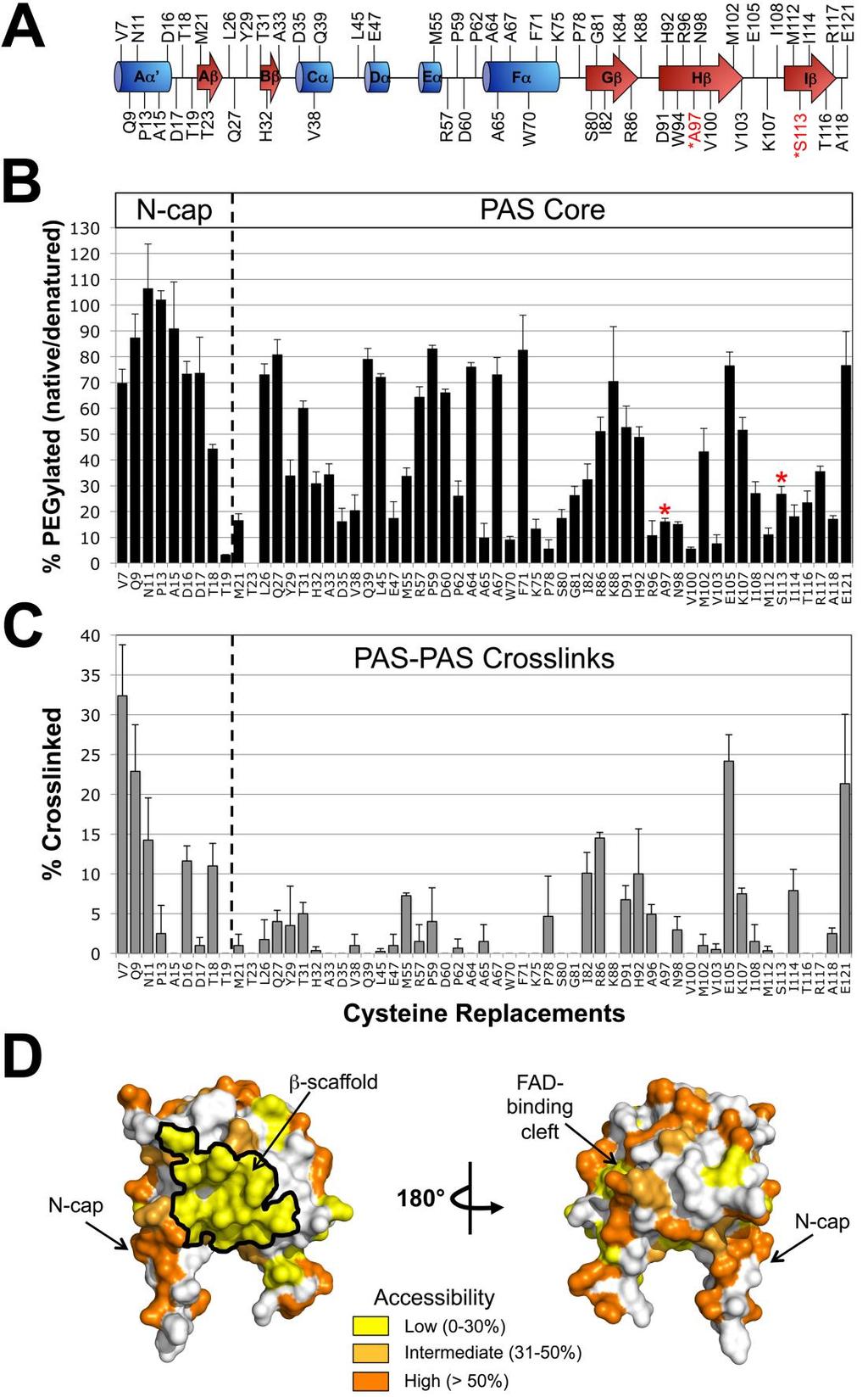
76 58
77 Figure 17. Probing the PAS domain for solvent accessibility and PAS-PAS proximity using PEGylation and disulfide crosslinking. A. Residues selected for cysteine replacement mapped onto the secondary structure of the Aer PAS domain. Residues in black font are predicted to be accessible on the surface of the Aer-PAS homology model; those in red font are predicted to face inwards towards the PAS interior, and were selected as surface-inaccessible controls. B. Extent of PEGylation for substituted cysteines in the Aer PAS core and N- terminal cap (N-cap). Red asterisks identify the surface-inaccessible controls. C. Extent of disulfide crosslinking between neighboring PAS domains. Error bars in B and C represent the standard deviation from multiple experiments. D. Aer-PAS homology model showing the distribution of the tested residues based on whether they had low (yellow shading, 0-30% PEGylation), intermediate (light orange, 31-50% PEGylation) or high (orange, > 50% PEGylation) accessibility. 59
78 PAS-PAS Crosslinking To determine if the area of low accessibility on the PAS β-scaffold was due to PAS-PAS interactions, each of the 59 PAS Cys substitutions that circumscribed the PAS domain was crosslinked in vivo by treating whole cells with 600 μm Cu(II)(1,10-phenanthroline)3 (CuPhe) for 20 min. Crosslinked products were identified by their migration on SDS-PAGE and detected with anti- Aer2-166 antisera. As anticipated, the interior-facing controls, A97C and S113C, did not crosslink (Fig. 17C). Notably, residues that clustered on the inaccessible PAS β-scaffold did not crosslink substantially (<8% dimers). This suggests that PAS-PAS interactions do not contribute to the inaccessible surface. The eight cysteine mutants that formed more than 10% dimers (Fig. 17C) also had high or intermediate accessibility to PEG-mal (Fig. 17B), indicating that some of these PAS-PAS interactions may have been transient, rather than stable. These residues included several in the flexible N-cap (V7C, Q9C, N11C, D16C and T18C), R86C in the β-scaffold, E105C in the PAS H-I loop, and E121C in the F1 loop (Fig. 17C). In the PAS core, the greatest extent of PAS-PAS crosslinking was observed for E105C. Our previous studies predict that only flexible regions of PAS can form PAS-PAS crosslinks within an Aer dimer because the PAS domains are separated by the HAMP four-helix bundle. Non-flexible PAS regions are more likely to contact another dimer within the trimer-of-dimers hexameric structure of Aer (Campbell et al., 2011). To differentiate intra- from inter-dimeric contacts for E105C, the aspartate receptor, Tar, was over-expressed from a 60
79 compatible plasmid to form mixed trimers-of-dimers with Aer (Gosink et al., 2006). In the presence of over-expressed Tar, Aer-E105C crosslinking decreased from 24% to 11%. This suggests that Aer-E105C crosslinking occurs between Aer dimers, a result that was also shown for E121C (Campbell et al., 2011). PAS-HAMP Interactions Defined by Disulfide Crosslinking We previously demonstrated crosslinking between Cys replacements in the PAS and HAMP domains of Aer (Campbell et al., 2010). For those experiments, Q248C was selected as the HAMP Cys probe because Q248C i) is located on the sequestered face of AS-2 (Figs. 16A and B), ii) is significantly more accessible in the absence of the PAS domain (Fig. 16C), and iii) does not significantly crosslink with itself within or between dimers [ 1% dimers, (Watts et al., 2008)]. Those initial studies demonstrated in vivo PAS-HAMP crosslinking between HAMP-Q248C and PAS-N98C or PAS-I114C (Campbell et al., 2010). The findings were possible because PAS-HAMP crosslinks can be differentiated from PAS-PAS and HAMP-HAMP crosslinks by their migration on SDS-PAGE. The different mobility is based on the N-terminal location of the PAS domain: Aer PAS-PAS crosslinked dimers migrate faster on SDS-PAGE than HAMP-HAMP crosslinked dimers, and PAS-HAMP crosslinked dimers have an intermediate mobility (Fig. 18A). Note that the latter point is only true if the PAS-HAMP crosslink forms between subunits, not within a subunit. An intra-subunit crosslink will migrate as a compact monomer (Bass et al., 2007). In the current study, we 61
80 identify additional PAS-HAMP pairs that can crosslink and use these data to define the PAS-HAMP interaction surface. Q248C was paired with 25 different PAS Cys residues at sites circumscribing the PAS domain (see Experimental Procedures for details). The 25 di-cys Aer constructs supported BT3312 aerotaxis in succinate minimal soft agar, with migration rates within the aerotactic range shown in Fig. S1. The Aer-Cys proteins were oxidized by treating whole cells with CuPhe as described above. Many of the substitutions produced small quantities of PAS-PAS and PAS-HAMP crosslinked dimers. However several residues preferentially produced PAS-HAMP crosslinked dimers: I82C, R96C, N98C, V100C, M112C and I114C on the PAS β-scaffold (Fig. 18A, right panel and Fig. 18B, left panel, gray bars) and V38C, in the N-cap hinge region. All of the PAS-HAMP crosslinks formed between, and not within, monomeric subunits. 62
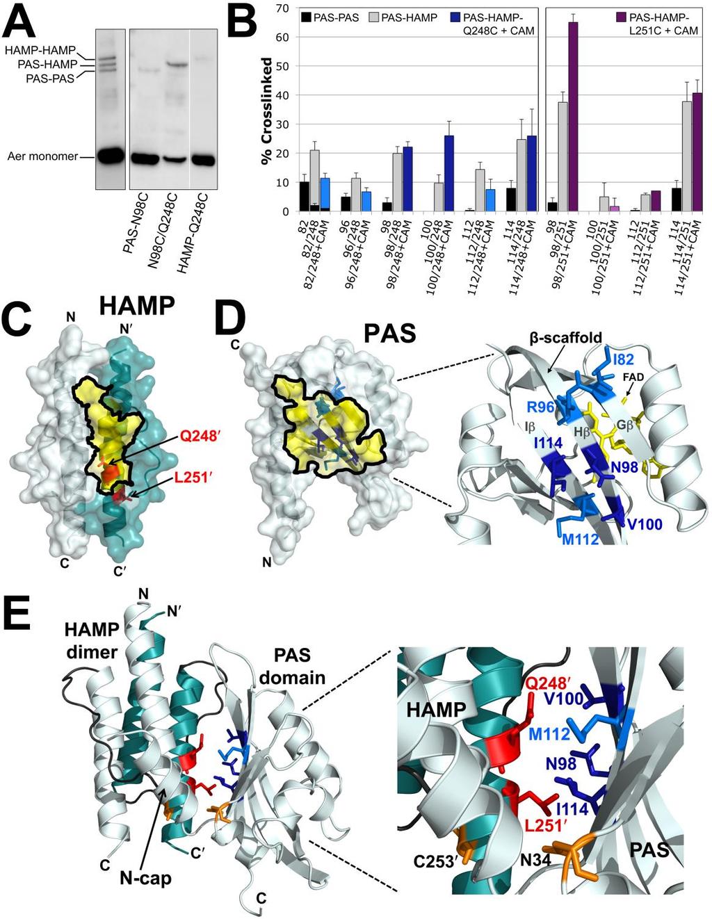
81 63
82 Figure 18. Disulfide crosslinking between the Aer PAS and HAMP domains. A. Representative examples comparing SDS-PAGE mobilities for Aer dimers crosslinked between PAS-PAS, PAS-HAMP and HAMP-HAMP. The left panel illustrates the separation of the three crosslinked bands for a di-cys mutant with similar band densities at each position (Aer-I82C/A245C). The right panel illustrates the separation of these bands for a di-cys mutant with preferential PAS- HAMP crosslinking (Aer-N98C/Q248C). In all cases, uncrosslinked monomer bands are near the bottom of the gels. B. Extent of disulfide formation for PAS residues that preferentially crosslinked with HAMP-Q248C (left panel) and -L251C (right panel). PAS-PAS crosslinking is indicated by black fill, PAS-HAMP crosslinking by gray fill, PAS-Q248C crosslinking after treatment with chloramphenicol in blue fill and PAS-L251C crosslinking after treatment with chloramphenicol in purple fill. Dark blue or dark purple fill indicates that the extent of PAS-HAMP crosslinking did not significantly decrease after chloramphenicol treatment, whereas lighter blue or purple fill indicates a decreased extent of PAS- HAMP crosslinking after chloramphenicol treatment. HAMP-Q248C and -L251C controls routinely produced 1% dimers and are not shown. Error bars represent the standard deviation from multiple experiments. C. HAMP homology model showing the location of HAMP-Q248 and -L251, which crosslinked with PAS, in relation to the region that was sequestered from PEG-mal (Fig. 16B, yellow shading). D. PAS homology model showing the β-scaffold positions of the PAS residues that preferentially crosslinked with HAMP-Q248C in relation to the region that was sequestered from PEG-mal (Fig. 17D, yellow shading). Residue colors match those used in the left panel of B. E. PAS-HAMP dimer model showing the proposed orientation of the PAS and HAMP domains based on PEGylation and PAS-HAMP crosslinking data. For clarity, the model includes the PAS domain from just one subunit (res ) and a HAMP dimer (monomers from both subunits, res ). The PAS model was manually oriented relative to the HAMP AS-2 helix of the cognate monomer to account for the crosslinking (B) and sequestration (C and D) data. Residue colors match those used in B, C and D. The locations of PAS-N34D and HAMP-C253R, which were previously identified as site-specific suppressors (Watts et al., 2004a), are shown in orange. Abbreviation: CAM, chloramphenicol. 64
83 Several of the residues, e.g., I82C, exhibited some PAS-PAS crosslinking, but the extent consistently decreased in the presence of HAMP-Q248C (Fig. 18B). This indicated that the side chains of the PAS residues collided more often with a HAMP domain than with another PAS domain. The PAS residues that preferentially crosslinked with HAMP-Q248C resided almost exclusively within the inaccessible region of the PAS β-scaffold (Fig. 18D). This region is notable because it includes the cluster of kinase-on lesions that we previously identified as components of the signaling pathway [(Campbell et al., 2010), and the red area in Fig. 20]. Aer undergoes a complex maturation process in which proper folding of the Aer PAS domain requires the presence of the HAMP domain (Herrmann et al., 2004). This process is easily destabilized by key mutations in either the PAS or HAMP domains (Buron-Barral et al., 2006b, Campbell et al., 2010). In the current study, we considered the possibility that some PAS-HAMP crosslinking occurred during the folding process before a mature Aer product was formed. To increase the fraction of mature Aer protein before crosslinking, new protein synthesis was inhibited with 500 µg ml -1 chloramphenicol for 15 min before adding CuPhe. After chloramphenicol treatment, three of the di-cys mutants containing Q248C (N98C/Q248C, V100C/Q248C and I114C/Q248C) had an equivalent or increased proportion of PAS-HAMP crosslinked dimers (Fig. 18B, dark blue bars). This indicates that N98, V100 and I114, which are located on the PAS H and I β-strands (Fig. 18D), are each proximal to HAMP AS-2 in the folded protein. To test whether PAS-HAMP disulfide crosslinks formed within or 65
84 between dimers, Aer-I114C/Q248C was expressed with increasing concentrations of Tar. The amount of crosslinked I114C-Q248C product was unaffected by increased Tar expression (not shown), indicating that PAS-HAMP disulfide bonds most likely occurred between cognate monomers of the same dimer. To identify HAMP residues other than Q248C that can crosslink with the PAS β-scaffold, we tested di-cys combinations with several other HAMP residues (see Experimental Procedures for details). Of these, only HAMP-L251C (Fig. 18C) showed preferential PAS-HAMP crosslinking (Fig. 18B, right panel). Like Q248C, L251C has favorable properties in that it does not significantly crosslink with cognate L251C either within or between dimers [ 1% dimers, (Watts et al., 2008)]. In the presence of chloramphenicol, HAMP-L251C preferentially crosslinked with PAS-N98C, -M112C, and -I114C, but not with - V100C (Fig. 18B, purple bars). The extent of crosslinking between HAMP-L251C and either PAS-N98C or PAS-I114C was higher than that between HAMP- Q248C and these PAS residues (Fig. 18B). This indicates that these PAS residues are closer to HAMP-L251C than they are to HAMP-Q248C. In contrast, crosslinking between HAMP-Q248C and either PAS-V100C or PAS-M112C was higher than that between HAMP-L251C and these PAS residues (Fig. 18B). This suggests that these PAS residues are closer to Q248C than they are to L251C. Using this information, homology models of the Aer PAS and HAMP domains were manually manipulated to obtain the best fit (Fig. 18E). To fit the data, the PAS domain was rotated ~180 around the PAS-HAMP interface such that the 66
85 PAS-HAMP interaction surfaces are now flipped relative to our previous PAS- HAMP models [e.g., in (Campbell et al., 2011, Watts et al., 2008)]. The revised PAS-HAMP orientation resolves an unexplained anomaly that was present in previous models. Pseudoreversion analysis previously identified PAS-N34D as an allele-specific suppressor of HAMP-C253R (Watts et al., 2004a). This implies close proximity between N34 and C253, but the previous PAS-HAMP models separated them. In the revised PAS-HAMP model, HAMP-C253 is in close proximity to PAS-N34 (Fig. 18E), resolving the anomaly. Comparison of the Kinase-on and Kinase-off States The solvent accessibility measurements in Figs. 19 and 20 were obtained under aerobic conditions and are expected to represent the kinase-off state of Aer (Repik et al., 2000a). To gain insight into changes that might occur on HAMP surfaces as a result of signaling, we re-measured PEG-mal reactivities for 34 HAMP residues in the presence of the PAS kinase-on lesion N85S [(Campbell et al., 2010), Fig. 19A]. N85 is located on the PAS Gβ strand, and its side chain is predicted to face the interior of the PAS domain, contacting the isoalloxazine ring of FAD [(Campbell et al., 2010), Fig. 14B]. N85 is a possible link from the bound FAD to the β-scaffold, and the N85S substitution results in a kinase-on output. Residues that were significantly more accessible in the presence of N85S formed a patch on the HAMP surface that included both AS-1 and cognate AS-2 residues (Fig. 19B, purple residues). This patch overlapped with AS-2 residues that were sequestered by the PAS domain in the kinase-off state (Fig. 16), 67
86 suggesting that the N85S lesion disrupts the interaction of PAS with HAMP. In contrast to the AS-1 and AS-2 residues with increased accessibility, residues with decreased accessibility in this region were located on a different face from the more accessible patch (Fig. 19B). The decrease in accessibility was greatest at the phase shift, where the PEGylation of D259C decreased from approximately 45% in the unstimulated state to approximately 3% in the presence of PAS-N85S. However, the face on which residues after N258 were located could not be determined because the three-dimensional structure of the phase shift is unknown. Notably, all residues that were significantly more accessible in the kinaseon state preceded the end of the HAMP domain (C253), while residues after the HAMP domain in the proximal signaling domain showed decreased accessibility. The proximal signaling and C-terminal kinase control domains are predicted to form an elongated antiparallel four-helix bundle in Aer (Fig. 14) that is analogous to the adaptation and protein interaction regions of other chemoreceptors (Falke & Piasta, 2014). Crosslinking studies of the proximal signaling domain indicate that this four-helix extends through the phase shift (res ). Our model pairs proximal signaling residue I257 with L505, and ends with H506 (aligned with L256) at the C-terminus of Aer [inferred from (Alexander & Zhulin, 2007), see Fig. 21]. Therefore, it is possible that residues following I257 that are less accessible in the kinase-on state form a more compact four-helix bundle. Conversely, increased accessibility in the HAMP domain in the kinase-on state may be associated with a more dynamic four-helix bundle. 68
. This differs from the prototypical membrane-bound chemoreceptor, in which HAMP is directly tethered through the membrane to a periplasmic sensor domain (Parkinson, 2010).")
87 Discussion The Aer PAS-HAMP Interaction Surface In this study, we investigated an unusual signaling mechanism in which a cytosolic PAS domain controls signaling by direct lateral interaction with a HAMP domain (Fig. 21). This differs from the prototypical membrane-bound chemoreceptor, in which HAMP is directly tethered through the membrane to a periplasmic sensor domain (Parkinson, 2010). Figure 19. Influence of the PAS kinase-on lesion, N85S, on the accessibility of residues in the HAMP and proximal signaling domains to PEG-mal. A. Histogram showing the average percent change in PEGylation for 34 Cys substitutions in Aer-N85S. Bars projecting to the right of the origin denote residues that became more accessible in the presence of N85S (significant increases are colored purple; p <0.05). Bars projecting to the left denote residues that became less accessible (significant decreases are colored red; p <0.05). Residues in gray had statistically insignificant changes. B. Location of residues from A that showed significant changes in accessibility (in the kinase-on state) when mapped onto models of the Aer-HAMP domain and part of the proximal signaling domain. The models include residues and Residues were omitted because of the phase shift in this region. Residues are modeled onto a 3.5 residue per turn coiled coil α-helix; the precise orientation of this region relative to HAMP is unknown. 69
88 Fiure 20. PAS space-filled model overlaying a region of previously determined kinase-on lesions [(Campbell et al., 2010), within the red line] with residues shown in the current study to be sequestered (dotted yellow line), or that preferentially crosslinked with the HAMP domain (blue). 70

89 Figure 21. Working model of Aer showing the relationship between PAS and HAMP in the kinase-off and kinase-on states, based on current and previous data. Colors match those of Fig. 18. The reduction of PAS-bound FAD elicits a conformational change that decreases interaction between the PAS domain and the HAMP domain. The relative orientation between the N-cap and PAS core also changes, perhaps altering the stability of PAS-HAMP interactions. Decreased PAS-HAMP interactions are accompanied by tilting of the HAMP helices and a more relaxed, dynamic HAMP structure. The HAMP, proximal signaling domain and protein interaction regions are in opposition across contiguous boundaries such that a relaxed HAMP domain results in a static proximal signaling domain and a dynamic protein interaction region. Relevant residues: N34D, C253R, allelespecific suppressors; , phase shift; pairs 262/500 and 256/506 are highlighted to show their relative latitudes on descending and ascending helices. See Results and Discussion for details. 71
90 In Aer, the measured solvent accessibilities of the PAS, HAMP and proximal signaling domains (Figs. 16 and 17) defined potential PAS-HAMP interaction surfaces. Data from PAS surface residues showed a hidden region on the PAS β-scaffold (1,370 Å 2, Fig. 17D) that was consistent with a HAMP footprint rather than the footprint of a cognate PAS domain. Specifically, none of the sequestered residues crosslinked substantially with another PAS domain (Fig. 17C) and any such interactions may be transient rather than stable. Notably, the hidden region overlapped both with a cluster of PAS kinase-on lesions that we previously identified as part of the aerotaxis signaling pathway (Campbell et al., 2010), and with PAS residues in this study that crosslinked with the HAMP domain (Fig. 17). Together, the data strongly support the hypothesis that the inaccessible PAS region forms the PAS component of the PAS-HAMP interface. Of the 70 HAMP and proximal signaling domain replacements tested, accessibilities inversely correlated with disulfide crosslinking in two regions: HAMP AS-1 and the proximal signaling domain region that followed the phaseshift. In contrast, most of HAMP AS-2 and the N-terminal proximal signaling domain did not inversely correlate with crosslinking. Notably, 10 of 11 AS-2 residues residing on the HAMP exterior had low accessibility to PEG-mal (Fig. 16). Four of these were likely shielded by the HAMP connector (Fig. 16B), but five of the remaining residues were sequestered by the PAS domain (Fig. 16B, boxed residues shaded yellow) and are proposed to form the HAMP component of the PAS-HAMP interface (Fig. 21). In support of this, HAMP AS-2 substitutions 72
91 Q248C and L251C crosslinked with residues on the cognate PAS β-scaffold (Fig. 18). A best fit of the crosslinking data required that the PAS domain be rotated ~180 relative to a previous model (Campbell et al., 2011, Watts et al., 2008). The location of allele-specific suppressors PAS-N34D and HAMP-C253R are now shown in close proximity (Fig. 18E) and in harmony with genetic studies (Watts et al., 2004a). PAS-HAMP crosslinking occurred between cognate subunits within dimers rather than between adjacent dimers because crosslinking was unaffected when collisions between Aer dimers were decreased by the presence of a second receptor (Tar). This is consistent with previous work indicating that HAMP dimers are centrally positioned, flanked by two PAS monomers (Campbell et al., 2011), and are required for PAS folding and FAD binding (Herrmann et al., 2004, Bibikov et al., 2000, Ma et al., 2005, Buron-Barral et al., 2006b). Interestingly, an arrangement similar to the Aer PAS β-scaffold- HAMP AS-2 α-helix interface (Fig. 18E) has been described for several proteins including the periplasmic sensing domain of the Sinorhizobium meliloti C4- dicarboxylate sensor DctB (Zhou et al., 2008) and the Vibrio harveyi quorum sensor LuxQ (Neiditch et al., 2006), where in both cases the β-scaffold of two PAS domains abuts a long α-helical spine. Changes in PAS N-cap Orientation During Signaling The PAS N-cap and the loop connecting the N-cap to the PAS core were highly accessible (Fig. 17) yet appear to be part of the Aer signaling pathway. Kinaseon lesions that define the Aer signaling pathway cluster not only on the PAS β- 73
92 scaffold and in the FAD cleft, but also in the N-cap (Campbell et al., 2010, Watts et al., 2006b). Truncating the first six N-cap residues bestows a kinase-on phenotype, while deleting the first 14 residues yields an inverted response phenotype (Watts et al., 2006b). Aer-L14 is significant in that it likely hydrogen bonds to N34, forming an unusual extended helix that is unbroken in structure, but discontinuous in sequence (Etzkorn et al., 2008, Key et al., 2007, Campbell et al., 2010, Watts et al., 2006b). In DcuS-PASc, an N248D substitution that is equivalent to Aer-N34D reorients the N-cap and activates the protein (Etzkorn et al., 2008). Notably, an N34D substitution in Aer is both kinase-on and an allelespecific suppressor of HAMP-C253R (Watts et al., 2004a). The crosslinking data from the current study indicate that N34 and C253 are in close proximity (Fig. 18E), and by homology to DcuS-PASc, the N-cap may reorient when PAS is in the kinase-on state (Fig. 21). In this scenario, N-cap reorientation would help destabilize PAS-HAMP interactions (Fig. 21), giving the HAMP domain more degrees of freedom and dynamic movement. Changes in HAMP Conformation and PAS-HAMP Interactions During Signaling We previously found that the kinase-on substitution, PAS-N85S, altered rates of crosslinking between HAMP AS-1 and AS-2 helices (Watts et al., 2011a). Rates decreased at the proximal end of HAMP, and increased at the distal end of HAMP. This was interpreted as either i) tilting of AS-2 with respect to AS-1, or ii) a more relaxed HAMP structure [Fig. 21, (Watts et al., 2011a)]. 74
93 The data from the current study reveal changes in PAS-HAMP interactions that are correlated with these states: the kinase-on substitution (PAS-N85S) exposed a patch of HAMP residues that had been hidden in the kinase-off state by the PAS domain (Fig. 19). This is consistent with a decrease in the strength of the PAS-HAMP AS-2 interaction in the kinase-on state, and a less compact, more dynamic conformation of HAMP. The dynamic conformation is likely to be favored by Aer-HAMP because it lacks strong hydrophobic residues at several key packing positions in the HAMP bundle (Watts et al., 2008, Parkinson, 2010). Still, some HAMP residues apparently remained in contact with PAS in the kinase-on state, because residues in the exposed patch remained less solvent accessible than they were in the PAS-less mutant (compare res. 241, 244 and 245 in Figs. 16C and 19A). However, PAS is effectively tethered to HAMP in the Aer dimer, so some collisional contacts are also expected. The impact of PAS-N85S on AS-2 accessibility was not uniform. In contrast to AS-2 residues that exhibited increased accessibility with N85S, a patch of residues at the distal end of AS-2 through the proximal signaling domain had decreased accessibility (Fig. 19). The proximal signaling domain links HAMP AS-2 to the kinase control domain and has a helical phase shift between residues 259 and 262; the shift is five residues downstream of the phase stutter at the HAMP-output junction (Brown et al., 1996, Parkinson, 2010). In Aer, the residues with decreased accessibility included not only residues throughout the proximal signaling domain toward the kinase control junction (N269), but also residues upstream, extending to the HAMP junction (C253) (Fig. 19). The 75
94 proximal signaling domain is also functionally distinguishable from the HAMP domain: missense mutations in the proximal signaling domain do not influence PAS-FAD binding (Ma et al., 2005, Buron-Barral et al., 2006b), nor are they suppressed by PAS suppressor lesions that rescue HAMP defects (Watts et al., 2004a). This suggests that the proximal signaling domain, unlike HAMP, does not associate with the PAS domain. The data from the current study are consistent with the dynamic bundle model (Parkinson, 2010, Zhou et al., 2009), although they are not definitive. The broad changes in accessibility associated with signaling (Fig. 19) are more easily explained by dynamics [regulated unfolding, (Schultz & Natarajan, 2013)] than by conformational changes in semi-rigid structures that mimic a specific pattern of changes propagated by the transmembrane helix of a chemoreceptor. In Aer, signaling caused opposite accessibility changes in the HAMP and proximal signaling domains (Figs. 19 and 21). This suggests an inverse structural relationship between these domains, such that when one is compact the other is dynamic. Inverted shifting between compact and dynamic structures has been proposed for the other E. coli chemoreceptors, whereby three contiguous segments have opposite structural transitions: the HAMP domain, the adaptation region and the protein interaction region of the kinase control module (Falke & Piasta, 2014, Parkinson, 2010). The first inversion is associated with a helical phase shift at the juncture between HAMP AS-2 and the adaptation region. This shift couples the helical bundles in opposition, so that increasing the packing stability of one bundle decreases the stability of the other. The second inversion 76
95 may occur at a glycine hinge that is equivalent to Aer residues G330, G331 and G429. The behavioral output for Aer indicates that the structural state of the protein interaction region is similar to the state of HAMP, as it is in other chemoreceptors (Fig. 21B). Switching to a more static HAMP domain would reverse the states of the other two regions, so the HAMP and protein interaction regions continue to share the same conformational profile [(Falke & Piasta, 2014), Fig. 21]. Recent studies on the Tsr receptor suggest that the HAMP domain acts as a brake that inhibits the default kinase-on state of the receptor. Thus, the HAMPindependent output state of the Tsr kinase control module is kinase-on, and HAMP must actively override this state in response to attractant stimuli (Ames et al., 2014). Given the homology among the Tsr, Tar, and Aer kinase-control modules, the default output for Aer is also likely to be kinase-on, although similar definitive analyses of Aer-HAMP deletions would be confounded by the requirement of HAMP for PAS maturation [(Herrmann et al., 2004, Buron-Barral et al., 2006b), J. S. Parkinson, personal communication]. Aer Signaling Model From the present and previous studies, we propose a complete pathway for Aer-mediated aerotaxis. When E. coli swims into a region where the ambient oxygen concentration cannot maintain the electron transport system, FAD bound to the Aer-PAS domain is reduced and protonated. A hydrogen-bond network linked to FAD is reorganized, resulting in a conformational change in the PAS β- 77
96 scaffold at the PAS-HAMP AS-2 interface [(Campbell et al., 2010); see also (Campbell et al., 2010, Key et al., 2007, Ukaegbu & Rosenzweig, 2009b)]. Altered PAS-HAMP interactions switch a patch of HAMP surface residues from low accessibility to high accessibility, consistent with decreased affinity between the PAS and HAMP domains. We propose that interactions between oxidized PAS and HAMP AS-2 promote a more ordered (static) HAMP structure and an active kinase-off output, whereas weaker PAS-HAMP interactions in the reduced state allow a more dynamic HAMP structure and kinase-on output (Fig. 21). Aer- HAMP controls the proximal signaling domain, and in turn, the kinase control domain. The tip of the kinase control domain is the protein interaction region, which shares the same relative state as HAMP (dynamic or static) [reviewed by (Falke & Piasta, 2014)]. After the bacteria approach the hypoxic region and FAD is reduced, the cells tumble briefly and swim in a different direction. This avoids anaerobiosis and ensures that cells move towards a higher oxygen concentration. It is the movement up the oxygen gradient that is dominant in determining net migration. In an aerobic environment, FAD becomes oxidized and PAS-HAMP interactions strengthen, resulting in a static HAMP domain and a kinase-off output. The static HAMP domain shifts the bias of the protein interaction region, which inhibits CheA kinase and suppresses changes in swimming direction. The model for direct signaling between the PAS and HAMP domains of Aer presents a new paradigm for controlling HAMP states. The paradigm suggests that signal-sensitive, intra-dimeric contacts between PAS and HAMP 78
97 AS-2 controls the static or dynamic nature of HAMP. This mechanism likely occurs in other proteins that have laterally interacting PAS and HAMP domains, and may be the mechanism by which other sensing domains can control HAMP activity in a wide variety of systems. 79
98 SUPPLEMENTARY INFORMATION DELINEATING PAS-HAMP INTERACTION SURFACES AND SIGNALING- ASSOCIATED CHANGES IN THE AEROTAXIS RECEPTOR AER Darysbel Garcia 1, Kylie J. Watts 1, Mark S. Johnson, and Barry L. Taylor* Division of Microbiology and Molecular Genetics, School of Medicine, Loma Linda University. Loma Linda, CA USA. *Corresponding author. 1 These two authors contributed equally to this investigation. 80
].")
99 Figure S1. Influence of PAS-Cys substitutions on Aer-mediated behavior in E. coli BT3312 (aer tsr). Aer-Cys mutants were tested in succinate minimal soft agar containing 50 μg. ml -1 ampicillin. Colony expansion was determined after hours of growth at 30 C and compared with wild-type Aer [as expressed from pgh1, (Rebbapragada et al., 1997a)]. Mutants with average colony diameters greater than 130% of WT Aer were designated as superswarmers. Mutants with average colony diameters less than 40% of WT have impaired behavior or are nonaerotactic (Watts et al., 2008). Only cells expressing Aer-W94C fell in this category and were non-aerotactic in minimal soft agar. In a temporal aerotaxis assay (Taylor et al., 2007), BT3312 cells expressing Aer-W94C were tumbly-biased in air, had a delayed response to nitrogen, and a showed a brief smooth-swimming response to oxygen. Experimental Procedures Bacterial Strains and Plasmids Cysteine-less (C-less) Aer (Aer-C193S/C203A/C253A) was expressed from pmb1 (Ma et al., 2004, Watts et al., 2006b), a ptrc99a-derivative that expresses Aer under the control of an IPTG-inducible ptrc promoter. All Aer-Cys 81
100 mutants in this study were derived from pmb1. The WT Tar expression plasmid, plc113, was a gift from John S. Parkinson and is a pacyc184-based plasmid that confers chloramphenicol resistance and carries a sodium salicylate-inducible promoter (Ames & Parkinson, 2006). Plasmids were expressed in E. coli BT3312, a strain that lacks the two aerotaxis receptors, Aer and Tsr [Δaer-1 Δtsr- 7021, (Repik et al., 2000] or in chemoreceptor-less BT3388 [aer::erm tsr-7021 tar-tap-5201 trg::tn10) (Yu et al., 2002)]. Mutant Construction A library of Aer mutants with single cysteine substitutions between residues 206 and 275 was previously constructed in pmb1 (Watts et al., 2008, Amin et al., 2006). Aer-N85S and additional Aer-Cys mutants were constructed by site-directed mutagenesis of pmb1 or pmb1-derived plasmids according to the instructions of the QuikChange II site-directed mutagenesis kit (Agilent Technologies, Santa Clara, CA). The N85S codon was introduced into plasmids that contained a single Cys codon substitution, whereas di-cys mutants were created from a pmb1-derivative that expressed either Aer-Q248C or Aer-L251C. Aer[ ] mutants were created by amplifying the coding region for residues from individual pmb1-derived plasmids using primers containing recognition sequences for AflIII and SalI, and ligating the products into ptrc99a with the NcoI-SalI DNA segment removed. Mutations were confirmed by sequencing the entire aer gene of each plasmid. 82
101 Expression and Aerotaxis Assays Plasmids were introduced into BT3312 (aer tsr) and Aer expression was confirmed by Western blotting with a 1 in 133,000 dilution of anti-aer2-166 antisera (Repik et al., 2000a). Aer[ ] mutants express stable protein (K.K. Gosink and J.S. Parkinson, personal communication), but have fewer epitopes than fulllength Aer, so were detected with a 1 in 50,000 dilution of anti-aer2-166 antisera. For Aer/Tar co-expression assays, Aer and Tar expression plasmids were introduced into BT3388 (aer tsr tar trg tap) and expressed as described (Campbell et al., 2011). Tar expression was confirmed using a 1 in 10,000 dilution of anti-tsr antisera [a gift from J. S. Parkinson, (Ames & Parkinson, 1994)]. Aerotaxis phenotypes were determined for each Aer mutant by inoculating cells into succinate minimal soft agar containing 50 μg ml -1 ampicillin, incubating the plates at 30 C for hours, and then observing colony morphologies (Taylor et al., 2007). In vivo Accessibility Assays using PEG-mal In vivo PEGylation assays were performed using an unpublished permeabilized cell protocol developed by Claudia A. Studdert at the Universidad Nacional de Mar del Plata, Argentina, but modified in this study to work optimally for Aer. BT3312 cells expressing each of the Aer-Cys mutants were grown at 30 C to mid-log phase in tryptone broth containing 100 μg ml -1 ampicillin, and induced for 3 hours with 50 μm IPTG. Aer-P211C, Aer-R235C and Aer-G240C have lower steady-state accumulation levels (Watts et al., 2008), as did Aer- 83
102 R57C, Aer-D60C and Aer-A97C (this study) and were induced with 100 μm IPTG. Two milliliters of each culture was centrifuged at 10,000 x g in each of two tubes (corresponding to denatured and native samples), then washed twice with 20 mm potassium phosphate [ph 7.0], 0.1 mm EDTA, and 0.1 mm MgCl2 buffer. The cells in each tube were resuspended in 1 ml of wash buffer and permeabilized by adding 50 μl of 1:4 toluene:ethanol. After 15 min of vigorous mixing at room temperature on an Eppendorf mixer (Eppendorf model 5432, Hauppauge, NY), the cells were centrifuged, the supernatant and excess fluid were removed, and the pellets were resuspended in 50 μl of wash buffer. Five mm PEG-mal (Laysan Bio, Arab, AL) was thoroughly mixed with the cells, and the tubes were incubated at 25 C for 15 min. PEGylation reactions were stopped by adding 100 μl of sample buffer with excess β-mercaptoethanol (native samples, 1.43 M β-mercaptoethanol) or with no reducing agent (denatured samples). The samples were then boiled for four min and analyzed by SDS- PAGE. A quenching control, in which 1.43 M β-mercaptoethanol was added before PEG-mal, efficiently quenched the PEGylation reaction (see Fig. 2). Bands were visualized on Western blots and quantified on a BioSpectrum digital imager (UVP, Upland, CA). For each residue, the proportion of PEGylated product was calculated by dividing the average density of the PEGylated band by the average densities of the non-pegylated plus PEGylated fractions from duplicate lanes. Reactions were repeated on at least two, but usually three or more, occasions. To compare extents of PEGylation in the presence and absence of the PAS domain or PAS-N85S, statistical analyses were carried out 84
103 using a two-tailed Student s t-test. A p value of less than 0.05 was considered statistically significant. In vivo Disulfide Crosslinking BT3312 cells expressing each of the di-cys and corresponding single-cys mutants were grown to mid-log phase in H1 minimal salts medium supplemented with 30 mm succinate, 0.1% (w/v) casamino acids and 100 μg ml -1 ampicillin, before being induced for 3 hours with 50 μm IPTG. Crosslinking was performed at 25 C by exposing whole cells to 600 μm Cu(II)(1,10-phenanthroline)3 (CuPhe) for 20 min, similar to that described previously (Amin et al., 2006, Hughson & Hazelbauer, 1996, Watts et al., 2008), but with modifications as described in (Lai & Hazelbauer, 2007, Taylor et al., 2007). The following 25 PAS-Cys mutants were tested for crosslinking with Q248C in di-cys receptors: T19C, M21C, T23C, H32C, N34C, D35C, T36C, V38C, L45C, M55C, K75C, P78C, S80C, I82C, K88C, N89C, R96C, N98C, V100C, V103C, I108C, M112C, I114C, A118C and E121C. PAS-Cys subsets were also tested for their ability to crosslink with R244C, G250C, M252C, C253, R254C and D259C. Aer-I114C/Q248C and Aer- E105C were also analyzed in BT3388, with Aer as the sole receptor, or in the presence of WT Tar (as expressed from plc113) with no induction or with 1.2 μm sodium salicylate induction. Crosslinked products were separated from monomers by SDS-PAGE and quantified on the UVP digital imager after Western blotting. Percent crosslinking was calculated by dividing the intensity of the crosslinked dimer band by the sum of the intensities of the monomer and dimer 85
104 bands, multiplied by 100. Aer-V260C (Watts et al., 2008) and C-less Aer (Ma et al., 2004) were used as positive and negative crosslinking controls, respectively. Dimer bands were never evident with C-less Aer, whereas the extent of dimerization for Aer-V260C was routinely 65% after 20 min. Reactions were repeated on two or more occasions. In silico Modeling Aer PAS and HAMP domain models were previously created from the coordinates of NifL PAS (2GJ3) and Af1503 HAMP (2ASX), respectively (Campbell et al., 2010, Watts et al., 2008). PyMOL viewer (The PyMOL Molecular Graphics System, Version 1.0, Schrödinger, LLC) was used to manipulate models, map experimental data and determine surface areas. The experimental results were used as a guide to manually manipulate the positions of the PAS and HAMP domains in an Aer dimer to obtain the best fit (Figs. 5 and 8). Acknowledgements We would like to thank John S. Parkinson for helpful suggestions and for providing a Tar expression plasmid and anti-tsr antisera, Claudia A. Studdert for helpful suggestions and for communicating an unpublished PEGylation protocol, Lauren A. Abraham for technical assistance, and Jennifer Ngo for constructing and testing several PAS-Cys mutants. This work was supported by the National Institute of General Medical Sciences of the National Institutes of Health under 86
105 Award Numbers RO1 GM to B. L. Taylor and 2 R25 GM (for support of D.G.). The content is solely the responsibility of the authors and does not represent the official views of the National Institutes of Health. 87
106 References Airola, M.V., N. Sukomon, D. Samanta, P.P. Borbat, J.H. Freed, K.J. Watts and B.R. Crane, (2013) HAMP domain conformers that propagate opposite signals in bacterial chemoreceptors. PLoS Biol 11: e Airola, M.V., K.J. Watts, A.M. Bilwes and B.R. Crane, (2010) Structure of concatenated HAMP domains provides a mechanism for signal transduction. Structure 18: Alexander, R.P. and I.B. Zhulin, (2007) Evolutionary genomics reveals conserved structural determinants of signaling and adaptation in microbial chemoreceptors. Proc Natl Acad Sci USA 104: Ames, P. and J.S. Parkinson, (1994) Constitutively signaling fragments of Tsr, the Escherichia coli serine chemoreceptor. J Bacteriol 176: Ames, P. and J.S. Parkinson, (2006) Conformational suppression of interreceptor signaling defects. Proc Natl Acad Sci USA 103: Ames, P., Q. Zhou and J.S. Parkinson, (2014) HAMP domain structural determinants for signalling and sensory adaptation in Tsr, the Escherichia coli serine chemoreceptor. Mol Microbiol 91: Amin, D.N., B.L. Taylor and M.S. Johnson, (2006) Topology and boundaries of the aerotaxis receptor Aer in the membrane of Escherichia coli. J Bacteriol 188: Aravind, L. and C.P. Ponting, (1999) The cytoplasmic helical linker domain of receptor histidine kinase and methyl-accepting proteins is common to many prokaryotic signalling proteins. FEMS Microbiol Lett 176: Bass, R.B., S.L. Butler, S.A. Chervitz, S.L. Gloor and J.J. Falke, (2007) Use of site-directed cysteine and disulfide chemistry to probe protein structure and dynamics: applications to soluble and transmembrane receptors of bacterial chemotaxis. Methods Enzymol 423: Bibikov, S.I., L.A. Barnes, Y. Gitin and J.S. Parkinson, (2000) Domain organization and flavin adenine dinucleotide-binding determinants in the aerotaxis signal transducer Aer of Escherichia coli. Proc Natl Acad Sci USA 97: Bibikov, S.I., R. Biran, K.E. Rudd and J.S. Parkinson, (1997) A signal transducer for aerotaxis in Escherichia coli. J Bacteriol 179:
107 Bibikov, S.I., A.C. Miller, K.K. Gosink and J.S. Parkinson, (2004) Methylationindependent aerotaxis mediated by the Escherichia coli Aer protein. J Bacteriol 186: Brown, J.H., C. Cohen and D.A. Parry, (1996) Heptad breaks in alpha-helical coiled coils: stutters and stammers. Proteins 26: Buron-Barral, M.D.C., K.K. Gosink and J.S. Parkinson, (2006) Loss- and gain-offunction mutations in the F1-HAMP region of the Escherichia coli aerotaxis transducer Aer. J Bacteriol 188: Butler, S.L. and J.J. Falke, (1998) Cysteine and disulfide scanning reveals two amphiphilic helices in the linker region of the aspartate chemoreceptor. Biochemistry 37: Campbell, A.J., K.J. Watts, M.S. Johnson and B.L. Taylor, (2010) Gain-offunction mutations cluster in distinct regions associated with the signalling pathway in the PAS domain of the aerotaxis receptor, Aer. Mol Microbiol 77: Campbell, A.J., K.J. Watts, M.S. Johnson and B.L. Taylor, (2011) Role of the F1 region in the Escherichia coli aerotaxis receptor Aer. J Bacteriol 193: Dunin-Horkawicz, S. and A.N. Lupas, (2010) Comprehensive analysis of HAMP domains: Implications for transmembrane signal transduction. J Mol Biol 397: Etzkorn, M., H. Kneuper, P. Dunnwald, V. Vijayan, J. Kramer, C. Griesinger, S. Becker, G. Unden and M. Baldus, (2008) Plasticity of the PAS domain and a potential role for signal transduction in the histidine kinase DcuS. Nat Struct Mol Biol 15: Falke, J.J. and K.N. Piasta, (2014) Architecture and signal transduction mechanism of the bacterial chemosensory array: Progress, controversies, and challenges. Curr Opin Struct Biol 29C: Ferris, H.U., S. Dunin-Horkawicz, L.G. Mondejar, M. Hulko, K. Hantke, J. Martin, J.E. Schultz, K. Zeth, A.N. Lupas and M. Coles, (2011) The mechanisms of HAMP-mediated signaling in transmembrane receptors. Structure 19: Gosink, K.K., M. del Carmen Buron-Barral and J.S. Parkinson, (2006) Signaling interactions between the aerotaxis transducer Aer and heterologous chemoreceptors in Escherichia coli. J Bacteriol 188:
108 Hazelbauer, G.L. and W.C. Lai, (2010) Bacterial chemoreceptors: providing enhanced features to two-component signaling. Curr Opin Microbiol 13: Herrmann, S., Q. Ma, M.S. Johnson, A.V. Repik and B.L. Taylor, (2004) PAS domain of the Aer redox sensor requires C-terminal residues for nativefold formation and flavin adenine dinucleotide binding. J Bacteriol 186: Hughson, A.G. and G.L. Hazelbauer, (1996) Detecting the conformational change of transmembrane signaling in a bacterial chemoreceptor by measuring effects on disulfide cross-linking in vivo. Proc Natl Acad Sci USA 93: Hulko, M., F. Berndt, M. Gruber, J.U. Linder, V. Truffault, A. Schultz, J. Martin, J.E. Schultz, A.N. Lupas and M. Coles, (2006) The HAMP domain structure implies helix rotation in transmembrane signaling. Cell 126: Key, J., M. Hefti, E.B. Purcell and K. Moffat, (2007) Structure of the redox sensor domain of Azotobacter vinelandii NifL at atomic resolution: signaling, dimerization, and mechanism. Biochemistry 46: Klose, D., N. Voskoboynikova, I. Orban-Glass, C. Rickert, M. Engelhard, J.P. Klare and H.J. Steinhoff, (2014) Light-induced switching of HAMP domain conformation and dynamics revealed by time-resolved EPR spectroscopy. FEBS Lett 588: Krell, T., J. Lacal, F. Munoz-Martinez, J.A. Reyes-Darias, B.H. Cadirci, C. Garcia- Fontana and J.L. Ramos, (2011) Diversity at its best: bacterial taxis. Environ Microbiol 13: Lai, R.Z. and J.S. Parkinson, (2014) Functional suppression of HAMP domain signaling defects in the E. coli serine chemoreceptor. J Mol Biol 426: Lai, W.C. and G.L. Hazelbauer, (2007) Analyzing transmembrane chemoreceptors using in vivo disulfide formation between introduced cysteines. Methods Enzymol 423: Lu, J. and C. Deutsch, (2001) Pegylation: a method for assessing topological accessibilities in Kv1.3. Biochemistry 40: Ma, Q., M.S. Johnson and B.L. Taylor, (2005) Genetic analysis of the HAMP domain of the Aer aerotaxis sensor localizes flavin adenine dinucleotidebinding determinants to the AS-2 helix. J Bacteriol 187:
109 Ma, Q., F. Roy, S. Herrmann, B.L. Taylor and M.S. Johnson, (2004) The Aer protein of Escherichia coli forms a homodimer independent of the signaling domain and FAD binding. J Bacteriol 186: Mechaly, A.E., N. Sassoon, J.M. Betton and P.M. Alzari, (2014) Segmental helical motions and dynamical asymmetry modulate histidine kinase autophosphorylation. PLoS Biol 12: e Mondejar, L.G., A. Lupas, A. Schultz and J.E. Schultz, (2012) HAMP domainmediated signal transduction probed with a Mycobacterial adenylyl cyclase as a reporter. J Biol Chem 287: Nambu, J.R., J.O. Lewis, K.A. Wharton, Jr. and S.T. Crews, (1991) The Drosophila single-minded gene encodes a helix-loop-helix protein that acts as a master regulator of CNS midline development. Cell 67: Neiditch, M.B., M.J. Federle, A.J. Pompeani, R.C. Kelly, D.L. Swem, P.D. Jeffrey, B.L. Bassler and F.M. Hughson, (2006) Ligand-induced asymmetry in histidine sensor kinase complex regulates quorum sensing. Cell 126: Parkinson, J.S., (2010) Signaling mechanisms of HAMP domains in chemoreceptors and sensor kinases. Annu Rev Microbiol 64: Parkinson, J.S., G.L. Hazelbauer and J.J. Falke, (2015) Signaling and sensory adaptation in Escherichia coli chemoreceptors: 2015 update. Trends Microbiol 23: Rebbapragada, A., M.S. Johnson, G.P. Harding, A.J. Zuccarelli, H.M. Fletcher, I.B. Zhulin and B.L. Taylor, (1997) The Aer protein and the serine chemoreceptor Tsr independently sense intracellular energy levels and transduce oxygen, redox, and energy signals for Escherichia coli behavior. Proc Natl Acad Sci USA 94: Repik, A., A. Rebbapragada, M.S. Johnson, J.O. Haznedar, I.B. Zhulin and B.L. Taylor, (2000) PAS domain residues involved in signal transduction by the Aer redox sensor of Escherichia coli. Mol Microbiol 36: Schultz, J.E. and J. Natarajan, (2013) Regulated unfolding: a basic principle of intraprotein signaling in modular proteins. Trends Biochem Sci 38: Swain, K.E. and J.J. Falke, (2007) Structure of the conserved HAMP domain in an intact, membrane-bound chemoreceptor: a disulfide mapping study. Biochemistry 46:
110 Swain, K.E., M.A. Gonzalez and J.J. Falke, (2009) Engineered socket study of signaling through a four-helix bundle: evidence for a yin-yang mechanism in the kinase control module of the aspartate receptor. Biochemistry 48: Taylor, B.L., (2007) Aer on the inside looking out: paradigm for a PAS-HAMP role in sensing oxygen, redox and energy. Mol Microbiol 65: Taylor, B.L., K.J. Watts and M.S. Johnson, (2007) Oxygen and redox sensing by two-component systems that regulate behavioral responses: behavioral assays and structural studies of Aer using in vivo disulfide cross-linking. Methods Enzymol 422: Taylor, B.L. and I.B. Zhulin, (1999) PAS domains: internal sensors of oxygen, redox potential, and light. Microbiol Mol Biol Rev 63: Ukaegbu, U.E. and A.C. Rosenzweig, (2009) Structure of the redox sensor domain of Methylococcus capsulatus (Bath) MmoS. Biochemistry 48: Wang, C., J. Sang, J. Wang, M. Su, J.S. Downey, Q. Wu, S. Wang, Y. Cai, X. Xu, J. Wu, D.B. Senadheera, D.G. Cvitkovitch, L. Chen, S.D. Goodman and A. Han, (2013) Mechanistic insights revealed by the crystal structure of a histidine kinase with signal transducer and sensor domains. PLoS Biol 11: e Wang, J., J. Sasaki, A.L. Tsai and J.L. Spudich, (2012) HAMP domain signal relay mechanism in a sensory rhodopsin-transducer complex. J Biol Chem 287: Watts, K.J., M.S. Johnson and B.L. Taylor, (2006a) Minimal requirements for oxygen sensing by the aerotaxis receptor Aer. Mol Microbiol 59: Watts, K.J., M.S. Johnson and B.L. Taylor, (2008) Structure-function relationships in the HAMP and proximal signaling domains of the aerotaxis receptor Aer. J Bacteriol 190: Watts, K.J., M.S. Johnson and B.L. Taylor, (2011) Different conformations of the kinase-on and kinase-off signaling states in the Aer HAMP domain. J Bacteriol 193: Watts, K.J., Q. Ma, M.S. Johnson and B.L. Taylor, (2004) Interactions between the PAS and HAMP domains of the Escherichia coli aerotaxis receptor Aer. J Bacteriol 186:
111 Watts, K.J., K. Sommer, S.L. Fry, M.S. Johnson and B.L. Taylor, (2006b) Function of the N-terminal cap of the PAS domain in signaling by the aerotaxis receptor Aer. J Bacteriol 188: Wuichet, K., R.P. Alexander and I.B. Zhulin, (2007) Comparative genomic and protein sequence analyses of a complex system controlling bacterial chemotaxis. Methods Enzymol 422: Yu, H.S., J.H. Saw, S. Hou, R.W. Larsen, K.J. Watts, M.S. Johnson, M.A. Zimmer, G.W. Ordal, B.L. Taylor and M. Alam, (2002) Aerotactic responses in bacteria to photoreleased oxygen. FEMS Microbiol Lett 217: Zhou, Q., P. Ames and J.S. Parkinson, (2009) Mutational analyses of HAMP helices suggest a dynamic bundle model of input-output signalling in chemoreceptors. Mol Microbiol 73: Zhou, Q., P. Ames and J.S. Parkinson, (2011) Biphasic control logic of HAMP domain signalling in the Escherichia coli serine chemoreceptor. Mol Microbiol 80: Zhou, Y.F., B. Nan, J. Nan, Q. Ma, S. Panjikar, Y.H. Liang, Y. Wang and X.D. Su, (2008) C4-dicarboxylates sensing mechanism revealed by the crystal structures of DctB sensor domain. J Mol Biol 383: Zhulin, I.B., (2001) The superfamily of chemotaxis transducers: from physiology to genomics and back. Adv Microb Physiol 45:
112 CHAPTER THREE GAS SENSING AND SIGNALING IN THE PAS-HEME DOMAIN OF THE PSEUDOMONAS AERUGINOSA AER2 RECEPTOR Darysbel Garcia, Emilie Orillard, Mark S. Johnson and Kylie J. Watts # Running title: Signaling in the PAS-Heme Domain of P. aeruginosa Aer2 Division of Microbiology and Molecular Genetics Loma Linda University Loma Linda, CA, 92350, USA # Corresponding author: Telephone: +1 (909) x83394 Fax: +1 (909) kwatts@llu.edu KEY WORDS: Chemoreceptor; PAS domain; signal transduction; Pseudomonas aeruginosa; heme; oxygen 94
113 This manuscript is based on experimental data acquired by Drs. Watts, Orillard, Johnson and myself. The work that I did not produce in this paper include; i) experiments on the proximal coordinating histidine, which were performed by Dr. Watts, ii) a few of the mutants were created by Drs. Watts and Orillard, and iii) determination of heme content using the pyridine hemochrome assay, which was performed by Drs. Johnson and Watts. A number of the experiments were performed by me after I left the lab. This was necessary in order to improve experimental standards deviations, and to acquire more reliable data using a new non-defective anaerobic hood. 95
114 Abstract The Aer2 chemoreceptor from Pseudomonas aeruginosa contains a PAS sensing domain that coordinates b-type heme and signals in response to the binding of O2, CO or NO. PAS-heme structures suggest that Aer2 uniquely coordinates heme via a His residue on a 310 helix (H234 on E ), stabilizes O2 binding via a Trp residue (W283), and signals via both W283 and an adjacent Leu residue (L264). Ligand binding may displace L264 and reorient W283 for hydrogen-bonding to the ligand. Here we clarified the mechanisms by which Aer2-PAS binds heme, regulates ligand-binding, and initiates conformational signaling. H234 coordinated heme, but additional hydrophobic residues in the heme cleft were also critical for stable heme-binding. O2 appeared to be the native Aer2 ligand (Kd of 16 M). With one exception, mutants that bound O2 could signal, whereas many mutants that bound CO could not. W283 stabilized O2-binding, but not CO-binding, and was required for signal initiation; W283 mutants that could not stabilize O2 were rapidly oxidized to Fe(III). W283F was the only Trp mutant that bound O2 with WT affinity. The size and nature of residue 264 was important for gas binding and signaling: L264W blocked O2 binding, L264A and L264G caused O2-mediated oxidation, and L264K formed a hexa-coordinate heme. Our data suggest that when O2 binds to Aer2, L264 may move concomitantly with W283 to initiate the conformational signal. The signal then propagates from the PAS domain to regulate the C-terminal HAMP and kinase control domains, ultimately modulating a cellular response. 96
115 Importance Pseudomonas aeruginosa is a ubiquitous environmental bacterium and opportunistic pathogen that infects multiple body sites including the lungs of cystic fibrosis patients. P. aeruginosa senses and responds to its environment via four chemosensory systems. Three of these systems regulate biofilm formation, twitching motility and chemotaxis. The role of the fourth system, Che2, is unclear but has been implicated in virulence. The Che2 system contains a chemoreceptor called Aer2, which contains a PAS sensing domain that binds heme and senses oxygen. Here we show that Aer2 uses unprecedented mechanisms to bind O2 and initiate signaling. These studies provide both the first functional corroboration of the Aer2-PAS signaling mechanism previously proposed from structure, as well as a signaling model for Aer2-PAS receptors. Introduction Pseudomonas aeruginosa is a common environmental bacterium and a significant cause of opportunistic human disease. It survives in complex environments with the aid of 26 chemoreceptors and four chemosensory systems that collectively sense environmental conditions and modify bacterial behavior. The roles of three of these chemosensory systems are known: one modulates type IV pili production and twitching motility (Pil-Chp system), another controls biofilm formation (Wsp system), and a third regulates flagella-mediated chemotaxis (Che system) (Kato et al., 2008, Sampedro et al., 2014). The role of the fourth chemosensory system, Che2 (PA0173-PA0179), is currently unknown. 97
116 Che2 expresses a complete set of chemosensory proteins (CheY2, CheA2, CheW2, CheR2, CheD and CheB2), including a chemoreceptor (PA0176) called Aer2 (previously called McpB). Aer2 was so named because it, along with classical Aer, was observed to mediate aerotaxis by P. aeruginosa (Hong et al., 2004b). However, we, and others, have not observed Aer2-mediated chemotaxis or aerotaxis in P. aeruginosa (Watts et al., 2011b, Guvener et al., 2006). Moreover, it is now understood that the response regulators (CheY proteins) of Che2-like systems do not bind to the bacterial flagella motor protein, FliM, to modulate swimming behavior [(Biswas et al., 2013, Hyakutake et al., 2005, Dasgupta & Dattagupta, 2008) and Watts et al., unpublished data]. This suggests that the primary role of Che2 is something other than the control of chemotaxis or aerotaxis. Notably, a role for Che2 in virulence has been suggested (Garvis et al., 2009, Schuster et al., 2004). The Che2 chemoreceptor, Aer2, has no membrane-spanning segments. However, during the early stationary phase of P. aeruginosa growth, Che2 proteins form a cluster at the cell pole that is held together solely by Aer2 (Guvener et al., 2006, Schuster et al., 2004). Importantly, Che2 proteins do not co-localize with Che (chemotaxis system) proteins (Guvener et al., 2006). Aer2 has an unusual architecture with a PAS sensory domain sandwiched between three N-terminal and two C-terminal HAMP domains (Fig. 22a). These domains precede a kinase control module that is typical of methyl-accepting chemoreceptors. The kinase control module has four predicted methylation sites (QEEE) and a C-terminal pentapeptide (GWEEF) for binding the adaptation 98
117 enzymes CheR2, CheB2 and CheD [Fig. 22a, (Garcia-Fontana et al., 2014)]. In P. aeruginosa, receptor deamidation/demethylation by CheB2 (and possibly CheD), as well as methylation by CheR2, is expected to fine-tune Aer2-mediated responses. The kinase control module of Aer2 has significant sequence identity with the kinase control modules of the major Escherichia coli chemoreceptors. Thus, Aer2 is able to control the E. coli chemotaxis pathway through direct interactions with the E. coli adapter protein, CheW, and the histidine kinase, CheA (Watts et al., 2011b). When Aer2 is expressed in otherwise chemoreceptor-less E. coli, it mediates repellent tumbling (signal-on) responses to O2, CO and NO (Watts et al., 2011b). Gas-bound Aer2 causes rapid autophosphorylation of bound CheA with subsequent phospho-transfer to CheY. Phospho-CheY in turn binds to the E. coli flagellar switch protein, FliM, causing a directional change in flagellar rotation from counterclockwise to clockwise, resulting in E. coli tumbling. The Aer2 gas response is initiated in the PAS (Per-ARNT-Sim) domain, which itself binds penta-coordinate b-type heme (Watts et al., 2011b). PAS domains are common sensing and signaling domains in nature. They have a broadly conserved structure that consists of a central antiparallel -sheet with five -strands (A, B, G, H, and I ) flanked by several -helices (C, D, E, and F ) (Moglich et al., 2009b). 99
 and a C-terminal pentapeptide (GWEEF) for binding adaptation enzymes.")
118 Figure 22. P. aeruginosa Aer2 and the structure of its PAS domain. (a) Model of an Aer2 dimer showing the PAS domain sandwiched between three N-terminal and two C-terminal HAMP domains. The C-terminal kinase control domain has four predicted methylation sites (QEEE) and a C-terminal pentapeptide (GWEEF) for binding adaptation enzymes. (b) Crystal structure of the Aer2 PAS domain in cartoon form with heme cofactor (shown as red sticks) and bound cyanide (shown as spheres) (PDB: 3VOL, (Sawai et al., 2012)). The Fe-CN bond angle is 137 (Sawai et al., 2012). The side chains of three amino acids relevant to this study, H234, L264 and W283 are shown as sticks. For clarity, the PAS structure is shown rotated 180 degrees around the x-axis compared with the orientation in Fig. 22a. (c) Cyanide-bound heme and a structural overlay showing the locations of the L264 and W283 side chains in both the unliganded (Fe 3+ heme, grey side chains, PDB: 4HI4, (Airola et al., 2013a)) and liganded (Fe 3+ -CN heme, colored side chains (Sawai et al., 2012)) Aer2 PAS domain. The position of the W283 nitrogen, which is predicted to bond with O2, is shown in blue. Abbreviations: res, residue; CN, cyanide. There are currently two structures for the Aer2 PAS domain: one contains cyanide bound to ferric heme [cyanomet, Fe 3+ -CN, PDB: 3VOL, (Sawai et al., 2012), Fig. 22b], and the other contains unliganded ferric heme [Fe 3+, PDB: 4HI4, (Airola et al., 2013a)]. These structures revealed several unusual PAS features, including an extended C /D helix, a short 310 helix called E (that replaces E ), 100
119 heme coordination via a His residue on E, and potential O2 stabilization via the indole group of a Trp residue on I (Fig. 22b). In contrast, other PAS domains with b-type heme, like those in E. coli DOS (EcDOS) or Sinorhizobium meliloti FixL (RmFixL), coordinate heme with a His residue on the F helix, and stabilize O2-binding via an Arg residue on G (Gilles-Gonzalez & Gonzalez, 2005). The two Aer2 PAS structures represent non-physiological heme states (cyanomet and ferric heme), but they do represent structures with and without ligand, and overlaying these two structures highlights several residues that may be important for conformational signaling. In the absence of ligand, the I Trp residue W283 appears to rotate ~90, and an adjacent Leu residue, L264 on Hβ, contracts towards the heme iron center to occupy the position where CN - was bound [Fig. 25c, (Airola et al., 2013a)]. The heme itself appears to shift ~2.0 Å upon ligand binding and the heme pocket adjusts accordingly (Airola et al., 2013a). The Aer2 PAS domain is flanked on either side by poly-hamp units (Fig. 22a). Individual HAMP domains form parallel four-helix bundles that are commonly found in prokaryotic proteins as signal-transducing modules (Dunin- Horkawicz & Lupas, 2010). In Aer2, there appears to be minimal PAS-HAMP interactions and, overall, Aer2 assumes a linear domain arrangement [Fig. 22a, (Airola et al., 2013a)]. This contrasts with the aerotaxis receptor, Aer, where sideon PAS-HAMP interactions allow PAS to control HAMP signaling state through direct interactions (Garcia et al., 2016). For Aer2, several structures have been solved for the N-terminal HAMP domains (Airola et al., 2013c, Airola et al., 2010b). Those structures show that HAMP 1 is separated from HAMP 2-3 by a 101
120 helical extension (Airola et al., 2010b, Airola et al., 2013c). HAMP 1 is also largely dispensable for Aer2 function (Watts et al., 2011b). In contrast, N-terminal HAMP 2-3, and C-terminal HAMP4-5, each form integrated di-hamp units that are indispensable for Aer2 function (Airola et al., 2010b, Watts et al., 2011b). The HAMP 1 and HAMP 2 structures represent signal-on and signal-off states, respectively (Airola et al., 2013c), lending support to the hypothesis that poly- HAMP chains relay signals by inter-converting HAMP signaling states along the HAMP chain. Based on experimental evidence, our current signaling model for Aer2 includes the following features: Aer2 PAS-heme binds O2, generating a conformational signal that is transmitted to the PAS I strand (Airola et al., 2013a, Watts et al., 2011b, Sawai et al., 2012). N-terminal HAMP 2-3 do not transmit signals, but function to stabilize the PAS signaling state by altering their conformations in response to PAS ligand binding (Watts et al., 2011b, Airola et al., 2013c). The PAS conformational signal is transmitted to C-terminal HAMP 4-5, which together function as a unit to inhibit signaling from the kinase control module (Watts et al., 2011b). Therefore, without a PAS ligand, the kinase control module conveys the signal-off state; but in the presence of PAS ligand, HAMP 4-5 no longer inhibits the kinase control module, resulting in a signal-on output and the autophosphorylation of bound CheA2. Without HAMP 4-5, the default state of the isolated kinase control module is signal-on (Watts et al., 2011b). The purpose of the current study is to clarify the mechanisms used by the Aer2 PAS domain to bind heme, regulate ligand binding, and initiate conformational signaling. We 102
121 provide evidence that i) the E His coordinates heme binding, ii) the hydrophobic heme pocket is crucial for stable heme binding, iii) O2 is the native ligand of the Aer2 PAS domain, iv) the unprecedented I Trp stabilizes O2-binding but not CObinding, and plays a pivotal role in signal initiation, and v) the Hβ Leu and other conserved PAS residues are important for heme binding, stable gas binding and signal transduction. Results Aer2 PAS Coordinates Heme with a Uniquely Positioned Histidine Residue Structural studies suggest that the Aer2 PAS domain from P. aeruginosa coordinates b-type heme via a His residue (H234) that resides on a short E helix [Fig. 22b, (Sawai et al., 2012, Airola et al., 2013a)]. In contrast, other PAS domains coordinate b-type heme with a His residue on the PAS F helix (Gilles- Gonzalez & Gonzalez, 2005, Kerby et al., 2008). Notably, E and F His residues are both highly conserved in Aer2-PAS homologs (Figs. 23a and S2). To test the contributions of each histidine to heme binding in P. aeruginosa Aer2, H234A (E His), H239A (F His) and H234A/H239A, were introduced into the PAS peptide, Aer2[ ]. Aer2[ ] is expressed with an N-terminal 6x- His tag and contains all necessary PAS heme-binding components (Watts et al., 2011b). The purified PAS-H234A peptide showed a significant heme-binding defect, whereas PAS-H239A retained wild-type (WT) heme content (Figs. 23b and c). This confirms that the E His is the predominant means of coordinating heme in Aer2. However, 20% of PAS-H234A molecules retained heme, and 103
122 PAS-H234A/H239A exhibited a significant decrease in heme content versus H234A alone (Fig. 23c, 6% heme, p <0.05). The dual His replacement peptide thus has a lower heme affinity, suggesting that H239 might contribute to heme coordination in the absence of H234. To determine the effect of the His substitutions on Aer2 signaling, mutations encoding H234A and H239A were introduced separately into fulllength aer2 in an E. coli expression plasmid. Both Aer2 mutants had steady-state expression levels comparable with WT Aer2 (Fig. 24a). When WT Aer2[1-679] is expressed in chemoreceptorless E. coli BT3388, it directs E. coli to tumble in the presence of O2 because Aer2 signaling activates the E. coli chemotaxis cascade (Watts et al., 2011b). When O2 is replaced with N2, Aer2 no longer signals, and after 5-10 sec, BT3388 cells resume smooth-swimming behavior (~2% of the cells tumble at any time) (Watts et al., 2011b). BT3388 cells expressing Aer2- H239A behaved like cells expressing WT Aer2: cells tumbled in air (20.9% O2) and had smooth-swimming behavior in N2. In contrast, Aer2-H234A orchestrated tumbling in air like WT Aer2, but cells remained tumbling-biased in N2 (~60% of cells tumbled in N2 versus ~2% for WT Aer2). Aer2-H234A therefore has a signal-on bias (Fig. 25a). The partial response may reflect the ability of a small proportion of heme-retaining molecules to respond to O2 changes. 104

123 Figure 23. Heme coordination in the Aer2 PAS domain. (a) Location of the E (H234) and F His (H239) side chains in the cyanomet structure of the Aer2 PAS domain (Sawai et al., 2012). Aer2-PAS structures indicate that the E His coordinates heme (Sawai et al., 2012, Airola et al., 2013a), whereas the F His coordinates heme in other PAS-heme proteins (Gilles-Gonzalez & Gonzalez, 2005, Kerby et al., 2008). Both His residues are highly conserved in Aer2-PAS homologs (see Fig. S2). (b) Purified Aer2 PAS[ ] peptides (imidazole bound, 2.6 to 4 mg ml -1 ) showing less red color in Aer2-H234A and Aer2- H234A/H239A compared with WT Aer2 and Aer2-H239A. (c) Heme content of PAS peptides with E and F His replacements, given as a percentage of WT PAS heme content, corrected for peptide concentration (see Materials and Methods). Abbreviations: CN, cyanide; WT, wild-type. When WT Aer2 is expressed in E. coli, cells respond to both O2 and CO (Watts et al., 2011b). To test for a CO response, E. coli BT3388 cells expressing Aer2 are monitored in CO temporal assays. In these assays, cells are perfused with N2 (to remove O2) until they resume smooth swimming, after which CO is perfused for 10 seconds. If the receptor can respond to CO, cells tumble, and continue to tumble for up to 30 sec after CO has been removed (Watts et al., 2011b). To 105
124 determine if Aer2-H239A can respond to CO, cells expressing Aer2-H239A were perfused with CO under anaerobic conditions. Similar to WT Aer2, cells expressing Aer2-H239A responded to CO by tumbling, and the tumbling persisted for ~30 seconds after CO was removed. However, a CO response could not be determined for Aer2-H234A, because cells expressing Aer2-H234A tumbled too extensively in the absence of O2. PAS Structures Suggest a Possible Signaling Mechanism Two PAS domain structures currently exist for Aer2, one with cyanide bound to ferric heme [cyanomet, Fe 3+ -CN (Sawai et al., 2012)], and another containing ferric heme without ligand [ferric heme, Fe 3+ (Airola et al., 2013a)]. The ligand-bound structure suggests that Aer2 stabilizes O2 binding via a Trp residue on the PAS I strand [W283, Fig. 23, (Sawai et al., 2012)]. Moreover, an overlay of the two PAS structures suggests that both the Iβ Trp and an adjacent Leu residue on Hβ (L264) reorient in response to ligand binding. In the absence of ligand, the indole group of the I Trp may rotate ~90, whereas the adjacent Hβ Leu residue contracts towards the heme iron center to occupy the position where CN - was bound [Fig. 23c, (Airola et al., 2013a)]. To determine the importance of W283 and L264 for Aer2 sensing and signaling, we performed sitedirected random mutagenesis on each residue and analyzed the effects on receptor function, heme binding and ligand binding. 106
![Figure 24. Steady-state cellular levels of full-length Aer2 proteins and PAS peptides in E. coli. (a) Steady-state levels of full-length Aer2 proteins compared with WT Aer2[1-679] in E. coli BT3388.](/docs-images/77/74646287/images/125-0.jpg "Aer2 expression was induced with 50 μm IPTG and protein levels were determined from Western blots, an example of which is shown in the inset box.")
125 Figure 24. Steady-state cellular levels of full-length Aer2 proteins and PAS peptides in E. coli. (a) Steady-state levels of full-length Aer2 proteins compared with WT Aer2[1-679] in E. coli BT3388. Aer2 expression was induced with 50 μm IPTG and protein levels were determined from Western blots, an example of which is shown in the inset box. Lanes are from the same gel; intervening lanes are represented by white space. (b) Steady-state levels of PAS peptides compared with WT PAS[ ] in E. coli BL21(DE3). Aer2 peptide expression was induced with 100 μm IPTG and protein levels were determined from Western blots (see the example in the inset box. Lanes are from the same gel; intervening lanes are represented by white space). Error bars represent the standard deviation from multiple experiments. 107
 against P. aeruginosa PAO1 Aer2 residues 175-290, and the top 100 sequences were aligned to create a WebLogo (http://weblogo.berkeley.edu (Crooks et al., 2004)).")
126 Figure S2. WebLogo sequence alignment of 100 Aer2 PAS domain-like sequences. Sequence homologs were acquired by performing an NCBI protein BLAST search ( against P. aeruginosa PAO1 Aer2 residues , and the top 100 sequences were aligned to create a WebLogo ( (Crooks et al., 2004)). The overall height of each letter stack indicates the sequence conservation at that position (measured in bits), while the height of each letter within the stack indicates the relative frequency of each amino acid at that position. Error bars are provided at twice the height of sample correction for positions with limited sequence information. Asterisks indicate the 16 conserved residues that were selected for site-directed alanine mutagenesis. The I Trp is Important for Gas Binding and Signal Initiation The PAS I Trp is 100% conserved in 100 Aer2 PAS-like sequences (Fig. S2) and may be essential for stabilizing heme-o2 binding. To determine the role of the I Trp in P. aeruginosa Aer2, we performed site-directed random mutagenesis on the W283 codon in the construct that expresses full-length Aer2. 108
127 Expression was induced in E. coli BT3388 with 200 M IPTG and individual mutants were screened under the microscope for behavioral defects. Mutants with non-wt behavior were sequenced to determine the amino acid substitution at W283. After several rounds of mutagenesis and screening, 12 amino acid changes were identified at W283 that altered behavior (Fig. 25a). W283H and W283Y were not identified during the screen, but they were specifically engineered since these amino acids stabilize O2-binding in other heme proteins (Podust et al., 2008, Kloek et al., 1994, Olson et al., 1988). W283A was similarly not identified during screening, but was created as part of the PAS alanine mutagenesis described below. All 15 of the W283 mutant proteins were stably expressed in E. coli BT3388 (Fig. 24a), but 12 of the receptors were signal-off receptors that did not respond to the addition or removal of O2 (Fig. 25a). Cells expressing these receptors swam smoothly in both the presence and absence of O2, even after induction with 1 mm IPTG to produce high cellular levels of Aer2. In contrast, cells expressing Aer2-W283F, L or I retained some functionality (Fig. 25a). Aer2-W283F and Aer2-W283L were signal-on biased mutants that orchestrated tumbling in air like cells expressing WT Aer2, but when air was removed, 50-80% of the cells continued to tumble (Fig. 25a). Cells expressing Aer2-W283I had an inverted phenotype where ~50% of the cells tumbled in N2, but became smooth swimming after ~30 seconds in air. Heme-CO binding does not require amino acid stabilization and should not require W283. However, only two of the 15 W283 mutants, Aer2-W283I and Aer2-W283V, responded to CO (Fig. 25a).Cells expressing these mutants tumbled when CO was added in either 109

128 N2 or air. It was not possible to determine whether cells expressing Aer2-W283F or Aer2-W283L responded to CO, because cells expressing these mutants tumbled too extensively to determine a CO response. Figure 25. Aer2 mutant phenotypes in temporal assays. (a) Effects of amino acid substitutions on Aer2-mediated behavior in E. coli BT3388. Signal-off mutants exhibited random swimming behavior (~2% tumbling) in both air and N2, whereas signal-on mutants tumbled constantly in both air and N2. Neither signal-off nor signal-on mutants responded to the introduction or removal of O2. Signal-off biased mutants responded to the introduction of O2 but adapted, unlike WT Aer2 in BT3388, which remains signal-on in the presence of O2. Signal-on biased mutants responded to the removal of O2, but at least 50% of the cells continued to tumble in N2. Residue substitutions marked by an asterisk resulted in receptors that could respond to CO, i.e., they directed cell tumbling in the presence of CO. CO responses could not be determined for signal-on biased and signal-on mutants. (b) Alanine mutants mapped onto the cyanomet structure of Aer2. Original side chains are shown as color-coded sticks based on the O2 responses listed in Fig. 25a. To determine the O2 and CO binding affinities of W283 mutants, W283- encoding mutations were transferred into the Aer2-PAS expression construct, 110
129 Aer2[ ], and the PAS peptides were purified on Ni-NTA agarose. W283 mutants that were analyzed included those that responded to O2 or CO, and 11 of the signal-off mutants; these were compared with WT Aer2 (which was determined to have an O2 Kd of 16 M, and a CO Kd of 2 M; see Fig. S3 for WT O2 and CO titrations). Unexpectedly, PAS peptides for nine of the 11 W283 signal-off mutants exhibited very low heme content when purified (3-37.5% of WT heme levels, Fig. 26a) and gas-binding affinities could not be determined. To test whether these heme-binding defects were also present in full-length receptors, we purified full-length Aer2-W283Y and Aer2-L264N (see below), both of which had PAS peptides with heme-binding defects (Fig. 26a). Neither of the purified full-length receptors showed heme binding (data not shown). The other signal-off mutants, W283C and W283V, had sufficient heme content to analyze (Fig. 26a). Aer2-W283C and Aer2-W283V did not respond to O2 and did not bind it; during O2 titrations, both mutants exhibited met-heme spectra with rapid oxidation from Fe(II) to Fe(III) heme (Fig. S3c; met-heme spectra were independently verified by oxidizing proteins with potassium ferricyanide and comparing with the spectra from O2 titrations). However, both mutants bound CO with WT affinity, and Aer2- W283V was able to respond to it (Figs. 25a and 26b). Of the three W283 mutants that responded to O2 in the behavioral assay (Aer2-W283F, L and I), only Aer2- W283F appeared to bind O2, and its O2 affinity was similar to that of WT Aer2- PAS (Fig. 26b). In contrast, Aer2-W283L and Aer2-W283I both responded to O2, but purified PAS peptides with these substitutions did not bind O2 (Figs. 25 and 26b) and were rapidly oxidized from Fe(II) to Fe(III) heme. Sawai et al. similarly 111
 Heme content of PAS peptides with amino acid substitutions, given as a percentage of WT PAS heme content, corrected for peptide concentration (see Materials and Methods).")
130 reported that purified full-length Aer2-W283L does not bind O2 (Sawai et al., 2012). Fig. 26. PAS peptide heme content and gas binding affinities. (a) Heme content of PAS peptides with amino acid substitutions, given as a percentage of WT PAS heme content, corrected for peptide concentration (see Materials and Methods). Values below 40% indicate a substantial heme-binding defect. Aer2[ ]- L264P contained no measureable heme. (b) PAS peptide O2 and CO binding affinities. A dash (-) indicates that O2- or CO-bound spectra were not observed, so binding affinities could not be determined. It is possible that O2 binding to these mutants is too transient to observe during in vitro O2 titrations, but sufficiently stable in vivo to generate a behavioral response. All of the W283 mutants tested bound CO, and with similar affinities to WT Aer2 (Fig. 26b), irrespective of whether the corresponding full-length receptors responded to CO or not (Fig. 25). Overall, these data indicate that the I Trp is important for stable heme- and O2-, but not CO-binding, and is important for signal initiation in the Aer2 PAS domain. 112
Excitation and Adaptation in Bacteria a Model Signal Transduction System that Controls Taxis and Spatial Pattern Formation
 Int. J. Mol. Sci. 2013, xx, 1-x; doi:10.3390/ OPEN ACCESS International Journal of Molecular Sciences ISSN 1422-0067 www.mdpi.com/journal/ijms Article Excitation and Adaptation in Bacteria a Model Signal
56:198:582 Biological Networks Lecture 11
 56:198:582 Biological Networks Lecture 11 Network Motifs in Signal Transduction Networks Signal transduction networks Signal transduction networks are composed of interactions between signaling proteins.
Excitation and Adaptation in Bacteria a Model Signal Transduction System that Controls Taxis and Spatial Pattern Formation
 Int. J. Mol. Sci. 2013, 14, 9205-9248; doi:10.3390/ijms14059205 OPEN ACCESS International Journal of Molecular Sciences ISSN 1422-0067 www.mdpi.com/journal/ijms Article Excitation and Adaptation in Bacteria
return in class, or Rm B
 Last lectures: Genetic Switches and Oscillators PS #2 due today bf before 3PM return in class, or Rm. 68 371B Naturally occurring: lambda lysis-lysogeny decision lactose operon in E. coli Engineered: genetic
Signal transduction in bacterial chemotaxis
 Signal transduction in bacterial chemotaxis Melinda D. Baker, 1 Peter M. Wolanin, 2 and Jeffry B. Stock 1,2 * Summary Motile bacteria respond to environmental cues to move to more favorable locations.
Dynamic receptor team formation can explain the high signal transduction gain in E. coli
 Dynamic receptor team formation can explain the high signal transduction gain in E coli Réka Albert, Yu-wen Chiu and Hans G Othmer School of Mathematics, University of Minnesota, Minneapolis, MN 55455
Aspartate chemosensory receptor signalling in Campylobacter jejuni. Author. Published. Journal Title DOI. Copyright Statement.
 Aspartate chemosensory receptor signalling in Campylobacter jejuni Author Korolik, Victoria Published 2010 Journal Title Virulence DOI https://doi.org/10.4161/viru.1.5.12735 Copyright Statement The Author(s)
Mutational Analysis of the Connector Segment in the HAMP Domain of Tsr, the Escherichia coli Serine Chemoreceptor
 JOURNAL OF BACTERIOLOGY, Oct. 2008, p. 6676 6685 Vol. 190, No. 20 0021-9193/08/$08.00 0 doi:10.1128/jb.00750-08 Copyright 2008, American Society for Microbiology. All Rights Reserved. Mutational Analysis
Diversity in Chemotaxis Mechanisms among the Bacteria and Archaea
 MICROBIOLOGY AND MOLECULAR BIOLOGY REVIEWS, June 2004, p. 301 319 Vol. 68, No. 2 1092-2172/04/$08.00 0 DOI: 10.1128/MMBR.68.2.301 319.2004 Copyright 2004, American Society for Microbiology. All Rights
Bacterial histidine kinase as signal sensor and transducer
 The International Journal of Biochemistry & Cell Biology xxx (2005) xxx xxx Molecules in focus Bacterial histidine kinase as signal sensor and transducer Ahmad Khorchid, Mitsuhiko Ikura Division of Molecular
Biophysics 490M Project
 Biophysics 490M Project Dan Han Department of Biochemistry Structure Exploration of aa 3 -type Cytochrome c Oxidase from Rhodobacter sphaeroides I. Introduction: All organisms need energy to live. They
Signal Transduction Phosphorylation Protein kinases. Misfolding diseases. Protein Engineering Lysozyme variants
 Signal Transduction Phosphorylation Protein kinases Misfolding diseases Protein Engineering Lysozyme variants Cells and Signals Regulation The cell must be able to respond to stimuli Cellular activities
THE ROLE OF THE TM2-HAMP JUNCTION IN CONTROL OF THE SIGNALING STATE OF THE ASPARTATE CHEMORECEPTOR OF E. COLI. A Dissertation GUS ALAN WRIGHT
 THE ROLE OF THE TM2-HAMP JUNCTION IN CONTROL OF THE SIGNALING STATE OF THE ASPARTATE CHEMORECEPTOR OF E. COLI A Dissertation by GUS ALAN WRIGHT Submitted to the Office of Graduate Studies of Texas A&M
Bacterial Chemotaxis
 Bacterial Chemotaxis Bacteria can be attracted/repelled by chemicals Mechanism? Chemoreceptors in bacteria. attractant Adler, 1969 Science READ! This is sensing, not metabolism Based on genetic approach!!!
The chemotaxis network of E. coli
 The chemotaxis network of E. coli Ned Wingreen Boulder Summer School 2007 Thanks to: Robert Endres, Clint Hansen, Juan Keymer, Yigal Meir, Mica Skoge, and Victor Sourjik Support from HFSP Adaptati Adaptati
Exam I Answer Key: Summer 2006, Semester C
 1. Which of the following tripeptides would migrate most rapidly towards the negative electrode if electrophoresis is carried out at ph 3.0? a. gly-gly-gly b. glu-glu-asp c. lys-glu-lys d. val-asn-lys
Gene regulation I Biochemistry 302. Bob Kelm February 25, 2005
 Gene regulation I Biochemistry 302 Bob Kelm February 25, 2005 Principles of gene regulation (cellular versus molecular level) Extracellular signals Chemical (e.g. hormones, growth factors) Environmental
Protein connectivity in chemotaxis receptor complexes. Abstract. Author Summary. Stephan Eismann 1,2, Robert G Endres 2,*,
 Protein connectivity in chemotaxis receptor complexes Stephan Eismann 1,2, Robert G Endres 2,*, 1 Department of Physics and Astronomy, University of Heidelberg, Heidelberg, Germany 2 Department of Life
A model of excitation and adaptation in bacterial chemotaxis
 Proc. Natl. Acad. Sci. USA Vol. 94, pp. 7263 7268, July 1997 Biochemistry A model of excitation and adaptation in bacterial chemotaxis PETER A. SPIRO*, JOHN S. PARKINSON, AND HANS G. OTHMER* Departments
Dynamic Receptor Team Formation Can Explain the High Signal Transduction Gain in Escherichia coli
 2650 Biophysical Journal Volume 86 May 2004 2650 2659 Dynamic Receptor Team Formation Can Explain the High Signal Transduction Gain in Escherichia coli Réka Albert, Yu-wen Chiu, and Hans G. Othmer School
Chemotaxis. Definition : The directed motion of organisms towards or away from chemical attractants or repellents.
 Bacterial Chemotaxis We will discuss the strategies of how bacteria swim towards food: o How do they detect the food source o How do they move at low Reynolds numbers o How do they control this movement
CHEM 3653 Exam # 1 (03/07/13)
") 1. Using phylogeny all living organisms can be divided into the following domains: A. Bacteria, Eukarya, and Vertebrate B. Archaea and Eukarya C. Bacteria, Eukarya, and Archaea D. Eukarya and Bacteria
1. Using phylogeny all living organisms can be divided into the following domains: A. Bacteria, Eukarya, and Vertebrate B. Archaea and Eukarya C. Bacteria, Eukarya, and Archaea D. Eukarya and Bacteria
Advanced Certificate in Principles in Protein Structure. You will be given a start time with your exam instructions
 BIRKBECK COLLEGE (University of London) Advanced Certificate in Principles in Protein Structure MSc Structural Molecular Biology Date: Thursday, 1st September 2011 Time: 3 hours You will be given a start
2. In regards to the fluid mosaic model, which of the following is TRUE?
 General Biology: Exam I Sample Questions 1. How many electrons are required to fill the valence shell of a neutral atom with an atomic number of 24? a. 0 the atom is inert b. 1 c. 2 d. 4 e. 6 2. In regards
Rex-Family Repressor/NADH Complex
 Kasey Royer Michelle Lukosi Rex-Family Repressor/NADH Complex Part A The biological sensing protein that we selected is the Rex-family repressor/nadh complex. We chose this sensor because it is a calcium
Protein Structure. W. M. Grogan, Ph.D. OBJECTIVES
 Protein Structure W. M. Grogan, Ph.D. OBJECTIVES 1. Describe the structure and characteristic properties of typical proteins. 2. List and describe the four levels of structure found in proteins. 3. Relate
Supplementary Figure 1. Aligned sequences of yeast IDH1 (top) and IDH2 (bottom) with isocitrate
 and IDH2 (bottom) with isocitrate") SUPPLEMENTARY FIGURE LEGENDS Supplementary Figure 1. Aligned sequences of yeast IDH1 (top) and IDH2 (bottom) with isocitrate dehydrogenase from Escherichia coli [ICD, pdb 1PB1, Mesecar, A. D., and Koshland,
SUPPLEMENTARY FIGURE LEGENDS Supplementary Figure 1. Aligned sequences of yeast IDH1 (top) and IDH2 (bottom) with isocitrate dehydrogenase from Escherichia coli [ICD, pdb 1PB1, Mesecar, A. D., and Koshland,
Heme-based NO sensors. HNOX: Heme-nitric oxide/oxygen binding domain/protein Bacterial
 Heme-based sensors HX: Heme-nitric oxide/oxygen binding domain/protein Bacterial 1 J. Inorg. Biochem. 99, 892 (2005). Soluble guanylate cyclase (sgc) is a nitric oxide () sensing hemoprotein that has been
LS1a Fall 2014 Problem Set #2 Due Monday 10/6 at 6 pm in the drop boxes on the Science Center 2 nd Floor
 LS1a Fall 2014 Problem Set #2 Due Monday 10/6 at 6 pm in the drop boxes on the Science Center 2 nd Floor Note: Adequate space is given for each answer. Questions that require a brief explanation should
Lecture 12. Metalloproteins - II
 Lecture 12 Metalloproteins - II Metalloenzymes Metalloproteins with one labile coordination site around the metal centre are known as metalloenzyme. As with all enzymes, the shape of the active site is
Function of the N-Terminal Cap of the PAS Domain in Signaling by the Aerotaxis Receptor Aer
 JOURNAL OF BACTERIOLOGY, Mar. 2006, p. 2154 2162 Vol. 188, No. 6 0021-9193/06/$08.00 0 doi:10.1128/jb.188.6.2154 2162.2006 Copyright 2006, American Society for Microbiology. All Rights Reserved. Function
Lectures by Kathleen Fitzpatrick
 Chapter 10 Chemotrophic Energy Metabolism: Aerobic Respiration Lectures by Kathleen Fitzpatrick Simon Fraser University Figure 10-1 Figure 10-6 Conversion of pyruvate The conversion of pyruvate to acetyl
Biochemistry Quiz Review 1I. 1. Of the 20 standard amino acids, only is not optically active. The reason is that its side chain.
 Biochemistry Quiz Review 1I A general note: Short answer questions are just that, short. Writing a paragraph filled with every term you can remember from class won t improve your answer just answer clearly,
Biological Opportunities with Solution Scattering. Brian R. Crane
 Biological Opportunities with Solution Scattering XDL 2011 Brian R. Crane Cornell University, Ithaca NY bc69@cornell.edu Bacterial Transmembrane Receptors Histidine kinases, adenylyl cyclases, methyl accepting
Regulation and signaling. Overview. Control of gene expression. Cells need to regulate the amounts of different proteins they express, depending on
 Regulation and signaling Overview Cells need to regulate the amounts of different proteins they express, depending on cell development (skin vs liver cell) cell stage environmental conditions (food, temperature,
Phenol Sensing by Escherichia coli Chemoreceptors: a Nonclassical Mechanism
 JOURNAL OF BACTERIOLOGY, Dec. 2011, p. 6597 6604 Vol. 193, No. 23 0021-9193/11/$12.00 doi:10.1128/jb.05987-11 Copyright 2011, American Society for Microbiology. All Rights Reserved. Phenol Sensing by Escherichia
Viewing and Analyzing Proteins, Ligands and their Complexes 2
 2 Viewing and Analyzing Proteins, Ligands and their Complexes 2 Overview Viewing the accessible surface Analyzing the properties of proteins containing thousands of atoms is best accomplished by representing
Effect of loss of CheC and other adaptational proteins on chemotactic behaviour in Bacillus subtilis
 Microbiology (2004), 150, 581 589 DOI 10.1099/mic.0.26463-0 Effect of loss of CheC and other adaptational proteins on chemotactic behaviour in Bacillus subtilis Michael M. Saulmon, Ece Karatan and George
Signaling Mechanisms of HAMP Domains in Chemoreceptors and Sensor Kinases
 Annu. Rev. Microbiol. 2010. 64:101 22 First published online as a Review in Advance on May 12, 2010 The Annual Review of Microbiology is online at micro.annualreviews.org This article s doi: 10.1146/annurev.micro.112408.134215
Courtesy of Elsevier. Used with permission.
 Chemistry 5.07 2013 Problem Set 5 Answers Problem 1 Succinate dehydrogenase (SDH) is a heterotetramer enzyme complex that catalyzes the oxidation of succinate to fumarate with concomitant reduction of
C a h p a t p e t r e r 6 E z n y z m y e m s
 Chapter 6 Enzymes 4. Examples of enzymatic reactions acid-base catalysis: give and take protons covalent catalysis: a transient covalent bond is formed between the enzyme and the substrate metal ion catalysis:
Patrick: An Introduction to Medicinal Chemistry 5e Chapter 04
 01) Which of the following statements is not true about receptors? a. Most receptors are proteins situated inside the cell. b. Receptors contain a hollow or cleft on their surface which is known as a binding
Structure-Function Relationships in the HAMP and Proximal Signaling Domains of the Aerotaxis Receptor Aer
 JOURNAL OF BACTERIOLOGY, Mar. 2008, p. 2118 2127 Vol. 190, No. 6 0021-9193/08/$08.00 0 doi:10.1128/jb.01858-07 Copyright 2008, American Society for Microbiology. All Rights Reserved. Structure-Function
Metabolism. Fermentation vs. Respiration. End products of fermentations are waste products and not fully.
 Outline: Metabolism Part I: Fermentations Part II: Respiration Part III: Metabolic Diversity Learning objectives are: Learn about respiratory metabolism, ATP generation by respiration linked (oxidative)
Reading Assignments. A. Genes and the Synthesis of Polypeptides. Lecture Series 7 From DNA to Protein: Genotype to Phenotype
 Lecture Series 7 From DNA to Protein: Genotype to Phenotype Reading Assignments Read Chapter 7 From DNA to Protein A. Genes and the Synthesis of Polypeptides Genes are made up of DNA and are expressed
Bacterial chemotaxis and the question of high gain in signal transduction. Réka Albert Department of Physics
 Bacterial chemotaxis and the question of high gain in signal transduction Réka Albert Department of Physics E. coli lives in the gut and takes up nutrients through its pores E. coli moves by rotating its
Biochemical bases for energy transformations. Biochemical bases for energy transformations. Nutrition 202 Animal Energetics R. D.
 Biochemical bases for energy transformations Biochemical bases for energy transformations Nutrition 202 Animal Energetics R. D. Sainz Lecture 02 Energy originally from radiant sun energy Captured in chemical
Genome Annotation Project Presentation
 Halogeometricum borinquense Genome Annotation Project Presentation Loci Hbor_05620 & Hbor_05470 Presented by: Mohammad Reza Najaf Tomaraei Hbor_05620 Basic Information DNA Coordinates: 527,512 528,261
Energy and Cellular Metabolism
 1 Chapter 4 About This Chapter Energy and Cellular Metabolism 2 Energy in biological systems Chemical reactions Enzymes Metabolism Figure 4.1 Energy transfer in the environment Table 4.1 Properties of
Membrane proteins Porins: FadL. Oriol Solà, Dimitri Ivancic, Daniel Folch, Marc Olivella
 Membrane proteins Porins: FadL Oriol Solà, Dimitri Ivancic, Daniel Folch, Marc Olivella INDEX 1. INTRODUCTION TO MEMBRANE PROTEINS 2. FADL: OUTER MEMBRANE TRANSPORT PROTEIN 3. MAIN FEATURES OF FADL STRUCTURE
Introduction. Gene expression is the combined process of :
 1 To know and explain: Regulation of Bacterial Gene Expression Constitutive ( house keeping) vs. Controllable genes OPERON structure and its role in gene regulation Regulation of Eukaryotic Gene Expression
Design Principles of a Bacterial Signalling Network
 Design Principles of a Bacterial Signalling Network Why is chemotaxis more complicated than needed? Jens Timmer Freiburg Institute for Advanced Studies Center for Systems Biology Center for Data Analysis
BACTERIAL PHYSIOLOGY SMALL GROUP. Monday, August 25, :00pm. Faculty: Adam Driks, Ph.D. Alan Wolfe, Ph.D.
 BACTERIAL PHYSIOLOGY SMALL GROUP Monday, August 25, 2014 1:00pm Faculty: Adam Driks, Ph.D. Alan Wolfe, Ph.D. Learning Goal To understand how bacterial physiology applies to the diagnosis and treatment
1. What is an ångstrom unit, and why is it used to describe molecular structures?
 1. What is an ångstrom unit, and why is it used to describe molecular structures? The ångstrom unit is a unit of distance suitable for measuring atomic scale objects. 1 ångstrom (Å) = 1 10-10 m. The diameter
Detailed description of overall and active site architecture of PPDC- 3dThDP, PPDC-2HE3dThDP, PPDC-3dThDP-PPA and PPDC- 3dThDP-POVA
 Online Supplemental Results Detailed description of overall and active site architecture of PPDC- 3dThDP, PPDC-2HE3dThDP, PPDC-3dThDP-PPA and PPDC- 3dThDP-POVA Structure solution and overall architecture
Biological Chemistry and Metabolic Pathways
 Biological Chemistry and Metabolic Pathways 1. Reaction a. Thermodynamics b. Kinetics 2. Enzyme a. Structure and Function b. Regulation of Activity c. Kinetics d. Inhibition 3. Metabolic Pathways a. REDOX
Model of Bacterial Band Formation in Aerotaxis
 3558 Biophysical Journal Volume 85 December 2003 3558 3574 Model of Bacterial Band Formation in Aerotaxis B. C. Mazzag,* I. B. Zhulin, y and A. Mogilner z *Department of Mathematics, Humboldt State University,
Evidence that Both Ligand Binding and Covalent Adaptation Drive a Two-state Equilibrium in the Aspartate Receptor Signaling Complex
 Published Online: 26 November, 2001 Supp Info: http://doi.org/10.1085/jgp.118.6.693 Downloaded from jgp.rupress.org on January 16, 2019 Evidence that Both Ligand Binding and Covalent Adaptation Drive a
Translation. A ribosome, mrna, and trna.
 Translation The basic processes of translation are conserved among prokaryotes and eukaryotes. Prokaryotic Translation A ribosome, mrna, and trna. In the initiation of translation in prokaryotes, the Shine-Dalgarno
Life 21 - Aerobic respiration Raven & Johnson Chapter 9 (parts)
") 1 Life 21 - Aerobic respiration Raven & Johnson Chapter 9 (parts) Objectives 1: Describe the overall action of the Krebs cycle in generating ATP, NADH and FADH 2 from acetyl-coa 2: Understand the generation
1 Life 21 - Aerobic respiration Raven & Johnson Chapter 9 (parts) Objectives 1: Describe the overall action of the Krebs cycle in generating ATP, NADH and FADH 2 from acetyl-coa 2: Understand the generation
Selective allosteric coupling in core chemotaxis signaling complexes
 Selective allosteric coupling in core chemotaxis signaling complexes Mingshan Li ( 李明山 ) and Gerald L. Hazelbauer 1 Department of Biochemistry, University of Missouri Columbia, Columbia, MO 65211 Edited
Repellent Taxis in Response to Nickel Ion Requires neither Ni 2 Transport nor the Periplasmic NikA Binding Protein
 JOURNAL OF BACTERIOLOGY, May 2010, p. 2633 2637 Vol. 192, No. 10 0021-9193/10/$12.00 doi:10.1128/jb.00854-09 Copyright 2010, American Society for Microbiology. All Rights Reserved. Repellent Taxis in Response
It has been well established that proper function and regulation
 Differences in the polar clustering of the high- and low-abundance chemoreceptors of Escherichia coli S. R. Lybarger and J. R. Maddock* Department of Biology, University of Michigan, Ann Arbor, MI 48109-1048
Systems biology of bacterial chemotaxis Melinda D Baker 1, Peter M Wolanin 2 and Jeffry B Stock 1
 Systems biology of bacterial chemotaxis Melinda D Baker 1, Peter M Wolanin 2 and Jeffry B Stock 1 Motile bacteria regulate chemotaxis through a highly conserved chemosensory signal-transduction system.
SUPPLEMENTARY INFORMATION
 doi:10.1038/nature17991 Supplementary Discussion Structural comparison with E. coli EmrE The DMT superfamily includes a wide variety of transporters with 4-10 TM segments 1. Since the subfamilies of the
Coupling metabolism and chemotaxis-dependent behaviours by energy taxis receptors
 University of Tennessee, Knoxville Trace: Tennessee Research and Creative Exchange Microbiology Publications and Other Works Microbiology 6-2010 Coupling metabolism and chemotaxis-dependent behaviours
Lecture 10: Cyclins, cyclin kinases and cell division
 Chem*3560 Lecture 10: Cyclins, cyclin kinases and cell division The eukaryotic cell cycle Actively growing mammalian cells divide roughly every 24 hours, and follow a precise sequence of events know as
Advanced Topics in RNA and DNA. DNA Microarrays Aptamers
 Quiz 1 Advanced Topics in RNA and DNA DNA Microarrays Aptamers 2 Quantifying mrna levels to asses protein expression 3 The DNA Microarray Experiment 4 Application of DNA Microarrays 5 Some applications
ABSTRACT. pathway of bacteria. In this system, cell-surface receptor proteins regulate a histidine
 ABSTRACT Title of Dissertation: BINDING INTERACTIONS IN THE BACTERIAL CHEMOTAXIS SIGNAL TRANSDUCTION PATHWAY Anna Kolesar Eaton, Doctor of Philosophy, 2008 Dissertation Directed By: Dr. Richard C. Stewart
SUPPLEMENTARY INFORMATION
 Supplementary Table 1: Data collection, phasing and refinement statistics ChbC/Ta 6 Br 12 Native ChbC Data collection Space group P4 3 2 1 2 P4 3 2 1 2 Cell dimensions a, c (Å) 132.75, 453.57 132.81, 452.95
Gene regulation II Biochemistry 302. February 27, 2006
 Gene regulation II Biochemistry 302 February 27, 2006 Molecular basis of inhibition of RNAP by Lac repressor 35 promoter site 10 promoter site CRP/DNA complex 60 Lewis, M. et al. (1996) Science 271:1247
Mutational analyses of HAMP helices suggest a dynamic bundle model of input output signalling in chemoreceptorsmmi_
 Molecular Microbiology (2009) 73(5), 801 814 doi:10.1111/j.1365-2958.2009.06819.x First published online 12 August 2009 Mutational analyses of HAMP helices suggest a dynamic bundle model of input output
Table 1. Crystallographic data collection, phasing and refinement statistics. Native Hg soaked Mn soaked 1 Mn soaked 2
 Table 1. Crystallographic data collection, phasing and refinement statistics Native Hg soaked Mn soaked 1 Mn soaked 2 Data collection Space group P2 1 2 1 2 1 P2 1 2 1 2 1 P2 1 2 1 2 1 P2 1 2 1 2 1 Cell
Transcription Regulation And Gene Expression in Eukaryotes UPSTREAM TRANSCRIPTION FACTORS
 Transcription Regulation And Gene Expression in Eukaryotes UPSTREAM TRANSCRIPTION FACTORS RG. Clerc March 26. 2008 UPSTREAM TRANSCRIPTION FACTORS Experimental approaches DNA binding domains (DBD) Transcription
Problem Set 1
 2006 7.012 Problem Set 1 Due before 5 PM on FRIDAY, September 15, 2006. Turn answers in to the box outside of 68-120. PLEASE WRITE YOUR ANSWERS ON THIS PRINTOUT. 1. For each of the following parts, pick
2015 AP Biology PRETEST Unit 3: Cellular Energetics Week of October
 Name: Class: _ Date: _ 2015 AP Biology PRETEST Unit 3: Cellular Energetics Week of 19-23 October Multiple Choice Identify the choice that best completes the statement or answers the question. 1) Which
CHEMISTRY OF THE HUMAN BODY
 CHEMISTRY OF THE HUMAN BODY (Sample Questions Key) WUCT 2018 The three questions below are meant to give a sense of the kinds of questions that might be asked on the exam in April 2018. The actual exam
RNA Synthesis and Processing
 RNA Synthesis and Processing Introduction Regulation of gene expression allows cells to adapt to environmental changes and is responsible for the distinct activities of the differentiated cell types that
Transmembrane Domains (TMDs) of ABC transporters
 of ABC transporters") Transmembrane Domains (TMDs) of ABC transporters Most ABC transporters contain heterodimeric TMDs (e.g. HisMQ, MalFG) TMDs show only limited sequence homology (high diversity) High degree of conservation
Transmembrane Domains (TMDs) of ABC transporters Most ABC transporters contain heterodimeric TMDs (e.g. HisMQ, MalFG) TMDs show only limited sequence homology (high diversity) High degree of conservation
Substrate Specificity of Alcohol Dehydrogenase
 0 Substrate Specificity of Alcohol Dehydrogenase Roshan Roshan Chikarmane and Jonathan White Department of Chemistry University of Oregon Eugene, OR 97403 April 26, 2014 Abstract: The substrate specificity
Honors Thesis: Characterization of the Che7 System of Myxococcus xanthus through a Yeast Two-Hybrid Assay
 Honors Thesis: Characterization of the Che7 System of Myxococcus xanthus through a Yeast Two-Hybrid Assay By: Erin Epperson and John Kirby* Georgia Institute of Technology School of Biology Atlanta, GA
15.2 Prokaryotic Transcription *
 OpenStax-CNX module: m52697 1 15.2 Prokaryotic Transcription * Shannon McDermott Based on Prokaryotic Transcription by OpenStax This work is produced by OpenStax-CNX and licensed under the Creative Commons
Potassium channel gating and structure!
 Reading: Potassium channel gating and structure Hille (3rd ed.) chapts 10, 13, 17 Doyle et al. The Structure of the Potassium Channel: Molecular Basis of K1 Conduction and Selectivity. Science 280:70-77
SUPPLEMENTARY INFORMATION
 Supplementary Results DNA binding property of the SRA domain was examined by an electrophoresis mobility shift assay (EMSA) using synthesized 12-bp oligonucleotide duplexes containing unmodified, hemi-methylated,
Systems Biology Across Scales: A Personal View XIV. Intra-cellular systems IV: Signal-transduction and networks. Sitabhra Sinha IMSc Chennai
 Systems Biology Across Scales: A Personal View XIV. Intra-cellular systems IV: Signal-transduction and networks Sitabhra Sinha IMSc Chennai Intra-cellular biochemical networks Metabolic networks Nodes:
BACTERIA AND ARCHAEA 10/15/2012
 BACTERIA AND ARCHAEA Chapter 27 KEY CONCEPTS: Structural and functional adaptations contribute to prokaryotic success Rapid reproduction, mutation, and genetic recombination promote genetic diversity in
Transmembrane Organization of the Bacillus subtilis Chemoreceptor McpB Deduced by Cysteine Disulfide Crosslinking
 doi:10.1016/s0022-2836(03)00834-9 J. Mol. Biol. (2003) 331, 941 949 Transmembrane Organization of the Bacillus subtilis Chemoreceptor McpB Deduced by Cysteine Disulfide Crosslinking Michael W. Bunn and
Advanced Higher Biology. Unit 1- Cells and Proteins 2c) Membrane Proteins
 Membrane Proteins") Advanced Higher Biology Unit 1- Cells and Proteins 2c) Membrane Proteins Membrane Structure Phospholipid bilayer Transmembrane protein Integral protein Movement of Molecules Across Membranes Phospholipid
Advanced Higher Biology Unit 1- Cells and Proteins 2c) Membrane Proteins Membrane Structure Phospholipid bilayer Transmembrane protein Integral protein Movement of Molecules Across Membranes Phospholipid
Attenuation of sensory receptor signaling by covalent modification
 Proc. Nati. Acad. Sci. USA Vol. 89, pp. 6756-676, August 1992 Biochemistry Attenuation of sensory receptor signaling by covalent modification (chemotaxis/signal transduction/phospborylation) KATHERINE
Overview of ion channel proteins. What do ion channels do? Three important points:
 Overview of ion channel proteins Protein Structure Membrane proteins & channels Specific channels Several hundred distinct types Organization Evolution We need to consider 1. Structure 2. Functions 3.
1.9 Practice Problems
 1.9 Practice Problems 1. Solution: B It s not only chlorophyll a but a combination of pigments. 2. Solution: D See at what wavelength rate of photosynthesis is the highest. 3. Solution: D It s a fact.
Review. Membrane proteins. Membrane transport
 Quiz 1 For problem set 11 Q1, you need the equation for the average lateral distance transversed (s) of a molecule in the membrane with respect to the diffusion constant (D) and time (t). s = (4 D t) 1/2
Initiation of translation in eukaryotic cells:connecting the head and tail
 Initiation of translation in eukaryotic cells:connecting the head and tail GCCRCCAUGG 1: Multiple initiation factors with distinct biochemical roles (linking, tethering, recruiting, and scanning) 2: 5
From molecules to behavior: E. coli s memory, computation and chemotaxis
 From molecules to behavior: E. coli s memory, computation and chemotaxis Yuhai Tu Physical Sciences & Computational Biology Center IBM T. J. Watson Research Center Acknowledgements: IBM Research Bernardo
Principles of Cellular Biology
 Principles of Cellular Biology آشنایی با مبانی اولیه سلول Biologists are interested in objects ranging in size from small molecules to the tallest trees: Cell Basic building blocks of life Understanding
Using Evolutionary Approaches To Study Biological Pathways. Pathways Have Evolved
 Pathways Have Evolved Using Evolutionary Approaches To Study Biological Pathways Orkun S. Soyer The Microsoft Research - University of Trento Centre for Computational and Systems Biology Protein-protein
Model Worksheet Teacher Key
 Introduction Despite the complexity of life on Earth, the most important large molecules found in all living things (biomolecules) can be classified into only four main categories: carbohydrates, lipids,
It s really this simple.
 Background Light harvesting complexes exist to facilitate and maximize the absorption capacity of the reaction centers (RC) as well as PSI and PSII Purple bacteria utilize these functions by having an
CHMI 2227 EL. Biochemistry I. Test January Prof : Eric R. Gauthier, Ph.D.
 CHMI 2227 EL Biochemistry I Test 1 26 January 2007 Prof : Eric R. Gauthier, Ph.D. Guidelines: 1) Duration: 55 min 2) 14 questions, on 7 pages. For 70 marks (5 marks per question). Worth 15 % of the final
NAME. EXAM I I. / 36 September 25, 2000 Biochemistry I II. / 26 BICH421/621 III. / 38 TOTAL /100
 EXAM I I. / 6 September 25, 2000 Biochemistry I II. / 26 BIH421/621 III. / 8 TOTAL /100 I. MULTIPLE HOIE (6 points) hoose the BEST answer to the question by circling the appropriate letter. 1. An amino