Minimal models for proteins and RNA: From folding to function
|
|
|
- Camilla Moore
- 6 years ago
- Views:
Transcription
1 Minimal models for proteins and RNA: From folding to function arxiv: v1 [q-bio.bm] 22 Aug 2008 David L. Pincus 1, Samuel S. Cho 1, Changbong Hyeon 3 and D. Thirumalai 1,2, August 22, Biophysics Program, Institute for Physical Science and Technology 2 Department of Chemistry and Biochemistry University of Maryland, College Park, MD Department of Chemistry, Chung-Ang University, Seoul , Republic of Korea Abstract We present a panoramic view of the utility of coarse-grained (CG) models to study folding and functions of proteins and RNA. Drawing largely on the methods developed in our group over the last twenty years, we describe a number of key applications ranging from folding of proteins with disulfide bonds to functions of molecular machines. After presenting the theoretical basis that justifies the use of CG models, we explore the biophysical basis for the emergence of a finite number of folds from lattice models. The lattice model simulations of approach to the folded state show that non-native interactions are relevant only early in the folding process - a finding that rationalizes the success of structure-based models that emphasize native interactions. Applications of off-lattice C α and models that explicitly consider side chains (C α -SCM) to folding of β-hairpin and effects of macromolecular crowding are briefly discussed. Successful applications of a new class of off-lattice models, referred to as the Self- Organized Polymer (SOP), intended to probe dynamics in large proteins is illustrated by describing the response of Green Fluorescent Protein (GFP) to mechanical force. The unfolding of GFP, at constant loading rate, occurs by the kinetic partitioning mechanism, involving a Corresponding author phone: ; fax: ; thirum@umd.edu 1
2 bifurcation in the pathways. The utility of the SOP model is further illustrated by applications that clarify the functions of the chaperonin GroEL and motion of the molecular motor kinesin. We also present two distinct models for RNA, namely, the Three Site Interaction (TIS) model and the SOP model, that probe forced unfolding and force quench refolding of a simple hairpin and Azoarcus ribozyme. The unfolding pathways of Azoarcus ribozyme depend on the loading rate, while constant force and constant loading rate simulations of the hairpin show that both forced-unfolding and force-quench refolding pathways are heterogeneous. The location of the transition state moves as force is varied. The predictions based on the SOP model show that force-induced unfolding pathways of the ribozyme can be dramatically changed by varying the loading rate. We conclude with a discussion of future prospects for the use of coarse-grained models in addressing problems of outstanding interest in biology. Introduction In order to carry out the myriad of cellular functions proteins 1,2 and RNA 3 have to fold to well defined three dimensional structures. Protein folding is a process by which a polypeptide chain made up of a linear sequence of amino acids self-assembles into a compact three dimensional structure. Experiments show that single domain proteins reach their native states on the time scales on the order of milliseconds, 4 which is rapid given the potential complexity of the folding process. Besides the intellectual challenge, solution of the protein folding problem will have important applications in the design of enzymes that can carry out non-biological reactions and in biotechnology. Moreover, the quest to understand how proteins fold has become important because misfolding and subsequent aggregation of proteins has been linked to a number of diseases (Alzheimer s disease, prion disorders, CJD, Parkinsons are few of the more common ones known to date). 5 9 In the last two decades, considerable progress has been made in attaining a global understanding of the mechanisms by which proteins fold thanks to breakthroughs in experiments, theory, and computations Fast folding experiments 4,11,20 22 and single molecule methods have begun to provide a direct glimpse into the initial stages of protein folding. These experiments show that there is a great diversity in the routes explored during the transitions from unfolded states to the folded state that were unanticipated in ensemble experiments. In particular, the use of mechanical force to generate folding trajectories shows that the pathways explored in the folding process 2
3 can vary greatly depending on the initial location in the folding landscape from which folding is commenced. 25 The advantage of single molecule experiments, which use force to initiate folding, is that they can explore regions of the energy landscape that are totally inaccessible in conventional methods in which folding processes are probed by changing denaturant concentration or temperature. 2 These increasingly sophisticated experiments have ushered in an era in which new theoretical models are needed to make quantitative and testable predictions. In contrast to the intense effort in deciphering the folding mechanism of proteins, the study of the self-assembly of RNA molecules began in earnest only after the landmark discovery that RNA can also perform catalytic activity In the intervening years, an increasing repertoire of cellular functions have been associated with RNA. 3 These include their role in replication, translational regulation, and viral propagation. Moreover, interactions of RNA with each other and with DNA and proteins are vital in many biological processes. Furthermore, the central chemical activity of ribosomes, namely, the formation of the peptide bond in the biosynthesis of polypeptide chains near the peptidyl transfer center, involves only RNA leading many to suggest that ribosomes are ribozymes The appreciation that RNA molecules play a major role in a number of cellular functions has made it important to establish their structure-function relationships. Just as in the case of proteins, the last fifteen years have also witnessed great strides in dissecting the complexity of RNA folding. 23,33,34 The number of experimentally determined high resolution RNA structures 30,31,35 continues to increase, enabling us to understand the interactions that stabilize the folded states. Single molecule and ensemble experiments using a variety of biophysical methods combined with theoretical techniques 14,34 have led to a conceptual framework for predicting various mechanisms by which RNA molecules fold. In order to make further progress new computational tools are required. Simulations of RNA molecules are difficult because their folding invariably requires counterions. Accounting for electrostatic interactions, which operate on multiple length scales, is a notoriously difficult problem. Nevertheless, as we document here, the principles that justify the use of minimal models for proteins can also be used to model RNA. Because functions of ribozymes and proteins are linked to folding, that may occur either spontaneously or in association with other biomolecules, we are inevitably lead to the question: How do these molecules fold? In this review, we describe insights into the folding mechanisms of proteins and RNA that have come from using coarse grained (CG) models. In principle, many of the 3
4 important questions in biomolecular folding and their functions can be addressed using all-atom Molecular Dynamics (MD) simulations in explicit water. 17 While this approach is valuable in many contexts, it is difficult to simulate the processes of interest described in this article reliably for long enough times to obtain insights or make testable predictions. As a result, there has been a great emphasis on developing CG models that capture the essential physics of the processes of interest. The major advantage of CG models, many of which were developed in our group over the past twenty years, is that accurate simulations can be carried out. The CG models have been of great importance in explaining a number of experimental observations, and they have also led to several successful predictions. Indeed, as the system size gets larger, as is the case for molecular machines for example, a straightforward MD approach cannot currently be used to follow the complex conformational changes the enzymes undergo during their reaction cycle. The use of CG models is not merely a convenience. Indeed, as we argue in the next section, there is a theoretical basis for using the structure-based models for folding and function. Here, we show using largely problems that we have solved, that simulations of CG model for complex problems accompanied by theoretical arguments have become the mainstay in addressing some of the outstanding issues in the folding and function of proteins and RNA. Rationale for developing Structure-Based CG models The use of coarse-grained models has a rich history in physics. In particular, models that capture the essence of a phenomena have been crucial in condensed matter physics 45 and soft matter science 46 - areas that are most closely related to the subject matter of the present article. For example, it is well known that spin systems are excellent models for a quantitative understanding of magnetism. Similarly, the complex phenomenon of superconductivity can be understood without accounting for all of the atomic details of the constituent matter. 47,48 In polymer physics, several universal properties, such as the dependence of the size, R g, of the polymer on the number of monomers, as well as the distribution of the end-to-end distances, only depend on the solvent quality and not on the details of the monomer structure 46,49,50 There are firm theoretical bases for using minimal models to describe complex phenomena such as those highlighted above. The concept of universality, embedded in the theory of critical phenomena 51 and expressed in renormalization group theory, 52 assures us that near the critical point the system is dominated by only one dominant (divergent) 4
5 length scale. Hence, the universal properties, such as the vanishing of the order parameter or the divergence of specific heat, depend only on dimensionality-determined critical exponents. Similarly, the mapping of the problem of a polymer in a good solvent (also referred to as self-avoiding walk) to an n-vector spin model with n 0 established a firm link between the universal behavior of polymers and critical phenomenon, 53 thereby explaining the Flory law for the variation of R g as a function of N, the number of monomers More importantly, such a mapping showed why the critical exponents, known in magnetic models, arise in the description of polymer properties, regardless of the chemical details of the monomers. In the context of biopolymers, phenomenological theories have helped rationalize the use of CG models. Although such theories are not as sound as the ones alluded to in the previous paragraph, they do take into account evolutionary considerations that are difficult to model with the same rigor as some of the phenomena in the physical and material world. The realization that evolved biopolymers such as RNA and proteins must be different came from theoretical studies of random heteropolymer and related models These studies showed that proteins made of random sequences cannot kinetically access the unique functional states on biologically relevant time scales. In particular, the dynamics of these models showed that typically random sequences would be stuck in metastable states for arbitrary long times, thus displaying glass-like behavior. 60,61 From these studies, it followed that the evolutionary process has resulted in proteins and RNA sequences that can fold and be (marginally) stable during their cellular life cycle. These ideas, that distinguish evolved proteins and those that are generated from random sequences, can be cast more precisely in terms of the characteristic temperatures that describe the potential conformational transitions in proteins. The temperatures that control foldability (efficient folding without being kinetically trapped in the competing basins of attraction (CBAs) for times so long that aberrant processes like aggregation become relevant) are the collapse temperature, T θ, 62 the folding transition temperature T F, and the glass transition temperature T g. 63 At the temperature T θ (named in honor of Flory), proteins collapse into compact structures from an expanded coil, and at T = T F they undergo a transition to the folded native state. The relaxation dynamics at the glass transition temperature T g slows down the conformational changes to a great extent, thus resulting in kinetic trapping in a large number of metastable minima. 64 Theoretical considerations were used to show that in foldable sequences T g < T F. 63,65 Alternatively, it was suggested that the avoidance of trapping in deep CBA s 5
6 for long times requires that T F T θ. 62 Indeed, it was shown based on the treatment of dynamics of heteropolymer models 60 and simple arguments that the two criteria are, in all likelihood, related. Using explicit calculations on a random hydrophobic-hydrophilic model 60 Thirumalai, Ashwin, and Bhattacharjee showed that βN + 1 T θ /T g = (1) 2 It follows from Eq. (1) that for a given N, (T θ /T g ) increases as the ratio (β) between the three and two body interaction strength increases. For T θ /T g 6, which coincides with the value for T F /T g proposed by Kaya and Chan, 66 we get from Eq. (1) β = 3/N. Thus, for proteins in the size range corresponding to protein L β 0.05 which shows that modest three-body interaction suffices to maximize T θ /T g, and hence T F /T g because max(t F /T g ) T θ /T g. We should emphasize that T g in Eq. (1) is a kinetic glass transition temperature and not the thermodynamic temperature at which conformational entropy vanishes. It is important to realize that the characteristic temperatures that describe foldable sequences depend on the entire free energy spectrum of protein conformations, which implies that the entropy of the misfolded states have to be included in the calculation of T F, T θ, and T g. 67 What is the connection between inequalities relating the characteristic temperatures (T g < T F T θ ) and models of proteins that exhibit protein-like behavior? It has been suggested that the energy landscape of foldable sequences is smooth and funnel -shaped so that they can be navigated efficiently. 68,69 We interpret funnel-shaped to mean that the gradient of the large dimensional energy landscape towards the native basin of attraction (NBA) is large enough that the biomolecule does not get kinetically trapped in the CBAs for long times during the folding process. However, sequences with perfectly smooth energy landscapes are difficult to realize because of energetic and topological frustration. 34,70 In proteins, the hydrophobic residues prefer to be sequestered in the interior while polar and charged residues are better accommodated on the surfaces where they can interact with water. Often these conflicting requirements cannot be simultaneously satisfied, and hence proteins and RNA can be energetically frustrated. In all likelihood, only evolved or well designed sequences can minimize energetic frustration. Even if a particular foldable sequence minimizes energetic conflicts, it is nearly impossible to eliminate topological frustration, especially in large proteins, which arises due to chain connectivity. 71,72 If the packing of locally formed structures 6
7 is in conflict with the global fold then the polypeptide or polynucleotide chain is topologically frustrated. 73 Both sources of frustration, energetic and topological, render the energy landscape rugged on length scales that are larger than those in which secondary structures ( (1 2) nm) form even if folding can be globally described using only two-states (i.e., folded and unfolded). These conflicting demands are minimized for sequences with a large gradient towards the native basins of attraction (NBA s). An immediate and crucial consequence of realizing that energetic frustration is minimized in natural proteins is that the strength of the interactions between amino acid residues that are present in the native state characterized by a free energy scale g N must be stronger than the non-native (i.e., those that are not in the native state) interactions (g NN ). The inequality g N /g NN assures us that the NBA is kinetically accessible under a wide range of external conditions. For the well designed sequences, non-native interactions play a role only in the initial stages of the folding process as early simulations using lattice models showed (see below). Fluctuation in g NN and the associated entropy of the non-native conformations (ones whose overlap with the native state is substantial) is related to the characteristic temperatures. 67 If g N /g NN > 1, then the gradient towards the native conformation from any part of the folding landscape would be greater than the spatial variation in the underlying roughness, which we view as a mathematical definition of the funnel-shaped landscapes. From these arguments, it follows that CG models that emphasize the role of native over non-native interactions can provide a valuable description of folding, assembly, and function(s) of biomolecules. The success of such an approach in a wide range of applications is a testimony to the use of CG models, and the underlying conceptual basis for their use. Protein Folding Lattice Models Lattice models were used in the early 1950s to calculate the universal properties of self-avoiding random walks. A familiar approach was advocated in the late eighties to study protein folding. 74,75 In applications to proteins, two simplifications are typically made. First, it is assumed that the polypeptide chain can be represented using only the C α atoms. Second, the connected C α atoms 7
8 are confined to the vertices of a suitable lattice. A large number of studies have been done using square or cubic lattices. 76 Fig. 1A provides an illustration of a model of a polypeptide chain in a cubic lattice. To satisfy the excluded volume condition, only one bead is allowed to occupy a lattice site. The energy of a conformation, specified by {r i } N i=1, is E [{r i }] = [ r i r j a] B ij, (2) i<j+3 where N is the number of beads in the chain, a is the lattice spacing, and B ij is the value of the contact interaction between beads i and j. Typically, the lattice spacing is chosen to correspond to the distance between C α atoms along the protein backbone ( 3.8 Å ). Several different forms for the elements of the contact matrix, B ij, have been used in many studies. Note that the discrete form of the energy makes it suitable only for Monte-Carlo simulation or exact enumeration provided that N is small (i.e., N 25 for square lattice and N 20 for cubic lattice). Despite the drastic simplifications, great insight into global folding mechanisms were obtained using lattice models. Their success in providing a caricature of the folding process can be attributed to their ability to capture certain global protein-like properties. The two most salient features of native protein structures are that they are compact and that protein interiors are largely made up of hydrophobic residues, while hydrophilic residues are better accommodated along a protein s surface. Foldable lattice sequences capture these features, and allow us to exhaustively simulate the thermodynamics and folding kinetics even when N is relatively large. Here, we give a few examples from simulations of lattice models that provided a conceptual understanding of the structure of sequence space and folding mechanisms, as well as specific predictions for the role of disulfide bonds in the folding process. Thinning of Sequence Space One of the interesting uses of lattice models was to provide a plausible physical basis for the thinning of sequence space, leading to foldable sequences. The Hydrophic-Polar (HP) model (two-letter code for amino acids), and exact enumeration of all possible conformations of the HP model, with N 25, allowed Camacho and Thirumalai 77 to confirm the well known results that the number of self-avoiding conformations and maximally compact structures grow exponentially with N. Because 8
9 a protein s folded structure is unique, it follows that specific native interactions among the large number of compact structures are required to mimic protein-like behavior. Surprisingly, it was found that the number of minimum energy compact structures (MECS) increased very slowly with N. It was conjectured that C MECS (N) ln(n). The generality of these results were subsequently confirmed using the three dimensional random bond (RB) model, 78 in which elements of the interaction matrix B ij were distributed according to a Gaussian with mean 0 or -0.1 (55% of residues are hydrophobic) and unit variance. Thus, imposing minimal restrictions on the protein structures (compactness and low energies) naturally leads to a sparse structure space. The clear implication of C MECS (N) ln(n) is that many sequences likely map onto the same fold. In an important article, Li et al. 79 explicitly showed that this is indeed the case by considering 27-mer HP models on a 3D cubic lattice. They found using simulations, that certain putative basins of attraction in the fold space served as attractors for a much larger number of sequences than others; such structures were considered more designable than others. Lindgard and Bohr 80 further substantiated these ideas by demonstrating that only very few compact folds are compatible with protein characteristics. All of these studies confirmed that the density of the structure space is sparse, and that each natural fold can be designed by many sequences. We parenthetically note that recent single molecule experiments, that use force-quench to initiate folding, have clearly revealed the role of MECS in directing protein folding from unfolded conformations (J. M. Fernandez, private communication). Foldability and Folding Mechanisms Although many sequences map onto the same structure, not all such sequences are viable as proteins. This is because of the dual requirements of thermodynamic stability and kinetic competence. Not only must a native protein be compact and be of low energy, but (under folding conditions) it must be able to adopt such a state on a biologically relevant time scale. Lattice models have also proven useful in understanding the kinetics of protein folding. Using the HP model 62 and subsequently the RB model, 81 it was shown that the parameter σ CT = (T θ T F )/T θ (3) 9
10 governs the foldability of proteins. They considered several 27 bead sequences and computed the mean first passage time, τ f, to the native conformation. Small changes in the value of σ CT resulted in dramatic (a few orders of magnitude) increases in τ f. Thus, the dual requirements of stability and kinetic accessibility of the folded state are best satisfied by those sequences that have relatively small values of σ CT. Similarly, lattice simulations have also shown 82 that foldable sequences have T F /T g 1.6. Here, T g is a kinetic glass transition temperature that is signaled by a large increase in the optimal folding time. These studies also provide numerical evidence for the relationship between T g, T F, and T θ. Lattice models have even been used to qualitatively understand the mechanisms for the folding of proteins, and in particular the kinetic partitioning mechanism (KPM). 83 According to KPM, a fraction of proteins Φ reach the NBA rapidly without being trapped in other competing basins of attraction (CBA). Only on longer time scales do fluctuations allow CBA trapped molecules to reach the NBA. Φ is sequence dependent and explicitly determined by σ CT. Thus, in general we can write the time dependence of the fraction of molecules that have not folded at time t, P u (t), as, P u (t) = Φ exp ( t/τ F ) + k a k exp ( t/τ k ), (4) where τ F is the time constant associated with the fast-folders, τ k is the escape time from the CBA labeled k, and a k is the fraction of molecules initially trapped in the k-th CBA. The simulations using off-lattice and lattice models showed that some trajectories reach the native state directly from random coil conformations, while others get trapped and require much longer times to reach the native state. The validity of KPM has been firmly established for the folding of both proteins and RNA. 34 Disulfide Bonds in Folding The refolding pathways of bovine pancreatic trypsin inhibitor (BPTI) were intensely scrutinized, because BPTI s native state can be characterized by three disulfide bonds between Cys residues. The native conformation of the 56 residue BPTI contains three disulfide bonds between residues 5,14,30,38,51, and 55, and is denoted by [30-51;5-55;14-38]. Experiments show that, under oxidizing conditions, the native state is formed reversibly from among the 75 possible intermediates consist- 10
11 ing of single, double, and triple disulfide bonds. The refolding pathways are characterized by the accumulation of the various intermediates. Early experiments showed that of the 75 possible intermediates only eight could be detected on the experimental time scale Most importantly, it was shown that three non-native states, the intermediates with disulfide bonds not present in the native state - are well populated. In particular, the non-native species [30-51;5-14] and [30-51;5-38] were involved in the productive pathway; this is, folding proceeds through either of these two kinetically equivalent intermediates. The key role of non-native intermediates in driving BPTI folding was challenged by Weissman and Kim (WK), 87,88 who used a rapid acid quenching method to disrupt the folding process, and determined the nature of populated intermediates. Based on these studies, it was argued that, in the productive pathway, only native intermediates play a significant role. Non-native intermediates may only be involved as required by disulfide chemistry in the last stages of the folding of BPTI; that is, they play a role in the formation of the precursor [30-51;5-55] from [30-51;14-38] (denoted by N sh sh and N, respectively). To resolve the apparent controversy between the distinct proposals for BPTI folding, 89 we introduced a theory based on the proximity rule and simulations based on lattice models. 90 basic concept of the proximity rule is that local events, governed largely by entropic considerations, determine the initial folding events. Because the conformations of the intermediates that determine the folding pathway are specified in terms of S-S bonds in BPTI, they can be used as a surrogate reaction coordinate. There are two ingredients in the proximity rule: (1) Under oxidizing conditions the probability of the S-S formation is viewed as forming loops of appropriate length. The probability of forming a loop of length l is 91 The P (l) [1 e ( l/lp) ]/l θ 3 (5) where θ 3 2.2, and l p is the persistence length of the protein. (2) The second component of the proximity rule is related to the kinetics of native state formation. Folding follows a three-stage kinetics. 62 (i) There is a rapid collapse of the chain to a set of compact conformations driven by a competition between hydrophobic forces and loop entropy. In BPTI, this is characterized by the formation of loops between Cys residues, so that the S-S bonds form. At the end of this stage, the most stable single disulfide species accumulate. (ii) In the second stage, intermediates with 11
12 single disulfide bonds rearrange to form native two-disulfide species. (iii) The rate-determining step involves the transition from the stable two-disulfide species to the native conformation. In this sequential progression bifurcations in the folding pathways are possible resulting in the parallel pathways to the native state. 90 The proximity rule and experimentally determined times for rearrangement of single S-S intermediates to double S-S species were use to map out the refolding pathways. 90 The theory showed that, on long time scales, only native-like species are populated, which is in accord with the WK experiments. In the process of formation of N sh sh and N, it is likely that non-native intermediates form transiently. The key prediction of our theory was that the native single disulfide intermediate [14-38] forms rapidly in the folding process. However, the instability of the intermediate [14-38] results in a decrease in its concentration where as those of the metastable native species [30-51] and [5-55] increase. The theoretical prediction was subsequently confirmed by Dadlez and Kim 92 who showed using oxidized glutathione that [14-38] is the first intermediate to form. The confirmation of this key theoretical prediction validates the proximity rule, and the general principle that native interactions should dominate the folding process even if non-native species accumulate transiently early in the folding process. To further highlight the relevance of non-native intermediates in the folding of proteins we used simple lattice models with disulfide bonds. 89 A two-dimensional lattice sequence consisting of hydrophobic (H), polar (P), and Cys (C) residues was simulated to incorporate the role of S-S bonds. If two beads are near neighbors on the lattice, they can form a S-S bond with associated energy gain of ɛ s with ɛ s > 0. This model is a variant of the HP model in which ɛ s /ɛ h (ɛ h is the interaction strength between the hydrophobic residues) controls the refolding pathway. Because of the simplicity of the model, it can be simulated in great detail to provide insights into the role of non-native intermediates in S-S driven folding. We considered a sequence of M = 23 monomers, of which four represent C sites. The native conformation is specified as [2-15;9-22]. The model sequence has six possible single and two disulfide intermediates including the native state (Fig. 2). There are three native intermediates and two non-native intermediates. The folding pathways in Fig. 3 are characterized by the time dependent concentration of the six species. Even in this simple model, the routes leading to the native state (N) shows evidence for multiple routes. Clearly, there are pathways that reach N exclusively via native-like intermediates. 12
13 In other routes, non-native intermediates are populated early in the folding process. At the shortest times, (measured in units of Monte Carlo Steps) t < 10 5 τ f (τ f is the folding time) single disulfide bonds accumulate. The probability of their formation can be predicted using P (l) (Eq. (5)). When t 10 1 τ f the non-native single disulfide intermediates rearrange to form the more stable native [9-22] and [2-15] species. Their stabilities are determined by entropy loss due to the favorable enthalpic gain arising from hydrophobic collapse. The single disulfide species act as nucleation sites for further consolidation of the native state. In the second stage of the assembly a non-native two-disulfide intermediate [2-9;15-22] forms transiently. Because this intermediate is unstable it quickly rearranges to the more stable native N state. Interestingly when t 0.01τ F there are two native-like intermediates, in which the disulfide bonds are in place but some other parts are not fully structured. This is the analogue of the N sh sh state in BPTI which only needs the nearly solvent-exposed [14-38] bond to form to reach N. In the final stage of folding, structural fluctuations that transiently break the native S-S bonds enable the transition to N. The transition involves transient formation of the non-native intermediate [2-9;15-22]. The two native-like intermediates I 1 and I 2 (Fig. 3) rearrange almost exclusively through the native intermediate. Several important conclusions for BPTI folding emerged from this study. (1) Non-native species form early in the folding process when the ordering is determined by entropic considerations. The current experiments on BPTI are far too slow to detect these intermediates. On the time scale of collapse, stable native-like single disulfide species form. This study also justifies the use of models that emphasize the role of native-interactions in driving the folding process. The structure based models, that discourage non-native contact formation, probably only quantitatively influences the description of the earliest events in the folding process. In order to obtain an accurate description of such processes a detailed characterization of the denatured state ensemble, in which non-native interactions may play a role, is required. (2) As the folding reaction progresses, native-like intermediates form so that the productive pathways largely contain native-like intermediates. (3) The rate-determining step involves an activated transition from native-like species, via a high free-energy non-native transition state to N. The transition involves rearrangement of the structure that does not involve the S-S bonds. We concluded that, although the folding pathways of BPTI can be described in terms of disulfide intermediates, a complete description requires accounting for hy- 13
14 drophobic and charge effects as well. The profound effect of point mutations in altering the folding rates and the pathways of BPTI folding 47 suggests that there are strong couplings between S-S bond formation and other forces that drive the native structure formation. These findings are in accord with predictions using lattice models. 89,93 Folding Using Off-Lattice Models Since the earliest efforts of Flory to understand conformational transitions in peptides, there has been considerable effort to develop off-lattice models for proteins. The pioneering work of Levitt and Warshel 53 showed that some aspects of refolding of BPTI can be captured using a simplified representation of polypeptide chains. This work, which in retrospect should be viewed as the first attempt to simulate globular folding using CG models, has laid the foundation for devising various off-lattice models. Spurred in part by theoretical arguments (see Introduction), Honeycutt and Thirumalai (HT) 94 developed a C α -representation of polypeptides for which rigorous simulations of thermodynamics and kinetics could be carried out. The HT model and variations have formed the basis of numerous studies that have profitably been used to gain insights into a number of increasingly complex problems. By using a few examples, we illustrate the power of these models and the need to develop appropriate variations depending on the problem at hand. C α Models The original HT model, 94 which used a three letter representation (hydrophobic, polar, and neutral) of polypeptides, was used to probe the energy landscapes of β-barrel structures. The typical energy function used in the simulations of C α -models is given by N φ N θ V α = V (θ i ) + V (φ i ) + V ij, (6) i=1 i=1 j>i+3 where V (θ i ) = k θ 2 (θ i θ 0 ) 2 and V (φ i ) = A(1 + cos φ i ) + B(1 + cos(3φ i )). Thus, bond angles are harmonically constrained about equilibrium values of θ 0 = 105 and the torsion potential contains three minima ( a global minimum corresponding to the trans-state and two slightly higher gauche minima ). 14
15 Two hydrophobic beads interacted via the following attractive potential: [ ( ) 12 ( ) ] 6 σ σ V ij = 4ε h. (7) r ij r ij Neutral beads interacted with neutral, hydrophilic, and hydrophobic beads via the short-ranged repulsive potential: ( ) 12 σ 4ε h. (8) r ij Hydrophilic beads interacted with hydrophilic and hydrophobic beads via a longer-ranged repulsive potential: [ ( ) 12 ( ) ] 6 σ σ 4ε l +. (9) r ij r ij Using this model, HT computed the spectrum of low energy conformations that showed that the native state is separated by an energy gap from native-like structures. However, the interplay between the entropy of the native-like conformations and the energy gap, 67 that can be succinctly expressed in terms of the characteristic temperatures, determines foldability. 58 An important result in the HT study is that sequences that are topologically and energetically frustrated can be trapped in native-like conformations for prolonged periods of time. Such conformations, which are functionally competent and kinetically accessible would render them metastable (Fig. 4). While many foldable sequences do not fall into this category, the metastability hypothesis is important in the context of aggregation-prone proteins. For example, it has been suggested that the normal cellular form of the mammalian prion protein, PrP C may well be metastable because regions of the C-terminal ordered structure are frustrated. 95 The energy landscape of the HT model is rugged. Indeed, refolding in such a landscape occurs by the KPM 71 (see Eq. (4)). While such a model accurately describes the folding of lysozyme, 73 there are a number of examples in which folding occurs by two-state kinetics. Because the folding landscape of such proteins is relatively smooth, it was realized that upon elimination of non-native interactions the folding efficiency could be enhanced. With this observation and the notion that native topology drives folding Clementi et. al, 70,96 devised structure-based Go models. In this class of models, the energy function is a variation of the one given in Eq. (6) except that interactions that are not present in the native state are repulsive. The resulting C α -Go model has been used 15
16 with success in probing the refolding of a large number of experimentally well-characterized proteins (e.g., CI2, 70 SH3 domain, 70 and Interlukin 97 ). These studies clearly show that simple models, with physically motivated approximations, provide valuable insights into protein folding kinetics. C α -SCM It is well known that, although proteins can tolerate large volume mutations in their core without being fully destabilized, their interior is densely packed. Indeed, a detailed analysis of the shapes of folded structures shows that single domain proteins are highly spherical. 98 In order to capture the packing of the largely hydrophobic core, it is important to go beyond the simple C α models. In addition, studies using lattice models with side chains showed that the extent of cooperativity is better captured if the interior is densely packed. 99 To provide a more realistic representation, Klimov and Thirumalai 100 represented a polypeptide chain using two interaction sites per amino acid residue (except Gly). One of the sites is the C α atom and the other represents the side chain. The sizes of the side chains were taken to be proportional to their van der Waals radii. The resulting C α -SCM was first applied to study the formation of a β-hairpin. To date this is the only study whose results quantitatively agree with thermodynamic measurements 101 and measurements of its folding kinetics. More importantly, they also showed that the transition to the ordered structure occurs over a very broad temperature range due to finite-size (16 residues) of the system. In silico mutational studies also showed that the mechanism of hairpin formation, that involves an interplay of collapse and turn formation, depends on the loop stiffness. This result, which was further developed using Φ-value analysis, was used to propose that the stiffness of the distal loop in the SH3 domain leads to a polarized transition state in its folding. 81 There are a variety of novel applications using the C α -SCM. Most noteworthy is the use of these models to probe the effects of molecular crowding on the stability and folding kinetics of WW domain, an all β-sheet protein. By modeling the crowding particles as spheres Cheung and Thirumalai 102 showed that crowding enhances the stability of the protein relative to the bulk. The folding rates also increase non-monotonically as the volume fraction is increased. These results were explained theoretically by approximately mimicking crowding effects by confinement. More recently, Cheung and coworkers have extended these treatments to larger proteins. 103,104 In collaboration with experimentalists, they have have shown that the ideas developed in the context of the small WW 16
17 domain also apply to larger systems. These impressive simulations further illustrate the use of C α -SCM in the study of problems that are realistic models for folding under cellular conditions. Self-Organized Polymer (SOP) Model for Single Molecule Force Spectroscopy The remarkable progress in using C α models and C α -SCM models has, in general, been restricted to relatively small proteins (N 100 residues). For N much larger than about 100 converged simulations become difficult to carry out, even for minimal models. However, many of the problems of current interest, such as protein-protein interactions, links between allosteric transitions and protein function, and movements in molecular machines often involve thousands of residues. In order to tackle a subset of these problems, we have devised a class of models that is even simpler to simulate than the well known C α and C α -SCM models. The resulting model has to be realistic enough to take into account the interactions that stabilize the native fold, yet be simple enough that within finite computational time one can trace the transition dynamics of large molecules. The self-organized polymer (SOP) model, a prototype for a new class of versatile coarse-grained structure-based models, is well suited to understanding dynamics at the spatial resolution that single-molecule force spectroscopy of large proteins provides. We have recently introduced the SOP model to study the response of proteins and RNA to mechanical force. 106,111,112 The reason for using the SOP model in force spectroscopy applications is the following: (i) Forced-unfolding and force-quench refolding lead to large conformational changes on the order (10-100) nm. Currently, single molecule experiments (laser optical tweezers or atomic force microscopy) cannot resolve structural changes below 1 nm. 25,36,113,114 As a result, details of the rupture of hydrogen bonds or local contacts between specific residues cannot be discerned from FEC s or the dynamics of the end-to-end distance (R) alone. Because only large changes in R are monitored, it is not crucial to model minor perturbative details due to local interactions such as bond-angle and various dihedral angle potentials. As shown in the literature on normal-mode models, 115 the inclusion of small details only affects the higher frequency modes, and the global dynamics are mainly determined by the low frequency normal modes Such modes, that are linked to function, are robust 117 as long as the topological constraints are not altered. (ii) In the context of mechanical unfolding as well as the folding of proteins, many of the details of the unfolding and folding pathways can be accurately computed by taking into account only the interactions that 17
18 stabilize the native fold. 100 Previous studies also suggested that it is crucial to take into account chain connectivity and attractive interactions that faithfully reproduce the contact map of a fold. The basic idea of the SOP model is to use the simplest possible Hamiltonian to simulate the lowresolution global dynamics for proteins of arbitrary size. The energy function for proteins in the SOP representation of polypeptide chains is V SOP = V FENE + V NON [ N 1 k = 2 R2 0 log 1 + i=1 N 3 N i=1 j=i+3 ( ri,i+1 ri,i+1) 0 2 ] + R 2 0 ( ) 6 N 2 σ ε l (1 ij) + r i,j i=1 ε l ( N 3 N i=1 j=i+3 σ r i,i+2 [ (r 0 ) 12 ( i,j r 0 i,j ε h 2 ij r i,j r i,j (10) ) 6 The first term in Eq. (10) is the finite extensible nonlinear elastic (FENE) potential for chain connectivity with parameters, k = 20 kcal/(mol Å 2 ), R 0 = 0.2 nm, r i,i+1 is the distance between neighboring beads at i and i+1, and r 0 i,i+1 is the distance in the native structure. The use of the FENE potential is more advantageous than the standard harmonic potential, especially for forcedstretching, because the fluctuations of r i,i+1 are strictly restricted around r 0 i,i+1with variations of ±R 0 to produce worm-like chain behavior. The Lennard-Jones potential is used to account for interactions that stabilize the native topology. A native contact is defined for bead pairs i and j such that i j > 2 and whose distance is less than 8 Å in the native state. We use ε h = 1 2 kcal/mol for native pairs, and ε l = 1 kcal/mol for nonnative pairs. In the current version, we have neglected nonnative attractions. ) 6 ] This should not qualitatively affect the results, because under tension such interactions are greatly destabilized. To ensure noncrossing of the chain, i,i+2 pairs interacted repulsively with σ = 3.8 Å. There are five parameters in the SOP force field. In principle, the ratio of ε h /ε l and R c can be adjusted to obtain realistic values of critical forces. For simplicity, we choose a uniform value of ε h for all protein constructs. ε h can be made sequence-dependent and ion-implicit if one wants to improve the simulation results. The time spent in calculating the Lennard-Jones forces scales as O(N 2 ). Drastic savings in computational time can be achieved by truncating forces due to the Lennard-Jones potential for interacting pairs with r ij > 3r 0 ij or 3σ to zero. We refer to the model as the self-organized polymer (SOP) model because it only uses the polymeric nature of the biomolecules and the crucial 18
19 topological constraints that arise from the specific fold. For probing forced-unfolding of proteins (or RNA), it is sufficient to only include attractive interactions between contacts that stabilize the native state. We believe none of the results will change qualitatively if this restriction is relaxed, i.e., if nonnative interactions are also taken into account. Forced-Unfolding and Force-Quench Refolding of GFP Recently, single molecule force experiments using AFM have been exploited to unravel GFP from its native structure. The measured force-extension curves (FEC s) were used to construct its partial energy landscape. 118 Two unfolding intermediates were identified; the first intermediate (GFP α) results from the disruption of H1 (Figure 5), and the second, GFP α β, was conjectured to be either unraveling of β1 from the N-terminus or β11 from the C-terminus. Precise assignment of the structural characteristics of the intermediate is difficult not only because of the complex topology of GFP but also because, unlike in RNA, secondary structures in proteins are typically unstable in the absence of tertiary interactions. Thus, it is impossible to obtain the unfolding pathways from the FEC alone. Mechanical Unfolding of GFP The native state of GFP (PDB file 1gfl in Figure 5A) consists of 11 β-strands, three helices, and two relatively long loops. A two-dimensional connectivity map of the β-strands shows that β4, β5, β6 and β7, β8, β9 are essentially disjointed from the rest of the structure (Fig. 5B). From the structure alone, we expect that the strands in the substructures (Dβ 1 [β4,β5,β6]) and (Dβ 2 [β7,β8,β9]) would unravel almost synchronously. We probed the structural changes that accompany the forcedunfolding of GFP using FEC s and the dynamics of rupture of contacts at v = 2.5µm/sec ( 2.5v AFM ), where v( v AFM ) is the pulling speed ( pulling speed used in AFM experiments ). The unfolding FEC s in a majority of molecules have several peaks (Fig. 5C) that represent unfolding of the specific secondary structural elements (SSE s). By using simulations to monitor contact (residue-residue) rupture, the structures that unravel can be unambiguously assigned to the FEC peaks. Unfolding begins with the rupture of H1 (leading to the intermediate GFP α), which results in the extension by about z 3.2 nm (Fig. 5C). The force required to disrupt H1 is about 50 pn (Fig. 5C) which 19
20 compares well with the experimental estimate of 35 pn. 118 In the second intermediate, GFP α β, β1 unfolds. 118 The value of the force required to unfold β1 is about 100 pn (Fig. 5C), which is also roughly in agreement with experiment. 118 After the initial events, the unfolding process is complex. For example, ruptured interactions between strands β2 and β3 transiently reform (Fig. 5D). The last two rips represent unraveling of Dβ 1 and Dβ 2 in which the strands in Dβ 1 and Dβ 2 unwind nearly simultaneously. Besides the dominant pathway (72%) described above (Fig. 5D top), a parallel unfolding route is navigated by some of the trajectories. 106 In the alternative pathways (28%) (Fig. 5D bottom) the C-terminal strand β11 unfolds after the formation of GFP α. In both the dominant and the subdominant routes, multiple intermediates are observed in simulations. To assess if the intermediates in the dominant pathway are too unstable to be detected experimentally, we calculated the accessible surface area of the sub-structures using the PDB coordinates for GFP. The structures of the intermediates are assumed to be the same upon rupture of the SSEs, and hence our estimate of surface area is a lower bound. The percentage of exposed hydrophobic residues in the intermediate [β2,β3,β11] is 25% compared to 17.4% for the native fold whereas in excess of 60% of the hydrophobic residues in Dβ 2 are solvent accessible. We conclude that the intermediate [β2,β3,β11] in which H1, β1-β3, and β11 partially unfold is stable enough to be detected. However, the lifetimes of the late stage intermediates are likely to be too short for experimental detection. In the subdominant unfolding route the barrel flattens after the rupture of β11 thus exposing in excess of 50% of hydrophobic residues. As a result, we predict that there are only two detectable intermediates. GFP Refolding Upon Force Quench The efficacy of the SOP model was further established by following refolding after quenching an applied force from a high value. To initiate refolding, we reduced the force on the fully stretched GFP to a quench force, f Q = 0. Formation of secondary structures and establishment of a large number of tertiary contacts occurs rapidly, in about 2.5 ms. 111 Subsequently, the molecule pauses in a metastable intermediate state in which all the secondary structural elements are formed but the characteristic barrel of the native state is absent. The transition from the metastable intermediate to the NBA, during which the barrel forms, is the rate limiting step. Native state formation is signaled 20
21 by the closure of the barrel and the accumulation of long-range contacts between H1 and the rest of the structure. Both the size and the end-to-end distance decrease nearly continuously and it is only in the final stages where a precipitous reduction takes place. The root mean square deviation of the intermediate from the native state is about 20 Å, whereas the final refolded structure deviates by only 3 Å from the native conformation. Contact formation at the residue level shows that the interaction between β3 and β11 and between β1 and β6 are responsible for barrel closing. The assembly of GFP appears to be hierarchical in the sense that the secondary structural elements form prior to the establishment of tertiary interactions. The force-quench refolding of GFP suggests that large proteins are more likely to follow hierarchical assembly than small globular proteins. A similar hierarchical mechanism was recently found in thermal refolding of GFP using C α -Go models. 119 From Folding to Function: Simulations Using SOP The potential link between large scale allosteric transitions and function is most vividly illustrated in biological nanomachines. 93,120,121 To fully understand the underlying mechanism of allostery it is important to dynamically monitor the structural changes that occur in the transition from one state to another. The great utility of the SOP model is that it can be used to probe structural changes in the reaction cycle of biological nanomachines, GroEL 107 and kinesin. 108,109 Chaperonin GroEL The misfolding of proteins and their subsequent aggregation is linked to fatal neurodegenerative diseases like Alzheimer s and prion diseases. 8,9,122 In the cellular environment molecular chaperones, such as trigger factor 123 or the GroEL-GroES chaperonin system powered by ATP molecules, 93 increase the yield of the native state for substrate proteins that are prone to misfold. 93,124 Thus, the normal operation of chaperonin systems are crucial to cellular function. The most well studied chaperonin is GroEL, that has two heptameric rings, stacked back-to-back. Substrate proteins are captured by GroEL in the T state (Fig. 6) while ATP-binding triggers a transition to the R state. The binding of the co-chaperonin GroES requires dramatic movements in the A domains which doubles the volume of the central cavity. Although structural and mutational studies have identified many residues that affect GroEL function, only few studies have explored the dynamics 21
22 of allosteric transitions between the various states. 125 To obtain a detailed understanding of the allosteric mechanism, beyond insights gained from comparison of static structures, 126 it is important to probe the transition dynamics of the entire molecular construct. We used the SOP Hamiltonian 111 to include electrostatic interactions between charged residues and the interactions of GroEL with its ligand, ATP. 107 The order of events was monitored in the allosteric transition initiated by ATP binding (T R) and ATP hydrolysis (R R ). By simulating the dynamics of ligand-induced conformational changes in the heptamer and in two adjacent subunits, we obtained an unprecedented view of the key interactions that drive the various allosteric transitions. 107 The transitions between states are induced with the assumption that the rate of conformational changes in the molecular machine is slower than the rate at which ligand-binding-induced strain propagates. In the simulations, the system Hamiltonian for the GroEL molecule is switched from one pre-equilibrated state to the other state (T R or R R ), and the position of each interaction center is updated using the Brownian dynamics algorithm 105,107 r i (t + δt) = r i (t) ri H({r} X)δt/ζ + ξ i (t), (11) where the random displacement satisfies the fluctuation dissipation theorem: ξ iα (t)ξ iβ (t) = 2 k BT ζ δtδ αβδ ij, (12) and the system Hamiltonian for the T R allosteric transition is changed from the H({r} T ) for pre-equilibration to the H({r} R) for production via a switching Hamiltonian, H({r} T R). The changes in the Hamiltonian amount to the changes in the equilibrium distance between the residues i and j, i.e., r 0 ij = r 0 ij(t ), r 0 ij = r 0 ij(r) and r 0 ij = r 0 ij(t R) = (1 f(t))r 0 ij(t ) + f(t)r 0 ij(r) for T and R states and for the T R transition. In the implementation in Hyeon et al. 107 we used f(t) = t/τ T R. A similar strategy that time-dependently combines two potentials of mean force has recently been used to probe the stepping dynamics of kinesin on a microtubule. 109 By controlling the value of τ T R, one can alter the rate of local dynamics from ATP binding or ATP hydrolysis. The simplicity of the SOP model allowed us to generate multiple trajectories to resolve the key events in the allosteric transitions. Below we briefly recapitulate the major results and important testable predictions made in our preliminary study. 22
23 Heptamer dynamics show that the A domains rotate counterclockwise in the T R transition and clockwise in R R transition: The clockwise rotation of the apical domain alters the nature of the lining of the SP binding sites (domain color-coded in magenta in Fig. 6). The dynamic changes in the angle associated with the hinge motion of the intermediate (I) domain, that is perpendicular to the A domain, lead to an expansion of the overall volume of the heptamer ring. In the R R transition, the A domain is erected, so that the SP binding sites are oriented upwards to provide binding interfaces for GroES. Some residues, notably ( Fig. 6 ), which are completely exposed on the exterior surface in the T state, move to the interior surface during the T R R transitions. Global T R and R R transitions follow two-state kinetics: Time-dependent changes in root mean square deviation (RMSD) with respect to a reference state (T, R, or R ), differ from molecule to molecule, suggestive of large heterogeneity. GroEL spends a substantial fraction of time (measured in terms of first passage time) in the transition state (TS) region during the T R transition. The ensemble average of the time-dependence of RMSD for both the T R and R R transitions follow single exponential kinetics. Despite a broad transition region, the allosteric transitions can be approximately described by a two-state model. Interestingly, during the allosteric transitions certain regions partially unfold (i.e., GroEL behaves as a soft machine that responds to external loads). The plastic motions, which are indicative of malleability of GroEL, are expected to be a fundamental characteristic of all biological machines. T R transition is triggered by a downward tilt of helices F and M in the I-domain followed by a multiple salt-bridge switching mechanism: Several residues in helices F ( ) and M ( ) in the I domain interact with the nucleotide-binding sites in the equatorial (E) domain thus creating a tight nucleotide binding pocket. Tilting of the F and M helices by 15 (Fig. 6) enables the favorable interactions to occur. The T R transition involves the formation and breakage of intra- and intersubunit contacts. The approximate order of events that drive the ATP-driven T R transition are the following (Fig. 6): (1) The ATP-binding-induced downward tilt of the F, M helices is the earliest event that accompanies the subsequent spectacular movement of GroEL. Upon the downward tilt of the F and M helices, the entrance to the ATP binding pocket gets narrow. In the T state E386, located at the tip of M helix, forms intersubunit salt-bridges with R284, R285, and R197. In the transition to the R state, these salt-bridges are disrupted 23
24 and a new intrasubunit salt-bridge with K80 forms simultaneously. The tilting of M helix must precede the formation of intersubunit salt-bridge between the charged residues E386 with K80. (2) At the residue level, the reversible formation and breaking of D83-K327 salt-bridge, in concert with the intersubunit salt-bridge switch associated with E386 and E257, are among the most significant events that dominate the T R transition. The coordinated global motion is orchestrated by a multiple salt-bridge switching mechanism, and partial unfolding and stretching of elements in the apical domain. The movement of the A domain results in the dispersion of the SP binding sites and also leads to the rupture of the E257- R268 intersubunit salt-bridge. To maintain the stable configuration in the R state, E257 engages in salt-bridge formation with positively charged residues that are initially buried at the interface of interapical domain in the T state. During the T R transitions E257 interacts partially with K245, K321, and R322 as evidenced by the decrease in their distances. The distance between E409-R501 salt-bridge remains constant ( 10 Å) throughout the whole allosteric transitions. This salt-bridge and two others (E408-K498 and E409-K498) might be important for enhancing positive intra-ring cooperativity and for stability of the chaperonins. In summary, coordinated dynamic changes in the network of salt-bridges drive the T R transition. R R transition involves a spectacular outside-in movement of K and L helices accompanied by interdomain salt-bridge formation K80-D359: The dynamics of the irreversible R R transition is propelled by substantial movements in the A domain helices K and L. These drive the dramatic conformational change in GroEL and result in doubling of the volume of the cavity. (1) Upon ATP hydrolysis the F and M helices rapidly tilt by an additional 10. Nearly simultaneously there is a small reduction in the P33-N153 distance. 107 These relatively small changes are the initial events in the R R transition. (2) In the subsequent step, the A domain undergoes significant conformational changes that are most vividly captured by the outsidein concerted movement of helices K and L. In the process, a number of largely polar and charged residues that are exposed to the exterior in the T state line the inside of the cavity in the R state. The outside-in motion of the K and L helices (Fig. 6) leads to the formation of an interdomain salt-bridge K80-D359. These spectacular changes alter the microenvironment of the cavity interior for the substrate protein (SP). The interaction between the SP and GroEL changes from being hydrophobic in the T state to being hydrophilic in the R state. 24
25 The clockwise rotation of the apical domain, which is triggered by a network of salt-bridges as well as interactions between hydrophobic residues at the interface of subunits, orients it in the upward direction so as to permit the binding of the mobile loop of GroES. Hydrophobic interactions between SP binding sites and GroES drive the R R transition. The hydrophilic residues, that are hidden on the side of apical domain in the T or the R state, now form an interior surface of GroEL (see the residue colored in yellow on the A domain in Figure 6). TSEs are broad: Disorder in the TSE structures is largely localized in the A domain which shows that the substructures in this domain partially unfold as the barrier crossings occur (Fig. 6 in Hyeon et al. 107 ). By comparison the E domain remains more or less structurally intact even at the transition state which suggests that the relative immobility of this domain is crucial to the function of this biological nanomachine. The dispersions in the TSE are also reflected in the heterogeneity of the distances between various salt-bridges in the transition states. The values of the contact distances, in the T R transition among the residues involved in the salt-bridge switching between K80, R197, and E386 at the TS have a very broad distribution which also shows that the R197-E386 is at least partially disrupted in the TS and that K80-E386 is partially formed. As summarized above, we probed the allosteric transitions in GroEL ( 3700 residues) using the SOP model, and produced a number of new predictions that can be tested experimentally. The transitions occur by a coordinated switch between networks of multiple salt-bridges. The most dramatic outside-in movement, the rearrangement of helices K and L of the A domain, occurs largely in the R R transition and results in intersubunit K80-D359 salt-bridge formation. In both transitions most of the conformational changes occur in the A domain with the E domain serving as a largely structurally static base that is needed for force transmission. These large scale conformational changes, which are difficult to capture using standard MD simulations, are intimately linked to function. Kinesin The study of unidirectional motility of kinesin motors began with the discovery in 1985 of the kinesin s ATPase activity coupled to the unidirectional transport motion of cellular organelles along microtubules (MTs). 127,128 The structural studies using X-ray crystallography and cryo- EM 132,133 structures show that the kinesin motor has two heavy chains and two light chains. The 25
26 heavy chain has a globular head (the motor domain) connected via a short, flexible neck linker to the stalk, which is a long, coiled-coil region that ends in a tail region formed with a light-chain. Single molecule experiments using optical tweezers and fluorescence dye 137,138 suggested that kinesin undergoes structural transitions resulting in an alternative binding of motor head to the microtubule binding sites that are 8-nm apart. The force-atp-velocity (or force-atp-randomness) relationship measured through the single molecule assays and kinetic ensemble experiments prompted several groups to decipher the energy landscape of motor dynamics by proposing and solving the phenomenological models that best describe the motility data However, understanding the working principle of kinesin motors based on the structural changes during the reaction cycle has been missing in the study of molecular motors. Despite the rapid improvement made in experimental spatial and temporal resolution, the level of observations on the kinesin dynamics using the present single molecule experiments alone is too crude to make final conclusions. In conjunction with the experiments, we should be able to further benefit from the structure-based approach. 108,109 In a recent study Hyeon and Onuchic (HO) 108 used the SOP model to understand the mechanochemistry of kinesin motors from a structural perspective. Treating the MT surface as a template for the interaction between the kinesin and MT, they showed that the topological constraint exclusively perturbs the ATP binding pocket of the leading head through the neck-linker when both heads of the kinesin motor are bound to the microtubule binding site. The internal tension exerted through the neck-linker deforms the nucleotide binding pocket from its native-like configuration (see structures in blue box in Fig. 7). Assuming that the binding affinity of the nucleotide to the binding pocket is maximized at the native-like configuration, the nucleotide binding to the leading head becomes chemically unfavorable. Unless the release of inorganic phosphate (P i ), leading to the dissociation of the trailing kinesin head from the microtubule binding site alleviates the deformation of leading head structure, the ATP binding pocket of leading head remains disrupted. Therefore, the high level of processivity, unique to the kinesin-1 motor, is achieved through the asymmetric strain induced regulation mechanism 143,144 between the two motor domains on the MT. Computational study using the simple structure based model clarifies the experimental proposal of the rearward strain regulation mechanism between the two motor heads. The above model can be extended to study the dynamic behavior of kinesin s stepping motion coupled to the geometry of MT surface (Figure 7). By exhaustively sampling the configurations of 26
27 kinesin tethered head on the surface of 13-protofilament MT by either modeling the neck-linker of the MT-bound head being ordered or being disordered, HO 109 constructed the two extreme cases of three-dimensional potentials of mean force (PMFs) felt by the tethered head. The power stroke of the kinesin motor was mimicked by switching the PMF from the one with a disordered (unzipped) neck-linker to the other with an ordered (zipped) neck-linker, and the stepping dynamics of kinesin tethered head was simulated using a diffusion dynamics of a quasi-particle on the time-varying PMF. If the rate of power stroke is slower than k p (20 µs) 1, the substep of kinesin stepping lends itself in the averaged time trace because of the sideway binding site of the MT. With an emphasis on the explicit MT topology in studying the kinesin dynamics, this work demonstrated the interplay between the emergence of substep and the rate of power stroke. It was also shown that the binding dynamics of kinesin to the MT is eased by a partial unfolding of kinesin structure. The two recent applications of the SOP model to the function of biological machines show the utility of C α simulations in elucidating dynamics features that are difficult to tease out experimentally. Furthermore, treatment of such large systems holds promise for providing detailed (albeit at a coarse-grained level) structural perspectives in these and related ATP-consuming machines. RNA Folding Folded RNA molecules have a complex architectural organization. 145 Many, not all, of the nucleotides engage in Watson-Crick base pairing, 146 while other regions form bulges, loops, etc. These structural motifs form tertiary interactions, and they give rise to a number of distinct folds whose stability can be dramatically altered by counterions. 147 At first glance it might appear that it is difficult to develop coarse-grained models for RNA, which are polyelectrolytes, that fold into compact structures as the electrostatic interactions are attenuated by adding counterions. Moreover, recent studies have shown valence, size, and shape of counterions profoundly influence RNA folding Despite the complexity, it is possible to devise physics-based models that capture the essential aspects of RNA folding and dynamics. In order to provide a framework for understanding and anticipating the outcomes of increasingly sophisticated experiments involving RNA we have developed two classes of models. These models are particularly useful in probing the effect of mechanical force in modulating the folding landscape of simple hairpins to ribozymes. In the following sections, 27
28 we discuss two coarse-graining strategies for representing RNA molecules (Fig. 8) and assess their usefulness in reproducing experimental observations. Three Interaction Site (TIS) Model 110 From the general architecture of RNA molecules, it is immediately clear that they are composed of a series of nucleotides that are connected together via chemically identical ribose sugars and charged phosphates that make up its backbone. Protruding from the backbone are four possible aromatic bases that may form hydrogen bonding interactions with other bases, typically following the wellknown Watson-Crick pairing rules. Local base-stacking interactions may also play an important role in stabilizing the folded structure. Taking into account the above mentioned cursory observations, we constructed a coarse-grained off-lattice model of RNA by representing each nucleotide by three beads with interaction sites corresponding to the ribose sugar group, the phosphate group, and the base. In the TIS model, the bases are covalently linked to the ribose center, and the sugar and phosphates make up the backbone. Therefore, an RNA molecule with N nucleotides is composed of 3N interaction centers. The potential energy of a conformation is given by: V T = V SR + V LR V SR = V Bonds + V Angles + V Dihedrals (13) V LR = V NC + V Elec + V Stack The short-range interactions (V SR ) include the bond angle, and dihedral terms (V Bonds, V Angles, and V Dihedrals, respectively) which account for the chain connectivity and the angular degrees of freedom as is commonly used in coarse-grained models of this type. 99 The long-range interactions (V LR ) are composed of the native interaction term, V NC, pairwise additive electrostatic term between the phosphates, V Elec, and base stacking interaction term that stabilize the hairpin, V Stack. We now describe the long-range interaction terms in detail. The native Go interaction term between the bases mimics the hydrophobicity of the purine/pyrimidine group, and a Lennard-Jones interaction between the nonbonded interaction centers is as follows: V NC = N 1 N i=1 j=i+1 V Bi B j (r) + N i=1 2N 1 m=1 V Bi (SP ) m (r) + 2N 4 2N 1 m=1 n=m+3 V (SP )m(sp ) n (r) (14) 28
29 A native contact is defined as two noncovalently bound beads provided they are within a cut-off distance r c (= 7.0 Å in the native structure. Two beads that are beyond r c in the native structure are considered to be nonnative. potential: Pairs of beads that are considered native have the following [ (r o ) 12 ( ij r o ) ] 6 ij V α,β (r) = C h 2 r r (15) For beads that are nonnative, the interactions are described by: V α,β (r) = C R [ (a r ) 12 + ( a r ) 6 ], (16) where a = 3.4 Å and C R = 1 kcal/mol. The electrostatic potential between the phosphate groups is assumed to be pairwise additive: V Elec = N 1 N i=1 j=i+1 V Pi P j (r) (17) We assume a Debye-Hückel interaction, which accounts for screening by condensed counterions and hydration effects, and it is given by: V Pi P j = z P i z Pj e 2 4πɛ 0 ɛ r r e r/l D (18) where z Pi = 1 is the charge on the phosphate ion, l D the Debye length, l D = ε r k B T/8πk elec e 2 I with k elec = JmC 2 and ε r = 10. To calculate the ionic strength, I = 1/2 i z2 i c i, the concentration of the ions, c i, is used. Since the Debye screening length T, the strength of the electrostatic interaction between the phosphate groups is temperature-dependent, even when we ignore the variations of ε with T. At room temperature (T 300 K), the electrostatic repulsion V Pi P j 0.5 kcal/mol between the phosphate groups at r 5.8 Å, which is the closest distance between them. It follows that the V elec between phosphate groups across the base pairing (r = Å) is almost negligible. Finally, it is well known that simple RNA secondary structures are stabilized largely by stacking interactions whose context-dependent values are known (16,17). The orientation dependent stacking 29
30 interaction term is taken to be: V i ({φ}, {ψ}, {r}; T ) = G i (T ) e αst{sin2 (φ 1i φ o 1i )+sin2 (φ 2i φ o 2i )+sin2 (φ 3i φ o 3i )+sin2 (φ 4i φ o 4i )} e βst{(r ij r o 1i )2 +(r i+1j 1 r o 2i )2 } e γst{sin2 (ψ 1i ψ o 1i )+sin2 (ψ 2i ψ o 2i )} (19) where G(T ) = H T S. The bond angles {φ} are φ 1i S i B i B j, φ 2i B i B j S j, φ 3i S i+1 B i+1 B j 1, and φ 4i B i+1 B j 1 S j 1. The distance between two paired bases r ij = B i B j, r i+1j 1 = B i+1 B j 1, and ψ 1i and ψ 2i are the dihedral angles formed by the four beads B i S i S i+1 B i+1 and B j 1 S j 1 S j B j, respectively. The superscript o refers to angles and distances in the PDB structure. The values of α st, β st and γ st are 1.0, 0.3Å 2 and 1.0 respectively. The values for H and S were taken from Turner s thermodynamic data set. 152,153 Once the appropriate model has been formulated, simulations are performed to follow the dynamics of the RNA molecule of interest for comparison to experiments. A combination of forced unfolding and force quench refolding of a number of RNA molecules has been used to map the energy landscape of RNA. These experiments identify kinetic barriers and the nature of intermediates by using mechanical unfolding or refolding trajectories that monitor end-to-end distance R(t) of the molecule in real time (t) or from force-extension curves (FEC s). The power of simulations is that they can be used to deduce structural details of the intermediates that cannot be unambiguously inferred using R(t) or FEC s. As such, forced-unfolding simulations are performed by applying a constant force to the bead at one end of the molecule under conditions that mimic the experimental conditions as closely as possible. We can then observe their dynamics in simulations to understand the microscopic view of how they behave. Forced Unfolding of P5GA Using the TIS Model To date laser optical tweezer experiments have used f to unfold or refold by force-quench by keeping T fixed. 154 A fuller understanding of RNA folding landscape can be achieved by varying T and f. Calculations using the TIS model for even a simple hairpin show that the phase diagram is rich when both T and f are varied. Using the fraction of native contacts, Q, as an order parameter, the diagram of states in the (f, T ) plane shows that the P5GA hairpin behaves approximately as 30
31 a two-state folder. In the absence of force f = 0 pn, the folding unfolding transition midpoint is at T m = 341 K. As force increases, T F decreases monotonically such that the transition midpoints (T m, f m ) form a phase boundary separating the folded ( Q > 0.5 and R < 3 nm) and unfolded states. The phase boundary is sharp at low T m and large f m, but it is broad at low force. The locus of points separating the unfolded and folded states is given by: ( ) α ) T f c f o (1 T m (20) where f 0 the critical force at low temperatures and α(= 6.4) is a sequence-dependent exponent. The large value of α suggests a weak first-order transition. The thermodynamic relation log K eq (f) = F UF /k B T + f x UF /k B T and the dependence of log K eq (K eq is computed as time averages of the traces in Fig. 10A) on f is used to estimate F UF and x UF, which is the equilibrium distance separating the native basin of attraction (NBA) and the basin corresponding to the ensemble of unfolded states (UBA). The transition midpoint K(f m ) = 1 gives f m 6 pn, which is in excellent agreement with the value obtained from the equilibrium phase diagram (Fig. 9). From the slope, log K eq (f)/ f = 1.79 pn 1, x UF 7.5 nm, we found, by extrapolation to f = 0, that F UF 6.2 kcal/mol under the assumption that x UF is constant and independent of f. In the RNA pulling experiments, 113 the time interval between the hopping transitions from folded to unfolded states at the midpoint of force was measured at a single temperature. We calculated the dynamics along the phase boundary (T m, f m ) to evaluate the variations in the free-energy profiles and the dynamics of transition from the NBA to UBA. Along the boundary (T m, f m ), there are substantial changes in the free-energy landscape. The free-energy barrier F increases dramatically at low T and high f. The weakly first-order phase transition at T T m and low f becomes increasingly strong as we move along the (T m, f m ) boundary to low T and high f. The two basins of attraction (NBA and UBA) are separated by a free-energy barrier whose height increases as force increases (or temperature decreases) along (T m, f m ). The hopping time τ h along (T m,f m ) is τ h = τ 0 exp ( F /k B T ). (21) To estimate the variations in τ h along the (T m, f m ) boundary, we performed three very long over- 31
32 damped Langevin simulations at T m = 305 K and f m = 6 pn. The unfolding/refolding time is observed to be 1-4 ms. From the free-energy profile, we find F /T 3, so that τ 0 = 0.05 to 0.2 ms. Consequently, τ h at T = 254 K and f = 12 pn is estimated to be 1-4 s, which is three orders of magnitude greater than at the higher T m and lower f m. These simulations showed that only by probing the dynamics over a wide range of (T, f) values can the entire energy landscape be constructed. To probe the structural transitions in the hairpin, we performed Brownian dynamics simulations at a constant force with T = 254 K. From the phase diagram, the equilibrium unfolding force at this temperature is 12 pn (Fig. 9). To monitor the complete unfolding of P5GA, in the time course of the simulations, we applied f = 42 pn to one end of the hairpin with the other end fixed. In contrast to thermal unfolding (or refolding), the initially closed hairpin unzips from the end to the loop region. The unzipping dynamics, monitored by the time dependence of R, shows quantized staircase-like jumps with substantial variations in step length, that depend on the initial conditions. The lifetimes associated with the intermediates vary greatly. The large dispersion reflects the heterogeneity of the mechanical unfolding pathways. Approach to the stretched state that occurs in a stepwise quantized manner, 155 as was first shown in lattice models of proteins. 155 Force-Quench Refolding 110 To monitor the dynamics of approach to the NBA, we initiated refolding from extended conformations with R = 13.5 nm, prepared by stretching at T = 290 K and f = 90 pn. Subsequently, we quenched the force to f = 0, and the approach to the native state was monitored. From the distribution of first passage times, the refolding kinetics follow exponential kinetics with the mean folding time of 191 µs, compared with 12.4 µs in the temperature quench. It is remarkable that, even though the final conditions (T = 290 K and f = 0) are the same as in thermal refolding, the time scale for hairpin formation upon force quench is significantly large than thermal refolding. The large difference arises because the molecules that are fully stretched with f f m and those that are generated at high T have vastly different initial conformations. Hence, they can navigate entirely different regions of the energy landscape in the approach to the native conformation. The distribution of R in the thermally denatured conformations is P (R) e βvtot(r)/k BT 0 (T 0 is the initial temperature), whereas in the ensemble of the stretched conformation have P (R) δ(r R s ) 32
33 where R s is the value of R when the hairpin is fully extended. The initially stretched conformations (R ext = 13.5 nm) do not overlap with the accessible regions of the canonical ensemble of thermally denatured conformations. 156 As a consequence, the regions of the free-energy landscape from which folding commences in force-jump folding are vastly different from those corresponding to the initial population of thermally equilibrated ensemble. The pathways explored by the hairpins en route to the NBA are heterogeneous. Different molecules reach the hairpin conformation by vastly different routes. Nevertheless, the time dependence of R shows that the approach to the native conformation occurs in stages. Upon release of force, there is a rapid initial decrease in R that results in the collapse of the hairpin. Surprisingly, this process takes an average of several microseconds, which is much longer than expectations based on theories of collapse kinetics of polymer coils. 157,158 In the second stage, the hairpin fluctuates in relatively compact state with R in the broad range (25-75 Å) for prolonged time periods. On these time scales, which vary considerably depending on the molecules, conformational search occurs among compact structures. The final stage is characterized by a further decrease in R that takes the molecules to the NBA. The last stage is the most cooperative and abrupt, whereas the first two stages appear to be much more continuous. Interestingly, similar relaxation patterns characterized by heterogeneous pathways and continuous collapse in the early stages have been observed in forcequench refolding of ubiquitin. 25 The multistage approach to the native stage is reminiscent of the three-stage refolding by Camacho-Thirumalai for protein refolding. 62 SOP Model for RNA Folding The TIS interaction model is not the simplest possible representation of RNA molecules, and one can further simplify the representation of RNA when the number of nucleotides is large. Instead of representing each nucleotide by three beads, like the protein counterparts, we can represent each nucleotide by a single bead. Such a model is similar to the SOP representation of proteins. The interactions stabilizing the native conformation are taken to be uniform. However, variations of this model are required for accurate modeling of RNA structures that have a subtle interplay between secondary and tertiary interactions. One of the computational bottlenecks of MD simulations is the computation of the torsion angle potential, largely because of the calculation of the trigonometric function in the energy function. 33
34 The repeated calculation of the dihedral angle potential term is sufficiently burdensome that some choose to use look-up tables so that its calculation are done only at the beginning of the program run. If the configuration of the torsion angle potential is not required then in simulation efficiency, an appreciable increase would be achieved, making such an approach attractive if it is reasonable. These arguments were the basis for the construction of the SOP model. In this very simple model, a single bead represents each nucleotide. Local interactions are defined by bond potentials and native contacts determine favorable long-range interactions. The Hamiltonian for the SOP model is the same as for proteins except the values of the parameters are different (see Table 1 in Hyeon et al. 111 ). Stretching Azoarcus Ribozyme SOP model simulations of the rip dynamics of the Azoarcus ribozyme were performed (Figure 11A). The structure of the (195 nt) Azoarcus ribozyme 159 (PDB code: 1u6b) is similar to the catalytic core of the T. thermophila ribozyme, including the presence of a pseudoknot. The size of this system in terms of the number of nucleotides allows exploration of the forced unfolding over a wide range of loading conditions. For the Azoarcus ribozyme, ten mechanical unfolding trajectories were generated at three loading rates. At the highest loading rate, the FEC has six conspicuous rips (red FEC in Figure 11B), whereas at the lower r f the number of peaks is reduced to between two and four. The structures in each rip were identified by comparing the FEC s (Figure 11B) with the history of rupture of contacts (Figure 11C). At the highest loading rate, the dominant unfolding pathway of the Azoarcus ribozyme is N [P 5] [P 6] [P 2] [P 4] [P 3] [P 1]. At medium loading rates, the ribozyme unfolds via N [P 1, P 5, P 6] [P 2] [P 4] [P 3], which leads to four rips in the FEC s. At the lowest loading rate, the number of rips is further reduced to two, which we identify with N [P 1, P 2, P 5, P 6] [P 3, P 4]. Unambiguously identifying the underlying pulling speeddependent conformational changes requires not only the FEC s, but also the history of rupture of contacts (Figure 11C). The simulations using the SOP model also showed that unfolding pathways can be altered by varying the loading rate. To understand the profound changes in the unfolding pathways as r f is varied, it is necessary to compare r f with r T, the rate at which the applied force propagates along RNA (or proteins). 111 In 34
35 both AFM and LOT experiments, force is applied to one end of the chain (3 end) while the other end is fixed. The initially applied tension propagates over time in a nonuniform fashion through a network of interactions that stabilize the native conformation. The variable λ = r T /r f determines the rupture history of the biomolecules. If λ 1, then the applied tension at the 5 end of the RNA propagates rapidly so that, even prior to the realization of the first rip, force along the chain is uniform. This situation pertains to the LOT experiments (low r f ). In the opposite limit, λ 1, the force is nonuniformly felt along the chain. In such a situation, unraveling of RNA begins in regions in which the value of local force exceeds the tertiary interactions. Such an event occurs close to the end at which the force is applied. The intuitive arguments given above were made precise by computing the rate of propagation of force along the Azoarcus ribozyme. To visualize the propagation of force, we computed the dynamics of alignment of the angles between the bond segment vector (r i,i+1 ) and the force direction during the unfolding process (Figures 11D-F). The nonuniformity in the local segmental alignment along the force direction, which results in a heterogeneous distribution of times in which segment vectors approximately align along the force direction, is most evident at the highest loading rate (Figure 11E). Interestingly, the dynamics of the force propagation occurs sequentially from one end of the chain to the other at high r f. Direct comparison of the differences in the alignment dynamics between the first (θ 1 ) and last angles (θ N 1 ) (see Figure 11D) illustrates the discrepancy in the force values between the 3 and 5 ends (Figure 11F). There is nonuniformity in the force values at the highest r f, whereas there is a more homogeneous alignment at low r f. The microscopic variations in the dynamics of tension propagation are reflected in the rupture kinetics of tertiary contacts (Figure 11C) and, hence, in the dynamics of the rips (Figure 11B). These results highlight an important prediction of the SOP model, that the very nature of the unfolding pathways can drastically change depending on the loading rate, r f. The dominant unfolding rate depends on r f, suggesting that the outcomes of unfolding by LOT and AFM experiments can be dramatically different. In addition, predictions of forced unfolding based on all-atom MD simulations should also be treated with caution unless, for topological reasons (as in the Ig27 domain from muscle protein titin), the unfolding pathways are robust to large variations in the loading rates. 35
36 Concluding Remarks We have presented a handful of applications to show the power of using simple coarse-grained structure-based models in the context of folding and functions of RNA and proteins. At a first glance it seems remarkable that such simple models can capture the complexity of self-assembly and, more impressively, describe in great detail the conformational dynamics of molecular machines. However, theoretical arguments and simulations of lattice models demonstrate that the dominance of native interactions that cooperatively stabilize the folded structures over non-native contacts (that occur more non-specifically) is the reason for the success of the structure-based approaches. There are several avenues that are likely to be explored using coarse grained models of increasing sophistication. First, experiments are starting to provide detailed information on the structures of unfolded states of proteins in the presence of denaturants such as urea and guanadinum hydrochloride. Direct simulations, therefore, require models of denaturants within the context of the CG models. Preliminary studies that tackle this challenging problem have already appeared. 160 Similarly, there is a challenge to model the counterion-dependent nature of unfolded states of ribozymes. This will require incorporating in an effective way counterion size and shape within the CG models. Second, it is increasingly clear that functions require interactions between biomolecules. Thus, the CG models will have to be expanded to include scales ranging from microns (DNA) to nanometers (RNA and proteins). Third, the brief description of the molecular machines given here shows a complex relationship between the mechanochemical cycles and functions. Explaining the linkage between the conformational changes for biological machines will require progress in establishing the validity of the CG models as well further developments in refining them. These and other challenges and progress to date show that the next ten years will witness an explosion in routinely using CG models to quantitatively understand many phenomena ranging from folding to function. Acknowledgments We thank several previous group members, notably, Prof. Carlos J. Camacho, Prof. Margaret S. Cheung, Prof. Ruxandra I. Dima and Dr. J. D. Honeycutt for valuable contributions. This work was supported in part by a grant from the National Science Foundation (CHE ). DLP 36
37 and SSC are each supported by Ruth L. Kirschstein National Research Service Awards from the National Institutes of Health. 37
38 References and Notes 1. Creighton, T. E. Proteins: Structure and Molecular Properties; W.H. Freeman: Fersht, A. Structure and Mechanism in Protein Science : A Guide to Enzyme Catalysis and Protein Folding; W. H. Freeman Company: Doudna, J.; Cech, T. Nature 2002, 418, Eaton, W. A.; Muñoz, V.; Hagen, S. J.; Jas, G. S.; Lapidus, L. J.; Henry, E. R.; Hofrichter, J. Ann. Rev. Biophys. Biomol. Struct. 2000, 29, Chiti, F.; Dobson, C. Annu. Rev. Biochem. 2006, 75, Thirumalai, D.; Klimov, D.; Dima, R. Curr. Op. Struct. Biol. 2003, 13, Treiber, D. K.; Williamson, J. R. Curr. Opin. Struct. Biol. 1999, 9, Dobson, C. M. Trends Biochem. Sci. 1999, 24, Selkoe, D. J. Nature 2003, 426, Fersht, A.; Daggett, V. Cell 2002, 108, Schuler, B.; Eaton, W. A. Curr. Opin. Struct. Biol. 2008, 18, Jackson, S. E. Folding Des. 1998, 3, R81 R Onuchic, J. N.; Wolynes, P. G. Curr. Opin. Struct. Biol. 2004, 14, Thirumalai, D.; Hyeon, C. Biochemistry 2005, 44, Shaknovich, E. Chem. Rev. 2006, 106, Kuhlman, B.; Dantas, G.; Ireton, G. C.; Varani, G.; Stoddard, B. L.; Baker, D. Science 2003, 302, Snow, C. D.; Sorin, E. J.; Rhee, Y. M.; Pande, V. S. Annu. Rev. Biophys. Biomol. Struct. 2005, 34,
39 18. Dobson, C. M.; Sali, A.; Karplus, M. Angewandte Chemie-International Edition 2005, 37, Shea, J. E.; Brooks III, C. L. Annu. Rev. Phys. Chem. 2001, 52, Jones, C. M.; Henry, E. R.; Hu, Y.; Chan, C. K.; Luck, S. D.; Bhuyan, A.; Roder, H.; Hofrichter, J.; Eaton, W. A. Proc. Natl. Acad. Sci. USA 1993, 90, Bieri, O.; Wirz, J.; Hellrung, B.; Schutkowski, M.; Drewello, M.; Kiefhaber, T. Proc. Natl. Acad. Sci. USA 1999, 96, Deniz, A. A.; Laurence, T. A.; Beligere, G. S.; Dahan, M.; Martin, A. B.; Chemla, D. S.; Dawson, P. E.; Schultz, P. G.; Weiss, S. Proc. Natl. Acad. Sci. USA 2000, 97, Sosnick, T.; Pan, T. Curr. Opin. Struct. Biol. 2003, 13, Rhoades, E.; Gussakovsky, E.; Haran, G. Proc. Natl. Acad. Sci. USA 2003, 100, Fernandez, J.; Li, H. Science 2004, 303, Cech, T. R.; Zaug, A. J.; Grabowski, P. J. Cell 1981, 27, Kruger, K.; Grabowski, P. J.; Zaug, A. J.; Sands, J.; Gottschling, D. E.; Cech, T. R. Cell 1982, 31, Guerrier-Takada, C.; Gardiner, K.; Marsh, T.; Pace, N.; Altman, S. Cell 1983, 35, Guerrier-Takada, C.; Altman, S. Science 1984, 223, Ban, N.; Nissen, P.; Hansen, J.; Moore, P. B.; Steitz, T. A. Science 2000, 289, Yusupov, M. M.; Yusupova, G. Z.; Baucom, A.; Lieberman, K.; Earnest, T. N.; Cate, J. H. D.; Noller, H. F. Science 2001, 292, Schluenzen, F.; Zarivach, R.; Harms, J.; Bashan, A.; Tocilj, A.; Albrecht, R.; Yonath, A.; Franceshi, F. Nature 2001, 413, Treiber, D. K.; Williamson, J. R. Curr. Opin. Struct. Biol. 2001, 11,
40 34. Thirumalai, D.; Woodson, S. A. Acc. Chem. Res. 1996, 29, Cate, J. H.; Doudna, J. A. Structure 1996, 4, Onoa, B.; Dumont, S.; Liphardt, J.; Smith, S. B.; Tinoco, Jr., I.; Bustamante, C. Science 2003, 299, Li, P. T. X.; Bustamante, C.; Tinoco, Jr., I. Proc. Natl. Acad. Sci. USA 2006, 103, Russell, R.; Herschlag, D. J. Mol. Biol. 2001, 308, Woodside, M.; Anthony, P.; Behnke-Parks, W.; Larizadeh, K.; Herschlag, D.; Block, S. Science 2006, 314, Zhuang, X.; Bartley, L.; Babcock, A.; Russell, R.; Ha, T.; Hershlag, D.; Chu, S. Science 2000, 288, Ma, H.; Proctor, D. J.; Kierzek, E.; Kierzek, R.; Bevilacqua, P. C.; Gruebele, M. J. Am. Chem. Soc. 2006, 128, Zarrinkar, P.; Williamson, J. Nat. Struct. Biol. 1994, 265, Koculi, E.; Thirumalai, D.; Woodson, S. J. Mol. Biol. 2006, 359, Pan, J.; Woodson, S. J. Mol. Biol. 1999, 294, Anderson, P. W. Basic Notions of Condensed Matter Physics; Westview Press: de Gennes, P. G. Scaling concepts in polymer physics; Cornell University Press: Ithaca and London, Zhang, J. X.; Goldenberg, D. P. Biochemistry 1993, 32, de Gennes, P. G. Superconductivity of Metals and Alloys; Westview Press: Oxford, Yamakawa, H. Modern Theory of Polymer Solutioins; Harper & Row: New York, Flory, P. J. Statistical Mechanics of Chain Molecules; New York Interscience Publishers:
41 51. Ma, S. K. Modern Theory of Critical Phenomena; W. A. Benjamin: Wilson, K. Rev. Mod. Phys. 1983, 55, Levitt, M.; Warshel, A. Nature 1975, 253, des Cloizeaux, J. J. Phys. (Paris) 1975, 36, de Gennes, P. G. Phys. Letters 1972, 38A, Daoud, M.; Cotton, J. P.; Farnoux, B.; Jannink, G.; Sarma, G.; Benoit, H.; Duplessix, R.; Picot, C.; de Gennes, P. G. Macromolecules 1975, 8, Bryngelson, J. D.; Wolynes, P. G. J. Phys. Chem. 1989, 93, Shakhnovich, E. I.; Gutin, A. M. Biophys. Chem. 1989, 34, Garel, T.; Orland, H.; Thirumalai, D. Analytical Theories of Protein Folding. In New Developments in the Theoretical Studies of Protein Folding; Elber, R., Ed.; World Scientific: Thirumalai, D.; Ashwin, V.; Bhattacharjee, J. K. Phys. Rev. Lett. 1996, 77, Takada, S.; Portman, J. J.; Wolynes, P. G. Proc. Natl. Acad. Sci. USA 1997, 94, Camacho, C.; Thirumalai, D. Proc. Natl. Acad. Sci. USA 1993, 90, Goldstein, R. A.; Luthey-Schulten, Z. A.; Wolynes, P. G. Proc. Natl. Acad. Sci. USA 1992, 89, Socci, N. D.; Onuchic, J. N. J. Chem. Phys. 1995, 103, Garel, T.; Leibler, L.; Orland, H. Journal De Physique II 1994, 4, Kaya, H.; Chan, H. S. Proteins 2000, 40, 637-6q Klimov, D. K.; Thirumalai, D. J. Chem. Phys. 1998, 109, Bryngelson, J.; Onuchic, J.; Socci, N.; Wolynes, P. Proteins: Structure, Function and Genetics 1995, 21,
42 69. Leopold, P. E.; Montal, M.; Onuchic, J. N. Proc. Natl. Acad. Sci. USA 1992, 89, Clementi, C.; Nymeyer, H.; Onuchic, J. N. J. Mol. Biol. 2000, 298, Guo, Z.; Thirumalai, D. Biopolymers 1995, 36, Thirumalai, D.; Woodson, S. A. RNA 2000, 6, Kiefhaber, T. Proc. Natl. Acad. Sci. USA 1995, 92, Lau, K. F.; Dill, K. A. Macromolecules 1989, 22, Chan, H. S.; Dill, K. A. J. Chem. Phys. 1989, 90, Dill, K. A.; Bromberg, S.; Yue, K. Z.; Fiebig, K. M.; Yee, D. P.; Thomas, P. D.; Chan, H. S. Prot. Sci. 1995, 4, Camacho, C. J.; Thirumalai, D. Phys. Rev. Lett. 1993, 71, Klimov, D.; Thirumalai, D. Proteins 1996, 26, Li, H.; Helling, R.; Winfreen, N.; Tang, C. Science 1996, 273, Lindgard, P.; Bohr, H. Phys. Rev. Lett. 1996, 77, Klimov, D. K.; Thirumalai, D. J. Mol. Biol. 2002, 317, Socci, N. D.; Onuchic, J. N.; Wolynes, P. G. J. Chem. Phys. 1996, 104, Thirumalai, D.; Klimov, D. Curr. Op. Struct. Biol. 1999, 9, Creighton, T. E. Science 1992, 256, Creighton, T. E. J. Mol. Biol. 1984, 179, Creighton, T. E. J. Mol. Biol. 1977, 113, Weissman, J. S.; Kim, P. S. Science 1991, 253, Weissman, J. S.; Kim, P. S. Proc. Natl. Acad. Sci. USA 1992, 89,
43 89. Camacho, C. J.; Thirumalai, D. Proteins: Structure, Function and Genetics 1995, 22, Camacho, C. J.; Thirumalai, D. Proc. Natl. Acad. Sci. USA 1995, 92, Thirumalai, D. J. Phys. Chem. B 1999, 103, Dadlez, M.; Kim, P. S. Biochemistry 1996, 35, Thirumalai, D.; Lorimer, G. H. Ann. Rev. Biophys. Biomol. Struct. 2001, 30, Honeycutt, J. D.; Thirumalai, D. Biopolymers 1992, 32, Dima, R. I.; Thirumalai, D. Proc. Natl. Acad. Sci. USA 2004, 101, Karanicolas, J.; Brooks III, C. L. Prot. Sci. 2002, 11, Gosavi, S.; Shavez, L. L.; Jennings, P. A.; Onuchic, J. N. J. Mol. Biol. 2006, 357, Dima, R. I.; Thirumalai, D. J. Phys. Chem. B 2004, 108, Klimov, D. K.; Betancourt, M. R.; Thirumalai, D. Folding Des. 1998, 3, Klimov, D. K.; Thirumalai, D. Proc. Natl. Acad. Sci. USA 2000, 97, Munoz, V.; Thompson, P. A.; Hofrichter, J.; Eaton, W. A. Nature 1997, 390, Cheung, M. S.; Thirumalai, D. J. Phys. Chem. B 2007, 111, Stagg, L.; Zhang, S. Q.; Cheung, M. S.; Wittung-Stafshede, P. Proc. Natl. Acad. Sci. USA 2007, 104, Homouz, D.; Perham, M.; Samiotakis, A.; Cheung, M. S.; Wittung-Stafshede, P. Proc. Natl. Acad. Sci. USA 2008, 105, Chen, J.; Dima, R. I.; Thirumalai, D. J. Mol. Biol. 2007, 374, Mickler, M.; Dima, R. I.; Dietz, H.; Hyeon, C.; Thirumalai, D.; Rief, M. Proc. Natl. Acad. Sci. USA 2007, 104,
44 107. Hyeon, C.; Lorimer, G. H.; Thirumalai, D. Proc. Natl. Acad. Sci. USA 2006, 103, Hyeon, C.; Onuchic, J. N. Proc. Natl. Acad. Sci. USA 2007, 104, Hyeon, C.; Onuchic, J. N. Proc. Natl. Acad. Sci. USA 2007, 104, Hyeon, C.; Thirumalai, D. Proc. Natl. Acad. Sci. USA 2005, 102, Hyeon, C.; Dima, R. I.; Thirumalai, D. Structure 2006, 14, Hyeon, C.; Thirumalai, D. Biophys. J. 2007, 92, Liphardt, J.; Onoa, B.; Smith, S. B.; Tinoco Jr., I.; Bustamante, C. Science 2001, 292, Marszalek, P. E.; Lu, H.; Li, H.; Carrion-Vazquez, M.; Oberhauser, A. F.; Schulten, K.; Fernandez, J. M. Nature 1999, 402, Bahar, I.; Rader, A. J. Curr. Opin. Struct. Biol. 2005, 15, Haliloglu, T.; Bahar, I.; Erman, B. Phys. Rev. Lett. 1997, 79, Zheng, W.; Brooks, B. R.; Thirumalai, D. Proc. Natl. Acad. Sci. USA 2006, 103, Dietz, H.; Rief, M. Proc. Natl. Acad. Sci. USA 2004, 101, Gosavi, S.; Whitford, P. C.; Jennings, P. A.; Onuchic, J. N. Proc. Natl. Acad. Sci. USA 2008, 105, Herbert, K. M.; Greenleaf, W. J.; Block, S. M. Annu. Rev. Biochem. 2008, 77, Vale, R. D.; Milligan, R. A. Science 2000, 288, Prusiner, S. B. Proc. Natl. Acad. Sci. USA 1998, 95, Teter, S. A.; Houry, W. A.; Ang, D.; Tradler, T.; Rockabrand, D.; Fischer, G.; Blum, P.; Georgopoulos, C.; Hartl, F. U. Cell 1999, 97,
45 124. Tehver, R.; Thirumalai, D. J. Mol. Biol. 2008, 377, Cui, Q.; Karplus, M. Prot. Sci. 2008, 17, Xu, Z.; Horwich, A. L.; Sigler, P. B. Nature 1997, 388, Brady, S. T. Nature 1985, 317, Vale, R. D.; Reese, T. S.; Sheetz, M. P. Cell 1985, 42, Sack, S.; Muller, J.; Alexander, M.; Manfred, T.; Mandelkow, E. M.; Brady, S. T.; Mandelkow, E. Biochemistry 1997, 36, Kozielski, F.; Sack, S.; Marx, A.; Thormählen, M.; Sch?nbrunn, E.; Biou, V.; Thompson, A.; Mandelkow, E. M.; Mandelkow, E. Cell 1997, 91, Kikkawa, M.; Okada, Y.; Hirokawa, N. Cell 2000, 100, Rice, S.; Lin, A. W.; Safer, D.; Hart, C. L.; Naber, N.; Carragher, B. O.; Cain, S. M.; Pechatnikova, E.; Wilson-Kubalek, E. M.; Whittaker, M.; Pate, E.; Cooke, R.; Taylor, E. M.; Milligan, R. A.; Vale, R. D. Nature 1999, 402, Kikkawa, M.; Hirokawa, N. EMBO J. 2006, 25, Schnitzer, M. J.; Block, S. M. Nature 1997, 388, Visscher, K.; Schnitzer, M. J.; Block, S. M. Nature 1999, 400, Block, S. M.; Asbury, C. L.; Shaevitz, J. W.; Lang, M. J. Proc. Natl. Acad. Sci. USA 2003, 100, Asbury, C. L.; Fehr, A. N.; Block, S. M. Science 2003, 302, Yildiz, A.; Tomishige, M.; Vale, R. D.; Selvin, P. R. Science 2004, 303, Fisher, M. E.; Kolomeisky, A. B. Proc. Natl. Acad. Sci. USA 2001, 98, Fisher, M. E.; Kim, Y. C. Proc. Natl. Acad. Sci. USA 2005, 102,
46 141. Kolomeisky, A. B.; Fisher, M. E. Ann. Rev. Phys. Chem. 2007, 58, Liepelt, S.; Lipowsky, R. Phys. Rev. Lett. 2007, 98, Guydosh, N. R.; Block, S. M. Proc. Natl. Acad. Sci. USA 2006, 103, Uemura, S.; Ishiwata, S. Nature Struct. Biol. 2003, 10, Tinoco Jr., I.; Bustamante, C. J. Mol. Biol. 1999, 293, Dima, R. I.; Hyeon, C.; Thirumalai, D. J. Mol. Biol. 2005, 347, Heilman-Miller, S. L.; Pan, J.; Thirumalai, D.; Woodson, S. A. J. Mol. Biol. 2001, 309, Koculi, E.; Lee, N. K.; Thirumalai, D.; Woodson, S. A. J. Mol. Biol. 2004, 341, Koculi, E.; Hyeon, C.; Thirumalai, D.; Woodson, S. A. J. Am. Chem. Soc. 2007, 129, Heilman-Miller, S. L.; Pan, J.; Thirumalai, D.; Woodson, S. A. J. Mol. Biol. 2001, 306, Pan, J.; Thirumalai, D.; Woodson, S. A. Proc. Natl. Acad. Sci. USA 1999, 96, Mathews, D.; Sabina, J.; Zuker, M.; Turner, D. J. Mol. Biol. 1999, 288, Walter, A. E.; Turner, D. H.; Kim, J.; Lyttle, M. H.; Muller, P.; Mathews, D. H.; Zuker, M. Proc. Natl. Acad. Sci. USA 1994, 91, Onoa, B.; Tinoco, Jr, I. Curr. Opin. Struct. Biol. 2004, 14, Klimov, D. K.; Thirumalai, D. Proc. Natl. Acad. Sci. USA 1999, 96, Hyeon, C.; Thirumalai, D. Biophys. J. 2006, 90, Thirumalai, D. J. Phys. I (Fr.) 1995, 5, Pitard, E.; Orland, H. Europhys. Lett. 1998, 41,
47 159. Rangan, P.; Masquida, B.; Westhof, E.; Woodson, S. A. Proc. Natl. Acad. Sci., USA 2003, 100, O Brien, E. P.; Ziv, G.; Haran, G.; Brooks, B.; Thirumalai, D. Proc. Natl. Acad. Sci. USA 2008, In Press Humphrey, W.; Dalke, A.; Schulten, K. J. Mol. Graph. 1996, 14, Mathematica; Wolfram Research, Inc.: Champaign, Illinois, Peng, Q.; Li, H. Proc. Natl. Acad. Sci. USA 2008, 105, ;. 47
48 Figure Captions. Fig. 1: Coarse grained representation of polypeptide chains. In a lattice model (A) beads are confined to occupying the vertices of a suitable lattice, while in an off-lattice model (B) beads of the chain can occupy any position consistent with the underlying ( typically continuous and differentiable ) Hamiltonian and equations of motion. The schematic representation in (A) shows a folded structure in a cubic lattice with N = 27. (Figures generate with VMD 161 and Mathematica 162 ). Fig. 2: The native conformation of a sequence of a 2D 23-mer lattice model to probe the role of disulfide bonds in folding. The sequence consisted of hydrophobic (H), polar (P), and Cys (C) residues. Exhaustive Monte Carlo simulations were used to examine the role of non-native intermediates in protein folding. 89 [2-15;9-22] form disulfide bonds in the native state (squares). Fig. 3: Camacho and Thirumalai 89 showed that there are many complex pathways leading to the native state [2-15;9-22]. The figure reveals that a nonzero number of trajectories pass the native-like intermediates (I 1 and I 2 ). Non-native intermediates are only sampled early in the folding reaction. Time was measured in Monte Carlo Steps (MCS). Fig. 4: Schematic of rugged folding landscape of a foldable sequence. The potentials of meanforce illustrate a central Native Basin of Attraction (NBA) flanked by two native-like metastable minima of slightly higher energy. The flanking minima are separated from the central minimum by transition states ( at left ). It is important to bear in mind that this is a simple illustration and that many foldable sequences do not get trapped in metastable minima. Nevertheless, the concept is important in the context of aggregation-prone proteins (e.g., PrP C ) (Figures generated with VMD 161 and Mathematica 162 ). Fig. 5: (A) Native structure of GFP (PDB ID 1GFL) that shows the characteristic barrel structure. (B) Illustration of the connectivity of the various secondary structure elements. (C) The force-extension curve extracted from constant-loading rate simulations at v = 2.5µm/s and with a spring constant of 35 pn/nm that is typical of the values used in simulations. (D) The primary unfolding pathways extracted from the simulations; 72% followed the dominant (top) pathway, while 28% followed an alternate (bottom) pathway. The partitioning shown here for GFP has also been observed in forced-unfolding of T4-lysozyme. 163 Fig. 6: The hemicycle of GroEL heptamer (cis-ring only), which is completed in about 6 secs at 48
49 37 C in the presence of substrate protein and ATP. Upon ATP binding GroEL undergoes T R transition, while interaction with GroES and subsequent ATP hydrolysis results in R R R transitions. X-ray structures of T and R have been determined. The R structure is known from cryo-em maps. At each stage of the mechanochemical cycle, defined by the chemical state of nucleotide and substrate protein, the GroEL structure changes dramatically. Top views of the GroEL heptamer at T, R, and R states are shown, and the nomenclature of domains and helices are also given in the structure of a single subunit. The full GroEL structure with double ring is shown in the right at the bottom. Fig. 7: Mechanochemical cycle of conventional kinesin (kin-1). (A) During the kinetic step shown in the blue box, ATP binding to the leading head is inhibited, which leads to the high level of processivity of the kinesin motor. This aspect is explained by the mechanochemistry due to the asymmetric strain induced regulation mechanism between the two motor domains on the microtubule (MT). The thermal ensemble of structures from the simulations shows that the nucleotide binding pocket of the leading head (L) is more disordered than that of the trailing head (T). Both are indicated by the green arrows. The conformation of L is maintained as long as T remains bound to the MT. The tension built on the neck-linker of the L leads to the disorder in the ATP binding pocket. (B) The kinetic step from (i) to (ii) enclosed in the green box denotes the stepping dynamics of kinesin motor, which is explained by the combined processes of power stroke and diffusional search of the next binding site. Because of the multiplicity of the MT binding sites, the pattern of time traces involving stepping dynamics can be affected by the rate of power stroke. Fig. 8: A schematic illustration of the various levels of coarse-graining for models of RNA. The detailed all-atom representation (top) can be reduced to include three beads for each nucleotide corresponding to the base, sugar, and phosphate moieties as in the TIS model (center). Further coarse-graining results in each bead being represented by a single nucleotide (bottom), and is referred to as the SOP model. The energy functions in the TIS and SOP models are shown in Eqs. 13 and 10, respectively. Fig. 9: Phase diagram for the P5GA hairpin in terms of f and T. This panel shows the diagram of states obtained using the fraction of native contacts as the order parameter. The values of the thermal average of the fraction of native contacts, Q, are color coded as indicated on the scale shown on the right. The dashed line is a fit to the locus of points in the (f, T ) plane that separates 49
50 the folded hairpin from the unfolded states (Eq. 20). Fig. 10: (A) Time traces of R at various values of constant force at T = 305 K. At f = 4.8 pn< f m 6 pn, R fluctuates around at low values which shows that the NBA is preferentially populated (first panel). At f f m (third panel) the hairpin hops between the folded state (low R value) and unfolded states (R 10 nm). The transitions occur over a short time interval. These time traces are similar to that seen in Fig.2-C of Liphardt et al. 113 (B) Logarithm of the equilibrium constant K eq (computed using the time traces in (A)) as a function of f. The red line is a fit with log K eq = f. (C) Equilibrium free energy profiles F (R) as a function of R at T = 305 K. The colors represent different f values that are displayed in the inset. The arrows give the location of the unfolded basin of attraction. Note that the transition state moves as a function f in accord with the Hammond postulate. Fig. 11: (A) Secondary structure of Azoarcus ribozyme. (B) Force-extension curves of Azoarcus ribozyme at three r f (v = 43µm/s, k s = 28pN/nm in red, v = 12.9µm/s, k s = 28 pn/nm in green, and v = 5.4µm/s, k s = 3.5 pn/nm in blue) obtained using the SOP model. (C) Contact rupture dynamics at three loading rates. The rips, resolved at the nucleotide level, are explicitly labeled. (D) Topology of Azoarcus ribozyme in the SOP representation. The first and the last alignment angles between the bond-vectors and the force direction are specified. (E) Time evolutions of cos Θ i (i = 1, 2,..., N 1) at three loading rates are shown. The values of cos Θ i are color-coded as indicated on the scale shown on the right of bottom panel. (F) Comparisons of the time evolution of cos Θ i (blue) and cos Θ N 1 (red) at three loading rates shows that the differences in the f c values at the opposite ends of the ribozyme are greater as r f increases. 50


51 Figures. A B Figure 1: 51

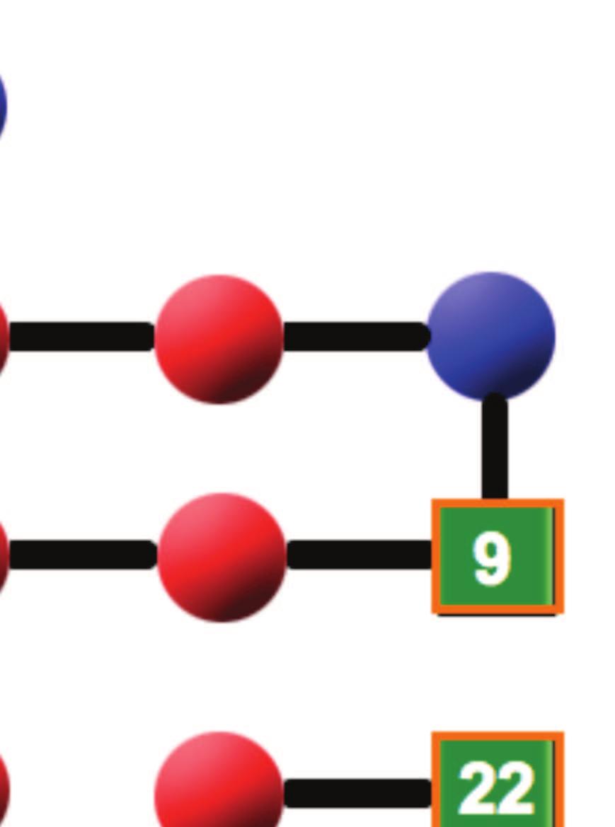
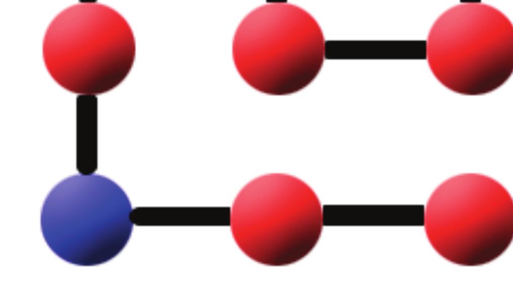
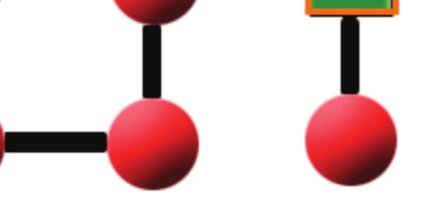
52 Figure 2: 52

53 Figure 3: 53


54 Figure 4: 54
55 Figure 5: 55

56 Figure 6: 56

57 Figure 7: 57

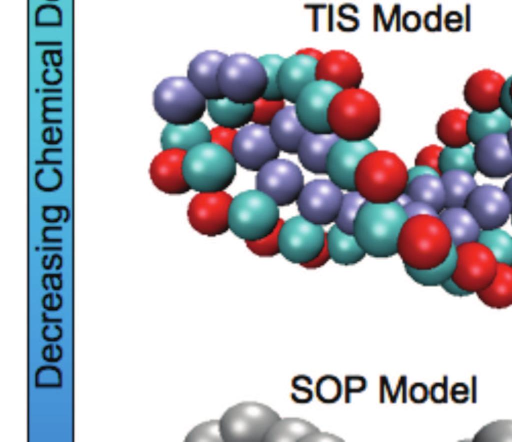

58 Figure 8: 58


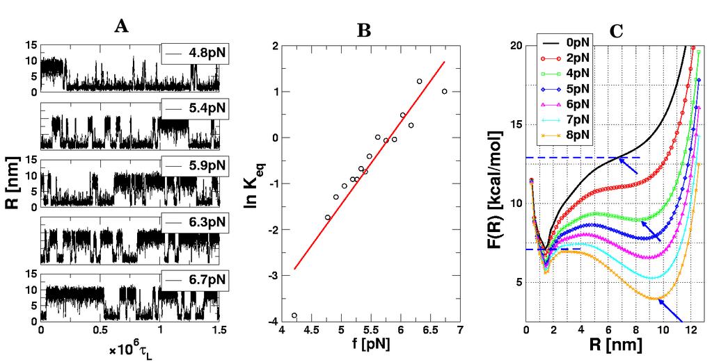
59 Figure 9: Figure 10: 59

60 Figure 11: 60
Protein Folding. I. Characteristics of proteins. C α
 I. Characteristics of proteins Protein Folding 1. Proteins are one of the most important molecules of life. They perform numerous functions, from storing oxygen in tissues or transporting it in a blood
arxiv: v1 [cond-mat.soft] 22 Oct 2007
![arxiv: v1 [cond-mat.soft] 22 Oct 2007](/thumbs/78/78401876.jpg "arxiv: v1 [cond-mat.soft] 22 Oct 2007") Conformational Transitions of Heteropolymers arxiv:0710.4095v1 [cond-mat.soft] 22 Oct 2007 Michael Bachmann and Wolfhard Janke Institut für Theoretische Physik, Universität Leipzig, Augustusplatz 10/11,
Conformational Transitions of Heteropolymers arxiv:0710.4095v1 [cond-mat.soft] 22 Oct 2007 Michael Bachmann and Wolfhard Janke Institut für Theoretische Physik, Universität Leipzig, Augustusplatz 10/11,
Protein Folding & Stability. Lecture 11: Margaret A. Daugherty. Fall How do we go from an unfolded polypeptide chain to a
 Lecture 11: Protein Folding & Stability Margaret A. Daugherty Fall 2004 How do we go from an unfolded polypeptide chain to a compact folded protein? (Folding of thioredoxin, F. Richards) Structure - Function
Lecture 11: Protein Folding & Stability
 Structure - Function Protein Folding: What we know Lecture 11: Protein Folding & Stability 1). Amino acid sequence dictates structure. 2). The native structure represents the lowest energy state for a
Protein Folding & Stability. Lecture 11: Margaret A. Daugherty. Fall Protein Folding: What we know. Protein Folding
 Lecture 11: Protein Folding & Stability Margaret A. Daugherty Fall 2003 Structure - Function Protein Folding: What we know 1). Amino acid sequence dictates structure. 2). The native structure represents
Many proteins spontaneously refold into native form in vitro with high fidelity and high speed.
 Macromolecular Processes 20. Protein Folding Composed of 50 500 amino acids linked in 1D sequence by the polypeptide backbone The amino acid physical and chemical properties of the 20 amino acids dictate
Protein Structure. W. M. Grogan, Ph.D. OBJECTIVES
 Protein Structure W. M. Grogan, Ph.D. OBJECTIVES 1. Describe the structure and characteristic properties of typical proteins. 2. List and describe the four levels of structure found in proteins. 3. Relate
The protein folding problem consists of two parts:
 Energetics and kinetics of protein folding The protein folding problem consists of two parts: 1)Creating a stable, well-defined structure that is significantly more stable than all other possible structures.
Research Paper 1. Correspondence: Devarajan Thirumalai
 Research Paper Protein folding kinetics: timescales, pathways and energy landscapes in terms of sequence-dependent properties Thomas Veitshans, Dmitri Klimov and Devarajan Thirumalai Background: Recent
Outline. The ensemble folding kinetics of protein G from an all-atom Monte Carlo simulation. Unfolded Folded. What is protein folding?
 The ensemble folding kinetics of protein G from an all-atom Monte Carlo simulation By Jun Shimada and Eugine Shaknovich Bill Hawse Dr. Bahar Elisa Sandvik and Mehrdad Safavian Outline Background on protein
arxiv:cond-mat/ v1 [cond-mat.stat-mech] 8 Nov 1996
![arxiv:cond-mat/ v1 [cond-mat.stat-mech] 8 Nov 1996](/thumbs/89/97526426.jpg "arxiv:cond-mat/ v1 [cond-mat.stat-mech] 8 Nov 1996") Protein Folding Kinetics: Time Scales, Pathways, and Energy arxiv:cond-mat/9611065v1 [cond-mat.stat-mech] 8 Nov 1996 Landscapes in Terms of Sequence Dependent Properties T. Veitshans 1, D. K. Klimov 2,
Protein Folding Kinetics: Time Scales, Pathways, and Energy arxiv:cond-mat/9611065v1 [cond-mat.stat-mech] 8 Nov 1996 Landscapes in Terms of Sequence Dependent Properties T. Veitshans 1, D. K. Klimov 2,
Proteins are not rigid structures: Protein dynamics, conformational variability, and thermodynamic stability
 Proteins are not rigid structures: Protein dynamics, conformational variability, and thermodynamic stability Dr. Andrew Lee UNC School of Pharmacy (Div. Chemical Biology and Medicinal Chemistry) UNC Med
THE TANGO ALGORITHM: SECONDARY STRUCTURE PROPENSITIES, STATISTICAL MECHANICS APPROXIMATION
 THE TANGO ALGORITHM: SECONDARY STRUCTURE PROPENSITIES, STATISTICAL MECHANICS APPROXIMATION AND CALIBRATION Calculation of turn and beta intrinsic propensities. A statistical analysis of a protein structure
Unfolding CspB by means of biased molecular dynamics
 Chapter 4 Unfolding CspB by means of biased molecular dynamics 4.1 Introduction Understanding the mechanism of protein folding has been a major challenge for the last twenty years, as pointed out in the
Swelling and Collapse of Single Polymer Molecules and Gels.
 Swelling and Collapse of Single Polymer Molecules and Gels. Coil-Globule Transition in Single Polymer Molecules. the coil-globule transition If polymer chains are not ideal, interactions of non-neighboring
Chap. 2. Polymers Introduction. - Polymers: synthetic materials <--> natural materials
 Chap. 2. Polymers 2.1. Introduction - Polymers: synthetic materials natural materials no gas phase, not simple liquid (much more viscous), not perfectly crystalline, etc 2.3. Polymer Chain Conformation
arxiv: v1 [q-bio.bm] 25 Feb 2014
![arxiv: v1 [q-bio.bm] 25 Feb 2014](/thumbs/88/114859085.jpg "arxiv: v1 [q-bio.bm] 25 Feb 2014") Development and applications of Coarse Grained models for RNA Changbong Hyeon School of Computational Sciences, Korea Institute for Advanced Study, Seoul 130-722, Republic of Korea Natalia A. Denesyuk
Development and applications of Coarse Grained models for RNA Changbong Hyeon School of Computational Sciences, Korea Institute for Advanced Study, Seoul 130-722, Republic of Korea Natalia A. Denesyuk
Biology Chemistry & Physics of Biomolecules. Examination #1. Proteins Module. September 29, Answer Key
 Biology 5357 Chemistry & Physics of Biomolecules Examination #1 Proteins Module September 29, 2017 Answer Key Question 1 (A) (5 points) Structure (b) is more common, as it contains the shorter connection
Lecture 2 and 3: Review of forces (ctd.) and elementary statistical mechanics. Contributions to protein stability
 and elementary statistical mechanics. Contributions to protein stability") Lecture 2 and 3: Review of forces (ctd.) and elementary statistical mechanics. Contributions to protein stability Part I. Review of forces Covalent bonds Non-covalent Interactions: Van der Waals Interactions
Lecture 2 and 3: Review of forces (ctd.) and elementary statistical mechanics. Contributions to protein stability Part I. Review of forces Covalent bonds Non-covalent Interactions: Van der Waals Interactions
Biochemistry,530:,, Introduc5on,to,Structural,Biology, Autumn,Quarter,2015,
 Biochemistry,530:,, Introduc5on,to,Structural,Biology, Autumn,Quarter,2015, Course,Informa5on, BIOC%530% GraduateAlevel,discussion,of,the,structure,,func5on,,and,chemistry,of,proteins,and, nucleic,acids,,control,of,enzyma5c,reac5ons.,please,see,the,course,syllabus,and,
Introduction to Computational Structural Biology
 Introduction to Computational Structural Biology Part I 1. Introduction The disciplinary character of Computational Structural Biology The mathematical background required and the topics covered Bibliography
Why Proteins Fold. How Proteins Fold? e - ΔG/kT. Protein Folding, Nonbonding Forces, and Free Energy
 Why Proteins Fold Proteins are the action superheroes of the body. As enzymes, they make reactions go a million times faster. As versatile transport vehicles, they carry oxygen and antibodies to fight
Quantitative Stability/Flexibility Relationships; Donald J. Jacobs, University of North Carolina at Charlotte Page 1 of 12
 Quantitative Stability/Flexibility Relationships; Donald J. Jacobs, University of North Carolina at Charlotte Page 1 of 12 The figure shows that the DCM when applied to the helix-coil transition, and solved
Archives of Biochemistry and Biophysics
 Archives of Biochemistry and Biophysics 531 (2013) 24 33 Contents lists available at SciVerse ScienceDirect Archives of Biochemistry and Biophysics journal homepage: www.elsevier.com/locate/yabbi Review
Molecular Mechanics. I. Quantum mechanical treatment of molecular systems
 Molecular Mechanics I. Quantum mechanical treatment of molecular systems The first principle approach for describing the properties of molecules, including proteins, involves quantum mechanics. For example,
Proteins polymer molecules, folded in complex structures. Konstantin Popov Department of Biochemistry and Biophysics
 Proteins polymer molecules, folded in complex structures Konstantin Popov Department of Biochemistry and Biophysics Outline General aspects of polymer theory Size and persistent length of ideal linear
Short Announcements. 1 st Quiz today: 15 minutes. Homework 3: Due next Wednesday.
 Short Announcements 1 st Quiz today: 15 minutes Homework 3: Due next Wednesday. Next Lecture, on Visualizing Molecular Dynamics (VMD) by Klaus Schulten Today s Lecture: Protein Folding, Misfolding, Aggregation
Energetics and Thermodynamics
 DNA/Protein structure function analysis and prediction Protein Folding and energetics: Introduction to folding Folding and flexibility (Ch. 6) Energetics and Thermodynamics 1 Active protein conformation
Lecture 2-3: Review of forces (ctd.) and elementary statistical mechanics. Contributions to protein stability
 and elementary statistical mechanics. Contributions to protein stability") Lecture 2-3: Review of forces (ctd.) and elementary statistical mechanics. Contributions to protein stability Part I. Review of forces Covalent bonds Non-covalent Interactions Van der Waals Interactions
Lecture 2-3: Review of forces (ctd.) and elementary statistical mechanics. Contributions to protein stability Part I. Review of forces Covalent bonds Non-covalent Interactions Van der Waals Interactions
Govardhan Reddy. John E. Straub. D. Thirumalai*
 1162 J. Phys. Chem. B 2009, 113, 1162 1172 Influence of Preformed Asp23-Lys28 Salt Bridge on the Conformational Fluctuations of Monomers and Dimers of Aβ Peptides with Implications for ates of Fibril Formation
NMR Characterization of Partially Folded and Unfolded Conformational Ensembles of Proteins
 Elisar Barbar Department of Chemistry and Biochemistry, Ohio University, Athens, OH 45701 NMR Characterization of Partially Folded and Unfolded Conformational Ensembles of Proteins Abstract: Studies of
Polypeptide Folding Using Monte Carlo Sampling, Concerted Rotation, and Continuum Solvation
 Polypeptide Folding Using Monte Carlo Sampling, Concerted Rotation, and Continuum Solvation Jakob P. Ulmschneider and William L. Jorgensen J.A.C.S. 2004, 126, 1849-1857 Presented by Laura L. Thomas and
arxiv:cond-mat/ v1 2 Feb 94
 cond-mat/9402010 Properties and Origins of Protein Secondary Structure Nicholas D. Socci (1), William S. Bialek (2), and José Nelson Onuchic (1) (1) Department of Physics, University of California at San
 Title Theory of solutions in the energy r of the molecular flexibility Author(s) Matubayasi, N; Nakahara, M Citation JOURNAL OF CHEMICAL PHYSICS (2003), 9702 Issue Date 2003-11-08 URL http://hdl.handle.net/2433/50354
arxiv:chem-ph/ v1 11 Nov 1994
 chem-ph/9411008 Funnels, Pathways and the Energy Landscape of Protein Folding: A Synthesis arxiv:chem-ph/9411008v1 11 Nov 1994 Joseph D. Bryngelson, Physical Sciences Laboratory, Division of Computer Research
Isothermal experiments characterize time-dependent aggregation and unfolding
 1 Energy Isothermal experiments characterize time-dependent aggregation and unfolding Technical ote Introduction Kinetic measurements have, for decades, given protein scientists insight into the mechanisms
Introduction The gramicidin A (ga) channel forms by head-to-head association of two monomers at their amino termini, one from each bilayer leaflet. Th
 channel forms by head-to-head association of two monomers at their amino termini, one from each bilayer leaflet. Th") Abstract When conductive, gramicidin monomers are linked by six hydrogen bonds. To understand the details of dissociation and how the channel transits from a state with 6H bonds to ones with 4H bonds or
Abstract When conductive, gramicidin monomers are linked by six hydrogen bonds. To understand the details of dissociation and how the channel transits from a state with 6H bonds to ones with 4H bonds or
Structuring of hydrophobic and hydrophilic polymers at interfaces Stephen Donaldson ChE 210D Final Project Abstract
 Structuring of hydrophobic and hydrophilic polymers at interfaces Stephen Donaldson ChE 210D Final Project Abstract In this work, a simplified Lennard-Jones (LJ) sphere model is used to simulate the aggregation,
Dihedral Angles. Homayoun Valafar. Department of Computer Science and Engineering, USC 02/03/10 CSCE 769
 Dihedral Angles Homayoun Valafar Department of Computer Science and Engineering, USC The precise definition of a dihedral or torsion angle can be found in spatial geometry Angle between to planes Dihedral
Biochemistry Prof. S. DasGupta Department of Chemistry Indian Institute of Technology Kharagpur. Lecture - 06 Protein Structure IV
 Biochemistry Prof. S. DasGupta Department of Chemistry Indian Institute of Technology Kharagpur Lecture - 06 Protein Structure IV We complete our discussion on Protein Structures today. And just to recap
Introduction to" Protein Structure
 Introduction to" Protein Structure Function, evolution & experimental methods Thomas Blicher, Center for Biological Sequence Analysis Learning Objectives Outline the basic levels of protein structure.
BIBC 100. Structural Biochemistry
 BIBC 100 Structural Biochemistry http://classes.biology.ucsd.edu/bibc100.wi14 Papers- Dialogue with Scientists Questions: Why? How? What? So What? Dialogue Structure to explain function Knowledge Food
Predicting free energy landscapes for complexes of double-stranded chain molecules
 JOURNAL OF CHEMICAL PHYSICS VOLUME 114, NUMBER 9 1 MARCH 2001 Predicting free energy landscapes for complexes of double-stranded chain molecules Wenbing Zhang and Shi-Jie Chen a) Department of Physics
Protein Dynamics. The space-filling structures of myoglobin and hemoglobin show that there are no pathways for O 2 to reach the heme iron.
 Protein Dynamics The space-filling structures of myoglobin and hemoglobin show that there are no pathways for O 2 to reach the heme iron. Below is myoglobin hydrated with 350 water molecules. Only a small
Contents. xiii. Preface v
 Contents Preface Chapter 1 Biological Macromolecules 1.1 General PrincipIes 1.1.1 Macrornolecules 1.2 1.1.2 Configuration and Conformation Molecular lnteractions in Macromolecular Structures 1.2.1 Weak
From Amino Acids to Proteins - in 4 Easy Steps
 From Amino Acids to Proteins - in 4 Easy Steps Although protein structure appears to be overwhelmingly complex, you can provide your students with a basic understanding of how proteins fold by focusing
F. Piazza Center for Molecular Biophysics and University of Orléans, France. Selected topic in Physical Biology. Lecture 1
 Zhou Pei-Yuan Centre for Applied Mathematics, Tsinghua University November 2013 F. Piazza Center for Molecular Biophysics and University of Orléans, France Selected topic in Physical Biology Lecture 1
Molecular Modelling. part of Bioinformatik von RNA- und Proteinstrukturen. Sonja Prohaska. Leipzig, SS Computational EvoDevo University Leipzig
 part of Bioinformatik von RNA- und Proteinstrukturen Computational EvoDevo University Leipzig Leipzig, SS 2011 Protein Structure levels or organization Primary structure: sequence of amino acids (from
Dana Alsulaibi. Jaleel G.Sweis. Mamoon Ahram
 15 Dana Alsulaibi Jaleel G.Sweis Mamoon Ahram Revision of last lectures: Proteins have four levels of structures. Primary,secondary, tertiary and quaternary. Primary structure is the order of amino acids
Distance Constraint Model; Donald J. Jacobs, University of North Carolina at Charlotte Page 1 of 11
 Distance Constraint Model; Donald J. Jacobs, University of North Carolina at Charlotte Page 1 of 11 Taking the advice of Lord Kelvin, the Father of Thermodynamics, I describe the protein molecule and other
Visualizing folding of proteins (1 3) and RNA (2) in terms of
 and RNA (2) in terms of") Can energy landscape roughness of proteins and RNA be measured by using mechanical unfolding experiments? Changbong Hyeon and D. Thirumalai Chemical Physics Program, Institute for Physical Science and
Can energy landscape roughness of proteins and RNA be measured by using mechanical unfolding experiments? Changbong Hyeon and D. Thirumalai Chemical Physics Program, Institute for Physical Science and
Flexibility of Protein Structure
 Flexibility of Protein Structure Proteins show varying degree of conformational flexibility Due to movements of atoms in molecules vibration in bond length and angles Reflects the existence of populations
arxiv:cond-mat/ v1 [cond-mat.soft] 5 May 1998
![arxiv:cond-mat/ v1 [cond-mat.soft] 5 May 1998](/thumbs/91/105426806.jpg "arxiv:cond-mat/ v1 [cond-mat.soft] 5 May 1998") Linking Rates of Folding in Lattice Models of Proteins with Underlying Thermodynamic Characteristics arxiv:cond-mat/9805061v1 [cond-mat.soft] 5 May 1998 D.K.Klimov and D.Thirumalai Institute for Physical
Linking Rates of Folding in Lattice Models of Proteins with Underlying Thermodynamic Characteristics arxiv:cond-mat/9805061v1 [cond-mat.soft] 5 May 1998 D.K.Klimov and D.Thirumalai Institute for Physical
Announcements & Lecture Points
 Announcements & Lecture Points Homework 3 (Klaus Schulten s Lecture): Due Wednesday. Quiz returned, next homework on Wednesday. Today s Lecture: Protein Folding, Misfolding, Aggregation. Experimental Approach
The Nature of Folded States of Globular Proteins
 The Nature of Folded States of Globular Proteins J. D. HONEYCUTT and D. THlRUMALAl* Biosym Technologies, Inc., 10065 Barnes Canyon Road, San Diego, California 921 21; and Department of Chemistry and Biochemistry,
Biotechnology of Proteins. The Source of Stability in Proteins (III) Fall 2015
 Fall 2015") Biotechnology of Proteins The Source of Stability in Proteins (III) Fall 2015 Conformational Entropy of Unfolding It is The factor that makes the greatest contribution to stabilization of the unfolded
Biotechnology of Proteins The Source of Stability in Proteins (III) Fall 2015 Conformational Entropy of Unfolding It is The factor that makes the greatest contribution to stabilization of the unfolded
Protein Folding experiments and theory
 Protein Folding experiments and theory 1, 2,and 3 Protein Structure Fig. 3-16 from Lehninger Biochemistry, 4 th ed. The 3D structure is not encoded at the single aa level Hydrogen Bonding Shared H atom
Biomolecules. Energetics in biology. Biomolecules inside the cell
 Biomolecules Energetics in biology Biomolecules inside the cell Energetics in biology The production of energy, its storage, and its use are central to the economy of the cell. Energy may be defined as
Polymer Solution Thermodynamics:
 Polymer Solution Thermodynamics: 3. Dilute Solutions with Volume Interactions Brownian particle Polymer coil Self-Avoiding Walk Models While the Gaussian coil model is useful for describing polymer solutions
Lecture 21 (11/3/17) Protein Stability, Folding, and Dynamics Hydrophobic effect drives protein folding
 Protein Stability, Folding, and Dynamics Hydrophobic effect drives protein folding") Reading: Ch4; 142-151 Problems: Ch4 (text); 14, 16 Ch6 (text); 1, 4 NEXT (after exam) Reading: Ch8; 310-312, 279-285, 285-289 Ch24; 957-961 Problems: Ch8 (text); 1,2,22 Ch8 (study-guide:facts); 1,2,3,4,5,9,10
Reading: Ch4; 142-151 Problems: Ch4 (text); 14, 16 Ch6 (text); 1, 4 NEXT (after exam) Reading: Ch8; 310-312, 279-285, 285-289 Ch24; 957-961 Problems: Ch8 (text); 1,2,22 Ch8 (study-guide:facts); 1,2,3,4,5,9,10
Biochemistry: Concepts and Connections
 Biochemistry: Concepts and Connections Dean R. Appling Spencer J. Anthony-Cahill Christopher K. Mathews Chapter 6 The Three Dimensional Structure of Proteins Cartoon representation of myoglobin, showing
Introduction to Comparative Protein Modeling. Chapter 4 Part I
 Introduction to Comparative Protein Modeling Chapter 4 Part I 1 Information on Proteins Each modeling study depends on the quality of the known experimental data. Basis of the model Search in the literature
Section Week 3. Junaid Malek, M.D.
 Section Week 3 Junaid Malek, M.D. Biological Polymers DA 4 monomers (building blocks), limited structure (double-helix) RA 4 monomers, greater flexibility, multiple structures Proteins 20 Amino Acids,
Local Interactions Dominate Folding in a Simple Protein Model
 J. Mol. Biol. (1996) 259, 988 994 Local Interactions Dominate Folding in a Simple Protein Model Ron Unger 1,2 * and John Moult 2 1 Department of Life Sciences Bar-Ilan University Ramat-Gan, 52900, Israel
ICCP Project 2 - Advanced Monte Carlo Methods Choose one of the three options below
 ICCP Project 2 - Advanced Monte Carlo Methods Choose one of the three options below Introduction In statistical physics Monte Carlo methods are considered to have started in the Manhattan project (1940
Dominant Paths in Protein Folding
 Dominant Paths in Protein Folding Henri Orland SPhT, CEA-Saclay France work in collaboration with P. Faccioli, F. Pederiva, M. Sega University of Trento Henri Orland Annecy meeting 2006 Outline Basic notions
BIOC : Homework 1 Due 10/10
 Contact information: Name: Student # BIOC530 2012: Homework 1 Due 10/10 Department Email address The following problems are based on David Baker s lectures of forces and protein folding. When numerical
arxiv:q-bio/ v2 [q-bio.bm] 31 Jan 2006
![arxiv:q-bio/ v2 [q-bio.bm] 31 Jan 2006](/thumbs/90/101401483.jpg "arxiv:q-bio/ v2 [q-bio.bm] 31 Jan 2006") Forced-unfolding and force-quench refolding of RNA hairpins arxiv:q-bio/0601043v2 [q-bio.bm] 31 Jan 2006 Changbong Hyeon 1 and D. Thirumalai 1,2 1 Biophysics Program Institute for Physical Science and
Forced-unfolding and force-quench refolding of RNA hairpins arxiv:q-bio/0601043v2 [q-bio.bm] 31 Jan 2006 Changbong Hyeon 1 and D. Thirumalai 1,2 1 Biophysics Program Institute for Physical Science and
= (-22) = +2kJ /mol
 = +2kJ /mol") Lecture 8: Thermodynamics & Protein Stability Assigned reading in Campbell: Chapter 4.4-4.6 Key Terms: DG = -RT lnk eq = DH - TDS Transition Curve, Melting Curve, Tm DH calculation DS calculation van der
Lecture 8: Thermodynamics & Protein Stability Assigned reading in Campbell: Chapter 4.4-4.6 Key Terms: DG = -RT lnk eq = DH - TDS Transition Curve, Melting Curve, Tm DH calculation DS calculation van der
Secondary structure stability, beta-sheet formation & stability
 Protein Physics 2016 Lecture 6, February 5 Secondary structure stability, beta-sheet formation & stability Magnus Andersson magnus.andersson@scilifelab.se Theoretical & Computational Biophysics Recap of
Modeling Background; Donald J. Jacobs, University of North Carolina at Charlotte Page 1 of 8
 Modeling Background; Donald J. Jacobs, University of North Carolina at Charlotte Page 1 of 8 Depending on thermodynamic and solvent conditions, the interrelationships between thermodynamic stability of
Molecular dynamics simulations of anti-aggregation effect of ibuprofen. Wenling E. Chang, Takako Takeda, E. Prabhu Raman, and Dmitri Klimov
 Biophysical Journal, Volume 98 Supporting Material Molecular dynamics simulations of anti-aggregation effect of ibuprofen Wenling E. Chang, Takako Takeda, E. Prabhu Raman, and Dmitri Klimov Supplemental
Untangling the Mechanics of Entangled Biopolymers
 Untangling the Mechanics of Entangled Biopolymers Rae M. Robertson-Anderson Physics Department University of San Diego students/postdocs: Cole Chapman, PhD Tobias Falzone, PhD Stephanie Gorczyca, USD 16
Paul Sigler et al, 1998.
 Biological systems are necessarily metastable. They are created, modulated, and destroyed according to a temporal plan that meets the survival needs of the cell, organism, and species...clearly, no biological
BCMP 201 Protein biochemistry
 BCMP 201 Protein biochemistry BCMP 201 Protein biochemistry with emphasis on the interrelated roles of protein structure, catalytic activity, and macromolecular interactions in biological processes. The
NMR studies of protein folding
 NMR studies of protein folding Juhi Juneja and Jayant B. Udgaonkar* National Centre for Biological Sciences, Tata Institute of Fundamental Research, GKVK Campus, Bangalore 560 065, India NMR spectroscopy
Protein Folding Prof. Eugene Shakhnovich
 Protein Folding Eugene Shakhnovich Department of Chemistry and Chemical Biology Harvard University 1 Proteins are folded on various scales As of now we know hundreds of thousands of sequences (Swissprot)
Quiz 2 Morphology of Complex Materials
 071003 Quiz 2 Morphology of Complex Materials 1) Explain the following terms: (for states comment on biological activity and relative size of the structure) a) Native State b) Unfolded State c) Denatured
Bioengineering 215. An Introduction to Molecular Dynamics for Biomolecules
 Bioengineering 215 An Introduction to Molecular Dynamics for Biomolecules David Parker May 18, 2007 ntroduction A principal tool to study biological molecules is molecular dynamics simulations (MD). MD
BSc and MSc Degree Examinations
 Examination Candidate Number: Desk Number: BSc and MSc Degree Examinations 2018-9 Department : BIOLOGY Title of Exam: Molecular Biology and Biochemistry Part I Time Allowed: 1 hour and 30 minutes Marking
Principles of Physical Biochemistry
 Principles of Physical Biochemistry Kensal E. van Hold e W. Curtis Johnso n P. Shing Ho Preface x i PART 1 MACROMOLECULAR STRUCTURE AND DYNAMICS 1 1 Biological Macromolecules 2 1.1 General Principles
(Entropic) Stochastic Resonance in Biological Systems at Mesoscale
 Stochastic Resonance in Biological Systems at Mesoscale") (Entropic) Stochastic Resonance in Biological Systems at Mesoscale Wokyung Sung Department of Physics, POSTECH, IBS center for Self-assembly and Complexity, Pohang, 790-784, South Korea As interconnected,
(Entropic) Stochastic Resonance in Biological Systems at Mesoscale Wokyung Sung Department of Physics, POSTECH, IBS center for Self-assembly and Complexity, Pohang, 790-784, South Korea As interconnected,
Advanced sampling. fluids of strongly orientation-dependent interactions (e.g., dipoles, hydrogen bonds)
") Advanced sampling ChE210D Today's lecture: methods for facilitating equilibration and sampling in complex, frustrated, or slow-evolving systems Difficult-to-simulate systems Practically speaking, one is
Advanced sampling ChE210D Today's lecture: methods for facilitating equilibration and sampling in complex, frustrated, or slow-evolving systems Difficult-to-simulate systems Practically speaking, one is
Lecture 26: Polymers: DNA Packing and Protein folding 26.1 Problem Set 4 due today. Reading for Lectures 22 24: PKT Chapter 8 [ ].
![Lecture 26: Polymers: DNA Packing and Protein folding 26.1 Problem Set 4 due today. Reading for Lectures 22 24: PKT Chapter 8 [ ].](/thumbs/84/90810734.jpg "Lecture 26: Polymers: DNA Packing and Protein folding 26.1 Problem Set 4 due today. Reading for Lectures 22 24: PKT Chapter 8 [ ].") Lecture 26: Polymers: DA Packing and Protein folding 26.1 Problem Set 4 due today. eading for Lectures 22 24: PKT hapter 8 DA Packing for Eukaryotes: The packing problem for the larger eukaryotic genomes
Lecture 26: Polymers: DA Packing and Protein folding 26.1 Problem Set 4 due today. eading for Lectures 22 24: PKT hapter 8 DA Packing for Eukaryotes: The packing problem for the larger eukaryotic genomes
Biomolecules: lecture 10
 Biomolecules: lecture 10 - understanding in detail how protein 3D structures form - realize that protein molecules are not static wire models but instead dynamic, where in principle every atom moves (yet
3. Solutions W = N!/(N A!N B!) (3.1) Using Stirling s approximation ln(n!) = NlnN N: ΔS mix = k (N A lnn + N B lnn N A lnn A N B lnn B ) (3.
 (3.1) Using Stirling s approximation ln(n!) = NlnN N: ΔS mix = k (N A lnn + N B lnn N A lnn A N B lnn B ) (3.") 3. Solutions Many biological processes occur between molecules in aqueous solution. In addition, many protein and nucleic acid molecules adopt three-dimensional structure ( fold ) in aqueous solution.
3. Solutions Many biological processes occur between molecules in aqueous solution. In addition, many protein and nucleic acid molecules adopt three-dimensional structure ( fold ) in aqueous solution.
Supplementary Information
 Supplementary Information Mechanical unzipping and rezipping of a single SNARE complex reveals hysteresis as a force-generating mechanism Duyoung Min, Kipom Kim, Changbong Hyeon, Yong Hoon Cho, Yeon-Kyun
Why Proteins Fold? (Parts of this presentation are based on work of Ashok Kolaskar) CS490B: Introduction to Bioinformatics Mar.
 CS490B: Introduction to Bioinformatics Mar.") Why Proteins Fold? (Parts of this presentation are based on work of Ashok Kolaskar) CS490B: Introduction to Bioinformatics Mar. 25, 2002 Molecular Dynamics: Introduction At physiological conditions, the
Why Proteins Fold? (Parts of this presentation are based on work of Ashok Kolaskar) CS490B: Introduction to Bioinformatics Mar. 25, 2002 Molecular Dynamics: Introduction At physiological conditions, the
Macromolecular Crowding
 Macromolecular Crowding Keng-Hwee Chiam Mathematical and Theoretical Biology Group Goodsell (1994) Macromolecular Crowding, Oct. 15, 2003 p.1/33 Outline What: introduction, definition Why: implications
Thermodynamics. Entropy and its Applications. Lecture 11. NC State University
 Thermodynamics Entropy and its Applications Lecture 11 NC State University System and surroundings Up to this point we have considered the system, but we have not concerned ourselves with the relationship
Molecular Mechanics, Dynamics & Docking
 Molecular Mechanics, Dynamics & Docking Lawrence Hunter, Ph.D. Director, Computational Bioscience Program University of Colorado School of Medicine Larry.Hunter@uchsc.edu http://compbio.uchsc.edu/hunter
Lattice protein models
 Lattice protein models Marc R. Roussel epartment of Chemistry and Biochemistry University of Lethbridge March 5, 2009 1 Model and assumptions The ideas developed in the last few lectures can be applied
NMR, X-ray Diffraction, Protein Structure, and RasMol
 NMR, X-ray Diffraction, Protein Structure, and RasMol Introduction So far we have been mostly concerned with the proteins themselves. The techniques (NMR or X-ray diffraction) used to determine a structure
The Molecular Dynamics Method
 The Molecular Dynamics Method Thermal motion of a lipid bilayer Water permeation through channels Selective sugar transport Potential Energy (hyper)surface What is Force? Energy U(x) F = d dx U(x) Conformation
Introduction to Polymer Physics
 Introduction to Polymer Physics Enrico Carlon, KU Leuven, Belgium February-May, 2016 Enrico Carlon, KU Leuven, Belgium Introduction to Polymer Physics February-May, 2016 1 / 28 Polymers in Chemistry and
EXAM I COURSE TFY4310 MOLECULAR BIOPHYSICS December Suggested resolution
 page 1 of 7 EXAM I COURSE TFY4310 MOLECULAR BIOPHYSICS December 2013 Suggested resolution Exercise 1. [total: 25 p] a) [t: 5 p] Describe the bonding [1.5 p] and the molecular orbitals [1.5 p] of the ethylene
Biophysics II. Hydrophobic Bio-molecules. Key points to be covered. Molecular Interactions in Bio-molecular Structures - van der Waals Interaction
 Biophysics II Key points to be covered By A/Prof. Xiang Yang Liu Biophysics & Micro/nanostructures Lab Department of Physics, NUS 1. van der Waals Interaction 2. Hydrogen bond 3. Hydrophilic vs hydrophobic
3 Biopolymers Uncorrelated chains - Freely jointed chain model
 3.4 Entropy When we talk about biopolymers, it is important to realize that the free energy of a biopolymer in thermal equilibrium is not constant. Unlike solids, biopolymers are characterized through
Aqueous solutions. Solubility of different compounds in water
 Aqueous solutions Solubility of different compounds in water The dissolution of molecules into water (in any solvent actually) causes a volume change of the solution; the size of this volume change is
Statistical Physics of The Symmetric Group. Mobolaji Williams Harvard Physics Oral Qualifying Exam Dec. 12, 2016
 Statistical Physics of The Symmetric Group Mobolaji Williams Harvard Physics Oral Qualifying Exam Dec. 12, 2016 1 Theoretical Physics of Living Systems Physics Particle Physics Condensed Matter Astrophysics