belonging to the Genus Pantoea
|
|
|
- Myra Holt
- 5 years ago
- Views:
Transcription
1 Emerging diseases of maize and onion caused by bacteria belonging to the Genus Pantoea by Teresa Goszczynska Submitted in partial fulfilment of the requirements for the degree Philosophiae Doctoriae in The Faculty of Natural and Agricultural Sciences University of Pretoria Pretoria Supervisor: Prof. T.A. Coutinho Co-supervisor: Prof. S.N. Venter May 2007 University of Pretoria
2 Emerging diseases of maize and onion caused by bacteria belonging to the Genus Pantoea by Teresa Goszczynska Supervisor: Prof. T.A. Coutinho Co-supervisor: Prof. S.N. Venter Department: Microbiology and Plant Pathology Degree: Ph. D. (Microbiology) SUMMARY Center rot of onion, caused by Pantoea ananatis, was first described in the USA, in P. ananatis is seed-borne in onions and it was suggested that it was introduced into the USA on infected seed lots from South Africa. Center rot has not been observed in South Africa and it was essential to determine if P. ananatis is present in local onion seed. Colonies resembling those of P. ananatis were isolated from four South African seed lots on PA 20, a new semiselective medium. Pathogenicity tests demonstrated that the South African and America strains induced the same symptoms on onion. Phenotypic and genotypic analyses identified the strains from seed as P. ananatis. In 2004/2005, an unreported disease of maize, brown stalk rot, was observed on commercial fields in South Africa. The representative strains induced disease symptoms similar to those observed in the field. The phenotypic and genotyping tests showed that the strains belonged to the genus Pantoea and separated them into two groups. The first group was identified as P. ananatis. The F-AFLP genomic fingerprints generated by the second group of strains, were distinctly different from those generated by known Pantoea species. To resolve the taxonomic position of Pantoea isolated i
3 from onion and maize, sixty-seven strains were subjected to a polyphasic study. The methods used included phenotypic characterisation, genomic fingerprinting, 16S rrna gene sequence analysis and DNA-DNA hybridisation. The results revealed that the strains belong to three different species within the genus Pantoea: P. ananatis, P. vagens and a novel species, Pantoea allii sp. nov. ii
4 ACKNOWLEDGMENTS I thank My husband Dariusz and daughter Dorota for their support. I am grateful for their patience and understanding during the past few years. Prof. Teresa Coutinho for everything, without her this project would not have been possible. Prof. Fanus Venter, for advice, support and DNA-DNA hybridisation. All my co-workers at the Bacterial Diseases Unit, Plant Protection Research Institute. Thank you Vallry, Harry and Zama for making of hundreds litres of media, plating endless plant extracts and helping with never-ending diagnostic work. All the students from the micro lab, especially Carrie Brady for help with AFLPs. Dr. Wilhelm Botha, from my Institute, for all tips on rep-pcr and Bionumerics. The Agricultural Research Council, the National Research Foundation and the University of Pretoria, for contributing funds and providing the facilities and opportunities needed to complete this project. iii
5 CONTENTS Summary Acknowledgements Contents i iii iv Preface 1 Chapter 1 The genus Pantoea in plant pathology 3 Introduction 3 The genus Pantoea 5 Pantoea species as pathogens 7 Pantoea species as beneficial microorganisms in agriculture 11 Isolation of Pantoea species from plants 12 Determination of pathogenicity 13 Phenotypic identification of Pantoea species using commercial systems 14 Detection of plant pathogenic bacteria using the polymerase chain reaction (PCR) 15 DNA fingerprinting-based methods used for identification and taxonomy of 16 bacteria 16S rrna gene sequence analysis 22 Multi-locus sequence analysis (MLSA) 24 Integration of various diagnostic methods for a polyphasic identification 25 Conclusions 27 Literature cited 28 Chapter 2 PA 20, a semi-selective medium for isolation and enumeration of Pantoea ananatis 50 Abstract 50 Introduction 51 iv
6 Materials and methods 51 Results and discussion 54 Acknowledgements 56 References 56 Tables 59 Figures 63 Chapter 3 Isolation and identification of Pantoea ananatis from onion seed in South Africa 67 Summary 67 Introduction 68 Materials and methods 69 Results 73 Discussion 76 Acknowledgements 78 References 79 Tables 83 Figures 89 Chapter 4 Isolation and identification of the causal agent of brown stalk rot, a new disease of maize in South Africa 93 Abstract 93 Introduction 95 Materials and methods 96 Results 100 Discussion 103 Acknowledgements 105 Literature cited 105 Tables 111 v
7 Figures 116 Chapter 5 Polyphasic characterisation of Pantoea strains from onion and maize and the description of Pantoea allii sp. nov. 125 Abstract 125 Introduction 126 Materials and methods 128 Results 132 Discussion 137 Description of Pantoea allii sp. nov. 138 Acknowledgements 140 Literature cited 140 Tables 146 Figures 153 Appendix A 170 vi
8 PREFACE Maize and onion are important agricultural crops in South Africa. The country is one of the biggest producers of onion seed in the world. In the 2004/2005 growing season 358 tons of onion seed were produced, of which 282 tons were for the export market. Maize is the most important grain crop in South Africa, being both the major animal feed grain and the staple food of the majority of the population. For the 2003/2004 marketing year maize was responsible for the second largest contribution to the gross value of agricultural production in the country. The South African maize industry is also the largest maize industry in Africa. Commercial farmers are cultivating nearly three million hectares of maize per year. In the past five years, South Africa produced between 7.2 and 10.1 million tons of maize per annum, with an average of 9.2 million tons. The main maize production areas in South Africa are the Free State, Northwest and Mpumalanga Provinces. These three provinces are responsible for 85% of the total maize produced in the country. Numerous fungal diseases cause excessive damage to maize and onion in South Africa and efforts to control the quality of these crops concentrated on the detection and control of fungal pathogens. Consequently, little is known about plant pathogenic bacteria that may be present. In 1981, leaf and seed stalk necrosis of onion, caused by Erwinia herbicola (syn. Pantoea agglomerans) was reported in the onion seed production areas. In 2000, leaf blight of onion, caused by Xanthomonas axonopodis pv. allii was observed in a few commercial fields of the Limpopo Province. These two diseases have not been observed again and no attempts have been made to screen the locally produced onion seed for the presence of P. agglomerans and X. axonopodis pv. allii. In 1997, a disease similar to leaf and seed stalk necrosis was observed in onion fields in Georgia, USA. The disease, named center rot, has occurred in commercial fields in Georgia every year since 1997 and accounted for 100% loss in some fields. The causal agent of center rot is a gram-negative, facultatively anaerobic, seed-borne bacterium, identified as Pantoea ananatis. The seed associated with the first outbreak of center rot of onion in Georgia, USA, was produced in South Africa and it was suggested that the center rot 1
9 pathogen was possibly introduced on infested seed lots. P. ananatis is the causal agent of bacterial blight and dieback of Eucalyptus in South Africa, but center rot of onion has not been reported from this country. The first goal of this study was to determine if pathogenic P. ananatis was present in South African onion seed and to compare such strains to those associated with center rot of onions in the USA. Nutrient agar is the common growth medium used to isolate P. ananatis from plant material and seed. Nutrient agar, however, is non-selective and many other organisms present as saprophytes or endophytes in plant material and in seed may hamper the detection of the target pathogen. In Chapter 2, a semi-selective medium, PA 20, is described, which suppresses growth of many saprophytic microorganisms and serves as a suitable medium for growth and enumeration of P. ananatis. The medium was specifically developed to detect this pathogen on onion seed. Chapter 3 describes the isolation and identification of P. ananatis from South African onion seed. In 2004 and 2005, a new disease was observed in commercial maize fields in the Northwest and Mpumalanga Provinces. Diseased plants were scattered throughout the fields and 10-70% of the crop was affected. Gram-negative bacteria producing yellow colonies were consistently isolated from diseased tissues and these were tentatively identified as belonging to the genus Pantoea. Two Pantoea species have been reported to cause diseases on maize worldwide. P. stewartii subsp. stewartii causes Stewart s wilt in Europe, Asia and the Americas. P. ananatis was described as an agent of leaf spot on maize in Brazil. The second goal of this study was to identify and characterise the causal agent of brown stalk rot of maize observed in South Africa. Chapter 4 describes the identification of the bacteria causing this new disease of maize. The third goal of this study was to characterise a collection of Pantoea strains from onion and maize using a polyphasic approach based on analyses of carbon source utilisation, physiological characteristics, fluorescent amplified fragment length polymorphism (F- AFLP), repetitive sequence based PCR (rep-pcr) genomic fingerprinting, 16S rrna gene sequence analysis and DNA-DNA hybridisations. The results are presented in Chapter 5. 2
10 CHAPTER 1 The Genus Pantoea in Plant Pathology; Literature Review INTRODUCTION The genus Pantoea was established in 1989 (Gavini et al., 1989) and it is classified in the family Enterobacteriaceae. Pantoea species are associated with plants as pathogens, saprophytes and epiphytes (Cottyn et al., 2001; Gavini et al., 1989; Gitaitis et al., 2000; Grimont and Grimont, 2005; Iimura and Hosono, 1996; Perombelon, 1981; Walcott et al., 2002). Strains of P. ananatis and P. agglomerans were reported to cause diseases in humans (De Beare et al., 2004; Gavini et al., 1989; Kratz et al., 2003; Lim et al., 2006; Maki et al., 1976). Bacteria belonging to the genus (Grimont and Grimont, 2005) are gram-negative, noncapsulated, non-spore forming straight rods; most are motile and are peritrichously flagellated; are facultatively anaerobic, oxidase negative and catalase positive; colonies on nutrient agar are round, smooth, with entire margins; colonies might be yellow, cream/white or beige. There are seven described species within the genus Pantoea and these include: P. agglomerans, P. ananatis, P. citrea, P. dispersa, P. punctata, P. terrea and P. stewartii, containing two subspecies, indologenes and stewartii (Grimont and Grimont, 2005). Bacterial wilt of maize was the first plant disease ever reported caused by a bacterium of the genus Pantoea. The disease is known as Stewart s wilt, to honour F. C. Stewart, who described the symptoms in 1897 on sweet corn in the USA (Stewart, 1897). At that time, the bacterium causing the disease was named Bacillus stewartii. It was also known as: Bacterium stewartii, Erwinia stewartii, Pseudobacterium stewartii, Pseudomonas stewartii, Phytobacterium stewartii and Xanthomonas stewartii (Brenner et al., 1984). The pathogen has been responsible for serious crop loses since its first discovery (Coplin et al., 2002). In 1993, the bacterium was transferred to the genus Pantoea as Pantoea stewartii subsp. stewartii (Mergaert et al., 1993). 3
11 Although the pink disease of pineapple was originally described in 1915 in Hawaii (Lyon et al., 1915), the pathogen responsible for this disease remained unknown until Cha et al. (1997) identified the bacterium causing the pink disease as Pantoea citrea, using molecular techniques. In 1928, Serrano (1928) reported brown rot of pineapple fruitlets, caused by P. ananatis, also in Hawaii. In the same year, Brown (1928) described a bacterial pocket disease of sugar beet, caused by P. agglomerans. In 1934, P. agglomerans was reported to induce galls on Gypsophila (Brown, 1934). During the following 47 years, only one report was published, in 1954, on P. ananatis parasitic on uredia of cereal rust (Pon et al., 1954). Since 1981, however, bacterial species within the genus Pantoea have become increasingly important as plant pathogens worldwide. Seventeen new diseases caused by P. ananatis, P. agglomerans, P. punctata and P. stewartii subsp. indologenes have been described on a variety of crops (Table 1), including beetroot (Burr et al., 1991), cantaloupe (Bruton et al., 1991), cotton (Medrano and Bell, 2006), Eucalyptus (Coutinho et al., 2002), honeydew melon (Wells et al., 1987), maize (Paccola- Meirelles et al., 2001), mandarin orange (Kageyama et al., 1992), millet (Frederickson, 1997), onion (Gitaitis and Gay, 1997), pear (Lindow et al., 1998), rice (Azegami et al., 1983; Cother et al., 2004) and sudangrass (Azad et al., 2000). Disease symptoms are diverse and include galls, rots, wilt, leaf blights, necrosis and spots, dieback and stem necrosis (Grimont and Grimont, 2005). In the case of infection of honeydew melons, pineapple and cantaloupe fruit, the symptoms occur only after harvest (Bruton et al., 1986; Wells et al., 1987; Bruton et al., 1991, Cha et al., 1997). Despite increasing frequency of plant diseases caused by Pantoea species, identification and characterisation of these bacteria were usually based on colony morphology, few physiological and biochemical tests and sometimes fatty acids analyses. Only P. ananatis strains causing dieback of Eucalyptus (Coutinho et al., 2002) and stem necrosis of rice (Cother et al., 2004) have been identified based on phenotypic and genotypic characteristics. Consequently, relatively little is known about isolation, identification, host specificity, genetic relatedness or epidemiology of these bacterial pathogens. This review focuses on the detection, pathogenicity, ecology and taxonomy of the bacteria belonging to the genus Pantoea. The role of Pantoea species in plant pathology is 4
12 demonstrated and a list of Pantoea species causing diseases is presented. The techniques used in the diagnosis of plant diseases, and the methods for the identification of bacterial pathogens are discussed. THE GENUS PANTOEA In the past, all phytopathogenic bacteria in the family Enterobacteriaceae belonged to the genus Erwinia. The genus was proposed by Winslow et al. (1917) for all plant-associated gram-negative, non-spore forming, fermentative, rod-shaped bacteria (Beji et al., 1988). In the 1960 s, Dye divided the genus Erwinia into four natural groups, founded on the type of disease symptoms produced on plants (Verdonck et al., 1987): amylovora (Dye, 1968) causing dry necrotic or wilt diseases; carotovora (Dye, 1969a) inducing soft rots; herbicola (Dye, 1969b) containing saprophytes and putative plant pathogens and atypical erwinias (Dye, 1969c). The Herbicola group, of interest in this review, consisted of Erwinia strains that usually produced a yellow pigment on general growth media, including nutrient agar (Dye, 1969b; Beji et al., 1998). In 1974, Lelliot (1974) included four species in the Herbicola group. These were: Erwinia uredovora (Dye, 1963b), E. milletiae (Dye, 1969b), E. stewartii (Dye, 1963a) and E. herbicola (E. herbicola var. herbicola and E. herbicola var. ananas) (Dye, 1969b). In 1971, Herbicola bacteria were involved in a nosocomial septicemia outbreak in the United States, when 40 of 378 patients died (Maki et al., 1976; Brenner et al., 1984). After comparing human and plant isolates, Ewing and Fife (1972) concluded that all strains belonged to a single species, which were named Enterobacter agglomerans. That conclusion was confirmed in 1986 by examining the DNA relatedness among 86 strains of Enterobacter agglomerans, isolated from humans, and the type strains of Erwinia herbicola and E. milletiae (Lind and Ursing, 1986). However, phytopathologists preferred to use the nomenclature of Dye while medical bacteriologists used the nomenclature of Edwig and Fife (Beji et al., 1988). This group of organisms was referred too in the literature as the Erwinia herbicola-enterobacter agglomerans complex (Grimont and Grimont, 2005). In 1984, Brenner et al. (1984) studied 124 strains isolated from plants and humans that belonged to the Erwinia herbicola-enterobacter agglomerans complex. The results of the 5
13 DNA-DNA hybridisations showed that 90 of these strains formed 13 hybridisation groups, numbered I-XIII. The authors demonstrated the immense genetic diversity of these bacteria and inadequacy of the nomenclature used at the time. In 1987, Verdonck et al. (1987) performed the numerical analysis of phenotypic features of 529 enterobacterial strains, including those assigned to the Erwinia herbicola-enterobacter agglomerans complex and other Erwinia species. The strains received as Enterobacter agglomerans, Erwinia herbicola, E. milletiae, E. ananas, E. uredovora and E. stewartii were dispersed into 23 phenotypic groups or phena. However, the type strains of Enterobacter agglomerans, Erwinia herbicola and E. milletiae grouped into a single phenon 8, the type strains of E. ananas and E. uredovora into phenon 12 and eight strains of E. stewartii into phenon 29. In 1988, Beji et al. (1988) studied the DNA relatedness of the type strain Enterobacter agglomerans, ATCC 27155, to 54 strains from the Erwinia herbicola-enterobacter agglomerans complex. These 54 strains represented phenons 7B and 8 of Verdonck et al. (1987) and hybridisation groups V and XIII of Brenner et al. (1984). Seventy-three reference strains belonging to different species of the family Enterobacteriaceae were also included. Erwinia herbicola-enterobacter agglomerans complex strains produced seven distinct protein profiles and showed 62-97% DNA binding to the Enterobacter agglomerans type strain, forming the hybridisation group The group included the type strains of Enterobacter agglomerans, Erwinia herbicola and E. milletiae. The authors concluded that these species names are subjective synonyms. In 1989, based on the research of Beji et al. (1988), a new genus, Pantoea, was proposed (Gavini et al., 1989), with the type species, Pantoea agglomerans. The new species accommodated the type strains of Enterobacter agglomerans, Erwinia herbicola and E. milletiae, as well as the other strains belonging to electrophoretic protein profile groups I and III to VI of Beji et al. (1988) and DNA hybridisation group XIII of Brenner et al. (1984). At the same time, the second species, P. dispersa, was proposed, for the strains of the DNA hybridisation group III of Brenner et al. (1984) and phenon 10 of Verdonck et al (1987). In 1992, bacterial strains were isolated from fruit and soil samples in Japan. The strains were classified as three new species within the genus Pantoea, namely P. citrea, P. punctata and 6
14 P. terrea (Kageyama at al., 1992). Grimont and Grimont (2005) suggested that additional taxonomic research was necessary to validate the placement of these three species within the genus Pantoea. The suggestion was based on the phylogenetic analysis of the 16S rrna gene sequences and the rpob gene sequences of all Pantoea species. P. citrea, P. punctata and P. terrea formed a separate cluster in phylogenetic trees that joined the cluster containing other described Pantoea species at a lower level (Grimont and Grimont, 2005). In 1993, Erwinia ananas and Erwinia uredovora were united as a single species and transferred to the genus Pantoea as P. ananatis (Mergaert at al., 1993). P. ananatis comprised strains belonging to the hybridisation group VI of Brenner et al. (1984) and phenon 12 of Verdonck et al. (1987). The species Pantoea stewartii (Mergaert at al., 1993) contained two groups of strains. The first group, received as Erwinia stewartii, was originally isolated from maize or from corn flea beetles and formed phenon 29 of Verdonck et al. (1987). The DNA binding values among these strains were 93 to 99% and they were named hybridisation group The second group, DNA hybridisation group 2632, included strains received as Erwinia herbicola and E. ananas that were part of phenon 13 of Verdonck et al. (1987), and were very similar biochemically to P. ananatis. The DNA binding values among the strains of these two groups were between 60 and 83% and they were assigned to two subspecies of P. stewartii: The hybridisation group 2715 was named P. stewartii subsp. stewartii and the hybridisation group 2632, P. stewartii subsp. indologenes. There are also four hybridisation groups from a study by Brenner et al. (1984); groups I, II, IV and V; which according to the latest edition of the Bergey s Manual of Systematic Bacteriology, belong to the genus Pantoea (Grimont and Grimont, 2005), but have not yet been described. PANTOEA SPECIES AS PLANT PATHOGENS Members of the genus Pantoea are mainly plant-pathogenic and plant-associated bacteria. A list of diseases caused by these pathogens is presented in Table 1. 7
15 Table 1. The diseases of plants caused by bacteria belonging to the genus Pantoea. Host and Pathogen Disease Name References Baby s breath (Gypsophila paniculata) P. agglomerans Galls Brown, 1934 Cooksey, 1986 Beetroot (Beta vulgaris) P. agglomerans Root galls Brown, 1928 Burr et al., 1991 Cotton (Gossypium hirsutum) P. agglomerans Seed and boll rot Medrano & Bell, 2006 Eucalyptus P. ananatis Bacterial blight and dieback Coutinho et al., 2002 Foxtail millet (Setaria italica) P. stewartii subsp. Leaf spot Mergaert et al., 1993 indologenes Maize (Zea mays) P. stewartii subsp. Stewart s wilt Stewart, 1897 stewartii P. ananatis Leaf spot Paccola-Meirelles et al., 2001 Mandarin orange (Citrus nobilis) P. citrea Pink disease Kageyama et al., 1992 P. punctata Brown spot Kageyama et al., 1992 Melon and cantaloupe (Cucumis melo) P. ananatis Soft rot Bruton et al., 1986 Wells et al., 1989 Postharvest rot Bruton et al., 1991 Onion (Allium cepa) P. agglomerans Leaf and seed stalk necrosis Leaf blight and bulb rot Hattingh & Walters, 1981 Edens et al., 2006 P. ananatis Leaf blight, seed stalk rot, and bulb decay, Gitaitis & Gay, 1997 Schwartz & Otto,
16 Host and Pathogen Disease Name References Center rot Walcott et al., 2002 Pear (Pyrus communis) P. agglomerans Russetting of pear fruits Lindow et al., 1998 Pearl millet (Penissetum glaucum, P americanum) P. agglomerans Necrosis of the leaf tips Frederickson, 1997 and margins P. stewartii subsp. indologenes Leaf spot Mergaert et al., 1993 Pineapple (Ananas cosmosus) P. ananatis Bacterial fruitlet brown rot Serrano, 1928 P. citrea Pink disease Lyon, 1915 Kageyama et al., 1992 Cha et al., 1997 Rice (Oryza sativa) P. ananatis Palea browning Azegami et al., 1983 Stem necrosis Cother et al., 2004 Rust on cereals P. ananatis Parasite on uredia Pon et al., 1954 Sudangrass (Sorghum sudanense) P. ananatis & P. stewartii subsp. indologenes Leaf blotch Azad et al., 2000 Sugarcane (Saccharum spp.) P. ananatis Soft rot Serrano, 1928 Many plant pathogenic Pantoea species are seed-borne and seed-transmitted, such as P. ananatis in onion (Walcott et al., 2002), sudangrass (Azad et al., 2000), rice (Tabei et al., 1988), buckwheat (Iimura and Hosono, 1996) and P. agglomerans in cotton (Medrano and Bell, 2006). P. stewartii subsp. stewartii, the causal agent of Stewart s wilt of maize, is also seed-borne and many countries have banned the importation of maize seed, unless they are certified free of P. stewartii (Coplin et al, 2002). 9
17 Pantoea stewartii subsp. stewartii infects its host plant systemically. It multiplies quickly and produces exopolysaccharide materials in the vascular system. Wilting of maize plants is caused by the blockage of vessels by exopolysaccharides (Grimont and Grimont, 2005; Pepper, 1967). The corn flea beetle (Chaetocnema publicaria) is a host of P. stewartii subsp. stewartii during winter. Throughout the growing season, the pathogen is spread from plant to plant and from field to field by the beetle. The epidemics of Stewart s wilt usually occur after mild winters that facilitate the survival of the insect vector (Grimont and Grimont, 2005; OEPP/EPPO, 1978). P. ananatis, causing center rot of onion, is transmitted via tobacco thrips, Frankliniella fusca (Gitaitis et al., 2003; Wells et al., 2002). P. ananatis has also been reported as a gut inhabitant of brown planthoppers, Nilaparvata lugens (Watanabe et al., 1996) and mulberry pyralids, Glyphodes pyloalis (Watanabe and Sato, 1999), but none of these insects are a vector of the pathogen. The tumorigenic P. agglomerans, which induces galls on Gypsophila and beetroot and P. citrea causing pink disease of pineapple, are spread mechanically and through infected plant propagation material (Cooksey, 1986; Burr et al., 1991; Cha et al., 1997). P. agglomerans strains that produce plant hormones may also induce diseases. For example, P. agglomerans strains producing indole acetic acid and cytokinin (Guo et al., 2001) cause galls formation on Gypsophila (Manulis et al., 1998) and russeting of pear fruits (Lindow et al., 1998). Some strains of indoleacetic acid-producing P. agglomerans increase the severity of the olive knot disease, caused by Pseudomonas savastanoi pv. savastanoi (Marchi et al., 2006). Some P. agglomerans and P. ananatis strains are ice nucleation active (Hirano and Upper, 2000). The ice nucleation active bacteria, when present on plant surfaces, increase the chances of frost injury at subzero temperatures (Hirano and Upper, 1995; Hirano and Upper, 2000; Lindow et al., 1982). P. agglomerans, found as an epiphyte on leaves of maize, was reported to enhance the frost injury in this host (Lindow et al., 1978). The ice nucleation active P. ananatis was isolated from the gemmisphere of tea, phyllosphere of many vegetables, the flowers of magnolia (Goto et al., 1988; Goto et al., 1993) as well as from wheat and barley (Newton and Hayward, 1986). 10
18 PANTOEA SPECIES AS BENEFICIAL MICROORGANISMS IN AGRICULTURE Not all Pantoea strains are plant pathogens. Many strains have been used for biological control of other plant pathogens. In general, an effective biological control agent has to be able to live and multiply in the same ecological niche as the pathogen (Özactan and Bora, 2004; Pusey, 1997; Vanneste et al., 1999). There are two main mechanisms, by which biological control strains hamper the growth of pathogens. The first mechanism is competition for sites, growth space and growth-limiting nutrients. P. agglomerans strains were reported to suppress the fire blight pathogen, Erwinia amylovora, in pear fruits and apple blossoms (Beer et al., 1984; Özactan and Bora, 2004). Application of naturally occurring Ice strains of, among others, P. agglomerans, significantly reduced frost injury to sensitive crops by (Hirano and Upper, 2000; Lindow, 1982; Lindow et al., 1983; Wilson and Lindow, 1994). The post-harvest control of blue mould on pome fruits, caused by Penicillum expansum was achieved by application of epiphytic P. agglomerans and P. ananatis isolated from the fruits and leaf surfaces of apples and pears (Nunes et al., 2001; Torres et al., 2005). The second biocontrol mechanism is production of bio-fungicides and bio-bactericides that inhibit the growth of pathogens. A strain of P. dispersa producing chitinase has been used as a biocontrol agent against fungal plant pathogens (Gohel et al., 2004). A strain of P. agglomerans is effective in controlling many banana pathogens (Gunasinghe et al., 2004). P. agglomerans strains control damping-off caused by Phythium species on canola, safflower, dry pea and sugar beet (Barding et al., 2003). A strain of a Pantoea species was reported to induce resistance in cucumber against Pseudomonas syringae pv. lachrymans, the agent of angular leaf spot (Hoon et al., 2006). A strain of P. citrea that contains the gene for albicdin detoxification has been reported to attenuate the pathogenicity of Xanthomonas albilineans to sugarcane (Zhang and Birch, 1997a). The gene is a promising candidate to transfer into sugarcane to confer a form of resistance against leaf scald (Zhang and Birch, 1997b). Many researchers isolated and identified free-leaving nitrogen-fixing bacteria from leaves and stems of diverse plants (Hirano and Upper, 2000; Ladha et al., 1983; Ruinen, 1975; Sprent and Sprent, 1990). In 2004, a nitrogen-fixing Pantoea sp. was isolated from sugarcane in Cuba that may be valuable in agriculture (Loiret et al., 2004). The strain of P. 11
19 agglomerans, isolated from the rhizosphere of wheat in Morocco, had a positive effect on the aggregation and macroporosity of rhizosphere soil. The ultimate goal of the researchers is to use that exopolysaccharide-producing bacterium as inoculant in wheat fields to regulate water stress and, therefore, improve yields (Amellal et al., 1998). ISOLATION OF PANTOEA SPECIES FROM PLANTS Plant disease diagnosis and the identification of pathogen consist of several steps and these usually include: isolation of a pathogen from diseased plant tissues; obtaining a pure culture of a suspected pathogen; microscopic examination, for example to determine the Gram-stain reaction; serology, if a pathogen-specific antibodies are available; phenotypic tests including nutritional, physiological and biochemical characterisation and DNA-based identification (Alvarez, 2004; Goszczynska et al., 2000; Houpikian and Raoult, 2002). Alvarez (2004) and Houpikian and Raoult (2002) wrote that although DNA-based techniques have become indispensable in the detection and identification of bacterial pathogens, they could not completely replace other, more traditional methods. For example, isolation of bacteria on agar media is crucial in plant pathology. A pure culture of a pathogen allows studying both known and emerging bacterial diseases. Purified bacterial strains can be characterised in detail and easily compared with similar strains isolated by other researchers. Isolation and obtaining a pure culture of bacteria remains the ultimate goal of pathogen identification (Houpikian and Raoult, 2002). Majority of plant pathogenic bacteria, including Pantoea species, can be easily isolated from diseased tissues, especially from freshly collected samples (Coplin et al., 2002; Goszczynska et al., 2000). When a new disease is observed, isolations should be done from many plants, to make sure that the particular bacterium is associated with a disease (Alvarez, 2004). Azad et al. (2000), for instance, used 151 affected plants in their study while investigating a new leaf blotch disease of sudangrass, caused by two Pantoea species. Media specifically selective for Pantoea strains are not yet available, but all media designed for the isolation of Enterobacteriaceae can be used for the isolation of Pantoea species and these include: MacConkey agar, Drigalski lactose agar, blood agar and Luria-Bertani (LB) agar (Grimont and Grimont, 2005). Although LB has been the most popular agar medium for 12
20 isolation of human pathogens, plant pathologists preferred nutrient agar (NA). Most of the plant pathogenic Pantoea species, in the first reports, were isolated on NA, including P. agglomerans from onion (Hattingh and Walters, 1981) and P. ananatis from Eucalyptus (Coutinho et al., 2002). Isolation of P. ananatis from onion seed on NA is difficult due to abundant growth of saprophytes. Walcott et al. (2002) tried to solve this problem by using a polyclonal antiserum against P. ananatis. Seed extracts were incubated with immunomagnetic beads coated with the antiserum to immuno-bind the pathogen. Then the beads with bound P. ananatis were plated on NA. However, 64% of colonies recovered from seed lots by immuno-plating were not P. ananatis. DETERMINATION OF PATHOGENICITY Determination of pathogenicity and fulfilment of Koch s postulates is a crucial step in the identification of plant pathogenic bacteria. Pathogenicity tests are not standardised and are dependant on the host-pathogen combination (Goszczynska et al., 2000). Many authors used several different inoculation methods to confirm pathogenicity of Pantoea species associated with disease symptoms. Coutinho et al. (2002) employed two inoculation techniques for P. ananatis causing bacterial blight and dieback of Eucalyptus. Three-month-old Eucalyptus plants were used in pathogenicity tests. A fine needle was dipped into a bacterial suspension and then gently inserted into the surface of young leaves and into the petioles of young leaves. All plants were incubated at temperatures between 20 and 23 o C and relative humidity between 80 and 90%. Paccola-Meirelles et al. (2001) investigating leaf spot of maize, also used two inoculation methods, but tests were performed on 15-, 30-, and 45-day old plants. In the first method, a bacterial suspension was sprayed onto leaves after carefully wounding them with carborundum or puncturing them with a sterile needle. In the second method, the bacterial suspension was injected into leaves with a syringe and 26-gauge needle. 13
21 Azad et al. (2000) inoculated sudangrass using five methods: suspensions were sprayed onto leaf surfaces, infiltrated into leaf tissues with a syringe, injected into stems, mixed with an abrasive material and applied to leaves with a cloth and bacterial colonies were applied directly to wounds made in leaves and stems. Three plant growth conditions were tested for each inoculation method, which varied in temperature and relative humidity. All inoculation methods and growth conditions were favourable for development of symptoms. The infection was most rapid, however, when plants were inoculated by infiltration of bacterial suspensions into the tissues, and by direct application of bacterial colonies to needle induced wounds. Symptoms appeared 2-3 days after inoculation on such plants when they were kept in a growth chamber with a constant temperature of 32 o C. In contrast, in a greenhouse with a lower temperature (20 o C), symptoms were not visible until 17 days after inoculation. Cother et al. (2004) reported that stem necrosis developed in rice following inoculation of inflorescences and stems. No lesions were present on spray-inoculated leaves. PHENOTYPIC IDENTIFICATION OF PANTOEA SPECIES If a bacterium induces disease symptoms in pathogenicity tests, computerised commercial identification systems can be employed for identification. Examples include: metabolic tests, API 20E and API 50CHE (BioMérieux, La Balme les Grottes, Montalieu Vercieu, France); substrate utilisation, Biolog (Biolog, Inc., Hayward, CA) and Pheno 100 (BioMérieux) and analysis of fatty methyl esters (MIDI, Newark, DE). The obtained results are compared to that in the built-in database, and the name of the species name is given by the programme. Alvarez (2004) cautioned that the phenotypic identification systems are not always correct, as not all bacterial species are included in the databases. This is particularly true in the case of Pantoea species. The phenotypic characteristics of named species and unnamed hybridisation groups of Pantoea are summarised in the latest edition of the Bergey s Manual of Systematic Bacteriology (Grimont and Grimont, 2005). The differentiation between Pantoea species based solely on phenotypic characteristics is sometimes difficult. For example, P. agglomerans and P. dispersa could be distinguished by only two tests, the ability to utilise malonate and erythitol (Gavini et al, 1989). P. ananatis and P. stewartii subsp. indologenes, two indole producing species of the genus, are only differentiated by the ability to produce acid from sorbitol and α-methyl-d-mannoside (Mergaert et al., 1993). 14
22 Tumorigenic P. agglomerans from Gypsophila and table beet (Burr et al., 1991; Cooksey et al., 1986), P. ananatis associated with a leaf spot disease of maize (Paccola-Meirelles et al., 2001), P. ananatis and P. stewartii causing leaf blotch on sudangrass (Azad et al., 2000) and P. agglomerans isolated from pearl millet (Frederickson, 1997), were identified solely by selected biochemical and physiological characteristics. Walcott et al. (2002) confirmed P. ananatis isolated from onion seed by the Hugh-Leifson assay, indole test and fatty acid methyl ester analysis. DETECTION OF PLANT PATHOGENIC BACTERIA USING THE POLYMERASE CHAIN REACTION (PCR) The first report about the polymerase chain reaction (PCR), titled Enzymatic amplification of beta-globulin genomic sequences and restriction site analysis for diagnosis of sickle cell anemia was published by Saiki et al. (1985). Two years later, Mullis and Falona (1987) published the paper Specific synthesis of DNA in vitro via a polymerase chain reaction. In 1988, Saiki et al. (1988) reported about the Primer directed enzymatic amplification of DNA with a thermostable DNA polymerase allowing them to make unlimited copies of a fragment of DNA. The PCR technique revolutionised research in wide-ranging fields of the biological sciences (Babalola, 2003). The impact of PCR on biology could be compared with the unravelling the structure of DNA and decoding of the genetic code. In 1995, K. Mullis received the Nobel Price in chemistry for the discovery of PCR. In the field of phytobacteriology, numerous research groups have reported the development of pathogen specific primers, allowing detection and identification of these pathogens in heterogeneous mixtures (Alvarez, 2004). Several primers pairs were developed for the specific detection and identification of P. stewartii subsp. stewartii in plant material (Blakemore et al., 1992; Blakemore et al., 1999; Coplin et al., 2002). The pathogen was easily detected in the tissue extracts from infected plants. The minimum number of P. stewartii cells needed for the detection was 200 CFU (Coplin et al., 2002). The PCR could be inhibited by many compounds present in the plant and seed extracts (Alvarez, 2004; Schaad et al., 1995). Immunocapture or immunomagnetic separation (IMS) has been used to overcome this problem. The role of the antibody in the IMS-PCR is to 15
23 capture the pathogen from a plant extract containing mixture of bacteria and PCR-inhibitory substances. The IMS-PCR assay developed to detect P. ananatis in onion seed, demonstrated detection and recovery of 10 1 to 10 4 CFU/ml of spiked seed wash (Walcott et al., 2002). Schaad et al. (1995) developed the Bio-PCR to enhance the sensitivity of PCR reaction. Bio-PCR detects living cells of pathogens, those that could cause a disease, as bacterial colonies are picked up from agar plates preceding the PCR reaction. A plant or seed extract is plated on semi-selective media, plates are incubated for two days (longer incubation time is required for the slow-growing bacteria) and then the bacterial growth is removed from the agar plates and suspended in sterile distilled water. This bacterial suspension is used as a template in the PCR with primers targeting specific pathogen. In the early 1990 s, Higuchi et al. (1992) demonstrated the simultaneous amplification and detection of specific DNA sequences. The technique, named the real-time PCR (Higuchi et al., 1993), consists of the amplification of DNA by PCR that is monitored using fluorescent technology, while the amplification is occurring (Valasek and Repa, 2005). This method is: extremely sensitive; detects less than five copies of a target sequence; very quick, results are obtained within min; performed in a close reaction environment so the chances for cross contaminations are minimised (Valasek and Repa, 2005). Many researchers pointed out that recent advances in the molecular-based diagnostic techniques, especially the real-time PCR, are invaluable in early and rapid detection of pathogens in propagation material, seed and diseased crops. Correct phytosanitary measures can be implemented at once, contributing to the crop biosecurity (Martin et al., 2000; Schaad et al., 2003; Strange and Scott, 2005). DNA FINGERPRINTING-BASED METHODS USED FOR IDENTIFICATION AND TAXONOMY OF BACTERIA The DNA-based, fingerprinting methods used for identification and taxonomy of bacteria can be divided into three groups. The first group is the restriction fragment analysis based on the digestion of the whole genome DNA with restriction enzymes. Examples include restriction fragment length polymorphisms (RFLP) and pulsed-field gel electrophoresis (PFGE). PCR-based typing techniques comprise the second group of fingerprinting methods with random amplified polymorphic DNA (RAPD) and repetitive sequence based PCR (Rep- PCR). Amplified fragment length polymorphism (AFLP) combines the PCR and the 16
24 digestion of DNA with restriction enzymes. All these techniques have been used by countless research groups for identification, differentiation and classification of almost every cultivable bacterial group. The perfect genomic fingerprinting technique should be applicable to all bacterial strains to be studied, reproducible, highly discriminatory, easy to do and fast (Olive and Bean, 1999; Vandamme et al., 1996). Genomic fingerprints obtained by using these techniques are usually complex and computerised analysis is necessary to interpret the results and to compare a large number of strains (de Bruijn et al., 1996; Gürtler and Mayall, 2001; Olive and Bean, 1999; Rademaker and de Bruijn, 1997; Savelkoul et al., 1999). One of the best software available for such analysis is BioNumerics (Applied Maths, Kortrijk, Belgium). None of the fingerprinting methods is perfect, of course, but some are more suitable for specific bacterial taxa than others are. Restriction fragment length polymorphism (RFLP). The restriction fragment analysis consists of four stages: extraction of the whole-genome DNA, digestion of DNA with restriction enzyme or enzymes, agarose gel electrophoresis of DNA fragments and interpretation of results (Vandamme et al., 1996). The selection of restriction enzymes and the digestion conditions has to be determined experimentally for the group of bacteria to be studied. Additionally, the obtained patterns of DNA fragments are usually complicated, difficult to compare and not always reliable (Vandamme et al., 1996). Despite these obvious drawbacks, RFLP was used successfully in 1989 in Australia, for identification of Ralstonia solanacearum race 2, the causal agent of Moko disease of banana (Hayward, 1996; Hyde et al., 1992). Pulsed-field gel electrophoresis (PFGE) also known as the low-frequency restriction fragment analysis. PFGE is regarded as the most discriminatory fingerprinting method available (Struelens et al., 2001). The PFGE technique is an improved RFLP. There are many differences, contributing to the enhancement in reproducibility, reliability and discrimination power (Olive and Bean, 1999; Trenover et al., 1995; Vandamme et al., 1996). The DNA is not extracted from the cells by conventional techniques. Bacterial strains are cultivated on an appropriate agar medium or in a broth and mixed with molten agarose to form agarose plugs. Bacterial cells in agarose plugs are lysed and digested with restriction enzymes in situ. The restriction enzymes are cutting DNA infrequently (recognise specific 17
25 combination of six to eight bases). Consequently, the number of obtained DNA fragments is smaller than in conventional RFLP, but they are large (10 to 800 kb). Special electrophoretic techniques are used to separate large DNA fragments, known as pulsed-field gel electrophoresis (PFGE). The PFGE procedure is simple to perform and the results are reproducible, but it is time consuming and therefore, not convenient for assessment of the large number of strains (Olive and Bean, 1999). Despite the time factor the PFGE remains the gold standard of DNAbased typing (Olive and Bean, 1999). Gürtler and Mayall (2001) were concerned about growing evidence regarding the negative effect of mobile genetic elements on the determination of bacterial relatedness by PFGE. Plasmids and transposons present in some strains, but absent in others of the same species, may contain restriction sites for infrequent cutting enzymes, for example, up to seven SmaI sites (Thal et al., 1997). PFGE patterns of such strains were different, but still the strains belonged to a single species. PFGE was evaluated to confirm the identity of P. stewartii subsp. stewartii (Coplin et al., 2002). After DNA digestion with SpeI and XbaI restriction enzymes, P. stewartii strains could be easily distinguished from related Erwinia and Pantoea species and from each other. PFGE analysis of P. stewartii revealed many common bands among strains from different geographic regions. The genome of P. stewartii strains was highly conserved (similarity from 60 to 100%) based on these common fragments. On the other hand, there was sufficient divergence in the PFGE profiles to differentiate between P. stewartii strains from different geographical regions, showing that the technique also may be a useful tool for population genetics and epidemiological studies. Random amplified polymorphic DNA (RAPD) assay. The RAPD fingerprinting is PCRbased. The technique was named by its developers as an arbitrary primed PCR (Welsh and McCleland, 1990; Williams et al., 1990). In RAPD, short (about 10 bases long), random primers are used to produce genomic fingerprints of bacterial strains. The primers that generate the best pattern for identification or discrimination of the studied bacteria have to be selected empirically (Olive and Bean, 1999). Trebaol et al. (2001) used 340 RAPD primers to investigate the genetic diversity in Xanthomonas cynarae, causing bacterial bract spot of artichoke. Among these 340 primers tested, only 40 produced reproducible and reliable fingerprints. 18
26 As the RAPD primers are arbitrary (not complimentary to any specific locus on the genome), the technique is very sensitive to changes in the reaction conditions, including the annealing temperature and reagents. That sensitivity makes RAPD banding patterns difficult to reproduce (Meunier et al., 1993; Olive and Bean, 1999; Welsh and McClelland, 1990). The RAPD technique is relatively easy to execute, quick and does not require expensive laboratory equipment. Any laboratory owning a thermocycler and an electrophoresis apparatus can do it. Hence, many studies contributed to standardisation of the procedure. Vogel et al. (1999) performed RAPD typing of Klebsiella pneumoniae, K. oxytoca, Serratia marcescens and Pseudomonas aeruginosa isolates using standardised reagents. The use of Ready-To-Go RAPD Analysis beads resulted in reproducible and stable banding patterns with a high discriminatory capacity. A sequencing primer, M13 used in RAPD fingerprinting assays also allows for standardisation of the procedure (Olive and Bean, 1999; Vila et al., 1996). In 2005, Rosetti and Giraffa (2005) reported that the strains of dairy lactic acid bacteria are rapidly identified by M13-generated, RAPD-PCR databases. A web-based database for the provisional identification of bacterial species using only genotypes was developed in Japan (Watanabe et al., 2002). The PCR reactions with DNA of all kinds of bacteria are performed using a set of four RAPD primers so all species can be compared on the same platform. The PCR products are separated, using a temperaturegradient gel electrophoresis and the images are processed to generate species-identification dots, named spiddos. The protocol was standardised to make the system reproducible and reliable. Repetitive sequence based PCR (Rep-PCR). The rep-pcr genomic fingerprints are produced by performing PCR with primers complimentary to repetitive DNA elements present within bacterial genomes. The PCR amplifies genomic regions located between the repetitive sequences, generating reproducible patterns characteristics for examined bacterial strains (de Bruijn et al., 1996; Versalovic et al., 1991). There are several families of repetitive sequences (de Bruijn, 1992; Versalovic et al., 1991; Versalovic et al., 1994), but three of them have been used extensively to characterise diverse bacterial species. These are (de Bruijn et al., 1996; Olive and Bean, 1999): the bp repetitive extragenic palindromic (REP) sequence (Stern et al., 1984); the bp enterobacterial repetitive 19
27 intergenic consensus (ERIC) sequence (Hulton et al., 1991) and the 154 bp BOX element (Koeuth et al., 1995). The discriminatory power of rep-pcr increases when strains are analysed with multiple sets of primers (Olive and Bean, 1999; Rademaker et al., 2000). Rep-PCR has only slightly less discriminatory power than PFGE (Barbier et al., 1996; Georghiou et al., 1995; Liu and Wu, 1997; Weigel et al., 2004), but unlike PFGE, is simple, quick and inexpensive. Olive and Bean (1999) wrote that rep-pcr is becoming the most widely used method of DNA typing. Purification of DNA from bacterial cells prior to rep-pcr is not essential. Many researchers obtained identical patterns using broth cultures, single colonies, extract from lesions and purified DNA as templates for the reaction (de Bruijn et al., 1996). Rep-PCR has been used to differentiate and identify, among many others, strains of Escherichia coli from water (dos Anjos Borges et al., 2003), soft-rot Erwinia from ornamental plants (Norman et al., 2003), Serratia marcencens causing the cucurbit yellow wine disease (Zhang et al., 2003) and Rhizobium meliloti (de Bruiin, 1992). A database of rep-pcr patterns of a large collection of Xanthomonas isolates coupled to a computer assisted phylogenetic analysis, has been used as a tool for strain diagnosis (de Bruijn et al., 1996; Rademaker and de Bruijn, 1997). Amplified fragment length polymorphism (AFLP). In 1993, Zabeau and Vos, (1993), patented a new technique for DNA fingerprinting of plant genomes, the selective restriction fragment amplification. Vos et al. (1995) described the method in details and named it the amplified fragment length polymorphism (AFLP). AFLP was adapted and optimised for the analysis of bacterial genomes and for the identification of bacteria by Janssen et al. (1996). The AFLP protocol is quite complicated and contains several stages (Janssen et al., 1996; Kassama et al., 2002; Olive and Bean, 1999; Savelkoul et al., 1999; Vandamme et al., 1996). Extraction and purification of DNA from pure bacterial cultures is essential, because the AFLP fingerprints produced by the direct use of DNA from e.g. soil are not instructive. The DNA (about 100 ng) is digested with two restriction enzymes to produce DNA fragments with two types of sticky ends, corresponding to the restriction enzymes used. One enzyme is a frequent cutter, for example MseI, recognising a 4 bp restriction site. The second enzyme recognises a 6 bp site (not so frequent cutter), EcoRI being the most popular. Short 20
28 nucleotides (adapters) are then ligated to the restriction fragments. These adapters contain a restriction site complementary to a sticky end and a sequence homologous to a primer for a subsequent PCR. Pre-amplification PCR is performed with primers complimentary to the sequences of an adapter and a restriction site. Pre-amplification PCR amplifies only restriction fragments that have an adapter ligated to both ends. Selective PCR amplification is done with selective primers that are complementary to the restriction sites and have one to three selective nucleotides at their 3 -ends. A selective primer complimentary to the MseI with two selective nucleotides GG will amplify only the MseI sites flanked by the CC nucleotides, thus reducing the complexity of DNA fingerprints. One of the selective primers, containing the normal frequency restriction site (EcoRI for example) is labelled with a fluorescent dye, to visualise obtained fingerprints on gels. The PCR products are separated by a polyacrylamide gel electrophoresis and visualised by using a laser light. The discriminatory power of AFLP is equal to that of PFGE for most bacterial taxa (Jureen et al., 2004; Keto-Timonen et al., 2003; Lindstedt et al., 2000, Olive and Bean, 1999). Keto- Timonen et al. (2003) reported that AFLP was faster than and not as laborious as PFGE when applied to the discrimination of Listeria isolates. In some cases, for example in differentiation of Streptococcus pyogenes strains (Desai et al., 1998), and recognition of individual strains of Xanthomonas axonopodis (Janssen et al., 1996), AFLP was superior to PFGE (Savelkoul et al., 1999). Countless researchers have used AFLP analysis in bacterial studies. The technique was applied to the identification and taxonomy of the soft rot bacteria Erwinia carotovora (syn. Pectobacterium carotovorum subsp. carotovorum) and E. chrysanthemi (syn. Dickeya spp) (Avrova et al., 2002). AFLP analysis of Klebsiella pneumoniae, K. oxytoca and other Klebsiella species allowed the discrimination of the species within the genus and recognition of epidemiologically non-related isolates (Jonas et al., 2004). Kassama et al. (2002) used AFLP for identification of bacterial isolates from urinary tract infections. The relatedness of 69 bacteria was determined by cluster analysis of the AFLP-generated fingerprints. The bacteria grouped into eight clusters, corresponding to eight bacterial species. Brady (2005) developed an AFLP-based typing system for the genus Pantoea. Seventy-nine strains included in the study, both reference strains of Pantoea species and suspected Pantoea from Eucalyptus, formed 15 distinct clusters in the dendrogram derived from the fingerprint data. 21
29 As the genus Pantoea contains seven species, the results indicated that some strains could belong to new, yet undescribed species. AFLP and rep-pcr genomic fingerprinting techniques were compared to DNA-DNA hybridisation studies (Rademaker et al., 2000). AFLP and rep-pcr genomic fingerprints (rep-pcr with three different primers sets) were produced for 178 Xanthomonas strains, belonging to 20 defined DNA-DNA homology groups within Xanthomonas. The authors calculated similarity values obtained from rep-pcr and AFLP generated fingerprints and compared them with the DNA-DNA homology values. The similarity values were highly correlated, suggesting that genomic fingerprinting using AFLP and rep-pcr shows real genotypic and phylogenetic relationships among Xanthomonas. The study gave evidence that rep-pcr and AFLP could be used for identification of bacterial strains, studying the taxonomic diversity of bacterial groups and determination of phylogenetic structure of bacterial populations (Rademaker et al., 2000). Other DNA-based typing methods. Apart from RFLP, PFGE, RAPD, rep-pcr and AFLP, the introduction of molecular biological techniques into the microbiology laboratory yielded a large variety of other DNA-based typing methods. Restriction fragment analysis of plasmids, PCR-based locus specific RFLP, ribotyping, cleavase fragment length polymorphism have all been used and evaluated as tools for taxonomic investigations (Gürter and Mayall, 2001; Houpikian and Raoult, 2004; Louws et al., 1999; Olive and Bean, 1999; Rademaker et al., 2000; Vandamme et al., 1996; Versalovic and Lupski 2002). 16S rrna GENE SEQUENCE ANALYSIS In the 1970 s, Sanger, Gilbert and Maxam developed a technique for DNA sequencing (Broughton, 2003). Today, almost 40 years later, it is difficult to imagine biological sciences without DNA sequences. There are over sequences of the 16S rrna gene available in the GenBank, the largest database of nucleotide sequences (National Center for Biotechnology Information, U.S. National Institute of Health, Bethesda, MD). Since the landmark studies by Woese in 1970 s (Woese et al., 1975) and 1980 s (Woese et al., 1985; Woese, 1987), the 16S rrna gene sequence has become an essential tool for bacterial classification and identification (Clarridge, 2004; Vandamme et al., 1996). Clarridge (2004) 22
30 compared the impact of the 16S rrna gene sequence on bacterial taxonomy with that of the Gram-stain, developed in 1884 by C. Gram (Broughton, 2003). The 16S rrna gene supports protein synthesis in bacteria, a fundamental element of every cell function (Noller et al., 2005). The 16S rrna gene (Clarridge, 2004) is present in all bacteria, the sequence is approximately 1,550 bp long, is the most conserved bacterial gene, contains both conserved and variable regions, variable regions are used for the comparative taxonomy and is easily amplified by PCR using universal primers (Weisburg et al., 1991). The gene could be amplified directly from diseased tissue, allowing characterisation and identification of bacteria that are difficult or impossible to culture (Avilla et al., 1998; Houpikian and Raoult, 2002; Relman, 1999) Kwon et al. (1997) and Hauben et al. (1998) showed, using 16S rdna sequences of plant associated strains, representing Erwinia, Pantoea and other Enterobacteriaceae, that three phylogenetic groups exist within Pantoea. Pantoea species group in a monophyletic unit that is closely related to the true Erwinia species. The results indicated that 16S rdna sequences could be used to differentiate and identify bacteria of the family Enterobacteriaceae, including the genus Pantoea. The 16S rdna sequences have been used, together with phenotypic characterisation, in identification of P. ananatis causing stem necrosis of rice (Azad et al., 2000), P. ananatis and P. agglomerans isolated from human hosts (De Beare et al. 2004; Kratz et al., 2003), and Pantoea species from the environment (Loiret et al., 2004; Torres et al., 2005). Coutinho et al. (2002) showed that bacteria causing bacterial blight and dieback of Eucalyptus are P. ananatis, by, among other methods, the analysis of the 16S rrna gene. The 16S rdna sequence is sometimes inadequate for the identification of closely related bacterial species because of: possible lateral transfers within the gene, the highly conserved nature of the gene and multiple copies of the 16S rrna gene within a single cell (Broughton, 2003; Cillia et al., 1996; Keswani and Whitman, 2001). Alternatively, several other genes have been examined for their potential as phylogenetic markers. These include the reca gene involved in recombination and DNA repair (Waleron et al., 2002), the groe, which encodes stress proteins (Harada and Ishikawa, 1997), and the gyrb gene encoding the ATPase domain of DNA gyrase (Dauga, 2002). 23
31 MULTI-LOCUS SEQUENCE ANALYSIS (MLSA) Analysis of a single gene sequence does not always lead to correct identification of a bacterial isolate. Van Berkum et al. (2003) pointed out that phylogeny of a gene is phylogeny of that gene, not of its host. The authors observed inconsistency in the 16S rrna, ITS and 23S rrna-derived phylogenetic relationships among bacterial strains of Rhizobiaceae. Fukushima et al. (2002) evaluated the gyrb and 16S rrna sequences of Salmonella, Shigella and Escherichia coli for identification purposes. The classification of bacterial strains obtained by the 16S rrna sequences analysis was different from that determined by the analysis of the gyrb gene sequences. The ad hoc committee for the re-evaluation of the species definition in bacteriology proposed that not one, but at least five genes sequences of any given strain should be analysed, to obtain a phylogenetic data for species identification (Stackebrandt et al., 2002). Such genes should be: protein coding (housekeeping genes), located at diverse chromosomal loci, present in a single copy, not prone to recombination and widely distributed among bacterial taxa (Stackebrandt et al., 2002; Zeigler, 2003). The multi-locus sequence analysis (MLSA), a method for the genotyping characterisation of diverse groups of prokaryotes (Gevers et al., 2005), stems from the multi-locus sequence typing (MLST), a method for the genotyping characterisation of prokaryotes at the infraspecific level (Gevers et al., 2005). MLST was developed to identify hypervirulent lineages of Neisseria meningitides (Maiden et al., 1998). This approach is used in clinical epidemiology for molecular typing of strains belonging to a single, described species (Gevers et al., 2005; Singh et al., 2006). The purpose of MLSA is to select the genes, which combined sequences, when analysed, could clearly divide the strains belonging to a genus into separate, previously described species. The division of strains into species obtained by MLSA should be congruent with other methods, specifically with the DNA-DNA hybridisation (Gevers et al., 2005). Zeigler (2003) compared 49 sequences of complete bacterial genomes belonging to 23 described species to find the genes valuable for predicting relatedness of whole genomes in bacteria. He selected 32 protein-encoding genes applying the following criteria: genes were 24
32 present in all 49 genomes, only one copy of the gene was present in each of the genomes, the genes were unique (no close paralogues to confuse analysis), the gene sequence had to be long enough to include informative phylogenetic data and short enough for routine sequencing. The 16S rrna gene was included as the most popular in phylogenetic studies. From the pool of 32 protein-coding genes, only eight showed an excellent ability for a species prediction. The best two genes for identification of diverse bacteria to a genus and species level were recn, a recombination and repair protein-encoding gene and dnax, a gene encoding two subunits of DNA polymerase III. The 16S rrna gene sequence had the poorest ability in assigning bacterial strains to the correct species. Naser et al. (2005) examined the usefulness of RNA polymerase α subunit (rpoa) and phenylalanyl trna synthase (phes) gene sequences as a tool for identification of enterococci. Ninety-six representative strains comprising all currently described Enterococcus species were analysed. All species were clearly differentiated based on their rpoa and phes sequences. The author concluded that these two genes could be successfully used for identification of Enterococcus. Richter et al. (2006) sequenced seven genes of various strains within Borrelia burgdorferi sensu lato. Only the strains for which DNA-DNA hybridisation values were available in the literature were selected. The delineation obtained with MLSA was fully congruent with that established by DNA-DNA reassociations. The results also revealed an existence of a novel species, Borrelia spielmanii sp. nov. INTEGRATION OF VARIOUS METHODS FOR A POLYPHASIC IDENDIFICATION Identification means assigning an unknown bacterial strain into one of the existing taxonomic groups (Vandamme et al., 1996). The basic unit of bacterial taxonomy is the species. The species is defined as a group of strains with approximately 70% or greater DNA-DNA relatedness and with 5 o C or less ΔT m (Wayne et al., 1987). This definition is regarded the golden rule in delineation of bacterial species (Vandamme et al., 1996). However, the DNA-DNA hybridisation is difficult to perform, slow, reproducibility between laboratories is questionable, the data cannot be stored in databases 25
33 and each experiment requires the inclusion of sometimes several reference strains (Stackebrandt, 2002). Rosselló-Mora and Amann (2001) provided more universal and not as rigid definition: The species is a monophyletic and genomically coherent cluster of individual organisms that show a high degree of overall similarity in many independent characteristics, and is diagnosable by a discriminative phenotypic property (Rosselló-Mora and Amann, 2001). The definition calls for using a polyphasic approach, including both phenotypic and genotypic techniques, to identify a bacterial strain to the species level. The ad hoc committee for the re-evaluation of the species definition in bacteriology suggested the following components should be considered for species delineation (Stackebrandt et al., 2002): the 16S rrna gene sequence (>1,300 bp), protein-coding gene sequences, genotyping using fingerprinting techniques targeting whole genomes (PFGE, RAPD, rep- PCR, AFLP), phenotypic characterisation using preferably commercial systems, MLSA and DNA-DNA reassociation. The polyphasic characterisation is compulsory for the description of novel species in the International Journal of Systematic and Evolutionary Microbiology (Kämpfer et al., 2003). Xanthomonas arboricola pv. fragariae, a novel pathovar causing a new disease of strawberry, bacterial leaf blight, was described following polyphasic characterisation (Janse et al., 2001). The authors used a variety of phenotypic techniques, pathogenicity tests, AFLP analysis and DNA-DNA hybridisation to compare the leaf blight-causing strains with reference strains of other Xanthomonas species. Coutinho et al. (2002) used a polyphasic approach to identify the causal agent of bacterial blight and dieback of Eucalyptus. Bacterial strains were characterised phenotypically by using the nutritional (Biolog) and physiological (API system) tests and fatty acid analysis. The 16S rdna sequences were compared with those of other Enterobacteriaceae from the GenBank and phylogenetic analysis revealed that the isolates belong to the genus Pantoea. DNA-DNA hybridisation between the strains from Eucalyptus and the type strain of P. ananatis identified the bacterium causing blight and dieback of Eucalyptus as P. ananatis. 26
34 Polyphasic analysis has been used to identify previously undescribed pathogens of passion fruit. Goncalves and Rosato (2000) isolated Xanthomonas campestris-like bacteria from passion fruit plants and fruits. The genetic diversity of 55 strains was examined using RAPD, rep-pcr, RFLP of the 16S-23S rdna intergenic spacer region, PFGE, AFLP and SDS- PAGE of whole cell proteins. Xanthomonas strains from passion fruit could not be classified by the analysis of these data. Only DNA-DNA hybridisation identified them as X. axonopodis pv. passiflorae. CONCLUSIONS A pure culture of the causal agent of the disease is essential for its identification. Isolation of Pantoea ananatis from onion seed on the general growth media such as nutrient agar, although possible, is difficult, due to abundant growth of other bacteria and fungi present in seed (Walcott et al., 2002). The development of an appropriate semi-selective medium would increase the efficiency of detection of P. ananatis in onion seed and other plant material. Pantoea strains should be classified and identified based on genotypic and phenotypic characteristics, as identification based on phenotypic tests only does not always lead to clear results (Gavini et al., 1989, Mergaert et al., 1993). PFGE is considered as the most-discriminatory DNA-based typing method (Struelens et al., 2001). Although it was used successfully to distinguish P. stewartii subsp. stewartii from related Pantoea species (Coplin et al., 2002), this technique is laborious and time consuming (Olive and Bean, 1999). RAPD fingerprints assays are quicker and easier to perform that PFGE. Unfortunately, the assay is difficult to standardise and the RAPD fingerprints are not always reproducible. Additionally, the RAPD primers that generate the best DNA patterns for identification and differentiation must be determined empirically for each taxon. Rep-PCR and AFLP genomic fingerprinting are proven as reliable methods, with a high degree of reproducibility, for the identification and diagnosis of plant pathogenic bacteria. The discriminatory power of AFLP is equal to that of PFGE (Lindstedt at al. 2000) and the rep-pcr is only slightly less discriminatory than PFGE (Weigel et al., 2004). The genotypic and phylogenetic relationships among microorganisms obtained by the analysis of the AFLP and rep-pcr fingerprints were highly correlated to that obtained using the DNA-DNA 27
35 hybridisation studies (Rademaker et al., 2000). These two techniques are easy to perform and relatively inexpensive. However, the results are difficult to compare between laboratories, as the choice of primers, restriction enzymes and gel systems varies between laboratories. Phylogenetic relationships between bacteria could be determined by comparing the sequences of the stable part of the genes. The gene most commonly used for taxonomic purposes is the 16S rrna gene. Although this gene has the poorest ability to predict genome relatedness at the species level, compared to 32 protein-encoding genes studied by Zeigler (2003), the 16S rrna gene sequence is available for a large number of strains of almost all bacterial species. The sequences are deposited in publicly accessible databases. The sequence of an unknown strain is easily compared with many previously deposited sequences, making identification quick and efficient. MLSA of housekeeping genes has a superior potential for species discrimination than the 16S rdna analysis (Zeigler, 2003). The analysis of several genes of diverse chromosomal loci has the potential to replace DNA-DNA hybridisation in the delineation of bacterial species (Gevers et al., 2005; Richter et al., 2006; Zeigler, 2003). MLSA is simple and portable between laboratories, but the cost of sequencing several gene fragments for each isolate makes it rather a distant option for the routine identification of bacteria. One technique is not sufficient for identification of plant pathogenic bacteria especially those causing previously undescribed diseases of new hosts. The identification of such bacteria requires a polyphasic approach, employing several genotypic and phenotypic methods (Alvarez, 2004; Coutinho et al., 2002; Vandamme et al., 1996). LITERATURE CITED Alvarez, A. M. (2004). Integrated approaches for detection of plant pathogenic bacteria and diagnosis of bacterial diseases. Annu Rev Phytopathol 42, Amellal, N., Burtin, G., Bartoli, F. & Heulin, T. (1998). Colonization of wheat roots by an exopolysaccharide-producing Pantoea agglomerans strain and its effect on rhizosphere soil aggregation. Appl Environ Microbiol 64,
36 Avila, F. J., Bruton, B. D., Fletcher, J., Sherwood, J. L., Pair, S. D. & Melcher, U. (1998). Polymerase chain reaction detection and phylogenetic characterization of an agent associated with yellow vine disease of cucurbits. Phytopathology 88, Avrova, A. O., Hyman, L. J., Toth, R. L. & Toth, I. K. (2002). Application of amplified fragment length polymorphism fingerprinting for taxonomy and identification of the soft rot bacteria Erwinia carotovora and Erwinia chrysanthemi. Appl Environ Microbiol 68, Azad, H. R., Holmes, G. J. & Cooksey, D. A. (2000). A new leaf blotch disease of sudangrass caused by Pantoea ananatis and Pantoea stewartii. Plant Dis 84, Azegami, K., Ozaki, K. & Matsuda, A. (1983). Bacterial palea browning, a new disease of rice caused by Erwinia herbicola. Bull Nat Inst Agri Sci Ser C 39, Babalola, O. O. (2003). Molecular techniques: An overview of methods for the detection of bacteria. Afr J Biotech 2, Barbier, N., Saulnier, P., Chachaty, E., Dumontier, S. & Andremont, A. (1996). Random amplified polymorphic DNA typing versus pulsed field gel electrophoresis for epidemiological typing of vancomycin-resistant enterococci. J Clin Microbiol 34, Barding, S. D., Huang, H. C., Liu, L. & Yanke, L. J. (2003). Control, by microbial seed treatment, of damping-off caused by Pythium sp. on canola, safflower, dry pea, and sugar beet. Can J Plant Pathol 25, Beer, S. V., Rundle, J. R. & Wodzinski, R. S. (1984). Interaction between E. amylovora and E. herbicola in vitro, in immature pear fruits and in apple blossoms. Acta Horticult 151, Beji, A., Mergaert, J., Gavini, F., Izard, D., Kersters, K., Leclerc, H. & De Ley, J. (1988). Subjective synonymy of Erwinia herbicola, Erwinia milletiae, and Enterobacter agglomerans and redefinition of the taxon by genotypic and phenotypic data. Intern J Syst Bacteriol 38,
37 Blakemore, E. J. A., Law, J. R. & Reeves, J. C. (1999). PCR identification of Erwinia stewartii and its comparison with two other methods. Seed Sci Technol 27, Blakemore, E. J. A., Reeves, J. C & Ball, S. F. L. (1992). Polymerase chain reaction used in the development of a DNA probe to identify Erwinia stewartii, a bacterial pathogen of maize. Seed Sci Technolog 20, Brady, C. L. (2005). Taxonomy of Pantoea associated with bacterial blight of Eucalyptus. Master Thesis. The Faculty of Natural and Agricultural Sciences, Department of Microbiology and Plant Pathology, University of Pretoria, Pretoria, South Africa. Brenner, D. J., Fanning, G. R., Knutson, J. K. L., Steigerwalt, A. G. & Krichevsky, M. I. (1984). Attempts to classify Herbicola group-enterobacter agglomerans strains by deoxyribonucleic acid hybridization and phenotypic tests. Int J Syst.Bacteriol 34, Brown, N. A. (1928). Bacterial pocket disease of the sugar beet. J Agric Res 37, Brown, N. A. (1934). A gall similar to crown gall, produced on Gypsophila by a new bacterium. J Agric Res 48, Broughton, W. J. (2003). Roses by other names: Taxonomy of the Rhizobiaceae. J. Bacteriol 185, Bruton, B. D., Wells, J. M. & Lester, G. E. (1986). Pathogenicity of Erwinia ananas to muskmelons in Texas. Phytopathology 76, Bruton, B. D., Wells, J. M., Lester, G. E. & Patterson, C. L. (1991). Pathogenicity and characterization of Erwinia ananas causing a postharvest disease of cantaloup fruit. Plant Dis 75, Burr, T. J., Katz, B. H., Abawi, G. S. & Crosier, D. C. (1991). Comparision of tumorigenic strains of Erwinia herbicola isolated from table beet with E. h. gypsophilae. Plant Dis 75,
38 Cha, J-S., Pujol, C. Ducusin, A. R. Macion, E. A. Hubbard, C. H. & Kado, C. I.. (1997). Studies on Pantoea citrea, the causal agent of pink disease of pineapple. J Phytopath 145, Cillia, V., Lafay, B. & Christen, R (1996). Sequence heterogeneities among 16S ribosomal RNA sequences, and their effect on phylogenetic analyses at the species level. Mol Biol Evol 13, Clarridge, J. E. (2004). Impact of 16S rrna gene sequence analysis for identification of bacteria on clinical microbiology and infectious diseases. Clin Microbiol Rev 17, Cooksey, D A. (1986). Galls of Gypsophila paniculata caused by Erwinia herbicola. Plant Dis 70, Coplin, D. L., Majerczak, D. R., Zhang, Y., Kim, W., Jock, S. & Geider. (2002). Identification of Pantoea stewartii subsp. stewartii by PCR and strain differentiation by PFGE. Plant Dis 86, Cother, E. J., Reinke, R., McKenzie, C., Lanoiselet, V. M. & Noble, D. H. (2004). An unusual stem necrosis of rice caused by Pantoea ananas and the first record of this pathogen on rice in Australia. Austral Plant Pathol 33, Cottyn, B., Regalado, E., Lanoot, B., De Cleene, M., Mew, T. W. & Swings, J. (2001). Bacterial populations associated with rice seed in the tropical environment. Phytopathology 91, Coutinho, T.A., Preisig, O., Mergaert, J., Cnockaert, M. C., Riedel, K.-H., Swings, J. & Wingfield, M. J. (2002). Bacterial blight and dieback of Eucalyptus species, hybrids and clones in South Africa. Plant Dis 86, Dauga, C. (2002). Evolution of the gyrb gene and the molecular phylogeny of Enterobacteriaceae: a model molecule for molecular systematic studies. Int J Syst Evol Microbiol 52,
39 De Beare, T., Verhelst, R., Labit, C., Verschraegen, G., Wauters, G., Claeys, G. & Vaneechoutte, M. (2004). Bacteremic infection with Pantoea ananatis. J Clin Microbiol 42, de Bruijn, F. J. (1992). Use of repetive (repetitive extragenic polyndromic and enterobacterial repetitive intergenic) sequences and the polymerase chain reaction to fingerprint the genomes of Rhizobium meliloti isolates and other soil bacteria. Appl Environ Microbiol 58, de Bruijn, F. J., Rademaker, J., Schneider, M., Rossbach, U. & Louws, F. J. (1996). Rep-PCR genomic fingerprinting of plant-associated bacteria and computer-assisted phylogenetic analysis, p In G. Stacey, B. Mullin and Gresshoff, P (ed.), Biology of plant-microbe interaction; Proceedings of the 8 th International Congress of Molecular Plant- Microbe Interactions. APS Press. Desai, M., Tanna, A., Wall, R., Efstratiou, A., George, R. & Stanley, J. (1998). Fluorescent amplified-fragment length polymorphism analysis of an outbreak of group A streptococcal invasive disease. J Clin Microbiol 36, dos Anjos Borges, L. G., Vechia, V. D. & Corção, G. (2003). Characterisation and genetic diversity via rep-pcr of Escherichia coli isolates from polluted waters in Southern Brazil. FEMS Microbiol Ecol 45, Dye. D. W. (1963a). The taxonomic position of Xanthomonas stewartii (Erw. Smith 1914) Dowson N Z J Sci 6, Dye. D. W. (1963b). The taxonomic position of Xanthomonas uredovorus. Pon et al N Z J Sci 6, Dye, D. W. (1968). A taxonomic study of the genus Erwinia. I. The amylovora group. N Z J Sci 11,
40 Dye, D. W. (1969a). A taxonomic study of the genus Erwinia. II. The carotovora group. N Z J Sci 12, Dye, D. W. (1969b). A taxonomic study of the genus Erwinia. III. The herbicola group. N Z J Sci 12, Dye, D. W. (1969c). A taxonomic study of the genus Erwinia. IV. Atypical erwinias. N Z J Sci 12, Edens, D. G., Gitaitis, R. D., Sanders, F. H. & Nischwitz, C. (2006). First report of Pantoea agglomerans causing a leaf blight and bulb rot of onions in Georgia. Plant Dis 90, Edwig, W. H. & Fife, M. A. (1972). Enterobacter agglomerans (Beijerinck) comb. Nov. (the herbicola-lathyri bacteria). Int J Syst Bacteriol 22, Frederickson, D. E. (1997). A disease of pearl millet in Zimbabwe caused by Pantoea agglomerans. Plant Dis 81, 959. Fukushima, M., Kakinuma, K. & Kawaguchi, R. (2002). Phylogenetic analysis of Salmonella, Shigella, and Escherichia coli strains on the basis of the gyrb gene sequence. J Clin Microbiol 40, Gavini, F., Mergaert, J., Beji, A., Mielcarek, C., Izard, D., Kersters, K. & De Ley, J. (1989). Transfer of Enterobacter agglomerans (Beijerinck 1888) Ewing and Fife 1972 to Pantoea gen. Nov. as Pantoea agglomerans comb. Nov. and description of Pantoea dispersa sp. nov. Intern J Syst Bacteriol 39, Georghiou, P. R., Hamill, R. J., Wright, C. E., Versalovic, J., Koeuth, T., Watson, D. A. & Lupski, J. R. (1995). Molecular epidemiology of infections due to Enterobacter aerogenes: identification of hospital-associated strains by molecular techniques. Clin Inf Dis 20,
41 Gevers, D., Cohan, F. M., Lavrence, J. G., Spratt, B. G., Coenye, T., Feil, E. J., Stackebrandt, E., Van de Peer, Y., Vandamme, P., Thompson, F. L. & Swings, J. (2005). Re-evaluating prokaryotic species. Nature Rev Microbiol 3, Gitaitis, R. D. & Gay, J. D. (1997). First report of a leaf blight, seed stalk rot, and bulb decay of onion by Pantoea ananas in Georgia. Plant Dis 81, Gitaitis, R. D., Walcott, R. R., Wells, M. L., Diaz Perez, J. C. & Sanders, F. H. (2003). Transmission of Pantoea ananatis, causal agent of center rot of onion, by tobacco thrips, Frankliniella fusca. Plant. Dis 87, Gitaitis, R. D., Zolobowska, L., Culpepper, S., Langston, D. & Walcott, R. (2000). Polymerase chain reaction detection of the onion pathogen Pantoea ananatis on various weeds and crops in Georgia, USA. In Proceeding of the International Conference on Plant Pathogenic Bacteria, 10 th (ed. S. H. De Boer), P1, Charlottetown, Prince Edward Island, Canada, pp , Kluwer Academic Publishers, Dordrecht, Netherlands. Gohel, V., Megha, C., Vyas, P. & Chhaptar, H. S. (2004). Strain improvement of chitinase producing isolate Pantoea dispersa for enhancing its biocontrol potential against fungal plant pathogens. Ann Microbiol 54, 503. Goncalves, E. R. & Rosato, Y. B. (2000). Genotypic characterisation of xanthomonad strains isolated from passion fruit plants (Passiflora spp. ) and their relatedness to different Xanthomonas species. Int j Syst Evol Microbiol 50, Goszczynska, T., Serfontein, J. J. & Serfontein, S. (2000). Introduction to practical phytobacteriology. SAFRINET, the Southern African loop of BioNet International, The Swiss Agency for Development and Cooperation, Isteg Scientific Publications, Irene, South Africa. Goto, M., Huang, B. L., Makino, T., Goto, T. & Inaba, T. (1988). A taxonomic study on ice nucleation active bacteria isolated from gemmisphere of tea (Thea sinensis L.), phyllosphere of vegetables and flowers of Magnolia denudare Desr. Ann Phytopathol Soc Jpn 54,
42 Goto, M., Komaba, M., Horikawa, T. & Nakamura, N. (1993). The role of ice nucleation active bacteria on frost damage of tea plants. Ann Phytopathol Soc Jpn 59, Grimont, P. A. D. & Grimont, F. (2005). Genus XXIII. Pantoea Gavini, Mergaert, Beji, Mielcarek, Izarc, Kersters and De Ley 1989b, 343 VP emend. Mergaert, Verdonck and Kersters 1993, 172. Pages in: Bergey s manual of systematic bacteriology, Volume two, Part B. D. J. Brenner, N. J. Krieg, J. T. Staley and G. M. Garrity, eds. Springer. Gunasinghe, R. N., Ikiriwatte, C. J. & Karunaratne, A. M. (2004). The use of Pantoea agglomerans and Flavobacterium sp. to control banana pathogens. The J Hortic Sci Biotech 79, Guo, M., Manulis, S., Barash, I. & Lichter, A. (2001). The operon for cytokinin biosynthesis of Erwinia herbicola pv. gypsophilae contains two promoters and is plantinduced. Can J Microbiol 47, Gürter, V. & Mayall, B. C. (2001). Genomic approaches to typing, taxonomy and evolution of bacterial isolates. Int J Syst Evol Microbiol 51, Harada, H. & Ishikawa, H. (1997). Phylogenetical relationship based on groe genes among phenotypically related Enterobacter, Pantoea, Klebsiella, Serratia and Erwinia species. J Gen Appl Microbiol 43, Hattingh, M. J. & Walters, D. F. (1981). Stalk and leaf necrosis of onion caused by Erwinia herbicola. Plant Dis 65, Hauben, l., Moore, E. R. B., Vauterin, L., Steenackers, M., Mergaert, J., Verdonck, L. & Swings, J. (1998). Phylogenetic position of phytopatogens within the Enterobacteriaceae. System Appl Microbiol 21, Hayward, A. C. (1996). Molecular biology in systematics and diagnosis of phytopathogenic prokaryotes. Phytoparasitica 24,
43 Higuchi, R., Dollinger, G., Walsh, P. S. & Griffith, R. (1992). Simultaneous amplification and detection of specific DNA sequences. Biotechnology 10, Higuchi, R., Fockler, C., Dollinger, G. & Watson, R. (1993). Kinetic PCR analysis: realtime monitoring of DNA amplification reaction. Biotechnology 11, Hirano, S. S. & Upper, C. D. (1995). Ecology of the ice nucleation active bacteria, p In R. E. Lee, Jr., G. R. Warren and L. V. Gusta (ed.), Biological ice nucleation and its applications. American Phytopathological Society, St. Paul, Minnesota, USA. Hirano, S. S. & Upper, C. D. (2000). Bacteria in the leaf ecosystem with emphasis on Pseudomonas syringae a pathogen, ice nucleus and epiphyte. Microbiol Molec Biol Rev 64, Hoon, H., De, J. R., Chun, K. B., Yong, K. K. & Woong, K. Y. (2006). Induced resistance against Pseudomonas syringae pv. lachrymans in cucumber by Pantoea spp. strain HB th World Congress of Soil Science, July 9-15, 2006, Philadelphia, Pennsylvania, USA. Houpikian, P. & Raoult, D. (2004). Traditional and molecular techniques for the study of emerging bacterial diseases: one laboratory perspective. Emerg Infect Dis 8, Hulton, C. S. J., Higgings, C. F. & Sharp, P. M. (1991). ERIC sequences, a novel family of repetitive elements in the genomes of Escherichia coli, Salmonella typhimurium, and other enterobacterial. Mol Microbiol 5, Hyde, K. D., McCulloch, B., Akiew, E., Peterson, R. A. & Diatloff, A. D. (1992). Strategies used to eradicate bacterial wilt of Heliconia (race 2) in Cairns, Australia, following introduction of the disease from Hawaii. Australas Plant Pathol 21, Iimura, K. & Hosono, A. (1996). Biochemical characteristics of Enterobacter agglomerans and related strains found in buckwheat seeds. Int J Food Microbiol 30, Janse, J. D., Rossi, M. P., Gorkink, R. F. J., Derks, J. H. J., Swings, J., Janssens, D. & Scortichini, M. (2001). Bacterial leaf blight of strawberry (Fragaria ananassa) caused by a 36
44 pathovar of Xanthomonas arboricola, not similar to Xanthomonas fragariae Kennedy & king. Description of the causal organism as Xanthomonas arboricola pv. fragariae (pv. nov.). Plant Pathol 50, Janssen, P., Coopman, R., Huys, G., Swings, J., Bleeker, M., Vos, P., Zabeau, M. & Kersters, K. (1996). Evaluation of the DNA fingerprinting method AFLP as a tool in bacterial taxonomy. Microbiology 142, Jonas, D., Spitzmüller, B., Daschner, F. D., Verhoef, J. & Brisse, S. (2004). Discrimination of Klebsiella pneumoniae and Klebsiella oxytoca phylogenetic groups and other Klebsiella species by use of amplified fragment length polymorphism. Res Microbiol 155, Jureen, R., Harthug, S., Sørnes, S., Digranes, A., Willems, R. J. L. & Langeland, N. (2004). Comparative analysis of amplified fragment length polymorphism and pulsed field gel electrophoresis in a hospital outbreak and subsequent endemicity of ampicillin-resistant Enterococcus faecium. FEMS Immunol Med Microbiol 40, Kageyama, B., Nakae, M., Yagi, S. & Sonoyama, T. (1992). Pantoea punctata sp. nov., Pantoea citrea sp. nov., and Pantoea terrea sp. nov. isolated from fruit and soil samples. Int J Syst Bacteriol 42, Kassama, Y., Rooney, P. J. & Goodacre, R. (2002). Fluorescent amplified fragment length polymorphism probalistic database for identification of bacterial isolates from urinary tract infections. J Clin Microbiol 40, Kämpfer, P., Bucholits, S., Albrecht, A., Busse, H-J. & Stackebrandt, E. (2003). Towards a standardized format for the description of a novel species (of an established genus): Ochrobactrum gallinifaecis sp. nov. Int J Syst Evol Microbiol 53, Keswani, J. & Whitman, W. B. (2001). Relationship of 16S rrna sequence similarity to DNA hybridization in prokaryates. Int J Syst Evol Microbiol 51,
45 Keto-Timonen, R. O., Autio, T. J. & Korkeala, H. J. (2003). An improved amplified fragment length polymorfism (AFLP) protocol for discrimination of Listeria isolates. Syst Appl Microbiol 26, Kratz, A., Greenberg, D., Barki, Y., Cohen, E., Lifshitz, M. & Soroka, D. (2003). Pantoea agglomerans as a cause of septic arthritis at palm tree thorn injury; case report and literature review. Arsch Dis Child 88, Koeuth, T., Versalovic, J. & Lupski, J. R. (1995). Differential subsequence conservation of interspersed repetitive Streptococcus pneumoniae BOX elements in diverse bacteria. Genome Res 5, Kwon, S. W., Go, S. J., Kang, H.W., Ryu, J. C. & Jo, J. K. (1997). Phylogenetic analysis of Erwinia species based on 16S rrna gene sequences. Int J Syt Bacteriol 47, Ladha, J.K., Barraquio, W. L. & Watanabe, I. (1983). Isolation and identification of nitrogen-fixing Enterobacter cloacae and Klebsiella planticola associated with rice plants. Can J Microbiol 29, Lelliott, R. A. (1974). Genus XII. Erwinia Winslow, Broadhurst, Buchanan, Krumwiede, Rogers ans Smith , p In R. E. Buchanan and N. E. Gibbons (ed.), Bergey s manual of determinative bacteriology, 8 th ed. The Wiliams & Wilkins Co., Baltimore. Lim, P-S., Chen, S-L., Tsai, C-Y. & Pai, M-A. (2006). Pantoea peritonitis in a patient receiving chronic ambulatory peritoneal dialysis (Case report). Nephrology 11, Lind, E., & Ursing, J. (1986). Clinical strains of Enterobacter agglomerans (synonyms: Erwinia herbicola, Erwinia milletiae) identified by DNA-DNA hybridization. Acta Pathol Microbiol Immunol Scand Sect B 94, Lindow, S. E. (1982). Population dynamics of epiphytic ice nucleation active bacteria on frost sensitive plants and frost control by means of antagonistic bacteria, p In P. H. 38
46 Li and A. Sakai (ed.)., Plant cold hardiness and freezing stress mechanism and crop implications, vol. 2. Academic Press, New York, N. Y. Lindow, S. E., Arny, D. C. & Upper, C. D. (1978). Erwinia herbicola: a bacterial ice nucleus active in increasing frost injury to corn. Phytopathology 68, Lindow, S. E., Arny, D. C. & Upper, C. D. (1982). Bacterial ice nucleation: a factor in frost injury to plants. Plant Physiol 70, Lindow, S. E., Arny, D. C. & Upper, C. D. (1983). Biological control of frost injury: an isolate of Erwinia herbicola antagonistic to ice nucleation active bacteria. Phytopathology 73, Lindow, S. E., Desurmont, C., Elkins, R., McGourty, G., Clark, E. & Brandl, M. T. (1998). Occurrence of indole-3 acetic acid-producing bacteria on pear trees and their association with fruit russet. Phytopathology 88, Lindstedt, B., Heir, E., Vardund, T. & Kapperund, G. (2000). Fluorescent amplifiedfragment length polymorphism genotyping of Salmonella enterica subsp. enterica serovars and comparison with pulsed-field electrophoresis typing. J Clin Microbiol 38, Liu, P. Y-F. & Wu, W.-L. (1997). Use of different PCR-based DNA fingerprinting techniques and pulsed-field gel electrophoresis to investigate the epidemiology of Acinetobacter calcoacetics-acinetobacter baumanni complex. Diagn Microbiol Infect Dis 28, Loiret, F. G., Ortega, E., Kleiner, D., Ortega-Rodes, P., Rodes, R. & Dong, Z. (2004). A putative new endophytic nitrogen-fixing Pantoea sp. from sugarcane. J Appl Microbiol 97, Lyon, H. L. (1915). A survey of the pineapple problems. Hawaii. Plant Rec 13,
47 Louws, F. J., Rademaker, J. L. W. & De Bruijn, F. J. (1999). The three D s of PCR based genomic analysis of phytobacteria: diversity, detection, and disease diagnosis. Annu Rev Phytopathol 37, Manulis, S., Haviv-Chesner, A., Brandl, M., Lindow, S.E. & Barash, I. (1998). Differential involvement of indole-3-acetic acid biosynthetic pathways in pathogenicity and epiphytic fitness of Erwinia herbicola pv. gypsophilae. Mol Plant Microbe Interact 11, Maki, D. G., Rhame, F. S., Mackel, D. C. & Bennett, J. V. (1976). Nationwide epidemic of septicemia caused by contaminated intravenous products. I. Epidemiologic and clinical features. Am J Med 60, Maiden, M. C., Bygraves, J. A., Feil, E., Morelli, G., Russel, J. E., Urwin, R., Zhang, Q., Zhou, J., Zurth, K., Caugant, D. A., Feavers, I. M., Achtman, M. & Spratt, B. G. (1998). Multilocus sequence typing: a portable approach to the identification of clones within populations of pathogenic microorganisms. Proc Natl Acad Sci USA 95, Marchi, G., Sisto, A., Cimmino, A., Andolfi, A., Cipriani, M. G., Evidente, A. & Surico, G. (2006). Interaction between Pseudomonas savastanoi pv. savastanoi and Pantoea agglomerans in olive knots. Plant Pathol online, doi: /j x. Martin, R. R., James, D. & Lévesque, C. A. (2000). Impacts of molecular diagnostic technologies on plant disease management. Annu Rev Phytopathol 38, Medrano, E. G. & Bell, A. A. (2006). Role of Pantoea agglomerans in opportunistic bacterial seed and ball rot of cotton (Gossypium hirsutum) grown in the field. J Appl Microbiol 102, Mergaert, J., Verdonck, L. & Kersters, K. (1993). Transfer of Erwinia ananas (synonym, Erwinia uredovora) and Erwinia stewartii to the genus Pantoea emend. as Pantoea ananas (Serrano 1928) comb. Nov. and Pantoea stewartii (Smith 1898) comb. nov., respectively, 40
48 and description of Pantoea stewartii subsp. indologenes subsp. nov. Int J Syst Bacteriol 43, Meunier, J. R. & Grimont, P. A. D. (1993). Factors affecting reproducibility of random amplified polymorphic DNA fingerprinting. Res Microbiol 144, Mullis, K. B. & Faloona, F. (1987). Specific synthesis of DNA in vitro via a polymerase chain reaction. Meth Enzymol 155, Naser, S. M., Thompson, F. L., Hoste, B., Gevers, D., Davyndt, P., Vancanneyt, M. & Swings, J. (2005). Application of multilocus sequence analysis (MLSA) for rapid identification of Enterococcus species based on rpoa and phes genes. Microbiology 151, Newton, D. & Hayward, A. C. (1986). Ice nucleation activity of some reference cultures of Pseudomonas syringae and field isolates of bacteria from wheat and barley in Queensland, Australia. Australas Plant Pathol 15, Noller, H. F., Hoang, L. & Fredrick, K. (2005). The 30S ribosomal P site: a function of 16S rrna. FEBS Lett 579, Norman, D. J., Yuen,J. M. F., Resendiz, R., & Boswell, J. (2003). Characterization of Erwinia population from nursery retention ponds and lakes infecting ornamental plants in Florida. Plant Dis 87, Nunes, C., Usall, J., Teixido, N. & Vinas, I. (2001). Biological control of postharvest pear diseases using a bacterium, Pantoea agglomerans CPA-2. Int J Food Microbiol 70, OEPP/EPPO. (1978). Data sheets on quarantine organisms No 54, Erwinia stewartii. Bull OEPP/EPPO 8 (2). Olive, D. M. & Bean, P. (1999). Principles and applications of methods for DNA-based typing of microbial organisms. J Clin Microbiol 37,
49 Özactan, H. & Bora, T. (2004). Biological control of fire blight in pear orchards with a formulation of Pantoea agglomerans strain Eh 24. Braz J Microbiol 35, Paccola-Meirelles, L. D., Ferreira, A. S., Merelles, W. F., Marriel, I. E. & Casela, C. R. (2001). Detection of bacterium associated with a leaf spot disease of maize in Brazil. J Phytopathology 149, Pepper, E. H. (1967). Stewart s bacterial wilt of corn. Monographs of the American Phytopathological Society No 4. American Phytopathological Society, St. Paul, Minnesota, USA. Perombelon, M. C. (1981). The ecology of erwinias on aerial plant surfaces, p In J. P. Blakeman (ed.), Microbial ecology of the phylloplane. Academic Press, New York, N. Y. Pon, D. S., Townsend, C. E., Wessman, G. E., Schmitt, C. G. & Kingsolver, C. H. (1954). A Xanthomonas parasitic on uredia of cereal rust. Phytopathology 44, Pusey, P. L. (1997). Crab apple blossoms as a model for research on biological control of fire blight. Phytopathology 87, Rademaker, J. L. W. & de Bruijn, F. J. (1997). Characterisation and classification of microbes by Rep-PCR genomic fingerprinting and computer-assisted pattern analysis. Pages in: DNA markers: Protocols, applications and overviews. G. Caetano-Annolles and P. M. Gresshoff, eds. Wiley, New York. Rademaker, J. L. W., Hoste, B., Louws, F. J., Kersters, K., Swings, J., Vauterin, L., Vauterin, P. & de Bruijn, F. J. (2000). Comparision of AFLP and rep-pcr genomic fingerprinting with DNA-DNA homology studies: Xanthomonas as a model system. Int J Syst Evol Microbiol 50, Relman, D. A. (1999). The search for unrecognised pathogens. Science 284,
50 Richter, D., Postic, D., Sertour, N., Livey, I., Matuschka, F-R. & Baranton, G. (2006). Delineation of Borrelia burgdorferi sensu lato species by multilocus sequence analysis and confirmation of the delineation of Borrelia spielmanni sp. nov. Int J Syst Evol Microbiol 56, Rosselló-Mora, R. & Amann, R. (2001). The species concept for prokaryotes. FEMS Microbiol Rev 25, Rossetti, L. & Giraffa G. (2005). Rapid identification of dairy lactic acid bacteria by M13- generated, RAPD-PCR fingerprint databases. J Microbiol Methods 63, Ruinen, J. (1975). Nitrogen fixation in the phyllosphere, p In W. D. P. Stewart (ed.), Nitrogen fixation by free-living micro-organisms. Cambridge University Press, Cambridge, U. K. Saiki, R. H., Gelfland, D. H. Stoffel, S., Scharf, S. J., Higuchi, R., Horn, G. T., Mullis, K. B. & Erlich, H. A. (1988). Primer directed enzymatic amplification of DNA with a thermostable DNA polymerase. Science 239, Saiki, R. H., Scharf, S., Faloona, F., Mullis, K. B., Horn, G. T., Ehrlich, H.A. & Arnheim N. (1985). Enzymatic amplification of beta-globulin genomic sequences and restriction site analysis for diagnosis of sickle cell anemia. Science 230, Savelkoul, P. H. M., Aarts, H. J. M., De Haas, J., Dijkshoorn, J., Buim, B., Otsen, M., Rademaker, J. L. W., Schouls, L. & Lenstra, J. A. (1999). Amplified-fragment length polymorphism analysis: the state of art. J Clin Microbiol 37, Schaad, N. W., Frederick, R. D., Shaw, J., Schneider, W. L., Hickson, R., Petrillo, M. D. & Luster, D. G. (2003). Advances in molecular-based diagnostics in meeting crop biosecurity and phytosanitary issues. Annu Rev Phytopathol 41, Schaad, N. W., Tamaki, S., Hatziloukas, E. & Panopoulas, N. (1995). A combined biological amplification (BIO-PCR) technique to detect Pseudomonas syringae pv. phaseolicola in bean seed extracts. Phytopathology 85,
51 Schwartz, H. F. & Otto, K. (2000). First report of a leaf blight and bulb decay of onion by Pantoea ananatis in Colorado. Plant Dis 84, 808. Serrano, F. B. (1928). Bacterial fruitlet brown-rot of pineapple in the Philippines. Philippine J Sci 36, Singh, A., Goering, R. V., Simjee, S., Foley, S. L. & Zervos, M. J. (2006). Application of molecular techniques to the study of hospital infection. Clin Microbiol Rev 19, Sprent, J. I. & Sprent, P. (1990). Nitrogen fixing organisms. Chapman and Hall, London, U. K. Stackebrandt, E. (2002). From species definition to species concept: population genetics is going to influence the systematic of Prokaryotes. WFCC Newsletter 35, 1-4. Stackebrandt, E., Frederiksen, W., Garrity, G. M. Grimont, P. A. D., Kämpfer, P., Maiden, M. C. J., Nesme, X., Rosselló-Mora, R., Swings, J., Trüper, H. G., Vauterin, L., Ward, A. C. & Whitman, W. B. (2002). Report of the ad-hoc committee for the reevaluation of the species definition in bacteriology. Int J Syst Evol Microbiol 52, Stern, M. J., Ames, G. F. L., Smith, N. H., Robinson, E. C. & Higgins, C. F. (1984). Repetitve extragenic palindromic sequences: a major component of the bacterial genome. Cell 37, Stewart, F. C. (1897). A bacterial disease of sweet corn. N Y Agric Exp Sta Bull 130, Strange, R. N. & Scott, P. R. (2005). Plant disease: a thread to global food security. Annu Rev Phytopathol 43, Struelens, M. J., De Ryck, R. & Deplano, A. (2001). Analysis of microbial genomic macrorestriction patterns by pulsed-field gel electrophoresis (PFGE) typing, p In 44
52 L. Dijkshoorn, K. J. Towner and M. Struelens (ed.), New approaches for the generation and analysis of microbial typing data. Elsevier Science B. V., Amsterdam, The Netherlands. Tabei, H., Azegami, K. & Fvkuda, T. (1988). Infection site of rice grain with Erwinia herbicola, the causal agent of bacterial palea browning of rice. Annu Phytopathol Soc Japan, 54, 637. Thal, L. A., Silverman, J., Donabedian, S. & Zervos, M. J. (1997). The effect of Tn916 insertions on contour-clamped homogenous electrophoresis patterns of Enterococcus faecalis. J Clin Microbiol 35, Torres, R., Teixidó, N., Ussal, J., Abadias, M. & Viñas, I. (2005). Post-harvest control of Penicillium expansum on pome fruits by the bacterium Pantoea ananatis CPA-3. The J Hort Sci Biotech 80, Trebaol, G., Manceau, C., Tirilly, Y. & Boury, S. (2001). Assessment of the genetic diversity among strains of Xanthomonas cynarae by randomly amplified polymorphic DNA analysis and development of specific characterized amplified regions for the rapid identification of X. cynarae. Appl Environ Microbiol 67, Trenover, F. C., Arbeit, R. D., Goering, R. V., Mickelsen, P. A., Murray, B. E., Persing, D. H. & Swaminathan, B. (1995). Interpreting chromosomal DNA restriction patterns produced by pulsed-field gel electrophoresis: criteria for bacterial strain typing. J Clin Microbiol 33, van Berkum, P., Terefework, Z., Paulin, L., Suomalainen, S., Lindström, K. & Eardly, B. D. (2003). Discordant phylogenics with the rrn loci of rhizobia. J Bacteriol 185, Valasek, M. A. & Repa, J. J. (2005). The power of real time PCR. Adv Physiol Educ 29,
53 Vandamme, P., Pot, B., Gillis, M., De Vos, P., Kersters, K. & Swings, J. (1996). Polyphasic taxonomy, a consensus approach to bacterial systematics. Microbiol Rev 60, Vanneste, J. L., Yu, J., Cornish, D. A., Voyle, M. D. & Melbourne, M. (1999). Designing a biological control of fire blight: Expression of a new peptide antibiotic gene in Erwinia amylovora and of the harpin gene in Erwinia herbicola. In M. T. Momol and H. Saygili (ed.), Proceeding of the Eight International Workshop on Fire Blight, ISHS Acta Horticulturae 489, Kusadasi, Turkey. Available online, Verdonck, L., Mergaert, J., Rijckaert, C., Swings, J., Kersters, K. & De Ley, J. (1987). Genus Erwinia: Numerical analysis of phenotypic features. Int J Syst Bacteriol 37, Versalovic, J., Koeuth, T. & Lupski, J. R. (1991). Distribution of repetitive DNA sequences in eubacteria and application to fingerprinting of bacterial genomes. Nucl Acids Res 19, Versalovic, J. & Lupski, J. R. (2002). Molecular detection and genotyping of pathogens: more accurate and rapid answers. Trends Microbiol 10, Versalovic, J., Schneider, M., de Bruijn, F. J. & Lupski, J. R. (1994). Genomic fingerprinting of bacteria using repetitive sequence based PCR (rep-pcr). Meth Cell Mol Biol 5, Vila, J., Marcos, M. A. & Jimenes de Anta, M. T. (1996). A comparative study of different PCR-based DNA fingerprinting techniques for typing of the Acinetobacter calcoaceticus-a. baumannii complex. J Med Microbiol 44, Vogel, L., Jones, G., Triep, S., Koek, A. & Dijkshoorn, L. (1999). RAPD typing of Klebsiella pneumoniae, Klebsiella oxytoca, Serratia marcescens and Pseudomonas aeruginosa isolates using standarized reagents. Clin Microbiol Infect 5,
54 Vos, P., Hogers, R., Bleeker, M., Reijans, M., van der Lee, T., Hornes, M., Frijters, A., Pot, J., Peleman, J., Kuiper, M. & Zabeau. M. (1995). AFLP; a new technique for DNA fingerprinting. Nucl Acids Res 23, Walcott, R. R., Gitaitis, R. D., Castro, A. C., Sanders Jr., F. H. & Diaz-Perez, J. C. (2002). Natural infestation of onion seed by Pantoea ananatis, causal agent of center rot. Plant Dis 86, Waleron, M., Waleron, K., Podhajska, A. J. & Lojkowska, E. (2002). Genotyping of bacteria belonging to the former Erwinia genus by PCR-RFLP analysis of a reca gene. Microbiology 148, Watanabe, K., Kawakita, H., & Sato, M. (1996). Epiphytic bacterium, Erwinia ananas, commonly isolated from rice plants and brown leafhoppers (Nilaparvata lugens) in hopperburn patches. Appl Entomol Zoology 31, Watanabe, T., Saito, A., Takeuchi, Y., Naimuddin, M. & Nishigaki, K. (2002). A database for the provisional identification of species using only genotypes: web-based genome profiling. Genome Biol 3, Watanabe, K. & Sato, M. (1999). Gut colonization by an ice nucleation active bacterium, Erwinia (Pantoea) ananas reduces the cold hardiness of mulberry pyralid larvae. Cryobiology 38, Wayne, L. G., Brenner, D. J., Colwell, R. R., Grimont, P. A. D., Kandler, O., Krichevsky, M. I., Moore, W. E. C., Murray, R. G. E., Stackebrandt, E., Starr, M. P. & Trϋper, H. G. (1987). Report on the Ad Hoc Committee on Reconciliation of Approaches to Bacterial Systematics. Int J Syst Bacteriol 37, Weigel, R. M., Qiao, B., Teferedegne, B., Suh, D. K., Barber, D. A., Isaacson, R. E. & White, B. A. (2004). Comparison of pulsed field gel electrophoresis and repetitive sequence polymerase chain reaction as genotyping methods for detection of genetic diversity and inferring transmission of Salmonella. Vet Microbiol 100,
55 Weisburg, W. G., Barns, S. M., Pelletier, D. A. & Lane, D. J. (1991). 16S ribosomal DNA amplification for phylogenetic study. J Bacteriol 173, Wells, M. L., Gitaitis, R. D. &Sanders, F. H. (2002). Association of tobacco thrips, Frankliniella fusca (Thysanoptera: Thripidae) with two species of bacteria of the genus Pantoea. Annu Entomol Soc Am, 95, Wells, J. M., Sheng, W. S., Ceponis, M. J. & Chen, T. A. (1987). Isolation and characterization of strains of Erwinia ananas from honeydew melons. Phytopathology 77, Welsh, J. & McClelland, M. (1990). Fingerprinting genomes using PCR with arbitrary primers. Nucleic Acids Res 18, Williams, J. G., Kubelic, A. R., Livak, K. J., Rafalsky, J. A. & Tingey, S. V. (1990). DNA polymorphisms amplified by arbitrary primers are useful genetic markers. Nucleic Acids Res 18, Wilson, M. & Lindow, S. E. (1994). Ecological similarity and coexistence of epithytic icenucleation (Ice +) Pseudomonas syringae and non-ice-nucleating (Ice ) biological control agent. Appl Environ Microbiol 60, Winslow, C. E. A., Broadhurst, R. E., Buchanan, R. E., Krumwiede, C., Rogers, L. A. Jr. & Smith, G. H. (1917). The families and genera of the bacteria. Preliminary report of the Committee of the Society of American Bacteriologists on Characterization and Classification of Bacterial Types. J Bacteriol 2, Woese, C. R., Fox, G. E., Zablen, L., Uchida, T., Bonene, L., Pechman, K., Lewis, B. J. & Stahl, D. (1975). Conservation of primary structure in 16S ribosomal RNA. Nature 254, Woese, C. R. (1987). Bacterial evolution. Microbiol Rev 51,
56 Woese, C. R., Stackebrandt, E., Macke, J. & Fox, G. E. (1985). A phylogenetic definition of the major eubacterial taxa. Syst Appl Microbiol 6, Zabeau, M. & Vos, P. (1993). Selective restriction fragment amplification: a general method for DNA fingerprinting. European Patent Office, publication A1. Zeigler, D. R. (2003). Gene sequences usefull for predicting relatedness of whole genomes in bacteria. Int J Syst Evol Microbiol 53, Zhang, L. & Birch, R. G. (1997a). Mechanism of biocontrol by Pantoea dispersa of sugar cane leaf scald disease caused by Xanthomonas albilineans. J Appl Microbiol 82, Zhang, L. & Birch, R. G. (1997b). The gene for albicidin detoxyfication from Pantoea dispersa encodes an esterase and attenuates pathogenicity of Xanthomonas albilineans to sugarcane. Proc Natl Acad Sci USA 94, Zhang, Q., Weyant, R., Steigerwalt, A. G., White, L. A., Melcher, U., Bruton, B. D., Pair, S. D., Mitchel, F. L. & Fletcher, J. (2003). Genotyping of Serratia marcescens strains associated with cucurbit yellow vine disease by repetitive elements-based polymerase chain reaction and DNA-DNA hybridization. Phytopathology 93,
57 CHAPTER 2 PA 20, a Semi-Selective Medium for Isolation and Enumeration of Pantoea ananatis As published in: Journal of Microbiological Methods Vol. 64, pages Abstract A semi-selective medium, PA 20, was developed for the isolation and enumeration of Pantoea ananatis from plant material, specifically from onion seed. The medium has a ph of 8.0 and contains NH 4 H 2 PO 4, K 2 HPO 4, magnesium sulphate, NaCl, D (+) arabitol, crystal violet, bromothymol blue and thallium nitrate. All P. ananatis strains from a variety of hosts produced characteristic yellow colonies in 6-7 days at 25 o C. Plating efficiencies on PA 20 in comparison to nutrient agar ranged from 92 to 112%. Recovery from naturally infested and artificially contaminated onion seed was high, with an almost total reduction of saprophytes. Keywords: PA 20 medium; Pantoea ananatis, Seed; Selective isolation 50
58 1. Introduction Pantoea ananatis is a pathogen causing diseases in a number of economically important plants including onion (Gitaitis and Gay, 1997; Schwartz and Otto, 2000), Eucalyptus (Coutinho et al., 2002), corn (Paccola-Meirelles et al., 2001), melons (Bruton et al., 1986; Wells et al., 1987), sudangrass (Azad et al., 2000), rice (Azegami et al., 1983) and pineapple (Serrano, 1928). P. ananatis is both seed-borne and seed-transmitted in sudangrass (Azad et al., 2000), rice (Tabei et al., 1988) and onions (Walcott et al., 2002). The disease of onion, caused by P. ananatis, was named center rot. Onion seed associated with the first outbreak of center rot in Georgia, USA (Gitaitis and Gay, 1997), was produced in South Africa and it was suspected that the bacterium was introduced into that country on infected seed lots (Walcott et al., 2002). In South Africa, efforts to control the quality of commercially produced onion seed mainly focus on the detection of fungal pathogens, and little is known about potential bacterial pathogens associated with these seeds. Nutrient Agar (NA) and yeast extract dextrose calcium carbonate (YDC) (Wilson et al., 1967) are the common growth media used to isolate P. ananatis from plant material and seed (Azad et al., 2000; Coutinho et al., 2002; Walcott et al., 2002). These media, however, are non-selective and many other organisms present as saprophytes or endophytes in plant material and in seed may hamper the detection of the target pathogen. In this paper, we describe a semi-selective medium, PA 20, which suppresses growth of many saprophytic microorganisms and serves as a suitable medium for growth and enumeration of P. ananatis. The medium was specifically developed to detect this pathogen on onion seed. 2. Materials and Methods 2.1. Bacterial strains Bacterial strains used in this study are listed in Table 1. Stock cultures of all isolates were maintained in milk-glycerol liquid medium (10% skim milk, 15% glycerol in distilled water) at 70 o C. Stock strains were transferred onto NA (Difco) plates and incubated at 25 o C to recover growing cultures. Cultures were routinely checked for purity and colony characteristics. 51
59 2.2. PA 20 development Criteria for the semi-selective medium were: differentiation between P. ananatis and other bacteria by colony morphology and inhibition of fungal and bacterial contaminants commonly found on onion seed, without hampering the growth of P. ananatis. Substrate utilisation profiles using the BIOLOG (BIOLOG, Inc., Hayward, CA) plates designed for gram negative bacteria revealed that P. ananatis strains utilised D (+) arabitol, while the majority of other bacteria listed in Table 1 did not (data not shown). A variety of media, based on the utilisation of D (+) arabitol and modification of Medium C of Dye (Dye, 1968) were prepared. Different concentrations of NaCl: 0.5; 1.0; 2.0; 3.0 and 4.0%, and a range of ph from 7.0 until 10.0 were evaluated. The ph indicator bromothymol blue was added to monitor ph changes during bacterial growth. Crystal violet and thallium nitrate were added (Ishimaru and Klos, 1984) to suppress other bacteria and fungi (Norris et al., 1976; Srivastava et al., 1976). A loopful of bacterial suspensions of the strains listed in Table 1 was streaked on the media in triplicate and observed daily for bacterial growth. The medium that satisfied the criteria specified above was named PA 20 and had the following composition per litre: NaCl, 20 g; K 2 HPO 4, 1 g; NH 4 H 2 PO 4, 1 g; MgSO 4 x 7H 2 O, 0.2 g; bromothymol blue, 1 ml of 1.6% aq. solution; crystal violet, 2 ml of 0.075% aq. solution; agar, 15 g. The ph was adjusted to 8.0 with 1.0 N NaOH. After autoclaving and cooling to 50 o C, 3 g of D (+) arabitol dissolved in 5 ml water and 2 ml of 1% aq solution of thallium nitrate were added. Both solutions were filter sterilised. Plates were stored at room temperature for 24 hours before use Colony morphology and plating efficiency on PA 20 Sixteen P. ananatis strains were selected to determine colony morphology and plating efficiency on PA 20 (Table 2). The strains were grown on NA for hours at 25 o C. Colonies were suspended in 9 ml quarter-strength, sterile Ringer (Oxoid) buffer (RB) to an absorbance density of 0.1 at 640 nm (UV-160A Shimazu spectrophotometer). Ten-fold serial dilutions were prepared in half-strength nutrient broth (Difco), and 0.1 ml of selected dilutions was spread-plated in triplicate on PA 20 and a non-selective NA (contained per litre: beef extract, 3.0 g; peptone, 5.0 g; agar, 15.0 g; ph 6.8 ± 0.2 at 25 o C). Plates were incubated at 25 o C. Colonies were assessed and counted after 7 days. Plating efficiencies 52
60 were determined by dividing the number of colonies growing on PA 20 by the number of colonies on NA, and multiplying the quotient by 100. The experiment was replicated twice PA 20 selectivity assessment Selectivity was evaluated by assaying pathogen-free onion seed artificially infested with P. ananatis strain Pans One seed lot contained a large number of saprophytic bacteria (5.4 x 10 6 CFU/gram) while the second seed lot had a low population of saprophytes (2.2 x 10 3 CFU/gram). For each seed lot two sub-samples, 5 g each, were placed in separate sterile mortars and crashed with pestles. Crashed seeds were transferred to separate sterile Erlenmeyer s flasks containing 100 ml of RB. One of the sub-samples of each seed-lot was spiked with a suspension of P. ananatis to produce an estimated population of 10 3 CFU/ml. Flasks were incubated for 30 min at room temperature on a rotary shaker. Ten-fold serial dilutions were made in half-strength nutrient broth, and 0.1 ml of each dilution (spiked and non-spiked) was spread on the surface of PA 20 and NA. Colonies were counted after 7 days incubation at 25 o C. Suspected P. ananatis (selection based on colony morphology), were purified and their identities confirmed by: oxidase test, indole production and Hugh-Leifson oxidation/fermentation (Walcott et al., 2002). Tests were replicated three times Isolations from naturally infested seed Five onion seedlots obtained from seed companies and farmers were tested for the presence of P. ananatis. Seed extracts were prepared as described above. For each sample, ten-fold dilution series were made in half-strength nutrient broth, and 0.1 ml of each dilution and direct seed extract was plated in triplicate on NA and PA 20. Plates were incubated at 25 o C and examined daily for the presence of characteristic P. ananatis colonies. Three suspected P. ananatis isolates were selected from each PA 20 plate (selection based on colony morphology) for further identification Identification of P. ananatis from naturally infested seed Isolates were purified and their identities confirmed by pigment production on NA, motility, cell morphology, Gram stain, oxidase, catalase, indole production, Hugh-Leifson oxidation/fermentation, gas production from glucose and hydrogen sulphide from cysteine 53
61 (Mergaert et al., 1993). The tests were done according to methods described by Fahy and Hayward, Pathogenicity was determined on onion (Allium cepa, cv. Granex 33) in greenhouse assays by using a stub inoculation test adapted from Fenwick and Guthrie, A sterile needle was dipped into the bacterial colony on NA (24-48 hours growth) and then the needle was inserted under the epidermis of a leaf. At least two leaves were inoculated per isolate. Inoculated plants were incubated in a greenhouse with 27 o C/23 o C day/night temperatures and observed daily for the development of symptoms. 3. Results and discussion 3.1. PA 20 development D (+) arabitol as a single carbon source, the ph, NaCl concentration and presence of thallium nitrate all contributed to the PA 20 selectivity. P. ananatis grew on PA media at ph 7.0; 8.0 and 9.0, but some saprophytes grew as well, sometimes producing large, slimy colonies. The addition of thallium nitrate inhibited saprophytic bacteria at ph 9.0 but not at ph 7.0. However, the growth of P. ananatis on PA medium at ph 9.0 containing thallium nitrate was also hampered. Only at ph 8.0 with thallium nitrate did P. ananatis grow well and saprophytes listed in Table 1 were inhibited. The concentration of NaCl influenced the colony size and the time of growth to achieve a colony large enough for recognition and enumeration. When the concentration of NaCl was lower or higher than 2% colonies of P. ananatis were either too small for evaluation (<2%) or took 10 or more days to appear on the media (>2%). Thus the medium, named PA 20, containing thallium nitrate, 2% NaCl and a ph 8.0 was selected for further evaluation Colony morphology on PA 20 and plating efficiency All strains of P. ananatis grew on PA 20 medium and colonies were visible after incubation for 6 to 7 days (Table 1). Colonies were yellow, 3-4 mm in diameter; shiny, dropshaped with small, granular, darker yellow inclusions within. P. ananatis decreased the ph of the medium (degradation of D (+) arabitol to acid) and produced a diagnostic yellow zone around individual colonies (Fig. 1). 54
62 Pseudomonas syringae, Pantoea agglomerans, Pectobacterium carotovorum subsp. carotovorum, Xanthomonas campestris and saprophytes isolated from onion seed did not grow on PA 20 medium. Pantoea stewartii subsp. indologenes produced small, 1-2 mm in diameter, yellow colonies with a dark green centre (Table 1). Plating efficiencies of the 16 P. ananatis strains ranged from 92% to 112% with a mean of 99.5% on PA 20 medium compared to NA. Results are presented in Table PA 20 selectivity assessment P. ananatis was not recovered from seed samples plated on NA or from non-amended seeds on a semi-selective PA 20. Plates of NA were overgrown by other bacteria and fungi. In contrast, on PA 20, saprophytic growth was reduced by % (Table 3, Fig. 2A and 2B) and P. ananatis was easily identified by characteristic, yellow, drop shaped colonies (Fig. 2B), that were all oxidase negative, indole positive and glucose fermentative. P. ananatis was recovered at the expected concentrations on PA 20 from both amended seed samples. The results are presented in Table Recovery from naturally infested seed P. ananatis was isolated on PA 20 medium from two of the five onion seed samples obtained from seed companies and farmers. The selection of suspected P. ananatis on NA was difficult, because many saprophytes produced similar yellow colonies. The detection on PA 20 was not hampered, because other microorganisms present in onion seed either did not grow on the medium or had distinctly different colony morphology. All suspected P. ananatis purified from PA 20 produced yellow colonies on NA, were gram-negative rods ( µm length and µm width), motile, oxidase negative, catalase and indole positive, and fermentatively utilised glucose. They did not produce gas from glucose and hydrogen sulphide from cysteine. The results of tests are characteristic for P. ananatis as described by Mergaert et al. (1993). Pathogenicity of P. ananatis to the onion cultivars Granex 33 is shown in Table 1. All pathogenic isolates induced identical symptoms on onion leaves. Two to four days after inoculation water soaked spots appeared on leaves that expanded into longitudinal, bleachedgreen lesions with chlorotic margins. 55
63 In conclusion, PA 20 is a useful semi-selective medium for the isolation and enumeration of P. ananatis from onion seed. Experiments demonstrated the efficiency of the medium in recovering P. ananatis from seed containing large number of other microorganisms. The medium should not, however, be used as a single diagnostic tool. P. ananatis strains nonpathogenic to onion are indistinguishable from pathogens on PA 20 (Table 1, Table 2). Identity of the pathogen must be confirmed using selected phenotypic characteristics and by conducting pathogenicity tests described in this paper. P. ananatis from hosts other then onion also grew on PA 20, showing its potential as a general-purpose medium for isolation of these bacteria. Acknowledgements We thank the Agricultural Research Council (ARC) and the National Research Foundation (NRF) for financial support. References Azad, H. R., Holmes, G. J., Cooksey, D.A., A new leaf blotch disease of sudangrass caused by Pantoea ananatis and Pantoea stewartii. Plant Dis. 84, Azegami, K., Ozaki, K., Matsuda, A., Bacterial palea browning, a new disease of rice caused by Erwinia herbicola. Bull. Nat. Inst. Agri. Sci. Ser. C 39, Bruton, B. D., Wells, J. M., Lester, G. E., Pathogenicity of Erwinia ananas to muskmelons in Texas. Phytopathology 76, Coutinho, T. A., Preisig, O., Mergaert, J., Cnockaert, M. C., Riedel, K.-H., Swings, J., Wingfield, M. J., Bacterial blight and dieback of Eucalyptus species, hybrids and clones in South Africa. Plant. Dis. 86, Dye, D. W A taxonomic study of the genus Erwinia. 1. The amylovora group. New Zealand J. Sci 11,
64 Fahy, P. C., Hayward, A. C., Media and methods for isolation and diagnostic test. In Fahy, P. C., Persley, G. J. (Eds.), Plant bacterial diseases, a diagnostic guide. Academic Press Australia, pp Fenwick, E. C., Guthrie, J. W., An improved Pseudomonas phaseolicola pathogenicity test. Abstr. Phytopathology 59, 11. Gitaitis, R. D., Gay, J. D., First report of a leaf blight, seed stalk rot, and bulb decay of onion by Pantoea ananas in Georgia. Plant Dis. 8, Ishimaru, C., Klos, E. J A new medium for detecting Erwinia amylovora and its use in epidemiological studies. Phytopathology 74, Mergaert, J., Verdonck, L., Kersters, K., Transfer of Erwinia ananas (synonym, Erwinia uredovora) and Erwinia stewartii to the genus Pantoea emend. as Pantoea ananas (Serrano 1928) comb. nov. and Pantoea stewartii (Smith 1898) comb. nov., respectively, and description of Pantoea stewartii subsp. indologenes subsp. nov. Int. J. Syst. Bacteriol. 43, Norris, P., Man, W. K., Hughes, M. N Toxicity and accumulation of thallium in bacteria and yeast. Arch. Microbiol. 110, Paccola-Meirelles, L. D., Ferreira, A. S., Merelles, W. F., Marriel, I. E., Casela, C. R., Detection of bacterium associated with a leaf spot disease of maize in Brazil. J. Phytopathology 149, Schwartz, H. F., Otto, K., First report of a leaf blight and bulb decay of onion by Pantoea ananatis in Colorado. Plant Dis. 84, 808. Serrano, F. B., Bacterial fruitlet brown-rot of pineapple in the Philippines. Philippine J. Sci. 36, Srivastava, T. N., Bajpai, K. K., Singh, K Anti-microbial activities of diaryl gallium, indium and thallium compounds. Indian J. Agric. Sci. 43,
65 Tabei, H., Azegami, K., Fvkuda, T., Infection site of rice grain with Erwinia herbicola, the causal agent of bacterial palea browning of rice. Ann. Phytopathol. Soc. Japan 54, 637. Walcott, R. R., Gitaitis, R. D., Castro, A. C., Sanders Jr., F. H., Diaz-Perez, J. C., Natural infestation of onion seed by Pantoea ananatis, causal agent of center rot. Plant Dis. 86, Wells, J. M., Sheng, W. S., Ceponis, M. J., Chen, T. A., Isolation and characterization of strains of Erwinia ananas from honeydew melons. Phytopathology 77, Wilson, E. E., Zeitoun, F. M., Frederickson, D. L., Bacterial phloem canker, a new disease of Persian walnut trees. Phytopathology 57,
66 Table 1. Bacterial strains a used in this study, their growth on PA 20 medium and their pathogenicity to onion (Allium cepa) cv. Granex 33. Bacterium and strain number Host Growth on PA 20 b Pathogenicity to onion in a stubinoculation test c P. ananatis LMG 2665 T Ananas cosmosus + - LMG 2676 Puccinia graminis + - LMG 20103, Eucalyptus + -, - ATCC Cucumis melo + - ATCC BAA 515 Allium cepa + - Blackshank 15, 24, 30 Hort. Hill 7, 24, 31, 32 Pans 99-8, 99-23, Allium cepa + +, -, - -, +, +, + -, -, + BD 321, 322, 323,234,325, Allium cepa + all - 326, 327, 328, 329 BD 377, 381, 383, 386, Allium cepa + all + 387, 388, 390, 392, 393 P. agglomerans LMG 1286 T knee laceration - - SUH 2 Allium cepa - + DAR Allium cepa - - P. stewartii subsp. indologenes LMG 2632 T Setaria italica + d - LMG 2671 Ananas cosmosus + d - Pseudomonas syringae Allium cepa - not tested Pectobacterium carotovorum Solanum tuberosum - not tested subsp. carotovorum Xanthomonas campestris pv. allii BD 142, 143, 211 Allium cepa - not tested Saprophytes, 22 isolates Allium cepa seed - all - 59
67 a LMG: BCCM/LMG Culture Collection, Universiteit Gent, Belgium; ATCC: American Type Culture Collection, Manassas, VA; DAR: Australian Collection of Plant Pathogenic Bacteria, Orange; BD: Plant Pathogenic and Plant Protecting Bacteria (PPPPB), ARC-PPRI, South Africa; Blackshank, Hort. Hill and Pans: R. Walcott, Department of Plant Pathology, University of Georgia, Athens; Saprophytes from onion seed: this study. b Growth on PA 20 medium from suspension of cells plated for isolated colonies. c A sterile needle was dipped into the bacterial colony on NA (24-48 hours growth) and then the needle was inserted under the epidermis of an onion leaf; results recorded after one week; pathogenic (+), non-pathogenic (-), not tested. d Grew on the medium but colony morphology was different from P. ananatis. 60
68 Table 2. Plating efficiency of P. ananatis strains on PA 20 medium relative to NA medium. Strain no Number of CFU a on: Plating efficiency NA PA 20 (%) on PA 20 b Strains from onion ATCC BAA BD BD BD BD BD Blackshank Hort. Hill Hort. Hill Pans Pans Average Strains from other hosts ATCC LMG 2665 T LMG LMG LMG Average 98.8 TOTAL AVERAGE 99.5 a Strains were pre-cultured on NA medium hours at 25 o C, colonies removed with sterile cotton swab and suspended in SR to an absorbance density of 0.1 at 640 nm; tenfold dilution series were prepared in half-strength nutrient broth and 0.1 ml volumes were plated on test media and incubated at 25 o C for 7 days. Figures in each row are from the same dilution. b Plating efficiency = colony forming units on PA 20 medium/colony forming units on NA x 100. Figures were calculated from the mean colony numbers of minimum five plate counts per strain. 61
69 Table 3. Recovery of P. ananatis on a semi-selective medium PA 20 from two pathogenfree onion seed lots spiked with Pans to a final concentration ~103 CFU/ml. NA was used as a control, general growth medium. Characteristic yellow, drop shaped colonies on PA 20 (Fig. 1) were considered as P. ananatis. Seed sample a CFU/ml b recovered on PA 20 evaluation NA medium PA 20 medium total CFU/ml total CFU/ml P. ananatis CFU/ml c not amended % reduction saprophytes A B amended % P. ananatis recovery A B a A - seed with low (2.2 x 10 3 cfu/gram) population of saprophytes and B - seed with high (5.4 x 10 6 cfu/gram) population of saprophytes. Not amended seed - 5 gram seed crashed with mortar and pestle, 100 ml of quarter-strength Ringer (Oxoid) buffer; amended seed - as above plus a bacterial suspension of strain Pans to a final concentration ~10 3 CFU/ml were mixed and incubated for 30 min at room temperature on a rotary shaker. b Values are the mean of three replications. Five ten-fold serial dilutions of each seed extracts were made in half-strength nutrient broth, 0.1 ml of each dilution and direct seed extract was plated on three plates of each of PA 20 and NA. Colonies were counted after 7 days incubation at 25 o C. c Identity of two suspected P. ananatis colonies from each PA 20 plate confirmed by physiological tests (oxidase negative, indole positive, glucose fermentative. 62
70 Fig. 1. Colony morphology of P. ananatis on PA 20 medium after 7 days of incubation at 25 o C. Colonies are yellow, 3-4 mm in diameter, shiny, drop shaped with small, granular, darker inclusions inside. Lighter zones around colonies on PA 20 are yellow and contrast with the dark blue of the medium. 63

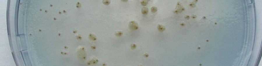
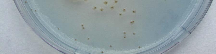
71 Fig. 1 64
72 Fig. 2. Comparison in selectivity between non-selective NA (left) and semi-selective medium PA 20 (right). Isolations were done from: A P. ananatis-free onion seed; B the same seed as in A spiked with P. ananatis. Note total suppression of non-target microflora on PA


73 A B Fig. 2 66
74 CHAPTER 3 Isolation and Identification of Pantoea ananatis from Onion Seed in South Africa As published in: Seed Science and Technology Vol. 43, pages Summary Center rot of onion, caused by the gram-negative, facultatively anaerobic bacterium, Pantoea ananatis, was first described in Georgia, USA, in The disease was later reported in Colorado and Michigan, but not in any other area of the world. P. ananatis is seed-borne and seed-transmitted in onions. A similar disease, leaf and seed stalk necrosis of onion, was reported in South Africa in 1981, and it was suggested that P. ananatis was responsible for that disease and was introduced into the USA on infected seed lots from South Africa. However, a representative strain of the leaf and seed stalk necrosis pathogen, isolated in South Africa in 1981, was identified as P. agglomerans in this study. As center rot of onion has not been observed in South Africa, and P. ananatis was not isolated from diseased plants, the main objectives of this research were to determine if pathogenic P. ananatis is present in local onion seed and to compare the South African strains to center rot causing strains from the USA. Colonies resembling those of P. ananatis were isolated from four seed lots on a semi-selective medium, PA 20. Pathogenicity tests demonstrated that the South African strains could induce the same symptoms on onion as those caused by the American strains. Physiological and biochemical characterization using Biolog, API 20E and API 50 CHE systems, and analysis of the 16S rrna gene sequences identified the strains from seed as P. ananatis. This is the first report on the presence of the center rot pathogen of onion, P. ananatis, in South African onion seed. 67
75 Introduction Center rot disease of onion was first observed in Georgia, USA, on Sweet Vidalia onions in 1997 (Gitaitis and Gay, 1997) and later in Colorado (Schwartz and Otto, 2000a) and Michigan (Mark et al., 2002). The disease has occurred in commercial fields in Georgia every year since 1997 and accounted for 100% loss in some fields (Walcott et al., 2002). The causal agent of center rot is a Gram-negative, facultatively anaerobic bacterium, identified as Pantoea ananatis (Gitaitis and Gay, 1997). The disease affects the center leaves of onions, which become water-soaked, soft, and bleached white as the rot progresses. Advanced stages of the disease result in complete wilting and bleaching of all leaves (Mark et al., 2002; Walcott et al., 2002). Walcott et al. (2002), reported natural infestation and transmission of P. ananatis in onion seed and implied that seed was the primary source of inoculum. This pathogen is known to be seed-borne and seed-transmitted in rice (Tabei et al., 1988), sudangrass (Azad et al., 2000) and buckwheat (Iimura and Hosono, 1996). In the 2004/2005 growing season 358 tons of onion seed were produced in South Africa, of which 282 tons were for the export market (Sansor Annual Report, 2005). Efforts to control the quality of commercially produced onion seed concentrated on the detection of fungal pathogens, and little is known about bacterial pathogens that may be present. P. ananatis is the causal agent of bacterial blight and dieback of Eucalyptus in South Africa (Coutinho et al., 2002) but center rot of onion has not been reported. However, in 1981, Erwinia herbicola was reported to cause a similar disease, stalk and leaf necrosis of onion in South Africa (Hattingh and Walters, 1981). In 1989, Beji et al. (1988) proposed transferring E. herbicola (syn. E. milletiae) to the new genus Pantoea (Gavini et al., 1989), that included species formerly classified as E. ananas, E. uredovora and E. stewartii (Mergaert et al., 1993). The seed associated with the first outbreak of center rot of onion in Georgia, USA, was produced in South Africa and Walcott et al. (2002) suggested that the center rot pathogen was possibly introduced on infested seed lots. The goal of this study was to determine if pathogenic P. ananatis was present in South African onion seed and to compare such strains to those associated with center rot of onions in the USA. 68
76 Materials and methods Media used for isolations Two different culture media were used for isolations. The first was a general growth medium, nutrient agar (NA) (Difco, Sparks, MD) as recommended by Walcott et al. (2002). Another was a semi-selective medium PA 20 (Goszczynska et al., 2006) developed specifically for isolation of P. ananatis from onion seed. PA 20 contained the following per liter: NaCl, 20 g; K 2 HPO 4, 1 g; NH 4 H 2 PO 4, 1 g; MgSO 4 x7h 2 O, 0.2 g, bromothymol blue, 1 ml of 1.6% aq. solution; crystal violet, 2 ml of 0.075% aq. solution; agar, 15 g. The ph was adjusted to 8.0 with 1.0 N NaOH. After autoclaving and cooling to 50 o C, 3 g of D (+) arabitol dissolved in 5 ml water and 2 ml of 1% aq solution of thallium nitrate were added. Both solutions were filter sterilized prior to adding them to the medium. Isolations from plants Diseased onion fields in the Small Karoo area of the Western Cape Province were identified by the onion seed producers in June-July and November-January between Disease symptoms on twenty-four experimental fields of Allium cepa, Granex type, were similar to leaf and stalk necrosis described by Hattingh and Walters (1981). Irregular oval spots, 4-10 cm long with a necrotic center and a dirty yellow or water-soaked margin lesions were found on onion leaves and seed stalks. Small water-soaked lesions were also present. In June-July 2003, nineteen onion fields of commercial bulb producers in the Northern and Free State Provinces were inspected. Onions from these fields were healthy. In April 2005, on one water-logged field in the Northern Province, where the cv. Granex 33 was grown, symptoms similar to that of center rot were observed. The youngest, center leaves of onions were chlorotic, becoming soft and wilted as the disease advanced. A light tan discoloration progressed into bulbs, resulting in the decay of the inner scales. The disease affected approximately 50% of the crop. Ten to twenty diseased plants were collected from each field and isolations were done from all collected plants. Tissue from lesion margins of infected leaf, seed stalk or bulb was removed with a sterile scalpel and macerated in 1 ml of sterile quarter-strength Ringer buffer (Oxoid, Basingstoke, Hampshire, England) for 20 min at 25 o C. Plant extracts were streaked onto NA and PA 20. NA and PA 20 plates were evaluated after four and seven days of incubation at 25 o C, respectively. Suspected colonies were purified by streaking onto NA and 69
77 cultures stored in milk-glycerol liquid medium (10% skim milk, 15% glycerol in distilled water) at 20 o C for further analysis. Isolations from seed Thirty-two seed samples were evaluated for the presence of P. ananatis: twenty-eight from A. cepa, two from A. fistulosum and two from A. porri. Fifteen A. cepa seed samples were commercial seed: six of cv. Granex 33, three of cv. Pegasus, two of cv. Pyramid, one of cv. Capricio and three of experimental onions (Granex type). The seed were produced in the Small Karoo area and harvested one or two years prior to testing. One sample, of an unknown cultivar, was from a farmer in the Northern Province, who produced the seed for his own use. The remaining twelve seed samples were harvested in January 2004 from umbels of experimental onion (Granex type) fields in the Small Karoo, in which leaf blight developed naturally. Twenty umbels were collected from each field and pooled into one sample. Seeds were extracted manually and stored in paper bags at 4 o C until processing. Isolations from seed were carried out as described previously (Goszczynska et al., 2006). For each seed lot, two sub-samples, 5 g each, were crushed and placed into separate sterile Erlenmeyer s flasks containing 100 ml of a quarter-strength Ringer buffer. Flasks were incubated for 30 min at 25 o C on a rotary shaker at 180 rpm/min. For each sample, four tenfold serial dilutions were made in half strength nutrient broth. A volume of 0.1 ml of each dilution was plated onto three plates each of NA and PA 20. Plates were incubated at 25 o C. Colonies were counted and evaluated after 7 days. Suspected colonies of the pathogen were purified by streaking onto NA and stored at 20 o C in milk-glycerol liquid medium for further analysis. Preliminary characterization of strains From each seed lot, 20 yellow colonies from NA and 20 yellow colonies from PA 20 were purified and used in preliminary pathogenicity and biochemical tests. The strains from seed washings were characterized by colony morphology, pigment production on NA, motility, cell morphology, Gram stain, oxidase reaction, catalase reaction, indole production, Hugh- Leifson oxidation/fermentation test, gas production from glucose and hydrogen sulphide production from cysteine (Mergaert et al., 1993), according to methods described by Fahy and Hayward (1983). Pathogenicity on onion was determined in green house assays by using a stub inoculation test (Goszczynska et al., 2005). A sterile needle was dipped into the bacterial colony on NA (24-48 hours growth) and then the needle was inserted under the 70
78 epidermis of a leaf. At least two leaves were inoculated per isolate. Inoculated plants were incubated in a greenhouse with 27 o C/23 o C day/night temperatures and observed daily for the development of symptoms. Bacterial strains The following bacterial strains from onion were characterized in the study: three strains isolated from South African seed (Table 1), the strain reported to cause leaf and seed stalk necrosis of onion in 1981 and four P. ananatis strains associated with center rot in the USA (Table 2). Two P. ananatis strains causing blight of Eucalyptus, and the type strains of P. ananatis LMG 2665 T and P. agglomerans LMG 1286 T were used as reference strains (Table 2). Stock cultures of all strains were maintained in milk-glycerol liquid medium at 20 o C. Stock strains were transferred onto NA plates and incubated at 25 o C to recover growing cultures. Cultures were routinely checked for purity and colony characteristics on NA. Pathogenicity tests Spray inoculations were performed with strains listed in Table 2. Strains were grown on NA at 25 o C for 24 hours. Bacterial suspensions were made in sterile distilled water to obtain approximately 10 7 colony forming units (CFU)/ml (as determined by dilution plating on NA). Six to eight week-old onion plants (A. cepa - cv. Granex 33 and cv. Pyramid; A. fistulosum - cv. White Welsh) and leek plants (A. porri - cv. Giant Italian) were spray inoculated to runoff with each bacterial suspension. Negative control plants were inoculated with sterile distilled water. Three plants were inoculated with each strain. Plants were incubated for 72 hours in a humidity chamber at 27 o C and relative humidity of 95%. Later plants were maintained in a greenhouse with 27 o C/23 o C day/night temperatures and observed daily for three weeks for the development of symptoms. The identities of the bacteria re-isolated from lesions were confirmed by colony morphology on NA and PA 20, Gram stain, oxidase reaction, Hugh-Leifson test, and indole production (Coutinho et al., 2002; Walcott et al., 2002). Biochemical and physiological tests Strains listed in Table 2 were characterized by sole carbon source utilization profiles, using Biolog GN Microplates together with Biolog MicroLog version 4.2 software (Biolog, Inc., Hayward, CA), according to the manufacturer s instructions. The strains were also tested with the API 20E and API 50CHE systems (BioMérieux, La Balme les Grottes, 71
79 Montalieu Vercieu, France) using the procedure recommended by the manufacturers. The results of the API 20E and API 50CHE tests were recorded after 24 and 48 hours of incubation at 30 o C respectively. Profiles were identified using the APILAB V4.0 identification program (BioMérieux). Amplification and sequencing of the 16S rrna gene Genomic DNA of strains was extracted by the cetyltrimethylammonium bromide (CTAB) method according to the DNA Miniprep protocol of Wilson (1989). Colonies from each strain were picked from the 48 hours NA plates and transferred into micro centrifuge tubes with sterile STE (10mM Tris-HCl, 1mM EDTA, 2M NaCl, ph 8.0) buffer. Cell lysis was obtained by incubation with sodium dodecyl sulfate and selective precipitation of cell debris and polysaccharides with CTAB/NaCl. DNA was extracted with phenol-chloroform-isoamyl alcohol, precipitated with isopropanol, washed with ethanol, air dried and dissolved in 100 μl of sterile water. Purified DNA was quantified using a Dyna Quant 200 fluorometer (Hoefer, San Francisco, CA) and Hoescht H intercalating dye (Polysciences, Warrington, PA). The DNA concentration was adjusted to ng μl -1 with sterile water. The 16S rrna gene fragment was amplified using the universal primers fd1 (5 -AGA GTT TGA TCC TGG CTC AG-3 ) and rd1 (5 -AAG GAG GTG ATC CAG CCG CA-3 ) (Weisburg et al., 1991). DNA was amplified in 50 μl reaction volumes containing polymerase chain reaction (PCR) buffer (10 mm Tris-HCl, ph 9.0 at 25 o C; 50 mm KCl, 0.1% Triton X-100); 1.5 mm MgCl 2 ; 150 μm dntps; 1.0 μm each primer; Taq polymerase (Promega Corp., Madison, WI) 1 unit per reaction volume and ng DNA template μl -1. Amplifications were performed according to Weisburg at al. (1991) in a Hybaid Omni Gene thermocycler (Teddington, England). The amplified PCR products were cut from the gel and purified using a QIAquick PCR purification kit (Qiagen, Hilden, Germany), and then cloned with the pgem-t Easy Vector System II kit (Promega, Madison, WI) according to the manufacturer s instructions. Plasmids with inserts were purified using the QIAprep Spin kit (Qiagen). The sequencing was performed using the Dyenamic ET Dye terminator cycle sequencing kit for MegaBACE with dideoxynucletide chain-termination chemistry. Sequences were analyzed using a MegaBACE 500 Sequencer (Amersham Biosciences, Buckinghamshire, England) with MegaBACE 500 Sequence Analyzer (version 2.4) software. 72
80 The GenBank/EMBL databases were used for homology searches using the BLAST program (National Center for Biotechnology Information, U.S. National Institute of Health, Bethesda, MD). Sequence analysis The 16S rdna nucleotide sequences obtained in this study were aligned with a selection of 16S rdna sequences of Pantoea species from GenBank with the DNAMAN software (version 5.2.9, Lynnon Biosoft, Quebec, Canada), using an optimal alignment and a dynamic method according to Feng and Doolitlle (1987). Phylogenetic trees were constructed with the neighbor joining method (Saitou and Nei, 1987) and evolutionary distances calculated according to the method of Jukes and Cantor (1969) and Kimura (1980), using the DNAMAN software package. Bootstrap analysis of the data, based on 1000 permutations was used to assess the stability of relationships. Results Recovery from plants P. ananatis was not isolated from diseased plants. The lesions on onions with symptoms similar to leaf and seed stalk necrosis were caused by Pseudomonas syringae, which was frequently isolated from these plants on a Milk-Tween medium (Goszczynska and Serfontein, 1998) and confirmed by pathogenicity tests and LOPAT characteristics (Lelliot et al., 1966). Yellow, mucoid colonies were consistently isolated on NA from diseased onions with center rot-like symptoms. Yellow bacteria were gram-negative rods, oxidase negative, catalase positive, fermentatively utilized glucose and produced gas from glucose. Five representative strains were identified as Enterobacter cloacae using the Biolog system, with a similarity index of Five strains were positive in the following tests on the API 20E strips: β-galactosidase, arginine dihydrolase, lysine decarboxylase, utilization of citrate, tryptophane deaminase, production of acetoin, and acid production from glucose, mannitol, rhamnose, sucrose, melibiose, amygdalin and arabinose. They were negative in the following tests: ornithine decarboxylase, H 2 S production, urease, indole production, gelatinase, acid from inositol and sorbitol. The strains induced leaf rot on onion leaves in pathogenicity tests. 73
81 Rot of onions in South Africa and its causal agent E. cloacae were similar to that reported by Bishop (1990) and Schwartz and Otto (2000b) in the USA. Recovery from seed Yellow bacteria were recovered from most seed samples on NA. Preliminary identification using biochemical tests showed these strains were not P. ananatis (data not shown). All were negative in a stub inoculation pathogenicity test. On PA 20, P. ananatis was isolated from four of 28 A. cepa seed samples tested, but not from seed of A. fistulosum or A. porri. Characteristic yellow colonies, 3-4 mm in diameter, shiny, drop-shaped with small, granular, darker inclusions were visible on a PA 20 medium after 6-7 days of incubation at 25 o C (Goszczynska et al., 2006). The number of P. ananatis-like colonies recovered from the infested seed lots ranged from 2.2 x 10 3 CFU/gram to 5.0 x 10 6 CFU/gram of seed (Table 1). The strains from onion seed produced yellow colonies on NA, were gram-negative rods ( µm length and µm width), motile, oxidase negative and catalase positive. All strains fermentatively utilized glucose and produced indole from tryptophane. They did not produce gas from glucose nor hydrogen sulphide from cysteine. One pathogenic strain per seed lot was selected for further characterization: BD 336 and PA 4 from two different experimental onion seed lots, harvested two years prior to testing and BD 390 from the seed produced by a farmer for his own use. None of the strains from the commercial seed cv. Granex 33 was pathogenic on onion cv. Granex 33 in a stub test and they were excluded from the study. Pathogenicity test All eight selected strains from onion were pathogenic to onion cv. Granex 33, but not to leek (Table 2). Strains BD 336, BD 310 and PA 4 did not produce disease symptoms on onion cv. Pyramid, all other strains did. Branching onion (A. fistulosum) cv. White Welsh was susceptible only to BD 287. All strains, including BD 287, induced identical symptoms on susceptible onion plants. Two to three days after inoculation, water-soaked spots appeared at the tips of leaves that expanded into longitudinal, bleached, green lesions with chlorotic margins. After two weeks, plants were completely wilted (Figure 1). Colonies recovered from onion plants with disease symptoms were yellow, gram-negative rods, oxidase negative and utilized glucose as a facultative anaerobe. All strains, except re-isolated BD 287 produced indole. 74
82 Two strains from Eucalyptus, LMG and LMG 20104, did not induce the disease on onions or leek. Control plants inoculated with water did not develop symptoms, nor were yellow bacteria isolated from leaves of these plants on NA and PA 20. Physiological and biochemical characterization The metabolic profiles of the eight strains from onion and two strains from Eucalyptus on Biolog GN microplates were most similar to the database profile of P. agglomerans, with a mean similarity index of The type strains of P. ananatis, LMG 2665 T and P. agglomerans, LMG 1286 T were both identified as P. agglomerans by Biolog with a similarity index 0.51 (P. ananatis was not in the Biolog database). There was consistency in utilization of 66 substrates but some strains varied in the utilization of glycogen, adonitol, turanose, α-hydroxybutyric acid, bromosuccinic acid, succinamic acid, D-serine and uridine. The type strain of P. agglomerans and BD 287 gave nearly identical reactions and differed from others by not utilizing 11 substrates: Tween 40, Tween 80, gentiobiose, lactulose, D- melibiose, D-raffinose, D-sorbitol, citric acid, formic acid, D-glucosamic acid and quinic acid. Strain BD 287 did not utilize D-arabitol and psicose and LMG 1286 T did not utilize α- lactose and acetic acid. Physiological and biochemical profiles obtained by using the API 20E and API 50CHE systems revealed that South African P. ananatis had similar biochemical characteristics and resembled the American and Eucalyptus strains and the type strain of P. ananatis (Table 3). In the API 20E tests, LMG 2665 T, BD 301, BD 309 and BD 315 were identical. African BD 390 and PA 4 differed by giving a positive reaction in the API 20E inositol test. Strain BD 310 did not produce β-galactosidase. The three profiles were identified by the APILAB program as Pantoea spp. 2. South African BD 336 differed from the others by not utilizing citrate and not producing acid from rhamnose and sucrose. However, in the API 50CHE acid from sucrose was produced after 48 hours. The profile was not recognized by the program. Unlike P.ananatis from onion and LMG 2665 T, two strains from Eucalyptus liquefied gelatin (Coutinho et al.,2002). P. agglomerans LMG 1286 T and BD 287 differed from the other strains by the inability to utilize citrate, produce indole from tryptophan and acid from sorbitol. There was consistency in the ability of the twelve strains to produce acid from 35 substrates (15 positives and 20 negatives) on the API 50CHE strips (Table 3). The type strain of P. 75
83 agglomerans and BD 287 differed from the other strains by not producing acid from glycerol, inositol, D-sorbitol, D-raffinose and D-arabitol. 16S rdna sequence analysis In this study, almost complete 16S rdna sequences (~1500 bp) were determined for the four South African and four American strains. The sequences were deposited into the GenBank database. Accession numbers are shown in Table 2. A BLAST search of the EMBL/GenBank database conducted with the sequences revealed a high degree of sequence identity ( 98%) with previously determined sequences of bacteria belonging to the genus Pantoea. The seven indole-producing strains had the highest homology to the 16S rdna of P. ananatis strains LMG (accession number AF364847) and LMG (AF364844), ranging from 98.9% for BD 309 and BD 336 to 99.7% for BD 301 and BD 315. The strain BD 287 (Hattingh and Walters, 1981) had the highest homology (99.7%) to the 16S rdna sequence of P. agglomerans strain LMG 2565 (Z96082). Figure 2 shows the phylogenetic relationship derived from a neighbor-joining analysis of the pairwise comparison among the 16S rdna sequences of eight strains from this study with nine sequences of four described species of the genus Pantoea. Klebsiella pneumoniae DSM T (X87276) was used as an outgroup taxon. Phylogenetic trees constructed by two different methods, with Jukes-Cantor and Kimura algorithms, were identical (not shown). Five strains, three American (BD 301, BD 310, BD 315) and two South African (BD 390, PA 4), formed a cluster with P. ananatis (syn. E. uredovora) at 72% confidence value from bootstrap analysis. The strain BD 287 grouped closely (77% confidence value) with P. agglomerans (syn. E. herbicola, E. milletiae). The sequences of the South African BD 336 and BD 309 from the USA clustered with P. agglomerans on phylogenetic trees. Some strains of P. ananatis from rice studied by Cother et al. (2004) also grouped with P. agglomerans on phylogenetic trees of the 16S rrna gene sequences. Discussion Walcott et al. (2002) suggested that the center rot pathogen, P. ananatis, was introduced to the USA on infested onion seed lots produced in South Africa. The suggestion was based on the description of a similar disease of onion, stalk and leaf necrosis, reported in South Africa 76
84 in 1981(Hattingh and Walters, 1981). The causal agent of leaf and seed stalk necrosis, although pathogenic to onion, is not P. ananatis. Nutritional and genotypic characteristics of a representative strain, BD 287, closely resembled those of the P. agglomerans type strain, LMG 1286 T (Table 3, Figure 2). Our results are supported by those of Verdonck et al. (1987) and Beji et al. (1988) who also identified BD 287 as P. agglomerans in their taxonomic investigations. In this study, P. ananatis pathogenic to onion was detected in three South African onion seed lots. Three strains isolated from onion seed lots induced the same symptoms on onions as those caused by strains isolated from diseased plants in the USA (Figure 1). There were, however, differences in susceptibility among onions cultivars (Table 2), providing an opportunity for the selection of tolerant material. Interestedly, P. ananatis causing leaf blight of Eucalyptus did not induce the disease on onions in pathogenicity tests done in this study, suggesting that Eucalyptus strains could be host specific (Azad et al., 2000). When comparing the South African strains, the type strain P. ananatis and the American strains associated with center rot, by means of the Biolog system, we observed similar results in the utilization of 95 different carbon sources. With API 20E and API 50CHE strips, the profiles for the majority of onion strains were also similar to each other and to the reference strains of P. ananatis. Three strains (BD 336, BD 309 and BD 310) varied slightly in a few biochemical characteristics (Table 3). The production of acetoin and acid from L-rhamnose and methyl-α-d mannopyranoside differed among the 18 strains of P. ananatis studied by Mergaert et al., 1993, while β-galactosidase and citrate utilization were reported to be positive for all strains. P. ananatis from Eucalyptus (this study, Coutinho et al., 2002) and rice (Cother et al., 2004), also showed a few biochemical differences from those characteristic for the species. The genotypic characterization of the three strains from onion seed confirmed that South African strains are P. ananatis. Although the seed strain BD 336, and the American BD 309 clustered with P. agglomerans on phylogenetic trees (Figure 2), their 16S rdna sequences were 98.8% homologous to the P. ananatis type strain. Additionally, all seed strains and strains from the USA were indole positive and their Biolog and API profiles were more similar to those of P. ananatis from Eucalyptus, LMG 2665 T, and those reported for 18 strains of P. ananatis examined by Mergaert et al. (1993). We concluded that the strains isolated from onion seed belong to this species. Our results showed that P. ananatis pathogenic to onion is present in South African onion seed. This, however, does not support the claim that the pathogen was introduced to the USA 77
85 on infected seed lots from South Africa in 1997 (Walcott et al., 2002). P. ananatis was detected in the USA on Vidalia onions several years prior to the 1997 epidemic (Mark et al., 2002). P. ananatis is widely distributed throughout Georgia on many types of weeds as epiphytes (Gitaitis et al., 2000) and it has been reported to cause diseases on muskmelon (Bruton et al., 1986), honeydew melon (Wells et al.,1987) and Sudangrass (Azad et al., 2000) in the USA. The center rot pathogen is transmitted via tobacco thrips, Frankliniella fusca (Wells et al., 2002; Gitaitis et al., 2003). Although P. ananatis from sudangrass did not cause extensive disease in other plant species studied by Azad et al. (2000), the possibility that P. ananatis was transferred from another host to onions by thrips cannot be excluded. P. ananatis was not isolated from diseased onion plants in this study. Leaf and seed stalk blights were caused by P. syringae, which was also present in the majority of surveyed onion seed lots (data not shown). Center rot-like symptoms observed on the water logged field in 2005 were caused by E. cloacae, an opportunistic pathogen and a common component of the micro flora in water, soil and on plant surfaces (Bishop, 1990; Schwartz and Otto, 2000b). Our survey of onion fields was limited to three provinces in South Africa, and we cannot conclude that the disease was not present in other areas of the country. P. ananatis was found in three of twenty-eight locally produced A. cepa seed lots, and is a potential source of inoculum for the development of center rot. The diseases on sudangrass (Azad et al., 2000), Eucalyptus (Coutinho et al., 2002) and onions (Walcott et al., 2002) were prevalent during spring and summer, when temperatures and relative humidity were high. Onion is a winter crop in South Africa, and the arid environmental conditions could be not conductive for the disease development (Webster et al., 1983). A rapid screening technique to detect this bacterium in seed has to be evaluated, and commercially important seeds should be tested to determine their levels of infestation. Acknowledgements We thank Mr. Harry Boroko for technical help and Ms Di James (University of Cape Town, Department of Molecular and Cellular Biology, Cape Town, South Africa) for sequencing of 16S rdnas. Research was supported by the Agricultural Research Council and the National Research Foundation. 78
86 References Azad, H. R., Holmes, G. J. and Cooksey, D.A. (2000). A new leaf blotch disease of Sudangrass caused by Pantoea ananatis and Pantoea stewartii. Plant Disease, 84, Beji, A., Mergaert, J., Gavini, F., Izard, D., Kersters, K., Leclerc, H. and De Ley, J. (1988). Subjective synonymy of Erwinia herbicola, Erwinia milletiae, and Enterobacter agglomerans and redefinition of the taxon by genotypic and phenotypic data. International Journal of Systematic Bacteriology, 38, Bishop, A. L. (1990). Internal decay of onions caused by Enterobacter cloacae. Plant Disease, 74, Bruton, B. D., Wells, J. M., Lester, G. E. and Patterson, C. L. (1991). Pathogenicity and characterization of Erwinia ananas causing a postharvest disease of cantaloup fruit. Plant Disease, 75, Cother, E. J., Reinke, R., McKenzie, C., Lanoiselet, V. M. and Noble, D. H. (2004). An unusual stem necrosis of rice caused by Pantoea ananas and the first record of this pathogen on rice in Australia. Australasian Plant Pathology, 33, Coutinho, T.A., Preisig, O., Mergaert, J., Cnockaert, M. C., Riedel, K.-H., Swings, J. and Wingfield, M. J. (2002). Bacterial blight and dieback of Eucalyptus species, hybrids and clones in South Africa. Plant Disease, 86, Fahy, P. C. and Hayward, A. C. (1983). Media and methods for isolation and diagnostic tests. In Plant bacterial diseases, a diagnostic guide. (eds. Fahy, P. C., and Persley, G. J.), pp , Academic Press Australia. Feng, D. F. and Doolittle, R. F. (1987). Progressive sequence alignment as a prerequisite to correct phylogenetic trees. Journal of Molecular Evolution, 25, Gavini, F., Mergaert, J., Beji, A., Mielcarek, C., Izard, D., Kersters, K. and De Ley, J. (1989). Transfer of Enterobacter agglomerans (Beijerinck 1888) Ewing and Fife 1972 to 79
87 Pantoea gen. Nov. as Pantoea agglomerans comb. Nov. and description of Pantoea dispersa sp. nov. International Journal of Systematic Bacteriology, 39, Gitaitis, R. D. and Gay, J. D. (1997). First report of a leaf blight, seed stalk rot, and bulb decay of onion by Pantoea ananas in Georgia. Plant Disease,. 81, Gitaitis, R. D., Walcott, R. R., Wells, M. L., Diaz Perez, J. C. and Sanders, F. H. (2003). Transmission of Pantoea ananatis, causal agent of center rot of onion, by tobacco thrips, Frankliniella fusca. Plant. Disease, 87, Gitaitis, R. D., Zolobowska, L., Culpepper, S., Langston, D. and Walcott, R. (2000). Polymerase chain reaction detection of the onion pathogen Pantoea ananatis on various weeds and crops in Georgia, USA. In Proceeding of the International Conference on Plant Pathogenic Bacteria, 10 th (ed. S. H. De Boer), P1, Charlottetown, Prince Edward Island, Canada, pp , Kluwer Academic Publishers, Dordrecht, Netherlands. Goszczynska, T. and Serfontein, J. J. (1998). Milk-Tween agar, a semi-selective medium for isolation and differentiation of Pseudomonas syringae pv. syringae, Pseudomonas syringae pv. phaseolicola and Xanthomonas axonopodis pv. phaseoli. Journal of Microbiological Methods, 32, Goszczynska, T., Venter, S. N. and Coutinho T. A. (2006). PA 20, a semiselective medium for isolation and enumeration of Pantoea ananatis. Journal of Microbiological Methods, 64, Hattingh, M. J. and Walters, D. F. (1981). Stalk and leaf necrosis of onion caused by Erwinia herbicola. Plant Disease, 65, Iimura, K. and Hosono, A. (1996). Biochemical characteristics of Enterobacter agglomerans and related strains found in buckwheat seeds. International Journal of Food Microbiology, 30, Jukes, T. H. and Cantor, C. R. (1969). Evolution of protein molecules, p In Munro, H. N. (ed.), Mammalian protein metabolism. Academic Press, New York, N. Y. 80
88 Kimura, M. (1980). A simple method for estimating evolutionary rates of base substitutions through comparative studies of nucleotide sequences. Journal of Molecular Evolution, 16, Lelliot, R. A., Billing, E. and Hayward, A. C. (1966). A determinative scheme for the fluorescent plant pathogenic Pseudomonas. Journal of Applied Bacteriology, 29, Mark, G. L., Gitaitis, R. D. and Lorbeer, J. W. (2002). Bacterial diseases of onion. In Allium crop science: recent advances, (eds. H. D. Rabinowitch and L. Currah), , CAB International, UK, Wallington. Mergaert, J., Verdonck, L. and Kersters, K. (1993). Transfer of Erwinia ananas (synonym, Erwinia uredovora) and Erwinia stewartii to the genus Pantoea emend. as Pantoea ananas (Serrano 1928) comb. Nov. and Pantoea stewartii (Smith 1898) comb. nov., respectively, and description of Pantoea stewartii subsp. indologenes subsp. nov. International Journal of Systematic Bacteriology, 43, Saitou, N. and Nei, M. (1987). The neighbor-joining method: a new method for reconstructing phylogenetic trees. Molecular Biological Evolution, 4, SANSOR Annual Report 2004/05. (2005). South African National Seed Organisation. South Africa, Pretoria. Schwartz, H. F. and Otto, K. (2000a). First report of a leaf blight and bulb decay of onion by Pantoea ananatis in Colorado. Plant Disease, 84, 808. Schwartz, H. F. and Otto, K. (2000b). First report of a bulb decay of onion by Enterobacter cloacae in Colorado. Plant Disease, 84: 808. Tabei, H., Azegami, K. and Fvkuda, T. (1988). Infection site of rice grain with Erwinia herbicola, the causal agent of bacterial palea browning of rice. Annuals of the Phytopathological Society of Japan, 54,
89 Verdonck, L., Mergaert, J., Rijckaert, C., Swings, J., Kersters, K. and De Ley, J. (1987). Genus Erwinia: Numerical analysis of phenotypic features. International Journal of Systematic Bacteriology, 37, Walcott, R. R., Gitaitis, R. D., Castro, A. C., Sanders Jr., F. H. and Diaz-Perez, J. C. (2002). Natural infestation of onion seed by Pantoea ananatis, causal agent of center rot. Plant Disease, 86, Webster, D. M., Atkin, J. D. and Cross, J. E. (1983). Bacterial blights of snap beans and their control. Plant Disease, 67, Weisburg, W. G., Barns, S. M., Pelletier, D. A. and Lane, D. J. (1991). 16S ribosomal DNA amplification for phylogenetic study. Journal of Bacteriology, 173, Wells, M. L., Gitaitis, R. D. and Sanders, F. H. (2002). Association of tobacco thrips, Frankliniella fusca (Thysanoptera: Thripidae) with two species of bacteria of the genus Pantoea. Annuals of Entomological Society of America, 95, Wells, J. M., Sheug, W. S., Ceponis, M. J. and Chen, T. A. (1987). Isolation and characterization of strains of Erwinia ananas from honeydew melons. Phytopathology, 77, Wilson, K. (1989). Preparation of genomic DNA from bacteria. In Current protocols in molecular biology, (eds. F. M. Ausubel, R. Brent, R. E. Kimgston, D. D. Moore, J. G. Seidman,J. A. Smith and K. Struhl), , John Wiley & Sons, Inc, New York, N.Y. 82
90 Table 1. Recovery of P. ananatis from naturally infested A. cepa seed on PA 20 medium a and its pathogenicity to onion (cv. Granex 33) in a stub inoculation pathogenicity test b. Seed lot Description Cultivar P. ananatis CFU/gram Pathogenicity to onion 20 of seed a strains per seed lot 1 Commercial, Granex x / 20-2 Experimental, Granex type 2.4 x / 20 BD Experimental, Granex type 2.2 x / 20 PA 4 4 Farmer own use, unknown 5.0 x / 20 BD 390 Strains selected for characterization a Characteristic yellow colonies, 3-4 mm in diameter, shiny, drop-shaped with small, granular, darker inclusions on PA 20 medium were regarded as P. ananatis. Colonies were counted after 7 days of incubation. Figures represent the mean of three replicates. b Results were recorded after 4 days. Positive reaction: water soaked spots that expanded into longitudinal, bleached-green lesions with chlorotic margins (Goszczynska et al., 2006). 83
91 Table 2. Bacterial strains characterized in this study and their pathogenicity to three onion cultivars. Pathogenicity to onion cultivars in a greenhouse test Strain a Origin 16S rdna GenBank A. cepa A. fistulosum accession number c cv. Granex 33 cv. Pyramid cv. White Welsh USA b BD 301 Blackshank 15, diseased onion AY BD 309 Horticulture Hill 24, diseased onion AY BD 310 Horticulture Hill 31, diseased onion AY BD 315 Pans , diseased onion AY South Africa BD 336 Onion seed AY BD 390 Onion seed AY PA 4 Onion seed AY BD 287 b SUH 2, leaf & stalk necrosis AY Reference strains LMG P. ananatis, Eucalyptus AF LMG P. ananatis, Eucalyptus AF LMG 1286 T P. agglomerans, type strain AJ nd nd nd LMG 2665 T P. ananatis, type strain Z96081 nd nd nd 84
92 a BD and PA strains: accession numbers of the Plant Pathogenic and Plant Protecting Bacteria (PPPPB) culture collection, ARC-PPRI, Pretoria, South Africa. American strains deposited by R. Walcott, Department of Plant Pathology, University of Georgia, Athens, USA. LMG strains: accession numbers of the BCCM/LMG Culture Collection, University Gent, Belgium. b Isolated in 1981, Hattingh and Walters (1981). c 16S rdna sequences of reference strains obtained from the GenBank database. nd not done 85
93 Table 3. Physiological and biochemical characteristics of the American and South African onion strains and reference strains of P. ananatis and P. agglomerans using the API 20E and API 50CHE systems. P. ananatis American strains South African strains P. agglomerans Characteristic a LMG2665 T LMG20103 BD 301 BD 309 BD 310 BD 315 BD336 BD390 PA4 LMG1286 T BD 287 LMG20104 API 20E ß-galactosidase Simmons citrate Production of Indole Acetoin Gelatinase API 50CHE Acid production Glycerol L-rhamnose Inositol D-sorbitol Methyl-α-D mannopyranoside Amygdalin
94 P. ananatis American strains South African strains P. agglomerans Characteristic a LMG2665 T LMG20103 BD 301 BD 309 BD 310 BD 315 BD336 BD390 PA4 LMG1286 T BD 287 LMG20104 D-cellobiose D-lactose D-melibiose D-raffinose D-fucose D-arabitol Potassium ± - ± ± gluconate Aesculin hydrolysis a API 20E tests data was recorded after 24 hours, API 50CHE data (acid production) after 48 hours. Incubation temperature was at 30 o C. Symbol (+) indicates positive reaction; symbol (-) negative reaction; symbol (±) uncertain. All strains produced acid from: L-arabinose, D-ribose, D-xylose, D-galactose, D-glucose, D-fructose, D-mannose, D-mannitol, N- acetyloglucosamine, gentibiose, arbutin, salicylin, D-maltose, sucrose and D-trehalose. All strains were negative in the following tests: arginine dihydrolase, lysine decarboxylase, ornithine decarboxylase, H 2 S, urease, tryptophane desaminase and acid production from: erythitol, D-arabinose, L-xylose, D-adonitol, methyl-b-d-xylopyranoside, L-sorbose, 87
95 dulcitol, methyl-α-d-glucopyranoside, inulin, D-melezitose, starch, glycogen, xylitol, D-turanose, D-lyxose, D-tagatose, L-fucose, L- arabitol, potassium 2-ketogluconate and potassium 5-ketogluconate. 88
96 Fig. 1. Symptoms on onion induced by the strains associated with center rot in the USA and by the South African strains isolated from onion seed. 89

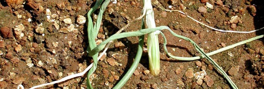
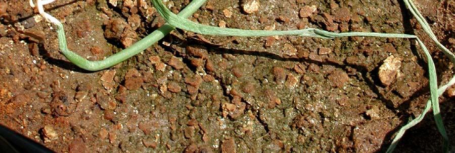
97 Fig. 1 90
98 Fig. 2. Phylogenetic tree showing the relationship among selected partial 16S rdna sequences from Pantoea species, including onion strains (in bold; South African strains from seed are underlined), based on pairwise comparisons using the Jukes-Cantor parameter. The sequence of Klebsiella pneumoniae was used as an outgroup taxon. The numbers at the nodes indicate the levels of bootstrap support based on data for replicates; only values that are greater then 50% are shown. Accession numbers (in brackets) and the sequences of P. ananatis, P. agglomerans, P. stewartii and K. pneumoniae were obtained from the GenGank/EMBL databases. 91
99 69 93 BD LMG 2565 P. agglomerans (Z96082) 100 LMG 2660 P. agglomerans (U80183) DSM 3493 T P. agglomerans (AJ233423) BD 336 BD 309 PA 4 58 LMG 2676 P. ananatis (U80209) BD BD 310 LMG P. ananatis (AF364844) BD 315 BD 390 LMG 2665 T P. ananatis (Z96081) LMG P. ananatis (AF364847) LMG 2632 T P. stewartii subsp. indologenes (Y13251) 89 LMG 2715 T P. stewartii subsp. stewartii (U80208) DSM T Klebsiella pneumoniae (X87276)
100 CHAPTER 4 Isolation and Identification of the Causal Agent of Brown Stalk Rot, a New Disease of Maize in South Africa As published in: Plant Disease Vol. 91, pages ABSTRACT During , an unreported disease of maize (Zea mays) was observed on commercial fields in the Northwest and Mpumalanga Provinces of South Africa. Infected plants were stunted, with a vertical crack at the fist internode. Inside the stem, a dark brown, narrow lesion was present along the crack. Internal browning inside the stem extended upwards, reaching the top internode in some plants. Seed cobs were underdeveloped. Diseased plants were scattered in the fields and 10-70% of the crop was affected. Gram-negative, facultatively anaerobic bacteria were consistently isolated from diseased tissues. Pathogenicity tests established that representative strains induced disease symptoms similar to those observed on maize plants in the field. Physiological and biochemical characterization using the API 20E and API 50CHE systems and 16S rrna gene sequence analyses showed that the strains belonged to the genus Pantoea. The results of these tests also separated the strains into two groups. The first group, giving a positive reaction in the indole test, was similar to P. ananatis. The second group of strains was indole negative and resembled P. agglomerans. The F-AFLP genomic fingerprints generated by the indole positive strains and P. ananatis reference strains were similar and clustered together in the dendrogram, confirming that the indole positive bacteria causing brown stalk rot on maize were P. ananatis. The F-AFLP fingerprints produced by the indole negative strains were distinctly different from those generated by P. ananatis, P. agglomerans, P. dispersa, P. citrea, P. stewartii subsp. stewartii and P. stewartii subsp. indologenes. The results indicated that indole negative bacteria causing brown stalk rot on maize might belong to a previously undescribed species of the genus Pantoea. This is the first report of a new disease on maize, brown stalk rot, caused by two bacterial species, P. ananatis and an undescribed Pantoea sp. 93
101 Additional keywords: corn, detection 94
102 Maize (Zea mays) is the most important grain crop in South Africa, being both the major animal feed grain and the staple food of the majority of the population. For the 2003/2004 marketing year maize was responsible for the second largest contribution to the gross value of agricultural production in the country. The South African maize industry is also the largest maize industry in Africa (39). Commercial farmers cultivate nearly three million hectares of maize per year. In the past five years, South Africa produced between 7.2 and 10.1 million tons of maize per annum, with an average of 9.2 million tons. The main maize production areas in South Africa are the Free State, Northwest and Mpumalanga Provinces. These three provinces are responsible for 85% of the total maize produced in the country (7, 39). Numerous fungal diseases cause excessive damage to maize in South Africa (12), especially Puccinia common rust (13), Cercospora gray leaf spot (20), Aureobasidium leaf spot (18), Fusarium (14, 15, 16) and Stenocarpella (13) stem, cob and root rots. By contrast, only two bacterial diseases have been reported on maize in the country, leaf streak (11) caused by Xanthomonas campestris pv. zeae and bacterial stalk rot (35), caused by Dickeya zeae. In January 2004, a new disease was observed in a single breeder s field in the Mpumalanga Province on an inbred female line. In December 2004 and January/February 2005 the same symptoms were also found in commercial maize fields in the Northwest and Mpumalanga Provinces. The disease was prevalent on the inbreed female and single cross female lines and a commercial hybrid SR 52. Affected plants were stunted and a vertical crack at the first internode was always present (Fig. 1A). In most diseased plants, the crack extended into the second or sometimes the third internode. When the crack was split open, a dark brown, narrow lesion was visible along the crack (Fig.1B). Internal browning inside the stem extended upwards, reaching the top internode in some plants (Fig. 1C). Seed cobs were underdeveloped. On one field, the leaf margins were soft, bleached-green and curled inwards (Fig. 1D). Diseased plants were scattered throughout the fields and 10-70% of the crop was affected. When examined under a microscope, cut edges of symptomatic tissues consistently exhibited bacterial streaming. Gram negative bacteria producing yellow colonies were consistently isolated from diseased tissues and these were tentatively identified as belonging to the genus Pantoea (32). Two bacterial species of Pantoea have been reported to cause diseases on maize and other Poaceae hosts. P. stewartii subsp. stewartii causes Stewart s wilt in Europe, Asia and the Americas, but not in South Africa (8, 33). P. ananatis was described as an agent of leaf spot on maize in Brazil (34), stem necrosis of rice in Australia 95
103 (9) and glume and grain discoloration of rice in Japan (4). P. ananatis and P. stewartii subsp. indologenes were reported to cause leaf blotch on sudangrass in California (3). The aim of this study was to identify and characterize the causal agent of brown stalk rot of maize observed in South Africa. MATERIALS AND METHODS Isolation of the causal agent from plants. Maize breeders and commercial farmers reported ten diseased maize fields throughout Mpumalanga and Northwest Provinces. Typical brown stalk rot symptoms were observed in these fields, on which the inbreed female, the single cross female lines and the hybrid SR 52 were cultivated. One hundred diseased plants with brown stalk rot symptoms, approximately ten plants per field, were collected in January 2004, December 2004 and February Isolations were done from all collected plants, from at least one stalk lesion per plant and from the necrotic stripes on leaves observed in one field. Small sections of stalk or leaf tissue with symptoms were cut aseptically from the margins of lesions and macerated in 1 ml of sterile distilled water for 20 min at 25 o C. Plant extracts were streaked onto nutrient agar (Difco, Sparks, MD), tryptone glucose extract agar (TGA) (Difco) and King s B (30) media. Plates were incubated at 25 o C and examined after 3 to 5 days for bacterial growth. On nutrient agar, bacterial colonies were very small, approximately 1 mm in diameter. On King s B, bacterial growth was very slimy and it was difficult to distinguish single colonies. On TGA, yellow, circular, well-separated colonies, 3-4 mm in diameter, were visible 2 to 3 days after plating. After the initial isolations from approximately 20 plants, subsequent isolations were made on TGA plates only. Plates were incubated at 25 o C and examined after 3 to 5 days for bacterial growth. Suspected colonies were purified by streaking onto TGA and cultures stored in milk-glycerol liquid medium (10% skim milk, 15% glycerol in distilled water) at 20 o C for further analysis. The same procedure was used to re-isolate bacteria from inoculated plants. Preliminary identification of bacteria. Ten to twenty yellow bacterial colonies per field isolated on TGA medium were purified and used in preliminary identification tests. The strains were characterized by colony morphology on TGA, motility, cell morphology, Gram stain, catalase and oxidase reaction, Hugh-Leifson oxidation/fermentation test, production of 96
104 indole, gas from glucose and hydrogen sulphide from cysteine (32), according to methods described by Fahy and Hayward (17). Bacterial strains. Bacterial strains from maize characterized in this study are listed in Table 1. Type strains of P. ananatis, P. agglomerans, P. dispersa, P. citrea, P. stewartii subsp. indologenes and P. stewartii subsp. stewartii and seven P. ananatis strains isolated from sudangrass, onion and Eucalyptus were used as reference strains (Table 1). P. stewartii subsp. stewartii is a quarantine organism in South Africa and only DNA was used in this study. Stock cultures of all strains were maintained in milk-glycerol liquid medium at 20 o C. Stock strains were transferred onto TGA plates and incubated at 25 o C to recover growing cultures. Cultures were routinely checked for purity and colony characteristics on TGA. Pathogenicity tests. Pathogenicity on maize was determined in greenhouse assays on maize cultivar SR 52, shown to be susceptible in the field. Thirteen strains from maize, two P. ananatis from Eucalyptus, five from onion and one from sudangrass were used in pathogenicity tests (Table 1). Each bacterial strain was grown on TGA plates for hours at 25 o C, and each strain was suspended in sterile distilled water to obtain approximately 10 7 CFU/ml. The concentrations were confirmed by dilution plating on TGA plates. Maize plants were inoculated immediately after preparation of the bacterial suspensions. In the first experiment, maize plants were four weeks old and in the second experiment, plants were nine weeks old. Four-week-old plants were inoculated using two methods: (a) suspensions of single strains were infiltrated into leaf tissues with a syringe and (b) suspensions of single strains were injected into stems below the first leaf whorl. Inoculated plants were placed in plastic bags and maintained in a greenhouse with natural light and 28/23 o C day/night temperature. After seven days the plastic bags were removed and plants were kept in the same greenhouse. Relative humidity (RH) fluctuated between 50 and 70%. Nine-week-old plants were inoculated by injecting a few drops of bacterial suspensions into the first internode of the stem. Stem-inoculated plants were maintained in a greenhouse with natural light and at temperatures and RH described above. At least two plants were inoculated with each bacterial strain per inoculation method. Each experiment continued for ten weeks. Control plants in all experiments were inoculated with sterile distilled water. Pathogenicity tests were performed twice. Bacteria re-isolated from symptomatic tissues were confirmed by colony morphology on TGA, Gram stain, indole production, and Huigh-Leifson and oxidase tests (10, 22). 97
105 Biochemical and physiological tests. Biochemical and physiological characteristics of the strains listed in Table 1 were examined with the API 20E and API 50CHE systems (BioMérieux, La Balme les Grottes, Montalieu Vercieu, France), according to the procedure recommended by the manufacturers. The results of the API 20E and API 50CHE tests were recorded after 24 and 48 hours of incubation at 30 o C, respectively. Profiles were identified using the APILAB V4.0 identification program (BioMérieux). The data for the P. stewartii subsp. stewartii strain LMG 2715 T was obtained from the paper by Mergaert et al. (32). The cluster analysis was performed with the combined API 20E (21 characters) and API 50CHE (49 characters) data, using the Bionumerics software (version 4.5, Applied Maths, Kortrijk, Belgium). Similarity matrixes were constructed with the Dice coefficient and cluster analyses were performed by the unweighted pair group method (UPGMA). The data was presented as an UPGMA dendrogram. DNA extraction. Genomic DNA of bacterial strains (Table 1) was extracted by using the GenElute Bacterial Genomic DNA Kit (Sigma, Steinheim, Germany), according to the manufacturer s instructions. Purified DNA was quantified using a Dyna Quant 200 fluorometer (Hoefer, San Francisco, CA) and Hoescht H intercalating dye (Polysciences, Warrington, PA). The DNA concentration was adjusted to ng μl -1 with sterile water. The DNA was stored at 20 o C until further analysis. Fluorescent amplified fragment length polymorphism analysis. All strains listed in Table 1 were examined by the fluorescent amplified fragment length polymorphism (F- AFLP)-based system for the identification of plant-associated species from the genus Pantoea (6). Genomic DNA, ng from each isolate, was digested with EcoRI and MseI (Roche, Mannheim, Germany) restriction enzymes, and then ligated to the respective adaptors for 2 hours at 20 o C. Ligation reactions were diluted 1:10 in nuclease-free water for preamplification with Eco-00 (5 -GAC TGC GTA CCA ATT C-3 ) and Mse-00 (5 -GAT GAG TCC TGA CTA A-3 ) primers (Inquaba, Pretoria, South Africa). Each 25 μl preamplification reaction contained PCR buffer (10 mm Tris-HCl, ph 9.0 at 25 o C; 50 mm KCl, 0.1% Triton X-100); 1.5 mm MgCl 2, 250 μm of each dntps; 100 pmol of each primer; 1 U Taq polymerase (Qiagen, Hilden, Germany) and 2 μl diluted ligation reaction. Amplification 98
106 conditions were as follows: initial denaturation at 94 o C for 3 min; 20 cycles at 94 o C for 30 s, 56 o C for 1 min and 72 o C for 1 min; and final elongation at 72 o C for 5 min. Each preamplification reaction was diluted 1:50 in nuclease-free water prior to selective amplification with Mse-CG (5 -GAT GAG TCC TGA CTA ACG-3 ) and fluorescently labeled Eco-G (5 - GAC TGC GTA CCA ATT CG-3 ) primers (Inquaba). A volume of selective PCR reactions was 20 μl, and each contained: PCR buffer; 1.5 mm MgCl 2 ; 250 μm of each dntps; 0.5 pmol Eco-G primer; 2.4 pmol Mse-CG primer; 1 U Taq polymerase and 5 μl diluted preamplification reaction. The selective PCR amplifications included the following steps: denaturation at 94 o C for 5 min; 9 cycles of denaturation at 94 o C for 30 s, initial annealing at 65 o C for 30s (annealing temperature was reduced by 1 o C per cycle until 56 o C was reached) and elongation at 72 o C for 1 min; 23 cycles at 94 o C for 30 s, 56 o C for 30 s and 72 o C for 1 min, followed by 5 min extension at 72 o C. Amplifications were performed at least twice for each strain in the Hybaid Omni Gene thermocycler (Teddington, England). The selective amplification reactions (1 μl) were mixed with an equal volume of formamide loading buffer (95% formamide, 20 mm EDTA, bromophenol blue), heated for 3 min at 90 o C and then chilled on ice for 10 min. A volume of μl of each mixture was loaded onto the gel. The AFLP products were separated in 8% Long Ranger (LI-COR Biosciences, Lincoln, NE) denaturing gels on a LI-COR IR automated sequencer. Electrophoresis was carried out for 4 hours at 1500 V in 0.8 x TBE buffer. Images were imported into BioNumerics software. Gels were standardized with a bp sizing standard (LI-COR) and the bands between 50 and 700 bp were analyzed (6). Similarity was calculated using the Dice correlation coefficient and cluster analysis was performed by the UPGMA method. The data was presented as an UPGMA dendrogram. 99
107 Amplification and sequencing of the 16S rrna gene. The 16S rrna gene fragment of thirteen strains from maize (Table 1) was amplified using the universal primers fd1 (5 - AGA GTT TGA TCC TGG CTC AG-3 ) and rd1 (5 -AAG GAG GTG ATC CAG CCG CA-3 ) (41). DNA was amplified in 50 μl reaction volumes containing PCR buffer (10 mm Tris-HCl, ph 9.0 at 25 o C; 50 mm KCl, 0.1% Triton X-100); 1.5 mm MgCl 2 ; 150 μm dntps; 1.0 μm each primer; Taq polymerase (Promega Corp., Madison, WI) 1 unit per reaction volume and ng DNA template μl -1. Amplifications were performed according to Weisburg at al. (41) in a Hybaid Omni Gene thermocycler. The amplified PCR products were cut from the gel and purified using a QIAquick PCR purification kit (Qiagen, Hilden, Germany), and then cloned with the pgem-t Easy Vector System II kit (Promega, Madison, WI) according to the manufacturer s instructions. Plasmids with inserts were purified using the QIAprep Spin kit (Qiagen). The sequencing was performed using the Dyenamic ET Dye terminator cycle sequencing kit for MegaBACE with dideoxynucletide chain-termination chemistry. Sequences were analyzed using a MegaBACE 500 Sequencer (Amersham Biosciences, Buckinghamshire, England) with MegaBACE 500 Sequence Analyzer (version 2.4) software. The GenBank/EMBL databases were used for homology searches using the BLAST program (National Center for Biotechnology Information, U.S. National Institute of Health, Bethesda, MD). Sequence analysis. The 16S rdna nucleotide sequences obtained in this study were aligned with a selection of 16S rdna sequences of Pantoea species from GenBank with the MAFFT software, ver (28). Phylogenetic trees were constructed with the neighbor joining method (36) and evolutionary distances calculated according to the method of Kimura (29), using the MEGA software package, version 3.1, Kumar, Tamura, Nei, 2004 (31). Bootstrap analysis of the data, based on 1000 permutations was used to assess the stability of relationships. RESULTS Isolation of the causal agent. Yellow bacterial colonies were consistently isolated from all diseased tissues on TGA medium. Colonies were circular, 2-3 mm in diameter, raised, regular and shiny. One hundred and thirty-seven colonies were purified on TGA and used in 100
108 preliminary identification tests. All strains were gram negative rods, motile, oxidase negative and catalase positive. They did not produce H 2 S from cysteine and gas from glucose. All strains fermentatively utilized glucose. The majority of isolates produced indole, with the exception of all strains isolated from a single field of cv. SR 52 maize in the Northwest Province. Pathogenicity test. When four-week-old seedlings were inoculated, no lesions or other disease symptoms developed within ten weeks on any of the maize inoculated with any of the bacterial cultures. When bacterial suspensions were injected into the stems of nine-week-old plants, all thirteen strains from maize produced brown stalk rot symptoms within six to seven weeks (Table 1). Four to six days after inoculation, small, 2-4 mm, light brown or dark green lesions developed around the inoculation point. Drops of yellow liquid slowly oozed from the lesion for two to three days. After 6 to 7 weeks, a single, long, vertical crack appeared on the injected internode. A dark brown, narrow lesion was present in an internal stem tissue along the crack. The internal browning within the stem was also observed in upper internodes. One strain from maize, BD 561, also induced the symptoms on leaves, similar to those observed in the field. P. ananatis from Eucalyptus, onion and sudangrass did not cause brown stalk rot on maize. However, four strains, LMG and LMG from Eucalyptus and BD 315 and PA 4 from onion produced the symptoms on maize leaves, when nine-week-old plants were stem-inoculated. Four to five weeks after injection, yellow lesions appeared on young leaves. Within a week, lesions spread along leaf veins and turned bleached-green. Tissues inside the stem appeared to be healthy. Colonies recovered from plants with disease symptoms were yellow, gram negative rods, oxidase negative and utilized glucose as a facultative anaerobe. All strains, except re-isolated BD 500, BD 502 and BD 639 (isolated from one field in the Northwest Province) produced indole. Control plants inoculated with water did not develop symptoms, nor were yellow bacteria isolated from leaves and stems of these plants on TGA. Physiological and biochemical characterization. The results of physiological and biochemical tests using the API 20E and API 50CHE systems separated thirteen strains from maize into two groups (Fig. 2). Ten indole positive strains had similar biochemical characteristics and resembled the type strain of P. ananatis LMG 2665 T and P. ananatis 101
109 reference strains from Eucalyptus, onion and sudangrass (Table 2). Unlike other P. ananatis used in the study and 18 strains of P. ananatis studied by Mergaert et al. (32), three strains from maize produced acid from D-lyxose. The profiles of all these strains were identified by APILAB program as Pantoea spp. 2. Three indole negative strains from maize, BD 500, BD 502 and BD 639, were similar to P. agglomerans LMG 1286 T, and formed a separate group on the dendrogram (Fig. 2). The ability to produce acid from inositol, D-melibiose, starch, glycogen, D-fucose and D-arabitol, distinguished three indole negative isolates from LMG 1286 T (Table 2). None of the 21 P. agglomerans strains studied by Beji et al. (5), 16 by Gavini et al. (19) and 70 strains studied by Verdonck et al. (40), produced acid from glycogen or D-fucose, but some did from starch, D-melibiose and D-arabitol. The profiles of LMG 1286 T, BD 500, BD 502 and BD 639 were identified as Pantoea spp. 3. The type strains of P. citrea, P. dispersa, P. stewartii subsp. indologenes and P. stewartii subsp. stewartii produced different profiles and did not cluster with maize strains on the UPGMA dendrogram constructed with the results of the API 20E and API 50CHE tests (Fig. 2). F-AFLP analysis. Strains from maize, P. ananatis reference strains and five type strains of other Pantoea species generated complex DNA fingerprints from extracted genomic DNA (Fig. 3). The selective primers Eco-G/Mse-CG (6) yielded well-defined DNA fingerprints, with an average of 80 bands per isolate. Amplified DNA fragments ranged in size from approximately 50 bp to slightly greater than 700 bp and revealed a high degree of genetic diversity among the strains. Although significant differences were observed between the major groups, within each group of strains the fingerprints correlated. Ten indole positive strains from maize generated fingerprints that were similar to P. ananatis and they were contained in a cluster with P. ananatis type strain, LMG 2665 T, and P. ananatis reference strains (Fig. 3). The similarity values among these strains were between 73 and 96%. F-AFLP fingerprints generated by the three indole negative strains from maize were almost identical (88 to 94% similarity), but clearly different from that produced by the indole positive isolates and the type strains of P. ananatis, P. agglomerans, P. citrea, P. dispersa, P. stewartii subsp. stewartii and P. stewartii subsp. indologenes. 102
110 16S rdna sequence analysis. The 16S rdna sequences of thirteen strains from maize obtained in this study were deposited in the GenBank database. Accession numbers are listed in Table 1. BLAST searches with the 1.5 kb sequences showed that 16S rdna of ten indole positive strains had the highest homology (99.3 to 99.9%) to the 16S rdna of P. ananatis strains LMG (accession number AF364847), LMG (AF364844) and PA 4 (AY530796). The 16S rdna sequence of the indole negative strains had the highest homology, 99.6%, to P. agglomerans DSM 3493 T (AJ233423). Figure 4 shows the phylogenetic relationship derived from a neighbor-joining analysis of the pairwise comparison among the 16S rdna sequences of thirteen strains from this study with fifteen sequences of five described species of the genus Pantoea. Enterobacter cloacae ATCC T (AJ251469) Klebsiella pneumoniae DSM T (X87276) and Escherichia coli ATCC T (X80725) were used as the outgroup taxons. Phylogenetic evaluation confirmed the division of strains from maize into two groups at 99% confidence value from bootstrap analysis. The tree demonstrated clearly that the sequences of all indole positive isolates from maize clustered with P. ananatis. Three indole negative strains grouped closely with P. agglomerans. However, the three sequences formed a subcluster in the tree, separate from P. agglomerans, at a high, 91% confidence value. DISCUSSION In this study, we describe a new disease on maize, brown stalk rot, caused by two species of bacteria belonging to the genus Pantoea, P. ananatis and what appears to be a previously undescribed Pantoea sp. Koch s postulates were confirmed using thirteen strains, which were subjected to further taxonomic investigations. Nutritional and biochemical characterization using the commercial systems API 20E and API 50CHE, showed that the ten strains from maize had similar biochemical characteristics and resembled the type strain of P. ananatis and P. ananatis reference strains (Fig 2, Table 2). Although all indole positive, some strains varied slightly in a few biochemical tests from that characteristic for the species (32), such as producing acid from D-lyxose but not from D- sorbitol or arbutin. Several P. ananatis from Eucalyptus (10), rice (9) and onion (22) also showed a few unusual biochemical properties. The genotypic characterization confirmed that the ten strains were P. ananatis. The 16S rdna sequences of all indole positive isolates were 99.3 to 99.9% homologous to that of P. ananatis and all clustered with P. ananatis in a 103
111 neighbor-joining phylogenetic tree (Fig. 4). The AFLP genomic fingerprinting technique was demonstrated as a rapid, discriminatory method not only to determine the taxonomic diversity of bacteria (37), but also to identify them to the species level (2, 24, 25, 27). The F- AFLP fingerprints produced by the ten indole positive strains using selective primers Eco- G/Mse-CG (6) strongly resembled that yielded by P. ananatis (Fig. 3). We conclude that the indole positive bacteria associated with brown stalk rot of maize in South Africa are P. ananatis. Nutritional and physiological characteristics of the three indole negative strains, BD 500, BD 502 and BD 639 were similar to P. agglomerans LMG 1286 T (Fig. 2, Table 2) and their 16S rdna sequences were 99.6% homologous to the P. agglomerans type strain. However, the F-AFLP fingerprints generated by these three strains, which were almost identical to each other, were distinctly different from that of P. agglomerans and other Pantoea species used in the study. BD 500, 502 and BD 639 could also be differentiated from LMG 1286 T by giving positive reaction in the tryptophane deaminase test and not producing acid from amygdalin on the API 20E strips (Table 2). Therefore, the taxonomic status of these three strains, that were isolated from only one field of maize with brown stalk rot (Table 1), needs to be further investigated. Two different bacterial species of the genus Pantoea were reported to cause similar disease symptoms on other crops. P. ananatis and P. agglomerans were isolated from onion (21, 23) and proved to induce indistinguishable symptoms on this host in pathogenicity tests (22). P. punctata and P. citrea were both isolated from mandarin orange (26). P. ananatis and P. stewartii subsp. indologenes were causing leaf blotch of sudangrass (3) and rot of pineapple (32, 38). P. ananatis and Pantoea spp. isolated from diseased maize produced identical brown stalk rot symptoms on maize SR 52 when the pathogens were injected into the stems of nine-weeks-old plants. However, the disease did not develop following leaf inoculation. Only one strain, P. ananatis BD 561, produced the leaf stripes observed in a field, but failed to induce stalk rot when the leaf was inoculated. The results suggested that brown rot develops in stalks if the pathogen enters into plants through the stem. The most damaging insect pests of maize in South Africa are several species of stalk borers (1). The larvae enter the stem by boring a hole, usually some distance above the ground and bores upward in the stem. Stalk borer was present on several maize fields with brown stalk rot and P. ananatis was occasionally isolated from the larvae in this study (data not shown). It is possible that stalk borers play a role in the spreading of the brown stalk rot pathogens. 104
112 The sudden appearance of brown stalk rot prompted investigations into potential sources of inoculum. A strain associated with the leaf blotch disease of sudangrass in the USA (3) was non pathogenic on maize, but two strains from onion generated leaf symptoms on this plant. Center rot disease of onion has not been found in South Africa on plants and the pathogen was only isolated from onion seed (22), eliminating that host as a source of disease on maize. Although two strains of P. ananatis from Eucalyptus used in the study did not induce stalk rot on maize, they produced lesions on leaves in pathogenicity tests when injected into the stem. Bacterial blight and dieback of Eucalyptus was first observed in South Africa in 1998 (10), six years before the appearance of brown stalk rot on maize. The possibility that P. ananatis was transmitted into maize from Eucalyptus by an unknown vector cannot be excluded. The impact of brown stalk rot on maize yield and quality has not been measured. Disease was most severe on inbreed and single cross female lines. This is of considerable concern, as the lines represent some of the crucial breeding stock on which the maize industry is based. More research is required to understand the epidemiology of this new disease, and to develop the management strategies to reduce its expansion. ACKNOWLEDGEMENTS We thank Mr. Harry Boroko for technical help and Ms Di James (University of Cape Town, Department of Molecular and Cellular Biology, Cape Town, South Africa) for sequencing of 16S rdnas. Research was supported by the Agricultural Research Council and the National Research Foundation. LITERATURE CITED 1. Annecke, D. P., and Moran, V. C Insects and mites of cultivated plants in South Africa. Butterworths, Durban, South Africa. 2. Avrova, A. O., Hyman, L. J., Toth, R. L., and Toth, I. K Application of amplified fragment length polymorphism fingerprinting for taxonomy and identification of the soft rot bacteria Erwinia carotovora and Erwinia chrysanthemi. Appl. Environ. Microbiol. 68:
113 3. Azad, H. R., Holmes, G. J., and Cooksey, D.A A new leaf blotch disease of Sudangrass caused by Pantoea ananatis and Pantoea stewartii. Plant Dis. 84: Azegami, K., Ozaki, K., Matsuda, A., and Ohata, K Bacterial palea browning, a new disease of rice caused by Erwinia herbicola. Bull. Nat. Inst. Agric. Sci. 37: Beji, A., Mergaert, J., Gavini, F., Izard, D., Kersters, K., Leclerc, H., and De Ley, J Subjective synonymy of Erwinia herbicola, Erwinia milletiae, and Enterobacter agglomerans and redefinition of the taxon by genotypic and phenotypic data. Int. J. Syst. Bacteriol. 38: Brady, C. L. (2005). Taxonomy of Pantoea associated with bacterial blight of Eucalyptus. Master Thesis. The Faculty of Natural and Agricultural Sciences, Department of Microbiology and Plant Pathology, University of Pretoria, Pretoria, South Africa. Online, publication: 7. CEC Maize: Area planted, production, yield per province Grain South Africa.Industry Info. Online, publication /2006/ 8. CMI Distributions Maps of Plant Diseases No. 41 (edition 4). CAB International, Wallingford, UK. 9. Cother, E. J., Reinke, R., McKenzie, C., Lanoiselet, V. M., and Noble, D. H An unusual stem necrosis of rice caused by Pantoea ananas and the first record of this pathogen on rice in Australia. Austral. Plant Path. 33: Coutinho, T.A., Preisig, O., Mergaert, J., Cnockaert, M. C., Riedel, K.-H., Swings, J., and Wingfield, M. J Bacterial blight and dieback of Eucalyptus species, hybrids and clones in South Africa. Plant Dis. 86: Coutinho, T. A., and Wallis, F. M Bacterial streak disease of maize (Zea mays L.) in South Africa. J. Phytopathology 133:
114 12. Crous, P. W., Phillips, A. J. L., and Baxter, A. P Phytopathogenic fungi from South Africa. University of Stellenbosch, Department of Plant Pathology Press, Stellenbosch, South Africa. 13. Doidge, E. M A preliminary checklist of plant diseases occurring in South Africa. Botanical Survey of South Africa, Memoir, 6: Doidge, E. M. and Bottomley, A. M A revised list of diseases occurring in South Africa. Botanical Survey of South Africa, Memoir 11: Doidge, E. M., Bottomley, A. M., Van Der Plank, J. E. and Pauer, G. D A revised list of plant diseases occurring in South Africa. Sci. Bull. Dep. Agric. S. Afr. 346: Du Toit, J. J Root rot of young maize plants the causal fungi. SA J. Agric. Sci. 11: Fahy, P. C., and Hayward, A. C Media and methods for isolation and diagnostic tests. Pages in: Plant bacterial diseases, a diagnostic guide. P. C Fahy and G. J. Persley eds. Academic Press Australia. 18. Flett, B. C., and Nowell, D. C First report of Aureobasidium zeae on maize in South Africa. SA J. Sci. 91:ii. 19. Gavini, F., Mergaert, J., Beji, A., Mielcarek, C., Izard, D., Kersters, K., and De Ley, J Transfer of Enterobacter agglomerans (Beijerinck 1888) Ewing and Fife 1972 to Pantoea gen. Nov. as Pantoea agglomerans comb. Nov. and description of Pantoea dispersa sp. nov. Int. J. Syst. Bacteriol. 39: Gevers, H. O., Lake, J. K., and Hohls, T Diallel cross analysis of resistance to grey leaf spot in maize. Plant Dis. 78:
115 21. Gitaitis, R. D., and Gay, J. D First report of a leaf blight, seed stalk rot, and bulb decay of onion by Pantoea ananas in Georgia. Plant Dis. 81: Goszczynska, T., Moloto, V. M., Venter, S. N., and Coutinho, T. A Isolation and identification of Pantoea ananatis from onion seed in South Africa. Seed Sci. Tech. 34: Hattingh, M. J., and Walters, D. F Stalk and leaf necrosis of onion caused by Erwinia herbicola. Plant Dis. 65: Janssen, P., Coopman, R., Huys, G., Swings, J., Bleeker, M., Vos, P., Zabeau, M., and Kersters, K Evaluation of the DNA fingerprinting method AFLP as a tool in bacterial taxonomy. Microbiology 142: Jonas, D., Spitzmüller, B., Daschner, F. D., Verhoef, J., and Brisse, S Discrimination of Klebsiella pneumoniae and Klebsiella oxytoca phylogenetic groups and other Klebsiella species by use of amplified fragment length polymorphism. Res. Microbiol. 155: Kageyama, B., Nakae, M., Yagi, S., and Sonoyama, T Pantoea punctata sp. nov., Pantoea citrea sp. nov., and Pantoea terrea sp. nov. isolated from fruit and soil samples. Int. J. Syst. Bacteriol.. 42: Kassama, Y., Rooney, P. J., and Goodacre, R Fluorescent amplified fragment length polymorphism probalistic database for identification of bacterial isolates from urinary tract infections. J. Clin. Microbiol. 40: Katoh, K., Kuma, K., Toh, H., and Miyata, T MAFFT: a novel method for rapid multiple sequence alignment based on fast Fourier transform. Nucl. Acids Res. 30: Kimura, M A simple method for estimating evolutionary rates of base substitutions through comparative studies of nucleotide sequences. J. Molec. Evol. 16:
116 30. King, E. O., Ward, M. K., and Raney, D. E Two simple media for the demonstration of pyocyanin and fluorescin. J. Lab. Clin. Med. 44: Kumar, S., Tamura, K., and Nei, M MEGA3: Integrated software for Molecular Evolutionary Genetics Analysis and sequence alignment. Brief. Bioinformatics 5: Mergaert, J., Verdonck, L., and Kersters, K Transfer of Erwinia ananas (synonym, Erwinia uredovora) and Erwinia stewartii to the genus Pantoea emend. as Pantoea ananas (Serrano 1928) comb. nov. and Pantoea stewartii (Smith 1898) comb. nov., respectively, and description of Pantoea stewartii subsp. indologenes subsp. nov. Int. J. Syst. Bacteriol. 43: OEPP/EPPO Data sheets on quarantine organisms No. 54, Erwinia stewartii. Bulletin OEPP/EPPO Bulletin 8 (2). 34. Paccola-Meirelles, L. D., Ferreira, A. S., Meirelles, W. F., Mariel, I. E., and Casela, C. R Detection of a bacterium associated with a leaf spot disease of maize in Brazil. J. Phytopatology 149: Pauer, G. D Erwinia carotovora f. sp. zeae, the bacterium causing stalk rot of maize in the Republic of South Africa. SA J. Agric. Sci. 7: Saitou, N., and Nei, M The neighbor-joining method: a new method for reconstructing phylogenetic trees. Molec. Biol. Evol. 4: Savelkoul, P. H. M., Aarts, H. J. M., dehaas, J., Dijkshoorn, L., Duim, B., Otsen, M., Rademaker, J. L. W., Schouls, L., and Lenstra, J. A Amplified fragment length polymorphism analysis: the state of an art. J. Clin. Microbiol. 37: Serrano, F. B Bacterial fruitlet brown rot of pineapples in the Philippines. Philipp. J. Sci. 36:
117 39. The maize-to-maize meal value chain Pages in: Food Pricing Monitoring Committee, Final Report, Part 4, Chapter 2. National Department of Agriculture, Pretoria, South Africa. Online publication: Verdonck, L., Mergaert, J., Rijckaert, C., Swings, J., Kersters, K., and De Ley, J Genus Erwinia: Numerical analysis of phenotypic features. Int. J. Syst. Bacteriol. 37: Weisburg, W. G., Barns, S. M., Pelletier, D. A., and Lane, D. J S ribosomal DNA amplification for phylogenetic study. J. Bacteriol. 173:
118 Table 1. Strains of Pantoea characterized in this study. Strain a Host and origin MP-Mpumalanga Province NW-Northwest Province 16S rdna (GenBank accession number) b Pathogenicity test on maize c Strains isolated from maize with brown stalk rot symptoms BD 435 Maize, stalk, MP, field 1 AY Brown stalk rot BD 442 Maize, stalk, MP, field 2 AY Brown stalk rot BD 500 Maize, stalk, NW, field 3 DQ Brown stalk rot BD 502 Maize, stalk, NW, field 3 DQ Brown stalk rot BD 639 Maize, stalk, NW, field 3 DQ Brown stalk rot BD 543 Maize, stalk, NW, field 4 DQ Brown stalk rot BD 561 Maize, leaf, NW, field 4 DQ Brown stalk rot Leaf stripe BD 577 Maize, stalk, MP, field 5 DQ Brown stalk rot BD 588 Maize, stalk, MP, field 6 DQ Brown stalk rot BD 602 Maize, stalk, MP, field 7 DQ Brown stalk rot BD 622 Maize, stalk, MP, field 8 DQ Brown stalk rot BD 640 Maize, stalk, MP, field 9 DQ Brown stalk rot BD 647 Maize, stalk, MP, field 10 DQ Brown stalk rot P. ananatis reference strains from other hosts BD 301 Onion, USA AY No symptoms BD 315 Onion, USA AY Leaf stripe BD 333 Onion, SA DQ No symptoms BD 336 Onion, SA AY No symptoms PA 4 Onion, SA AY Leaf stripe Sudangrass, USA - No symptoms LMG Eucalyptus, SA AF Leaf stripe LMG Eucalyptus, SA AF Leaf stripe Type strains LMG 2665 T P. ananatis Z LMG 1286 T P. agglomerans AJ LMG 2603 T P. dispersa DQ LMG 2632 T P. stewartii subsp. Y
119 Strain a Host and origin MP-Mpumalanga Province NW-Northwest Province 16S rdna (GenBank accession number) b Pathogenicity test on maize c indologenes LMG 2715 T P. stewartii subsp. stewartii U LMG T P. citrea - - a BD and PA strains: accession numbers of the Plant Pathogenic and Plant Protecting Bacteria (PPPPB) culture collection, ARC-PPRI, Pretoria, South Africa; BD 301 and BD 315 were deposited by R. Walcott, Department of Plant Pathology, University of Georgia, Athens. LMG strains: accession numbers of the BCCM/LMG Culture Collection, University Gent, Belgium. Strain from sudangrass: D.A. Cooksey, Department of Plant Pathology, University of California, Riverside. * P. stewartii subsp. stewartii is a quarantine organism in South Africa, and only DNA was used in this study. b 16S rdna sequences of strains from maize were obtained in this study; accession numbers and sequences of other strains were obtained from the GenBank databases. c Symptoms induced on the stem-inoculated maize SR 52 within six to seven weeks, when bacterial suspensions were injected into the stems of nine-week-old plants. - not done 112
120 Table 2. Physiological and biochemical characteristics of the strains isolated from maize with brown stalk rot, and reference strains of P. ananatis and P. agglomerans using the API 20E and API 50CHE systems. Characteristic a P. ananatis LMG 2665 T P. ananatis from onion, Eucalyptus BD435,442,543,561, 577,588,602,622,640, P. agglomerans LMG 1286 T BD 500, 502, 639 from maize Type strain and sudangrass 647 from maize Type strain API 20E Citrate utilization + d Tryptophane deaminase Indole Gelatinase - d d - + Acid from Inositol + d d - + D-sorbitol + + d - - L-rhamnose + d D-sucrose + d Amygdalin + + d + - API 50CHE Acid from Glycerol ± ± L-rhamnose + d Inositol
121 Characteristic a P. ananatis LMG 2665 T P. ananatis from onion, Eucalyptus BD435,442,543,561, 577,588,602,622,640, P. agglomerans LMG 1286 T BD 500, 502, 639 from maize Type strain and sudangrass 647 from maize Type strain D-sorbitol + + d - - Methyl-α-D- + d d - - mannopyranoside Amygdalin - d d - - Arbutin + + d + d D-cellobiose d D-maltose + d d + + D-lactose D-melibiose Sucrose + d D-raffinose + d Starch d Glycogen d Gentibiose + + d ± + D-lyxose - - d - - D-fucose ± D-arabitol Potassium gluconate - d d
122 Characteristic a P. ananatis LMG 2665 T P. ananatis from onion, Eucalyptus BD435,442,543,561, 577,588,602,622,640, P. agglomerans LMG 1286 T BD 500, 502, 639 from maize Type strain and sudangrass 647 from maize Type strain Aesculin hydrolysis + d d + d a API 20E test data was recorded after 24 hours, API 50CHE test data was recorded after 48 hours. Incubation temperature was at 30 o C. Symbol (+) indicates positive reaction; symbol (-) negative reaction; symbol (±) weak positive reaction; symbol (d) reaction differs. All strains gave positive reactions in the following tests: API 20E ß-galactosidase, acetoin and acid from D-glucose, D-mannitol, D- melibiose, L-arabinose; API 50CHE acid production from: L-arabinose, D-ribose, D-xylose, D-galactose, D-glucose, D-fructose, D- mannose, D-mannitol, N-acetyloglucosamine, salicylin and D-trehalose. All strains were negative in the following tests: API 20E arginine dihydrolase, lysine decarboxylase, ornithine decarboxylase, H 2 S, urease; API 50 CHE acid production from: erythitol, D-arabinose, L-xylose, D-adonitol, methyl-ß-d-xylopyranoside, L-sorbose, dulcitol, methyl-α-d-glucopyranoside, inulin, D-melezitose, xylitol, D-turanose, D-tagatose, L-fucose, L-arabitol, potassium 2- ketogluconate and potassium 5-ketogluconate. 115
123 Fig. 1. Brown stalk rot symptoms on maize observed in a field. A, vertical crack on the first internode of the stem; B, long, brown lesion along the crack; C, internal browning inside the stem; D, curling of the leaf edges. 116


124 A B Fig

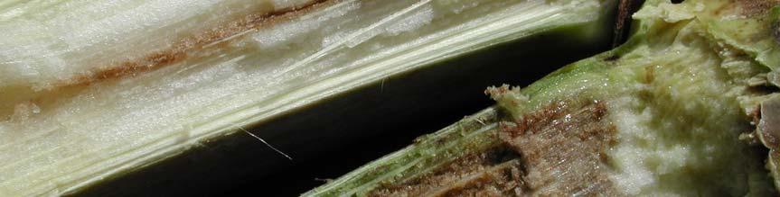


125 C D Fig
126 Fig. 2. A dendrogram based on the results of the API 20E and API 50CHE systems (BioMérieux, La Balme les Grottes, Montalieu Vercieu, France) tests, showing relationships among bacterial strains isolated from maize with brown stalk rot, P. ananatis reference strains, P. agglomerans, P. citrea, P. dispersa, P. stewartii subsp. stewartii and P. stewartii subsp. indologenes. Similarity matrixes were constructed using the Dice coefficient and cluster analyses were performed by the unweighted pair group method with the Bionumerics program (version 4.5, Applied Maths, Kortrijk, Belgium). Strains from maize are in bold (indole negative strains are underlined). The data for the P. stewartii subsp. stewartii LMG 2715 T was obtained from the paper by Mergaert et al. (32). 119
127 LMG P. ananatis Eucalyptus LMG 2665 T P. ananatis PA 4 P. ananatis onion BD 315 P. ananatis onion BD 561 BD P. ananatis sudangrass BD 301 P. ananatis onion BD 640 BD 588 BD 647 BD 435 BD 442 BD 577 BD 602 LMG P. ananatis Eucalyptus BD 543 BD 333 P. ananatis onion BD 336 P. ananatis onion LMG 2603 T P. dispersa LMG 2632 T P. stewartii subsp. indologenes LMG T P. citrea BD 500 BD 502 BD 639 LMG 1286 T P. agglomerans LMG 2715 T P. stewartii subsp. stewartii 120
128 Fig. 3. A dendrogram showing similarity among the fluorescent amplified fragment length polymorphism (F-AFLP) DNA fingerprints of bacterial strains isolated from maize with brown stalk rot, P. ananatis reference strains, P. agglomerans, P. citrea, P. dispersa, P. stewartii subsp. stewartii and P. stewartii subsp. indologenes. The fingerprints were generated using the selective primers Eco-G/Mse-CG (6). Similarity matrixes were constructed using the Dice coefficient and cluster analyses were performed by the unweighted pair group method with the Bionumerics program (version 4.5, Applied Maths, Kortrijk, Belgium). Strains from maize are in bold (indole negative strains are underlined). 121
129 BD 640 BD 647 BD 315 P. ananatis onion BD 435 BD 543 BD 301 P. ananatis onion BD 561 BD 588 BD 622 BD 442 BD 577 BD 602 BD 336 P. ananatis onion PA 4 P. ananatis onion BD 333 P. ananatis onion LMG P. ananatis Eucalyptus LMG 2665 T P. ananatis P. ananatis sudangrass LMG P. ananatis Eucalyptus LMG 2632 T LMG 2715 T P. stewartii subsp. indologenes P. stewartii subsp. stewartii BD 500 BD 639 BD 502 LMG 1286 T LMG 2603 T P. agglomerans P. dispersa LMG T P. citrea 122
130 Fig. 4. Phylogenetic tree showing the relationship among selected partial 16S rdna sequences from Pantoea species and strains isolated from maize with brown stalk rot (in bold, indole negative strains are underlined). Phylogenetic tree was constructed with the neighbor-joining method (39) and evolutionary distances calculated according to the method of Kimura (32), using the MEGA software package, version 3.1; Kumar, Tamura, Nei, 2004 (34). The sequences of Enterobacter cloacae, Klebsiella pneumoniae and Escherichia coli were used as outgroup taxons. The numbers at the nodes indicate the levels of bootstrap support based on data for replicates, only values greater than 90 % are shown. Accession numbers (in brackets) and the sequences of P. ananatis, P. agglomerans, P. dispersa, P. stewartii, E. cloacae, K. pneumoniae and E. coli were obtained from the GenBank/EMBL databases. Bar represents genetic distance. 123
131 90 BD 435 BD 442 PA 4 P. ananatis (AY530796) LMG 2676 P. ananatis (U80209) BD 640 LMG P. ananatis (AF364844) BD 647 BD 301 P. ananatis (AY579209) BD 315 P. ananatis (AY579212) BD 602 BD 588 BD 333 P. ananatis (DQ512490) LMG 2665 T P. ananatis (Z96081) LMG P. ananatis (AF364847) BD 561 BD 577 BD 622 BD LMG 2676 P. ananatis (U80209) LMG 2565 P. agglomerans (Z96082) LMG 2660 P. agglomerans (U80183) DSM 3493 T P. agglomerans (AJ233423) BD BD 502 BD 639 LMG 2715 T P. stewartii subsp. stewartii (U80208) 99 LMG 2632 T P. stewartii subsp. indologenes (Y13251) LMG 2603 T P. dispersa (DQ504305) 99 ATCC T Enterobacter cloacae (AJ251469) DSM T Klebsiella pneumoniae (X87276) ATCC T Escherichia coli (X80725)
132 CHAPTER 5 Polyphasic Characterisation of Pantoea Strains from Onion and Maize, and the Description of Pantoea allii sp. nov. Abstract Bacteria from the genus Pantoea have become increasingly important plant pathogens around the world. In South Africa, they cause diseases of two economically important crops, Eucalyptus and maize. The strains inducing center rot of onion have only been found in onion seed in this country, and have not yet been associated with any disease outbreak. The identity and taxonomic position of Pantoea isolates from onion and maize has not been fully determined. Forty-seven strains were subjected to a polyphasic study that included phenotypic characterisation, analysis of the F-AFLP patterns, rep-pcr genomic fingerprinting, 16S rdna gene sequences and DNA-DNA hybridisation. The results revealed that the strains belong to three different species within the genus Pantoea. Majority of strains were identified as P. ananatis. Indole-negative strains from maize were identified as P. vagens. Some strains from onion, previously classified as P. ananatis, were shown to form a novel species. The name Pantoea allii sp. nov. is proposed for these strains (type strain BD 390 T ) and the description of the species is presented. 125
133 INTRODUCTION Bacteria belonging to the genus Pantoea are common on plant surfaces, in seeds, soil, and water and have been isolated from humans and animals (Gavini et al., 1989; Mergaert et al., 1993). Many microbiologists have studied the taxonomy of the genus. The studies included the numerical analysis of phenotypic characteristics (Verdonck et al., 1987), 16S rrna gene sequence analysis (Hauben et al., 1998), fatty acid and protein profile analyses, DNA-DNA hybridisation (Brenner et al., 1984; Beji et al., 1988; Gavini et al., 1989, Mergaert et al., 1993) and AFLP fingerprints analysis (Brady, 2005). There are seven described species within the genus Pantoea: P. agglomerans, P. ananatis, P. citrea, P. dispersa, P. punctata, P. terrea, and P. stewartii containing two subspecies, indologenes and stewartii. There are also several hybridisation groups from a study by Brenner et al. (1984), and a protein profile group of Beji et al. (1988), that according to the latest edition of the Bergey s manual of systematic bacteriology belong to the genus Pantoea (Grimont and Grimont, 2005). Plant pathogenic Pantoea species cause diseases on diverse crops, such as pineapple (Serrano, 1928; Kageyama et al., 1992), melons (Bruton et al., 1986; Wells et al., 1987), cantaloupe (Bruton et al., 1991) sudangrass (Azad et al., 2000), Eucalyptus (Coutinho et al., 2002), rice (Azegami et al., 1983; Cother et al., 2004), mandarin orange (Kageyama et al., 1992), sugarcane (Serrano, 1928), beets (Brown, 1928), Gypsophila (Brown, 1934), onion (Hattingh and Walters, 1981; Gitaitis and Gay, 1997; Schwartz and Otto, 2000; Goszczynska et al., 2006) and maize (Elliott, 1941; Paccola-Meirelles et al., 2001; Goszczynska et al., 2007). Disease symptoms are diverse and include galls, rots, wilt, leaf blights, necrosis and spots, dieback and stem necrosis (Grimont and Grimont, 2005). However, not all Pantoea strains are plant pathogens, and some have been used for biological control of Erwinia amylovora (Beer et al., 1984) and Xanthomonas albilineans (Zhang and Birch, 1997). Some strains of P. agglomerans (Nunes et al., 2001) and P. ananatis (Torres et al., 2005) are effective for the biological control of post harvest pome fruit diseases caused by fungi. Diseases of onion are characterised by leaf blight, central leaf rot, seed stalk necrosis and rot, and bulb decay and these symptoms can lead to economically significant loses (Hattingh and Walters, 1981; Walcott et al., 2002). These diseases are caused by P. ananatis (Gitaitis and Gay, 1997) and P. agglomerans (Hattingh and Walters, 1981). The strains of P. ananatis 126
134 inducing center rot of onion have only been found in onion seed in South Africa, and have not yet been associated with any disease outbreak (Goszczynska et al., 2006). Brown stalk rot of maize was first observed in South Africa in 2004 and was caused by P. ananatis and an unknown Pantoea sp. similar phenotypically to P. agglomerans (Goszczynska et al., 2007). Affected plants were stunted, with a vertical crack at the first internode, surrounded by a dark brown, narrow lesion. Internal browning inside the stem extended upwards, reaching the top internode in some plants. Seed cobs were underdeveloped. Diseased plants were scattered over the fields and 10-70% of the crop was affected (Goszczynska et al., 2007). The diseases of onion and maize caused by Pantoea species were recorded for the first time during the last decade. Their taxonomic position has not been fully clarified. Bacteria isolated from onion in the USA and South Africa was considered to be P. ananatis. This classification was based only on biochemical and physiological characteristics (Gitaitis and Gay, 1997; Schwartz and Otto, 2000; Walcott et al., 2002) and the analysis of the 16S rdna sequences (Goszczynska et al., 2006). Such methods are often insufficient for species delineation according to the recommendation of Wayne et al. (1987) and Stackebrandt et al. (2002). Pantoea strains isolated from maize with brown stalk rot symptoms in South Africa were additionally examined by using fluorescent amplified fragment length polymorphism analysis (F-AFLP) (Goszczynska et al., 2007). The majority of strains isolated from diseased maize were P. ananatis. Several isolates, although similar to P. agglomerans on the basis on biochemical tests and 16S rdna sequences, produced F-AFLP fingerprints that were distinctly different from that generated by P. ananatis, P. agglomerans, P. dispersa, P. citrea, P. stewartii subsp. stewartii and P. stewartii subsp. indologenes. The results indicated that these bacteria belong to a previously undescribed species of the genus Pantoea and their taxonomic status needed to be further investigated. Bacteria recently isolated from Eucalyptus in Uganda were proposed to form a new species within a genus Pantoea, P. vagens (unpublished results(goszczynska et al., 2007).). The type strain of P. vagens, BCC 105 T was included in this study. The present research was initiated to characterise a collection of Pantoea strains from onion and maize by a polyphasic approach based on analyses of carbon source utilisation, 127
135 physiological characteristics, 16S rrna gene sequence analysis, DNA-DNA hybridisation and F-AFLP. Based on the results obtained, the new species Pantoea allii is proposed. MATERIALS AND METHODS Bacterial strains. Sixty-seven bacterial strains used in the study are listed in Table 1. Twenty-four strains were originally isolated from onion plants and seed in the USA and South Africa. Twenty-three strains were isolated from maize with brown stalk rot in South Africa. Twenty Pantoea strains, including the type strains of P. ananatis, P. agglomerans, P. citrea, P. dispersa, P. vagens, P. stewartii subsp. indologenes and P. stewartii subsp. stewartii were used as reference strains. The strains were routinely cultured on tryptone glucose extract agar (TGA) (Difco, Sparks, MD), at 26 o C and preserved in milk glycerol liquid medium (10% skim milk, 15% glycerol in distilled water) at 20 o C. All cultures were routinely checked for purity and colony characteristics on TGA. Pathogenicity tests. Pathogenicity on onion cv. Granex 33 was determined in green house assays by using a stub inoculation test (Goszczynska et al., 2006). A sterile needle was dipped into the bacterial colony on TGA (24-48 hours growth) and then the needle was inserted under the epidermis of a leaf. At least two leaves were inoculated per isolate. Inoculated plants were incubated in a greenhouse with 27 o C/23 o C day/night temperatures and observed daily for the development of symptoms. Pathogenicity on maize was determined in greenhouse assays on a susceptible maize cultivar SR 52 (Goszczynska et al., 2007). Nine-week-old plants were inoculated by injecting a few drops of bacterial suspensions into the first internode of the stem. Maize plants were maintained in a greenhouse with natural light and at temperatures and RH described above. At least two plants were inoculated with each bacterial strain per inoculation method. This experiment continued for ten weeks. Three control plants in all experiments were inoculated with sterile distilled water. Phenotypic methods Morphological and physiological characteristics. Strains from onion and maize were characterised by colony morphology, pigment production on TGA, motility, cell 128
136 morphology, Gram-stain, oxidase reaction, catalase reaction, indole production, Hugh- Leifson oxidation/fermentation test, gas production from glucose, hydrogen sulphide production from cysteine, growth at different temperatures (4, 30, 37, 40 and 44 o C), and tolerance to NaCl (Mergaert et al., 1993). The tests were performed according to methods described by Fahy and Hayward (1983). Utilisation of carbon sources. All strains listed in Table 1 were characterised phenotypically by the Biolog GN2 microplate system (Biolog, Inc., Hayward, CA), according to the manufacturer s instructions. The inoculated microplates were incubated at 30 o C for 24 hours. Plates were then scored visually for carbon source utilisation patterns. The data was entered into the Bionumerics software (version 4.5, Applied Maths, Kortrijk, Belgium). Similarity matrixes were constructed using the Pearson coefficient. Groups of strains were defined by the unweighted pair group method (UPGMA) using the same software. Biochemical tests. Biochemical characteristics of the strains listed in Table 1 were examined with the API 20E system (BioMérieux, La Balme les Grottes, Montalieu Vercieu, France), according to the procedure recommended by the manufacturers. Selected strains were also tested with the API 50CHE system. The results of the API 20E and API 50CHE tests were recorded after 24 and 48 hours of incubation at 30 o C respectively. Two cluster analyses were performed using the Bionumerics software. The first analysis was done with the API 20E data (21 characters) for all strains listed in Table 1 and the second analysis with the API 50CHE (49 characters) data for selected strains. Similarity matrixes were constructed with the Dice coefficient and cluster analyses were performed by the UPGMA method. The data was presented as UPGMA dendrograms. 129
137 Molecular methods DNA extraction. Genomic DNA of bacterial strains (Table 1) was extracted by using the GenElute Bacterial Genomic DNA Kit (Sigma, Steinheim, Germany), according to the manufacturer s instructions. Purified DNA was quantified using a Dyna Quant 200 fluorometer (Hoefer, San Francisco, CA) and Hoescht H intercalating dye (Polysciences, Warrington, PA). The DNA was stored at 20 o C until further analysis. Fluorescent amplified fragment length polymorphism (F-AFLP). All strains listed in Table 1 were examined by the fluorescent amplified fragment length polymorphism (F- AFLP)-based system for the identification of plant-associated species from the genus Pantoea, as described by Brady et al. (2006). Genomic DNA, ng from each isolate, was digested with EcoRI and MseI (Roche, Mannheim, Germany) restriction enzymes, and then ligated to the respective adaptors. Preamplification PCR reactions were done with Eco-00 (5 -GAC TGC GTA CCA ATT C-3 ) and Mse-00 (5 -GAT GAG TCC TGA CTA A-3 ) primers. Selective PCR amplifications were performed with Mse-CG (5 -GAT GAG TCC TGA CTA ACG-3 ) and fluorescently labelled Eco-G (5 -GAC TGC GTA CCA ATT CG-3 ) primers. Amplifications were done at least twice for each strain in the Hybaid Omni Gene thermocycler (Teddington, England) according to the Brady (2005) specifications. The selective amplification reactions (1 μl) were mixed with an equal volume of formamide loading buffer (95% formamide, 20 mm EDTA, bromophenol blue), heated for 3 min at 90 o C and then chilled on ice for 10 min. A volume of μl of each mixture was loaded onto the gel. The AFLP products were separated in 8% Long Ranger (LI-COR Biosciences, Lincoln, NE) denaturing gels on a LI-COR IR automated sequencer. Electrophoresis was carried out for 4 hours at 1500 V in 0.8 x TBE buffer. Images were imported into BioNumerics software. Gels were standardized with a bp sizing standard (LI-COR) and the bands between 50 and 700 bp were analysed (Brady, 2005). Similarity of the AFLP fingerprints was calculated using the Dice correlation coefficient and the cluster analysis was performed using the unweighted pair group method (UPGMA). The results were presented as an UPGMA dendrogram. 130
138 Repetitive sequence based PCR genomic fingerprinting. Repetitive sequence based polymerase chain reaction (rep-pcr) genomic fingerprints were obtained for all strains listed in Table 1. PCR was performed in 10 μl volumes containing PCR buffer (10 mm Tris HCl, ph 9.0 at 25 o C; 50 mm KCl; 0.1% Triton X-100); 3.5 mm MgCl 2 ; 150 μm dntps; 1.0 μm primer, 2.5% DMSO; Taq polymerase (Promega, Madison, WI) 0.15 units per reaction volume and ng template μl -1. Primers used in the PCR corresponded to the prokaryotic enterobacterial repetitive interogenic consensus (ERIC2) and the BOX1A subunit of the BOX element (Rademaker and de Bruijn, 1997). PCR reaction volumes were overlaid with 10 μl sterile mineral oil and PCR amplifications performed according to Rademaker and de Bruijn (1997) in a Hybaid Omni Gene thermocycler. The rep-pcr products were separated in 1.5% agarose in 1 x TBE buffer at 80 V. Gels were stained in ethidium bromide (10 mg ml -1 ) for 10 min and destained for 15 min in water. Images were imported into BioNumerics software. The similarity between strains was calculated using the Pearson s correlation coefficient applied to the entire densitometric curves of the gel tracks. Gels were standardized with DNA molecular weight marker VI (Roche, Steinheim, Germany). All PCR reactions were repeated at least twice. Cluster analysis was performed by the UPGMA method and the data presented as an UPGMA dendrogram. 16S rrna gene sequence analysis. The 16S rrna gene fragments of nine strains from onion and twelve strains from maize were amplified in previous studies (Goszczynska et al., 2006 and 2007). The GenBank accession numbers (National Center for Biotechnology Information, U.S. National Institute of Health, Bethesda, MD) are shown in Fig. 4. Ilse Cleenwerck (Ghent University, Belgium) provided the 16S rdna sequences of P. citrea, P. punctata and P. terrea. Carrie Brady (University of Pretoria) supplied the sequence of P. vagens BD 105 T. The 16S rdna nucleotide sequences of strains from maize and onion were aligned with a selection of 16S rdna sequences of Pantoea species from GenBank with the MAFFT software, ver (Katoh et al., 2002). Phylogenetic trees were constructed with the neighbor joining method (Saitou and Nei, 1987) and evolutionary distances calculated 131
139 according to the method of Kimura (1980), using the MEGA software package, version 3.1, Kumar, Tamura, Nei, 2004 (Kumar et al., 2004). Bootstrap analysis of the data, based on 1000 permutations was used to assess the stability of relationships. DNA-DNA hybridisation. The DNA was extracted from the five strains from onion (BD 309, BD 310, BD 377, BD 390, PA 4) three strains from maize (BD 435, BD 442, BD 639), BCC 105 from Eucalyptus, and the type strains of P. ananatis LMG 2665 T, P. agglomerans LMG 1286 T, P. stewartii subsp. stewartii LMG 2715 T, P. dispersa LMG 2603 T and P. vagens BCC 105 T, using the method described by Wilson (1989) and modified by Cleenwerck et al (2002). DNA-DNA hybridisations were performed with photo-biotin-labelled probes in microplate wells (Ezaki et al., 1989; Goris et al., 1998) as described by Cleenwerk et al. (2002). Fluorescent measurements were done with a HTS7000 BioAssay Reader (Perkin-Elmer Cetus, Norwalk, CT). The hybridisation temperature was 45 o C. Reciprocal experiments were performed for every pair of strains, and the means from reciprocal tests were presented. RESULTS Pathogenicity tests. All Pantoea strains isolated from onion induced identical symptoms on onion leaves cv. Granex 33 in pathogenicity tests. Two to four days after inoculation water soaked spots appeared on leaves that expanded into longitudinal, bleached-green lesions with chlorotic margins (Fig. 1). All Pantoea from maize produced brown stalk rot symptoms within six to seven weeks (Fig. 2). Four to six days after inoculation, small, 2-4 mm, light brown or dark green lesions developed around the inoculation point. Additionally, drops of yellow liquid slowly oozed from the lesion for two to three days. After 6 to 7 weeks, a single, long, vertical crack appeared on the injected internode. A dark brown, narrow lesion was present in the internal stem tissue along the crack. The internal browning within the stem was also observed in upper internodes. Morphological and physiological characteristics. The strains from onion and maize produced yellow colonies on TGA, were gram-negative rods ( µm length and
140 µm width), motile, oxidase negative and catalase positive. All strains fermentatively utilised glucose. They did not produce gas from glucose nor hydrogen sulphide from cysteine. Majority of strains produced indole from tryptophane, with the exception of Pantoea sp. from maize. Cells grew on nutrient agar containing up to 6% NaCl. Pantoea strains should be classified and identified mainly based on genotypic characteristics, as identification based on phenotypic tests does not always lead to clear results (Gavini et al., 1989, Mergaert et al., 1993). Thus, the results of molecular characterisation of the strains from onion and maize are presented first, followed by the phenotypic description. Molecular characterisation F-AFLP analysis. Pantoea strains used in the study generated complex DNA fingerprints from extracted genomic DNA (Fig. 3). The selective primers Eco-G/Mse-CG (Brady, 2005) yielded well-defined DNA fingerprints, with an average of 80 bands per isolate. Amplified DNA fragments ranged in size from approximately 50 bp to slightly greater than 700 bp. A total of 101 fragments were generated and all these fragments were polymorphic. Sixtyseven Pantoea strains formed seven clusters. Majority of strains from onion and maize clustered with P. ananatis type strain LMG 2665 T and P. ananatis reference strains. The similarity values among these strains were between 73 and 98%. The strains from maize, classified as Pantoea sp. in a previous study (Goszczynska et al., 2006), formed a cluster with P. vagens BCC 105 T. The F-AFLP fingerprints obtained for these strains showed 87 to 94% similarity. Five strains from onion, indistinguishable from P. ananatis by biochemical and morphological characteristics, generated F-AFLP patterns that were clearly different from those produced by other isolates used in the study, including the type strains of P. ananatis, P. agglomerans, P. citrea, P. dispersa, P. vagens, P. stewartii subsp. stewartii and P. stewartii subsp. indologenes. Fingerprints of BD 304, BD 309, BD 377, BD 380 and BD 390 were 70 to 92% similar to each other. Rep-PCR genomic fingerprinting. The rep-pcr fragments amplified with the BOX1A and ERIC2 primers, ranged in size from approximately 230 bp to greater than 3 kb. The banding 133
141 pattern revealed a high degree of genetic diversity among the strains. Although significant differences were observed between the major groups, within each group of strains fingerprints were similar (Fig. 4). The groups of strains obtained by the analysis of the rep- PCR data were identical to that obtained by using the F-AFLP fingerprints. The DNA fingerprint similarity among most of isolates from onion, maize, and P. ananatis reference strains ranged from 50% to 99% and all these strains clustered together on the rep- PCR dendrogram. The indole-negative strains from maize (Goszczynska et al., 2007) produced characteristic and unique rep-pcr fingerprints. BD 502 and BD 639 formed a separate cluster on the dendrogram with the type strain of P. vagens. A group of five strains from onion (BD 304, BD 309, BD 377, BD 380 and BD 390), generated rep-pcr fingerprints that were similar to each other (50 to 80%), but distinctly different from other strains. The same five isolates also grouped together on the F-AFLP dendrogram. 16S rrna gene sequence analysis. Figure 5 shows the phylogenetic relationship derived from a neighbor-joining analysis of the pairwise comparison among the 16S rdna sequences of twelve strains from maize and nine strains from onion. The 16S rdna sequences of fourteen Pantoea reference strains, including the type strains of seven described species within the genus were included in the analysis. The multiple sequence alignment of thirty-five 16S rdna sequences used in the study is presented in Appendix A. Phylogenetic evaluation confirmed the division of strains from maize and onion into three groups. Nine maize and five onion strains, that produced genomic fingerprints similar to P. ananatis, clustered with the 16S rdna sequences of P. ananatis. Two indole negative isolates from maize grouped closely, at 62% confidence value, with P. vagens BCC 105 T. Although the sequences of three onion isolates, BD 309, BD 377 and BD 390 were more than 99% similar to that of P. ananatis, they formed a separate cluster in the tree, at a high, 81% confidence value. DNA-DNA hybridisation. Between two and four strains from each F-AFLP and rep-pcr cluster were selected for the DNA-DNA hybridisation. The results of DNA-DNA hybridisations of all examined strains are shown in Table
142 DNA-DNA binding values obtained for four indole positive strains, BD 310 and PA 4 from onion and BD 435 and BD 442 from maize and P. ananatis LMG 2665 T, varied between 90 and 100%. Since 70% DNA binding value was recommended as the minimum level of genotypic relatedness within a species (Wayne et al., 1987), these four isolates were identified as P. ananatis. Strain BD 639 showed 89% DNA relatedness to BCC 105 T. These two strains displayed an intermediate level of DNA similarity, 63 to 65%, to the type strain of P. agglomerans, and low levels to the known Pantoea species. Therefore, BD 639, and two indole negative strains, BD 500 and BD 502 (Goszczynska et al., 2007) that grouped with BCC 105 T in the F-AFLP, rep-pcr and 16S rdna dendrograms, were identified as P. vagens. DNA-DNA hybridisation data revealed that three strains from onion, BD 309, BD 377 and BD 390 displayed a high level of DNA relatedness, 90 to 100%, and low levels of relatedness to the known Pantoea species and P. vagens. The name Pantoea allii sp. nov. is proposed for this taxon. Phenotypic characteristics Utilisation of carbon sources. On the UPGMA dendrogram of the Biolog data (Fig. 6), the strains used in the study formed four clusters. The biggest group consisted of 54 strains and included all strains identified as P. ananatis by molecular fingerprinting, P. stewartii subsp. indologenes and P. allii sp. nov. There was consistency in utilisation of 69 substrates on the GN2 plates among these strains, 41 positive and 28 negative reactions. No single test was able to differentiate between P. ananatis and P. allii. P. stewartii subsp. indologenes, however, could be distinguished from these two other species by its inability to utilise L- rhamnose, L-histidine and D,L,α-glycerol phosphate. The P. vagens strains from maize and the P. vagens type strain BCC 105 T clustered with P. agglomerans. The isolates within a cluster gave 41 positive and 27 negative reactions in Biolog GN2 plates. P. vagens differed from P. agglomerans by its ability to utilise nine substrates: D-melibiose, turanose, L-ornithine, D-serine, L-threonine and four acids, formic, α-hydroxybutyric, α-ketoglutaric and succinamic. 135
143 API 20E. The results of physiological and biochemical tests using the API 20E system separated 67 strains (Table 1) into six groups (Fig. 7). All P. ananatis and P. allii fell within a single cluster. These two species could not be distinguished from each other based on API 20E tests. Five P. ananatis strains isolated from onion seed did not produce acid from sucrose, unlike the other P. ananatis used in the study and 18 strains of P. ananatis studied by Mergaert et al. (1993). Three strains of P. vagens formed a separate group in the dendrogram (Fig. 7). They gave positive reactions in the following tests: β-galactosidase, acetoin, gelatinase and produced acid from D-glucose, D-mannitol, inositol, L-rhamnose, D-sucrose, D-melibiose, and L- arabinose. They did not produce acid from D-sorbitol and amygdalin, indole from tryptophane, arginine dihydrolase, lysine decarboxylase, ornithine decarboxylase, citrate, H 2 S and urease. Although in another study (Goszczynska et al., 2007) three strains from maize differed from other Pantoea species by giving a positive reaction in tryptophane deaminase test, the type strain of P. vagens, BCC 105 T, was negative for this test. In contrast to other indole negative Pantoea species, P. vagens did not produce acid from amygdalin, and could be distinguished by that characteristic. API 50CHE. Thirty-three strains were examined by the analysis of anaerobic acid production from 48 substrates and the ability to hydrolyse esculin in the API 50CHE strips. Contrary to Biolog and API 20E, P. allii could be differentiated from P. ananatis and other Pantoea species by the API 50CHE tests (Fig. 7). Five P. allii strains produced acid from at least one of the following compounds; D-adonitol, methyl-β-d-xylopyranoside, L-sorbose, inulin and xylitol, while all other Pantoea isolates did not. Production of acid from D- adonitol, methyl-ß-d-xylopyranoside, L-sorbose and inulin, were not reported for members of the genus (Gavini et al., 1989; Mergaert et al., 1993). Biochemical characteristics of P. vagens were additionally defined by the API 50CHE. The isolates produced acid from D-fucose, unlike other strains used in the study. 136
144 DISCUSSION Diseases caused by bacteria belonging to the genus Pantoea are emerging diseases in South Africa, and may have an increasing economic impact in the future. The causal agents, isolated from onion (Hattingh and Walters, 1981), Eucalyptus (Coutinho et al., 2002), onion seed (Goszczynska et al., 2006) and most recently from maize (Goszczynska et al., 2007), have been described as P. agglomerans, P. ananatis and Pantoea sp. closely related to P. agglomerans, respectively. The species allocation, with the exception P. ananatis from Eucalyptus, was based on a biochemical and physiological characterisation, analysis of the 16S rdna sequences and, in the case of maize pathogens, F-AFLP genomic fingerprinting. Such a taxonomic system is insufficient for species delineation according to the recommendation of Wayne et al. (1987) and Stackebrandt et al. (2002). This study was performed to determine the accurate taxonomic position of bacteria from onion and maize in the genus Pantoea. To achieve this, a collection of strains from South African maize and strains pathogenic to onion isolated in South Africa and the USA were examined using a variety of methods, recommended for the taxonomic description of bacterial species (Stackebrandt et al., 2002). Phenotypic characterisation confirmed that strains from maize and onion belong to the genus Pantoea and indicated, as expected, that at least two bacterial species were present. The 16S rdna sequence analysis, however, revealed that three species could be present, as some strains from onion, thought to be P. ananatis, formed a separate cluster in the 16S rdna tree (Fig. 5). However, 16S rdna sequence analysis is known to be of limited value in terms of taxonomic resolution at the species level for members of Enterobacteriaceae, due to possible lateral transfers within the gene (Rayssiguier et al., 1989) and multiple copies of the 16S rrna gene (Cillia et al., 1996). Taxonomic relationships at the species level were assessed by rep-pcr and F-AFLP genomic fingerprints analysis and DNA-DNA hybridisation. All indole positive Pantoea from maize and the majority of strains from onion generated genomic fingerprints that were similar to that produced by the reference strains of P. ananatis (Fig. 3, Fig. 4). Four strains of that group, two from onion and two from maize, showed % DNA binding to P. ananatis type strain LMG 2665 T (Table 2). As strains with similar genomic fingerprints are 137
145 genomically closely related (Gavini et al., 1989; Rademaker et al., 2000), all strains within P. ananatis rep-pcr and F-AFLP clusters are thus identified as P. ananatis. The indole negative strains from maize, although similar to P. agglomerans by phenotypic tests and the 16S rdna sequences, produced genomic fingerprints that clustered with P. vagens BCC 105 T (Fig. 3, Fig. 4). BD 639 showed a high level, 89%, of DNA relatedness to BCC 105 T. Although DNA biding ratios of these two strains to P. agglomerans were between 63 and 65%, they should not be classified as a subspecies of P. agglomerans. The fingerprints of P. vagens were distinctly different from that of P. agglomerans, further supporting the establishment of a separate species to contain them. The strains BD 390 and BD 309 were identified in a previous study as P. ananatis (Goszczynska et al., 2006). However, the genomic fingerprints generated by BD 309, BD 390 and three other isolates from onion, did not resemble that produced by P. ananatis, and formed a separate cluster in the F-AFLP and rep-pcr dendrograms (Fig. 3, Fig. 4). The DNA-DNA hybridisation values among three strains of this cluster were between 90 and 100%, confirming that they belong to a single species. The most closely related type strain was P. ananatis with 44 to 57% DNA reassociation, below the 70% threshold allowed for species delineation (Wayne et al., 1987). The name Pantoea allii sp. nov. is proposed for this species. Table 3 gives phenotypic characteristics useful in the differentiation of the species of the genus Pantoea. P. vagens could not be easily differentiated from P. agglomerans and P. allii from P. ananatis by an exclusive biochemical or physiological tests. A similar problem was reported by Gavini et al. (1989) with P. agglomerans versus P. dispersa and by Mergaert et al. (1993) concerning P. ananatis versus P. stewartii subsp. indologenes. Pantoea strains should be classified and identified mainly based on genotypic characteristics, as identification based on phenotypic tests does not always lead to clear results (Gavini et al., 1989, Mergaert et al., 1993). Description of Pantoea allii sp. nov. Pantoea allii [al li.i. N.L. adj. allii from Allium, the genus of onion (Allium cepa L.). 138
146 The description below is based on the data obtained for the five strains, BD 304, BD 309, BD 377, BD 380 and BD 390, isolated from onion plants with centre rot symptoms in the USA and from onion seed in South Africa. In pathogenicity tests, strains induce symptoms identical to those caused by P. ananatis on onion plants. Gram-negative, non-capsulated, non-spore forming straight rods, motile. Colonies on nutrient agar and TGA are yellow, smooth, round, and convex with entire margins. They are facultatively anaerobic, oxidase negative and catalase positive. Cells tolerate up to 6% NaCl and grow at 30 o C, 37 o C and 40 o C, but not at 4 o C and 44 o C. P. allii does not produce arginine dihydrolase, lysine decarboxylase, ornithine decarboxylase, H 2 S, urease, tryptophane deaminase and gas from glucose. Citrate is utilised. Indole, acetoin, and β-galactosidase are produced. P. allii strains produce acid from at least one of the following carbon sources: D-adonitol, methyl-β-d-xylopyranoside, L-sorbose, inulin and xylitol, while P. ananatis do not produce acid from this compounds. Other biochemical characteristics of P. allii at 30 o C are shown in Table 3. The following carbon sources are utilised at 30 o C (as assessed by Biolog GN microplates), by 100% of strains: dextrin, tween 40, tween 80, N-acetyl-D-glucosamine, L-arabinose, D- arabitol, D-cellobiose, D-fructose, D-galactose, gentiobiose, α-d-glucose, inositol, α-dlactose, lactulose, D-mannitol, D-mannose, D-melibiose, β-methyl-d-glucoside, D-raffinose, L-rhamnose, sucrose, D-trehalose, pyruvic acid methyl ester, succinic acid mono-methyl ester, acetic acid, cis-aconitic acid, citric acid, D-galacturonic acid, D-gluconic acid, D- glucosaminic acid, D-glucuronic acid, D,L-lactic acid, quinic acid, D-saccharic acid, succinic acid, bromosuccinic acid, D-alanine, L-alanine, L-alanyl-glutamic acid, L-histidine, L- proline, L-serine, urocanic acid, inosine, thymidine, glycerol, D,L,α-glycerol phosphate, α- D-glucose-1-phosphate and D-glucose-6-phosphate. The following carbon substrates are not utilised: α-cyclodextrin, N-acetyl-D-galactosamine, erythitol, L-fucose, turanose, xylitol, D- galactonic acid lactone, β-hydroxybutyric acid, p-hydroxyphenylacetic acid, itaconic acid, α- ketobutyric acid, α-ketoglutaric acid, α-ketovaleric acid, malonic acid, propionic acid, sebacic acid, L-alaninamide, hydroxy-l-proline, L-leucine, L-phenylalanine, L-pyroglutamic acid, L-threonine, D,L-carnitine, γ-aminobutyric acid, phenylethylamine, putrescine, 2-139
147 aminoethanol and 2,3-butanediol. P. allii varies in the utilisation of the following carbon sources in the Biolog GN plates: glycogen, adonitol, maltose, D-psicose, D-sorbitol, formic acid, α-hydroxybutyric acid, γ-hydroxybutyric acid, succinamic acid, glucuronamide, L- ornithine and D-serine. The proposed type strain is BD 390, isolated from onion seed in South Africa. ACKNOWLEDGEMENTS Thank the University of Ghent, Belgium, for doing DNA-DNA hybridisations. LITERATURE CITED Azad, H. R., Holmes, G. J. & Cooksey, D. A. (2000). A new leaf blotch disease of sudangrass caused by Pantoea ananatis and Pantoea stewartii. Plant Dis 84, Azegami, K., Ozaki, K. & Matsuda, A. (1983). Bacterial palea browning, a new disease of rice caused by Erwinia herbicola. Bull Nat Inst Agri Sci Ser C 39, Beer, S. V., Rundle, J. R. & Wodzinski, R. S. (1984). Interaction between E. amylovora and E. herbicola in vitro, in immature pear fruits and in apple blossoms. Acta Horticult 151, Beji, A., Mergaert, J., Gavini, F., Izard, D., Kersters, K., Leclerc, H. & De Ley, J. (1988). Subjective synonymy of Erwinia herbicola, Erwinia milletiae, and Enterobacter agglomerans and redefinition of the taxon by genotypic and phenotypic data. Intern J Syst Bacteriol 38, Brady, C. L. (2005). Taxonomy of Pantoea associated with bacterial blight of Eucalyptus. Master Thesis. The Faculty of Natural and Agricultural Sciences, Department of Microbiology and Plant Pathology, University of Pretoria, Pretoria, South Africa. 140
148 Brenner, D. J., Fanning, G. R., Knutson, J. K. L., Steigerwalt, A. G. & Krichevsky, M. I. (1984). Attempts to classify Herbicola group-enterobacter agglomerans strains by deoxyribonucleic acid hybridisation and phenotypic tests. Int J Syst.Bacteriol 34, Brown, N. A. (1928). Bacterial pocket disease of the sugar beet. J Agric Res 37, Brown, N. A. (1934). A gall similar to crown gall, produced on Gypsophila by a new bacterium. J Agric Res 48, Bruton, B. D., Wells, J. M. & Lester, G. E. (1986). Pathogenicity of Erwinia ananas to muskmelons in Texas. Phytopathology 76, Bruton, B. D., Wells, J. M., Lester, G. E. & Patterson, C. L. (1991). Pathogenicity and characterization of Erwinia ananas causing a postharvest disease of cantaloup fruit. Plant Dis 75, Cillia, V., Lafay, B. & Christen, R (1996). Sequence heterogeneities among 16S ribosomal RNA sequences, and their effect on phylogenetic analyses at the species level. Mol Biol Evol 13, Cleenwerck, I., Vandemeulebroecke, K., Janssens, D. & Swings, J. (2002). Reexamination of the genus Acetobacter, with description of Acetobacter cerevisiae sp. nov. and Acetobacter malorum sp. nov. Int J Syst Bacteriol 52, Cother, E. J., Reinke, R., McKenzie, C., Lanoiselet, V. M. & Noble, D. H. (2004). An unusual stem necrosis of rice caused by Pantoea ananas and the first record of this pathogen on rice in Australia. Austral Plant Pathol 33, Coutinho, T.A., Preisig, O., Mergaert, J., Cnockaert, M. C., Riedel, K.-H., Swings, J. & Wingfield, M. J. (2002). Bacterial blight and dieback of Eucalyptus species, hybrids and clones in South Africa. Plant Dis 86, Elliott, C. (1941). Bacterial wilt of dent corn inbreds. Phytopathology 32,
149 Ezaki, T., Hashimoto, Y. & Yabuuchi, E. (1989). Fluorometric deoxyribonucleic aciddeoxyribonucleic acid hybridisation in microdilution wells as an alternative to memrane filter hybridisation in which radioisotopes are used to determine genetic relatedness among bacterial strains. Int J Syst Bacteriol 39, Fahy, P. C. & Hayward, A. C. (1983). Media and methods for isolation and diagnostic tests. In Plant bacterial diseases, a diagnostic guide. (eds. Fahy, P. C., and Persley, G. J.), pp , Academic Press Australia. Gavini, F., Mergaert, J., Beji, A., Mielcarek, C., Izard, D., Kersters, K. & De Ley, J. (1989). Transfer of Enterobacter agglomerans (Beijerinck 1888) Ewing and Fife 1972 to Pantoea gen. Nov. as Pantoea agglomerans comb. Nov. and description of Pantoea dispersa sp. nov. Intern J Syst Bacteriol 39, Gitaitis, R. D. & Gay, J. D. (1997). First report of a leaf blight, seed stalk rot, and bulb decay of onion by Pantoea ananas in Georgia. Plant Dis 8, Goris, J., Suzuki, K.-I., De Vos, P., Nakase, T. & Kersters, K. (1998). Evaluation of a microplate DNA-DNA hybridisation method compared with the initial renaturation method. Can J Microbiol 44, Goszczynska, T., Moloto, V. M., Venter, S. N. & Coutinho, T. A. (2006). Isolation and identification of Pantoea ananatis from onion seed in South Africa. Seed Sci & Tech 34, Goszczynska, T., Botha, W.J., Venter, S. N. & Coutinho, T. A. (2007). Isolation and identification of the causal agent of brown stalk rot, a new disease of maize in South Africa. Plant Dis 91, Grimont, P. A. D. & Grimont, F. (2005). Genus XXIII. Pantoea Gavini, Mergaert, Beji, Mielcarek, Izarc, Kersters and De Ley 1989b, 343 VP emend. Mergaert, Verdonck and Kersters 1993, 172. Pages in: Bergey s manual of systematic bacteriology, Volume two, Part B. D. J. Brenner, N. J. Krieg, J. T. Staley and G. M. Garrity, eds. Springer. 142
150 Hattingh, M. J. & Walters, D. F. (1981). Stalk and leaf necrosis of onion caused by Erwinia herbicola. Plant Dis 65, Hauben, l., Moore, E. R. B., Vauterin, L., Steenackers, M., Mergaert, J., Verdonck, L. & Swings, J. (1998). Phylogenetic position of phytopatogens within the Enterobacteriaceae. System Appl Microbiol 21, Kageyama, B., Nakae, M., Yagi, S. & Sonoyama, T. (1992). Pantoea punctata sp. nov., Pantoea citrea sp. nov., and Pantoea terrea sp. nov. isolated from fruit and soil samples. Int J Syst Bacteriol 42, Katoh, K., Kuma, K., Toh, H. & Miyata, T. (2002). MAFFT: a novel method for rapid multiple sequence alignment based on fast Fourier transform. Nucl Acids Res 30, Kimura, M. (1980). A simple method for estimating evolutionary rates of base substitutions through comparative studies of nucleotide sequences. J Molec Evol 16, Kumar, S., Tamura, K. & Nei, M. (2004). MEGA3: Integrated software for Molecular Evolutionary Genetics Analysis and sequence alignment. Brief Bioinformatics 5, Mergaert, J., Verdonck, L. & Kersters, K. (1993). Transfer of Erwinia ananas (synonym, Erwinia uredovora) and Erwinia stewartii to the genus Pantoea emend. as Pantoea ananas (Serrano 1928) comb. Nov. and Pantoea stewartii (Smith 1898) comb. nov., respectively, and description of Pantoea stewartii subsp. indologenes subsp. nov. Int J Syst Bacteriol 43, Nunes, C., Usall, J., Teixido, N. & Vinas, I. (2001). Biological control of postharvest pear diseases using a bacterium, Pantoea agglomerans CPA-2. Int J Food Microbiol 70, Paccola-Meirelles, L. D., Ferreira, A. S., Merelles, W. F., Marriel, I. E. & Casela, C. R. (2001). Detection of bacterium associated with a leaf spot disease of maize in Brazil. J Phytopathology 149,
151 Rademaker, J. L. W. & de Bruijn, F. J. (1997). Characterisation and classification of microbes by Rep-PCR genomic fingerprinting and computer-assisted pattern analysis. Pages in: DNA markers: Protocols, applications and overviews. G. Caetano-Annolles and P. M. Gresshoff, eds. Wiley, New York. Rademaker, J. L. W., Hoste, B., Louws, F. J., Kersters, K., Swings, J., Vauterin, L., Vauterin, P. & de Bruijn, F. J. (2000). Comparision of AFLP and rep-pcr genomic fingerprinting with DNA-DNA homology studies: Xanthomonas as a model system. Int J Syst Evol Microbiol 50, Rayssiguier, C., Thaller, D. S. & Radman, M. (1989). The barrier to recombination between Escherichia coli and Salmonella typhimurium is disrupted in mismatch repair mutants. Nature 342, Saitou, N. & Nei, M. (1987). The neighbor-joining method: a new method for reconstructing phylogenetic trees. Molec Biol Evol, 4, Schwartz, H. F. & Otto, K. (2000). First report of a leaf blight and bulb decay of onion by Pantoea ananatis in Colorado. Plant Dis 84, 808. Serrano, F. B. (1928). Bacterial fruitlet brown-rot of pineapple in the Philippines. Philippine J Sci 36, Stackebrandt, E., Frederiksen, W., Garrity, G. M. & 10 other authors (2002). Report of the ad-hoc committee for the re-evaluation of the species definition in bacteriology. Int J Syst Evol Microbiol 52, Torres, R., Teixidó, N., Ussal, J., Abadias, M. & Viñas, I. (2005). Post-harvest control of Penicillium expansum on pome fruits by the bacterium Pantoea ananatis CPA-3. The J Hort Sci Biotech 80, Verdonck, L., Mergaert, J., Rijckaert, C., Swings, J., Kersters, K. & De Ley, J. (1987). Genus Erwinia: Numerical analysis of phenotypic features. Int J Syst Bacteriol 37,
152 Walcott, R. R., Gitaitis, R. D., Castro, A. C., Sanders Jr., F. H. & Diaz-Perez, J. C. (2002). Natural infestation of onion seed by Pantoea ananatis, causal agent of center rot. Plant Dis 86, Wayne, L. G., Brenner, D. J., Colwell, R. R., Grimont, P. A. D., Kandler, O., Krichevsky, M. I., Moore, W. E. C., Murray, R. G. E., Stackebrandt, E., Starr, M. P. & Trϋper, H. G. (1987). Report on the Ad Hoc Committee on Reconciliation of Approaches to Bacterial Systematics. Int J Syst Bacteriol 37, Wells, J. M., Sheng, W. S., Ceponis, M. J. & Chen, T. A. (1987). Isolation and characterization of strains of Erwinia ananas from honeydew melons. Phytopathology 77, Wilson, K. (1989). Preparation of genomic DNA from bacteria. Pages in: Current protocols in molecular biology. F. M. Ausubel, R. Brent, R. E. Kimston, D. D. Moore, J. G. Seidman, J. A. Smith and K. Sthrul, eds. John Wiley & Sons, Inc, New York, N. Y. Zhang, L. & Birch, R. G. (1997). Mechanism of biocontrol by Pantoea dispersa of sugar cane leaf scald disease caused by Xanthomonas albilineans. J Appl Microbiol 82,
153 Table 1. Pantoea strains used in this study. Strain Other strain Species (as received) Host Location Source designations P. ananatis Sorghum sudanense USA, California D. Azad BCC 105 T Pantoea vagens. Eucalyptus Uganda T. A. Coutinho BD P. ananatis Allium cepa USA R. Gitaitis BD 287 SUH 2 P. agglomerans Allium cepa South Africa PPPPB BD 295 ATCC BAA 517 P. ananatis Allium cepa USA, Colorado ATCC BD 296 ATCC BAA 516 P. ananatis Allium cepa USA, Colorado ATCC BD 301 Blackshank 15 P. ananatis Allium cepa USA, Georgia R. Walcott BD 304 Blackshank 24 P. ananatis Allium cepa USA, Georgia R. Walcott BD 305 Blackshank 30 P. ananatis Allium cepa USA, Georgia R. Walcott BD 309 Hort. Hill 24 P. ananatis Allium cepa USA, Georgia R. Walcott BD 310 Hort. Hill 31 P. ananatis Allium cepa USA, Georgia R. Walcott BD 311 Hort. Hill 32 P. ananatis Allium cepa USA, Georgia R. Walcott BD 312 Pans P. ananatis Allium cepa USA, Georgia R. Walcott BD 315 Pans P. ananatis Allium cepa USA, Georgia R. Walcott BD 317 Pans P. ananatis Allium cepa USA, Georgia R. Walcott BD 325 P. ananatis Allium cepa South Africa R. Walcott BD 326 P. ananatis Allium cepa South Africa PPPPB BD 331 P. ananatis Allium cepa South Africa PPPPB 146
154 Strain Other strain Species (as received) Host Location Source designations BD 333 P. ananatis Allium cepa South Africa PPPPB BD 334 P. ananatis Allium cepa South Africa PPPPB BD 335 P. ananatis Allium cepa South Africa PPPPB BD 336 P. ananatis Allium cepa South Africa PPPPB BD 377 P. ananatis Allium cepa South Africa PPPPB BD 380 P. ananatis Allium cepa South Africa PPPPB BD 390 P. ananatis Allium cepa South Africa PPPPB BD 435 P. ananatis Zea mays South Africa PPPPB BD 442 P. ananatis Zea mays South Africa PPPPB BD 478 P. ananatis Zea mays South Africa PPPPB BD 494 P. ananatis Zea mays South Africa PPPPB BD 515 P. ananatis Zea mays South Africa PPPPB BD 527 P. ananatis Zea mays South Africa PPPPB BD 541 P. ananatis Zea mays South Africa PPPPB BD 543 P. ananatis Zea mays South Africa PPPPB BD 551 P. ananatis Zea mays South Africa PPPPB BD 556 P. ananatis Zea mays South Africa PPPPB BD 561 P. ananatis Zea mays South Africa PPPPB BD 577 P. ananatis Zea mays South Africa PPPPB 147
155 Strain Other strain Species (as received) Host Location Source designations BD 588 P. ananatis Zea mays South Africa PPPPB BD 596 P. ananatis Zea mays South Africa PPPPB BD 602 P. ananatis Zea mays South Africa PPPPB BD 614 P. ananatis Zea mays South Africa PPPPB BD 620 P. ananatis Zea mays South Africa PPPPB BD 622 P. ananatis Zea mays South Africa PPPPB BD 629 P. ananatis Zea mays South Africa PPPPB BD 639 Pantoea sp. Zea mays South Africa PPPPB BD 640 P. ananatis Zea mays South Africa PPPPB BD 647 P. ananatis Zea mays South Africa PPPPB CTB 1061 P. ananatis Oryza sativa Japan CTB CTB 1135 P. ananatis Oryza sativa Japan CTB DAR P. agglomerans Pyrrus communis Australia ACPPB DAR P. agglomerans Allium cepa Australia ACPPB LMG 1286T P. agglomerans Human Zimbabwe BCCM/LMG LMG P. ananatis Eucalyptus South Africa BCCM/LMG LMG P. ananatis Eucalyptus South Africa BCCM/LMG LMG 22049T P. citrea Mandarin orange Japan BCCM/LMG LMG 2565 P. agglomerans Cereals Canada BCCM/LMG 148
156 Strain Other strain Species (as received) Host Location Source designations LMG 2603T P. dispersa Soil Japan BCCM/LMG LMG 2632 P. stewartii subsp. Setaria italica India BCCM/LMG indologenes LMG 2665T P. ananatis Ananas cosmosus Hawaii BCCM/LMG LMG 2671 P. stewartii subsp. Ananas cosmosus Hawaii BCCM/LMG indologenes LMG 2676 P. ananatis Puccinia graminis USA BCCM/LMG LMG 2713 P. stewartii subsp. Zea mays USA BCCM/LMG stewartii LMG 2715T P. stewartii subsp. Zea mays USA BCCM/LMG stewartii LMG 2749 P. dispersa Human BCCM/LMG PA 3 P. ananatis Allium cepa South Africa PPPPB PA 4 P. ananatis Allium cepa South Africa PPPPB BCCM/LMG Culture Collection: Universiteit Gent, Belgium; ATCC: American Type Culture Collection, Manassas, VA; ACPPB: Australian Collection of Plant Pathogenic Bacteria, Orange; PPPPB: Plant Pathogenic and Plant Protecting Bacteria, ARC-PPRI, South Africa; R. Walcott: Department of Plant Pathology, University of Georgia, Athens; T. A. Coutinho: Department of Microbiology and Plant Pathology, Forestry and Agricultural Biotechnology Institute (FABI), University of Pretoria, Pretoria, South Africa; R. Gitaitis: Department of Plant Pathology, University of Georgia; CTB: Centre Technique du Bois, Paris, France. 149
157 Table 2. DNA binding values of examined Pantoea strains. The tests were performed at the University of Ghent, Belgium. Strain P. ananatis 1 LMG 2665 T BD BD BD PA P. allii sp. nov. 6 BD BD 390T BD P. agglomerans 9 LMG 1286 T P. vagens 10 BCC 105 T BD P. stewartii subsp. stewartii 12 LMG 2715 T P. dispersa 13 LMG 2603 T
158 Table 3. Differential characteristics of Pantoea allii and some biochemically similar species within the genus Pantoea. Characteristic API 20E tests Citrate utilisation Tryptophane deaminase d - - Indole Gelatinase + d - + d - Acid from: Inositol d d - + d - D-sorbitol + d L-rhamnose + d d + + d D-sucrose + d Amygdalin d API 50CHE tests Esculin hydrolysis + d d d + - Acid from: D-adonitol d Methyl-β-D-xylopyranoside d L-sorbose d Inositol d + D-sorbitol + d Amygdalin + d D-lactose Inulin d Starch d - - Glycogen d - - Xylitol D-fucose , positive reaction for at least 90% of the strains;,negative reaction for at least 90 % of the strains; d, 11-89% strains positive. 1, Pantoea allii; 2, P. ananatis; 3, P. stewartii subsp. indologenes; 4, P. vagens; 5, P. agglomerans; 6, P. dispersa 151
159 Data for P. ananatis and P. stewartii subsp. indologenes are from this study and Mergaert et al. (1993); for P. agglomerans and P. dispersa are from this study and Gavini et al. (1989). 152
160 Fig. 1. The symptoms induced by Pantoea ananatis (A) and P. allii (B) strains isolated from onion on onion leaves in pathogenicity tests. Two to four days after inoculation water soaked spots appeared on leaves that expanded into longitudinal, bleached-green lesions with chlorotic margins. 153

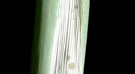


161 A B Fig
162 Fig. 2. The symptoms produced by P. ananatis isolated from maize with brown stalk rot on maize SR 52. A, Four to six days after inoculation, small, 2-4 mm, light brown or dark green lesions developed around the inoculation point. Additionally, drops of yellow liquid slowly oozed from the lesion for two to three days. B, after 6 to 7 weeks, a single, long, vertical crack appeared on the injected internode. C, A dark brown, narrow lesion was present in an internal stem tissue along the crack. D, Browning of the internal tissue in an upper internode. The symptoms induced by P. vagens were indistinguishable from those induced by P. ananatis. 155


163 A B Fig


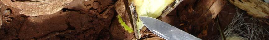
164 C D Fig
belonging to the Genus Pantoea
 Emerging diseases of maize and onion caused by bacteria belonging to the Genus Pantoea by Teresa Goszczynska Submitted in partial fulfilment of the requirements for the degree Philosophiae Doctoriae in
Bacterial spot of pepper and tomato
 Website to brush up on bacterial diseases Bacterial spot of pepper and tomato http://www.apsnet.org/edcenter/intropp/lessons/prokaryotes/pages/bacterialspot.aspx Potato blackleg and soft rot http://www.apsnet.org/edcenter/intropp/lessons/prokaryotes/pages/blacklegpotato.aspx
Taxonomy of Pantoea associated with bacterial blight of Eucalyptus
 Taxonomy of Pantoea associated with bacterial blight of Eucalyptus by Carrie Louise Brady Submitted in partial fulfilment of the requirements for the degree Magister Scientiae in The Faculty of Natural
Unit G: Pest Management. Lesson 2: Managing Crop Diseases
 Unit G: Pest Management Lesson 2: Managing Crop Diseases 1 Terms Abiotic disease Bacteria Biotic disease Cultural disease control Disease avoidance Disease resistance Disease tolerance Fungi Infectious
DEVELOPMENT OF AN AFLP-BASED TYPING SYSTEM FOR THE GENUS PANTOEA
 DEVELOPMENT OF AN AFLP-BASED TYPING SYSTEM FOR THE GENUS PANTOEA 5.1) Introduction: Since the first description of Pantoea ananatis in 1928 (Serrano, 1928), this bacterium has been subsequently isolated
Lab tomorrow: Bacterial Diseases. Bacteria
 Lab tomorrow: Bacterial Diseases Quiz: Koch s Postulates (p. 17-19), Botrytis Predisposition (p. 97)., And, intros for Bacteria (pp 67-69), Biocontrol of Crown Gall (p. 117), and Observation of Viral Movement
Unit D: Controlling Pests and Diseases in the Orchard. Lesson 5: Identify and Control Diseases in the Orchard
 Unit D: Controlling Pests and Diseases in the Orchard Lesson 5: Identify and Control Diseases in the Orchard 1 Terms Abiotic disease Bacteria Biotic diseases Cultural disease control Disease avoidance
BACTERIAL DISEASES OF EUCALYPTUS
 BACTERIAL DISEASES OF EUCALYPTUS T.A. Coutinho, S.N. Venter, J. Roux, X.D. Zhou and M.J. Wingfield ACPP APPS, Darwin 2011 INTRODUCTION Estimated 18m ha of Eucalyptus planted in 80 countries Numerous fungal
THE IDENTIFICATION OF TWO UNKNOWN BACTERIA AFUA WILLIAMS BIO 3302 TEST TUBE 3 PROF. N. HAQUE 5/14/18
 THE IDENTIFICATION OF TWO UNKNOWN BACTERIA AFUA WILLIAMS BIO 3302 TEST TUBE 3 PROF. N. HAQUE Introduction: The identification of bacteria is important in order for us to differentiate one microorganism
Chapter 8. Summarizing Discussion
 Chapter 8 Summarizing Discussion Chapter 8 This thesis focuses on the ecology and pathogenicity of biovar 3 Dickeya sp. provisionally called D. solani, a blackleg and soft rot pathogen, recently introduced
North American Bramble Growers Research Foundation 2016 Report. Fire Blight: An Emerging Problem for Blackberry Growers in the Mid-South
 North American Bramble Growers Research Foundation 2016 Report Fire Blight: An Emerging Problem for Blackberry Growers in the Mid-South Principal Investigator: Burt Bluhm University of Arkansas Department
Identification of culturable endophytes isolated from apple tissues with antagonism towards Neonectria ditissima
 Identification of culturable endophytes isolated from apple tissues with antagonism towards Neonectria ditissima Jing Liu, Hayley Ridgway & Eirian Jones Background Apple production in NZ widely cultivated
Plant Disease Introduction. Larry A. Sagers Utah State University Extension Regional Horticulturist
 Plant Disease Introduction Larry A. Sagers Utah State University Extension Regional Horticulturist Plant Pathology Basics Disease Anything that interferes with normal plant function Plant Pathology Basics
Pink Disease of Pineapple
 Feature Story March 2003 Pink Disease of Pineapple Clarence I. Kado University of California Department of Plant Pathology One Shields Ave Davis, CA 95616 Contact: cikado@ucdavis.edu Next to mangos and
ENTEROBACTER AEROGENES UNKNOWN BACTERIA FLOW CHART UNKNOWN LAB REPORT, MICROBIOLOGY ENTEROBACTER AEROGENES
 ENTEROBACTER AEROGENES UNKNOWN BACTERIA PDF UNKNOWN LAB REPORT, MICROBIOLOGY ENTEROBACTER AEROGENES IDENTIFICATION OF AN UNKNOWN BACTERIAL SPECIES OF 1 / 5 2 / 5 3 / 5 enterobacter aerogenes unknown bacteria
Ch 10. Classification of Microorganisms
 Ch 10 Classification of Microorganisms Student Learning Outcomes Define taxonomy, taxon, and phylogeny. List the characteristics of the Bacteria, Archaea, and Eukarya domains. Differentiate among eukaryotic,
MORPHOLOGICAL, CULTURAL AND PATHOGENIC CHARACTERISTICS OF MACROPHOMINA PHASEOLINA ISOLATES FROM SUGAR BEET
 MORPHOLOGICAL, CULTURAL AND PATHOGENIC CHARACTERISTICS OF MACROPHOMINA PHASEOLINA ISOLATES FROM SUGAR BEET Stojšin, V., Budakov, D., Bagi, F., Đuragin, N., Marinkov, R. Department for Environmental and
Morphological and Biochemical Properties Useful for Identification of Erwinia chrysanthemi
 International Journal of Current Microbiology and Applied Sciences ISSN: 2319-7706 Volume 6 Number 10 (2017) pp. 343-348 Journal homepage: http://www.ijcmas.com Original Research Article https://doi.org/10.20546/ijcmas.2017.610.042
Fully approved by The South African Department of Agriculture, Forestry & Fisheries Registration Number: B4807
 ExploGrow is a revolutionary 100% organic biofertiliser in a league of its own, comprising 17 highly complex micro-organisms, environmentally friendly soil ameliorant and plant growth stimulant, with increased
Effects of postharvest onion curing parameters on bulb rot caused by Pantoea agglomerans, Pantoea ananatis and Pantoea allii in storage
 Doi: 10.1111/ppa.12438 Effects of postharvest onion curing parameters on bulb rot caused by Pantoea agglomerans, Pantoea ananatis and Pantoea allii in storage C. Vahling-Armstrong a, J. K. S. Dung b, J.
Plant disease. Plant Diseases: Learning objectives: Plant Disease: Any physiological or structural abnormality that is harmful to the plant
 Plant disease Plant Diseases: Identification and Control Melodie Putnam Extension Plant Pathologist Learning objectives: Difference between biotic and abiotic diseases and their manifestation Difference
Prereq: Concurrent 3 CH
 0201107 0201101 General Biology (1) General Biology (1) is an introductory course which covers the basics of cell biology in a traditional order, from the structure and function of molecules to the structure
Basic Plant Pathology for Franklin County Master Gardener Volunteers. Nancy J. Taylor Plant Pathology Department Ohio State University
 Basic Plant Pathology for Franklin County Master Gardener Volunteers Nancy J. Taylor Plant Pathology Department Ohio State University https://www.youtube.com/watch?v=h4 PuP_QbD14 Mildews Mildews Mildews
Plant Disease Introduction
 Utah State University DigitalCommons@USU All Archived Publications Archived USU Extension Publications 6-30-2006 Plant Disease Introduction Larry A. Sagers Utah State University Follow this and additional
Isolation optimization of bacterial endophytes from cucumber plants and evaluation of their effects on growth promotion and biocontrol
 Isolation optimization of bacterial endophytes from cucumber plants and evaluation of their effects on growth promotion and biocontrol Ozaktan H., Gül A., Çakır B., Yolageldi L., Akköprü A., Fakhraei D.,
AGROBACTERIUM. First described by Smith and Townsend (1907) Responsible for crown gall. Performed Koch's postulates
 Responsible for crown gall. Performed Koch's postulates") AGROBACTERIUM First described by Smith and Townsend (1907) Responsible for crown gall Performed Koch's postulates The disease is worldwide in distribution Speciation was based on pathogenicity Agrobacterium
AGROBACTERIUM First described by Smith and Townsend (1907) Responsible for crown gall Performed Koch's postulates The disease is worldwide in distribution Speciation was based on pathogenicity Agrobacterium
Curriculum Links. AQA GCE Biology. AS level
 Curriculum Links AQA GCE Biology Unit 2 BIOL2 The variety of living organisms 3.2.1 Living organisms vary and this variation is influenced by genetic and environmental factors Causes of variation 3.2.2
Distance Learning course Plant pathology and entomology Covered topics
 Distance Learning course Plant pathology and entomology Covered topics The distance learning course Plant pathology and entomology consist of four online modules that treat with the main groups of plant
CRISPR-SeroSeq: A Developing Technique for Salmonella Subtyping
 Department of Biological Sciences Seminar Blog Seminar Date: 3/23/18 Speaker: Dr. Nikki Shariat, Gettysburg College Title: Probing Salmonella population diversity using CRISPRs CRISPR-SeroSeq: A Developing
Szent István University Faculty of Horticultural Sciences Plant Pathology Department
 Szent István University Faculty of Horticultural Sciences Plant Pathology Department NOVEL ANTAGONISTIC BACTERIA AS PROSPECTIVE AGENTS FOR THE BIOCONTROL OF SOME PLANT BACTERIAL DISEASES THESIS OF Ph.D.
Chapter 19. Microbial Taxonomy
 Chapter 19 Microbial Taxonomy 12-17-2008 Taxonomy science of biological classification consists of three separate but interrelated parts classification arrangement of organisms into groups (taxa; s.,taxon)
Taxonomy. Content. How to determine & classify a species. Phylogeny and evolution
 Taxonomy Content Why Taxonomy? How to determine & classify a species Domains versus Kingdoms Phylogeny and evolution Why Taxonomy? Classification Arrangement in groups or taxa (taxon = group) Nomenclature
Assist. Prof. Martina Šeruga Musić acad. year 2016/17
 Assist. Prof. Martina Šeruga Musić acad. year 2016/17 PHYTOPATHOGENIC BACTERIA there are more than 100 species of known phytopathogenic bacteria genera Agrobacterium, Erwinia, Ralstonia, Pseudomonas, Xanthomonas,
High Performance Biology
 High Performance Biology Frequently Asked Questions (FAQ s) +61 3 5133 9118 What is Bacstim 100? Bacstim 100 is a microbial based product containing 5 different spore forming Bacillus spp. strains. The
TABLE OF CONTENTS CHAPTER NO. TITLE PAGE NO. LIST OF TABLES LIST OF FIGURES 1 INTRODUCTION AIM AND SCOPE OF THE PRESENT INVESTIGATION 7
 viii TABLE OF CONTENTS ABSTRACT LIST OF TABLES LIST OF FIGURES iii xxiii xxviii 1 INTRODUCTION 1 1.1 AIM AND SCOPE OF THE PRESENT INVESTIGATION 7 2 LITERATURE REVIEW 8 2.1 AN OVERVIEW OF TEA 8 2.2 TEA
Assessment Schedule 2016 Biology: Demonstrate understanding of biological ideas relating to micro-organisms (90927)
") NCEA Level 1 Biology (90927) 2016 page 1 of 5 Assessment Schedule 2016 Biology: Demonstrate understanding of biological ideas relating to micro-organisms (90927) Evidence Statement Question One No response
NCEA Level 1 Biology (90927) 2016 page 1 of 5 Assessment Schedule 2016 Biology: Demonstrate understanding of biological ideas relating to micro-organisms (90927) Evidence Statement Question One No response
Commercial microbial inoculants with endophytes (an overview)
") Commercial microbial inoculants with endophytes (an overview) Matthias Döring INOQ GmbH, Germany Vorname Name www.inoq.de 1 Inquiries about products with ENDOPHYTES by: EU commission of agriculture German
Commercial microbial inoculants with endophytes (an overview) Matthias Döring INOQ GmbH, Germany Vorname Name www.inoq.de 1 Inquiries about products with ENDOPHYTES by: EU commission of agriculture German
INTRODUCTION bioactive compounds Pigmentation chromobacteria water soluble water insoluble
 INTRODUCTION So far we have witnessed several useful applications of microbes including applications in food and the bioremediation of the environment. Besides consuming the desired substrate (oil) and
Microbiology / Active Lecture Questions Chapter 10 Classification of Microorganisms 1 Chapter 10 Classification of Microorganisms
 1 2 Bergey s Manual of Systematic Bacteriology differs from Bergey s Manual of Determinative Bacteriology in that the former a. groups bacteria into species. b. groups bacteria according to phylogenetic
Laboratory Exercise # 7: Aseptic Technique
 Laboratory Exercise # 7: Aseptic Technique Purpose: The purpose of this laboratory exercise is to acquaint the student with the procedures of aseptic transfer of microbiological cultures. ntroduction:
Bacterial Diseases of Fruit Trees
 Bacterial Diseases of Fruit Trees Steven Swain Environmental Horticulture Advisor UCCE Marin & Sonoma Counties The Diseases Fire blight Erwinia amylovora Common name after the scorched appearance of leaves
NORTHERN ILLINOIS UNIVERSITY. Screening of Chemical Libraries in Search of Inhibitors of Aflatoxin Biosynthesis. A Thesis Submitted to the
 NORTHERN ILLINOIS UNIVERSITY Screening of Chemical Libraries in Search of Inhibitors of Aflatoxin Biosynthesis A Thesis Submitted to the University Honors Program In Partial Fulfillment of the Requirements
Basics of Plant Pathology. Pam Roberts Katherine Hendricks Southwest Florida Research and Education Center
 Basics of Plant Pathology Pam Roberts Katherine Hendricks Southwest Florida Research and Education Center Historical Significance of Plant Diseases Sri Lanka France Late Blight on potato caused the Irish
Associative and Endophytic Nitrogen-fixing Bacteria and Cyanobacterial Associations
 Associative and Endophytic Nitrogen-fixing Bacteria and Cyanobacterial Associations Edited by Claudine Elmerich Institut Pasteur, Paris, France and William E. Newton Department of Biochemistry Virginia
Microbial Activity in the Rhizosphere
 K. G. Mukerji C. Manoharachary J. Singh (Eds.) Microbial Activity in the Rhizosphere With 35 Figures 4y Springer 1 Rhizosphere Biology - an Overview 1 Chakravarthula Manoharachary, Krishna G. Mukerji 1.1
Cm W)aøs A.3.1. Blast (Pyricularia grisea) Description: The disease caused by fungal infection.
aøs A.3.1. Blast (Pyricularia grisea) Description: The disease caused by fungal infection.") A.3. Diseases A.3.1. Cm W)aøs Blast (Pyricularia grisea) Picture 100 : Leaf blast. Description: The disease caused by fungal infection. Leaf blast: An infected leaf has diamond shaped or elliptical or
A.3. Diseases A.3.1. Cm W)aøs Blast (Pyricularia grisea) Picture 100 : Leaf blast. Description: The disease caused by fungal infection. Leaf blast: An infected leaf has diamond shaped or elliptical or
Worksheet for Morgan/Carter Laboratory #13 Bacteriology
 Worksheet for Morgan/Carter Laboratory #13 Bacteriology Ex. 13-1: INVESTIGATING CHARACTERISTICS OF BACTERIA Lab Study A: Colony Morphology Table 13.1 Characteristics of Bacterial Colonies Name of Bacteria
Microbial Taxonomy. C. Microbes usually have few distinguishing properties that relate them, so a hierarchical taxonomy mainly has not been possible.
 Microbial Taxonomy 1. Traditional taxonomy or the classification through identification and nomenclature of microbes, both "prokaryote" and eucaryote, is in a mess we are stuck with it for traditional
Diagnostics and genetic variation of an invasive microsporidium (Nosema ceranae) in honey bees (Apis mellifera)
 in honey bees (Apis mellifera)") Diagnostics and genetic variation of an invasive microsporidium (Nosema ceranae) in honey bees (Apis mellifera) Dr. M. M. Hamiduzzaman School of Environmental Sciences University of Guelph, Canada Importance
Diagnostics and genetic variation of an invasive microsporidium (Nosema ceranae) in honey bees (Apis mellifera) Dr. M. M. Hamiduzzaman School of Environmental Sciences University of Guelph, Canada Importance
Ecology of Infectious Disease
 Ecology of Infectious Disease What is the basis of community robustness (resistance to invasion)? How does robustness influence disease development? The Microbial Context: Microbial Interactions Affect
Introduction to Microbiology. CLS 212: Medical Microbiology Miss Zeina Alkudmani
 Introduction to Microbiology CLS 212: Medical Microbiology Miss Zeina Alkudmani Microbiology Micro- means very small (that needs a microscope to see). Microbiology is the study of very small living organisms.
Title: Plant Nitrogen Speaker: Bill Pan. online.wsu.edu
 Title: Plant Nitrogen Speaker: Bill Pan online.wsu.edu Lesson 2.3 Plant Nitrogen Nitrogen distribution in the soil-plantatmosphere Chemical N forms and oxidation states Biological roles of N in plants
Investigation 3: Comparing DNA Sequences to Understand Evolutionary Relationships with BLAST
 Investigation 3: Comparing DNA Sequences to Understand Evolutionary Relationships with BLAST Introduction Bioinformatics is a powerful tool which can be used to determine evolutionary relationships and
Q1) Germ theory of diseases. Q2) Louis Pasteur. Q3) Bacillus. Q4) Cyanobacteria. Q5) Viroids. Q6) Prions Q7) TMV Q8) T4
 Germ theory of diseases. Q2) Louis Pasteur. Q3) Bacillus. Q4) Cyanobacteria. Q5) Viroids. Q6) Prions Q7) TMV Q8) T4") (DMB01) ASSIGNMENT - 1 Introduction Microorganisms Q1) Germ theory of diseases Q2) Louis Pasteur Q3) Bacillus Q4) Cyanobacteria Q5) Viroids Q6) Prions Q7) TMV Q8) T4 (DMB01) ASSIGNMENT - 2 Introduction
(DMB01) ASSIGNMENT - 1 Introduction Microorganisms Q1) Germ theory of diseases Q2) Louis Pasteur Q3) Bacillus Q4) Cyanobacteria Q5) Viroids Q6) Prions Q7) TMV Q8) T4 (DMB01) ASSIGNMENT - 2 Introduction
Diseases of Sesame. Dr. S. Parthasarathy, M.Sc. (Ag)., Ph.D., FBSS.
., Ph.D., FBSS.") Diseases of Sesame Dr. S. Parthasarathy, M.Sc. (Ag)., Ph.D., FBSS. Assistant Professor (Plant Pathology), Dept. of Plant Pathology, College of Agricultural Technology, Theni Diseases of Sesame Charcoal
Diseases of Sesame Dr. S. Parthasarathy, M.Sc. (Ag)., Ph.D., FBSS. Assistant Professor (Plant Pathology), Dept. of Plant Pathology, College of Agricultural Technology, Theni Diseases of Sesame Charcoal
(DMB 01) M.Sc. (Previous) DEGREE EXAMINATION, DECEMBER First Year. Microbiology. Paper I INTRODUCTION TO MICROORGANISMS
 M.Sc. (Previous) DEGREE EXAMINATION, DECEMBER First Year. Microbiology. Paper I INTRODUCTION TO MICROORGANISMS") wk 7 (DMB 01) Paper I INTRODUCTION TO MICROORGANISMS PART A (5 8 = 40 marks) 1. Explain the growth of microbiology in the twentieth century. 2. Describe the structure of eukaryotic cell with a neat-labeled
wk 7 (DMB 01) Paper I INTRODUCTION TO MICROORGANISMS PART A (5 8 = 40 marks) 1. Explain the growth of microbiology in the twentieth century. 2. Describe the structure of eukaryotic cell with a neat-labeled
Microbes usually have few distinguishing properties that relate them, so a hierarchical taxonomy mainly has not been possible.
 Microbial Taxonomy Traditional taxonomy or the classification through identification and nomenclature of microbes, both "prokaryote" and eukaryote, has been in a mess we were stuck with it for traditional
Microbial Taxonomy. Slowly evolving molecules (e.g., rrna) used for large-scale structure; "fast- clock" molecules for fine-structure.
 used for large-scale structure; fast- clock molecules for fine-structure.") Microbial Taxonomy Traditional taxonomy or the classification through identification and nomenclature of microbes, both "prokaryote" and eukaryote, has been in a mess we were stuck with it for traditional
Microbial Taxonomy Traditional taxonomy or the classification through identification and nomenclature of microbes, both "prokaryote" and eukaryote, has been in a mess we were stuck with it for traditional
Salmonella enteritidis Identification and Isolation
 Department of Microbiology, Qom Branch, Islamic Azad University. Qom, Iran Start Here Advisor Dr.Mohsen Zargar Consulting Advisor Dr.Taghi Salehi Zahraei Presented by Zeinab Yazdanpanah 1 Outcome Enterobacteriaceae
KILGORE COLLEGE BIOLOGY DEPARTMENT Biology 2421 Syllabus
 COURSE: BIOL 2421 (4-3-4) TITLE: CATALOG DESCRIPTION: Microbiology and Pathology A study of the morphology, physiology, genetics, taxonomy and control of microorganisms. This course includes a study of
Use of the 3M Molecular Detection System for Salmonella and Listeria spp.
 Use of the 3M Molecular Detection System for Salmonella and Listeria spp. March 11, 213 Prof Steve Forsythe Pathogen Research Centre, School of Science and Technology Nottingham Trent University Clifton
Welcome to the Iowa Certified Nursery Professional Training program Module 7: Introduction to Plant Diseases and Insects.
 Welcome to the Iowa Certified Nursery Professional Training program Module 7: Introduction to Plant Diseases and Insects. 1 After completing this module you should: 1. Understand the causes of abssiotic
Gram negative bacilli
 Gram negative bacilli 1-Enterobacteriaceae Gram negative bacilli-rods Enterobacteriaceae Are everywhere Part of normal flora of humans and most animals They are cause of -30-35% septisemia -more than 70%
A. Incorrect! In the binomial naming convention the Kingdom is not part of the name.
 Microbiology Problem Drill 08: Classification of Microorganisms No. 1 of 10 1. In the binomial system of naming which term is always written in lowercase? (A) Kingdom (B) Domain (C) Genus (D) Specific
OCR Biology Checklist
 Topic 1. Cell level systems Video: Eukaryotic and prokaryotic cells Compare the structure of animal and plant cells. Label typical and atypical prokaryotic cells. Compare prokaryotic and eukaryotic cells.
OCR Biology Checklist
 Topic 1. Cell level systems Video: Eukaryotic and prokaryotic cells Compare the structure of animal and plant cells. Label typical and atypical prokaryotic cells. Compare prokaryotic and eukaryotic cells.
Cryotherapy: A New Method to Eliminate Pathogens from Sweetpotato Propagation Materials
 Cryotherapy: A New Method to Eliminate Pathogens from Sweetpotato Propagation Materials Margaret Worthington Graduate Group in Horticulture and Agronomy University of California, Davis April 14, 2009 http://www.judithbarathart.com
Overview of the major bacterial pathogens The major bacterial pathogens are presented in this table:
 Practical Microbiology 30/11/2018 University of Sulaimani college of Pharmacy Year2 Lab. 5: Overview of the major bacterial pathogens The major bacterial pathogens are presented in this table: Major Bacterial
Plant and animal cells (eukaryotic cells) have a cell membrane, cytoplasm and genetic material enclosed in a nucleus.
 have a cell membrane, cytoplasm and genetic material enclosed in a nucleus.") 4.1 Cell biology Cells are the basic unit of all forms of life. In this section we explore how structural differences between types of cells enables them to perform specific functions within the organism.
4.1 Cell biology Cells are the basic unit of all forms of life. In this section we explore how structural differences between types of cells enables them to perform specific functions within the organism.
Useful Propagation Terms. Propagation The application of specific biological principles and concepts in the multiplication of plants.
 Useful Propagation Terms Propagation The application of specific biological principles and concepts in the multiplication of plants. Adventitious Typically describes new organs such as roots that develop
Bacteria. The Three Types of Important Heterotrophic Bacteria
 Bacteria Kingdom Monera Prokaryote (their genetic material is not bound with a membrane) Classified according to shape - Spherical (cocci) - Spiral - Rod Shaped -TWO TYPES: Heterotrophic (organism that
Microbiology Helmut Pospiech
 Microbiology http://researchmagazine.uga.edu/summer2002/bacteria.htm 05.04.2018 Helmut Pospiech The Species Concept in Microbiology No universally accepted concept of species for prokaryotes Current definition
Pharmaceutical Microbiology Forum Newsletter Vol. 12 (4) Page 3 of 14 (NCIMB 8545, CIP NBRC. Salmonella enterica ssp typhimurium
 Page 3 of 14 (NCIMB 8545, CIP NBRC. Salmonella enterica ssp typhimurium") Page 3 of 14 Continued from page 2 Table 2. Absence of Specified Details Media Growth Promotion Organisms for Trypticase Soy Staphylococcus aureus Escherichia coli Pseudomonas aeruginosa Salmonella Staphylococcus
Page 3 of 14 Continued from page 2 Table 2. Absence of Specified Details Media Growth Promotion Organisms for Trypticase Soy Staphylococcus aureus Escherichia coli Pseudomonas aeruginosa Salmonella Staphylococcus
Introduction to Microbiology BIOL 220 Summer Session I, 1996 Exam # 1
 Name I. Multiple Choice (1 point each) Introduction to Microbiology BIOL 220 Summer Session I, 1996 Exam # 1 B 1. Which is possessed by eukaryotes but not by prokaryotes? A. Cell wall B. Distinct nucleus
Lab Exercise 5: Pure culture techniques
 Lab Exercise 5: Pure culture techniques OBJECTIVES 1. Perform a streak-plate to separate the cells of a mixed culture so that discrete colonies can be isolated. 2. Perform a pour-plate (loop) dilution
INTRODUCTION MATERIALS & METHODS
 Evaluation of Three Bacterial Transport Systems, The New Copan M40 Transystem, Remel Bactiswab And Medical Wire & Equipment Transwab, for Maintenance of Aerobic Fastidious and Non-Fastidious Organisms
CHARACTERIZATION OF BACTERIAL PATHOGEN CAUSING WILT AND LEAF BLIGHT ON CORN (Zea mays) BY PHYSIOLOGICAL, BIOCHEMICAL AND MOLECULAR METHODS
 BY PHYSIOLOGICAL, BIOCHEMICAL AND MOLECULAR METHODS") 286 AGRIVITA VOLUME 34 No. 3 OCTOBER - 2012 ISSN : 0126-0537 CHARACTERIZATION OF BACTERIAL PATHOGEN CAUSING WILT AND LEAF BLIGHT ON CORN (Zea mays) BY PHYSIOLOGICAL, BIOCHEMICAL AND MOLECULAR METHODS Lilis
286 AGRIVITA VOLUME 34 No. 3 OCTOBER - 2012 ISSN : 0126-0537 CHARACTERIZATION OF BACTERIAL PATHOGEN CAUSING WILT AND LEAF BLIGHT ON CORN (Zea mays) BY PHYSIOLOGICAL, BIOCHEMICAL AND MOLECULAR METHODS Lilis
Kingdom Monera(Archaebacteria & Eubacteria)
") Kingdom Monera(Archaebacteria & All bacteria are prokaryotes Characteristics: 1. No nucleus Eubacteria) 2. No membrane bound organelles 3. Smaller & less ribosomes 4. Most are smaller than eukaryotes 5.
Kingdom Monera(Archaebacteria & All bacteria are prokaryotes Characteristics: 1. No nucleus Eubacteria) 2. No membrane bound organelles 3. Smaller & less ribosomes 4. Most are smaller than eukaryotes 5.
In vitro the effect of intestinal normal flora on some pathogenic bacteria.
 In vitro the effect of intestinal normal flora on some pathogenic bacteria. Abstract: Dr.abbass shaker Ali adel Leena abd Al-Redha The effect of two types of intestinal bacterial normal floral ( and klebsiella)
The impact of Agrobacterium tumefaciens and other soil borne disease causing agents of economic importance in production of roses
 The impact of Agrobacterium tumefaciens and other soil borne disease causing agents of economic importance in production of roses Video conference on global competitiveness of the flower industry in the
Biology Assessment. Eligible Texas Essential Knowledge and Skills
 Biology Assessment Eligible Texas Essential Knowledge and Skills STAAR Biology Assessment Reporting Category 1: Cell Structure and Function The student will demonstrate an understanding of biomolecules
Basidiomycota (the club fungi)
") Basidiomycota in lab tomorrow Quiz (Lab manual pages 9-15 Isolation of fungal pathogens and 53-59 Ascos III, and intro pages for Basidiomycota (pp. 61-63) and Race I.D. of Wheat Stem Rust (p. 111). Look
Basidiomycota in lab tomorrow Quiz (Lab manual pages 9-15 Isolation of fungal pathogens and 53-59 Ascos III, and intro pages for Basidiomycota (pp. 61-63) and Race I.D. of Wheat Stem Rust (p. 111). Look
STAAR Biology Assessment
 STAAR Biology Assessment Reporting Category 1: Cell Structure and Function The student will demonstrate an understanding of biomolecules as building blocks of cells, and that cells are the basic unit of
Document category: There is no restriction on the circulation of this document
 GA2-06 Agenda Item 2 Issued: 16 January 2018 CIMMYT Position on gene editing: An example to support the development of a common position on gene editing Purpose This document provides CIMMYT s Position
Why Calcium is So Important
 Why Calcium is So Important Calcium - A Transportation Problem By Dr. Lynette Morgan As hydroponic growers we like to think that by supplying our plants with all the nutrients they need in the right ratios,
BIOAG'L SCI + PEST MGMT- BSPM (BSPM)
") Bioag'l Sci + Pest Mgmt-BSPM (BSPM) 1 BIOAG'L SCI + PEST MGMT- BSPM (BSPM) Courses BSPM 102 Insects, Science, and Society (GT-SC2) Credits: 3 (3-0-0) How insects develop, behave, and affect human activity.
Bioag'l Sci + Pest Mgmt-BSPM (BSPM) 1 BIOAG'L SCI + PEST MGMT- BSPM (BSPM) Courses BSPM 102 Insects, Science, and Society (GT-SC2) Credits: 3 (3-0-0) How insects develop, behave, and affect human activity.
VCE BIOLOGY Relationship between the key knowledge and key skills of the Study Design and the Study Design
 VCE BIOLOGY 2006 2014 Relationship between the key knowledge and key skills of the 2000 2005 Study Design and the 2006 2014 Study Design The following table provides a comparison of the key knowledge (and
7. M2/1 Subfamily Caesalpinoideae. A flower of Bauhinia sp. shows floral morphology typical of the species in the subfamily Caesalpinoideae.
 SLIDE NOTES AND EXPLANATIONS 1. M1/1 The Nitrogen Cycle. Gaseous nitrogen in the air is converted into a biologically useful form through biological nitrogen fixation in legumes and through chemical fixation
Diagnosing Plant Problems. A strategy to get started
 + Diagnosing Plant Problems A strategy to get started + Causes of plant damage Living factors Pests such as insects, mites, rodents, mammals Pathogens that cause disease such as fungi, bacteria, viruses,
Lassen Community College Course Outline
 Lassen Community College Course Outline AGR 20 Introduction to Plant Science 4.0 Units I. Catalog Description This course is an introduction to plant science including structure, growth processes, propagation,
EVALUATION OF WILD JUGLANS SPECIES FOR CROWN GALL RESISTANCE
 EVALUATION OF WILD JUGLANS SPECIES FOR CROWN GALL RESISTANCE Ed Stover, Malendia Maccree, Malli Aradhya, Ali E. McClean, and Daniel A. Kluepfel INTRODUCTION Crown Gall disease of walnut is caused by the
pglo/amp R Bacterial Transformation Lab
 pglo/amp R Bacterial Transformation Lab Name: Date: Purpose: To gain an understanding of the techniques of culturing E. coli bacteria and transforming E. coli bacteria using genetic engineering. Introduction:
Proteus & Pseudomonas
 Proteus & Pseudomonas Ahmad Ausama Al-Kazzaz Anas Huthaifa AL-Dewachi Ameer Saadallah Zacko Al-Ta i Supervised by: Dr. Khalid Ahmad Ausama Al-Kazzaz Proteus Proteus is a genus of Gram-negative Proteobacteria
Express PRA 1) for Syndrome basses richesses (SBR) Prepared by: Julius Kühn-Institut, Institute for Plant Health: 11 July, 2012.
 for Syndrome basses richesses (SBR) Prepared by: Julius Kühn-Institut, Institute for Plant Health: 11 July, 2012.") Express PRA 1) for Prepared by: Julius Kühn-Institut, Institute for Plant Health: 11 July, 2012. Initiation: Dr. Gritta Schrader: translated by Elke Vogt-Arndt Occurrence in Baden-Württemberg Phytosanitary
Express PRA 1) for Prepared by: Julius Kühn-Institut, Institute for Plant Health: 11 July, 2012. Initiation: Dr. Gritta Schrader: translated by Elke Vogt-Arndt Occurrence in Baden-Württemberg Phytosanitary
TIME-LINE OF INFECTION
 Review of Lecture 8: Getting inside the host is a critical step in disease development Fungal pathogens use contact and chemical tropisms to guide their way to a site where infection is possible Pathogens
Universiteit van Pretoria University of Pretoria. Mikrobiologie 251 Toets Maart 2012 Microbiology 251 Test March Examiners: Dr L Moleleki
 Universiteit van Pretoria University of Pretoria Mikrobiologie 251 Toets Maart 2012 Microbiology 251 Test March 2012 Tyd: 1 uur Time: 1 hour Eksaminatore: Dr L Moleleki Examiners: Dr L Moleleki Beantwoord
Allelopathy In Trees
 Allelopathy In Trees by Dr. Kim D Coder, Daniel B. Warnell School of Forest Resources, University of Georgia 4/99 Trees have developed in ecological systems filled with many other organisms. The environment,
Basidiomycetes (the club fungi)
") Basidiomycetes in lab tomorrow Quiz (Lab manual pages 7-13 Isolation of fungal pathogens and 51-57 Ascos III, and intro pages for Basidiomycetes (pp. 59-61) and Race I.D. of Wheat Stem Rust (p. 109). Look
Basidiomycetes in lab tomorrow Quiz (Lab manual pages 7-13 Isolation of fungal pathogens and 51-57 Ascos III, and intro pages for Basidiomycetes (pp. 59-61) and Race I.D. of Wheat Stem Rust (p. 109). Look
Todd A.Steinlage, Alaska Department of Natural Resources, Division of Agriculture, Plant Materials Center
 Tomato Spotted Wilt Virus in Alaska Greenhouses and Nurseries Todd A.Steinlage, Alaska Department of Natural Resources, Division of Agriculture, Plant Materials Center Tomato spotted wilt virus (TSWV)