Faironia difasciculata, a new gymnosperm from the Early Carboniferous (Mississippian) of Montagne Noire, France
|
|
|
- Joanna Phillips
- 5 years ago
- Views:
Transcription
 of Montagne Noire, France.")


1 Faironia difasciculata, a new gymnosperm from the Early Carboniferous (Mississippian) of Montagne Noire, France Anne-Laure Decombeix, Jean Galtier, Brigitte Meyer-Berthaud To cite this version: Anne-Laure Decombeix, Jean Galtier, Brigitte Meyer-Berthaud. Faironia difasciculata, a new gymnosperm from the Early Carboniferous (Mississippian) of Montagne Noire, France. Review of Palaeobotany and Palynology, Elsevier, 2006, 142 (3), pp < /j.revpalbo >. <hal > HAL Id: hal Submitted on 9 Nov 2006 HAL is a multi-disciplinary open access archive for the deposit and dissemination of scientific research documents, whether they are published or not. The documents may come from teaching and research institutions in France or abroad, or from public or private research centers. L archive ouverte pluridisciplinaire HAL, est destinée au dépôt et à la diffusion de documents scientifiques de niveau recherche, publiés ou non, émanant des établissements d enseignement et de recherche français ou étrangers, des laboratoires publics ou privés.
2 Faironia difasciculata, a new gymnosperm from the Early Carboniferous (Mississippian) of Montagne Noire, France. Anne-Laure Decombeix *, Jean Galtier and Brigitte Meyer-Berthaud Botanique et bioinformatique de l'architecture des plantes (UMR 5120 CNRS- CIRAD), TA40/PS2, CIRAD, Boulevard de la Lironde, Montpellier cedex 5, France Abstract A new taxon of probable gymnosperm affinities is described from the base of the Carboniferous (Mississippian, Middle Tournaisian) of Montagne Noire, southern France. It is based on a permineralised stem showing vascular and cortical tissues, and one attached petiole base. Faironia difasciculata gen. et sp. nov. shows the combination of a broad eustele and dense wood with a multifascicular leaf trace, originating from two non-adjacent axial strands. The Kalymma-type petiole base resembles that of the calamopityan pteridosperms but anatomy of the stele, wood and phloem more closely compares to that of early arborescent gymnosperms such as Pitus, Eristophyton or Megalomyelon the taxonomic position of which needs to be re-evaluated. Faironia is the sixth genus of seed plant affinities based on stems reported from the Montagne Noire deposits that, to date, comprise the most diverse assemblage of permineralised seed plant remains from just above the Devonian/Carboniferous boundary. Keywords: Mississippian, Early Carboniferous; early gymnosperm; permineralised stem; petiole base; eustele; dense wood. * Corresponding author: Fax: addresses: anne-laure.decombeix@cirad.fr (AL. Decombeix), jean.galtier@cirad.fr (J. Galtier), meyerberthaud@cirad.fr (B. Meyer-Berthaud) 1
3 1. Introduction The discovery of the lycopsid cone Lepidostrobus brownii by Schimper (Schimper, 1870) was the starting point of palaeobotanical investigation of the Lydiennes Formation of Montagne Noire (southern France). More than thirty taxa representing lycopsids, sphenopsids, ferns and lignophytes are presently known from plant fragments preserved as permineralisations or, rarely, as compressions. Lignophytes (i.e. progymnosperms and seed plants) represent an essential component of this Mid-Tournaisian flora and, particularly, stems and leaves of pteridosperm affinities are the most common fossils with a high proportion of calamopityan axes (Galtier et al., 1988). The abundance of vegetative remains of these plants in contrast to the rarity of possible corresponding fertile organs (Galtier and Rowe, 1989, 1991; Rowe and Galtier, 1990) is noteworthy. Within the context of a study on Early Carboniferous lignophytes we describe in this paper a new specimen from the Tournaisian of Montagne Noire which presents an original combination of characters. Although this specimen has previously been cited and figured in two papers (Galtier, 1988, Figure 3.10 F; and New taxon 1 in Galtier and Meyer-Berthaud, 2006), no formal description has yet been given. Following the seed plant precursors (Gerrienne et al., 2004) and earliest seed plants of Devonian age (Fairon-Demaret and Scheckler, 1987; Rothwell et al., 1989; Serbet and Rothwell, 1992), there are more than forty taxa of putative seed plants of Early Carboniferous age known as anatomically preserved stems. Their reproductive structures being not clearly identified, these plants (with the exception of Heterangium) are not used by phylogenetic analyses (e.g. Doyle and Donoghue, 1992; Rothwell and Serbet, 1994). However they document an impressive diversification of their vegetative morphology, habit and size while their systematic position remains unclear. The new plant described in this paper combines features known in different groups of lignophytes and it significantly increases our understanding of early seed plants diversity. 2. Materials and methods The Lydiennes Formation corresponds to sedimentary deposits in a shallow sea and consists of alternating beds of argilites and radiolarian cherts containing 2
4 phosphatic nodules. Horizons containing plant fossils are considered middle Tournaisian in age (Tn2a-Tn2b) based on the conodonts (Galtier et al., 1988). The fossils are found loose in the matrix or occasionally included in the nodules. Specimen MN 266, described in this study, is an anatomically preserved stem included in a nodule that was collected on a natural outcrop, near S t Nazaire de Ladarez (Hérault, France). The specimen was about 7 cm long and 5 cm wide; it was cut in 5 segments labeled A, B, C, D, and E. The organic matter being partly replaced by minerals, standard peel sections were of poor quality; therefore the specimen was studied using polished surfaces and wafers under reflected light, and by thin sections. Observations were carried out using an Olympus BO61 stereomicroscope and an Olympus BX60 microscope. Photographs were made with an Olympus DP12 Microscope Digital Camera System and processed using Adobe Photoshop 6.0. Drawings of the transverse sections and of the leaf emission pattern were made with a Wild camera lucida attached on a stereomicroscope. Measurement of cells and tissues were conducted using SigmaScan software. Mean dimensions are given for one hundred measurements of each parameter. The specimen and slides are kept in the AMAP Laboratory, Paleobotanical Collections, Université Montpellier II. 3. Systematic palaeobotany Faironia gen. nov. Diagnosis: Stem with primary and secondary vascular tissues and primary cortex. Eustele consisting of numerous discrete axial strands of primary xylem at the periphery of a broad circular pith. Ratio of primary xylem diameter to stem diameter high (about 1:2). Leaves helically arranged. Each leaf initially supplied by two traces originating separately from two non-adjacent axial strands. The paired leaf traces divide in the cortex supplying the petiole base with at least six vascular bundles. Axial primary xylem strands sometimes separated from the secondary xylem by parenchyma. Axial strands and leaf trace bundles mesarch. Metaxylem tracheids with scalariform to multiseriate bordered pitting on all walls. Secondary xylem tracheids with multiseriate bordered pits on radial walls. Rays uni to quadriseriate, not high. Secondary phloem present, including fibres. Narrow cortex consisting of an inner 3
5 zone of parenchyma with sclerotic nests and an outer zone of sparganum-type fibre bundles. Type species: Faironia difasciculata sp. nov. Faironia difasciculata sp. nov. (Plates I-III) Diagnosis: Stem with a circular eustele 21 mm wide, composed of more than 40 axial strands and incipient leaf traces. Primary xylem strands peripheral, in contact with the secondary xylem, or separated from the secondary xylem by one-four, rarely more, layers of parenchyma cells. Primary xylem strands mesarch, µm in diameter consisting of metaxylem tracheids up to 50 µm wide and very small protoxylem elements. Homogeneous pith of polygonal parenchyma cells µm wide and up to 250 µm high. Tracheids of secondary xylem rectangular to polygonal, 14-67µm (average 39 µm) in transverse section. Multiseriate pits 11 µm in average diameter with inclined elliptical apertures. Rays uni-to multiseriate (up to 4 cells and 92 µm wide), expanded in the innermost part of secondary xylem, relatively short (up to 28 cells and 650 µm high). Ray parenchyma cells procumbent, rectangular, µm long radially, 18 µm wide tangentially and 30 µm high. Secondary phloem with axial elements of parenchyma (20-35 µm wide), ray cells (17-34 µm wide), probable sieve cells, and fibres µm wide and µm long. Secondary phloem surrounded by a peripheral zone of parenchyma containing groups of large and short thick-walled cells (up to 70 µm wide). Parenchymatous cells of inner cortex compressed with cells µm wide tangentially and µm wide radially. In petiole base, ground tissue of isodiametric cells 129 µm in diameter, and scattered sclerotic nests up to 200 µm in diameter. Outer cortex of sparganum type 0.5 mm thick. Initial pair of leaf traces widely separated (about 5mm) at the periphery of the pith with at least four axial strands in the interval. Production of the pair of leaf traces asymmetrical, one leaf trace diverging first. Each leaf trace at first circular, 250 µm wide with 1 protoxylem strand, becoming radially elongated (310x400 µm in transverse section) with division of the protoxylem strand when crossing the wood. In the inner cortex each leaf trace enlarging to form a 600x1000 µm wide bundle that divides twice resulting in a petiole vascular supply of six bundles. The two lateral bundles of the petiole vascular supply larger with four or more protoxylem strands. Tracheids of leaf traces up to 67µm in diameter, pitted on all walls. Petiole base 2.5 cm broad tangentially. 4
6 Holotype: Specimen MN 266 (Plates I-III). Type locality: Outcrop at Puech Capel, south of S t Nazaire de Ladarez, Hérault, southern France (see map in Scott et al., 1984, figure 18b). Horizon: nodule in radiolarian chert from the Lydiennes Formation of Montagne Noire; mid Tournaisian age (Tn2a-Tn2b). Repository: Specimen and slides provisionally stored in the AMAP Laboratory, CIRAD, are part of the Paleobotanical collections, Service des Collections, Université Montpellier II, France. Etymology: Derivation of the generic name: in honour of our distinguished colleague Dr. Muriel Fairon-Demaret to whom the present volume is dedicated in the year of her official retirement. Derivation of the specific epithet: refers to the double origin of the leaf trace. 4. Description 4.1. General features Faironia is represented by a single specimen of anatomically preserved stem showing one petiole base attached (Plate I, 1; Fig. 1). It measures 6.3 cm long and about 3.4 cm in diameter. The centre of the specimen displays a prominent eustele with a large number of discrete primary xylem strands at the periphery of a broad parenchymatous pith. The ratio of stelar tissue diameter to total stem diameter, petiole base excluded, is around 1:2 (calculated from section illustrated in Fig. 1C). The surrounding ring of wood is dense, of the "pycnoxylic" type. A few elements of the secondary phloem are preserved. The cortex is narrow with an outer zone of the sparganum-type. There is no evidence of a periderm. Leaf traces depart in pairs. The petiole base vascular supply consists of 6 bundles arranged tangentially Stele In the proximal part of the specimen that shows no sign of compression, the eustele is broadly circular in transverse section (Plate I, 2; Fig.1B). It is 2.1 cm wide and comprises about 45 mesarch primary xylem strands regularly distributed around a pith exclusively composed of parenchymatous cells. Pith cells are polygonal in transverse section (Plate I, 4). The central cells display a wide range of size and include the largest elements. Peripheral cells are smaller 5
7 (Plate I, 3). In longitudinal section, pith cells are rectangular to polygonal and some are arranged in short vertical rows (Plate I, 5). Mean dimensions are 149 µm wide ( µm) x 144 µm high ( µm). The two to four outermost rows of pith cells in contact with the wood comprise narrower elements (50-90µm wide, µm high). In cross section, the mesarch primary xylem strands are circular, triangular, or tangentially elongated and range from 80 to 280 µm in diameter. They either lie in contact with the secondary xylem (Plate I, 3) or are immersed in the outer part of the pith. In the latter case, they are separated from the secondary xylem by 1-4 layers of parenchyma cells, rarely more (Plate I, 6, 7). An accurate delineation of the immersed xylem strands is often difficult. The largest, outermost, tracheids of metaxylem have thin walls and their diameter is about the same as that of the surrounding parenchyma cells (Plate I, 7). Immersed strands look generally smaller in diameter than those against the secondary xylem. Their number is about equal to that of strands in contact with the wood. Tangential pairs of immersed strands (Plate I, 6) and the elongate shape of some peripheral strands (Plate I, 3) suggest that they may divide during their course. The available material has not permitted us to reconstruct the vertical course of the primary xylem strands. We suspect but cannot demonstrate that the peripheral and immersed strands are part of the same axial strands that have a sinuous course and get in contact with the secondary xylem when about to produce a leaf trace. The occurrence of paired strands lying in a radial to oblique-radial plane at the periphery of the pith is related to leaf trace departure (see below) (Plate III, 1). In the primary xylem strands, protoxylem tracheids identified from their small diameter in cross section are 7-9 µm wide. Metaxylem tracheids are a little smaller than the secondary xylem tracheids with an average diameter of 27 µm (15-50 µm). In longitudinal section, their walls display scalariform thickening to multiseriate bordered pitting (Plate I, 8) Secondary xylem The secondary xylem is up to 6 mm thick, devoid of growth rings and composed of secondary xylem tracheids and parenchymatous rays. Secondary xylem tracheids are polygonal in cross section (Plate II, 2). At 5 mm from the pith both their average radial and tangential diameters are 39 µm with respective maximal values of 58 µm and 67 µm. In radial section tracheid walls display 2-4 rows of crowded bordered 6
8 pits (Plate II, 5). Pits are hexagonal, range from 6-17 µm (average: 11 µm) in diameter, and show inclined elliptical apertures. In transverse section rays separate 3-16 rows of tracheids and a large number cross the wood entirely. Their widening when close to the pith (Plate I, 3) results mostly from the enlargement of their cells. A tangential section, at 1 mm from the pith, shows rays 1-4 cells broad (up to 92 µm wide) and short, not exceeding 28 cells and 650 µm high (Plate II, 3, 4); the average size for 100 rays is 1.5 cell broad and 6.5 cells high. About 60% are uniseriate. Parenchyma ray cells display variable shapes. In tangential section, they are 18 µm broad and 30 µm high in average dimensions. In radial section they are longer ( µm) than high. Cross-fields display crowded oval pits arranged in two horizontal rows Cambium and secondary phloem Between the secondary xylem and inner cortex, a µm wide zone represents the location of the vascular cambium and phloem (Plate II, 1). The inner part of the zone consists of three types of poorly preserved thin-walled elements among which are scattered groups of small radially aligned thick-walled cells (Plate II, 6-7). The latter cells range µm in diameter, are elongated in longitudinal section ( µm high); and represent fibers (Plate II, 7, 8). Among the thinwalled cells, those that are elongated in longitudinal section may represent sieve cells; the shorter ones, with transverse walls, form the axial parenchyma. The third type of thin-walled elements is represented by µm wide ray cells that are radially elongated in transverse section and best seen when crossing groups of fibers (Plate II, 7). This inner part is interpreted as the secondary phloem. Cells of the vascular cambium, identifiable in radial longitudinal section by their narrow diameter, may be observable at the periphery of the secondary xylem. The outer part of the zone consists of parenchyma cells and groups of large thickwalled cells with no particular orientation in transverse section. The thick-walled cells are up to 70 µm in diameter, appear short in longitudinal section and represent sclereids (Plate II, 6, 8). The composition and spatial arrangement of the cells in this outer part suggest that it was not produced by the cambium Cortex 7
9 The cortex is preserved in about one-third of the stem circumference. Except in the petiole base, it is characteristically very narrow, about 1 mm thick in transverse section (Fig. 1A-C, Plate II, 1). The part of the inner cortex against the phloem zone comprises large parenchyma cells measuring up to 130 µm in diameter that appear isodiametric to moderately compressed in transverse section. The outer part of the inner cortex comprises tangentially stretched cells ( x µm) that are significantly more compressed than those of the inner part (Plate II, 1). In radial longitudinal section all parenchyma cells measure up to 220 µm high. The inner cortex has occasional sclerotic nests of sclereids. The outer cortex is of the sparganum-type. In transverse section, radial bands of sclerenchyma fibres about 500 µm in radial dimension (Plate II, 1; Plate III, 8) alternate with zones of parenchyma cells. Sclerenchyma cells range µm in diameter and the ratio of sclerenchyma to parenchyma is variable, about 1:0.8. It is higher in the petiole base Leaf trace emission and petiole base Leaf traces are emitted in pairs (Fig. 2). The two traces depart at a different vertical level, the one opposite the direction of the ontogenetic spiral (a, Fig.2A-C) diverging more proximally. As a result their pattern of branching as seen in transverse sections is asymmetrical (Fig. 2D).The two traces of a pair arise along a radial-oblique plane from two strands separated by 4 to 5 axial strands (Fig. 2A, B). Trace emission is generally associated with the occurrence of a "deeply immersed" (about 600 µm from the inner edge of the secondary xylem) strand (Plate III, 1). Successive pairs of traces are emitted according to a spiral phyllotaxy (Fig. 1) at an angle of corresponding to a Fibonacci series. Successive leaf traces are numbered 1 to 8 in acropetal order on Fig. 1A-D; however, due to the destruction of a large part of the cortex, traces 2, 3 and 5 are missing. The internode length is about 20 mm. If correctly interpreted this correspond to a 2/5 phyllotaxis. Initially each trace consists of a xylem bundle about 250 µm wide in transverse section, with a single mesarch protoxylem strand (a, Fig. 2A; b, Fig. 2B; Plate III, 1). As it crosses the secondary xylem, the trace is accompanied by parenchyma on its adaxial side. It gets radially elongated (310 µm tangentially, 400 µm radially in transverse section) and displays 2 radially oriented mesarch strands of protoxylem (b, Fig. 2C; Plate III, 2). In the inner cortex, each trace of a pair is proximally oval with 8
10 a diameter that has increased up to 0.6 x 1 mm (a, Fig. 2C). Each trace then becomes curved and produces a bundle laterally (Plate III, 4). The petiole vascular supply at this stage comprises four bundles, two small abaxial and two large lateral ones (Fig. 2D). This is followed by a further division of the two lateral bundles to form a petiolar vascular supply of 6 bundles (Fig. 2D; Plate III, 5). The 6 petiolar bundles are arranged in a C-shaped pattern, the concave part of which faces the centre of the stem (Fig. 1A; Plate III, 5). They are radially elongated with various shapes and sizes (0.8 to 2.4 mm in radial dimension). The two lateral ones are larger, more elongate and contain four or more protoxylem strands (ai and bi, Fig. 2D; Plate III, 7). It is possible that each of these lateral bundles divides again higher up inside the petiole to result in a leaf vascular supply of 8 or more bundles. Of the four other bundles, the two median ones which resulted from the first division are larger than the other two (aiii and biii, Fig. 2D). They are radially elongated and contain two to three protoxylem strands (Plate III, 6). The vascular bundle tracheids are µm wide in cross section (average 37 µm). Their walls display scalariform thickenings to multiseriate pitting (Plate III, 3). In the most complete transverse section, the petiole base measures about 2.5 cm tangentially (Plate III, 5). The six vascular bundles are embedded in a parenchymatous ground tissue and surrounded by sclerotic nests that are relatively small (up to 200 µm diameter) and consist of a central core of small thick-walled cells surrounded by large radiating parenchyma cells. A continuous zone of sclerenchyma fibres developed between the stem cortex and the leaf base. 5. Discussion Two sets of questions are raised by Faironia difasciculata. One is related to the nature of the organ represented by the type specimen, its position in the plant, and the type of growth form we can infer for the species. The second deals with its taxonomic affinities and significance in relation to the patterns of diversification of the early seed plants Nature of the type specimen 9
11 The wide pith and relatively large petiole base comprising 6 vascular bundles indicate that the Faironia stem produced a large primary body during its ontogenetic trajectory, a character expected for main axes, but that cannot be ruled out for distal branches as shown by the twigs of Pitus included in the species P. dayi (Gordon, 1935). If the size of the pith is correlated with the morphogenetic potentialities of the apex, a trait assessed for a wide range of plants (Eggert, 1961, 1962; Scheckler, 1978), we can expect that the Faironia axis described in this paper had a high potential in terms of primary growth, quantified by the number and/or size of organs produced and the large number of axial primary xylem strands present. The narrow ring of wood and persistence of a petiole base, in turn, are suggestive of a young axis, in an early phase of development. But the fact that wood production, even limited in this specimen, resulted in the extensive crushing of the cortex without provoking the initiation of a periderm suggests that secondary growth was limited. In P. dayi by contrast, a periderm is described at all levels of the distal twigs except the very terminal portions with a narrow pith (Gordon, 1935; Speck and Rowe, 1994). For Faironia, the lack of periderm is a severe constraint in terms of stem longevity suggesting that the type specimen was probably a determinate axis. Whether it represents the main stem (trunk) of a short-lived plant, or the determinate distal branch of a long-lived tree that expressed a periderm at proximal levels, but not distally, is an unanswered question that requires additional material to be resolved Taxonomy The specimen exhibits the unique combination of a eustelic organization and dense wood with a multifascicular leaf trace, originating from two non-adjacent axial strands, which justifies its assignment to a new genus. It represents a stem with a wide pith and limited development of wood providing useful information both on the primary body and secondary growth potential of the plant. Collectively, these characters allow detailed comparison with stems of Late Devonian and Early Carboniferous lignophytes presumed to be seed plants based on their compound megaphylls (Table 1). The earliest known seed plants, of Late Devonian age, are characterised by slender protostelic stems that do not exceed 25 mm in diameter and bear highly divided fronds (Matten et al., 1980, 1984; May and Matten, 1983; Fairon-Demaret and Scheckler, 1987; Rothwell et al., 1989). During the Tournaisian, about 40 taxa based on anatomically preserved stems with presumed seed plant 10
12 affinities document an impressive diversification of morphology and habit with at least two size classes (Galtier, 1988; Galtier and Meyer-Berthaud, 2006). The majority are interpreted as small semi-self supporting plants with a generally protostelic stem, manoxylic wood and a prominent hypodermis. They correspond to members of the calamopityan, buteoxylalean and lyginopteridalean pteridosperms. In contrast, arborescent forms like Pitus resemble the archaeopteridalean progymnosperms in having a eustelic primary vascular system comprising numerous discrete sympodial strands and the potential for developing large amounts of pycnoxylic wood. Following Galtier (1992) and Galtier and Meyer-Berthaud (2006), a manoxylic wood comprises wide tracheids (up to 150 µm or more in diameter) and rays that are both broad (more than 5 cells) and tall (up to 200 cells); pycnoxylic woods are characterized by narrow tracheids (less than 50 µm in diameter) and short rays Comparison with early arborescent seed plants An exhaustive list of Mississippian (Early Carboniferous) pteridosperm taxa of putative arborescent habit has recently been compiled by Galtier and Meyer- Berthaud (2006). It includes a number of relatively well-known genera, most originally described from late Tournaisian and Viséan Scottish localities (e.g. Pitus, Eristophyton, Bilignea, Stanwoodia, Endoxylon) together with more obscure taxa like the Tournaisian genera Aporoxylon, Pycnoxylon, Cauloxylon and Megalomyelon. To date, Pitus and Eristophyton, which exhibit trunks exceeding 25 cm in diameter, are the most widely represented ones, both geographically and stratigraphically (Decombeix et al., 2005). Faironia presents a number of similarities with these early putative arborescent seed plants: (1) the stelar organization, which consists of a broad eustele comprising a large number of discrete primary xylem strands at the periphery of a circular parenchymatous pith (about 40 strands around a 20 mm wide pith in Faironia); (2) the small diameter of the primary and secondary xylem tracheids, with wood tracheids (average diameter 39 µm in Faironia) exceeding metaxylem tracheids (average diameter 27 µm in Faironia); (3) the relatively small rays of secondary xylem (average 1.5 cell wide and 6.5 cells high in Faironia); (4) the complex secondary phloem consisting of fibres, different types of thin-walled cells and ray cells. Interestingly, these characters are also shared with the arborescent progymnosperm genus Archaeopteris. However our specimen which has a petiole 11
13 base still attached does not show any evidence of a periderm, a tissue that has been recognized in Pitus, Eristophyton, Stanwoodia, Endoxylon, and Bilignea (Scott, 1902, 1924; Gordon, 1935; Galtier et al., 1993a) Among the five species of Pitus reported by Gordon (1935), P. dayi comprises stems comparable in size to the Faironia specimen. These range from 8-25 mm in diameter and are regarded by Gordon as "small twigs, presumably the ultimate branches of some large stems". The stelar organization in Faironia resembles that of Pitus, with a number of primary xylem strands separated from the secondary xylem. But our plant does not show the deeply immersed strands that characterize all species of Pitus including P. dayi and that occur in all types of axes in the genus, except possibly the largest ones (Gordon, 1935). Faironia also differs from Pitus by its smaller rays, double leaf trace and vascular supply of the petiole base, with six bundles instead of the three reported in Pitus dayi. At last, a periderm is initiated very early in the young twigs of P. dayi (Speck and Rowe, 1994), a condition that is not realized in Faironia. The new plant is also similar to Eristophyton waltonii (Lacey, 1953) in stelar characteristics (broad stele comprising primary xylem strands separated from the secondary xylem), size of individual leaf traces, and in its multifascicular petiole anatomy resembling that of Lyginorachis waltonii, the probable petiole of Eristophyton waltonii according to Long (1987). However E. waltonii differs by its broader rays, sclerotic nests in the pith, simple origin of leaf traces and occurrence of pitting on the tangential walls of the secondary xylem tracheids (Galtier and Scott, 1990; Lacey, 1953). Faironia is characterised by its double leaf trace origin, a feature recognized in Pycnoxylon leptodesmon, Cauloxylon ambiguum and Megalomyelon myriodesmon (Cribbs, 1938, 1939, 1940), three taxa founded on broader decorticated woody stems but unfortunately lacking information on cortex and petiole anatomy. Megalomyelon and Cauloxylon exhibit the broadest eusteles with up to 100 primary xylem strands around a pith exceeding 3 cm in diameter. This may reflect a maximum epidogenetic phase of development in these arborescent plants that correspond to values without equivalent in present woody gymnosperms of coniferous type. In both Megalomyelon and Cauloxylon, the leaf traces are emitted by two non-adjacent axial strands with 1-4 intercalary strands. Another similarity with Faironia is the location of the primary xylem strands in a submarginal zone, detached from the secondary xylem but 12
14 never in the central part of the pith like in Pitus. Differences include the size of rays, up to 90 cells high in both species and up to 7 cells wide in Cauloxylon, and the presence of numerous sclerotic nests in the pith of Cauloxylon. One cannot exclude the possibility that Faironia was a young stage of development of a plant that resulted in a tree showing the characteristics of Megalomyelon or Cauloxylon. Other putative arborescent gymnosperms of Tournaisian age like Archeopitys (Scott and Jeffrey, 1914), Eristophyton beinertianum (Scott, 1902), Bilignea solida (Scott, 1924) differ by features of stelar organisation whilst information on cortex and petiole anatomy is lacking in the first two taxa; B. solida also differs in its simple Lyginorachis-type of petiole (Bateman and Rothwell, 1990) Comparison with Calamopityaceae Faironia shows a multifascicular petiole base similar to the Kalymma-type petioles considered as characteristic of the Calamopityaceae (Barnard and Long, 1975). In addition, the leaf trace is double in origin, a rare feature also recognised in the calamopityan genera Diichnia (Beck et al., 1992; Read, 1936), Galtiera (Beck and Stein, 1987), Stenomyelon bifasciculare (Meyer-Berthaud, 1984), Calamopitys americana (Galtier and Beck, 1995) and Calamopitys schweitzeri (Galtier, Meyer- Berthaud and Beck, 1993b). However our new plant differs by the origin of the paired leaf traces from two non-adjacent axial strands (instead of two adjacent strands in Diichnia and a single strand in Galtiera, Stenomyelon and Calamopitys). Most important, Faironia is also distinct from Diichnia, Calamopitys and related genera in major features of the stele, wood, phloem and cortex organization (Table 1): (1) in calamopityan pteridosperms, the steles range from solid (Stenomyelon) or parenchymatised protosteles (Calamopitys) to small eusteles (Diichnia and some Calamopitys); small eusteles are polygonal and show a small number of primary xylem strands (Galtier, 1988), which is a very different situation from that in Faironia; (2) the primary and secondary xylem tracheids are large in calamopityans, generally exceeding 100 µm in diameter, with metaxylem tracheids broader than wood tracheids, the reverse of the situation in Faironia; (3) in calamopityans the rays are both high and wide; in combination with the large diameter of tracheids, this results in a typical manoxylic wood quite distinct from the wood observed in our new plant; (4) thick-walled cells and fibres have not yet been described in the phloem of calamopityans; (5) at last, the broad cortex and small eustele in the calamopityans 13
15 result in low values (1/5 to 1/10) of the stele diameter/stem diameter ratio (Galtier, 1988), which contrasts with the situation in Faironia (1/2). In addition to the calamopityans, the other early seed plants, including Elkinsia (Rothwell et al., 1989) and putative members of the Tournaisian Lyginopteridaceae and Buteoxylonaceae (Barnard and Long, 1973, 1975), are characterised by protostelic stems with a manoxylic wood and are therefore quite distinct from Faironia Systematic affinities Faironia is another example of an early pteridosperm stem that is difficult to classify, especially in the absence of fertile parts. The available information on Kalymma-type petiole anatomy would allow us to refer this plant to the Calamopityaceae within the Lyginopteridales according to the proposal of Barnard and Long (1975). However we do not agree with the use of a single character of petiole anatomy as a basis for a family level taxon. This is supported by the fact that multifascicular petioles have been recognised in Heterangium and Eristophyton without resulting in the attribution of these taxa to the Calamopityaceae. As discussed above our new plant shows a significant number of features in common with other early seed plants like Pitus and Eristophyton suggesting closer affinities with the latter than with the Calamopityaceae. However, these taxa have been attributed to the Lyginopteridaceae by Barnard and Long (1975), also on the basis of their Lyginorachis-type petiole anatomy, and this cannot be applied to our new plant. It is evident that the systematics of Late Devonian/Early Carboniferous seed fern families are badly in need of revision (Serbet and Rothwell, 1992). Barnard and Long (1975) used the order Lyginopteridales (including the four families Lyginopteridaceae, Calamopityaceae, Medullosaceae, Buteoxylonaceae) for the group normally referred to as the Pteridospermopsida. In contrast Rothwell et al. (1989) used the class Pteridospermopsida, including the Elkinsiales and the families Lyginopteridaceae, Calamopityaceae, Buteoxylonaceae and Stenokoleaceae while Taylor and Taylor (1993) refer all the seed ferns to the division Pteridospermophyta. Work in progress on other members of early Carboniferous arborescent seed plants (Decombeix et al., 2005; Galtier and Meyer-Berthaud, 2006) is expected to result in the definition of a new group of plants. In this paper Faironia is provisionally referred to as incertae sedis within the Pteridospermopsida. 14
16 6. Conclusion Faironia is a new genus of presumed seed plant affinities founded on a single stem presenting a previously unknown combination of anatomical characters. Faironia shares foliar traits (e.g. petioles of Kalymma-type, double origin of leaf traces) with the Calamopityaceae but its vascular body, which comprises a broad eustele and a dense wood, is similar to that of the arborescent pteridosperms of the Pitus-Eristophyton complex to which we suspect that they are more closely related. Considering stem growth trajectory, Faironia also appears unique. It is presently unknown whether the limited production of wood and lack of periderm in the typespecimen characterizes the entire plant or just distal short-lived branches. In any case, Faironia is characterized by a broader primary body than the Calamopityaceae, but has a more constrained secondary growth than the arborescent pteridosperms of Pitus-type. The unique combination of characters present in Faironia increases our knowledge of the anatomical diversity of Pitus-like seed plants, of which about 10 genera are currently recognised in the Tournaisian of Europe and USA (Galtier and Meyer-Berthaud, 2006). Faironia is the sixth genus of seed plant affinities based on stems reported from the middle Tournaisian of Montagne Noire and the third, with Lyginopitys and Triichnia, to be unknown elsewhere (Galtier et al., 1988; Galtier & Beck, 1992). The deposits of Montagne Noire document one of the most diverse assemblages of spermatophytes of Mississippian age. In regard to the paucity of localities yielding plants around the D/C boundary, the ongoing study of these deposits is of primary importance for understanding the patterns of evolution of seed plants, at a time when the progymnosperm genus Archaeopteris that dominated Late Devonian forests, was becoming extinct. 15
17 Acknowledgements We thank J. Guiraud for technical assistance, N. Rowe for discussion on Faironia mechanical properties and P. Gerrienne for inviting us to contribute to the present volume in honour of Muriel Fairon-Demaret. 16
18 References Barnard, P. D. W., Long, A. G., On the structure of a petrified stem and some associated seeds from the Lower Carboniferous rocks of East Lothian, Scotland. Trans. R Soc. London 69, Barnard, P. D. W., Long, A. G., Triradioxylon a new genus of Lower Carboniferous petrified stems and petioles together with a review of the classification of early Pterophytina. Trans. R. Soc. Edinburgh 69, Bateman, R. M., Rothwell, G. W., A reappraisal of the Dinantian floras at Oxroad Bay, East Lothian, Scotland.1. Floristics and the development of whole-plant concepts. Trans. R. Soc. Edinburgh: Earth Sciences 81, Beck, C. B., Stein, W. E., Galtiera bostonensis, gen. et sp. nov., a protostelic Calamopityacean from the New Albany Shales of Kentucky. Can. J. Bot. 65, Beck, C. B., Galtier, J., Stein, W. E. J., A reinvestigation of Diichnia Read from the New Albany Shales of Kentucky. Rev. Palaeobot. Palynol. 75, Cribbs, J. E., A new fossil plant from the Reed Springs Formation of Southwestern Missouri. Am. J. Bot. 25, Cribbs, J. E., Cauloxylon ambiguum, gen. et sp. nov., a new fossil plant from the Reed Springs Formation of southwestern Missouri. Am. J. Bot. 26, Cribbs, J. E., Structure of fossil stem with Pityean affinity from the Reed Springs Formation of Missouri. Bot. Gaz. 101, Decombeix, A.-L., Meyer-Berthaud, B., Rowe, N. P., Galtier, J., Diversity of large woody lignophytes preceding the extinction of Archaeopteris: new data from the middle Tournaisian of Thuringia (Germany). Rev. Palaeobot. Palynol. 137, Doyle, J. A., Donoghue, M. J., Fossil and seed plant phylogeny reanalyzed. Brittonia 44, Eggert, D. A., The ontogeny of Carboniferous arborescent Lycopsida. Palaeontographica 108B, Eggert, D. A., The ontogeny of Carboniferous arborescent Sphenopsida. Palaeontographica 110B,
19 Fairon-Demaret, M., Scheckler, S. E., Typification and redescription of Moresnetia zalesskyi Stockmans, 1948, an early seed plant from the Upper Famennian of Belgium. Bull. Ins. R. Sci. Nat. Belg., Sci. Terre 57, Galtier, J., Morphology and Phylogenetic Relationships of Early Pteridosperms in Beck, C.B. (Ed.), Origin and Evolution of Gymnosperms. Columbia University Press. New-York pp Galtier, J., On the earliest arborescent gymnosperms. Cour. Forsch.-Inst. Senckenberg 147, Galtier, J., Beck, C. B., Triichnia, a new eustelic calamopityacean from the Lower Carboniferous of France. Palaeontographica 224B, Galtier, J., Beck, C. B., A reinvestigation of Calamopitys americana with a description of two species from the Lower Carboniferous of U.S.A. and France. Palaeontographica 237B, Galtier, J., Rowe, N. P., A primitive seed-like structure and its implications for early gymnosperm evolution. Nature 340, Galtier, J., Rowe, N. P., A new permineralised seed-like structure from the basalmost Carboniferous of France. Neues Jahrb. Geol. Paläontol. Abh. 183, Galtier, J., Meyer-Berthaud, B., The diversification of early arborescent seed ferns. J. Torrey Bot. Soc. 133 (1): Galtier, J., Scott, A. C., On Eristophyton and other gymnosperms from the Lower Carboniferous of Castelton Bay, East Lothian, Scotland. Geobios 23, Galtier, J., Meyer-Berthaud, B., Rowe, N. P., Tournaisian plants from the "lydiennes" Formation of the Montagne Noire (France). Cour. Forsh.-Inst. Senckenberg 100, Galtier, J., Meyer-Berthaud, B., Beck, C. B., 1993b. Large Calamopitys stems from the Tournaisian of France. Palaeontographica 230, Galtier, J., Brown, R. E., Scott, A. C., Rex, G. M., Rowe, N. P., 1993a. A late Dinantian flora from Weaklaw, East Lothian, Scotland. Spec. Pap. Paleontol. 49, Gerrienne, P., Meyer-Berthaud, B., Fairon-Demaret, M., Streel, M., Steemans, P., Runcaria, a Middle Devonian Seed Plant Precursor. Science 306,
20 Gordon, W. T., The genus Pitys, Witham, emend. Trans. R. Soc. Edinburgh 58, Lacey, W. S., Scottish Lower Carboniferous Plants: Eristophyton waltonii sp. nov. and Endoxylon zonatum (Kidston) Scott from Dumbartonshire. Ann. Bot. 68, Long, A. G., Observations on Eristophyton Zalessky, Lyginorachis waltonii Calder, and Cladoxylon edromense sp. nov. from the Lower Carboniferous Cementstone group of Scotland. Trans. R. Soc. Edinburgh 78, Matten, L. C., Lacey, W. S., May, B. I., Lucas, R. C., A megafossil flora from the uppermost Devonian near Ballyheigue, Co. Kerry, Ireland. Rev. Palaeobot. Palynol. 29, Matten, L. C., Tanner, W. R., Lacey, W. S., Additions to the silicified Upper Devonian/Lower Carboniferous flora from Ballyheigue, Ireland. Rev. Palaeobot. Palynol. 43, May, B.I. and Matten L.C., A probable pteridosperm from the uppermost Devonian, near Ballyheigue, County Kerry, Ireland. Bot. J. Linn. Soc. 86: Meyer-Berthaud, B., Stenomyelon, from the Upper Tournaisian of the Montagne Noire (France). Canadian Journal of Botany 62 (11), Read, C. B., The flora of the New Albany shale, 1. Diichnia kentuckiensis, a new representative of the Calamopityeae. U.S. Geol. Surv. Prof. Pap. 185H, Rothwell, G. W., Serbet, R., Lignophyte Phylogeny and the Evolution of Spermatophytes: A Numerical Cladistic Analysis. Syst. Bot. 19 (3), Rothwell, G. W., Scheckler, S. E., Gillespie, W. H., Elkinsia gen. nov., a Late Devonian gymnosperm with cupulate ovules. Bot. Gaz. 150, Rowe, N. P., Galtier, J., A Lower Carboniferous Plant Assemblage from La Serre (Montagne Noire, France). Part II. Gymnosperms. Rev. Palaeobot. Palynol. 63, Scheckler, S. E., Ontogeny of progymnosperms. II. Shoots of Upper Devonian Archaeopteridales. Can. J. Bot. 56 (24), Schimper, W., Traité de Paléontologie végétale. Tome 2. Paris. 19
21 Scott, A. C., Galtier, J., Clayton, G., Distribution of anatomically-preserved floras in the Lower Carboniferous in Western Europe. Trans. R. Soc. Edinburgh 75, Scott, D. H., On the primary structure of certain Palaeozoic stems with the Dadoxylon type of wood. Trans. R. Soc. Edinburgh 40 (II), Scott, D. H., Fossil plants of the Calamopitys type, from the Carboniferous rocks of Scotland. Trans. R. Soc. Edinburgh 53 (III), Scott, D.H. and Jeffrey E.C., On fossil plants, showing structure, from the base of the Waverley Shale of Kentucky. Phil. Trans. R. Soc. B 205: Serbet, R., Rothwell, G. W., Characterizing the most primitive seed ferns. I. A reconstruction of Elkinsia polymorpha. Int. J. Plant. Sci. 153 (4), Speck, T., Rowe, N. P., Biomechanical Analysis of Pitus dayi: Early Seed Plant Vegetative Morphology and Its Implications on Growth Habit. J. Plant Res. 107, Taylor, T. N., Taylor, E. L., The biology and evolution of fossil plants. Prentice Hall, Englewood Cliffs, New Jersey, USA. 20
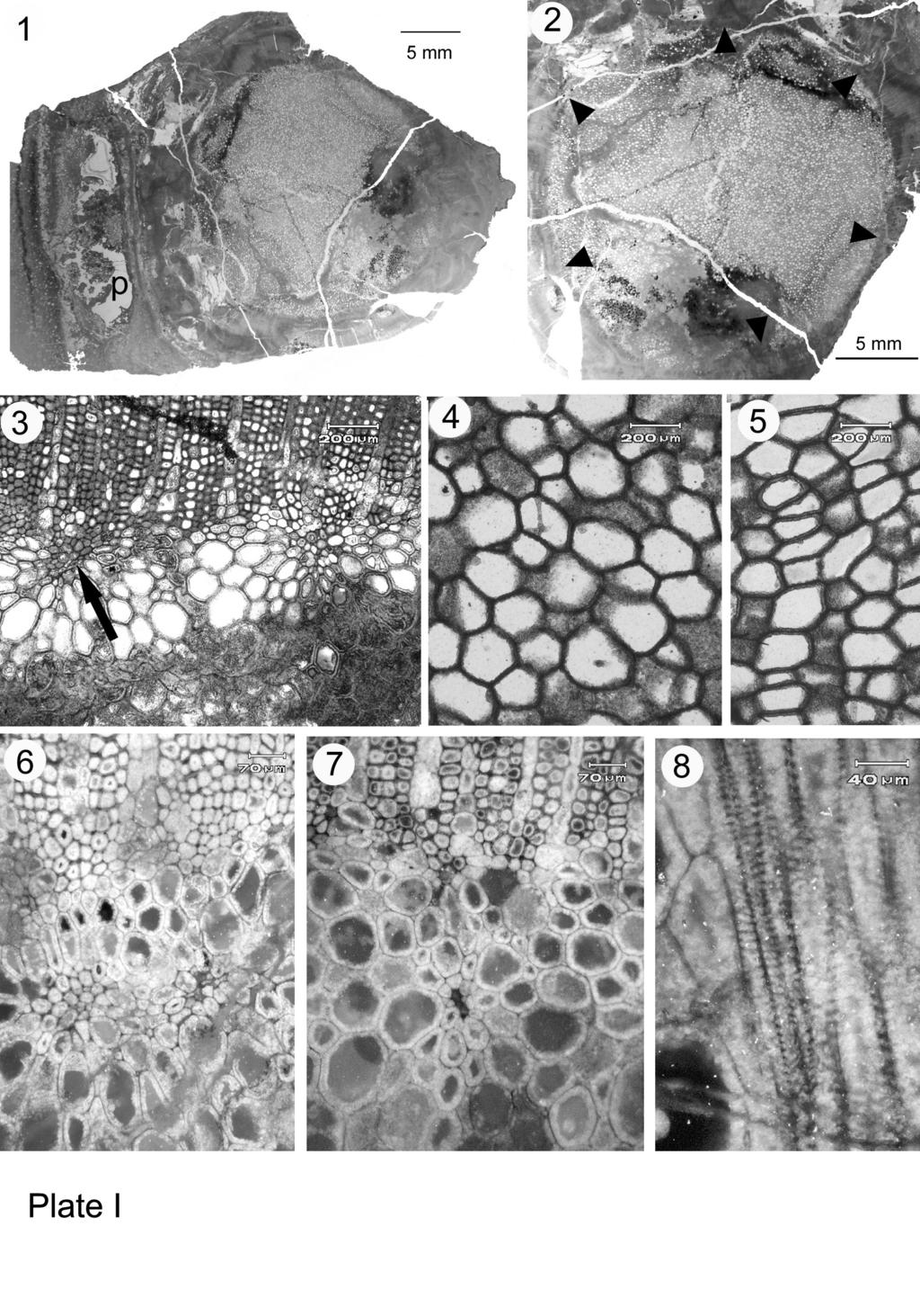
22 Plate legends Plate I. Faironia difasciculata gen. et sp. nov. from the Early Mississippian of France - General view and primary vascular tissues 1. Transverse section of stem with petiole base (p) corresponding to Fig. 1b, MN266- BS General view of stele with circular parenchymatous pith; arrowheads outline the stele border, transverse section. MN266-BS01 3. Transverse section of the pith-secondary xylem boundary showing two primary xylem strands in contact with the secondary xylem, the one at arrow tangentially elongated. MN266-AS Parenchymatous pith cells in transverse section. MN266-AS Parenchymatous pith cells in longitudinal section. MN266-C2LR Tangential pair of slightly immersed primary xylem strands, transverse section. MN266-AS Small immersed primary xylem strand, transverse section. MN266-AS Scalariform pitting on primary xylem tracheids walls, longitudinal section. MN266-C2LR02 Plate II. Faironia difasciculata gen. et sp. nov. from the Early Mississippian of France - Secondary tissues and cortex. 1. Transverse section through the secondary xylem (X2), phloem (P2), inner cortex (IC) that comprises an external zone of compressed cells, and the sparganum outer cortex (OC). MN266-DIO1. 2. Transverse section of secondary xylem showing a triseriate and a uniseriate ray. MN266-BS01 3. Longitudinal tangential section through secondary xylem showing various ray sizes. MN266-CLT Detail of rays, tangential section. MN266-CLT Multiseriate pitting of the radial walls of secondary xylem tracheids. MN266- C2LR02 6. Transverse section showing the external part of the secondary xylem (X2) and the phloem zone below, Sc: sclereids; arrow indicates radially aligned cells in the secondary phloem, MN266-BS01 21
23 7. Detail of the secondary phloem showing a group of radially aligned fibres (f) and probable ray cells (R), transverse section. MN266-BS01 8. Radial longitudinal section showing secondary xylem (X2) and phloem zone with fibers (f) and sclereids (Sc). MN266-C2LR02 Plate III. Faironia difasciculata gen. et sp. nov. from the Early Mississippian of France Leaf trace emission. Note: Leaf trace refers to one trace of a pair. 1. Transverse section at the level of a leaf trace departure showing the leaf trace (uppermost strand, at LT), a strand lying radially just below the LT, and a "deeply" immersed strand at arrow. MN266-BS Radially elongated leaf trace showing two protoxylem strands when crossing the wood, transverse section. MN266-EI Radial ornamentation of the tracheid walls of the leaf trace. MN266-C2LR Leaf trace dividing in the cortex; small abaxial bundle on the left, large lateral bundle on the right, transverse section. MN266-BS General view of the petiole base in transverse section, X2: secondary xylem, roman numerals indicate the location of the 6 vascular bundles originating from two leaf traces, a and b. MN266-AS01 6. Small vascular bundle in the leaf base, with 2 protoxylem strands, transverse section. MN266-AS Two bundles in the leaf base, the large lateral one (I) may divide again, transverse section. MN266-AS Sparganum and non-distorted ground-tissue in the petiole base, transverse section. MN266-AS01. 22
24 Figure legend Fig.1 Faironia difasciculata gen. et sp. nov. from the Early Mississippian of France Transverse sections in ascending order (A-D) of the stem illustrating the position of the vascular supplies to different leaves. Vascular supplies are numbered 1-8 following their order of emission along the ontogenetic spiral. Hypothesized location of leaves 2, 3 and 5 indicated in parentheses on figure B. (MN266; A: AS01, B: BS01, C: DI01, D: EI01). Fig. 2 Faironia difasciculata gen. et sp. nov. from the Early Mississippian of France A-C: Camera lucida drawings at three successive levels illustrating the divergence of a pair of leaf traces a and b (MN266 BS01, DI01, EI01). D: Drawing of the successive stages of trace division leading to the formation of the petiole vascular supply (composite of traces to leaves 7, 4 and 1). Bundles in the petiole base are numbered I-III for traces a and b. 23
25 Table legend Table1. Faironia difasciculata gen. et sp. nov. from the Early Mississippian of France Comparison of some significant anatomical characters in Faironia difasciculata and in some calamopityan and lyginopteridalean seed ferns. Cal eura: Calamopitys eurameriana, Cal sch: Calamopitys schweitzeri, Dii ken: Diichnia kentuckiensis, MN266: Faironia difasciculata, Eri wal: Eristophyton waltonii, Pit day: Pitus dayi, Meg myr: Megalomyelon myriodesmon. sppro: super-parenchymatised protostele, e: eustele. 24
26

27

28
29
30
31 Cal eura Cal sch Dii ken MN 266 Eri wal Pit day Meg myr Wood thickness (mm) Stele diameter (mm) x22 Stele type sppro e e e e e e Stele/stem diameter 1/7 1/10 1/5.5 1/1.5 1/2 Number of sympodial strands Metaxylem tracheids maximum diameter (µm) Wood tracheids maximum diameter (µm) Maximum ray width (cells) µm Maximum ray height (cells) > > mm >5 4 > Maximum number of pit rows Secondary phloem without (-) or f f f with fibres (f) Origin of leaf trace Intercalary strands Maximum number of bundles ?6 3 per leaf base Maximum width of petiole ?12 3 vascular supply (mm) Internode length (mm)
A tree without leaves
 A tree without leaves Brigitte Meyer-Berthaud, Anne-Laure Decombeix To cite this version: Brigitte Meyer-Berthaud, Anne-Laure Decombeix. A tree without leaves. Nature, Nature Publishing Group, 2007, 446
The puzzle presented by the famous stumps of Gilboa, New York, finds a solution in the
 PALAEOBOTANY A tree without leaves Brigitte Meyer-Berthaud and Anne-Laure Decombeix The puzzle presented by the famous stumps of Gilboa, New York, finds a solution in the discovery of two fossil specimens
A new Late Devonian genus with seed plant affinities
 Wang and Liu BMC Evolutionary Biology (2015) 15:28 DOI 10.1186/s12862-015-0292-6 RESEARCH ARTICLE Open Access A new Late Devonian genus with seed plant affinities Deming Wang * and Le Liu Abstract Background:
 Question 1: State the location and function of different types of meristem. Meristems are specialised regions of plant growth. The meristems mark the regions where active cell division and rapid division
Class XI Chapter 6 Anatomy of Flowering Plants Biology
 Class XI Chapter 6 Anatomy of Flowering Plants Biology Question 1: State the location and function of different types of meristem. Meristems are specialised regions of plant growth. The meristems mark
Primary Internal structure & Normal Secondary growth in Sunflower stem
 Primary Internal structure & Normal Secondary growth in Sunflower stem B. Sc. II - Botany Dr. (Miss) Kalpana R. Datar Assistant Professor DEPARTMENT OF BOTANY Willingdon College, Sangli. kalpana_datar@yahoo.com.
Visit For All NCERT solutions, CBSE sample papers, Question papers, Notes for Class 6 to 12. Chapter-6 ANATOMY OF FLOWERING PLANTS
 Chapter-6 ANATOMY OF FLOWERING PLANTS POINTS TO REMEMBER Anatomy : Anatomy is the study of internal structure of organisms. Plant anatomy includes organisation and structure of tissues. Tissue : A group
Plants. Tissues, Organs, and Systems
 Plants Tissues, Organs, and Systems Meristematic cells Specialized cells that are responsible for producing specialized cells, they produce three types of tissue in the body of a plant. Meristematic Cells
Plant Anatomy and Tissue Structures
 Plant Anatomy and Tissue Structures The Two Major Plant Systems Reproductive shoot (flower) Terminal bud Node Internode Angiosperm plants have threse major organs: Roots Stems Leaves & Flowers Terminal
Anatomy of Flowering Plants. K C Meena PGT Biology
 Anatomy of Flowering Plants K C Meena PGT Biology Tissues A group of similar cells performing same function. Types of plant tissues - Meristematic tissues and permanent tissues. Meristematic tissues Have
Angiosperms: Dicotyledons
 Angiosperms: Dicotyledons This section contains anatomical descriptions of stem and twig xylem, as well as the bark and pith regions of 244 dicotyledonous species belonging to 61 families. Angiosperms:
Introduction to Botany. Lecture 25
 Introduction to Botany. Lecture 25 Alexey Shipunov Minot State University November 2, 2015 Shipunov (MSU) Introduction to Botany. Lecture 25 November 2, 2015 1 / 33 Outline 1 Questions and answers 2 Stem
IX. PRIMARY STEM STRUCTURE AND DEVELOPMENT Bot 404 Fall 2004
 IX. PRIMARY STEM STRUCTURE AND DEVELOPMENT Bot 404 Fall 2004 A. Shoot apex -plants have an open system of growth, therefore the ability (at least potentially) to continue growth because there is a meristem
TRILOBOXYLON ARNOLD11 FROM THE MIDDLE DEVONIAN OF WESTERN NEW YORK
 CONTRIBUTIONS FROM THE MUSEUM OF PALEONTOLOGY THE UNIVERSITY OF MICHIGAN VOL. 26, No. 12, p. 257-288 December 31, 1983 TRILOBOXYLON ARNOLD11 FROM THE MIDDLE DEVONIAN OF WESTERN NEW YORK WILLIAM E. STEIN
ANATOMY OF PLANTS Introduction: The study of gross internal structure of plant organs by the technique of section cutting is called plant anatomy.
 ANATOMY OF PLANTS Introduction: The study of gross internal structure of plant organs by the technique of section cutting is called plant anatomy. (Pandey, 2002). Various plant organ viz. root, stem, leaves,
Downloaded from
 POINTS TO REMEMBER : 6. Anatomy of Flowering Plants Study of internal structure of plant is called anatomy. In plants cells are the basic unit. Cells organized into tissues and tissues organized into organs.
A CALLIXYLON (ARCHAEOPTERIDALES, PROGYMNOSPERMOPSIDA) TRUNK WITH PRESERVED SECONDARY
 TRUNK WITH PRESERVED SECONDARY") American Journal of Botany 100(11): 2219 2230. 2013. A CALLIXYLON (ARCHAEOPTERIDALES, PROGYMNOSPERMOPSIDA) TRUNK WITH PRESERVED SECONDARY PHLOEM FROM THE LATE DEVONIAN OF MOROCCO 1 A NNE-LAURE D ECOMBEIX
American Journal of Botany 100(11): 2219 2230. 2013. A CALLIXYLON (ARCHAEOPTERIDALES, PROGYMNOSPERMOPSIDA) TRUNK WITH PRESERVED SECONDARY PHLOEM FROM THE LATE DEVONIAN OF MOROCCO 1 A NNE-LAURE D ECOMBEIX
[279] A NOTE ON THE ORIGIN OF LATERAL ROOTS AND THE STRUCTURE OF THE ROOT-APEX OF LYGINOPTERIS OLDHAMIA
![[279] A NOTE ON THE ORIGIN OF LATERAL ROOTS AND THE STRUCTURE OF THE ROOT-APEX OF LYGINOPTERIS OLDHAMIA](/thumbs/83/87080534.jpg "[279] A NOTE ON THE ORIGIN OF LATERAL ROOTS AND THE STRUCTURE OF THE ROOT-APEX OF LYGINOPTERIS OLDHAMIA") [279] A NOTE ON THE ORIGIN OF LATERAL ROOTS AND THE STRUCTURE OF THE ROOT-APEX OF LYGINOPTERIS OLDHAMIA BY A. C. HALKET (With Plate XI and i figure in the text) E 'GlNOPTERis oi.dh.imi.i, a plant of the
[279] A NOTE ON THE ORIGIN OF LATERAL ROOTS AND THE STRUCTURE OF THE ROOT-APEX OF LYGINOPTERIS OLDHAMIA BY A. C. HALKET (With Plate XI and i figure in the text) E 'GlNOPTERis oi.dh.imi.i, a plant of the
TEXT The name stele has been derived from Greek word meaning pillar or column. Sachs in 1875 proposed that the vascular system of the plant body is a
 TEXT The name stele has been derived from Greek word meaning pillar or column. Sachs in 1875 proposed that the vascular system of the plant body is a continuous system. For the first time, the stelar organisation
Plant Tissues and Organs. Topic 13 Plant Science Subtopics , ,
 Plant Tissues and Organs Topic 13 Plant Science Subtopics 13.1.2, 13.1.3, 13.1.4 Objectives: List and describe the major plant organs their structure and function List and describe the major types of plant
Topic 2: Plant Structure & Growth Ch. 35 Angiosperms are the most complex plants. They are composed of cells, tissues, organs and organ systems.
 Topic 2: Plant Structure & Growth Ch. 35 Angiosperms are the most complex plants. They are composed of cells, tissues, organs and organ systems. Fig. 35.8 Plant Cells pp.798-802 Types of plant cells Include:
Plant Anatomy Lab 7 - Stems II
 Plant Anatomy Lab 7 - Stems II This exercise continues the previous lab in studying primary growth in the stem. We will be looking at stems from a number of different plant species, and emphasize (1) the
2 Description of the Specimens. 1 Materials and Methods. ZHOU Yi-Long 1, LIU Xian-Wei 2, WANG Shi-Jun 1*, CHEN Gui-Ren 3, TIAN Bao-Lin 3
 Acta Botanica Sinica 2004, 46 (11): 1261-1268 http://www.chineseplantscience.com On the Anatomical Structures of the Lepidodendralean Stem Lepidodendron tripunctatum from Coal Balls of Taiyuan Formation
NOTES ON GINKGO BILOBA'
 NOTES ON GINKGO BILOBA' WALTER WV. TUPPER (WITH PLATE xx) Among the gymnosperms, one of the groups most interesting from a morphological standpoint is the Ginkgoales, the only living representative of
Lecture 19. A Sieve Plate with large Sieve Pores. Secondary Phloem. Secondary phloem (cont d)
") Lecture 19 Secondary phloem (cont d) Secondary Phloem in Tilia americana (American Basswood) Secondary Phloem of Tilia Stained with Toluidine Blue & viewed with Crossed Polarizers. Secondary Phloem A Sieve
Lecture 19 Secondary phloem (cont d) Secondary Phloem in Tilia americana (American Basswood) Secondary Phloem of Tilia Stained with Toluidine Blue & viewed with Crossed Polarizers. Secondary Phloem A Sieve
THE OHIO JOURNAL OF SCIENCE
 THE OHIO JOURNAL OF SCIENCE VOL. XXIV JULY, 1924 No.. 4 THE VASCULAR ANATOMY OF CALAMOVILFA LONGIFOLIA.* ERNEST LINCOLN STOVER Eastern Illinois State Teachers' College The present study of the anatomy
Plant Anatomy: roots, stems and leaves
 Plant Anatomy: roots, stems and leaves The plant body has a hierarchy of organs, tissues and cells Plants, like animals, have organs composed of different tissues, which are composed of cells. Tissue is
CHAPTER 6 ANATOMY OF FLOWERING PLANTS MULTIPLE CHOICE QUESTIONS
 ANATOMY OF FLOWERING PLANTS 27 27 CHAPTER 6 ANATOMY OF FLOWERING PLANTS MULTIPLE CHOICE QUESTIONS 1. A transverse section of stem is stained first with safranin and then with fast green following the usual
Structural Botany Laboratory 3 Simplest and Earliest Land Plants
 Structural Botany Laboratory 3 Simplest and Earliest Land Plants At one time all of the most ancient (i.e., fossils of the Silurian and Devonian) and simple vascular plants were placed in the class Psilopsida.
Plant Structure. Lab Exercise 24. Objectives. Introduction
 Lab Exercise Plant Structure Objectives - Be able to identify plant organs and give their functions. - Learn distinguishing characteristics between monocot and dicot plants. - Understand the anatomy of
UNIT 6 - STRUCTURES OF FLOWERING PLANTS & THEIR FUNCTIONS
 6.1 Plant Tissues A tissue is a group of cells with common function, structures or both. In plants we can find 2 types of tissues: Meristem Permanent tissues Meristem is found in regions with continuous
SESSION 6: SUPPORT AND TRANSPORT SYSTEMS IN PLANTS PART 1
 SESSION 6: SUPPORT AND TRANSPORT SYSTEMS IN PLANTS PART 1 KEY CONCEPTS In this session we will focus on summarising what you need to know about: - Anatomy of dicotyledonous plants Root and stem: distribution
2/25/2013. o Plants take up water and minerals from below ground o Plants take up CO2 and light from above ground THREE BASIC PLANT ORGANS ROOTS
 o Plants take up water and minerals from below ground o Plants take up CO2 and light from above ground THREE BASIC PLANT ORGANS o Roots o Stems o Leaves ROOTS o Anchor plant o Absorb water and minerals
PHARMACOBOTANY LECTURE 5. PLANT TISSUES III.
 PHARMACOBOTANY LECTURE 5. PLANT TISSUES III. VASCULAR TISSUES VASCULAR TISSUES Xylem transporting water and mineral substances from the root upwards to other plant organs Phloem carries photosynthetic
The Shoot System: Primary Stem Structure - 1
 The Shoot System: Primary Stem Structure - 1 Shoot System The shoot system comprises the leaves and stems of plants. Leaves are located at nodes on the stem; the distance along the stem between nodes is
Plant Structure And Growth
 Plant Structure And Growth The Plant Body is Composed of Cells and Tissues Tissue systems (Like Organs) made up of tissues Made up of cells Plant Tissue Systems Ground Tissue System Ø photosynthesis Ø
Chapter #35~ Plant Structure and Growth
 Chapter #35~ Plant Structure and Growth What part of a plant is represented by each of these: Carrot Celery Red Pepper Tomato Lettuce Garbanzo Bean Angiosperm structure Three basic organs: Roots (root
STEMS Anytime you use something made of wood, you re using something made from the stem of a plant. Stems are linear structures with attached leaves
 STEMS OUTLINE External Form of a Woody Twig Stem Origin and Development Stem Tissue Patterns Herbaceous Dicotyledonous Stems Woody Dicotyledonous Stems Monocotyledonous Stems Specialized Stems Wood and
Plant Structure. Objectives At the end of this sub section students should be able to:
 Name: 3.2 Organisation and the Vascular Structures 3.2.1 Flowering plant structure and root structure Objectives At the end of this sub section students should be able to: 1. Label a diagram of the external
Secondary growth in stems
 Secondary growth in stems Secondary growth Some of the meristematic cells in plants with secondary growth keep their meristematic state and become cells of the cambium. The addition of secondary vascular
ROOTS. Syllabus Theme A Plant Structure and Function. Root systems. Primary Growth of Roots. Taproot system. Fibrous root system.
 Syllabus Theme A lant Structure and Function A2: Structure and function of the basic plant organs ampbell & Reece hap. 35 Selected page numbers ROOTS Functions Anchors the vascular plant Absorbs minerals
Lab Exercise 4: Primary Growth and Tissues in Stems
 Lab Exercise 4: Primary Growth and Tissues in Stems Tissues of the plant body can be classified in a variety of ways: functionally (based on the tissue function, e.g. vascular tissue ), morphologically
TARGET STUDY MATERIAL
 TARGET STUDY MATERIAL Plus-1 Botany VOL I TARGET EDUCATIONAL INSTITUTION Target Educational institution is the one and only Entrance coaching and CBSE 10 th coaching centre at Mukkam with advanced technologies
Chapter 35~ Plant Structure and Growth
 Chapter 35~ Plant Structure and Growth Plant Organization Plant morphology is based on plant s evolutionary history Need to draw in nutrients from the ground and the air Plant Organs Root system = roots
Exercise 12. Procedure. Aim: To study anatomy of stem and root of monocots and dicots.
 Aim: To study anatomy of stem and root of monocots and dicots. Principle: The study of internal morphology, i.e., cells of various tissues in an organ of a living body is called Anatomy. Tissue, which
VARIATION IN THE SIZE OF RAY PITS OF CONIFERS.*
 VARIATION IN THE SIZE OF RAY PITS OF CONIFERS.* FOREST B. H. BROWN. Since Haeckel proposed the word Ecology in 88, there has been an ever growing interest in the influence which environmental factors may
Plant Anatomy. By Umanga Chapagain
 Plant Anatomy By Umanga Chapagain PLANT ANATOMY The science of the structure of the organized plant body learned by dissection is called Plant Anatomy. In general, Plant Anatomy refers to study of internal
Plants. Plant Form and Function. Tissue Systems 6/4/2012. Chapter 17. Herbaceous (nonwoody) Woody. Flowering plants can be divided into two groups:
 Woody. Flowering plants can be divided into two groups:") Monocots Dicots 6/4/2012 Plants Plant Form and Function Chapter 17 Herbaceous (nonwoody) In temperate climates, aerial parts die back Woody In temperate climates, aerial parts persist The Plant Body Functions
Monocots Dicots 6/4/2012 Plants Plant Form and Function Chapter 17 Herbaceous (nonwoody) In temperate climates, aerial parts die back Woody In temperate climates, aerial parts persist The Plant Body Functions
Plant Anatomy: roots, stems and leaves
 Plant Anatomy: roots, stems and leaves The plant body has a hierarchy of organs, tissues and cells Plants, like animals, have organs composed of different tissues, which are composed of cells. Tissue is
The Petiolar Structure of Christella dentata (Forssk.) Brownsey & Jermy (Thelypteridaceae, Pteridophyta)
 Brownsey & Jermy (Thelypteridaceae, Pteridophyta)") Ethnobotanical Leaflets 12: 96-102. 2008. The Petiolar Structure of Christella dentata (Forssk.) Brownsey & Jermy (Thelypteridaceae, Pteridophyta) KAMINI SRIVASTAVA, M.Sc, D.Phil Department of Botany,
Ethnobotanical Leaflets 12: 96-102. 2008. The Petiolar Structure of Christella dentata (Forssk.) Brownsey & Jermy (Thelypteridaceae, Pteridophyta) KAMINI SRIVASTAVA, M.Sc, D.Phil Department of Botany,
Chapter 29: Plant Tissues
 Chapter 29: Plant Tissues Shoots and Roots Shoots (Leaves and Stem) Produce food by photosynthesis Carry out reproductive functions Roots Anchor the plant Penetrate the soil and absorb water and dissolved
A group of cells with common origin is called a tissue. The cells of a tissue usually perform a common function.
 Anatomy of Flowering Plants Tissues A group of cells with common origin is called a tissue. The cells of a tissue usually perform a common function. Types of Tissue: There are two main types of plant tissues,
Paleontological Society
 Paleontological Society New Occurrence of Periastron reticulatum Unger emend. Beck, an Enigmatic Mississippian Fossil Plant Author(s): Robert A. Gastaldo Reviewed work(s): Source: Journal of Paleontology,
STRUCTURAL BOTANY LABORATORY 3. Simplest and Earliest Land Plants. Fig. 1. Generalized relationships of Vascular Plants (Rothwell, 1994)
") STRUCTURAL BOTANY LABORATORY 3 Simplest and Earliest Land Plants At one time all of the most ancient (i.e., fossils of the Silurian and Devonian) and simple vascular plants were placed in the class Psilopsida.
STRUCTURAL BOTANY LABORATORY 3 Simplest and Earliest Land Plants At one time all of the most ancient (i.e., fossils of the Silurian and Devonian) and simple vascular plants were placed in the class Psilopsida.
Botany Physiology. Due Date Code Period Earned Points
 Botany Physiology Name C/By Due Date Code Period Earned Points Bot Phys 5N5 Stem Forms Bot Phys 5-05 Identify the major forms of stems in plants I. Identify the major forms of stems in plants A. internal
Primate cranium morphology through ontogenesis and phylogenesis: factorial analysis of global variation
 Primate cranium morphology through ontogenesis and phylogenesis: factorial analysis of global variation Nicole Petit-Maire, Jean-François Ponge To cite this version: Nicole Petit-Maire, Jean-François Ponge.
MAGNOLIA botany. evergreen ; spicy odor of blooms; chambered pith; hairy leaves(lower epidermis) & petioles
 & petioles") MAGNOLIA botany Angiosperm: primitive, ancestral (Not Eudicot, Not Monocot): order Ranales: family Magnoliaceae: Magnolia grandiflora (southern magnolia) evergreen ; spicy odor of blooms; chambered pith;
MAGNOLIA botany Angiosperm: primitive, ancestral (Not Eudicot, Not Monocot): order Ranales: family Magnoliaceae: Magnolia grandiflora (southern magnolia) evergreen ; spicy odor of blooms; chambered pith;
1 Conducting Units: Tracheids and Vessels 1.1 Evolutionary Specialization
 1 Conducting Units: Tracheids and Vessels 1.1 Evolutionary Specialization The development of upright land plants depended on the development of a waterconducting system. Many of the earliest land plants,
with others and thus regenerate a functioning conductive system. Regeneration
 388 BOTANY: SINNOTT AND BLOCH PROC. N. A. S. VISIBLE EXPRESSION OF CYTOPLASMIC PA TTERN IN THE DIFFERENTIATION OF XYLEM STRANDS BY EDMUND W. SINOTT AND ROBERT BLOCH OsBORN BOTANCAL LABORATORY, YALE UNIVERSITY
The significance of Runcaria (Middle Devonian, Belgium) in the evolution of seed plants.
 in the evolution of seed plants.") The significance of Runcaria (Middle Devonian, Belgium) in the evolution of seed plants. [L'importance de Runcaria (Dévonien moyen, Belgique) pour l'évolution des plantes à graines] Philippe GERRIENNE
The significance of Runcaria (Middle Devonian, Belgium) in the evolution of seed plants. [L'importance de Runcaria (Dévonien moyen, Belgique) pour l'évolution des plantes à graines] Philippe GERRIENNE
A COMPARATIVE STUDY OF THE PRIMARY VASCULAR SYSTE~1 CONIFERS. III. STELAR EVOLUTION IN GYMNOSPERMS 1
 Amer. J. Bot. 55(4): 464-472. 1!16'>. A COMPARATIVE STUDY OF THE PRIMARY VASCULAR SYSTE~1 CONIFERS. III. STELAR EVOLUTION IN GYMNOSPERMS 1 OF KADAMBARI K. NAMBOODIRI 2 AND CHARLES B. BECK Department of
I. Lycopodiales: The Vegetative Features of the Sporophyte Phase
 Lab II. Lycopodiales: the Clubmosses I. Lycopodiales: The Vegetative Features of the Sporophyte Phase The clubmosses (traditionally classified as species of the genus Lycopodium) are low, evergreen plants
Plant Structure and Function
 Plant Structure and Function A Meridian Biology AP Study Guide by John Ho and Tim Qi Plant Terms Growth: Growth Types Type Location Description Primary Primary Vertical growth (up-down), dominant direction
The three principal organs of seed plants are roots, stems, and leaves.
 23 1 Specialized Tissues in Plants Seed Plant Structure The three principal organs of seed plants are roots, stems, and leaves. 1 of 34 23 1 Specialized Tissues in Plants Seed Plant Structure Roots: absorb
Honors Biology I Ch 29 Plant Structure & Function
 3 Basic types of plant cells Honors Biology I Ch 29 Plant Structure & Function 1) Parenchyma cells- loosely packed or cells with a and thin, Involved in metabolic functions 2) Collenchyma cells- thicker
Smart Bolometer: Toward Monolithic Bolometer with Smart Functions
 Smart Bolometer: Toward Monolithic Bolometer with Smart Functions Matthieu Denoual, Gilles Allègre, Patrick Attia, Olivier De Sagazan To cite this version: Matthieu Denoual, Gilles Allègre, Patrick Attia,
Effects of Sun-Blotch on the Anatomy of the Avocado Stem
 California Avocado Association 1935 Yearbook 20: 125-129 Effects of Sun-Blotch on the Anatomy of the Avocado Stem Charles A. Schroeder Because of the comparatively recent discovery of the avocado disease
Case report on the article Water nanoelectrolysis: A simple model, Journal of Applied Physics (2017) 122,
 122,") Case report on the article Water nanoelectrolysis: A simple model, Journal of Applied Physics (2017) 122, 244902 Juan Olives, Zoubida Hammadi, Roger Morin, Laurent Lapena To cite this version: Juan Olives,
Case report on the article Water nanoelectrolysis: A simple model, Journal of Applied Physics (2017) 122, 244902 Juan Olives, Zoubida Hammadi, Roger Morin, Laurent Lapena To cite this version: Juan Olives,
NOTES: CH 35 - Plant Structure & Growth
 NOTES: CH 35 - Plant Structure & Growth In their evolutionary journey, plants adapted to the problems of a terrestrial existence as they moved from water to land ANGIOSPERMS (flowering plants) -most diverse
Wood Anatomy Lab What is wood? The lateral meristems Cell types Vessels Tracheids
 Wood Anatomy Lab Objectives of the Lab: 1) Learn to recognize major cell types and features of wood including: tracheids vessels rays axial parenchyma pits tyloses resin canals 2) Look at wood in three
Stems and Transport in Vascular Plants. Herbaceous Stems. Herbaceous Dicot Stem 3/12/2012. Chapter 34. Basic Tissues in Herbaceous Stems.
 Bud scale Terminal bud Stems and Transport in Plants One year's growth Terminal bud scale scars Axillary bud Leaf scar Node Internode Node Chapter 34 Lenticels Terminal bud scale scars Bundle scars A Woody
Plant Structure and Growth
 Plant Structure and Growth A. Flowering Plant Parts: The flowering plants or are the most diverse group of plants. They are divided into 2 classes and. Examples of monocots: Examples of dicots: The morphology
Non Permanent Tissues - Meristematic Tissue
 PLANT TISSUES Non Permanent Tissues - Meristematic Tissue Undifferentiated plant cells that are continually dividing by mitosis Large thin walled cells No vacuole Dense cytoplasm Large nucleus Found at
Methylation-associated PHOX2B gene silencing is a rare event in human neuroblastoma.
 Methylation-associated PHOX2B gene silencing is a rare event in human neuroblastoma. Loïc De Pontual, Delphine Trochet, Franck Bourdeaut, Sophie Thomas, Heather Etchevers, Agnes Chompret, Véronique Minard,
THE ROOTS OF WILD RICE. ZIZANIA AQUATICA L.
 THE ROOTS OF WILD RICE. ZIZANIA AQUATICA L. E. L. STOVER, Eastern Illinois State Teachers College. This grass grows from Maine to Minnesota in aquatic habitats (2 and 5). It is common in marsh lands all
PLANTS FORM AND FUNCTION PLANT MORPHOLOGY PART I: BASIC MORPHOLOGY. Plant Form & Function Activity #1 page 1
 AP BIOLOGY PLANTS FORM AND FUNCTION ACTIVITY #1 NAME DATE HOUR PLANT MORPHOLOGY PART I: BASIC MORPHOLOGY Plant Form & Function Activity #1 page 1 PART II: ROOTS 1. Examine the examples of the two root
NUCLEUS ALBUMINOUS CELL SIEVE CELL SIEVE AREA SMALL PORES SIEVE CELL PHLOEM L.S. SUGAR
 NUCLEUS? ALBUMINOUS CELL SUGAR SIEVE CELL SIEVE AREA SMALL PORES SIEVE CELL PHLOEM L.S. NUCLEUS S ALBUMINOUS CELL SUGAR SIEVE? CELL SIEVE PLATE LARGE PORES SIEVE? CELL PHLOEM L.S. NUCLEUS? ALBUMINOUS CELL
Stelar evolution. B. Sc. III Botany
 Stelar evolution B. Sc. III Botany Dr. (Miss) Kalpana R. Datar Assistant Professor DEPARTMENT OF BOTANY WILLINGDON COLLEGE, SANGLI. kalpana_datar@yahoo.com Stelar Evolution Stele is unit of vascular system.
Topic 23. The Ferns and Their Relatives
 Topic 23. The Ferns and Their Relatives Domain: Eukarya Kingdom: Plantae Ferns Leptosporangiate Ferns Psilophytes Genus: Psilotum Horsetails Genus: Equisetum In this treatment we lump the Psilophytes and
A Branched Cone of Equisetum, 113
 A Branched Cone of Equisetum, 113 6. E. C. Jeffrey. " The Development, Structure and Affinities of the genus Equisetum." Memoirs of the Boston Society of Natural History, Vol. V., No. 5, 1899. 7. R. Kidston.
A new simple recursive algorithm for finding prime numbers using Rosser s theorem
 A new simple recursive algorithm for finding prime numbers using Rosser s theorem Rédoane Daoudi To cite this version: Rédoane Daoudi. A new simple recursive algorithm for finding prime numbers using Rosser
Characterization of the local Electrical Properties of Electrical Machine Parts with non-trivial Geometry
 Characterization of the local Electrical Properties of Electrical Machine Parts with non-trivial Geometry Laure Arbenz, Abdelkader Benabou, Stéphane Clenet, Jean Claude Mipo, Pierre Faverolle To cite this
2.1 PLANT TISSUE HALIMAHTUN SAEDIAH BT ABU BAKAR KOLEJ TEKNOLOGI TIMUR
 2.1 PLANT TISSUE HALIMAHTUN SAEDIAH BT ABU BAKAR KOLEJ TEKNOLOGI TIMUR GENERAL Plant cell are differentiated possessing structural adaptations that make specific functions possible. Modifications of cell
CHAPTER 6 ANATOMY OF FLOWERING PLANTS
 84 BIOLOGY CHAPTER 6 ANATOMY OF FLOWERING PLANTS 6.1 The Tissues 6.2 The Tissue System 6.3 Anatomy of Dicotyledonous and Monocotyledonous Plants 6.4 Secondary Growth You can very easily see the structural
Chapter 28 Active Reading Guide Plant Structure and Growth
 Name: AP Biology Mr. Croft Chapter 28 Active Reading Guide Plant Structure and Growth In this unit on plants, the challenge for students will be to learn the new vocabulary. As we work through this unit,
Easter bracelets for years
 Easter bracelets for 5700000 years Denis Roegel To cite this version: Denis Roegel. Easter bracelets for 5700000 years. [Research Report] 2014. HAL Id: hal-01009457 https://hal.inria.fr/hal-01009457
CHAPTER 6 ANATOMY OF FLOWERING PLANTS
 84 BIOLOGY CHAPTER 6 ANATOMY OF FLOWERING PLANTS 6.1 The Tissues 6.2 The Tissue System 6.3 Anatomy of Dicotyledonous and Monocotyledonous Plants 6.4 Secondary Growth You can very easily see the structural
Evolution of the cooperation and consequences of a decrease in plant diversity on the root symbiont diversity
 Evolution of the cooperation and consequences of a decrease in plant diversity on the root symbiont diversity Marie Duhamel To cite this version: Marie Duhamel. Evolution of the cooperation and consequences
CAMBIUM, meristem, heartwood, and lenticel are
 Examining the Structures of a Tree CAMBIUM, meristem, heartwood, and lenticel are some terms that may be new to you. These terms are used to describe various tree structures. Not surprisingly, many terms
WHAT DO you think of when you
 Stem Anatomy WHAT DO you think of when you think of a stem? Do you think of a flower stalk, the trees in your area, or a soybean stalk? Most people probably visualize something like the flower or the bean
Lecture 4 Root Put line under your answer! There is only one correct answer in the multiple choice questions
 Lecture 4 Root Put line under your answer! There is only one correct answer in the multiple choice questions 1. The perception of gravity by a root is thought to take place in a) root hairs b) the region
WOOD AND BARK ANATOMY OF. Rancho Santa Ana Botanic Garden and Department of Biology, Pomona College Claremont, California 91711
 ALISO 12(3), 1989, pp. 485-495 WOOD AND BARK ANATOMY OF DEGENERIA SHERWIN CARLQUIST Rancho Santa Ana Botanic Garden and Department of Biology, Pomona College Claremont, California 91711 ABSTRACT Wood anatomy
BIOL 305L Laboratory One
 Please print Full name clearly: BIOL 305L Laboratory One General plant anatomy a great place to start! Introduction Botany is the science of plant life. Traditionally, the science included the study of
MODal ENergy Analysis
 MODal ENergy Analysis Nicolas Totaro, Jean-Louis Guyader To cite this version: Nicolas Totaro, Jean-Louis Guyader. MODal ENergy Analysis. RASD, Jul 2013, Pise, Italy. 2013. HAL Id: hal-00841467
Multiple sensor fault detection in heat exchanger system
 Multiple sensor fault detection in heat exchanger system Abdel Aïtouche, Didier Maquin, Frédéric Busson To cite this version: Abdel Aïtouche, Didier Maquin, Frédéric Busson. Multiple sensor fault detection
[92] THE NEW EVIDENCE OF ISOSPORY IN PALAEOZOIC SEED PLANTS
![[92] THE NEW EVIDENCE OF ISOSPORY IN PALAEOZOIC SEED PLANTS](/thumbs/92/110747357.jpg "[92] THE NEW EVIDENCE OF ISOSPORY IN PALAEOZOIC SEED PLANTS") [92] THE NEW EVIDENCE OF ISOSPORY IN PALAEOZOIC SEED PLANTS BY M. BENSON (With 4 figures in the text) AN intensive study of some sections of the Palaeozoic fructifica- J\ tion Schnetzia Bennieana Kidston,
[92] THE NEW EVIDENCE OF ISOSPORY IN PALAEOZOIC SEED PLANTS BY M. BENSON (With 4 figures in the text) AN intensive study of some sections of the Palaeozoic fructifica- J\ tion Schnetzia Bennieana Kidston,
Plant Structure and Function Extension
 Plant Structure and Function Extension NGSSS: SC.912.L.14.7 Relate the structure of each of the major plant organs and tissues to physiological processes. (AA) Part 1A: Leaves The leaf of a plant serves
2.5 : Cells are grouped into tissue
 2.5 : Cells are grouped into tissue 1 CELL STRUCTURE AND FUNCTIONS Prokaryotic and eukaryotic cells Structures & functions: Cell membrane and organelles Animal Cells are grouped into tissue Plant Cell
STATISTICAL ENERGY ANALYSIS: CORRELATION BETWEEN DIFFUSE FIELD AND ENERGY EQUIPARTITION
 STATISTICAL ENERGY ANALYSIS: CORRELATION BETWEEN DIFFUSE FIELD AND ENERGY EQUIPARTITION Thibault Lafont, Alain Le Bot, Nicolas Totaro To cite this version: Thibault Lafont, Alain Le Bot, Nicolas Totaro.
Histology and Anatomy of Flowering Plants
 Histology and Anatomy of Flowering Plants Very Short Answer Type Questions 1. The transverse section of a plant material shows the following anatomical features: a) The vascular bundles are conjoint, scattered