Exopodites, Epipodites and Gills in Crustaceans
|
|
|
- Branden Hicks
- 5 years ago
- Views:
Transcription
1 Arthropod Systematics & Phylogeny (2) Museum für Tierkunde Dresden, eissn , Exopodites, Epipodites and Gills in Crustaceans Ge o f f A. Bo x s h a l l 1 & Da m i à Ja u m e 2 1 Department of Zoology, The Natural History Museum, Cromwell Road, London SW7 5BD, United Kingdom [g.boxshall@nhm.ac.uk] 2 IMEDEA (CSIC-UIB), Instituto Mediterráneo de Estudios Avanzados, c / Miquel Marquès, 21, Esporles (Illes Balears), Spain [d.jaume@uib.es] Received 01.iii.2009, accepted 22.v Published online at on 25.viii > Abstract The structure of the outer parts of the maxillae and post-maxillary limbs is compared across the major crustacean groups. New anatomical observations are presented on the musculature of selected limbs of key taxa and general patterns in limb structure for the Crustacea are discussed. Exopodites vary in form but are typically provided with musculature, whereas epipodites and other exites lack musculature in all post-maxillary limbs. Within the Crustacea, only the Myodocopa possesses an epipodite on the maxilla. New evidence from developmental genetics, from embryology, and from new Palaeozoic fossils is integrated into a wider consideration of the homology of exites (outer lobes). This evidence supports the homology of the distal epipodite of anostracan branchiopods with the epipodite-podobranch complex of malacostracans. The evidence for the homology of pre-epipodites across the Crustacea is less robust, as is the evidence that the possession of a proximal pre-epipodite and a distal epipodite is the ancestral malacostracan condition. The widely assumed homology of the peracaridan oostegite with the pre-epipodite is questioned: little supporting evidence exists and possible differences in underlying control mechanisms need further loration. > Key words Comparative anatomy, limb structure, musculature, Crustacea. 1. Introduction The main axis of crustacean post-antennulary limbs originally comprises a proximal protopodal part plus two distal rami, an outer exopodite and an inner endopodite or telopodite (see Bo x s h a l l 2004 for review). The medial (inner) surface of a trunk limb can be produced to form a series of endites, including the proximal gnathobase. Similarly, the lateral (outer) surface of a trunk limb may be produced to form one or more exites, or outer lobes. In this paper we focus on the lateral compartment of the limb, i.e. on the exopodite and the exites of all kinds, and we compare the amazing diversity of lateral limb structures ressed throughout the Crustacea. We also focus primarily on the maxilla and post-maxillary trunk limbs because, as indicated by the cephalocaridan condition (Sa n d- e r s 1963; He s s l e r 1964), the limbs posterior to the maxillule are derived from a common pattern. However, where relevant we will include information on maxillulary structure. In addition we consider crustacean gills since many of the structures referred to as gills, a functionally based but anatomically imprecise term, are modified exites carried on the limbs. Other gills are modifications of the body wall adjacent to limb bases. The main aims of this paper are to lore the structure of the lateral compartment of limbs and to reexamine the evidence supporting the identification of homologous structures in different crustacean groups. The evidence base comprises some new anatomical observations on selected crustaceans, integrated with the new data emerging from morphological studies on fossil arthropods, particularly those from the Palaeozoic, from gene ression patterns as revealed by recent evolutionary-development studies, and from embryological studies. In addition, we seek to address some of the terminology problems by identifying new
2 230 Bo x s h a l l & Ja u m e: Exopodites, epipodites and gills in crustaceans criteria that might serve to strengthen the standard definitions. 2. Materials and methods 2.1. Material studied Amphionides reynaudi (H. Milne Edwards, 1833) material from collections of NHM, London: Reg. No Anaspides tasmaniae (Thomson, 1893) material from collections of NHM, London: Reg. Nos Andaniotes linearis K.H. Barnard, 1932 material from collections of NHM, London: Reg. Nos Argulus foliaceus (Linnaeus, 1758) adult female serial sectioned (transverse sections) at 8 μm, stained with Mallory s trichrome. Argulus japonicus Thiele, 1900 unregistered material from collections of NHM, London. Bentheuphausia amblyops G.O. Sars, 1885 material from collections of NHM, London: Reg. Nos VIII Nebalia pugettensis Clark, 1932 ovigerous female from collections of NHM, London: Reg. No Phreatogammarus fragilis (Chilton, 1882) material from collections of NHM, London: Reg. Nos Proasellus banyulensis (Racovitza, 1919) collected at Son Regalat, Bellpuig, Artà, Mallorca (Balearic Islands) by D. Jaume. Pseuderichthus larva of Pseudosquilla sp. from collections of NHM, London: Reg. No Polycope sp. material from collection of NHM, London: Reg. No Spelaeomysis bottazzii Caroli, 1924 material collected from Zinzulusa Cave, Lecce, Italy by D. Jaume, G. Boxshall & G. Belmonte. Sphaeromides raymondi Dollfus, 1897 males from collections of NHM, London: Reg. Nos Tulumella sp. material collected from the Exuma Cays, Bahamas, by T.M. Iliffe, G.A. Boxshall and D. Jaume Methods Dissected appendages were observed as temporary mounts in lactophenol on a Leitz Diaplan microscope equipped with differential interference optics. Anatomical drawings were made with the aid of a camera lucida. Material for SEM was washed in distilled water, dehydrated through graded acetone series, critical point dried using liquid carbon dioxide as the exchange medium, mounted on aluminium stubs and sputter coated with palladium. Coated material was examined on a Phillips XL30 Field Emission Scanning Electron microscope operated at 5 kv. When describing limb axes we used proximal-distal to describe the axis from the origin on the body to its tip, anterior-posterior for the axis parallel to the longitudinal anterior-posterior axis of the body, and lateral-medial (and outer-inner) to describe parts of limbs lying away from or closer to the vertical plane on the longitudinal anterior-posterior axis. Dorsal and ventral are used topologically, with reference to the ventral nerve cord. 3. The exopodite The exopodite is the outer ramus of the biramous arthropodan leg and has traditionally been defined by its origin on the distal part of the protopodite (the basis), lateral to the endopodite. The exopodite has been distinguished from other outer structures (exites of various kinds) on limbs by the possession of musculature inserting within it: such musculature is typically lacking in exites. Exopodites are divided into segments in some arthropods and the presence of intrinsic musculature allows segments to move relative to one another. Recently, the developmental approach employed by Wo l f f & Sc h o lt z (2008) has generated a new criterion the exopodite and endopodite are formed by a secondary subdivision of the growth zone of the main limb axis whereas lateral outgrowths, such as exites, result from the establishment of new axes. The earliest ression of the Distal-less gene in the tips of biramous limb buds is irrespective of the form of the adult limb, i.e. whether it is stenopodial or phyllopodial (Ol e s e n et al. 2001). As noted by Wo l f f & Sc h o lt z (2008), the process of exopodite and endopodite formation by subdivision of the primary limb axis is reflected by the transformation of the initially undivided Distal-less ression into two separate domains representing the tips of the two rami (Wi l l i a m s 2004). The mechanism producing this split is currently unknown but suggested likely scenarios are the suppression of Distal-less ression in the area between the exopodal and endopodal domains, or apoptosis (Wolff & Scholtz 2008) Exopodites in non-crustacean Arthropoda Comparison of the form of the exopodite across key fossil and Recent arthropod taxa led Bo x s h a l l (2004) to suggest that the ancestral state of the arthropodan exopodite was probably two-segmented. In early Palaeozoic arthropods, such as trilobites, the trunk limbs are typically biramous, with a well developed inner walking branch (the endopodite) and a well-developed, more-lamellate outer branch (the exopodite)
3 Arthropod Systematics & Phylogeny 67 (2) 231 (Edgecombe & Ra m s k ö l d 1996). Marrellomorphs, such as the Cambrian Marrella and the Silurian Xylokorys, also retain biramous post-antennulary limbs (Whittington 1971; Si v e t e r et al. 2007a) and their exopodites are well developed and apparently multisegmented. The basal arachnomorphan Sanctacaris from the Cambrian has slender but not conspicuously segmented exopodites on its prosomal limbs (Briggs & Co l l i n s 1988; and see also Bo x s h a l l 2004). In chelicerates, exopodites are rarely retained. Among extant chelicerates the Xiphosura retain exopodites on the flap-like, opisthosomal limbs (see Bo x s h a l l 2004: fig. 4C). The study of Limulus development by Mi t t m a n n & Sc h o lt z (2001) revealed the presence of transient, laterally-located, points of Distal-less ression on the developing prosomal limbs buds. This ression pattern was interpreted by Mi t t m a n n & Sc h o lt z (2001) and by Wo l f f & Sc h o lt z (2008) as evidence of the vestiges of epipodites but by Bo x s h a l l (2004) as vestiges of exopodites. We favour the latter interpretation because it is congruent with data on fossil chelicerates, in particular on the recently discovered Offacolus, from the Silurian Herefordshire Lagerstätte. The prosoma of Offacolus carries a pair of small uniramous chelicerae followed by six pairs of limbs, the first five of which each carry a large exopodite in addition to the walking limb branch (the endopodite); the sixth being uniramous (Sutton et al. 2002). It is the exact correspondence of the Distal-less ression pattern in Limulus (absent on chelicerae, present on pedipalps and walking legs 1 to 4, absent in chilaria) with the exopodite ression pattern in Offacolus (absent on chelicerae, present on limbs 2 to 6, absent in limb 7) that we find compelling. Extrapolating from their study on the clonal composition of amphipod trunk limbs, Wo l f f & Sc h o lt z (2008) stated that it was more reasonable to interpret the two branches in many Cambrian arthropod limbs as a uniramous limb with an exite, going on to conclude that a true biramous limb comprising an endopodite and an exopodite evolved as a result of a split of the initial limb bud within euarthropods, probably either in the lineage of the Mandibulata or that of the Tetraconata. This challenges the view of the biramous limb as a euarthropodan apomorphy. One implication of this re-interpretation is that the structures identified as exopodites on the prosomal limbs of Offacolus, for example, represent epipodites (a special type of exite, see below). Given their large size, cylindrical construction and the changes of angle between adjacent podomeres, which indicate an apparent segmented state, we consider this highly unlikely. The segmented state of the exopodite is especially significant since the segments (podomeres) do not resemble the annulations of annulate structures in arthropods, such as malacostracan antennules. Wo l f & Sc h o lt z s (2008) suggestion is partly based on their interpretation of the development of the flabellum on the fourth walking leg of Limulus polyphemus, in which Distal-less ression in the pedipalps to fourth walking legs inclusive is concentrated in a large inner group of cells that gives rise to the endopodite and a small outer group of cells where it is only transient, except in walking leg 4 where the group of cells gives rise to the flabellum of the adult (Mi t t m a n n & Sc h o lt z 2001). Wo l f f & Sc h o lt z (2008) infer from the timing of development of the flabellum, after the normal limb axis has been established, that it represents a secondary axis rather than a subdivision of the primary limb bud (which helps to define a ramus). We do not share this interpretation and we hypothesise that the late ression of Distal-less on the flabellum (and its serial homologues on the more anterior limbs), relative to that on the endopodite is secondary a result of heterochrony. We consider it likely, given the absence of an ressed exopodite on the pedipalps and walking limbs 1 to 3, that the ression of Distal-less marking the exopodal bud, as well as being weak and transient, is also secondarily delayed in all the post-cheliceral prosomal limbs. The flabellum thus represents the delayed exopodal bud of walking leg 4, its ancestral adult condition being indicated by the equivalent walking limb of Offacolus Branchiopoda In the Crustacea the form of the exopodite is variable. Within the Branchiopoda the exopodite of trunk limbs is unsegmented and lamella-like both in fossils such as Rehbachiella (Wa l o s s e k 1993) and in extant Anostraca and Notostraca (Ol e s e n 2007). The presence of muscles originating in the undivided protopodal part of the limb and inserting proximally within the exopodite as, for example, in the anostracan Thamnocephalus (Wi l l i a m s 2008) helps to define this exopodite as a primary ramus. When considering the development of limbs in crustaceans, Williams (2007) wrote Although the exopod and, much more rarely, the endopod can be lost in certain taxa apparent fusions or splitting of these branches do not occur. However, just in the Branchiopoda there are several examples of exopodites that are divided into dorsal and ventral lobes. Trunk limbs from species representing three diplostracan taxa, Laevicaudata, Spinicaudata and Cyclestherida, figured in Ol e s e n (2007: figs. 10D,H,I) all show such exopodites divided into dorsal and ventral lobes. In Lynceus the ventrally-extended exopodal lobe appears almost flagellum-like (Ma rt i n 1992: fig. 38A). In all three taxa the bilobate exopodites have a long outer margin which carries a row of well-developed, closely-set, plumose setae extending from the
4 232 Bo x s h a l l & Ja u m e: Exopodites, epipodites and gills in crustaceans dorsal to the ventral extremity. This setal array runs continuously along the margin, in close proximity to the inner surface of the carapace valves with which it is functionally linked. Motions of the setose exopodite create flow fields in the volume of water retained within the carapace, enhancing respiratory and/or osmotic exchange across the modified inner surface of the carapace (Ma rt i n 1992). In female branchiopods the divided exopodites of the trunk limbs can fulfil an additional reproductive function. In lynceids (Laevicaudata) the dorsal lobes of the exopodites of trunk limbs 9 and 10 are elongate and, in conjunction with lateral flaps on the body wall, function to retain the egg mass in the brood chamber within the carapace (Ma rt i n 1992). In the Spinicaudata the dorsal exopodal lobes are modified on trunk limbs 9 to 11 and form the so-called dorsal filaments to which the eggs are attached (Ma rt i n 1992). In female Notostraca the eleventh pair of trunk limbs is modified as an egg-bearing oostegopod, and the undivided exopodite serves as a lid closing off the concave pouch where the eggs are carried (Fry e r 1988) Branchiura The anterior trunk limbs of the Branchiura carry structures referred to as flagella, the homology of which has been controversial. Wi l s o n (1902) considered the flagellum to be protopodal in origin although Bo u v i e r (1898) had correctly demonstrated that it arises at the base of the exopodite. In Dolops and in most species of Argulus the flagellum is present only on the first two pairs of thoracopods and takes the form of a mediallydirected lobe originating on the posterodorsal margin of the laterally-directed exopodite (Fig. 1). Each flagellum originates from the extreme proximal part of the exopodite, close to its articulation with the basis (Fig. 2). Each is reflexed medially, lying over the posterodorsal surface of both coxa and basis, and carries along its free surface a row of well-developed, closely-set, plumose setae. This setal array lies in close proximity to the under surface of the laterally-projecting carapace lobes of Argulus. Motions of the swimming legs generate water flow across the modified ventral surface of the carapace, enhancing osmotic exchange through the so-called respiratory areas (Ha a s e 1975). The exopodal nature of the flagellum is clearly demonstrated by its origin on the exopodite and by the presence of a short intrinsic muscle inserting within its base (Fig. 3). As in the case of the diplostracan taxa, the subdivided exopodite can be functionally linked with the presence of modified areas on the adjacent surface of the carapace. Flagella are absent in the genus Chonopeltis, which is characterised by reduced carapace lobes and osed swimming legs, and in some species of Argulus, particularly those from the marine environment. It would be interesting to analyse in detail the correlation between the absence of flagella in Argulus species and the salinity regime inhabited Cephalocarida In the trunk limb of the Cephalocarida the structure identified by Sa n d e r s (1963) and He s s l e r (1964) as the exopodite is two-segmented and contains five intrinsic muscles within the proximal segment, four of which (He s s l e r 1964: fig. 3, ext 1 3 and exj) insert around the proximal rim of the distal segment. However, the exopodite carries a flattened outer lobe basally on the margin of the first segment. This leaf-like lobe was referred to as the pseudepipod by Sa n d e r s (1963) because it inserts on the proximal exopodal segment rather than on the coxa, but other authors have regarded it as an epipodite. Ri c h t e r (2002), for example, considered that the site of origin of the pseudepipod and the presence of setae are not strong enough evidence to reject the possibility of homology with the branchiopod and malacostracan epipodite. We regard the musculature as an important additional line of evidence. We accept that the pseudepipod represents a subdivision of the exopodite because the presence of muscles (originating within the protopod) inserting within it (He s s l e r 1964: fig. 3, psf 1 3 ) confirms its derivation as part of a ramus (typically supplied with musculature) rather than as an epipodite or other outer lobe (typically lacking musculature). The pseudepipod of cephalocaridans resembles the flagellum of the branchiuran fish lice in its location at the base of the exopodite and in the presence of muscles inserting within it Podocopa The Podocopa and Myodocopa are treated separately here in accord with the emerging evidence that the Ostracoda is not a monophyletic taxon (e.g. Re g i e r et al. 2008). McKe n z i e et al. (1999) referred to the branchial plates of podocopan mandibles, maxillules and maxillae as epipodial plates. This interpretation has been quite widespread, although there are several others; Ha n s e n (1925) for example regarded the branchial plate of the mandible as an exopodite, that of the maxillule as an epipodite, and that of the fifth limb as a pre-epipodite. Ho r n e (2005) pointed out that the so-called branchial plate on the mandible is carried on the basis and is supplied with intrinsic muscles originating in the basis. He reinterpreted it as the exopodite. Given the musculature pattern, we agree and note that no epipodites are known from the mandibles
 on first and second thoracic limbs.")
, showing muscle within base of flagellum.")
 also concluded that the branchial plates of podocopan maxillules and maxillae (= fifth limbs) are modified exopodites, and showed that they are supplied with muscles originating in")
5 Arthropod Systematics & Phylogeny 67 (2) ba fl co ba fl fl sm Figs Argulus japonicus (Branchiura) female. 1: Trunk with head and carapace lobes removed, scanning electron micrograph in dorsal view showing flagella (arrowed) on first and second thoracic limbs. 2: Thoracic limbs 1 and 2, scanning electron micrograph in dorsal view showing origin of flagella dorsally at base of exopodite. Abbreviations: ba = basis, co = coxa, = exopodite, fl = flagellum. 3 Fig. 3. Slightly oblique section through flagellum on first thoracic limb of Argulus foliaceus (Branchiura), showing muscle within base of flagellum. Abbreviations: = exopodite, fl = flagellum, sm = striated muscle within flagellum. of any members of the Podocopa, or from any crustacean. Ho r n e (2005) also concluded that the branchial plates of podocopan maxillules and maxillae (= fifth limbs) are modified exopodites, and showed that they are supplied with muscles originating in the basis, as for example in Eucypris virens (see Ho r n e 2005: fig. 8). We agree that the branchial plates are exopodites. The podocopan sixth limb is primitively biramous but the exopodite is reduced. It is represented by an elongate segment in the sigilloidean Saipanetta, by a small setose lobe in the bairdioidean Neonesidea, by one or two setae in some other taxa, or is lacking (Horne 2005). The exopodites on the maxillules and maxillae of podocopans sometimes show subdivision into a posterior lobe bearing many plumose setae and an anterior lobe bearing a few anteriorly-directed setae (often referred to as reflexed setae ) that lack setules (Ho r n e 2005). Only the posterior lobe of the exopodite appears to be involved in generating water flow Myodocopa Myodocopans carry a reflexed lobe on the basis of the mandibular palp and, although sometimes referred to as an epipod or epipodial plate, this is the exopodite (Horne 2005). The maxillule carries a onesegmented setose exopodite in myodocopans such as Azygocypridina (Bo x s h a l l 1997: fig. 13.2b) and in the cladocopine Metapolycope duplex the maxillulary exopodite is apparently divided into two segments by a weak diagonal suture during the first instar only, although subsequent instars have a one-segmented exopodite (Kornicker & Iliffe 1989). Horne (2005) suggested that the two-segmented state may be regarded as a plesiomorphic character state within the Crustacea and commented that this may be taken as evidence that the Myodocopa are a very early offshoot of the crown-group Crustacea. The maxillae and sixth limbs in myodocopans are biramous, each with a small setose, one-segmented exopodite (Bo x s h a l l 1997; Co h e n et al. 1998; Ko r- n i c k e r 2000) Malacostraca The exopodite on the pereopods of both leptostracan and archaeostracan Phyllocarida is unsegmented and lamellate (Sa r s 1896; Br i g g s et al. 2004). Internally the exopodite has conspicuous afferent and efferent
6 234 Bo x s h a l l & Ja u m e: Exopodites, epipodites and gills in crustaceans channels, typical of the haemolymph circulation system of such respiratory structures, as in Nebalia (Va n- n i e r et al. 1996) and Dahlella (Sh u et al. 1999), and there may be traces of such structures preserved in the Silurian fossil Cinerocaris (see below). The exopodite is muscular, with short muscles that originate in the protopodal part of the limb passing into and inserting within the exopodite (Fig. 4). In stomatopods (Hoplocarida) the pereopodal exopodites are missing on the anterior five pairs (the maxillipeds) and, according to Claus (1871), they are apparently represented by the slender, two-segmented, inner, stenopodial ramus on pereopods 6 to 8, which undergoes rotation during development. In the pereopods of eumalacostracans the fundamentally two-segmented exopodite is commonly flagellate with an annulated distal segment (Fig. 5). There are intrinsic muscles present in the proximal segment and these insert on or close to the telescoped proximal rim of the distal segment. Typically no musculature extends much beyond the proximal rim towards the flagellate tip of the distal segment (cf. Bo x s h a l l 2004: fig. 5F). A well-developed, multi-annulate exopodal flagellum is retained at least in some of the pereopods in many malacostracan groups, including the Anaspidacea, a few genera of Bathynellacea (for example Paraiberobathynella, Sinobathynella and Billibathynella), the dendrobranchiate Decapoda, the Lophogastrida, Cumacea, Tanaidacea and Mysida. In other malacostracan taxa the distal segment of the two-segmented exopodite is undivided, as for example in the pereopods of most of the Bathynellacea (family Parabathynellidae), the Euphausiacea and the Thermosbaenacea (Hessler 1982; Wagner 1994; Cam a c h o 2004). Rarely the entire exopodite is lamellate, as in the maxilliped of mysids such as Spelaeomysis (Fig. 6) and Stygiomysis (Wa g n e r 1992), and in some Bathynellacea (the family Bathynellidae) the exopodite is unsegmented. In the Spelaeogriphacea the exopodites of the posterior pereopods are transformed into gill-like structures (Gr i n d l e y & He s s l e r 1971). The exopodite of pereopods 2 to 8 of the Carboniferous syncarid Palaeocaris secretanae has an undivided distal segment (Pe r r i e r et al. 2006), indicating that its annulate state in Recent syncarids might be secondarily derived within the group. However, we consider it probable that the distal exopodal segment was annulate in the ancestral stock of the Eumalacostraca. Interpreting the status of reduced exopodites in some eumalacostracans remains problematic, partly because of changes in muscle signature patterns. In the highly derived maxilliped of Amphionides, for example, the exopodite is flattened, lamellate and unsegmented but is bipartite with a broad proximal section and a slender, tapering distal section (Fig. 7). Unusually, the intrinsic musculature is located somewhat distally within the exopodite, forming a broad fan spanning the transition region where the broad base and slender tip merge. We infer that this region represents the plane of the original articulation between proximal and distal segments of a two-segmented exopodite. The exopodite of the maxilla in decapod malacostracans typically forms a well developed lamellate outer lobe, known as the scaphognathite or bailer (Sc h r a m 1986). It has setose margins and typically functions to create water flow across the gills. The maxilla of Amphionides shows extreme development of the scaphognathite: it dominates the limb and the endopod and endites are all profoundly atrophied (Fig. 8). All the musculature serves to move the scaphognathite, with the extrinsic muscles moving the entire limb and the intrinsic muscles moving just the scaphognathite. A lamellate exopodal lobe is also found in the Mysida, Euphausiacea (Figs. 9 10) and the Lophogastrida (Ma n t o n 1928), but is usually considerably smaller than in the decapods. In most other eumalacostracans, including syncarids, the exopodite of the maxilla is reduced or absent. In the stomatopods Ha n s e n (1925) identified a small sub-triangular protruding plate on the outer margin of the third maxillary segment as representing the exopodite. After examination of both larval and adult stomatopod maxillae, we find no evidence to substantiate this suggestion. In the Phyllocarida the exopodite is absent in the archaeostracan Cinerocaris magnifica (Br i g g s et al. 2004) but in the Leptostraca a slender exopodite is present (Sa r s 1896). The exopodite of the maxilla in malacostracans has on occasion been misinterpreted as an epipodite or referred to as an exite, but no malacostracans, fossil or extant, are known to possess an epipodite on the maxilla (Ha n s e n 1925). Figs Malacostraca. 4: Second pereopod of ovigerous female of Nebalia pugettensis (Leptostraca), showing limb-intrinsic musculature supplying exopodite but no muscles entering epipodite. 5: First pereopod of Spelaeomysis bottazzii (Mysida) showing intrinsic musculature. 6: Maxilliped of Spelaeomysis bottazzii showing reduced, lamellate exopodite, well developed epipodite and intrinsic musculature. 7: Maxilliped of Amphionides reynaudi (Amphionidacea) showing intrinsic musculature in foliaceous exopodite only. 8: Maxilla of Amphionides reynaudi showing intrinsic musculature. 9: Maxilla of Spelaeomysis bottazzii showing intrinsic musculature. 10: Maxilla of Bentheuphausia amblyops (Euphausiacea) showing intrinsic musculature. Abbreviations: ba = basis, co = coxa, eff = major efferent channel of haemolymph system, en = endite, = endopodite, epi = epipodite, epi-dl = dorsal lobe of epipodite, epi-vl = ventral lobe of epipodite, = exopodite.


7 Arthropod Systematics & Phylogeny 67 (2) co epi-dl eff epi ba epi-vl eff 7 8 epi en 10 9 ba
8 236 Bo x s h a l l & Ja u m e: Exopodites, epipodites and gills in crustaceans 3.8. Loss of the exopodite Tab. 1. Maximum ression of the presence of exopodites on thoracic limbs of different malacostracan taxa. * based on Dendrobranchiata (data from Pe r e z-fa r fa n t e & Ke n s l e y 1997); v = possible vestige of exopodite in Atlantasellus (data from Ja u m e 2001); + 1 in manca stage only of certain Apseudomorpha (data from Bǎ c e s c u & Pe t r e s c u 1999); +² data from St e e l e & Steele (1991). Taxon All post-antennulary limbs are primitively biramous, ressing both exopodite and endopodite, in at least some taxa within the Crustacea (Bo x s h a l l 2004). However, the exopodite is not ressed in many limbs in particular crustacean groups and in extant hexapods and myriapods the post-antennulary limbs are also uniramous. Ol e s e n et al. (2001) noted that the absence of the exopodite in the thoracopods of the haplopodan branchiopod Leptodora kindtii was the result of the suppressed bifurcation of the early limb bud. The development of the uniramous pereopods in which the exopodite is not ressed, was compared with that of the biramous pleopods in the amphipod Orchestia cavimana by Wo l f f & Sc h o lt z (2008). They showed that uniramous pereopods are formed by the suppression of the split into exopodite and endopodite of the primary growth zone of the main limb axis. Comparing the clonal composition of the embryonic pereopods and pleopods, Wo l f f & Sc h o lt z (2008) showed that the same population of cells (identical genealogical background) which forms the exopodite in the biramous pleopods contributes to the outer part of the endopodite of the uniramous pereopods along most of the proximo-distal axis but not to the tip. The failure of ression of the exopodite in development results in the exopodal cell columns being conscripted to contribute to the endopodite. We consider the single ressed ramus to be the endopodite because it externally resembles a typical malacostracan pereopodal endopodite, comprising ischium, merus, carpus, propodus and dactylus, and, internally, amphipod pereopods show the musculature pattern typical of the endopodite of biramous pereopods in other peracaridans (cf. He s s l e r 1982). In terms of its basic organization the ramus is an endopodite. Presumably, the failure of development of the exopodite resulted in the population of cells that would have formed the exopodite becoming an unloited resource that was subsequently recruited to contribute to the endopodite. We interpret this simply as efficient use of resources. Amphipods are traditionally regarded as lacking pereopodal exopodites (e.g. Ri c h t e r & Sc h o lt z 2001), but St e e l e & St e e l e (1991) noted that the seventh pereopod (eighth thoracic limb) of some amphipods carries a gill-like exopodite on the basis not an epipodite on the coxa. This suggested homology requires further loration: it was not addressed by Wo l f f & Sc h o lt z (2008) since the seventh pereopods of Orchestia lack gills. In addition to amphipods, isopods are traditionally regarded as not ressing exopodites on any thoracic limbs (Tab. 1), although Jaume (2001) noted the presence of a small setose lobe on the basis of the fifth pereopods in Atlantasellus which he interpreted as possibly representing the exopodite. Tanaidaceans retain an exopodite on the second and third thoracic limbs (chelipeds and first pereopods) but an exopodite is also ressed transiently on the sixth and seventh thoracic limbs during the manca stage of certain apseudomorph tanaidaceans (Gu t u & Si e g 1999). 4. Exites outer lobes Th1 Th2 Th3 Th4 Th5 Th6 Th7 Th8 mxp Leptostraca Hoplocarida Anaspidacea Bathynellacea Decapoda* Amphionidacea Euphausiacea Thermosbaenacea Lophogastrida Mysidacea Amphipoda +² Isopoda v Bochusacea Mictacea Tanaidacea Cumacea Spelaeogriphacea Exite is employed here as a general term for any outer lobe originating on the protopodal part of a limb. It encompasses a variety of lobate structures for which a plethora of terms has been used, including epipodite, podobranch gill, coxal plate, pre-epipodite, mastigobranch, exognath, epipodial plate, pseudoexopod and branchial plate. Some of these terms are no longer in use, others are synonyms. We seek below to identify positional, structural, genetic and developmental criteria that allow the most useful of these terms to be defined unambiguously. We consider that positional and developmental criteria provide the strongest evidence of homology, and are, therefore, of the greatest utility. Although using the ression pattern of certain
9 Arthropod Systematics & Phylogeny 67 (2) 237 genes, such as Distal-less, is problematic, we find the restricted but congruent patterns of ression of other genes, for example, nubbin and apterous, or trachealess and ventral veinless, to be highly informative. In the case of Distal-less it is sometimes the absence of ression at a particular stage of development that is informative, rather than its presence. plr post arth ant arth 4.1. Epipodites and pre-epipodites Branchiopoda Anostracans alone among the Branchiopoda carry more than a single exite on the post-cephalic trunk limbs. Fossil branchiopods, such as the Cambrian Rehbachiella and the Devonian Lepidocaris lack any exites at all on any limbs of any known growth stages (Wa l o s s e k 1993; Sc o u r f i e l d 1926). Recent anostracans, however, may have either two or three exites on the trunk limbs, of which the proximal two are often referred to as pre-epipodites. The distalmost is referred to here as the epipodite of the Branchiopoda. The epipodite in branchiopods is a flattened, lamellate lobe which is typically carried distally on the outer margin of the protopodal part of the trunk limb, adjacent to the origin of the exopodite. It lacks setation along its free outer margin and lacks musculature. The epipodite also exhibits distinctive gene ression patterns: it strongly resses nubbin, apterous (Av e r o f & Co h e n 1997), trachealess (Mi t c h e l l & Cr e w s 2002) and ventral veinless (Fr a n c h-ma r r o et al. 2006), but only weakly resses Distal-less (Williams et al. 2002; Williams 1998). In Triops longicaudatus, a notostracan, Distal-less is never strongly ressed in the epipodite (Wi l l i a m s 1998). Similarly in the cyclestheridan Cyclestheria hislopi the epipodite shows no Distal-less ression (Ol e s e n et al. 2001). However, in the anostracan Thamnocephalus platyurus there is some Distal-less ression in the epipodite early in development, although this is subsequently down-regulated (Williams et al. 2002). This is the single epipodite found in non-anostracan branchiopods. In addition to the epipodite, two pre-epipodites are found in members of one anostracan family, the Chirocephalidae. In other anostracan families, such as the Artemiidae, Thamnocephalidae and Streptocephalidae, only a single pre-epipodite is found but morphogenetic data indicate that it always develops from paired rudiments (Wi l l i a m s 2007). On the basis of parsimony, Williams (2007) inferred that the ancestral state for the Anostraca was for paired rudiments to form a single pre-epipodite in the adult, and that the state of having two separate pre-epipodites was autapomorphic to the derived family Chirocephalidae. We consider that the embryological evidence of the st double origin of the pre-epipodite strongly supports the inference that two pre-epipodites were present in the shared ancestor of the Recent Anostraca, although this is not necessarily indicative of the ancestral state of the crown-group Crustacea. The anostracan pre-epipodite can be identified morphologically by its double origin. It does not ress the genes nubbin, apterous, trachealess and ventral veinless, but it maintains its Distal-less ression, unlike the true epipodite (Wi l l i a m s et al. 2002). In the Branchiopoda, the epipodite and preepipodites appear first as the transverse ridge which constitutes the horizontally organised limb bud (with the distal part located laterally) is differentiating into endite lobes and rami (see Mø l l e r et al. 2004). In the chirocephalid Eubranchipus grubii the epipodite and both pre-epipodites are already ressed in the form of distinct, somewhat flattened lobes by the time the trunk limbs swing into their vertical adult orientation (Mø l l e r et al. 2004) Malacostraca co ba podo Fig. 11. Schematic showing origins of gills in Malacostraca (adapted from Ho n g 1988). Abbreviations: ant arth = anterior arthrobranch, ba = basis, co = coxa, = endopodite, epi = epipodite, = exopodite, plr = pleurobranch, podo = podobranch, post arth = posterior arthrobranch, st = sternal gill. In decapods the podobranch gill and the epipodite typically share a common base (Fig. 11) arising from the lateral compartment of the coxa in post-maxillary limbs (Ho n g 1988; Ha u p t & Ri c h t e r 2008). This epipoditepodobranch complex provides a distinctive and highly recognisable morphological signature that is valuable for comparative studies, although the closeness of the association between podobranch and epipodite varies epi
.")
10 238 Bo x s h a l l & Ja u m e: Exopodites, epipodites and gills in crustaceans Tab. 2. Maximum ression of the presence of the epipoditepodobranch complex on thoracic limbs of different malacostracan taxa. * based on Dendrobranchiata (data from Pe r e z-fa r- fante & Kensley 1997). Taxon Th1 Th2 Th3 Th4 Th5 Th6 Th7 Th8 mxp Leptostraca Hoplocarida Anaspidacea Bathynellacea Decapoda* Amphionidacea + Euphausiacea Thermosbaenacea + Lophogastrida Mysidacea + Amphipoda Isopoda + Tanaidacea + Cumacea + Spelaeogriphacea + Bochusacea Mictacea ba with taxon within the Decapoda (Tay l o r & Tay l o r 1989). In brachyurans the epipodite first appears as a rounded bud on the outer surface of coxa of the limb (Hong 1988), typically during the zoeal phase. It elongates during successive moults within the zoeal phase and the podobranch appears at a subsequent moult, as a simple bud, located basally on the epipodite. The epipodites of the various limbs do not necessarily appear at the same stage, so in brachyurans, for example, the appearance of the epipodite bud on the second maxilliped is commonly delayed relative to the first and third maxillipeds. In decapods the epipodite forms as a bud-like outgrowth on the lateral surface of the coxa, at each subsequent moult it lengthens, and, finally, setae are added distally and the podobranch bud appears proximally (Ho n g 1988). In the decapod Pacifastacus lenuisculus the epipodite-podobranch complex is bilobed early in development. Da m e n et al. (2002) apparently found strong ression of the gene pdm/nubbin throughout the epipod/gill. However, using the same model decapod species, Fr a n c h-ma r r o et al. (2006) found ression of pdm/nubbin only in the posterior lobe, which corresponds to the epipodite of the adult. The epipodite lobe also resses engrailed, but only in the posterior half (Fr a n c h-ma r r o et al. 2006). The epipodite, like both rami, spans the antero-posterior compartment boundary and engrailed-ressing cells are found posterior to the boundary only. The anterior lobe, which corepi-podo Fig. 12. Bentheuphausia amblyops (Euphausiacea). First pereopod with endopod not drawn, showing epipodite-podobranch complex and intrinsic musculature within exopodite. Abbreviations: ba = basis, epi-podo = epipodite-podobranch complex, = exopodite. responds to the podobranch, lacks any engrailed ressing cells and, at least according to Fr a n c h-ma r- r o et al. (2006), does not ress pdm/nubbin. Euphausiaceans have gills on their pereopods (Figs ). The gills of the more posterior pereopods tend to be larger and more complex than those on the anterior limbs, but all originate as outgrowths from the epipodite (Sa r s 1896). The origin of the gill as an outgrowth from the coxal epipodite is robust evidence that the euphausiacean gill is the homologue of the decapod epipodite-podobranch complex. Similarly, in lophogastrids such as Gnathophausia, the complex branched coxal gills arise from a common base with the epipodite (Sa r s 1896). Again, we infer from the shared base originating on the pereopodal coxa, that the gills of the Lophogastrida are homologues of the decapod epipodite-podobranch complex. Amphipods also have pereopodal gills (Tab. 2). In a perceptive review of gill structure in gammaridean amphipods, St e e l e & St e e l e (1991) noted that the gill on the last thoracic limb, pereopod 7, originates on the basis and concluded that it represents a modified exopodite rather than an epipodite. We note here that the exopodites on the posterior pereopods of spelaeogriphaceans are also gill-like. St e e l e & St e e l e (1991) also noted that some amphipods have bilobed coxal
 showing increasing complexity of podobranch in more posterior legs (from Sa r s 1896).")
11 Arthropod Systematics & Phylogeny 67 (2) 239 P6 P5 P2 P1 P7 P3 P1 mxp Fig. 13. Gills along pereopod series of Bentheuphausia amblyops (Euphausiacea) showing increasing complexity of podobranch in more posterior legs (from Sa r s 1896). Abbreviations: mxp = maxilliped, P1 7 = pereopods 1 7 (P4 not figured). gills comprising a small, outer accessory lobe and a large, lamellate gill lobe. It is possible that the bilobed structure represents the epipodite-podobranch complex. The distribution of bilobed gills within the Amphipoda led St e e l e & St e e l e (1991) to suggest that this was the primitive state and that in most amphipods the outer lobe was lost. We note that vestiges of an apparently bilobed structure can be found in many other amphipods, such as the Sebidae (Ja u m e et al. 2009). In some female caprellids the epipodite can be ressed, together with the oostegite, even though the pereopod itself is lacking apart from the coxal part which is largely incorporated into the body wall. In the model amphipod Parhyale hawaiensis, the epipodite strongly resses the genes trachealess and ventral veinless (Fr a n c h-ma r r o et al. 2006). It also shows the typical ression pattern of engrailed, in the posterior compartment of the epipodite only (Br o w n e et al. 2005). Isopods lack pereopodal epipodites but typically retain a well developed epipodite on the maxilliped (Tab. 2). In the cirolanid Sphaeromides raymondi the maxilliped is described as possessing three well developed outer lobes in ovigerous females (Ra c o v i t z a 1912: fig. V). In males (Fig. 14) and immature females the coxa of the maxilliped is produced into a triangular outer lobe but the other lobes are lacking. In the immature female, the triangular coxal lobe was interpreted as representing the epipodite by Ra c o v i t z a (1912: labelled ep in his fig. VI). However, we consider this lobe to be an extension of the coxa rather than an epipodite, because a muscle originating in the trunk inserts within it and it is not delimited from the coxa, so the surface ornamentation of cuticular crescents is continuous over the surface of both the coxa and the lobe. No ovigerous females of Sphaeromides were available for study but we consider that the distal outer coxal lobe represents the epipodite and that the third outer lobe, which is carried on the basis, represents a lateral ansion of the segment margin, rather than a vestigial exopodite. It is interesting to note that both the lateral ansion of the basis and the true epipodite are found only in ovigerous females. A similar lateral ansion of the maxilliped basis is found also in the asellid Proasellus (Fig. 15, lat), but in both sexes. There are reports of an oostegite on the maxilliped of ovigerous females in some isopods (e.g. St o c h et al. 1996). This would be remarkable since oostegites are unknown on the maxilliped for any other peracaridan taxa (Tab. 3). In Proasellus banyulensis the maxilliped of the ovigerous female carries a large foliaceous epipodite on the outer margin of the coxa (Fig. 15, epi). This epipodite has a setose distal margin and is identical in both sexes. There is, however, an additional lobate structure on the coxa of the maxilliped of the female in Proasellus. A slender, gnathobase-like lobe originates on the medial side of the coxa (Fig. 15) and extends posteriorly so that its setose tip can pass dorsal to the anteriormost of the true oostegites and penetrate the anterior part of the marsupium. This lobe is muscular (Ma g n i e z 1974; present account) and it was referred to as the Wasserstrudelapparat by Ma g n i e z (1974), in clear reference to its presumed function (= water-vortex-apparatus). This lobe is absent in males
 showing muscle inserting within triangular lobe on coxa. Abbreviations: coxl = coxal lobe, = endopodite.")
12 240 Bo x s h a l l & Ja u m e: Exopodites, epipodites and gills in crustaceans coxl icl lat co ba epi Fig. 14. Maxilliped of male Sphaeromides raymondi (Isopoda) showing muscle inserting within triangular lobe on coxa. Abbreviations: coxl = coxal lobe, = endopodite. but typically retain a well developed epipodite on the maxilliped (Tab. 2). In most of these taxa the epipodite is a simple lobe but in cumaceans it can be a complex structure forming a multi-lobate gill which may represent the epipodite-podobranch complex. Functionally it works together with the exopodite, which forms the siphon and maintains respiratory water flow through the branchial chamber (Bă c e s c u & Pe t r e s c u 1999). Recent hoplocarids typically have one epipodite on the coxa of each of the first five thoracic limbs. Cl a u s (1883) inferred that these epipodites are respiratory and Burnett & Hessler (1973) presented strong erimental evidence based on studies of the circulation system supplying the epipodites in Hemisquilla ensigera, indicating that they have a primary respiratory function. From their position on the limb and their structure, we infer that these relatively simple lamellar structures are the homologues of the epipodite-podobranch complex. Uniquely amongst the Recent Malacostraca, two epipodites are found on the pereopods of some Anaspidacea. Published examples of amphipods apparently possessing two coxal gills on some pereopods, such as the second gnathopod of particular Phreatogammarus species (cf. Ch a p m a n 2003), have been re-interpreted as representing a normal coxal epipodite plus a foliaceous, stalked, sternal gill (Bréhier & Jaume pers. comm.). In anaspidaceans the two epipodites origiand non-ovigerous females a pattern of ression similar to that of true oostegites. Influenced by this ression pattern, some authors refer to this lobe as an oostegite, particularly in members of the Stenasellidae where the inner lobe is greatly anded and foliaceous in form and lacks setae (Ma g n i e z 1974: fig. 2B, o), as is typical for isopod oostegites. Isopods are unique among the peracaridans in showing this sexually dimorphic, inner coxal lobe on the maxilliped in Asellidae and Stenasellidae. Interestingly, in the Cirolanidae, Aegidae and Cymothoidae the epipodite of the maxilliped is also sexually dimorphic, being apparently reduced or absent in all life stages except brooding females (Br u s c a & Wi l s o n 1991). Finally, the lateral ansion on the basis is also sexually dimorphic in Sphaeromides (Ra c o v i t z a 1912) [although it is not dimorphic in Proasellus (Fig. 15, lat)]. In all of these taxa, aspects of the ressed form of the maxilliped fluctuate in concert with the reproductive cycle of the adult female and, presumably, the mechanism controlling this is similar to that inferred below for oostegites (see section 5). Despite the similar pattern of ression we do not consider the sexually dimorphic, inner coxal lobes on the maxilliped to be true oostegites (i.e. serial homologues of the oostegites on the pereopod series), because of the presence of intrinsic muscles inserting within them. Oostegites lack musculature. Thermosbaenaceans, tanaidaceans, spelaeogriphaceans and cumaceans all lack pereopodal epipodites Fig. 15. Right maxilliped of ovigerous female of Proasellus ba nyulensis (Isopoda) anterior (= dorsal) view in anatomical position, showing foliaceous epipodite, inner coxal lobe with setose tip, and anded lateral margin of basis. Abbreviations: ba = basis, co = coxa, = endopodite, epi = epipodite, icl = inner coxal lobe, lat = lateral ansion on basis.
13 Arthropod Systematics & Phylogeny 67 (2) 241 nate close together, but separately, on the outer coxal margin and in life largely overlap, thereby functioning as a double lamella (Ca n n o n & Ma n t o n 1929). Different interpretations are possible: (1) these structures may be subdivisions of a single marginal lobe and both may represent the true crustacean epipodite; (2) the distal lobe is the epipodite and the proximal lobe is a pre-epipodite; (3) the double structure is the result of a duplication event. The second of these, the presence of a distal epipodite and a proximal pre-epipodite, has been widely accepted as the ancestral malacostracan condition (Ca l m a n 1909; Ha n s e n 1925; Siewing 1963; Da h l 1983). Currently we lack unequivocal evidence that would enable us to identify the correct interpretation, although the presence of a single coxal epipodite in the Carboniferous Palaeocaris and in the bathynellaceans should suggest to us at least the possibility that the presence of two lobes is a secondarily derived state within the Syncarida. There is no strong evidence that the proximal lobe of anaspidacean syncarids is the homologue of the pre-epipodite in anostracan branchiopods. However, information on ression patterns in Recent anaspidaceans of genes such as engrailed, trachealess and apterous is needed in order to resolve the uncertainty regarding the origin and homology of these lobes. Leptostracans typically have a single, large, lamellate epipodite on each of the pereopods (Tab. 2). It can be bilobed; the epipodite in Nebalia and Speonebalia, for example, is produced both dorsally and ventrally into flattened lobes (Sa r s 1885; Bo w m a n et al. 1985). In Paranebalia, however, the epipodite is reduced and epipodites are absent in Nebaliella. The epipodite in Nebalia appears relatively late in development after the limbs have completed the swing down to their vertical, adult-orientation (Ol e s e n & Wa l o s s e k 2000). It begins development as a triangular-shaped rudiment but becomes a bilobed lamella in the adult. No muscles enter or insert on the epipodite in leptostracans (Fig. 4), it lacks marginal setation, and no Distal-less ression is found in the early anlagen of the epipodites (Pabst & Scholtz 2009). Epipodites have also been described on the pereopods of a Silurian archaeostracan. Br i g g s et al. (2004) described extensive lateral lamellate structures on the pereopods of Cinerocaris magnifica. They found three dorsal and two ventral flap-like structures originating laterally on the limb stem (protopod) and all were presumed to be delicate because only the outer rim of each was preserved. Br i g g s et al. (2004) refer to the whole ensemble as presumably representing some combination of exopod and epipods. The most posteriorly-located of these dorsal and ventral structures are joined and share a more proximal attachment to the limb. We find this structure astonishingly similar to the dorso-ventrally bilobed epipodite on the pereopods Tab. 3. Maximum ression of presence of oostegites on thoracic limbs in peracaridan taxa. r = rudimentary. Taxon of Recent leptostracans such as Nebalia and Dahlella in which the dorsal and ventral lobes are subdivided horizontally by a marked haemolymph channel (see Va n n i e r et al. 1996: fig. 7A; Sh u et al. 1999: fig. 9A). The lamellate exopodite forms a second ventrally-directed lobe in these genera and may be homologous with the second, ventrally-directed flap-like lobe in Cinerocaris. The linear thickening strut in Cinerocaris (see Briggs et al. 2004) is positioned centrally, similar to the main haemolymph channel in the exopodite of Recent leptostracans. The remaining difference between the pereopods of Cinerocaris and Dahlella is the apparent presence of two additional dorsal, flaplike structures. These may be additional, overlapping epipodite-like structures (pre-epipodites), or duplicated epipodites, or they may be preservational artefacts since only the rims are preserved and it is possible that these rims represent the defining margins of the conspicuous afferent and efferent haemolymph channels that are found on these respiratory structures in living phyllocarids (see Sh u et al. 1999) Podocopa No epipodites were recognised for any limbs of the Podocopa by Ho r n e (2005) in his review of ostracod limb structure Myodocopa Th1 Th2 Th3 Th4 Th5 Th6 Th7 Th8 mxp Lophogastrida Amphipoda Isopoda Bochusacea Mictacea Tanaidacea Cumacea r Spelaeogriphacea Mysidacea Epipodites have been reported on the maxillae (as fifth limbs) and sixth limbs of some Myodocopa. The maxilla of myodocopans carries a large bran - chial plate on the outer margin of the protopod which Box s h a l l (1997) interpreted as being precoxal in origin in Azygocypridina, but he found the limb highly modified, compact and difficult to interpret. Ho r n e
 who interpreted the proximal part of the limb as an undivided coxa. This obviously has implications for elucidating the homology of the branchial plates.")
14 242 Bo x s h a l l & Ja u m e: Exopodites, epipodites and gills in crustaceans (2005) considered the recognition of a precoxa in myodocopan maxilla to be contentious citing Coh e n et al. (1998) who interpreted the proximal part of the limb as an undivided coxa. This obviously has implications for elucidating the homology of the branchial plates. Hor n e (2005) interpreted the maxillary branchial plate as a coxal epipodite in the cladocopines Polycope and Metapolycope because it was carried on an undivided coxa, proximal to the coxabasis joint (Fig. 16). No extant crustaceans other than myodocopans have an epipodite on the maxilla. Adopting Ho r n e s (2005) interpretation of the part of the maxilla proximal to the coxa-basis joint as an undivided (or at least incompletely divided) coxa, we conclude that the outer lobe in Azygocypridina (a myodocopidan) is homologous with that in Polycope (a cladocopine halocypridan). Interestingly, branchial plates were also described in two Silurian myodocopans, the cylindroleberid Colymbosathon (Si v e t e r et al. 2003) and the nymphatelinid Nymphatelina gravida (Si v e t e r et al. 2007c). In Nymphatelina a similar epipodite is carried on both the maxilla and the sixth limb, suggesting serial homology Copepoda, Remipedia, Mystacocarida, Branchiura, Thecostraca and Tantulocarida No epipodites are found on the maxilla or post-maxillary trunk limbs of any of these taxa. The single isolated outer seta found on the lateral margin of the coxa in the maxilla of certain calanoid copepods has been referred to as probably representing the epipodite (Hu y s & Bo x s h a l l 1991) but there is no direct evidence in support of this interpretation, only an assumption of serial homology with the setose epipodite of copepod maxillules Pleopodal epipodites or exites There are reports of pleopodal epipodites in some malacostracans. For example, a small setose outer lobe is present on the protopodal part of the first to fifth pleopods in some isopods belonging to the Flabellifera, such as Bathynomus (Mi l n e Ed wa r d s & Bo u v i e r 1902: plt. 6, figs. 2, 3, 5), and in the Phreatocoidea (Ni c h o l l s 1943). This structure is referred to both as epipodite-like and as an epipodite in Sc h r a m (1986). The pleopods or the uropods of some amphipods, such as Thalassostygius and Metahadzia, carry a process at the outer distal angle of the protopod immediately adjacent to the base of the exopodite (Vo n k 1990; No t e n b o o m 1988), but the homology of such processes is unclear. In a new phreatogammarid am- epi Fig. 16. Maxilla of male Polycope sp. (Myodocopa), showing epipodite on lateral margin of protopodal part proximal to coxa-basis joint. Abbreviations: ba = basis, co = coxa, = endopodite, epi = epipodite, = exopodite. phipod from Chile, the third pleopod of the adult male displays a conspicuous, finger-like process on the lateral margin of the protopod of the limb (Bréhier & Jaume pers. comm.). This is a secondary sexual character of uncertain homology, but is possibly novel. In an unidentified species of the leptostracan Nebalia, Pa b s t & Sc h o lt z (2009) noted the presence of a tapering process at the outer distal angle of the protopod, immediately adjacent to the base of the exopodite, in the first three pairs of pleopods. This tapering process was not visible on the developing pleopod of Nebalia pugettensis at the stage illustrated by Will i a m s (2004), but the process on the pereopod illustrated by Wi l l i a m s (2004: fig. 8) is identical to that illustrated on the pleopod by Pa b s t & Sc h o lt z (2009: fig. 3A,C). Pa b s t & Sc h o lt z interpreted these tapering processes on the pleopods as homologues of pereopodal epipodite anlagen and, therefore, as evidence of serial homology in the basal malacostracan lineage, prior to the differentiation of the trunk limbs into pereopods and pleopods. It is noteworthy here that the first pleopod of the archaeostracan Cinerocaris apparently possesses at least one flap-like structure (possibly two), in serial homology with the epipodite of the pereopods (Br i g g s et al. 2004). We accept the suggested homology of these rudimentary processes in the Leptostraca. The presence of ba co
 taken by Jørgen Berge, showing oostegite, epipodite and coxal plate.")
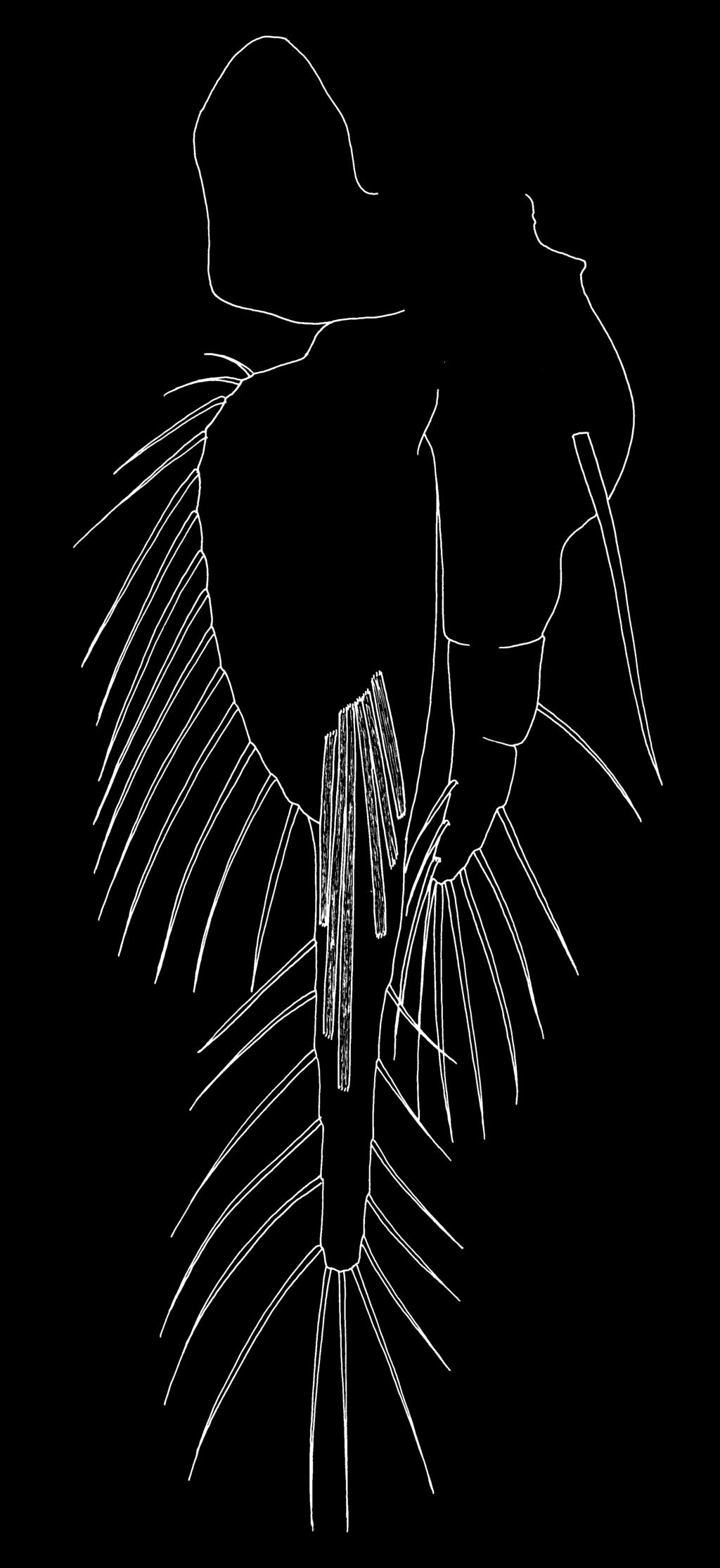
15 Arthropod Systematics & Phylogeny 67 (2) 243 oost epi co ba cxpl Fig. 17. Scanning electron micrograph of pereopod of Andaniotes linearis (Amphipoda; Stegocephalidae) taken by Jørgen Berge, showing oostegite, epipodite and coxal plate. Abbreviations: ba = basis, co = coxa, cxpl = coxal plate, = en do podite, epi = epipodite, oost = oos tegite. a vestige of the epipodite in pleopods is entirely consistent with the occurrence of epipodites on more than just the first eight post-maxillary limbs in anostracan branchiopods. Expression data for genes such as engrailed, trachealess or apterous might help to resolve any remaining uncertainty Coxal plates of Amphipoda Coxal plates are flattened outgrowths of the coxae of the pereopods and are characteristic of amphipods (Fig. 17). Functionally they extend the margins of the pereon ventrolaterally, effectively increasing the lateral compression of the body, shielding an inner channel through which water flows, and affording protection to the gills and oostegites (Li n c o l n 1979). In the embryo of the hyalid amphipod Parhyale hawaiensis the nascent coxal plate becomes apparent at stage 21 (120 h: 48% in scheme of Br o w n e et al. 2005) arising proximally on the outer surface of the coxa. This is about the same time as the epipodite first appears, located distally on the coxa. In the talitrid Orchestia cavimana Un g e r e r & Wo l f f (2005) concluded that the coxal plate and gill (= our epipodite) first appear proximolaterally as a shared anlage (labelled Cxpl+Gi in their figs. 2A and 3A) on pereopods 2 to 7. By the next stage of development the coxal plates and gills (Un g e r e r & Wo l f f 2005: fig. 4C) have separate insertions and the longitudinally-orientated, proximo-laterally originating coxal plate is clearly distinct from the somewhat transversely-orientated, distally-located gill. Just prior to hatching, the coxal plates of pereopods 1 to 7 are described as having developed into broad imbricate shields covering the insertions of the limbs and the gills, which have become convoluted by this stage. Although mislabelled as Cpl+Gi by Un g e r e r & Wo l f f (2005: fig. 3A), the swelling on pereopod 1 is the anlage of the coxal plate alone because amphipods do not have a gill on the first pereopod (no gill is present on the seventh either). Un g e r e r & Wo l f f (2005) showed shared anlagen for coxal plate and gill on the very early stage embryonic pereopods 2 to 6 of Orchestia cavimana. Additional evidence of this common developmental origin comes from study of the clonal composition of the embryonic limb of O. cavimana (Wo l f f & Sc h o lt z 2008), which showed that both the coxal plate and the gill are formed by basal clones from the dorsal-most cell columns (descendents of cells abcd7 to abcd9). These columns also contribute to the protopodal segments and the tergites. As pointed out by Wo l f f & Sc h o lt z (2008), clonal composition data help to identify the coxal plate as an outer lobe or exite, but currently we lack sufficient comparative clonal data for us to use this information in identifying potential homologues in other malacostracan taxa. Is the amphipod coxal plate homologous with any other proximally-located, crustacean exite? Its developmental origin as part of a common anlage with the amphipod gill might suggest that the whole coxal plate + gill complex is homologous with the epipoditepodobranch complex described above for decapods, euphausiaceans and lophogastrids. However, it also raises the intriguing possibility that the bilobed gill of amphipods (noted by St e e l e & St e e l e 1991, see above) represents the entire epipodite-podobranch complex, in which case, could the amphipod coxal plate be homologue of the anaspidacean proximal epipodite? More evidence is needed to answer these questions.
16 244 Bo x s h a l l & Ja u m e: Exopodites, epipodites and gills in crustaceans 4.3. Coxal plates of Isopoda Dr e y e r & Wä g e l e (2002) described the coxal plates of isopod pereopods, noting that the lateral protrusion of the plate with the sharp longitudinal keel that separates the dorsal from the ventral surface of the plate is unique for these [Scutocoxifera] isopods. The sclerotised coxal plates of isopods such as Idothea extend medially, to meet in the ventral midline, however coxal plates are absent in asellotes and phreatoicids. The coxal origin of the plate during embryogenesis was demonstrated by Gr u n e r (1954) in Porcellio. Dr e y e r & Wä g e l e (2002) considered that isopod coxal plates evolved within the Isopoda because they are absent in basal taxa such as the Phreatoicidea and Asellota, although they are present in the Calabozoidea (Br u s c a & Wi l s o n 1991), the supposed sister group of the Asellota (after Wä g e l e 1989). Br u s c a & Wi l s o n (1991) also considered the coxal plates to be derived within the Isopoda a phylogenetic scheme that would imply that they cannot be homologues of amphipod coxal plates The epipodites of Tanazios Tanazios is a Silurian arthropod interpreted as a probable stem-lineage crustacean by Si v e t e r et al. (2007b) although Bo x s h a l l (2007) considered that it should be classified as a member of the Labrophora, and its position could shed light on deep mandibulatan phylogeny. All its post-mandibular limbs are similarly patterned, with an endopodite and exopodite plus two exites on the outer margin of the protopodal part of the limb. These exites, although slender and rather pointed in form, are identified as epipodites by Si v e t e r et al. (2007b), but their narrow shape and relatively small size suggest that these exites are not primarily respiratory in function. The presence of two epipodites in this labrophoran contributes to the body of evidence suggesting that the presence of both an epipodite and a pre-epipodite on the trunk limbs was a more widely distributed character state in the Palaeozoic than previously realised. It could be interpreted as evidence that such a state was basic to the groundplan of the crown group Crustacea The epipodites of Yicaris Zh a n g et al. (2007) reported epipodites on the trunk limbs of the Cambrian arthropod Yicaris, which they classified as a crown-group crustacean. Three of these exite structures are arrayed proximo-distally along the lateral margin of the protopodal part (the basipod of Zh a n g et al.) of the post-mandibular trunk limbs. They were homologised with the epipodite plus pre-epipodite of anostracan Branchiopoda, and a groundplan of three epipodites per limb was suggested for the Eucrustacea (Zh a n g et al. 2007). The discovery of a series of larval stages revealed the pattern of development of individual epipodites: each commences development as a spine which then ands to form a leaf-like lamella on the lateral margin. These leaf-like structures pass through a stage with an extremely restricted proximal connection with the limb base (Zh a n g et al. 2007: fig. 1h), consistent with having developed from a marginal spine. Bo x s h a l l (2007) considered that this pattern of development was significantly different from that of crustacean epipodites, which first appear in development as unarmed, rounded, tissue-containing lobes, and that this raised serious doubt over the homology of these structures with crustacean epipodites plus preepipodites. We consider that these developmental differences are significant. We recognise that the epipodite vestige found on the anterior pleopods of Nebalia by Pa b s t & Sc h o lt z (2009) has the form of a tapering spiniform lobe, but this is a broad-based ansion of the outer distal angle of the protopod and does not articulate with the segment. In addition, we note a difference in timing, with the epipodite and pre-epipodite anlagen appearing simultaneously, and much earlier, in anostracan embryos (Mø l l e r et al. 2004) than in Yicaris where these structures appear to be added sequentially during the post-embryonic, larval development, together with other setal elements. The absence of epipodite-like structures on limbs elsewhere within the Cambrian arthropod fauna also implies a phylogenetic isolation and suggesting to us that the structures in Yicaris represent an independently-derived exite series Pleopodal gills The pleopods of stomatopods are typically broad, biramous flaps and carry tufted branching gills. These pleopodal gills originate on the exopodite, close to its base, as can be seen most readily in late larval stages (Fig. 18). Similar, highly branching, tufted pleopodal gills are present in the isopod Bathynomus (Flabellifera), however, these originate along the outer margin and near the base of the endopodite only (Mi l n e Edwa r d s & Bo u v i e r 1902: plt. 6, figs. 2 7). A respiratory function for these structures can be inferred from the external morphology and from the enhanced endopodal circulation system elucidated by Mi l n e Ed wa r d s & Bo u v i e r (1902). The five pairs of pleopods in aquatic Isopoda are generally involved in respiration and/ or osmo-regulation, although in some groups anterior pairs may be more robust and provide some protection
 incorrect attempt to reinterpret this as the exopodite, Ha n s e n s terminology is still employed by specialists such as Ma u c h l")
17 Arthropod Systematics & Phylogeny 67 (2) 245 Sa r s (1885) and Ha n s e n (1925) both described a lobe, referred to respectively as the exognath or pseudexopod, on the outer margin of the maxillule in some adult euphausiaceans and, despite He e g a a r d s (1948) incorrect attempt to reinterpret this as the exopodite, Ha n s e n s terminology is still employed by specialists such as Ma u c h l i n e (1967). In Bentheuphausia amblyops the pseudexopod forms a fleshy extension of the lateral margin and is produced into a small dorsal lobe (Figs ). No muscles pass into the pseudexopod and the tissue it contains appears granular with large nuclei and dense cytoplasm, as is typical of highly active cells. In some species the pseudexopod carries setae distally, around its ventral extremity, as well as an ornamentation of fine surface setules. The true exopodite, which is ressed transiently during larval development, is absent in the adult of Bentheuphausia. However, Pseudeuphausia sinica exhibits a unique condition in which the larval exopodite persists into the adult and is present together with a well-developed pseudexopod (Ma u c h l i n e 1967). The possession of an exite, referred to as the pseudexopod, on the coxa of maxillule is common to mysidaceans, syncarids and euphausiaceans. Although not referred to as a pseudexopod, atyid decapods of the genus Typhlatya possess a very similar outer coxal lobe on the maxillule (Ja u m e & Br é h i e r 2005: fig. 9A). The pseudexopod appears to have a similar valvefor the delicate, respiratory pleopods located posteriorly (Ro m a n & Da l e n s 1999). The exopodal gills on the pleopods of stomatopods and the endopodal gills on the pleopods of aquatic isopods are independently derived. Their origins on the rami indicate that neither can be inferred to be serially homologous with the malacostracan pereopodal epipodite. In some terrestrial isopods the respiratory pleopods show a most remarkable adaptation, forming a pleopodal lung which is closed off externally by a spiracle and extends through the pleonal tissues as a mass of branching internal respiratory tubules (Fe r r a r a et al. 1997). This trachea-like system has arisen within the Oniscidea as an adaptation to terrestrialisation and is independent of the trachea systems found in insects and in myriapods. In insects the tracheal system arises during development from tracheal placodes, cell clusters which invaginate and migrate to form the primary tracheal branches (Ma n n i n g & Kr a s n o w 1993). Homologues of the Drosophila tracheal inducer genes, such as the transcription factors trachealess and ventral veinless, were shown by Fr a n c h-ma r r o et al. (2006) to be ressed in crustacean epipodites leading them to speculate on the possibility of an evolutionary relationship between insect tracheae and crustacean epipodites. prp Fig. 18. Second pleopod of pseuderichthus larva of Pseudosquilla (Stomatopoda), showing gill on inner margin of first exopodal segment. Abbreviations: = endopodite, = exopodite, plg = pleopodal gill, prp = protopod. plg 4.7. Outer lobes on crustacean maxillules Various outer lobes have been described from crustacean maxillules, some of which were referred to by Ha n s e n (1925) as pseudexopods Pseudexopod on the malacostracan maxillule The maxillule of mysids and lophogastrids carries a broad, laterally-directed lobe arising from the posterior face of the coxa and referred to as the pseudexopod by Ha n s e n (1925). This flap-like lobe is described as delicate and its margins are ornamented with fine setules. According to Ca n n o n & Ma n t o n (1927), this pseudexopod functions as a valve controlling water flow during feeding activity. The outer margin of the protopod of the maxillule of the syncarid Paranaspides lacustris is extended as a thin movable plate, also termed the pseudexopod by Han s e n (1925). This plate extends to meet the lateral ridge of the maxilla so as to completely cover the space between the maxilla and the maxillule. According to Can n o n & Ma n t o n (1929), the pseudexopod acts as a valve helping to control water flow during feeding. This structure appears to be derived simply as an extension of the lateral margin to form a flap.

18 246 Bo x s h a l l & Ja u m e: Exopodites, epipodites and gills in crustaceans px px Figs Pseudexopod on maxillule of Bentheuphausia amblyops (Euphausiacea). 19: Posterior view showing musculature. 20: Anterior view showing fine setulation on surface of pseudexopod. Abbreviations: = endopodite, px = pseudexopod. like function, controlling water flow in mysidaceans and syncarids, but in euphausiaceans the nature of the pseudexopod tissues suggests to us an osmoregulatory or ionic exchange function. All of these structures appear to be derived as extensions of the lateral coxal margin of the maxillule and are considered here to be exites rather than serial homologues of the epipodite of post-maxillary limbs, although this remains a remote possibility. We have insufficient evidence to determine whether these structures are homologous but their presence in all four taxa supports the inference that they are homologous Coxal epipodite and basal exite of the copepod maxillule Copepods have a setose lobe on the outer margin of the coxa of the maxillule which is referred to as the epipodite (Hu y s & Bo x s h a l l 1991). This is typically prominent in calanoid copepods where it functions to create water flow during feeding, but is reduced (= less prominent and represented by fewer setae) or lost in other orders. Uniquely, the basis also carries a defined outer lobe in the order Platycopioida, which carries a maximum of two setae (Hu y s & Bo x s h a l l 1991). This exite is represented by just a single seta on the outer margin of the basis in calanoid, misophrioid and basal harpacticoid copepods, and is lost in the remaining orders Coxal epipodite of the myodocopan maxillule Boxshall (1997) interpreted the single seta originating near the lateral margin of the coxa of the maxillule in Azygocypridina as possibly representing a reduced epipodite. According to Horne (2005), more convincing evidence of a coxal epipodite can be found in the cylindroleberidoidean Cycloleberis squamiger and the cypridinoid Skogsbergia squamosa, both of which have a flattened, but unarmed lobe arising from the maxillulary coxa (Ko r n i c k e r 1974, 1975). 5. Oostegites The Peracarida is traditionally characterised by the possession of a ventral brood pouch, or marsupium, formed by the oostegites in the adult female (Si e w- i n g 1963). Oostegites are medially-directed, lobate outgrowths from the pereopodal coxae. In amphipods they develop gradually in the instars preceding the onset of sexual maturity but in isopods they appear fully formed at the moult into the adult. The oostegites overlap or interlock to provide an enclosure within which eggs and embryos develop, and different numbers of oostegites are ressed in the different peracaridan groups (Tab. 3). In caprellid amphipods two pairs of oostegites can be present in adult females even when the corresponding pereopod is lacking (apart from the coxa, which is largely incorporated into the body wall). In amphipods, for example Hyalella azteca, the oostegites first appear as small lamellae at the sixth stage female, become progressively larger at successive moults, and attain their definitive, marginally setose form in the ninth stage, the adult (Ge i s l e r 1944). We infer that gradual development of the oostegites, as exemplified by the amphipods, is the primitive pattern rather than the sudden appearance model. Oostegite form and development are highly variable and there
Københavns Universitet
 university of copenhagen Københavns Universitet The unique dorsal brood pouch of Thermosbaenacea (Crustacea, Malacostraca) and description of an advanced developmental stage of Tulumella unidens from the
A new asellote isopod of the genus Microjanira Schiecke & Fresi, 1970 (Crustacea: Isopoda: Asellota: Janiridae) from Japan
 from Japan") Bull. Kitakyushu Mus. Nat. Hist. Hum. Hist., Ser. A, 6: 13-18, March 31, 2008 A new asellote isopod of the genus Microjanira Schiecke & Fresi, 1970 (Crustacea: Isopoda: Asellota: Janiridae) from Japan
Bull. Kitakyushu Mus. Nat. Hist. Hum. Hist., Ser. A, 6: 13-18, March 31, 2008 A new asellote isopod of the genus Microjanira Schiecke & Fresi, 1970 (Crustacea: Isopoda: Asellota: Janiridae) from Japan
^ ^ LIBRARY Division of Crustace;
 /re/// ^ ^ LIBRARY Division of Crustace; Larval Development of Helice tridens wuana Rathbun ^Y RTEBRAT and H. tridens tridens de Haan (Crustacea, Brachyura) ZOOLOGY Reared in the Laboratory Crustacea By
Jùrgen Olesen. Acta Zoologica (Stockholm) 80: 163±184 (April 1999)
 80: 163±184 (April 1999)") Acta Zoologica (Stockholm) 80: 163±184 (April 1999) Larval and post-larval development of the branchiopod clam shrimp Cyclestheria hislopi (Baird, 1859) (Crustacea, Branchiopoda, Conchostraca, Spinicaudata)
Acta Zoologica (Stockholm) 80: 163±184 (April 1999) Larval and post-larval development of the branchiopod clam shrimp Cyclestheria hislopi (Baird, 1859) (Crustacea, Branchiopoda, Conchostraca, Spinicaudata)
H. MILNE EDWARDS (Crustacea, Brachyura) Reared in the Laboratory
 Reared in the Laboratory") Larval Development of Sesarma (Holometopus) dehaani H. MILNE EDWARDS (Crustacea, Brachyura) Reared in the Laboratory By Keiji BABA and Keisuke MIYATA Reprinted from the Memoirs of the Faculty of Education,
Larval Development of Sesarma (Holometopus) dehaani H. MILNE EDWARDS (Crustacea, Brachyura) Reared in the Laboratory By Keiji BABA and Keisuke MIYATA Reprinted from the Memoirs of the Faculty of Education,
Contumacious Beasts: A Story of Two Diastylidae (Cumacea) from Arctic Waters
 from Arctic Waters") The University of Maine DigitalCommons@UMaine Marine Sciences Faculty Scholarship School of Marine Sciences 2-1-2000 Contumacious Beasts: A Story of Two Diastylidae (Cumacea) from Arctic Waters S. Gerken
The University of Maine DigitalCommons@UMaine Marine Sciences Faculty Scholarship School of Marine Sciences 2-1-2000 Contumacious Beasts: A Story of Two Diastylidae (Cumacea) from Arctic Waters S. Gerken
DESCRIPTIONS OF TEN XANTHOIDEAN (CRUSTACEA: DECAPODA: BRACHYURA) FIRST STAGE ZOEAS FROM INHACA ISLAND, MOZAMBIQUE
 FIRST STAGE ZOEAS FROM INHACA ISLAND, MOZAMBIQUE") THE RAFFLES BULLETIN OF ZOOLOGY 2003 THE RAFFLES BULLETIN OF ZOOLOGY 2003 51(2): 323-378 DESCRIPTIONS OF TEN XANTHOIDEAN (CRUSTACEA: DECAPODA: BRACHYURA) FIRST STAGE ZOEAS FROM INHACA ISLAND, MOZAMBIQUE
THE RAFFLES BULLETIN OF ZOOLOGY 2003 THE RAFFLES BULLETIN OF ZOOLOGY 2003 51(2): 323-378 DESCRIPTIONS OF TEN XANTHOIDEAN (CRUSTACEA: DECAPODA: BRACHYURA) FIRST STAGE ZOEAS FROM INHACA ISLAND, MOZAMBIQUE
Uropods of Eumalacostraca (Crustacea s.l.: Malacostraca) and their phylogenetic significance
 and their phylogenetic significance") Arthropod Systematics & Phylogeny 181 70 (3) 181 206 Senckenberg Gesellschaft für Naturforschung, eissn 1864-8312, 14.12.2012 Uropods of Eumalacostraca (Crustacea s.l.: Malacostraca) and their phylogenetic
Arthropod Systematics & Phylogeny 181 70 (3) 181 206 Senckenberg Gesellschaft für Naturforschung, eissn 1864-8312, 14.12.2012 Uropods of Eumalacostraca (Crustacea s.l.: Malacostraca) and their phylogenetic
Post-embryonic development of remipede crustaceans
 EVOLUTION & DEVELOPMENT 9:2, 117 121 (2007) Post-embryonic development of remipede crustaceans Stefan Koenemann, a, Frederick R. Schram, b Armin Bloechl, a Thomas M. Iliffe, c Mario Hoenemann, a and Christoph
Bulletin Zoölogisch Museum
 Bulletin Zoölogisch Museum UNIVERSITEIT VAN AMSTERDAM Vol.11 No. 15 1988 Redescription of johanna Monod, 1926 Virgin Isls (Isopoda) from St. John, Hans Georg Müller Summary Based on the type material,
Lobetelson mclaughlinae n. gen., n. sp., a new genus and species of belotelsonid malacostracan from the Pennsylvanian of the Mazon Creek area
 Lobetelson mclaughlinae n. gen., n. sp., a new genus and species of belotelsonid malacostracan from the Pennsylvanian of the Mazon Creek area Frederick R. SCHRAM Department of Biology, University of Washington,
LARVAL DEVELOPMENT OF THE GHOST SHRIMP CALLICHIRUS ISLAGRANDE (DECAPODA: THALASSINIDEA: CALLIANASSIDAE) UNDER LABORATORY CONDITIONS
 UNDER LABORATORY CONDITIONS") JOURNAL OF CRUSTACEAN BIOLOGY, 20(1): 100 117, 2000 LARVAL DEVELOPMENT OF THE GHOST SHRIMP CALLICHIRUS ISLAGRANDE (DECAPODA: THALASSINIDEA: CALLIANASSIDAE) UNDER LABORATORY CONDITIONS Karen M. Strasser
JOURNAL OF CRUSTACEAN BIOLOGY, 20(1): 100 117, 2000 LARVAL DEVELOPMENT OF THE GHOST SHRIMP CALLICHIRUS ISLAGRANDE (DECAPODA: THALASSINIDEA: CALLIANASSIDAE) UNDER LABORATORY CONDITIONS Karen M. Strasser
LARVAL DEVELOPMENT OF THE CRUSTACEAN THOR - j ^-J- \ ^L, BY A. C. BROAD 4 ^ M" 0 ^ Duke University Marine Laboratory, Beaufort, North Carolina
 OL.C. IUDIL&LAU&I!***XP&T**/. LARVAL DEVELOPMENT OF THE CRUSTACEAN THOR - j ^-J- \ ^L, FLORIDANjm. KINGSLEY BY A. C. BROAD 4 ^ M" 0 ^ Duke University Marine Laboratory, Beaufort, North Carolina OIVISlOiM
Functional morphology of giant mole crab larvae: a possible case of defensive enrollment
 Rudolf et al. Zoological Letters (2016) 2:17 DOI 10.1186/s40851-016-0052-5 RESEARCH ARTICLE Open Access Functional morphology of giant mole crab larvae: a possible case of defensive enrollment Nicole R.
JØRGEN OLESEN*, STEFAN RICHTER and GERHARD SCHOLTZ. Humboldt-Universität zu Berlin, Institut für Biologie, Vergleichende Zoologie, Berlin, Germany
 Int. J. Dev. Biol. 45: 869-876 (2001) Original Article The evolutionary transformation of phyllopodous to stenopodous limbs in the Branchiopoda (Crustacea) - Is there a common mechanism for early limb
Pacific Tanaidacea (Crustacea): Revision of the Genus Agathotanais with Description of Three New Species
: Revision of the Genus Agathotanais with Description of Three New Species") Copyright Australian Museum, 1999 Records of the Australian Museum (1999) Vol. 51: 99 112. ISSN 0067-1975 Pacific Tanaidacea (Crustacea): Revision of the Genus Agathotanais with Description of Three New
Copyright Australian Museum, 1999 Records of the Australian Museum (1999) Vol. 51: 99 112. ISSN 0067-1975 Pacific Tanaidacea (Crustacea): Revision of the Genus Agathotanais with Description of Three New
Phylogeny of Branchiopoda (Crustacea) Character Evolution and Contribution of Uniquely Preserved Fossils
 Character Evolution and Contribution of Uniquely Preserved Fossils") Arthropod Systematics & Phylogeny 3 67 (1) 3 39 Museum für Tierkunde Dresden, eissn 1864-8312, 17.6.2009 Phylogeny of Branchiopoda (Crustacea) Character Evolution and Contribution of Uniquely Preserved
Arthropod Systematics & Phylogeny 3 67 (1) 3 39 Museum für Tierkunde Dresden, eissn 1864-8312, 17.6.2009 Phylogeny of Branchiopoda (Crustacea) Character Evolution and Contribution of Uniquely Preserved
Chapter 12: Aquatic Mandibulates
 Chapter 12: Aquatic Mandibulates Phylum Arthropoda Subphylum: Crustacea (Latin crusta = shell) Class: Malacostraca Order: Decapoda Order: Euphausiacea Order: Amphipoda Order: Isopoda Class: Maxillopoda
Crustacean classification: on-going controversies and unresolved problems*
 Zootaxa 1668: 313 325 (2007) www.mapress.com/zootaxa/ Copyright 2007 Magnolia Press ISSN 1175-5326 (print edition) ZOOTAXA ISSN 1175-5334 (online edition) Crustacean classification: on-going controversies
A new species of the genus Apseudomorpha Miller (Crustacea, Peracarida, Tanaidacea) from Korea
 from Korea") Journal of Species Research 5(3):385-392, 2016 A new species of the genus Apseudomorpha Miller (Crustacea, Peracarida, Tanaidacea) from Korea Hosung Hwang 1 and Il-Hoi Kim 2, * 1 National Institute of
Journal of Species Research 5(3):385-392, 2016 A new species of the genus Apseudomorpha Miller (Crustacea, Peracarida, Tanaidacea) from Korea Hosung Hwang 1 and Il-Hoi Kim 2, * 1 National Institute of
On the Ontogeny of Leptodora kindtii (Crustacea, Branchiopoda, Cladocera), with Notes on the Phylogeny of the Cladocera
, with Notes on the Phylogeny of the Cladocera") JOURNAL OF MORPHOLOGY 256:235 259 (2003) On the Ontogeny of Leptodora kindtii (Crustacea, Branchiopoda, Cladocera), with Notes on the Phylogeny of the Cladocera Jørgen Olesen, 1,2 * Stefan Richter, 1 and
JOURNAL OF MORPHOLOGY 256:235 259 (2003) On the Ontogeny of Leptodora kindtii (Crustacea, Branchiopoda, Cladocera), with Notes on the Phylogeny of the Cladocera Jørgen Olesen, 1,2 * Stefan Richter, 1 and
Carolin Haug 1*, Wafaa S Sallam 2, Andreas Maas 3, Dieter Waloszek 3, Verena Kutschera 3 and Joachim T Haug 1
 Haug et al. Frontiers in Zoology 2012, 9:31 RESEARCH Open Access Tagmatization in Stomatopoda reconsidering functional units of modern-day mantis shrimps (Verunipeltata, Hoplocarida) and implications for
Larval development of a mangrove crab (Perisesarma bidens DeHaan) (Crustacea: Brachyura: Sesarminae)
 (Crustacea: Brachyura: Sesarminae)") Bangladesh]. Fish. Res., 4(1), 2000: 43-56 Larval development of a mangrove crab (Perisesarma bidens DeHaan) (Crustacea: Brachyura: Sesarminae) M. Sirajul Islam* and Shigemitsu Shokita Department of Chemistly,
Bangladesh]. Fish. Res., 4(1), 2000: 43-56 Larval development of a mangrove crab (Perisesarma bidens DeHaan) (Crustacea: Brachyura: Sesarminae) M. Sirajul Islam* and Shigemitsu Shokita Department of Chemistly,
A new genus of leucosiid crabs (Crustacea, Decapoda, Brachyura) from the Red Sea
 from the Red Sea") A new genus of leucosiid crabs (Crustacea, Decapoda, Brachyura) from the Red Sea Hironori KOMATSU Department of Biological Sciences, Graduate School of Science, The University of Tokyo, 7-3-1 Hongo, Bunkyo-ku,
A new genus of leucosiid crabs (Crustacea, Decapoda, Brachyura) from the Red Sea Hironori KOMATSU Department of Biological Sciences, Graduate School of Science, The University of Tokyo, 7-3-1 Hongo, Bunkyo-ku,
The functional head of the Cambrian radiodontan (stem-group Euarthropoda) Amplectobelua symbrachiata
 Amplectobelua symbrachiata") Cong et al. BMC Evolutionary Biology (2017) 17:208 DOI 10.1186/s12862-017-1049-1 RESEARCH ARTICLE The functional head of the Cambrian radiodontan (stem-group Euarthropoda) Amplectobelua symbrachiata Peiyun
Cong et al. BMC Evolutionary Biology (2017) 17:208 DOI 10.1186/s12862-017-1049-1 RESEARCH ARTICLE The functional head of the Cambrian radiodontan (stem-group Euarthropoda) Amplectobelua symbrachiata Peiyun
Research Article. Farhana Shafiq GHORY*, Feroz Akhtar SIDDIQUI
 Research Article Turk J Zool 33 (2009) 35-45 TÜBİTAK doi:10.3906/zoo-0702-7 Zoeal Stages of Two Majoidea Crabs, Doclea muricata (Herbst, 1788) and Acanthonyx limbatus (A. Milne Edwards, 1862), Reared under
TWO NEW SPECIES OF CARIDINA (CRUSTACEA: DECAPODA: ATYIDAE) FROM HUNAN PROVINCE, CHINA
 FROM HUNAN PROVINCE, CHINA") THE RAFFLES BULLETIN OF ZOOLOGY 997 45( ): 23 33 TWO NEW SPECIES OF CARIDINA (CRUSTACEA: DECAPODA: ATYIDAE) FROM HUNAN PROVINCE, CHINA Zhao Liang Guo and Sammy De Grave ABSTRACT. The present contribution
THE RAFFLES BULLETIN OF ZOOLOGY 997 45( ): 23 33 TWO NEW SPECIES OF CARIDINA (CRUSTACEA: DECAPODA: ATYIDAE) FROM HUNAN PROVINCE, CHINA Zhao Liang Guo and Sammy De Grave ABSTRACT. The present contribution
Kakati, V.S. Karwar Research Center of C.M.F.R.I., Karwar , Karnataka, India ABSTRACT
 The Seventh Indian FISheries Forum Proceedings Eds. C. Vasudevappa. Y. Basavaraju. D. 5eeflappa, S. Ayyappan & S. Ravichandra Reddy(2005) Published by AFSIB Mangalore. ICAR, UAS(B). KVAFSU(B) & FFT(8)
Larval development PARAPENAEOPSIS STYLIFERA (H. MILNE EDWARDS)
") IX Larval development PARAPENAEOPSIS STYLIFERA (H. MILNE EDWARDS) M.S. Muthu N. N. Pillai K- V. George Parapenaeopsis stylifera was reared from the egg to the postlarval stage in the Narakkal laboratory.
IX Larval development PARAPENAEOPSIS STYLIFERA (H. MILNE EDWARDS) M.S. Muthu N. N. Pillai K- V. George Parapenaeopsis stylifera was reared from the egg to the postlarval stage in the Narakkal laboratory.
The Circulatory System in Malacostraca Evaluating Character Evolution on the Basis of Differing Phylogenetic Hypotheses
 Arthropod Systematics & Phylogeny 6 () Museum für Tierkunde Dresden, eissn 86-8,.6.9 The Circulatory System in Malacostraca Evaluating Character Evolution on the Basis of Differing Phylogenetic Hypotheses
Detonella papillicornis
 Detonella papillicornis A sow bug Phylum: Arthropoda, Crustacea Class: Malacostraca Order: Isopoda, Oniscidea Family: Detonidae Taxonomy: Detonella papillicornis was originally described by Richardson
A new genus and two new species of cave-dwelling Misophrioid copepods from the Balearic Islands (Mediterranean)
") This article was downloaded by: [Red de Bibliotecas del CSIC] On: 25 February 2015, At: 03:08 Publisher: Taylor & Francis Informa Ltd Registered in England and Wales Registered Number: 1072954 Registered
This article was downloaded by: [Red de Bibliotecas del CSIC] On: 25 February 2015, At: 03:08 Publisher: Taylor & Francis Informa Ltd Registered in England and Wales Registered Number: 1072954 Registered
Description of laboratory reared first zoea of Callinectes danae Smith (Crustacea, Decapoda, Portunidae)
") Description of laboratory reared first zoea of Callinectes danae Smith (Crustacea, Decapoda, Portunidae) Cheruparambil Sankarankutty 1 Sung Yun Hong 2 Kwang Bong Kim 2 ABSTRACT. A detailed description
Description of laboratory reared first zoea of Callinectes danae Smith (Crustacea, Decapoda, Portunidae) Cheruparambil Sankarankutty 1 Sung Yun Hong 2 Kwang Bong Kim 2 ABSTRACT. A detailed description
A new species of Caprellinoides (Crustacea: Amphipoda: Phtisicidae) from the Antarctic
 from the Antarctic") Helgol Mar Res (2001) 55:212 220 DOI 10.1007/s101520100082 ORIGINAL ARTICLE José Manuel Guerra-García A new species of Caprellinoides (Crustacea: Amphipoda: Phtisicidae) from the Antarctic Received: 20
Helgol Mar Res (2001) 55:212 220 DOI 10.1007/s101520100082 ORIGINAL ARTICLE José Manuel Guerra-García A new species of Caprellinoides (Crustacea: Amphipoda: Phtisicidae) from the Antarctic Received: 20
Outline. v Definition and major characteristics of animals v Dividing animals into groups based on: v Animal Phylogeny
 BIOSC 041 Overview of Animal Diversity: Animal Body Plans Reference: Chapter 32 Outline v Definition and major characteristics of animals v Dividing animals into groups based on: Body symmetry Tissues
Gulf Research Reports
 Gulf Research Reports Volume 6 Issue 4 January 1980 Lepidactylus triarticulatus N. Sp., A New Haustoriid Amphipod from the Northern Gulf of Mexico Philip B. Robertson Lamar University Charles R. Shelton
8/23/2014. Introduction to Animal Diversity
 Introduction to Animal Diversity Chapter 32 Objectives List the characteristics that combine to define animals Summarize key events of the Paleozoic, Mesozoic, and Cenozoic eras Distinguish between the
Modern Evolutionary Classification. Section 18-2 pgs
 Modern Evolutionary Classification Section 18-2 pgs 451-455 Modern Evolutionary Classification In a sense, organisms determine who belongs to their species by choosing with whom they will mate. Taxonomic
18.4 Embryonic development involves cell division, cell differentiation, and morphogenesis
 18.4 Embryonic development involves cell division, cell differentiation, and morphogenesis An organism arises from a fertilized egg cell as the result of three interrelated processes: cell division, cell
PLEOMOTHRA FRAGILIS N. SP. (REMIPEDIA) FROM THE BAHAMAS, WITH REMARKS ON MORPHOLOGIC REDUCTIONS AND POSTNAUPLIAR DEVELOPMENT
 FROM THE BAHAMAS, WITH REMARKS ON MORPHOLOGIC REDUCTIONS AND POSTNAUPLIAR DEVELOPMENT") JOURNAL OF CRUSTACEAN BIOLOGY, 28(1): 128 136, 2008 PLEOMOTHRA FRAGILIS N. SP. (REMIPEDIA) FROM THE BAHAMAS, WITH REMARKS ON MORPHOLOGIC REDUCTIONS AND POSTNAUPLIAR DEVELOPMENT Stefan Koenemann, Maren
JOURNAL OF CRUSTACEAN BIOLOGY, 28(1): 128 136, 2008 PLEOMOTHRA FRAGILIS N. SP. (REMIPEDIA) FROM THE BAHAMAS, WITH REMARKS ON MORPHOLOGIC REDUCTIONS AND POSTNAUPLIAR DEVELOPMENT Stefan Koenemann, Maren
Evolution of the Arthropod Mandible:
 Evolution of the Arthropod Mandible: a molecular developmental perspective Joshua Frederick Coulcher UCL Submitted for the Degree of Doctor of Philosophy September 2011 Declaration I, Joshua Frederick
Cap n collar differentiates the mandible from the maxilla in the beetle Tribolium castaneum
 Coulcher and Telford EvoDevo 2012, 3:25 RESEARCH Open Access Cap n collar differentiates the mandible from the maxilla in the beetle Tribolium castaneum Joshua F Coulcher and Maximilian J Telford * Abstract
mmi%mmii^hm^^ fifz Dynoides m^y^^^'yn
 A New Species of Sphaeromatid Isopod Crustacean of the genus Dynoides collected from Tachibana Bay, Shikoku, Southern Japan Noboru Nunomura Toyama Science Museum mmi%mmii^hm^^ fifz Dynoides m^y^^^'yn mi
ZOOLOGISCHE MEDEDELINGEN
 ZOOLOGISCHE MEDEDELINGEN UITGEGEVEN DOOR HET RIJKSMUSEUM VAN NATUURLIJKE HISTORIE TE LEIDEN (MINISTERIE VAN CULTUUR, RECREATIE EN MAATSCHAPPELIJK WERK) Deel 43 no. 11 20 december 1968 PARABOMOLOCHUS GLOBICEPS
; A new rock borlnsr isopod crustacean belong-lng to the genus Sphaeroma collected trom Tanabe Bay, Kii Peninsula* Middle Japan*
 ; A new rock borlnsr isopod crustacean belong-lng to the genus Sphaeroma ^ collected trom Tanabe Bay, Kii Peninsula* Middle Japan* Noboru Nunomura Toyama Science Museum Reprinted from Bdletin of the Toyama
v Scientists have identified 1.3 million living species of animals v The definition of an animal
 Biosc 41 9/10 Announcements BIOSC 041 v Genetics review: group problem sets Groups of 3-4 Correct answer presented to class = 2 pts extra credit Incorrect attempt = 1 pt extra credit v Lecture: Animal
Redescription of the larval stages of Upobebiapusilla (Petagna, 1792) (Thalassinidea, Upogebiidae) from laboratory-reared material
 (Thalassinidea, Upogebiidae) from laboratory-reared material") Invertebrate Reproduction and Development, 4: 1 (200) 8-90 Balaban, PhiladelphiaRehovot 0168-8170/0/$05.00 8 200 Balaban Redescription of the larval stages of Upobebiapusilla (Petagna, 1792) (Thalassinidea,
Invertebrate Reproduction and Development, 4: 1 (200) 8-90 Balaban, PhiladelphiaRehovot 0168-8170/0/$05.00 8 200 Balaban Redescription of the larval stages of Upobebiapusilla (Petagna, 1792) (Thalassinidea,
Peracarid monophyly and interordinal phylogeny inferred from nuclear small-subunit ribosomal DNA sequences (Crustacea: Malacostraca: Peracarida)
") Peracarid monophyly and interordinal phylogeny inferred from nuclear small-subunit ribosomal DNA sequences (Crustacea: Malacostraca: Peracarida) Author(s): Trisha Spears, Ronald W. DeBry, Lawrence G. Abele,
Peracarid monophyly and interordinal phylogeny inferred from nuclear small-subunit ribosomal DNA sequences (Crustacea: Malacostraca: Peracarida) Author(s): Trisha Spears, Ronald W. DeBry, Lawrence G. Abele,
Classification and Phylogeny
 Classification and Phylogeny The diversity of life is great. To communicate about it, there must be a scheme for organization. There are many species that would be difficult to organize without a scheme
Biosc 41 9/10 Announcements
 Biosc 41 9/10 Announcements v Genetics review: group problem sets Groups of 3-4 Correct answer presented to class = 2 pts extra credit Incorrect attempt = 1 pt extra credit v Lecture: Animal Body Plans
Gulf Research Reports
 Gulf Research Reports Volume 7 Issue 2 January 982 Tanaidacea (Crustacea: Peracardia) of the Gulf of Mexico. II. The Occurrence of Halmyrapseudes bahamensis Băcescu and Gutu, 974 (Apseudidae) in the Eastern
Kingdom Animalia. Zoology the study of animals
 Kingdom Animalia Zoology the study of animals Summary Animals are multicellular and eukaryotic. consume and digest organic materials thereby being heterotrophs. Most are motile at some time in their lives.
Frank D. Ferrari, Viatcheslav N. Ivanenko, and Hans-Uwe Dahms
 BODY ARCHITECTURE AND RELATIONSHIPS AMONG BASAL COPEPODS Frank D. Ferrari, Viatcheslav N. Ivanenko, and Hans-Uwe Dahms (FDF, correspondence, ferrarif@si.edu) IZ/MSC, MRC-534, National Museum of Natural
Classification and Phylogeny
 Classification and Phylogeny The diversity it of life is great. To communicate about it, there must be a scheme for organization. There are many species that would be difficult to organize without a scheme
Phylogenetic analysis. Characters
 Typical steps: Phylogenetic analysis Selection of taxa. Selection of characters. Construction of data matrix: character coding. Estimating the best-fitting tree (model) from the data matrix: phylogenetic
A new species of Scherocumella (Crustacea, Cumacea) from a coral lagoon of Lifou, New Caledonia
 from a coral lagoon of Lifou, New Caledonia") new species of Scherocumella (Crustacea, Cumacea) from a coral lagoon of Lifou, New Caledonia Jordi CORER Carrer Gran, 90, E-08310 rgentona (Spain) corbera@sct.ictnet.es Corbera J. 2004. new species of
new species of Scherocumella (Crustacea, Cumacea) from a coral lagoon of Lifou, New Caledonia Jordi CORER Carrer Gran, 90, E-08310 rgentona (Spain) corbera@sct.ictnet.es Corbera J. 2004. new species of
LABORATORY REARED LARVAL FORMS OF HIPPOLYSMATA {EXHIPPOLYSMATA) ENSIROSTRIS KEMP (DECAPODA : HIPPOLYTIDAE)
 ENSIROSTRIS KEMP (DECAPODA : HIPPOLYTIDAE)") ^. mar. biol. Ass. India, 1974, 16 (2): 594-608 LABORATORY REARED LARVAL FORMS OF HIPPOLYSMATA {EXHIPPOLYSMATA) ENSIROSTRIS KEMP (DECAPODA : HIPPOLYTIDAE) N. N. PlLLAI Central Marine Fisheries Research
^. mar. biol. Ass. India, 1974, 16 (2): 594-608 LABORATORY REARED LARVAL FORMS OF HIPPOLYSMATA {EXHIPPOLYSMATA) ENSIROSTRIS KEMP (DECAPODA : HIPPOLYTIDAE) N. N. PlLLAI Central Marine Fisheries Research
A new species of Cirolana
 A new species of Cirolana Leach, 1818 (Crustacea, Isopoda, Cirolanidae) from the western Ross Sea, Antarctica, the first record of the genus from polar waters Niel L. BRUCE Marine Biodiversity and Biosecurity,
The post-embryonic development of Remipedia (Crustacea) additional results and new insights
 additional results and new insights") Dev Genes Evol (2009) 219:131 145 DOI 10.1007/s00427-009-0273-0 ORIGINAL ARTICLE The post-embryonic development of Remipedia (Crustacea) additional results and new insights Stefan Koenemann & Jørgen Olesen
Dev Genes Evol (2009) 219:131 145 DOI 10.1007/s00427-009-0273-0 ORIGINAL ARTICLE The post-embryonic development of Remipedia (Crustacea) additional results and new insights Stefan Koenemann & Jørgen Olesen
Paul F. CLARK. Peter K. L. NG. Department of Zoology, The Natural History Muséum Cromwell Road, London, SW7 5BD (U.K.)
") The larval development of the poisonous mosaic crab, Lophozozymus pictor (Fabricius, 1798) (Crustacea, Decapoda, Brachyura, Xanthidae, Zosiminae), with comments on familial characters for first stage zoeas
The larval development of the poisonous mosaic crab, Lophozozymus pictor (Fabricius, 1798) (Crustacea, Decapoda, Brachyura, Xanthidae, Zosiminae), with comments on familial characters for first stage zoeas
Comparative morphology of the first zoea of twelve brachyuran species (Crustacea: Decapoda) from the Amazon region
 from the Amazon region") http://dx.doi.org/10.1590/s1984-46702013000300004 Comparative morphology of the first zoea of twelve brachyuran species (Crustacea: Decapoda) from the Amazon region Adelson S. de Souza 1, Rauquírio M.
http://dx.doi.org/10.1590/s1984-46702013000300004 Comparative morphology of the first zoea of twelve brachyuran species (Crustacea: Decapoda) from the Amazon region Adelson S. de Souza 1, Rauquírio M.
Comparative Mouthpart Morphology and Evolution of the Carnivorous Heptageniidae (Ephemeroptera) 1
 1") l Aquatic Insects, Vol. 8 (1986). No. 2, pp. 83-89 0165-0424/86/0802-0083 $3.00 Swets & Zeitlinger Comparative Mouthpart Morphology and Evolution of the Carnivorous Heptageniidae (Ephemeroptera) 1 by W.
l Aquatic Insects, Vol. 8 (1986). No. 2, pp. 83-89 0165-0424/86/0802-0083 $3.00 Swets & Zeitlinger Comparative Mouthpart Morphology and Evolution of the Carnivorous Heptageniidae (Ephemeroptera) 1 by W.
Radiodonta e.g. Anomallocaris
 CAMBRIAN Terreneuvian Epoch 2 Epoch 3 Furongian e.g. Anomallocaris Gilled lobopodians Bivalved arthropods Trilobitomorpha Eurypterida Bradoriida Branchiopoda Remipedia Swimming with flap-like structures
Biologists have used many approaches to estimating the evolutionary history of organisms and using that history to construct classifications.
 Phylogenetic Inference Biologists have used many approaches to estimating the evolutionary history of organisms and using that history to construct classifications. Willi Hennig developed d the techniques
A New Talitrid Genus and Species, Lowryella wadai, from Estuarine Reed Marshes of Western Japan (Crustacea: Amphipoda: Talitridae)
") Species Diversity 21: 143 149 25 November 2016 DOI: 10.12782/sd.21.2.143 A New Talitrid Genus and Species, Lowryella wadai, from Estuarine Reed Marshes of Western Japan (Crustacea: Amphipoda: Talitridae)
Species Diversity 21: 143 149 25 November 2016 DOI: 10.12782/sd.21.2.143 A New Talitrid Genus and Species, Lowryella wadai, from Estuarine Reed Marshes of Western Japan (Crustacea: Amphipoda: Talitridae)
CRUSTACEANA STUDIES ON DECAPOD LARVAL DEVELOPMENT INTERNATIONAL JOURNAL OF CRUSTACEAN RESEARCH. » Mm% SUPPLEMENT 2 LEIDEN E. J.
 J Reprinted from: CRUSTACEANA INTERNATIONAL JOURNAL OF CRUSTACEAN RESEARCH SUPPLEMENT 2 STUDIES ON DECAPOD LARVAL DEVELOPMENT» Mm% LEIDEN E. J. BRILL 1968 A COMPARISON OF THE LARVAL DEVELOPMENT OF METASESARMA
ESS 345 Ichthyology. Systematic Ichthyology Part II Not in Book
 ESS 345 Ichthyology Systematic Ichthyology Part II Not in Book Thought for today: Now, here, you see, it takes all the running you can do, to keep in the same place. If you want to get somewhere else,
A trilo b itomorp h origin for the Cru s t acea
 A trilo b itomorp h origin for the Cru s t acea ROB E RT R. HESSLE R AND WI LLIAM A. NEWMAN HessIer, R. R., & Newman, W. A. 1 9 7 5 0 7 1 5 : A trilobitomorph origin for the Crustacea. Fossils and Strata;
Patterns of Evolution
 Patterns of Evolution A tree that represents an estimate (hypothesis) of evolutionary relatedness is a phylogeny Classifications can be based on groupings within a phylogeny Groupings can be categorized
Chapter 16: Reconstructing and Using Phylogenies
 Chapter Review 1. Use the phylogenetic tree shown at the right to complete the following. a. Explain how many clades are indicated: Three: (1) chimpanzee/human, (2) chimpanzee/ human/gorilla, and (3)chimpanzee/human/
A MEGASECOPTERON FROM UPPER CARBONIFEROUS BY F. M. CARPENTER. In I962 Professor F. Stockmans, of the Institut Royal des Sciences STRATA IN SPAIN
 A MEGASECOPTERON FROM UPPER CARBONIFEROUS STRATA IN SPAIN BY F. M. CARPENTER Harvard University In I962 Professor F. Stockmans, of the Institut Royal des Sciences Naturelles de. Belgique, kindly sent me
EARLY POST-EMBRYONIC DEVELOPMENT OF MARINE CHELICERATES AND CRUSTACEANS WITH A NAUPLIUS
 EARLY POST-EMBRYONIC DEVELOPMENT OF MARINE CHELICERATES AND CRUSTACEANS WITH A NAUPLIUS BY FRANK D. FERRARI 1,5 ), JOHN FORNSHELL 1,6 ), ALEJANDRO A. VAGELLI 2,7 ), V. N. IVANENKO 3,8 ) and HANS-UWE DAHMS
Classifications can be based on groupings g within a phylogeny
 Patterns of Evolution A tree that represents an estimate (hypothesis) of evolutionary relatedness is a phylogeny Classifications can be based on groupings g within a phylogeny y Groupings can be categorized
Classification, Phylogeny yand Evolutionary History
 Classification, Phylogeny yand Evolutionary History The diversity of life is great. To communicate about it, there must be a scheme for organization. There are many species that would be difficult to organize
The History of Life. Fossils and Ancient Life (page 417) How Fossils Form (page 418) Interpreting Fossil Evidence (pages ) Chapter 17
 How Fossils Form (page 418) Interpreting Fossil Evidence (pages ) Chapter 17") Chapter 17 The History of Life Section 17 1 The Fossil Record (pages 417 422) This section explains how fossils form and how they can be interpreted. It also describes the geologic time scale that is used
Chapter 17 The History of Life Section 17 1 The Fossil Record (pages 417 422) This section explains how fossils form and how they can be interpreted. It also describes the geologic time scale that is used
Postembryonic development of Pyromaia tuberculata (Lockington, 1877): a review of larval and postlarval morphology*
: a review of larval and postlarval morphology*") SCI. MAR., 67 (2): 201-214 SCIENTIA MARINA 2003 Postembryonic development of Pyromaia tuberculata (Lockington, 1877): a review of larval and postlarval morphology* TOMÁS A. LUPPI and EDUARDO D. SPIVAK
SCI. MAR., 67 (2): 201-214 SCIENTIA MARINA 2003 Postembryonic development of Pyromaia tuberculata (Lockington, 1877): a review of larval and postlarval morphology* TOMÁS A. LUPPI and EDUARDO D. SPIVAK
Phylogenetic Analysis
 Phylogenetic Analysis Aristotle Through classification, one might discover the essence and purpose of species. Nelson & Platnick (1981) Systematics and Biogeography Carl Linnaeus Swedish botanist (1700s)
Phylogenetic Analysis
 Phylogenetic Analysis Aristotle Through classification, one might discover the essence and purpose of species. Nelson & Platnick (1981) Systematics and Biogeography Carl Linnaeus Swedish botanist (1700s)
Phylogenetic Analysis
 Phylogenetic Analysis Aristotle Through classification, one might discover the essence and purpose of species. Nelson & Platnick (1981) Systematics and Biogeography Carl Linnaeus Swedish botanist (1700s)
A new genus and new species of Agathotanaidae (Crustacea, Tanaidacea) from West Australia
 from West Australia") ZooKeys 243 15 26 (2012) A new genus and new species of Agathotanaidae (Crustacea, Tanaidacea) from West Australia 15 doi: 10.3897/zookeys.243.3408 www.zookeys.org Research article A peer-reviewed open-access
ZooKeys 243 15 26 (2012) A new genus and new species of Agathotanaidae (Crustacea, Tanaidacea) from West Australia 15 doi: 10.3897/zookeys.243.3408 www.zookeys.org Research article A peer-reviewed open-access
SYNCARID CRUSTACEANS FROM THE MONTCEAU LAGERSTÄTTE (UPPER CARBONIFEROUS; FRANCE)
") [Palaeontology, Vol. 49, Part 3, 2006, pp. 647 672] SYNCARID CRUSTACEANS FROM THE MONTCEAU LAGERSTÄTTE (UPPER CARBONIFEROUS; FRANCE) by VINCENT PERRIER*, JEAN VANNIER*, PATRICK R. RACHEBOEUF, SYLVAIN CHARBONNIER*,
[Palaeontology, Vol. 49, Part 3, 2006, pp. 647 672] SYNCARID CRUSTACEANS FROM THE MONTCEAU LAGERSTÄTTE (UPPER CARBONIFEROUS; FRANCE) by VINCENT PERRIER*, JEAN VANNIER*, PATRICK R. RACHEBOEUF, SYLVAIN CHARBONNIER*,
Asterocheres hirsutus, a new species of parasitic copepod (Siphonostomatoida: Asterocheridae) associated with an Antarctic hexactinellid sponge
 associated with an Antarctic hexactinellid sponge") Helgol Mar Res (2005) 59: 315 322 DOI 10.1007/s10152-005-0007-2 ORIGINAL ARTICLE Mª Eugenia Bandera Æ Mercedes Conradi Pablo J. Lo pez-gonza lez Asterocheres hirsutus, a new species of parasitic copepod
Helgol Mar Res (2005) 59: 315 322 DOI 10.1007/s10152-005-0007-2 ORIGINAL ARTICLE Mª Eugenia Bandera Æ Mercedes Conradi Pablo J. Lo pez-gonza lez Asterocheres hirsutus, a new species of parasitic copepod
MASTER OF ARTS GERTRUDE MELLEN HOOPER THE GRADUATE SCHOOL UNIVERSITY OF ILLINOIS STUDIES ON THE REGENERATION OF CRUSTACEAN APPENDAGES THESIS
 STUDIES ON THE REGENERATION OF CRUSTACEAN APPENDAGES BY GERTRUDE MELLEN HOOPER A. B. Jackson College, 191 5. THESIS Submitted in Partial Fulfillment of the Requirements for the Degree of MASTER OF ARTS
Amira A. AL-Hosary PhD of infectious diseases Department of Animal Medicine (Infectious Diseases) Faculty of Veterinary Medicine Assiut
 Faculty of Veterinary Medicine Assiut") Amira A. AL-Hosary PhD of infectious diseases Department of Animal Medicine (Infectious Diseases) Faculty of Veterinary Medicine Assiut University-Egypt Phylogenetic analysis Phylogenetic Basics: Biological
Amira A. AL-Hosary PhD of infectious diseases Department of Animal Medicine (Infectious Diseases) Faculty of Veterinary Medicine Assiut University-Egypt Phylogenetic analysis Phylogenetic Basics: Biological
Mandibulate convergence in an armoured Cambrian stem chelicerate
 Aria and Caron BMC Evolutionary Biology (2017) 17:261 DOI 10.1186/s12862-017-1088-7 RESEARCH ARTICLE Mandibulate convergence in an armoured Cambrian stem chelicerate Cédric Aria 1,4* and Jean-Bernard Caron
Global diversity of spelaeogriphaceans & thermosbaenaceans (Crustacea; Spelaeogriphacea & Thermosbaenacea) in freshwater
 in freshwater") Hydrobiologia (2008) 595:219 224 DOI 10.1007/s10750-007-9017-1 FRESHWATER ANIMAL DIVERSITY ASSESSMENT Global diversity of spelaeogriphaceans & thermosbaenaceans (Crustacea; Spelaeogriphacea & Thermosbaenacea)
Hydrobiologia (2008) 595:219 224 DOI 10.1007/s10750-007-9017-1 FRESHWATER ANIMAL DIVERSITY ASSESSMENT Global diversity of spelaeogriphaceans & thermosbaenaceans (Crustacea; Spelaeogriphacea & Thermosbaenacea)
Heredity and Evolution
 Heredity and Variation Heredity and Evolution Living organisms have certain recognisable heritable features such as height, complexion, colour of hair and eyes, shape of nose and chin etc. These are called
Aoridae (Amphipoda: Corophioidea) from the Austral Isles
 from the Austral Isles") Copyright Australian Museum, 2005 Records of the Australian Museum (2005) Vol. 57: 355 360. ISSN 0067-1975 Aoridae (Amphipoda: Corophioidea) from the Austral Isles ALAN A. MYERS Department of Zoology and
Copyright Australian Museum, 2005 Records of the Australian Museum (2005) Vol. 57: 355 360. ISSN 0067-1975 Aoridae (Amphipoda: Corophioidea) from the Austral Isles ALAN A. MYERS Department of Zoology and
On the systematic of the water mite Piona annulata (Thor, 1900) (Acari, Hydrachnidia: Pionidae)
 (Acari, Hydrachnidia: Pionidae)") ISSN 2336-9744 (online) ISSN 2337-0173 (print) The journal is available on line at www.biotaxa.org/em Correspondence On the systematic of the water mite Piona annulata (Thor, 1900) (Acari, Hydrachnidia:
ISSN 2336-9744 (online) ISSN 2337-0173 (print) The journal is available on line at www.biotaxa.org/em Correspondence On the systematic of the water mite Piona annulata (Thor, 1900) (Acari, Hydrachnidia:
Integrative Biology 200A "PRINCIPLES OF PHYLOGENETICS" Spring 2012 University of California, Berkeley
 Integrative Biology 200A "PRINCIPLES OF PHYLOGENETICS" Spring 2012 University of California, Berkeley B.D. Mishler Feb. 7, 2012. Morphological data IV -- ontogeny & structure of plants The last frontier
Lecture 11 Friday, October 21, 2011
 Lecture 11 Friday, October 21, 2011 Phylogenetic tree (phylogeny) Darwin and classification: In the Origin, Darwin said that descent from a common ancestral species could explain why the Linnaean system
ZOOTAXA. Deep-sea Oplophoridae (Crustacea Caridea) from the southwestern Brazil. Magnolia Press Auckland, New Zealand IRENE CARDOSO & PAULO YOUNG
 from the southwestern Brazil. Magnolia Press Auckland, New Zealand IRENE CARDOSO & PAULO YOUNG") Deep-sea Oplophoridae (Crustacea Caridea) from the southwestern Brazil IRENE CARDOSO & PAULO YOUNG Magnolia Press Auckland, New Zealand IRENE CARDOSO & PAULO YOUNG Deep-sea Oplophoridae (Crustacea Caridea)
Deep-sea Oplophoridae (Crustacea Caridea) from the southwestern Brazil IRENE CARDOSO & PAULO YOUNG Magnolia Press Auckland, New Zealand IRENE CARDOSO & PAULO YOUNG Deep-sea Oplophoridae (Crustacea Caridea)
Brachylicoa lui, a new species of parapseudid tanaidacean (Crustacea: Peracarida: Apseudomorpha), from the Hawaiian Islands, with a taxonomic key
, from the Hawaiian Islands, with a taxonomic key") Zoosyst. Evol. 93 (2) 2017, 413 435 DOI 10.3897/zse.93.13824 museum für naturkunde Brachylicoa lui, a new species of parapseudid tanaidacean (Crustacea: Peracarida: Apseudomorpha), from the Hawaiian Islands,
Zoosyst. Evol. 93 (2) 2017, 413 435 DOI 10.3897/zse.93.13824 museum für naturkunde Brachylicoa lui, a new species of parapseudid tanaidacean (Crustacea: Peracarida: Apseudomorpha), from the Hawaiian Islands,
Dr. Amira A. AL-Hosary
 Phylogenetic analysis Amira A. AL-Hosary PhD of infectious diseases Department of Animal Medicine (Infectious Diseases) Faculty of Veterinary Medicine Assiut University-Egypt Phylogenetic Basics: Biological
8/23/2014. Phylogeny and the Tree of Life
 Phylogeny and the Tree of Life Chapter 26 Objectives Explain the following characteristics of the Linnaean system of classification: a. binomial nomenclature b. hierarchical classification List the major
A new species of Uristes Dana, 1849 (Amphipoda: Lysianassoidea: Uristidae) from the Beagle Channel, Argentina
 from the Beagle Channel, Argentina") A new species of Uristes Dana, 1849 (Amphipoda: Lysianassoidea: Uristidae) from the Beagle Channel, Argentina Source: Proceedings of the Biological Society of Washington, 120(4):446-458. Published By:
A new species of Uristes Dana, 1849 (Amphipoda: Lysianassoidea: Uristidae) from the Beagle Channel, Argentina Source: Proceedings of the Biological Society of Washington, 120(4):446-458. Published By:
Chapter 32, 10 th edition Q1.Which characteristic below is shared by plants, fungi, and animals? ( Concept 32.1)
") Chapter 32, 10 th edition Q1.Which characteristic below is shared by plants, fungi, and animals? ( Concept 32.1) A) They are multicellular eukaryotes. B) They are heterotrophs. C) Their cells are supported
Chapter 32, 10 th edition Q1.Which characteristic below is shared by plants, fungi, and animals? ( Concept 32.1) A) They are multicellular eukaryotes. B) They are heterotrophs. C) Their cells are supported
Received January 22, 2004; accepted in principle June 2, 2004; accepted for publication July 13, 2004; published online July 23, 2004
 Complete larval and early juvenile development of the mangrove crab Perisesarma fasciatum (Crustacea: Brachyura: Sesarmidae) from Singapore, with a larval comparison of Parasesarma and Perisesarma GUILLERMO
NSUWorks. Nova Southeastern University
 Nova Southeastern University NSUWorks Marine & Environmental Sciences Faculty Articles Department of Marine and Environmental Sciences 1-1-1979 A Redescription of the Wood-Rasping Amphipod Trophichelura
Chapter 26. Phylogeny and the Tree of Life. Lecture Presentations by Nicole Tunbridge and Kathleen Fitzpatrick Pearson Education, Inc.
 Chapter 26 Phylogeny and the Tree of Life Lecture Presentations by Nicole Tunbridge and Kathleen Fitzpatrick Investigating the Tree of Life Phylogeny is the evolutionary history of a species or group of