6. Analytical Electron Microscopy
|
|
|
- Delphia Hopkins
- 5 years ago
- Views:
Transcription
1 Physical Principles of Electron Microscopy 6. Analytical Electron Microscopy Ray Egerton University of Alberta and National Institute of Nanotechnology Edmonton, Canada
2 The aim of analytical electron microscopy (AEM) TEM and SEM imaging à physical structure of the specimen. Analytical electron microscopy à elemental ( chemical ) structure BSE imaging in SEM, HAADF imaging in STEM are Z-dependent but we need a more precise Z-specific signal to distinguish between all elements in the Periodic Table. Inner electron shells of atoms have energies that can be measured by spectroscopy and which are highly element-specific. Either x-ray or electron spectroscopy can be used.
3 Bohr model of the atom electron orbits and shells energy levels
4 Bohr model for a hydrogenic atom of atomic number Z Neglecting screening and interaction with other electrons, K(Ze)(e)/r 2 = mv 2 /r, K = 1/(4πε 0 ) electrostatic = centripetal force (mv) r = n (h/2π) quantization of angular momentum h = Js = Planck constant, n = principal quantum number r n = n 2 (h/2π) 2 /(KmZe 2 ) = n 2 a 0 / Z, a 0 = 1 st Bohr radius E n = mv 2 /2 K(Ze)(e)/r n = KZe 2 /(2r n ) K Ze 2 /r n = K Z e 2 /(2r n ) = R (Z 2 /n 2 ) R = 13.6 ev = (first) ionization energy energy of hydrogen
5 Bohr model versus measured K-shell ionization energies
6 Real atoms A more realistic physical model of the atom uses wave mechanics, treating the atomic electrons as de Broglie waves. Analysis then involves solving the Schrödinger wave equation to determine the electron wavefunctions, represented by orbitals (pictured as charge-density clouds) that replace the concept of particle orbits. An exact solution is possible for hydrogen and results in binding energies that are identical to those predicted by Bohr. Approximate methods are used for other Z, and in most cases the calculated energy levels are close to those determined from optical spectroscopy (which involves transitions between levels). Other wave-mechanical principles determine the maximum number of electrons in each atomic shell: 2 for the K-shell, 8 for the L-shell, 18 for the M-shell etc. Because an atom in its ground state represents the minimum-energy configuration, electrons fill these shells in sequence (with increasing Z), starting with the K-shell. The measured energy levels differ substantially between different elements, giving photon energies (energy-level differences) that can be used to identify each element. Except for H and He, these photon energies are above 50 ev and lie within the x-ray region of the electromagnetic spectrum.
7 X-ray Emission Spectroscopy When a primary electron enters a TEM or SEM specimen, it is sometimes scattered inelastically by an inner-shell (e.g. K-shell) electron, causing the latter to undergo a transition to a higher-energy state, leaving the atom with an electron vacancy (hole) in its inner shell. However, the scattering atom remains in this excited state for only briefly: within about s, another atomic electron fills the inner-shell vacancy by making a downward transition from a higher energy level. In this de-excitation process, energy can be released in the form of a characteristic x-ray photon whose energy (hf ) is given by the difference in binding energy between the upper (n u ) and lower (n l ) levels. n u K-emission implies n l = 1, L implies n l = 2, M implies n l = 3 etc. A Greek subscript represents the change in quantum number: α means (n u n l ) = 1, β means (n u n l ) = 2, γ means (n u n l ) = 3. n l
8 X-ray emission spectrum X-ray emission spectrum (number of x-ray photons as a function of photon energy) recorded from a TEM specimen (NiO thin film on a Mo grid), showing characteristic peaks due to the elements C, O, Ni, Mo, and Fe. For Ni and Mo, both K- and L-peaks are visible.
9 Bremstrahlung background The characteristic peaks in the x-ray emission spectrum are superimposed on a continuous background that arises from the bremsstrahlung process (Bremstrahlen = braking radiation, implying deceleration of the electron). If a primary electron passes close to an atomic nucleus, it is elastically scattered and follows a curved path. During its deflection, the electron experiences a Coulomb force and a resulting centripetal acceleration toward the nucleus. Being a charged particle, it must emit electromagnetic radiation, with an amount of energy that depends on the impact parameter of the electron. Impact parameter is a continuous variable, slightly different for each primary electron, so the photons emitted have a broad range of energy and form a continuous background to the characteristic peaks in the x-ray emission spectrum.
10
11 X-ray Energy-Dispersive Spectroscopy (XEDS) In x-ray energy-dispersive (EDX) spectroscopy, the dispersive device is a semiconductor diode, fabricated from a single crystal of silicon and similar to the BSE detector in an SEM. If an x-ray photon enters and penetrates to the transition region (between p- and n-doped material), it can release a considerable number of outer-shell (valence) electrons from a particular atom. This process is equivalent to exciting electrons from the valence to the conduction band (creation of electron-hole pairs) and results in electrical conduction by both electrons and holes for a brief period of time. With a reverse-bias voltage applied to the diode, this conduction causes a charge to flow, proportional to the number N of electron-hole pairs generated. If all of the photon energy (h f ) goes into creating electron-hole (e-h) pairs, each pair requiring an average energy ΔE, energy conservation implies: N = h f / ΔE For silicon, ΔE 4 ev (> energy gap between valence and conduction bands), therefore a Cu-Kα photon creates about (8000eV)/(4eV) = 2000 e-h pairs.
12 Si(Li) EDX detector (schematic) and its signal-processing circuitry
13 EDX pulse processing and display pulse-height analysis (PHA) multichannel analyser (MCA) Voltage output signals of (a) the FET preamplifer, (b) the pulse-processor circuit, (c) the analog-to-digital converter (ADC), giving (d) the spectrum display.
14 Count-rate limitation PHA requires a conversion time to analyze the height of each pulse. X-ray photons arrive at random and if another x-ray photon arrives within this conversion time a false reading would occur, so the PHA circuit ignores such double leading to dead time which increases as photon-arrival rate increases. The beam current in the TEM or SEM should be kept low enough to ensure that the dead time is less than about 50%, otherwise the number of photons measured in a given recording time starts to fall.
 is sufficient, and the design allows large-area detectors.")
15 Silicon drift detector (SDD) has largely replaced the Si(Li) detector because count rate can be higher, thermoelectric cooling (instead of liq. N 2 ) is sufficient, and the design allows large-area detectors.

16 Dead time versus input count rate Courtesy of N.J. Zaluzec, M&M meeting 2003
 detector with its")

17 Si(Li) and SDD EDX-spectrometers Si(Li) detector with its liquid-nitrogen reservoir Large-area SDD system (with liq. N 2 cooling) installed in an fei TEM
18 Requirements for quantitative spectroscopy Characteristic X-rays are emitted isotropically (over 4π steradians) Collection solid angle = (detector area)/(distance from sample) ~ 0.1 steradian with Si(Li) detector ~ 1 steradian for large-area SDD detector Especially in the TEM, where high spatial resolution is often the aim, the beam current is low (e.g. < 1 na for a 1nm-diameter probe). So the x-ray count rate may be low. Poisson statistics: if a characteristic peak contains N counts, the variance is N and standard deviation is N 1/2 (shot noise).. Signal/noise ratio: SNR = N/N 1/2 = N 1/2 = 0.1 (10% accuracy) if N = 100.
19 EDX peak width and artifacts Peak FWHM: eV determined by e-h statistics. System peaks minimized by: Be tip to TEM specimen holder No TEM objective aperture Thick condenser apertures Test specimen: NiO thin film on a Mo grid. Mo peaks come from the grid + stray radiation outside the probe (hole count). Fe peak is from TEM polepieces.
20 Quantitative x-ray analysis in the TEM The number N A of x-ray photons in a characteristic peak depends on: * elemental concentration n A (atoms of element A per unit volume) ß needed * specimen thickness t (proportional to t for a thin specimen) * number N e of electrons passing through the sample during recording time, * ionization cross section σ A for creating a hole in an inner shell of element A. (this cross section can be interpreted as a target area for inelastic scattering; the value of σ A depends on Z and the type of inner shell: K, L, etc.) fluorescence yield ω A (not every inner-shell vacancy gives x-ray emission, the release of Auger electrons may occur instead). collection efficiency η, dependent on the solid angle of the x-ray detector so that N A = (n A t) σ A ω A η N e where n A t = areal density (atoms/area) n A /n B = [(σ B ω B )/(σ A ω A )] (N A /N B ) = k AB (N A /N B ) ß k-factor method
21 Quantitative x-ray analysis in the SEM Electrons penetrate until they are absorbed in the specimen. X-rays are created within the interaction volume but may be absorbed (photoelectric effect), depending on creation depth and absorption coefficient. This coefficient depends on chemical composition, which is unknown. But can first ignore x-ray absorption and get first estimate of composition, then correct for absorption, giving a better estimate of composition, etc. Another complication is x-ray fluorescence, which can be calculated if the composition is known, so the ZAF procedure includes this in the iteration. The electron microprobe is similar to the SEM but is optimised for high beam current rather than high resolution (limited for the x-ray signal by beam spread). It is capable of < 10-4 composition accuracy (on a flat specimen) using ZAF and normallyuses a wavelength-dispersive x-ray (WDX) spectrometer, which is based on x-ray diffraction and gives superior (~ 1eV) energy resolution so that background subtraction to characteristic peaks can be more precise.
22 Wavelength-dispersive x-ray instruments SEM + WDX spectrometer AEI EMMA-4 WDX-TEM modern EPMA (Jeol)
23 X-ray wavelength-dispersive spectroscopy (XWDS) Bragg law for x-rays: n λ = 2 d sin θ i θ i is changed by moving the analysing crystal and the x-ray detector (twice as fast) around the Rowland circle. To cover the wavelength range λ = 0.1nm to 1nm, several crystals (e.g. LiF, quartz, mica, organic) are used. Collection efficiency is low but high beam current compensates for this. θ i
24 WDX spectra Narrow peaks (less overlap), low background, Z down to 3 (Li) Left: WDX spectrum of MoS 2 showing Mo L-peaks and S K-peaks. Right: EDX spectrum, where Mo and S peaks overlap and are not resolved.
25 Auger spectroscopy Even though XWDS makes light elements detectable, the sensitivity is reduced because the de-excitation process for low-z elements involves mainly Auger emission. In Auger spectroscopy, these electrons are analysed in an electron spectrometer and their characteristic energies permit elemental analysis. However, these energies are typically a few hundred ev and these electrons are strongly scattered and absorbed in a solid, as they have a shallow (~2nm) escape depth. This requires ultrahigh vacuum (UHV) and in-situ surface preparation otherwise the spectrometer sees only the hydrocarbon contamination layer on a technical (air-exposed) surface. This possible in special surface-science equipment (e.g. Auger microscope) but not in a typical TEM or SEM. E Auger emission
26 Mean free path λ i for inelastic scattering as a function of incident-electron energy (above the Fermi level) The solid curve represents a least-squares fit to experimental data from many inorganic materials
27 Electron energy-loss spectroscopy (EELS) For every emitted x-ray or Auger electron, an inner-shell vacancy must be created by inelastic scattering of a primary electron. We can measure this scattering by putting an electron spectrometer in the path of the electrons transmitted through a thin specimen, to form an electron energy-loss spectrum. The energy losses are characteristic of the specimen. Inner-shell scattering gives an ionization edge at the binding (ionization) energy of each atomic shell. Identification of these edges indicates the elements present in the thin specimen. Their integrated intensity is a measure of the concentration of each element. n A / n B = (I A / I B ) (σ B /σ A ) Similar to the k-factor method of EDX analysis except that the cross sections (σ B and σ A ) can be calculated. Fluorescence yield does not matter since number of energy-loss electrons = number of x-ray photons + number of Auger electrons. Scattering by outer-shell (valence or conduction) electrons gives a plasmon peak with an energy loss (5 ev 30 ev) that depends on the electron density because the scattering causes a plasma oscillation of frequency f, equivalent to creating a photon of energy hf.
28 Electron energy-loss spectrum of YBCO superconductor showing Ba M- and N-edges, oxygen K-edge and weak ionization edges from copper and yttrium. The plasmon peak represents inelastic scattering from valence electrons. Courtesy of D.H. Shin (Ph.D. thesis, Cornell University).
29 Electron energy-loss spectrometer A high-resolution spectrometer is desirable, to give a resolution < 10-5 ~ 1eV. For a magnetic induction B perpendicular to the incoming electrons (speed v) evb = F = mv 2 /R giving circular path of radius R = mv/eb that depends on the energy loss E of the electron due to inelastic scattering in the specimen.
, allows high spatial resolution Disadvantages: very thin specimen, more difficult")
30 Electron energy-loss spectrometer below a TEM column Advantages of EELS: energy resolution better than 1eV (can give bonding information), allows high spatial resolution Disadvantages: very thin specimen, more difficult technique
Chemical Analysis in TEM: XEDS, EELS and EFTEM. HRTEM PhD course Lecture 5
 Chemical Analysis in TEM: XEDS, EELS and EFTEM HRTEM PhD course Lecture 5 1 Part IV Subject Chapter Prio x-ray spectrometry 32 1 Spectra and mapping 33 2 Qualitative XEDS 34 1 Quantitative XEDS 35.1-35.4
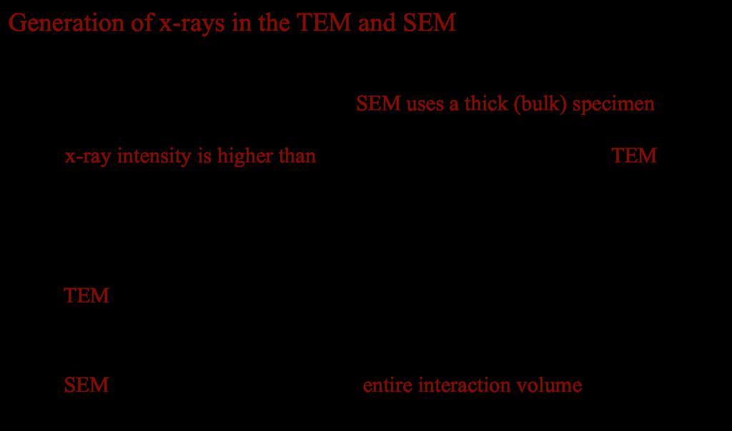
Generation of X-Rays in the SEM specimen
 Generation of X-Rays in the SEM specimen The electron beam generates X-ray photons in the beam-specimen interaction volume beneath the specimen surface. Some X-ray photons emerging from the specimen have
Praktikum zur. Materialanalytik
 Praktikum zur Materialanalytik Energy Dispersive X-ray Spectroscopy B513 Stand: 19.10.2016 Contents 1 Introduction... 2 2. Fundamental Physics and Notation... 3 2.1. Alignments of the microscope... 3 2.2.
SEM. Chemical Analysis in the. Elastic and Inelastic scattering. Chemical analysis in the SEM. Chemical analysis in the SEM
 THE UNIVERSITY Chemical Analysis in the SEM Ian Jones Centre for Electron Microscopy OF BIRMINGHAM Elastic and Inelastic scattering Electron interacts with one of the orbital electrons Secondary electrons,
An Introduction to Diffraction and Scattering. School of Chemistry The University of Sydney
 An Introduction to Diffraction and Scattering Brendan J. Kennedy School of Chemistry The University of Sydney 1) Strong forces 2) Weak forces Types of Forces 3) Electromagnetic forces 4) Gravity Types
Electron and electromagnetic radiation
 Electron and electromagnetic radiation Generation and interactions with matter Stimuli Interaction with sample Response Stimuli Waves and energy The energy is propotional to 1/λ and 1/λ 2 λ λ 1 Electromagnetic
Introduction to EDX. Energy Dispersive X-ray Microanalysis (EDS, Energy dispersive Spectroscopy) Basics of EDX
 Basics of EDX") Introduction to EDX Energy Dispersive X-ray Microanalysis (EDS, Energy dispersive Spectroscopy) EDX Marco Cantoni 1 Basics of EDX a) Generation of X-rays b) Detection Si(Li) Detector, SDD Detector, EDS
Introduction to EDX Energy Dispersive X-ray Microanalysis (EDS, Energy dispersive Spectroscopy) EDX Marco Cantoni 1 Basics of EDX a) Generation of X-rays b) Detection Si(Li) Detector, SDD Detector, EDS
4. Inelastic Scattering
 1 4. Inelastic Scattering Some inelastic scattering processes A vast range of inelastic scattering processes can occur during illumination of a specimen with a highenergy electron beam. In principle, many
MT Electron microscopy Scanning electron microscopy and electron probe microanalysis
 MT-0.6026 Electron microscopy Scanning electron microscopy and electron probe microanalysis Eero Haimi Research Manager Outline 1. Introduction Basics of scanning electron microscopy (SEM) and electron
MT Electron microscopy Scanning electron microscopy and electron probe microanalysis
 MT-0.6026 Electron microscopy Scanning electron microscopy and electron probe microanalysis Eero Haimi Research Manager Outline 1. Introduction Basics of scanning electron microscopy (SEM) and electron
Chemistry Instrumental Analysis Lecture 19 Chapter 12. Chem 4631
 Chemistry 4631 Instrumental Analysis Lecture 19 Chapter 12 There are three major techniques used for elemental analysis: Optical spectrometry Mass spectrometry X-ray spectrometry X-ray Techniques include:
Chapter 37 Early Quantum Theory and Models of the Atom
 Chapter 37 Early Quantum Theory and Models of the Atom Units of Chapter 37 37-7 Wave Nature of Matter 37-8 Electron Microscopes 37-9 Early Models of the Atom 37-10 Atomic Spectra: Key to the Structure
KMÜ 396 MATERIALS SCIENCE AND TECH. I PRESENTATION ELECTRON ENERGY LOSS SPECTROSCOPY (EELS) TUĞÇE SEZGİN
 TUĞÇE SEZGİN") KMÜ 396 MATERIALS SCIENCE AND TECH. I PRESENTATION ELECTRON ENERGY LOSS SPECTROSCOPY (EELS) TUĞÇE SEZGİN 20970725 HACETTEPE UNIVERSITY DEPARTMENT OF CHEMICAL ENGINEERING, SPRING 2011,APRIL,ANKARA CONTENTS
KMÜ 396 MATERIALS SCIENCE AND TECH. I PRESENTATION ELECTRON ENERGY LOSS SPECTROSCOPY (EELS) TUĞÇE SEZGİN 20970725 HACETTEPE UNIVERSITY DEPARTMENT OF CHEMICAL ENGINEERING, SPRING 2011,APRIL,ANKARA CONTENTS
EDS User School. Principles of Electron Beam Microanalysis
 EDS User School Principles of Electron Beam Microanalysis Outline 1.) Beam-specimen interactions 2.) EDS spectra: Origin of Bremsstrahlung and characteristic peaks 3.) Moseley s law 4.) Characteristic
Particle nature of light & Quantization
 Particle nature of light & Quantization A quantity is quantized if its possible values are limited to a discrete set. An example from classical physics is the allowed frequencies of standing waves on a
CHEM-E5225 :Electron Microscopy X-Ray Spectrometry
 CHEM-E5225 :Electron Microscopy X-Ray Spectrometry 2016.11 Yanling Ge Outline X-ray Spectrometry X-ray Spectra and Images Qualitative and Quantitative X-ray Analysis and Imaging Discussion of homework
X-Ray Photoelectron Spectroscopy (XPS)
") X-Ray Photoelectron Spectroscopy (XPS) Louis Scudiero http://www.wsu.edu/~scudiero; 5-2669 Electron Spectroscopy for Chemical Analysis (ESCA) The basic principle of the photoelectric effect was enunciated
X-Ray Photoelectron Spectroscopy (XPS) Louis Scudiero http://www.wsu.edu/~scudiero; 5-2669 Electron Spectroscopy for Chemical Analysis (ESCA) The basic principle of the photoelectric effect was enunciated
hν' Φ e - Gamma spectroscopy - Prelab questions 1. What characteristics distinguish x-rays from gamma rays? Is either more intrinsically dangerous?
 Gamma spectroscopy - Prelab questions 1. What characteristics distinguish x-rays from gamma rays? Is either more intrinsically dangerous? 2. Briefly discuss dead time in a detector. What factors are important
UNIT : QUANTUM THEORY AND THE ATOM
 Name St.No. Date(YY/MM/DD) / / Section UNIT 102-10: QUANTUM THEORY AND THE ATOM OBJECTIVES Atomic Spectra for Hydrogen, Mercury and Neon. 1. To observe various atomic spectra with a diffraction grating
Electron Microscopy I
 Characterization of Catalysts and Surfaces Characterization Techniques in Heterogeneous Catalysis Electron Microscopy I Introduction Properties of electrons Electron-matter interactions and their applications
Electron Microprobe Analysis 1 Nilanjan Chatterjee, Ph.D. Principal Research Scientist
 12.141 Electron Microprobe Analysis 1 Nilanjan Chatterjee, Ph.D. Principal Research Scientist Massachusetts Institute of Technology Electron Microprobe Facility Department of Earth, Atmospheric and Planetary
Electron Microprobe Analysis 1 Nilanjan Chatterjee, Ph.D. Principal Research Scientist
 12.141 Electron Microprobe Analysis 1 Nilanjan Chatterjee, Ph.D. Principal Research Scientist Massachusetts Institute of Technology Electron Microprobe Facility Department of Earth, Atmospheric and Planetary
Electronic Structure of Atoms. Chapter 6
 Electronic Structure of Atoms Chapter 6 Electronic Structure of Atoms 1. The Wave Nature of Light All waves have: a) characteristic wavelength, λ b) amplitude, A Electronic Structure of Atoms 1. The Wave
Electron probe microanalysis - Electron microprobe analysis EPMA (EMPA) What s EPMA all about? What can you learn?
 What s EPMA all about? What can you learn?") Electron probe microanalysis - Electron microprobe analysis EPMA (EMPA) What s EPMA all about? What can you learn? EPMA - what is it? Precise and accurate quantitative chemical analyses of micron-size
Electron probe microanalysis - Electron microprobe analysis EPMA (EMPA) What s EPMA all about? What can you learn? EPMA - what is it? Precise and accurate quantitative chemical analyses of micron-size
Transmission Electron Microscopy
 L. Reimer H. Kohl Transmission Electron Microscopy Physics of Image Formation Fifth Edition el Springer Contents 1 Introduction... 1 1.1 Transmission Electron Microscopy... 1 1.1.1 Conventional Transmission
MANIPAL INSTITUTE OF TECHNOLOGY
 SCHEME OF EVAUATION MANIPA INSTITUTE OF TECHNOOGY MANIPA UNIVERSITY, MANIPA SECOND SEMESTER B.Tech. END-SEMESTER EXAMINATION - MAY SUBJECT: ENGINEERING PHYSICS (PHY/) Time: 3 Hrs. Max. Marks: 5 Note: Answer
X-Ray Photoelectron Spectroscopy (XPS)
") X-Ray Photoelectron Spectroscopy (XPS) Louis Scudiero http://www.wsu.edu/~scudiero; 5-2669 Fulmer 261A Electron Spectroscopy for Chemical Analysis (ESCA) The basic principle of the photoelectric effect
X-Ray Photoelectron Spectroscopy (XPS) Louis Scudiero http://www.wsu.edu/~scudiero; 5-2669 Fulmer 261A Electron Spectroscopy for Chemical Analysis (ESCA) The basic principle of the photoelectric effect
CHARACTERIZATION of NANOMATERIALS KHP
 CHARACTERIZATION of NANOMATERIALS Overview of the most common nanocharacterization techniques MAIN CHARACTERIZATION TECHNIQUES: 1.Transmission Electron Microscope (TEM) 2. Scanning Electron Microscope
Chapter 37 Early Quantum Theory and Models of the Atom. Copyright 2009 Pearson Education, Inc.
 Chapter 37 Early Quantum Theory and Models of the Atom Planck s Quantum Hypothesis; Blackbody Radiation Photon Theory of Light and the Photoelectric Effect Energy, Mass, and Momentum of a Photon Compton
X-Ray Emission and Absorption
 X-Ray Emission and Absorption Author: Mike Nill Alex Bryant February 6, 20 Abstract X-rays were produced by two bench-top diffractometers using a copper target. Various nickel filters were placed in front
Gaetano L Episcopo. Scanning Electron Microscopy Focus Ion Beam and. Pulsed Plasma Deposition
 Gaetano L Episcopo Scanning Electron Microscopy Focus Ion Beam and Pulsed Plasma Deposition Hystorical background Scientific discoveries 1897: J. Thomson discovers the electron. 1924: L. de Broglie propose
Physics 1C Lecture 29A. Finish off Ch. 28 Start Ch. 29
 Physics 1C Lecture 29A Finish off Ch. 28 Start Ch. 29 Particle in a Box Let s consider a particle confined to a one-dimensional region in space. Following the quantum mechanics approach, we need to find
EEE4106Z Radiation Interactions & Detection
 EEE4106Z Radiation Interactions & Detection 2. Radiation Detection Dr. Steve Peterson 5.14 RW James Department of Physics University of Cape Town steve.peterson@uct.ac.za May 06, 2015 EEE4106Z :: Radiation
Chapter Six: X-Rays. 6.1 Discovery of X-rays
 Chapter Six: X-Rays 6.1 Discovery of X-rays In late 1895, a German physicist, W. C. Roentgen was working with a cathode ray tube in his laboratory. He was working with tubes similar to our fluorescent
MSE 321 Structural Characterization
 Auger Spectroscopy Auger Electron Spectroscopy (AES) Scanning Auger Microscopy (SAM) Incident Electron Ejected Electron Auger Electron Initial State Intermediate State Final State Physical Electronics
X-Ray Photoelectron Spectroscopy (XPS)-2
-2") X-Ray Photoelectron Spectroscopy (XPS)-2 Louis Scudiero http://www.wsu.edu/~scudiero; 5-2669 Fulmer 261A Electron Spectroscopy for Chemical Analysis (ESCA) The 3 step model: 1.Optical excitation 2.Transport
X-Ray Photoelectron Spectroscopy (XPS)-2 Louis Scudiero http://www.wsu.edu/~scudiero; 5-2669 Fulmer 261A Electron Spectroscopy for Chemical Analysis (ESCA) The 3 step model: 1.Optical excitation 2.Transport
XRF books: Analytical Chemistry, Kellner/Mermet/Otto/etc. 3 rd year XRF Spectroscopy Dr. Alan Ryder (R222, Physical Chemistry) 2 lectures:
 2 lectures:") 1 3 rd year XRF Spectroscopy Dr. Alan Ryder (R222, Physical Chemistry) 2 lectures: XRF spectroscopy 1 exam question. Notes on: www.nuigalway.ie/nanoscale/3rdspectroscopy.html XRF books: Analytical Chemistry,
1 3 rd year XRF Spectroscopy Dr. Alan Ryder (R222, Physical Chemistry) 2 lectures: XRF spectroscopy 1 exam question. Notes on: www.nuigalway.ie/nanoscale/3rdspectroscopy.html XRF books: Analytical Chemistry,
Massachusetts Institute of Technology. Dr. Nilanjan Chatterjee
 Massachusetts Institute of Technology Dr. Nilanjan Chatterjee Electron Probe Micro-Analysis (EPMA) Imaging and micrometer-scale chemical compositional analysis of solids Signals produced in The Electron
1 P a g e h t t p s : / / w w w. c i e n o t e s. c o m / Physics (A-level)
") 1 P a g e h t t p s : / / w w w. c i e n o t e s. c o m / Physics (A-level) Electromagnetic induction (Chapter 23): For a straight wire, the induced current or e.m.f. depends on: The magnitude of the magnetic
1 P a g e h t t p s : / / w w w. c i e n o t e s. c o m / Physics (A-level) Electromagnetic induction (Chapter 23): For a straight wire, the induced current or e.m.f. depends on: The magnitude of the magnetic
Semiconductor Physics and Devices
 Introduction to Quantum Mechanics In order to understand the current-voltage characteristics, we need some knowledge of electron behavior in semiconductor when the electron is subjected to various potential
Planck s Quantum Hypothesis Blackbody Radiation
 Planck s Quantum Hypothesis Blackbody Radiation The spectrum of blackbody radiation has been measured(next slide); it is found that the frequency of peak intensity increases linearly with temperature.
Understanding X-rays: The electromagnetic spectrum
 Understanding X-rays: The electromagnetic spectrum 1 ULa 13.61 kev 0.09 nm BeKa 0.11 kev 11.27 nm E = hn = h c l where, E : energy, h : Planck's constant, n : frequency c : speed of light in vacuum, l
THE NATURE OF THE ATOM. alpha particle source
 chapter THE NATURE OF THE ATOM www.tutor-homework.com (for tutoring, homework help, or help with online classes) Section 30.1 Rutherford Scattering and the Nuclear Atom 1. Which model of atomic structure
h p λ = mν Back to de Broglie and the electron as a wave you will learn more about this Equation in CHEM* 2060
 Back to de Broglie and the electron as a wave λ = mν h = h p you will learn more about this Equation in CHEM* 2060 We will soon see that the energies (speed for now if you like) of the electrons in the
X-ray spectroscopy: Experimental studies of Moseley s law (K-line x-ray fluorescence) and x-ray material s composition determination
 and x-ray material s composition determination") Uppsala University Department of Physics and Astronomy Laboratory exercise X-ray spectroscopy: Experimental studies of Moseley s law (K-line x-ray fluorescence) and x-ray material s composition determination
Uppsala University Department of Physics and Astronomy Laboratory exercise X-ray spectroscopy: Experimental studies of Moseley s law (K-line x-ray fluorescence) and x-ray material s composition determination
Lecture 5. X-ray Photoemission Spectroscopy (XPS)
") Lecture 5 X-ray Photoemission Spectroscopy (XPS) 5. Photoemission Spectroscopy (XPS) 5. Principles 5.2 Interpretation 5.3 Instrumentation 5.4 XPS vs UV Photoelectron Spectroscopy (UPS) 5.5 Auger Electron
Lecture 5 X-ray Photoemission Spectroscopy (XPS) 5. Photoemission Spectroscopy (XPS) 5. Principles 5.2 Interpretation 5.3 Instrumentation 5.4 XPS vs UV Photoelectron Spectroscopy (UPS) 5.5 Auger Electron
Quantum and Atomic Physics - Multiple Choice
 PSI AP Physics 2 Name 1. The Cathode Ray Tube experiment is associated with: (A) J. J. Thomson (B) J. S. Townsend (C) M. Plank (D) A. H. Compton 2. The electron charge was measured the first time in: (A)
Shell Atomic Model and Energy Levels
 Shell Atomic Model and Energy Levels (higher energy, deeper excitation) - Radio waves: Not absorbed and pass through tissue un-attenuated - Microwaves : Energies of Photos enough to cause molecular rotation
Electron Microprobe Analysis and Scanning Electron Microscopy
 Electron Microprobe Analysis and Scanning Electron Microscopy Electron microprobe analysis (EMPA) Analytical technique in which a beam of electrons is focused on a sample surface, producing X-rays from
Atom Physics. Chapter 30. DR JJ UiTM-Cutnell & Johnson 7th ed. 1. Model of an atom-the recent model. Nuclear radius r m
 Chapter 30 Atom Physics DR JJ UiTM-Cutnell & Johnson 7th ed. 1 30.1 Rutherford Scattering and the Nuclear Atom Model of an atom-the recent model Nuclear radius r 10-15 m Electron s position radius r 10-10
EMISSION SPECTROSCOPY
 IFM The Department of Physics, Chemistry and Biology LAB 57 EMISSION SPECTROSCOPY NAME PERSONAL NUMBER DATE APPROVED I. OBJECTIVES - Understand the principle of atomic emission spectra. - Know how to acquire
Complete nomenclature for electron orbitals
 Complete nomenclature for electron orbitals Bohr s model worked but it lacked a satisfactory reason why. De Broglie suggested that all particles have a wave nature. u l=h/p Enter de Broglie again It was
X-ray Energy Spectroscopy (XES).
.") X-ray Energy Spectroscopy (XES). X-ray fluorescence as an analytical tool for element analysis is based on 3 fundamental parameters: A. Specificity: In determining an x-ray emission energy E certainty
X-ray Energy Spectroscopy (XES). X-ray fluorescence as an analytical tool for element analysis is based on 3 fundamental parameters: A. Specificity: In determining an x-ray emission energy E certainty
HOW TO APPROACH SCANNING ELECTRON MICROSCOPY AND ENERGY DISPERSIVE SPECTROSCOPY ANALYSIS. SCSAM Short Course Amir Avishai
 HOW TO APPROACH SCANNING ELECTRON MICROSCOPY AND ENERGY DISPERSIVE SPECTROSCOPY ANALYSIS SCSAM Short Course Amir Avishai RESEARCH QUESTIONS Sea Shell Cast Iron EDS+SE Fe Cr C Objective Ability to ask the
object objective lens eyepiece lens
 Advancing Physics G495 June 2015 SET #1 ANSWERS Field and Particle Pictures Seeing with electrons The compound optical microscope Q1. Before attempting this question it may be helpful to review ray diagram
X-ray Absorption Spectroscopy
 X-ray Absorption Spectroscopy Nikki Truss November 26, 2012 Abstract In these experiments, some aspects of x-ray absorption spectroscopy were investigated. The x-ray spectrum of molybdenum was recorded
Electron Spettroscopies
 Electron Spettroscopies Spettroscopy allows to characterize a material from the point of view of: chemical composition, electronic states and magnetism, electronic, roto-vibrational and magnetic excitations.
EDS Mapping. Ian Harvey Fall Practical Electron Microscopy
 EDS Mapping Ian Harvey Fall 2008 1 From: Energy Dispersive X-ray Microanalysis, An Introduction Kevex Corp. 1988 Characteristic X-ray generation p.2 1 http://www.small-world.net/efs.htm X-ray generation
Photoelectric Effect Experiment
 Experiment 1 Purpose The photoelectric effect is a key experiment in modern physics. In this experiment light is used to excite electrons that (given sufficient energy) can escape from a material producing
Electron Spectroscopy
 Electron Spectroscopy Photoelectron spectroscopy is based upon a single photon in/electron out process. The energy of a photon is given by the Einstein relation : E = h ν where h - Planck constant ( 6.62
is the minimum stopping potential for which the current between the plates reduces to zero.
 Module 1 :Quantum Mechanics Chapter 2 : Introduction to Quantum ideas Introduction to Quantum ideas We will now consider some experiments and their implications, which introduce us to quantum ideas. The
and another with a peak frequency ω 2
 Physics Qualifying Examination Part I 7-Minute Questions September 13, 2014 1. A sealed container is divided into two volumes by a moveable piston. There are N A molecules on one side and N B molecules
= 6 (1/ nm) So what is probability of finding electron tunneled into a barrier 3 ev high?
 So what is probability of finding electron tunneled into a barrier 3 ev high?") STM STM With a scanning tunneling microscope, images of surfaces with atomic resolution can be readily obtained. An STM uses quantum tunneling of electrons to map the density of electrons on the surface
STM STM With a scanning tunneling microscope, images of surfaces with atomic resolution can be readily obtained. An STM uses quantum tunneling of electrons to map the density of electrons on the surface
ATOMIC STRUCTURE, ELECTRONS, AND PERIODICITY
 ATOMIC STRUCTURE, ELECTRONS, AND PERIODICITY All matter is made of atoms. There are a limited number of types of atoms; these are the elements. (EU 1.A) Development of Atomic Theory Atoms are so small
Understanding X-rays: The electromagnetic spectrum
 Understanding X-rays: The electromagnetic spectrum 1 ULa 13.61 kev 0.09 nm BeKa 0.11 kev 11.27 nm E = hn = h c l where, E : energy, h : Planck's constant, n : frequency c : speed of light in vacuum, l
X-Ray Photoelectron Spectroscopy (XPS)-2
-2") X-Ray Photoelectron Spectroscopy (XPS)-2 Louis Scudiero http://www.wsu.edu/~pchemlab ; 5-2669 Fulmer 261A Electron Spectroscopy for Chemical Analysis (ESCA) The 3 step model: 1.Optical excitation 2.Transport
X-Ray Photoelectron Spectroscopy (XPS)-2 Louis Scudiero http://www.wsu.edu/~pchemlab ; 5-2669 Fulmer 261A Electron Spectroscopy for Chemical Analysis (ESCA) The 3 step model: 1.Optical excitation 2.Transport
Chapter 28. Atomic Physics
 Chapter 28 Atomic Physics Quantum Numbers and Atomic Structure The characteristic wavelengths emitted by a hot gas can be understood using quantum numbers. No two electrons can have the same set of quantum
Notes on x-ray scattering - M. Le Tacon, B. Keimer (06/2015)
") Notes on x-ray scattering - M. Le Tacon, B. Keimer (06/2015) Interaction of x-ray with matter: - Photoelectric absorption - Elastic (coherent) scattering (Thomson Scattering) - Inelastic (incoherent) scattering
Notes on x-ray scattering - M. Le Tacon, B. Keimer (06/2015) Interaction of x-ray with matter: - Photoelectric absorption - Elastic (coherent) scattering (Thomson Scattering) - Inelastic (incoherent) scattering
Stellar Astrophysics: The Interaction of Light and Matter
 Stellar Astrophysics: The Interaction of Light and Matter The Photoelectric Effect Methods of electron emission Thermionic emission: Application of heat allows electrons to gain enough energy to escape
X-RAY PRODUCTION. Prepared by:- EN KAMARUL AMIN BIN ABDULLAH
 X-RAY PRODUCTION Prepared by:- EN KAMARUL AMIN BIN ABDULLAH OBJECTIVES Discuss the process of x-ray being produced (conditions) Explain the principles of energy conversion in x-ray production (how energy
Photoelectron spectroscopy Instrumentation. Nanomaterials characterization 2
 Photoelectron spectroscopy Instrumentation Nanomaterials characterization 2 RNDr. Věra V Vodičkov ková,, PhD. Photoelectron Spectroscopy general scheme Impact of X-ray emitted from source to the sample
Chapter 10: Wave Properties of Particles
 Chapter 10: Wave Properties of Particles Particles such as electrons may demonstrate wave properties under certain conditions. The electron microscope uses these properties to produce magnified images
The Bohr Model of Hydrogen
 The Bohr Model of Hydrogen Suppose you wanted to identify and measure the energy high energy photons. One way to do this is to make a calorimeter. The CMS experiment s electromagnetic calorimeter is made
Chapter 6: Electronic Structure of Atoms
 Chapter 6: Electronic Structure of Atoms Learning Outcomes: Calculate the wavelength of electromagnetic radiation given its frequency or its frequency given its wavelength. Order the common kinds of radiation
Advanced Lab Course. X-Ray Photoelectron Spectroscopy 1 INTRODUCTION 1 2 BASICS 1 3 EXPERIMENT Qualitative analysis Chemical Shifts 7
 Advanced Lab Course X-Ray Photoelectron Spectroscopy M210 As of: 2015-04-01 Aim: Chemical analysis of surfaces. Content 1 INTRODUCTION 1 2 BASICS 1 3 EXPERIMENT 3 3.1 Qualitative analysis 6 3.2 Chemical
Diffraction Gratings, Atomic Spectra. Prof. Shawhan (substituting for Prof. Hall) November 14, 2016
 November 14, 2016") Diffraction Gratings, Atomic Spectra Prof. Shawhan (substituting for Prof. Hall) November 14, 2016 1 Increase number of slits: 2 Visual Comparisons 3 4 8 2 Diffraction Grating Note: despite the name, this
Diffraction Gratings, Atomic Spectra Prof. Shawhan (substituting for Prof. Hall) November 14, 2016 1 Increase number of slits: 2 Visual Comparisons 3 4 8 2 Diffraction Grating Note: despite the name, this
Introduction to X-ray Photoelectron Spectroscopy (XPS) XPS which makes use of the photoelectric effect, was developed in the mid-1960
 XPS which makes use of the photoelectric effect, was developed in the mid-1960") Introduction to X-ray Photoelectron Spectroscopy (XPS) X-ray Photoelectron Spectroscopy (XPS), also known as Electron Spectroscopy for Chemical Analysis (ESCA) is a widely used technique to investigate
Introduction to X-ray Photoelectron Spectroscopy (XPS) X-ray Photoelectron Spectroscopy (XPS), also known as Electron Spectroscopy for Chemical Analysis (ESCA) is a widely used technique to investigate
Skoog Chapter 6 Introduction to Spectrometric Methods
 Skoog Chapter 6 Introduction to Spectrometric Methods General Properties of Electromagnetic Radiation (EM) Wave Properties of EM Quantum Mechanical Properties of EM Quantitative Aspects of Spectrochemical
Interactions with Matter
 Manetic Lenses Manetic fields can displace electrons Manetic field can be produced by passin an electrical current throuh coils of wire Manetic field strenth can be increased by usin a soft ferromanetic
X-RAY SPECTRA. Theory:
 12 Oct 18 X-ray.1 X-RAY SPECTRA In this experiment, a number of measurements involving x-rays will be made. The spectrum of x-rays emitted from a molybdenum target will be measured, and the experimental
X Rays & Crystals. Characterizing Mineral Chemistry & Structure. J.D. Price
 X Rays & Crystals Characterizing Mineral Chemistry & Structure J.D. Price Light - electromagnetic spectrum Wave behavior vs. particle behavior If atoms are on the 10-10 m scale, we need to use sufficiently
X-ray Photoelectron Spectroscopy (XPS)
") X-ray Photoelectron Spectroscopy (XPS) As part of the course Characterization of Catalysts and Surfaces Prof. Dr. Markus Ammann Paul Scherrer Institut markus.ammann@psi.ch Resource for further reading:
X-ray Photoelectron Spectroscopy (XPS) As part of the course Characterization of Catalysts and Surfaces Prof. Dr. Markus Ammann Paul Scherrer Institut markus.ammann@psi.ch Resource for further reading:
Technical University of Denmark. Center for Electron Nanoscopy. Advanced TEM (16 September 2010) Microanalysis in the electron microscope
 Microanalysis in the electron microscope") Microanalysis in the electron microscope MH1 1 Technical University of Denmark Center for Electron Nanoscopy Advanced TEM (16 September 2010) Microanalysis in the electron microscope Dr C B Boothroyd Synopsis
Microanalysis in the electron microscope MH1 1 Technical University of Denmark Center for Electron Nanoscopy Advanced TEM (16 September 2010) Microanalysis in the electron microscope Dr C B Boothroyd Synopsis
CHEM*3440. X-Ray Energies. Bremsstrahlung Radiation. X-ray Line Spectra. Chemical Instrumentation. X-Ray Spectroscopy. Topic 13
 X-Ray Energies very short wavelength radiation 0.1Å to 10 nm (100 Å) CHEM*3440 Chemical Instrumentation Topic 13 X-Ray Spectroscopy Visible - Ultraviolet (UV) - Vacuum UV (VUV) - Extreme UV (XUV) - Soft
Quantum physics. Anyone who is not shocked by the quantum theory has not understood it. Niels Bohr, Nobel Price in 1922 ( )
") Quantum physics Anyone who is not shocked by the quantum theory has not understood it. Niels Bohr, Nobel Price in 1922 (1885-1962) I can safely say that nobody understand quantum physics Richard Feynman
Quantum physics Anyone who is not shocked by the quantum theory has not understood it. Niels Bohr, Nobel Price in 1922 (1885-1962) I can safely say that nobody understand quantum physics Richard Feynman
Lecture 20 Optical Characterization 2
 Lecture 20 Optical Characterization 2 Schroder: Chapters 2, 7, 10 1/68 Announcements Homework 5/6: Is online now. Due Wednesday May 30th at 10:00am. I will return it the following Wednesday (6 th June).
DO PHYSICS ONLINE 9.4 ROM IDEAS TO IMPLEMENTATION MINDMAP SUMMARIES
 DO PHYSICS ONLINE 9.4 ROM IDEAS TO IMPLEMENTATION MINDMAP SUMMARIES 1 13/14 ELECTRIC POTENTIAL V [V] Measure of charge imbalance + 6 V + + + + + + - 3 V + 6 V + 3 V + + + + 15 V 0 V - V - - + 6 V -14 V
Lecture 4. The Bohr model of the atom. De Broglie theory. The Davisson-Germer experiment
 Lecture 4 The Bohr model of the atom De Broglie theory The Davisson-Germer experiment Objectives Learn about electron energy levels in atoms and how Bohr's model can be used to determine the energy levels
Chapter 39. Particles Behaving as Waves
 Chapter 39 Particles Behaving as Waves 39.1 Electron Waves Light has a dual nature. Light exhibits both wave and particle characteristics. Louis de Broglie postulated in 1924 that if nature is symmetric,
Röntgenpraktikum. M. Oehzelt. (based on the diploma thesis of T. Haber [1])
![Röntgenpraktikum. M. Oehzelt. (based on the diploma thesis of T. Haber [1])](/thumbs/91/106501237.jpg "Röntgenpraktikum. M. Oehzelt. (based on the diploma thesis of T. Haber [1])") Röntgenpraktikum M. Oehzelt (based on the diploma thesis of T. Haber [1]) October 21, 2004 Contents 1 Fundamentals 2 1.1 X-Ray Radiation......................... 2 1.1.1 Bremsstrahlung......................
Röntgenpraktikum M. Oehzelt (based on the diploma thesis of T. Haber [1]) October 21, 2004 Contents 1 Fundamentals 2 1.1 X-Ray Radiation......................... 2 1.1.1 Bremsstrahlung......................
Name: (a) What core levels are responsible for the three photoelectron peaks in Fig. 1?
 What core levels are responsible for the three photoelectron peaks in Fig. 1?") Physics 243A--Surface Physics of Materials: Spectroscopy Final Examination December 16, 2014 (3 problems, 100 points total, open book, open notes and handouts) Name: [1] (50 points), including Figures
Physics 243A--Surface Physics of Materials: Spectroscopy Final Examination December 16, 2014 (3 problems, 100 points total, open book, open notes and handouts) Name: [1] (50 points), including Figures
X-ray Absorption and Emission Prepared By Jose Hodak for BSAC program 2008
 X-ray Absorption and Emission Prepared By Jose Hodak for BSAC program 2008 1- A bit of History: Wilhelm Conrad Röntgen discovered 1895 the X-rays. 1901 he was honored by the Noble prize for physics. In
Ch 7 Quantum Theory of the Atom (light and atomic structure)
") Ch 7 Quantum Theory of the Atom (light and atomic structure) Electromagnetic Radiation - Electromagnetic radiation consists of oscillations in electric and magnetic fields. The oscillations can be described
Ch 7 Quantum Theory of the Atom (light and atomic structure) Electromagnetic Radiation - Electromagnetic radiation consists of oscillations in electric and magnetic fields. The oscillations can be described
5) Surface photoelectron spectroscopy. For MChem, Spring, Dr. Qiao Chen (room 3R506) University of Sussex.
 Surface photoelectron spectroscopy. For MChem, Spring, Dr. Qiao Chen (room 3R506) University of Sussex.") For MChem, Spring, 2009 5) Surface photoelectron spectroscopy Dr. Qiao Chen (room 3R506) http://www.sussex.ac.uk/users/qc25/ University of Sussex Today s topics 1. Element analysis with XPS Binding energy,
For MChem, Spring, 2009 5) Surface photoelectron spectroscopy Dr. Qiao Chen (room 3R506) http://www.sussex.ac.uk/users/qc25/ University of Sussex Today s topics 1. Element analysis with XPS Binding energy,
Chap. 1 (Introduction), Chap. 2 (Components and Circuits)
, Chap. 2 (Components and Circuits)") CHEM 455 The class describes the principles and applications of modern analytical instruments. Emphasis is placed upon the theoretical basis of each type of instrument, its optimal area of application,
CHEM 455 The class describes the principles and applications of modern analytical instruments. Emphasis is placed upon the theoretical basis of each type of instrument, its optimal area of application,
X-ray Spectroscopy. Danny Bennett and Maeve Madigan. October 12, 2015
 X-ray Spectroscopy Danny Bennett and Maeve Madigan October 12, 2015 Abstract Various X-ray spectra were obtained, and their properties were investigated. The characteristic peaks were identified for a
SECTION A Quantum Physics and Atom Models
 AP Physics Multiple Choice Practice Modern Physics SECTION A Quantum Physics and Atom Models 1. Light of a single frequency falls on a photoelectric material but no electrons are emitted. Electrons may
Accounts for certain objects being colored. Used in medicine (examples?) Allows us to learn about structure of the atom
 Allows us to learn about structure of the atom") 1.1 Interaction of Light and Matter Accounts for certain objects being colored Used in medicine (examples?) 1.2 Wavelike Properties of Light Wavelength, : peak to peak distance Amplitude: height of the
1.1 Interaction of Light and Matter Accounts for certain objects being colored Used in medicine (examples?) 1.2 Wavelike Properties of Light Wavelength, : peak to peak distance Amplitude: height of the
Chemistry (
 Question 2.1: (i) Calculate the number of electrons which will together weigh one gram. (ii) Calculate the mass and charge of one mole of electrons. Answer 2.1: (i) Mass of one electron = 9.10939 10 31
Quantum Condensed Matter Physics Lecture 12
 Quantum Condensed Matter Physics Lecture 12 David Ritchie QCMP Lent/Easter 2016 http://www.sp.phy.cam.ac.uk/drp2/home 12.1 QCMP Course Contents 1. Classical models for electrons in solids 2. Sommerfeld