Outline. Fundamentals of ultrasound Focusing in ultrasound Ultrasonic blood flow estimation Nonlinear ultrasonics
|
|
|
- Timothy Craig
- 5 years ago
- Views:
Transcription
1 生醫超音波技術 台大電機系李百祺
2 Outline Fundamentals of ultrasound Focusing in ultrasound Ultrasonic blood flow estimation Nonlinear ultrasonics
3 What is ultrasound?
4 Characteristics of Ultrasound A mechanical wave: Characterized by pressure, particle velocity and displacement. Density change of the propagating medium. But it is still a wave, i.e., there is reflection, refraction, scattering, diffraction, attenuation etc.

5 Basics of Acoustic Waves Longitudinal Wave:
6 Basics of Acoustic Waves Shear Wave:
7 Characteristics of Ultrasound A mechanical wave: Characterized by pressure, particle velocity and displacement. Density change of the propagating medium. But it is still a wave, i.e., there is reflection, refraction, scattering, diffraction, attenuation etc.
8 Sound Velocity and Density Change B ( x ) = c0 + (1 + ) u( x) 2A Phase velocity Nonlinearity Particle velocity Finite Amplitude Distortion
9 When Peak Pressure Is Very High Shock Wave
10 Characteristics of Ultrasound A mechanical wave: Characterized by pressure, particle velocity and displacement. Density change of the propagating medium. But it is still a wave, i.e., there is reflection, refraction, scattering, diffraction, attenuation etc.
11 Reflection Low Density to High Density High Density to Low Density
12 Refraction

13 Acoustic Scattering
14 Diffraction
15 Characteristics of Ultrasound Sound wave with frequencies higher than the audible range (>20-25kHz): Typical frequency range for biomedical applications: MHz. c=f λ. Sound (propagation) speed in soft tissues are around 1500m/sec. It becomes higher in hard tissues (e.g., bone).
16
17 Characteristics of Ultrasound Affected by the elastic properties of the propagating medium: Various modes of propagation. Hooke slaw: T=eS (tensor form in 3D). c = B / ρ Characteri stic impedance : Z 0 = ρc
18
19 Bio-Effects Heating Cavitation
20 Ultrasound Heating Bio-transfer equation:
21 Bio-Effects Heating Cavitation
22 Cavitation Formation and behavior of gas bubbles in acoustic fields. Transient cavitation: sudden growth and collapse of bubbles, resulting shock waves and very high temperatures.
23 Other Acoustic Phenomena Radiation force. Sonoluminescence. etc.

24 Radiation Force An ultrasonic wave exerts a static force on an interface or in a medium where there is a decrease in power in the wave propagation direction.
25 Other Acoustic Phenomena Radiation force. Sonoluminescence. etc.

26 Sonoluminescence Weak emission of light observable when high intensity ultrasound passing through a medium containing dissolved gases.
27 ,etc.
28 What can ultrasound do in medicine and biology?
29 Ultrasound in Medicine and Biology Diagnostics (as a wave): Imaging. Blood flow measurements. Bone density (indirect). etc.
30 Ultrasonic Imaging
31 Ultrasound in Medicine and Biology Therapeutics: Heat generation: Hyperthermia. HIFU. Shock wave Lithotripsy. etc.

32 Hyperthermia is a method of treating cancerous tissue by elevating the tissue temperature to 42.5 C or above, and maintaining this for minutes. Hyperthermia

33 Hyperthermia
34 HIFU High Intensity Focused Ultrasound. In the focal point, the sudden and intense absorption of the ultrasound beam creates a sudden elevation of the temperature (from 85 to 100 C) which destroys the cells located in the targeted zone.


35 HIFU for Prostate Cancer

36 Image Guided HIFU
37 Ultrasound in Medicine and Biology Therapeutics: Heat generation: Hyperthermia. HIFU. Shock wave Lithotripsy. etc.

38 Extracorporeal Lithotripsy The use of shock waves to destroy stones in the body.
39 Extracorporeal Lithotripsy
40 Ultrasound in Medicine and Biology Sonoluminescence. Radiation force. Cavitation. Cosmetics. etc.
41 Bio-Effects and Safety Requirements
42 Basics Safety regulations. Physical parameters vs. Bio-effects. Measurement techniques. Dose: Energy absorption in tissue. Temperature rise, cell damage. Dosimetry: measurements of such effects. Exposure: Characteristics of ultrasound field. Pressure, intensity, power. Exposimetry: measurements of temporal/spatial characterisitics.
43 Bio-Effects Temperature rise and cell damage (cavitation). FDA Track I: Pre-amendments. I SPTA (720 mw/cm 2 ) and I SPPA (190 W/cm 2 ). FDA Track III: TI (Thermal Index) and MI (Mechanical Index). ALARA (as low as reasonably achievable).
44 Bio-Effects Thermal index (TI): TIS, TIB, TIC. Analytical. Mechanical Index (MI): Experimental. TI Destruction of bubble with different sizes at various frequencies. MI W W P o deg 03. f c
45 Imaging and Focusing
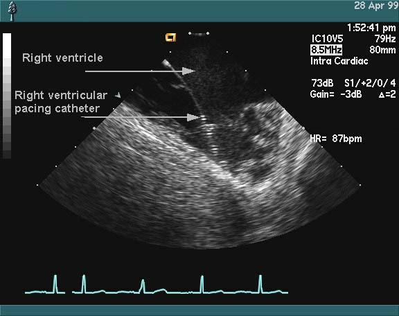
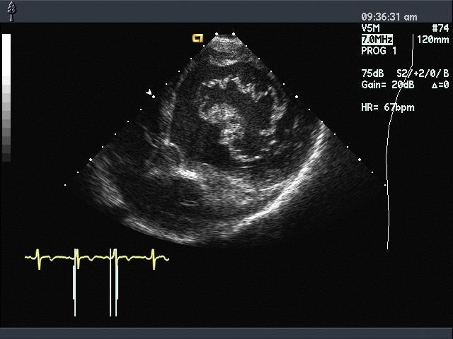
46
47
48

49 Real-Time Imaging
50
51
52
53 ransesophageal Echocardiogram (TEE)
54
55

56
 OB/GYN, vascular, cardiac,")

57 Clinical Applications (From OB/GYN, vascular, cardiac, transcranial, abdominal, musculoskeletal, endo-vaginal, endo-rectal, ocular, intra-vascular, etc.
58 Characteristics of Diagnostic Ultrasound Non-invasive. Safe (under regulations). Real-time. Reflection mode (similar to RADAR). Blood flow imaging. Access. Portable. Body type dependent.
59 Function Modes A-mode (A-scan, 1D). B-mode (Gray scale, 2D). 3D ultrasound. M-mode (motion) Color Doppler (2D, blood flow). Spectral Doppler (localized, blood flow). Audio Doppler.
60 A-Scan (Amplitude, 1D)
61 B-Scan (Brightness, 2D)
62 2D Scan Formats linear sector curved linear easy access limited view limited access wide view easy access wide view

63 3D Ultrasound
")
64 M-Mode (Motion)
65 Transducers: Generation and Detection of Sound Waves

66 Ultrasonic Array Transducers (From
67 Transducer Energy conversion: electrical mechanical. Generation and detection (speaker and microphone). Medical ultrasound: same device in MHz range. Piezoelectricity: electrical polarization mechanical strain. PZT, PVDF and composite materials are commonly used.

68 Anisotropy Piezoelectricity Poling Curie temperature: C.
69 Piezoelectricity P P+ P a2 a1 a2+ a2 a1+ a1 L L dipole strength of unit cell P = volume of unit cell P PS es q( a2 a1) 2 L ( a2+ a1) e: Piezoelectric stress constant
70 Detection of Ultrasound Reciprocal to generation. impinging wave electrode piezoelectric medium z=0 z=l L V ( t ) = E ( z, t ) dz 0 electrode V(t) surface area A
71 Design Considerations Bandwidth and sensitivity. PZT body Ring down backing PZT body PZT backing body matching layer
72 Acoustic Lens Fixed geometric elevational focusing.


73 1-D and 2-D Arrays
74 Focusing and Diffraction

75 B-mode Imaging 取自 rf signal envelop

76 Linear Scanning

77 Beam Formation Using Arrays Focusing:


78 Curved Linear Scanning

79 Sector Steering
80 How is the resolution determined?
81 ocusing Beam Formation Transducer To form a beam of sound wave such that only the objects along the beam direction are illuminated and possibly detected. Mainlobe Sidelobe


82 y Nomenclature beam pattern z Good Focusing x Poor Focusing x: Lateral, azimuthal, scan y: Elevational, non-scan z: Axial, range, depth Beam pattern Radiation pattern Diffraction pattern Focusing pattern
83 Pulsed Wave (PW) vs. Continuous Wave (CW)

84 Radiation Pattern
85 How to focus?
86 Beamforming Manipulation of transmit and receive apertures. Trade-off between performance/cost to achieve: Steer and focus the transmit beam. Dynamically steer and focus the receive beam. Provide accurate delay and apodization. Provide dynamic receive control.
87 Focusing Single Zone Focusing Multi-Zone Focusing Dynamic Focusing
88 Imaging Model ransmitter transducer propagation in the body transducer receiver display A-scan: 2βz R( x, y, z ) e 2z V ( t ) = k B( x, y, z ) p( t ) dx dy dz z c B-scan: 2z S ( x, t ) = k R( x, y, z ) B( x x, y, z ) p( t )dx dy dz c Scanning Convolution (Correlation vs. Convolution)
89 Imaging Model 2z 2z 2z p ( t ) = A( t )cos( 2πf0 ( t )) c c c Ideally, S ( x, t ) = R( x, y0, ct / 2) In practice, B( ) : A( ) : determined by diffraction determined by transducer bandwidth


90 Unfocused Focused Focused at ½ range Focused with twice the aperture
91 Axial Intensity Unfocused Focused Focused at ½ range Focused with 2X aperture
92 Implementation of Focusing Using Arrays
93 Beam Formation Using Arrays N i = 1 O( t ) = S ( t ( x, R, )) i τ i θ Delay and Sum
94 Propagating Delays τ( x, R, θ) = i ( 2 ( x Rsinθ) R cos θ) 2 2 i + c 1/ 2 2 R x i = c R In Fresnel region 2 2 R x x x i i i 2 τ( xi, R, θ) 1 + sinθ sin θ 2 2 c 2R R 2R R x = 1 sinθ + c R 2x i sinθ R x 2 R x sinθ x cos θ cos θ = + 2 2R c c 2Rc i i i i 1/ 2 Effective aperture size: 2a 2a cosθ
95 Propagating Delays Transmit: τ T ( x, R, θ) i x i sinθ = + c x cos 2 2 i 2Rc θ Receive: τ R ( x, R, θ) i 2R x i sinθ = + c c x cos 2 2 i 2Rc θ
96 Array Sampled Aperture w -a a d 2aw/d sinθ λ/d 0 λ/d λ/w
97 sinθ Array Steering and Grating Lobes 2aw/d λ/d-sinθ 0 sinθ 0 λ/d-sinθ 0 λ/w d primary beam secondary beam 2 λ / d d λ /2


98 Grating Lobes No Grating Lobes With Grating Lobes

99 Beam Sampling sinθ λ 4a
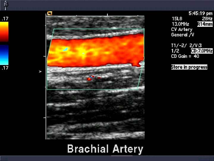
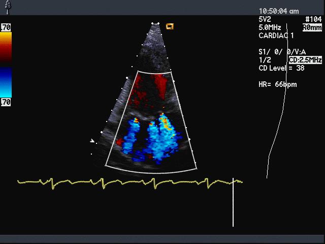
100 Real-Time Image Formation
101 Scan Conversion Acquired data may not be on the display grid. Acquired grid Display grid
102 Scan Conversion sinθ x R y acquired converted
103 Scan Conversion a(i,j) a(i,j+1) p a(i+1,j) a(i+1,j+1) original grid raster grid acquired data display pixel p( m, n) = c c m, n, i+ 1, j+ 1 m, n, i, j a( i, j) + c a( i + 1, j + 1) m, n, i+ 1, j a( i + 1, j) + c m, n, i, j+ 1 a( i, j + 1) +

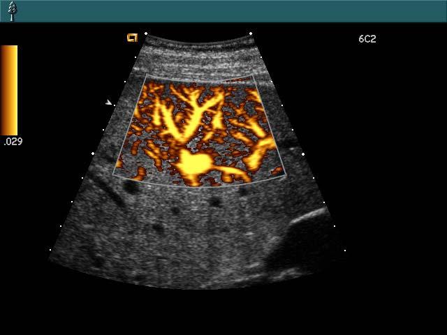
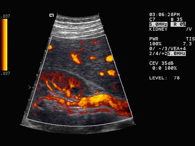
104 Moiré Pattern
105 Temporal Resolution (Frame Rate) Frame rate=1/frame time. Frame time=number of lines * line time. Line time=(2*maximum depth)/sound velocity. Sound velocity is around 1540 m/s. High frame rate is required for real-time imaging.
106 Temporal Resolution Display standard: NTSC: 30 Hz. PAL: 25 Hz (2:1 interlace). 24 Hz for movie. The actual acoustic frame rate may be higher or lower. But should be high enough to have minimal flickering. Essence of real-time imaging: direct interaction.
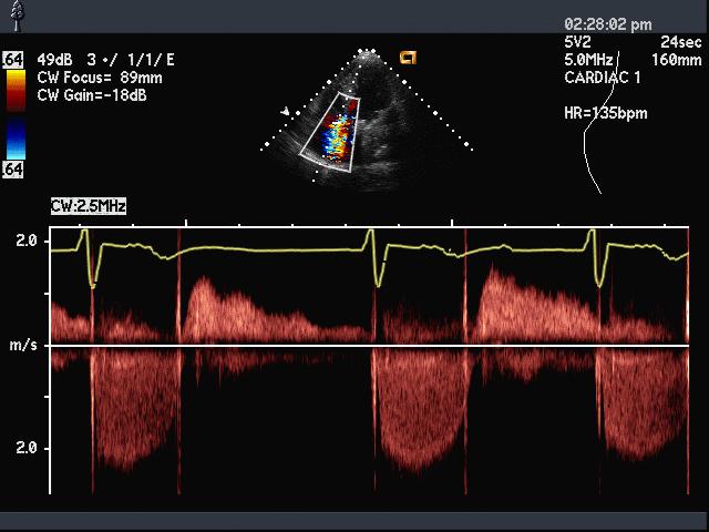
107 Temporal Resolution For an actual frame rate lower than 30 Hz, interpolation is used. For an actual frame rate higher than 30 Hz, information can be displayed during playback. Even at 30 Hz, it is still possibly undersampling.
108 Doppler Techniques for Motion Estimation
109 Color Doppler Mode
110
111
112
113 Power Doppler
114
115
116 PW Doppler (Spectral Doppler)
117
118 CW Doppler (Spectral Doppler)
119
120 Doppler Effect
121 Doppler Principles receiver source receiver Relative motion of the source causes a change in received frequency. Blood flow velocity is measured by detecting Doppler frequency shifts.
122 Doppler Equations where f c v s s is is and f is f f d d = f f s s vr + v c v ( vr + v c the carrier frequency, the sound velocity in blood, v d r s s the Doppler frequency shift, s ) are source and receiver velocities.
123 Doppler Ultrasound Primary scattering site: red blood cell. The platelet is too small and the number of leukocytes is not significant. The red blood cell size is around several microns. Thus, scattering and speckle are also present. The red blood cells in a sample volume are assumed to move in unison.
124 Doppler Equations f d = 2vf c s cos θ v c Typical physiological flows (5-10m/sec at most) are much slower than sound velocity in the body (~1500m/sec). Doppler shift is doubled due to round-trip propagation. Only parallel flows can be detected. θ
125 Flow Pattern vs. Velocity Profile ultrasound beam 0 velocity or Doppler shift
126 Flow Pattern vs. Velocity Profile ultrasound beam 0 velocity or Doppler shift
127 Flow Pattern vs. Velocity Profile ultrasound beam 0 velocity or Doppler shift
128 Doppler Spectrum Estimation original Doppler shifted demodulated f s f s +f d f d Short-time Fourier transform (Spectral Doppler). Correlation based estimation (Color Doppler).

129 Continuous Wave (CW) Doppler T R time time velocity
130 CW Doppler CW oscillator filter spectral estimation CW transmitter demodulator amplifier audio conversion signal processing T R speaker display
131 CW Doppler Array CW and AUX CW (half transmit, half receive). Mainly for Cardiology. Good velocity (frequency) resolution. No range resolution. Flows along the same direction are all detected. Frequency downshift due to attenuation can be ignored.
132 CW Doppler Processing original spectrum -f 0 -f d -f 0 f 0 f 0 +f d demodulated -2f 0 -f d f d demodulated and filtered f d
133 CW Doppler Processing Time-interval histogram ppt FFT. Mode-based spectrum estimation (AR), timefrequency analysis. Magnitudes are converted in db and displayed. Post-processing similar to B-mode.
134 Audio Doppler For typical blood velocities and carrier frequencies, the Doppler shifts from blood happen to be in the human audible range (near DC to 20KHz). Positive shifts in one channel and negative ones in the other. Hilbert transform. Right Clinically useful. Left

135 PW Blood Flow Measurements 取自
136 PW System Diagram CW oscillator W transmitter (gated CW) filter sample&hold demod./lpf gating audio conversion spectral estimation signal processing transducer amplifier speaker display

137 Pulsed Wave (PW) Doppler time PRI
138 Velocity Ambiguity 2f max 1 PRI 2f no aliasing max 4vmaxfs 1 = c PRI v max λ 4 PRI aliasing v max -v max v max
139
140 Range Ambiguity c PRI / 2 PRI T 0 c T 0 / 2 c ( PRI + T ) 0 OR? / 2
141 Pulse Wave (PW) Doppler Pulse-echo method, similar to B-mode. Post-processing similar to CW. Adjustable range resolution (gate). Maximum detectable velocity is λ/(4*pri). Maximum depth is (c*pri)/ point FFT.
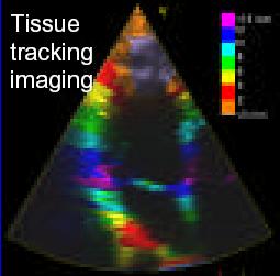
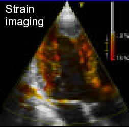
142 Color Doppler multiple gates ultiple firings
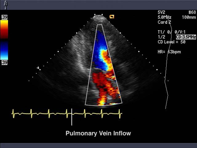
143 Color Doppler Similar to B-mode, except that each line is fired multiple times (5-15). Correlation processing. Multiple range gates along each line. Real-time two-dimensional flow imaging. Poor velocity (frequency) resolution.
144 Color Doppler Use efficient time domain correlation techniques to calculate flow characteristics. Auto-correlation of the Doppler signal. Commonly derived parameters are mean velocity (including directionality), variance and energy (power). f
145 Color Doppler: Mean Velocity R( t ) S ( t + τ) S ( τ) dτ j t R( t ) ω P ( ω) e dω ω = = ωp ( ω) dω P ( ω) dω ω = θ ( 0 ) θ( T ) θ( 0) θ( T ) = T T
146 Color Doppler: Variance 2 σ = 2 ( ω ω ) P ( ω) dω P ( ω) dω = ω ω AT ( ) A ( 0 ) σ = 2 T T 2 1 R( T ) R ( 0)
147 Color Doppler: Energy E = P( ω) dω = R(0) f
148 Color Doppler Flow parameters are mapped into colors for display (1D or 2D). Choice of map affects the presentation of Color Doppler images.
149 Color Doppler: Signal Processing beam former high pass filter autocorrelator parameter estimator signal and display processor Significant frame rate reduction. Small color boxes are often used to increase frame rate. Sophisticated systems utilize multiple beam formation to further increase frame rate.
150 Doppler: Complications Non-trivial wall filters are required to remove interference from slow-moving objects. Adequate signal processing capabilities and sufficient dynamic range are necessary to detect weak flows. Conflicts with frame rate requirements. Only parallel flow is detectable.
151 Doppler: Tissue Motion Imaging Doppler principles can be used to visualize cardiac motion. Higher signal levels allow simpler wall filters and less number of firing. Suitable for cardiac applications.
152 Doppler Tissue Imaging Heart motion parameters: Velocity: v=dw/dt. Displacement w: temporal integration of v. Strain rate: r=dv/dz. Strain s: temporal integration of r.


153 Doppler Tissue Imaging Integration in time Derivation in space Integration in time


154 Doppler Tissue Imaging
155 Ultrasonic Nonlinear Imaging- Tissue Harmonic Imaging
156 Sound Velocity and Density Change B ( x ) = c0 + (1 + ) u( x) 2A Phase velocity Nonlinearity Particle velocity Finite Amplitude Distortion
157 Non-linear Parameter B/A ( ) ( ) L = = = 2 0 ; ; ρ ρ ρ ρ ρ ρ ρ ρ ρ ρ s s P P P P = ρ ρ ρ ρ ρ ρ B A P P B/A defines non-linearity of the medium. The larger the B/A, the higher the nonlinear response.
158 B/A Parameters: Typical Values Typical values: Water:5.5+/-0.3. Liver: Fat: Muscle: 7.5. B/A imaging may be used for tissue characterization.
159 Tissue Non-linearity Tissue harmonics are virtually zero at the probe face.the intensity continues to increase until attenuation dominates. The higher the intensity is, the more tissue harmonics are generated. Such a mechanism automatically increase the difference between signal and acoustic noise.


160 Comparison of Radiation Patterns Beam Patterns transducer transducer db f 0 f 0 Lateral Position (mm)
161 What If We Use the Second Harmonic Signal for Imaging?
162 Advantages of Tissue Harmonic Imaging Low sidelobes. Better spatial resolution compared to fundamental imaging at the original frequency. Less affected by tissue inhomogeneities better performance on technically difficult bodies.
163 Tissue Harmonic Imaging Performance of ultrasound has been sub-optimal on technically difficult bodies. Most recent new developments have bigger impact on technically satisfactory bodies. Poor image quality leads to uncertainty in diagnosis and costly repeat examinations. Tissue harmonic imaging has been successful on difficult bodies.


164 Reduction of Imaging Artifacts


165 Reduction of Imaging Artifacts
Doppler echocardiography & Magnetic Resonance Imaging. Doppler echocardiography. History: - Langevin developed sonar.
 1 Doppler echocardiography & Magnetic Resonance Imaging History: - Langevin developed sonar. - 1940s development of pulse-echo. - 1950s development of mode A and B. - 1957 development of continuous wave
Lesson 05: Intensity Measurements and Bioeffects. This lesson contains 18 slides plus 6 multiple-choice questions.
 Lesson 05: Intensity Measurements and Bioeffects This lesson contains 18 slides plus 6 multiple-choice questions. This lesson was derived from pages 22 through 24 in the textbook: Intensity Measurements
31545 Medical Imaging systems
 31545 Medical Imaging systems Lecture 2: Ultrasound physics Jørgen Arendt Jensen Department of Electrical Engineering (DTU Elektro) Biomedical Engineering Group Technical University of Denmark September
Outline of today Medical Imaging systems. Wave types. 1. Discussion assignment on B-mode imaging
 Outline of today 3545 Medical Imaging systems. Discussion assignment on B-mode imaging Lecture : Ultrasound physics. Derivation of wave equation and role of speed of sound Jørgen Arendt Jensen Department
Basic System. Basic System. Sonosite 180. Acuson Sequoia. Echo occurs at t=2z/c where c is approximately 1500 m/s or 1.5 mm/µs
 Bioengineering 280A Principles of Biomedical Imaging Fall Quarter 2007 Ultrasound Lecture Sonosite 80 From Suetens 2002 Acuson Sequoia Basic System Basic System Echo occurs at t=2/c where c is approximately
Workshop 2: Acoustic Output Measurements
 37 th th UIA Symposium, Washington DC Workshop 2: Acoustic Output Measurements Mark Hodnett Senior Research Scientist Quality of Life Division National Physical Laboratory Teddington Middlesex, UK Workshop
Bioengineering 280A Principles of Biomedical Imaging. Fall Quarter 2006 Ultrasound Lecture 1
 Bioengineering 280A Principles of Biomedical Imaging Fall Quarter 2006 Ultrasound Lecture 1 From Suetens 2002 1 Basic System Echo occurs at t=2z/c where c is approximately 1500 m/s or 1.5 mm/µs Macovski
Physical principles of Harmonic Imaging Min Joo Choi, PhD
 Physical principles of Harmonic Imaging Min Joo Choi, PhD Department Biomedical Engineering College of Medicine, Cheju National University School of Medicine, King s College of London, University of London
EE 5345 Biomedical Instrumentation Lecture 6: slides
 EE 5345 Biomedical Instrumentation Lecture 6: slides 129-147 Carlos E. Davila, Electrical Engineering Dept. Southern Methodist University slides can be viewed at: http:// www.seas.smu.edu/~cd/ee5345.html
31545 Medical Imaging systems
 Simulation of ultrasound systems and non-linear imaging 545 Medical Imaging systems Lecture 9: Simulation of ultrasound systems and non-linear imaging Jørgen Arendt Jensen Department of Electrical Engineering
The Physics of Doppler Ultrasound. HET408 Medical Imaging
 The Physics of Doppler Ultrasound HET408 Medical Imaging 1 The Doppler Principle The basis of Doppler ultrasonography is the fact that reflected/scattered ultrasonic waves from a moving interface will
EL-GY 6813/BE-GY 6203 Medical Imaging, Fall 2016 Final Exam
 EL-GY 6813/BE-GY 6203 Medical Imaging, Fall 2016 Final Exam (closed book, 1 sheets of notes double sided allowed, no calculator or other electronic devices allowed) 1. Ultrasound Physics (15 pt) A) (9
Pixel-based Beamforming for Ultrasound Imaging
 Pixel-based Beamforming for Ultrasound Imaging Richard W. Prager and Nghia Q. Nguyen Department of Engineering Outline v Introduction of Ultrasound Imaging v Image Formation and Beamforming v New Time-delay
Angular Spectrum Decomposition Analysis of Second Harmonic Ultrasound Propagation and its Relation to Tissue Harmonic Imaging
 The 4 th International Workshop on Ultrasonic and Advanced Methods for Nondestructive Testing and Material Characterization, June 9, 006 at ABSTRACT Angular Spectrum Decomposition Analysis of Second Harmonic
Technical University of Denmark
 Technical University of Denmark Page 1 of 11 pages Written test, 9 December 2010 Course name: Introduction to medical imaging Course no. 31540 Aids allowed: none. "Weighting": All problems weight equally.
PD233: Design of Biomedical Devices and Systems
 PD233: Design of Biomedical Devices and Systems Lecture-9 Medical Diagnostic Imaging Ultrasound Dr. Manish Arora CPDM, IISc Course Website: http://cpdm.iisc.ac.in/utsaah/courses/ Ultrasound Physics Acoustic
Structure of Biological Materials
 ELEC ENG 3BA3: Structure of Biological Materials Notes for Lecture #19 Monday, November 22, 2010 6.5 Nuclear medicine imaging Nuclear imaging produces images of the distribution of radiopharmaceuticals
EXEMPLARY PROBLEMS APPENDIX B CHAPTER 1
 APPENDIX B EXEMPLARY PROBLEMS CHAPTER 1 1.1 A two - dimensional planar acoustic wave is defined by the function U = A e j ( ω t kx x ky y ). This wave impinges upon a perfect reflecting line that is parallel
Supplement (videos)
") Supplement (videos) Ruben s tube (sound): http://www.youtube.com/watch?v=gpcquuwqayw Doppler US (diagnostic use): http://www.youtube.com/watch?v=fgxzg-j_hfw http://www.youtube.com/watch?v=upsmenyoju8 High
Supplement (videos) Ruben s tube (sound): http://www.youtube.com/watch?v=gpcquuwqayw Doppler US (diagnostic use): http://www.youtube.com/watch?v=fgxzg-j_hfw http://www.youtube.com/watch?v=upsmenyoju8 High
Ultrasonic Measurement of Minute Displacement of Object Cyclically Actuated by Acoustic Radiation Force
 Jpn. J. Appl. Phys. Vol. 42 (2003) pp. 4608 4612 Part 1, No. 7A, July 2003 #2003 The Japan Society of Applied Physics Ultrasonic Measurement of Minute Displacement of Object Cyclically Actuated by Acoustic
Sound wave bends as it hits an interface at an oblique angle. 4. Reflection. Sound wave bounces back to probe
 : Ultrasound imaging and x-rays 1. How does ultrasound imaging work?. What is ionizing electromagnetic radiation? Definition of ionizing radiation 3. How are x-rays produced? Bremsstrahlung Auger electron
Physics and Knobology
 Physics and Knobology Cameron Jones, MD 02/01/2012 SOUND: Series of pressure waves traveling through a medium What is ULTRASOUND Physics Words WAVELENGTH: Distance traveled in one cycle FREQUENCY: number
High Intensity Focused Ultrasound Test & Measurement Techniques
 High Intensity Focused Ultrasound Test & Measurement Techniques Shmuel Ben-Ezra June 2011 What's HIFU??? 2 What's HIFU??? High Intensity Focused Ultrasound Plays an increasing role in medical-therapeutic
N db compared with that in the single pulse harmonic imaging mode, whereas
 26 at UMass Dartmouth, N. Dartmouth, MA Proceedings published in www.ndt.net Acoustic nonlinear imaging and its application in tissue characterization * Dong Zhang and Xiu-fen Gong Lab. of Modern Acoustics,
Mandatory Assignment 2013 INF-GEO4310
 Mandatory Assignment 2013 INF-GEO4310 Deadline for submission: 12-Nov-2013 e-mail the answers in one pdf file to vikashp@ifi.uio.no Part I: Multiple choice questions Multiple choice geometrical optics
Output intensity measurement on a diagnostic ultrasound machine using a calibrated thermoacoustic sensor
 Institute of Physics Publishing Journal of Physics: Conference Series 1 (2004) 140 145 doi:10.1088/1742-6596/1/1/032 Advanced Metrology for Ultrasound in Medicine Output intensity measurement on a diagnostic
Chapter 12 Sound in Medicine
 Infrasound < 0 Hz Earthquake, atmospheric pressure changes, blower in ventilator Not audible Headaches and physiological disturbances Sound 0 ~ 0,000 Hz Audible Ultrasound > 0 khz Not audible Medical imaging,
ISUOG Basic Training The Principles of Doppler Ultrasound. Juriy Wladimiroff
 ISUOG Basic Training The Principles of Doppler Ultrasound Juriy Wladimiroff Learning objectives At the end of this session, you will be able to understand the principles of: Doppler effect Doppler shift
ULTRASOUND. Ultrasound physical phenomenon properties basics of medical applications, History. History
 Ultrasound physical phenomenon properties basics of medical applications, ULTRASOUND History History Dr. Leopold Auenbrugger 76 - medical doctor first suggests the method of percussion in diagnostics Dr.
IEEE Ultrasonic symposium 2002
 IEEE Ultrasonic symposium 2002 Short Course 6: Flow Measurements Hans Torp Department of Circulation and Medical Imaging TU, orway Internet-site for short course: http://www.ifbt.ntnu.no/~hanst/flowmeas02/index.html
Chapter 2. Interaction with Soft Tissue
 Chapter 2 Interaction with Soft Tissue Ultrasound interacts with human soft tissue in several predictable ways that allow engineers to design instruments that provide diagnostic information that forms
Medical Imaging Physics Spring Quarter Week 3-2
 Medical Imaging Physics Spring Quarter Week 3-2 Ultrasound Daor Balzar balzar@du.edu www.du.edu/~balzar Outline Ultrasound Light, Eyes and Vision Reading assignment: CSG 12; D 15 Homework D 12: 5,6 and
DOPPLER-BASED ULTRASONIC BLOOD VELOCITY ESTIMATION
 DOPPLER-BASED ULTRASONIC BLOOD VELOCITY ESTIMATION BY TOROS ARIKAN THESIS Submitted in partial fulfillment of the requirements for the degree of Bachelor of Science in Electrical and Computer Engineering
Today s menu. Last lecture. Measurement of volume flow rate. Measurement of volume flow rate (cont d...) Differential pressure flow meters
 Differential pressure flow meters") Last lecture Analog-to-digital conversion (Ch. 1.1). Introduction to flow measurement systems (Ch. 12.1). Today s menu Measurement of volume flow rate Differential pressure flowmeters Mechanical flowmeters
Last lecture Analog-to-digital conversion (Ch. 1.1). Introduction to flow measurement systems (Ch. 12.1). Today s menu Measurement of volume flow rate Differential pressure flowmeters Mechanical flowmeters
ECMUS The Safety Committee of EFSUMB : Tutorial
 Doppler ultrasound devices Safety Aspects (update 2011) Basic Terminology Amplitude coded colour Doppler special colour Doppler technique which characterises the Doppler signal echo strength, also called
DEVIL PHYSICS THE BADDEST CLASS ON CAMPUS IB PHYSICS
 DEVIL PHYSICS THE BADDEST CLASS ON CAMPUS IB PHYSICS TSOKOS OPTION I-2 MEDICAL IMAGING Reading Activity Answers IB Assessment Statements Option I-2, Medical Imaging: X-Rays I.2.1. I.2.2. I.2.3. Define
The physics US and MRI. Prof. Peter Bogner
 The physics US and MRI Prof. Peter Bogner Sound waves mechanical disturbance, a pressure wave moves along longitudinal wave compression rarefaction zones c = nl, (c: velocity, n: frequency, l: wavelength
ISUOG Basic Training Physical Principles of Ultrasound including Safety
 ISUOG Physical Principles of Ultrasound including Safety Goals To understand Physics of ultrasound How is a b-mode image is generated The effects of ultrasound on human tissue To know Settings of your
Signal Loss. A1 A L[Neper] = ln or L[dB] = 20log 1. Proportional loss of signal amplitude with increasing propagation distance: = α d
![Signal Loss. A1 A L[Neper] = ln or L[dB] = 20log 1. Proportional loss of signal amplitude with increasing propagation distance: = α d](/thumbs/89/98517557.jpg "Signal Loss. A1 A L[Neper] = ln or L[dB] = 20log 1. Proportional loss of signal amplitude with increasing propagation distance: = α d") Part 6 ATTENUATION Signal Loss Loss of signal amplitude: A1 A L[Neper] = ln or L[dB] = 0log 1 A A A 1 is the amplitude without loss A is the amplitude with loss Proportional loss of signal amplitude with
Part 6 ATTENUATION Signal Loss Loss of signal amplitude: A1 A L[Neper] = ln or L[dB] = 0log 1 A A A 1 is the amplitude without loss A is the amplitude with loss Proportional loss of signal amplitude with
A Brief Introduction to Medical Imaging. Outline
 A Brief Introduction to Medical Imaging Outline General Goals Linear Imaging Systems An Example, The Pin Hole Camera Radiations and Their Interactions with Matter Coherent vs. Incoherent Imaging Length
Microwave-induced thermoacoustic tomography using multi-sector scanning
 Microwave-induced thermoacoustic tomography using multi-sector scanning Minghua Xu, Geng Ku, and Lihong V. Wang a) Optical Imaging Laboratory, Biomedical Engineering Program, Texas A&M University, 3120
3. Which of the following statements is (are) TRUE about detector crystals in Anger cameras?
 TRUE about detector crystals in Anger cameras?") BioE 1330 - Exam 2 11/13/2018 Answer Sheet - Correct answer is A for all questions 1. Unlike CT, in nuclear medicine A. Bremsstrahlung is not used to produce high-energy photons. B. signal can be increased
BioE 1330 - Exam 2 11/13/2018 Answer Sheet - Correct answer is A for all questions 1. Unlike CT, in nuclear medicine A. Bremsstrahlung is not used to produce high-energy photons. B. signal can be increased
Chapter 1. The Nature of Sound. The Nature of Sound
 Chapter 1 The Nature of Sound Waves carry energy, not matter, from one place to another. Sound waves carry energy as packets of particle compressions. Much like a spring that is compressed, energy is stored
Donald School Journal of Ultrasound Physical Bases in Obstetrics of Medical and Ultrasound Gynecology, April-June 2009;3(2):1-9
:1-9") Donald School Journal of Ultrasound Physical Bases in Obstetrics of Medical and Ultrasound Gynecology, April-June 2009;3(2):1-9 Physical Bases of Medical Ultrasound Jasminka Brnjas-Kraljevic Department
Donald School Journal of Ultrasound Physical Bases in Obstetrics of Medical and Ultrasound Gynecology, April-June 2009;3(2):1-9 Physical Bases of Medical Ultrasound Jasminka Brnjas-Kraljevic Department
I have nothing to disclose
 Critical Ultrasound for Patient Care April 6-8, 2016 Sonoma, CA Critical Ultrasound for Patient Care I have nothing to disclose April 6-8, 2016 Sonoma, CA UC SF University of California San Francisco UC
Theoretical questions for the final exam ED 2012.
 Theoretical questions for the final exam ED 2012. 1. Radiation a) Properties and types of radiation b) Physical parameters of radiation 2. Law of attenuation of radiation a) Experimental interpretation
Final exam questions ED
 Final exam questions ED 2015-2016 1. Radiation a) Properties and types of radiation b) Physical parameters of radiation 2. Law of attenuation of radiation a) Experimental interpretation of the law b) Forms
SCATTERING OF ULTRASONIC WAVE ON A MODEL OF THE ARTERY J. WÓJCIK, T. POWAŁOWSKI, R. TYMKIEWICZ A. LAMERS, Z. TRAWIŃSKI
 ARCHIVES OF ACOUSTICS 31, 4, 471 479 (2006) SCATTERING OF ULTRASONIC WAVE ON A MODEL OF THE ARTERY J. WÓJCIK, T. POWAŁOWSKI, R. TYMKIEWICZ A. LAMERS, Z. TRAWIŃSKI Institute of Fundamental Technological
Today s menu. Last lecture. Ultrasonic measurement systems. What is Ultrasound (cont d...)? What is ultrasound?
? What is ultrasound?") Last lecture Measurement of volume flow rate Differential pressure flowmeters Mechanical flowmeters Vortex flowmeters Measurement of mass flow Measurement of tricky flows" Today s menu Ultrasonic measurement
Last lecture Measurement of volume flow rate Differential pressure flowmeters Mechanical flowmeters Vortex flowmeters Measurement of mass flow Measurement of tricky flows" Today s menu Ultrasonic measurement
Simulation of Contrast Agent Enhanced Ultrasound Imaging based on Field II
 Simulation of Contrast Agent Enhanced Ultrasound Imaging based on Field II Tobias Gehrke, Heinrich M. Overhoff Medical Engineering Laboratory, University of Applied Sciences Gelsenkirchen tobias.gehrke@fh-gelsenkirchen.de
Session: P3B MEDICAL IMAGING Chair: N. de Jong Erasmus Medical Centre P3B-1
 using ultrasound data to test the performance of this algorithm and compare it to currently accepted delay estimators implementing a variety of sub-sample interpolation methods. Simulation results show
NUMERICAL METHODS FOR NONLINEAR WAVE PROPAGATION IN ULTRASOUND
 NUMERICAL METHODS FOR NONLINEAR WAVE PROPAGATION IN ULTRASOUND by Gianmarco F. Pinton Department of Biomedical Engineering Duke University Date: Approved: Gregg E. Trahey, Ph.D., Supervisor Kathryn R.
(INCLUDING THIS FRONT PAGE)
") I'IFIITIIBIFI UNIVERSITY OF SCIEI'ICE RITD TECHNOLOGY FACULTY OF HEALTH AND APPLIED SCIENCES DEPARTMENT OF NATURAL AND APPLIED SCIENCES QUALIFICATION: BACHELOR OF SCIENCE (MAJOR AND MINOR) QUALIFICATION
I'IFIITIIBIFI UNIVERSITY OF SCIEI'ICE RITD TECHNOLOGY FACULTY OF HEALTH AND APPLIED SCIENCES DEPARTMENT OF NATURAL AND APPLIED SCIENCES QUALIFICATION: BACHELOR OF SCIENCE (MAJOR AND MINOR) QUALIFICATION
BNG/ECE 487 FINAL (W16)
") BNG/ECE 487 FINAL (W16) NAME: 4 Problems for 100 pts This exam is closed-everything (no notes, books, etc.). Calculators are permitted. Possibly useful formulas and tables are provided on this page. Fourier
BNG/ECE 487 FINAL (W16) NAME: 4 Problems for 100 pts This exam is closed-everything (no notes, books, etc.). Calculators are permitted. Possibly useful formulas and tables are provided on this page. Fourier
Take-Home Final Exam Last Possible Due Date: Dec. 21, 2004, 5 p.m.
 Take-Home Final Exam Last Possible Due Date: Dec 1, 004, 5 pm Your solutions to the exam should be handed in to the instructor (BIRB 1088) or to Eve Gochis, the MRI lab administrator (BIRB 107) no later
Today s menu. Last lecture. A/D conversion. A/D conversion (cont d...) Sampling
 Sampling") Last lecture Capacitive sensing elements. Inductive sensing elements. Reactive Deflection bridges. Electromagnetic sensing elements. Thermoelectric sensing elements. Elastic sensing elements. Piezoelectric
Last lecture Capacitive sensing elements. Inductive sensing elements. Reactive Deflection bridges. Electromagnetic sensing elements. Thermoelectric sensing elements. Elastic sensing elements. Piezoelectric
Introduction to Medical Imaging. Medical Imaging
 Introduction to Medical Imaging BME/EECS 516 Douglas C. Noll Medical Imaging Non-invasive visualization of internal organs, tissue, etc. I typically don t include endoscopy as an imaging modality Image
Nanoscale Energy Conversion and Information Processing Devices - NanoNice - Photoacoustic response in mesoscopic systems
 Nanoscale Energy Conversion and Information Processing Devices - NanoNice - Photoacoustic response in mesoscopic systems Photonics group W. Claeys, S. Dilhair, S. Grauby, JM. Rampnoux, L. Patino Lopez,
DETERMINATION OF THE B/A OF BIOLOGICAL MEDIA BY MEASURING AND MODELING NONLINEAR DISTORTION OF PULSED ACOUSTIC WAVE IN TWO-LAYER MEDIA TAMARA KUJAWSKA
 DETERMINATION OF THE B/A OF BIOLOGICAL MEDIA BY MEASURING AND MODELING NONLINEAR DISTORTION OF PULSED ACOUSTIC WAVE IN TWO-LAYER MEDIA TAMARA KUJAWSKA Institute of Fundamental Technological Research Polish
Shear waves in solid-state materials
 Shear waves in solid-state materials TEAS Related topics Ultrasonic transmission measurement, propagation of ultrasound waves, ultrasound wave modes, shear waves, longitudinal and transverse waves, modulus
glass Calculate the magnitude of the Young modulus for glass. State your answer to (a) in terms of SI fundamental units.
 in terms of SI fundamental units.") Q1.The term ultrasound refers to vibrations in a material that occur at frequencies too high to be detected by a human ear. When ultrasound waves move through a solid, both longitudinal and transverse
Q1.The term ultrasound refers to vibrations in a material that occur at frequencies too high to be detected by a human ear. When ultrasound waves move through a solid, both longitudinal and transverse
Medical Biophysics II. Final exam theoretical questions 2013.
 Medical Biophysics II. Final exam theoretical questions 2013. 1. Early atomic models. Rutherford-experiment. Franck-Hertz experiment. Bohr model of atom. 2. Quantum mechanical atomic model. Quantum numbers.
Investigation of Transverse Oscillation Method
 Downloaded from orbit.dtu.dk on: Nov 01, 2018 Investigation of Transverse Oscillation Method Udesen, Jesper; Jensen, Jørgen Arendt Published in: I E E E Transactions on Ultrasonics, Ferroelectrics and
University of Cyprus. Reflectance and Diffuse Spectroscopy
 University of Cyprus Biomedical Imaging and Applied Optics Reflectance and Diffuse Spectroscopy Spectroscopy What is it? from the Greek: spectro = color + scope = look at or observe = measuring/recording
Tissue harmonic ultrasonic imaging
 C. R. Acad. Sci. Paris, t. 2, Série IV, p. 1139 1151, 2001 Physique appliquée/applied physics (Biophysique/Biophysics) IMAGERIE ACOUSTIQUE ET OPTIQUE DES MILIEUX BIOLOGIQUES Michalakis A. AVERKIOU ATL
INF5410 Array signal processing. Ch. 3: Apertures and Arrays
 INF5410 Array signal processing. Ch. 3: Apertures and Arrays Endrias G. Asgedom Department of Informatics, University of Oslo February 2012 Outline Finite Continuous Apetrures Apertures and Arrays Aperture
Digital Beamforming in Ultrasound Imaging
 Digital Beamforming in Ultrasound Imaging Sverre Holm Vingmed Sound AS, Research Department, Vollsveien 3C, N-34 Lysaker, Norway, Department of Informatics, University of Oslo, Norway ABSTRACT In medical
Biomedical Instrumentation System
 BME/EECS 458 - Biomedical Instrumentation & Design Matt O Donnell I.0 Introduction What is a biomedical instrument? To many it s an EKG machine, to others it s a chemical biosensor, and to some it s a
PROPERTY STUDY ON EMATS WITH VISUALIZATION OF ULTRASONIC PROPAGATION
 More Info at Open Access Database www.ndt.net/?id=18576 PROPERTY STUDY ON EMATS WITH VISUALIZATION OF ULTRASONIC PROPAGATION T. Yamamoto, T. Furukawa, I. Komura Japan Power Engineering and Inspection Corporation,
General Physics (PHY 2130)
") General Physics (PHY 2130) Lecture XII Sound sound waves Doppler effect Standing waves Light Reflection and refraction Lightning Review Last lecture: 1. Vibration and waves Hooke s law Potential energy
General Physics (PHY 2130) Lecture XII Sound sound waves Doppler effect Standing waves Light Reflection and refraction Lightning Review Last lecture: 1. Vibration and waves Hooke s law Potential energy
SUBSURFACE WAVES IN SOLIDS WITH CURVED SURFACE AND ACOUSTICAL IMPEDANCE ON IT
 SUBSURFACE WAVES IN SOLIDS WITH CURVED SURFACE AND ACOUSTICAL IMPEDANCE ON IT A. Baev, P. Prokhorenko, and M. Asadchaya Institute of Applied Physics, Minsk, Belarus Abstract: This paper presents the results
Department of Engineering Science. Robin Cleveland
 Department of Engineering Science Robin Cleveland robin.cleveland@eng.ox.ac.uk 1 Engineering Science What is Engineering? What is Engineering Science at Oxford? Biomedical Ultrasound Applications 2 Scientists
A Comparative Evaluation of Four Acoustic Hydrophones in High Intensity Focused Ultrasound (HIFU) Field Measurements
 Field Measurements") A Comparative Evaluation of Four Acoustic Hydrophones in High Intensity Focused Ultrasound (HIFU) Field Measurements Yunbo Liu and Keith Wear Yunbo.Liu@fda.hhs.gov Food and Drug Administration, Silver
A Comparative Evaluation of Four Acoustic Hydrophones in High Intensity Focused Ultrasound (HIFU) Field Measurements Yunbo Liu and Keith Wear Yunbo.Liu@fda.hhs.gov Food and Drug Administration, Silver
General Physics (PHY 2130)
") General Physics (PHY 2130) Lecture XII Sound sound waves Doppler effect Standing waves Light Reflection and refraction http://www.physics.wayne.edu/~apetrov/phy2130/ Lightning Review Last lecture: 1. Vibration
General Physics (PHY 2130) Lecture XII Sound sound waves Doppler effect Standing waves Light Reflection and refraction http://www.physics.wayne.edu/~apetrov/phy2130/ Lightning Review Last lecture: 1. Vibration
MEDT8007 Simulation methods in ultrasound imaging
 1 MEDT8007 Simulation methods in ultrasound imaging Dynamic objects Lasse Løvstakken Department of Circulation and Medical Imaging Norwegian University of Science and Technology 2 Lecture outline Simple
Apodization. Gibbs Artifact. Bioengineering 280A Principles of Biomedical Imaging. Fall Quarter 2013 MRI Lecture 5. rect(k x )
") Bioengineering 280A Principles of Biomedical Imaging Fall Quarter 2013 MRI Lecture 5 GE Medical Systems 2003 Gibbs Artifact Apodization rect(k ) Hanning Window h(k )=1/2(1+cos(2πk ) 256256 image 256128
Bioengineering 280A Principles of Biomedical Imaging Fall Quarter 2013 MRI Lecture 5 GE Medical Systems 2003 Gibbs Artifact Apodization rect(k ) Hanning Window h(k )=1/2(1+cos(2πk ) 256256 image 256128
Piezoelectric Materials and Devices
 Piezoelectric Materials and Devices Applications in Engineering and Medical Sciences M. S. VIJAYA CRC Press Taylor & Francis Croup Boca Raton London NewYork CRC Press is an imprint of the Taylor & Francis
PHASED-ARRAY FOCUSING WITH LONGITUDINAL GUIDED WAVES IN A VISCOELASTIC COATED HOLLOW CYLINDER
 PHASED-ARRAY FOCUSING WITH LONGITUDINAL GUIDED WAVES IN A VISCOELASTIC COATED HOLLOW CYLINDER W. Luo, J. L. Rose, J. K. Van Velsor, J. Mu Department of Engineering Science & Mechanics, The Pennsylvania
UNIVERSITY OF SOUTHAMPTON
 UNIVERSITY OF SOUTHAMPTON PHYS2023W1 SEMESTER 1 EXAMINATION 2016-2017 WAVE PHYSICS Duration: 120 MINS (2 hours) This paper contains 9 questions. Answers to Section A and Section B must be in separate answer
CHAPTER 1 INTRODUCTION TO ACOUSTIC SATURATION
 CHAPTER 1 INTRODUCTION TO ACOUSTIC SATURATION Acoustic saturation states that as the input voltage to a focused ultrasonic transducer increases, there exists a limit of the acoustic pressure at the transducer
Dept Electrical Engineering Chang Gung University, Taiwan
 Biomedical Optics Hsiao-Lung Chan, Ph.D. Dept Electrical Engineering Chang Gung University, Taiwan chanhl@mail.cgu.edu.tw Outline Essential optical principle Light propagation in biological tissue Blood
APPLICATION OF THE KHOKHLOV-ZABOLOTSKAYA-KUZNETSOV EQUATION TO MODELING HIGH-INTENSITY FOCUSED ULTRASOUND BEAMS
 Boston University OpenBU Mechanical Engineering http://open.bu.edu ENG: Mechanical Engineering: Theses & Dissertations 2010 APPLICATION OF THE KHOKHLOV-ZABOLOTSKAYA-KUZNETSOV EQUATION TO MODELING HIGH-INTENSITY
Spectral Velocity Estimation in the Transverse Direction
 Paper presented at the IEEE International Ultrasonics Symposium, Prague, Czech Republic, 3: Spectral Velocity Estimation in the Transverse Direction Jørgen Arendt Jensen Center for Fast Ultrasound Imaging,
EFIT SIMULATIONS FOR ULTRASONIC NDE
 EFIT SIMULATIONS FOR ULTRASONIC NDE René Marklein, Karl-Jörg Langenberg, Klaus Mayer (University of Kassel, Department of Electrical and Computer Engineering, Electromagnetic Field Theory, Wilhelmshöher
A small object is placed a distance 2.0 cm from a thin convex lens. The focal length of the lens is 5.0 cm.
 TC [66 marks] This question is about a converging (convex) lens. A small object is placed a distance 2.0 cm from a thin convex lens. The focal length of the lens is 5.0 cm. (i) Deduce the magnification
Technical University of Denmark
 Technical University of Denmark Page 1 of 10 pages Written test, 12 December 2012 Course name: Introduction to medical imaging Course no. 31540 Aids allowed: None. Pocket calculator not allowed "Weighting":
Phononic Crystals. J.H. Page
 Phononic Crystals J.H. Page University of Manitoba with Suxia Yang and M.L. Cowan at U of M, Ping Sheng and C.T. Chan at HKUST, & Zhengyou Liu at Wuhan University. We study ultrasonic waves in complex
K-space. Spin-Warp Pulse Sequence. At each point in time, the received signal is the Fourier transform of the object s(t) = M( k x
 = M( k x") Bioengineering 280A Principles of Biomedical Imaging Fall Quarter 2015 MRI Lecture 4 k (t) = γ 2π k y (t) = γ 2π K-space At each point in time, the received signal is the Fourier transform of the object
Bioengineering 280A Principles of Biomedical Imaging Fall Quarter 2015 MRI Lecture 4 k (t) = γ 2π k y (t) = γ 2π K-space At each point in time, the received signal is the Fourier transform of the object
Part III Minor Option in Medical Physics 2018 Examples Sheet
 Part III Minor Option in Medical Physics 2018 Examples Sheet Any errors or comments should be addressed sent to: seb53@cam.ac.uk URLs that may be useful: Stanford Event Generation Simulator: http://tinyurl.com/pkg476r
Finite Element Modeling of Ultrasonic Transducers for Polymer Characterization
 Excerpt from the Proceedings of the COMSOL Conference 2009 Milan Finite Element Modeling of Ultrasonic Transducers for Polymer Characterization Serena De Paolis *, Francesca Lionetto and Alfonso Maffezzoli
Vector blood velocity estimation in medical ultrasound
 Vector blood velocity estimation in medical ultrasound Jørgen Arendt Jensen, Fredrik Gran Ørsted DTU, Building 348, Technical University o Denmark, DK-2800 Kgs. Lyngby, Denmark Jesper Udesen, Michael Bachmann
Light Propagation in Free Space
 Intro Light Propagation in Free Space Helmholtz Equation 1-D Propagation Plane waves Plane wave propagation Light Propagation in Free Space 3-D Propagation Spherical Waves Huygen s Principle Each point
AQA Physics /7408
 AQA Physics - 7407/7408 Module 10: Medical physics You should be able to demonstrate and show your understanding of: 10.1 Physics of the eye 10.1.1 Physics of vision The eye as an optical refracting system,
Practical Results of Ultrasonic Imaging by Inverse Wave Field Extrapolation
 ECNDT 2006 - Th.2.3.1 Practical Results of Ultrasonic Imaging by Inverse Wave Field Extrapolation Niels PÖRTZGEN, RTD Group, Rotterdam, The Netherlands Abstract: Array technology in non-destructive inspection
SIMULATION OF ULTRASONIC NDT IN COMPOSITE RADIUS
 SIMULATION OF ULTRASONIC NDT IN COMPOSITE RADIUS N. Dominguez 1, O. Grellou 2, S. Van-der-Veen 2 1 European Aeronautic Defense and Space Company (EADS), Innovation Works Dept., 1 rue Marius Terce, 325
Electrical Engineering 3BA3: Structure of Biological Materials
 Electrical Engineering 3BA3: Structure of Biological Materials Day Class Instructor: Dr. I. C. BRUCE Duration of Examination: 3 Hours McMaster University Final Examination December, 2004 This examination
Doppler Ultrasound: from basics to practice
 Doppler Ultrasound: from basics to practice Poster No.: C-1643 Congress: ECR 2016 Type: Educational Exhibit Authors: J. A. Abreu, A. Vasquez, J. Romero, H. Rivera; Bogota/CO Keywords: Ultrasound physics,
Sound Touch Elastography
 White Paper Sound Touch Elastography A New Solution for Ultrasound Elastography Sound Touch Elastography A New Solution for Ultrasound Elastography Shuangshuang Li Introduction In recent years there has
Lecture 11: Introduction to diffraction of light
 Lecture 11: Introduction to diffraction of light Diffraction of waves in everyday life and applications Diffraction in everyday life Diffraction in applications Spectroscopy: physics, chemistry, medicine,