Nuclear Magnetic Resonance Spectroscopy
|
|
|
- Dulcie Horn
- 6 years ago
- Views:
Transcription

1 Nuclear Magnetic Resonance Spectroscopy Structural Elucidation Nuclear magnetic resonance spectroscopy is the name given to the technique which exploits the magnetic properties of nuclei and measures their radio frequency absorptions. Two very important techniques are proton NMR and carbon-13 NMR, although some other nuclei can be measured as well. The impact of NMR Spectroscopy on the natural sciences is substantial. It can be used to study mixtures of analyte; to understand dynamic effects such as change in temperature and reaction mechanisms; it can be used in the solution and solid state; and critically it is an invaluable tool in understanding protein and nucleic acid structure and their function. 1 NMR Spectroscopy


2 Nuclear Magnetic Resonance Explained NMR and MRI: Go to see movie serieshttp:// 2 NMR Spectroscopy
3 NMR Spectroscopy NMR an Overview NMR only works for nuclei that have the quantum mechanical property of SPIN. Spin exists for nuclei with odd atomic mass. The spin properties of nuclei give rise to a parameter called the chemical shift. oupling constants and relaxation times for a chemical give connectivity information between atoms. NMR can be used to follow chemical dynamics and chemical reactions. NMR samples can be solid, liquid, or gas, as well as mixture of chemicals Sample quantities in the range of micro- to milligram can be analyzed by NMR spectroscopy. Proton 1 NMR Spectroscopy 1 NMR yield hydrogen type in organic compounds. i.e., -, - 2, NMR spectrum yield environment of the hydrogen atoms via the chemical shift. 1 NMR spectrum yield relative number of hydrogen atoms area of the signals 1 NMR spectrum yield number of neighboring hydrogens via the splitting pattern of the signals. arbon NMR Spectroscopy 13 NMR spectrum yield type of carbon in molecule. ie., Alkyl, aromatic, carbonyl 13 NMR spectrum provide the electronic environment around each type of carbon. 13 NMR chemical shifts tells the number of neighbor around a carbon nuclei 3 NMR Spectroscopy
 can spin on their axes.")

4 Basic Theory of NMR An electron may be in a position of spin up or spin down. Subatomic particles (electrons, protons and neutrons) can spin on their axes. Some nuclei, such as 12, possess spins that are paired against each other, which results in the nucleus of the atom having no overall spin. Other nuclei such as those found in 1 and 13 possess an overall spin. 4 NMR Spectroscopy


 3.")
5 NMR active Nuclei and spin state The rules for determining the net spin of a nucleus- 1. If the neutrons and protons are both even, then the nucleus has NO spin. 2. If the neutrons plus protons is odd, then it has a half-integer spin (i.e. 1/2, 3/2, 5/2) 3. If the neutrons and protons are both odd, then it has an integer spin (i.e. 1, 2, 3) NMR Active Nuclei 5 NMR Spectroscopy


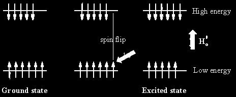
6 The NMR Transition The overall spin, I, is important. Quantum mechanics tells us that a nucleus of spin I will have 2I + 1 possible orientations. A nucleus with spin 1/2 will have 2 possible orientations. A nucleus with spin 3/2 has 4 orientation. In the absence of an external magnetic field, these orientations are of equal energy. If a magnetic field is applied, then the energy levels split. Each level is given a magnetic quantum number, m s. No Magnetic Field Presence of Magnetic Field 6 NMR Spectroscopy

7 The NMR Transition If a magnetic field is applied, then the energy levels split to a higher spin state and a lower spin state. The difference in population determines the strength of the signal. When the nucleus is in a magnetic field, the initial populations of the energy levels are determined by thermodynamics, as described by the Boltzmann distribution. This is very important, and it means that the lower energy level will contain slightly more nuclei than the higher level. It is possible to excite these nuclei into the higher level with electromagnetic radiation. The frequency of radiation needed is determined by the difference in energy between the energy levels. 7 NMR Spectroscopy
8 Energy of Transition The nucleus has a positive charge and spins. This generates a small magnetic field. The nucleus therefore possesses a magnetic moment, µ, which is proportional to its spin, I. µ = g I h / 2 p (magnetic moment) The constant, g, is called the gyromagnetic (magnetogyric) ratio and is a fundamental nuclear constant which has a different value for every nucleus. h is Planck s constant. This, g, is defined as the ratio of its magnetic dipole moment and its angular momentum. The energy of a particular energy level is given by; E = -(g h / 2 p ) µ B (Energy of spin states) Where B is the strength of the magnetic field at the nucleus. 8 NMR Spectroscopy
9 Energy of Transition The difference in energy between levels (the transition energy) can be found from DE = -(g h B / 2 p ) (Energy of transition) Recall that µ = g I h / 2 p rearranging for µ / I yields the equation D E = - µ B / I This means that if the magnetic field, B, is increased, so is DE. It also means that if a nucleus has a relatively large gyromagnetic ratio, then D E is correspondingly large. If you had trouble understanding this section, see the movie recommended at the end of this section. Also look up absorption of radiation by a nucleus in a magnetic field and review the concepts presented. 9 NMR Spectroscopy
10 Spin Flip in a Magnetic Field What happens to a charged particle in a magnetic field? In a magnetic field, a nucleus (of spin 1/2) will have a tendency to occupy a lower energy level (i.e. its magnetic moment does not oppose the applied field). The nucleus is spinning on its axis. In the presence of a magnetic field, this axis of rotation will precess around the magnetic field; The frequency of precession is termed the Larmor frequency, which is identical to the transition frequency. The potential energy of the precessing nucleus is given by: E = - µ B cos q where q is the angle between the direction of the applied field and the axis of nuclear rotation. 10 NMR Spectroscopy
11 Spin Flip in a Magnetic Field What happens to a charged particle in a magnetic field? If energy is absorbed by the nucleus, then the angle of precession, q, will change. For a nucleus of spin 1/2, absorption of radiation "flips" the magnetic moment so that it opposes the applied field (the higher energy state). It is important to realize that only a small proportion of "target" nuclei are in the lower energy state (and can absorb radiation). There is the possibility that by exciting these nuclei, the populations of the higher and lower energy levels will become equal. If this occurs, then there will be no further absorption of radiation. The spin system is saturated. The possibility of saturation means that we must be aware of the relaxation processes which return nuclei to the lower energy state. 11 NMR Spectroscopy
12 Upon Excitation The energy difference between nuclear spin states is small compared with the average kinetic energy of room temperature samples, and the +1/2 and -1/2 states are nearly equally populated. Indeed, in a field of 2.34 T (100Mz Proton) the excess population of the lower energy state is only six nuclei per million. Although this is a very small difference, when we consider the number of atoms in a practical sample (remember the size of Avogadro's number), the numerical excess in the lower energy state is sufficient for selective and sensitive spectroscopic measurements. The diagram illustrates the macroscopic magnetization of a sample containing large numbers of spin 1/2 nuclei at equilibrium in a strong external magnetic field (Bo). A slight excess of +1/2 spin states precess randomly in alignment with the external field and a smaller population of -1/2 spin states precess randomly in an opposite alignment. An overall net magnetization therefore lies along the z- axis. 12 NMR Spectroscopy

13 Upon Excitation The diagram and animation on the right show the changes in net macroscopic magnetization that occur as energy is introduced by rf irradiation at right angles to the external field. It is convenient to show the rf transmitter on the x-axis and the receiver-detector coil on the y-axis. First, the net magnetization shifts away from the z-axis and toward the y-axis. This occurs because some of the +1/2 nuclei are excited to the -1/2 state, and the precession about the z-axis becomes coherent (non-random), generating a significant y component to the net magnetization (M). After irradiation the nuclear spins return to equilibrium in a process called relaxation. As the xy coherence disappears and the population of the +1/2 state increases, energy is released and detected by the receiver. The net magnetization spirals back, and eventually the equilibrium state is reestablished. 13 NMR Spectroscopy
14 Relaxation Pathways For NMR spectroscopy to be practical, an efficient mechanism for nuclei in the higher energy -1/2 spin state to return to the lower energy +1/2 state must exist. In other words, the spin population imbalance existing at equilibrium must be restored if spectroscopic observations are to continue. Now an isolated spinning nucleus will not spontaneouly change its spin state in the absence of external perturbation. Indeed, hydrogen gas ( 2 ) exists as two stable spin isomers: ortho (parallel proton spins) and para (antiparallel spins). NMR spectroscopy is normally carried out in a liquid phase (solution or neat) so that there is close contact of sample molecules with a rapidly shifting crowd of other molecules (Brownian motion). This thermal motion of atoms and molecules generates local fluctuating electromagnetic fields, having components that match the Larmor frequency of the nucleus being studied. These local fields stimulate emission/absorption events that establish spin equilibrium, the excess spin energy being detected as it is released. This relaxation mechanism is called Spin-Lattice Relaxation (or Longitudinal Relaxation). The efficiency of spin-lattice relaxation depends on factors that influence molecular movement in the lattice, such as viscosity and temperature. The relaxation process is kinetically first order, and the reciprocal of the rate constant is a characteristic variable designated T 1, the spin-lattice relaxation time. In non-viscous liquids at room temperature T 1 ranges from 0.1 to 20 sec. A larger T 1 indicates a slower or more inefficient spin relaxation. Another relaxation mechanism called spin-spin relaxation (or transverse relaxation) is characterized by a relaxation time T 2. This process, which is actually a spin exchange, will not be discussed here. 14 NMR Spectroscopy


15 Pulse and Listen In a given strong external magnetic field, each structurally distinct set of hydrogens in a molecule has a characteristic resonance frequency, just as different size chimes have different characteristic frequencies. The drawing below depicts a set of four chimes, with the frequency of each designated by a colored sine wave. To reveal the frequency of a chime it is strike with a mallet and measure the sound emitted. This procedure can be repeated for each chime in the group so that all the characteristic frequencies are identified. An alternative means of acquiring the same information is to strike all the chimes simultaneously, and to subject the complex collection of frequencies produced to mathematical analysis. In the following diagram, the four frequencies assigned to our set of chimes are added together to give a complex summation wave. This is a straightforward conversion; and the reverse transformation, while not as simple, is readily accomplished, provided the combination signal is adequately examined and characterized. 15 NMR Spectroscopy
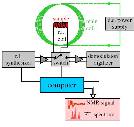
16 Pulse Versus ontinuous Wave A W NMR spectrometer functions by irradiating each set of distinct nuclei in turn, a process analagous to striking each chime independently. For a high resolution spectrum this must be done slowly, and a 12 ppm sweep of the proton region takes from 5 to 10 minute O 16 NMR Spectroscopy
17 Pulse Versus ontinuous Wave It has proven much more efficient to excite all the proton nuclei in a molecule at the same time, followed by mathematical analysis of the complex RF resonance frequencies emitted as they relax back to the equilibrium state. This is the principle on which a pulse Fourier transform spectrometer operates. By exposing the sample to a very short (10 to 100 μsec), relatively strong (about 10,000 times that used for a W spectrometer) burst of rf energy along the x-axis, as described above, all of the protons in the sample are excited simultaneously. The macroscopic magnetization model remains useful if we recognize it is a combination of magnetization vectors for all the nuclei that have been excited. 17 NMR Spectroscopy
18 Free Induction Decay and Fourier Transform The overlapping resonance signals generated as the excited protons relax are collected by a computer and subjected to a Fourier transform mathematical analysis. As shown in the diagram on the left, the Fourier transform analysis, abbreviated FT, converts the complex time domain signal emitted by the sample into the frequency (or field) domain spectrum we are accustomed to seeing. In this fashion a complete spectrum can be acquired in a few seconds. Because the relaxation mechanism is a first order process, the RF signal emitted by the sample decays exponentially. This is called a free induction decay signal, abbreviated FID. Since, the FID signal collected after one pulse, may be stored and averaged with the FID's from many other identical pulses prior to the Fourier transform, the nmr signal strength from a small sample may be enhanced to provide a useable spectrum. This has been essential to acquiring spectra from low abundance isotopes, such as 13. In practice, the pulse FT experiment has proven so versatile that many variations of the technique, suited to special purposes, have been devised and used effectively. 18 NMR Spectroscopy
19 1 NMR Spectrum Proton 1 NMR Spectroscopy 1 NMR yield hydrogen type in organic compounds. i.e., -, - 2, NMR spectrum yield environment of the hydrogen atoms via the chemical shift. 1 NMR spectrum yield relative number of hydrogen atoms area of the signals 1 NMR spectrum yield number of neighboring hydrogen via the splitting pattern of the signals. 19 NMR Spectroscopy

20 hemical Shift The magnetic field at the nucleus is not equal to the applied magnetic field; electrons around the nucleus shield it from the applied field. The difference between the applied magnetic field and the field at the nucleus is termed the nuclear shielding. onsider the s-electrons in a molecule. They have spherical symmetry and circulate in the applied field, producing a magnetic field which opposes the applied field. This means that the applied field strength must be increased for the nucleus to absorb at its transition frequency. This upfield shift is also termed diamagnetic shift. Electrons in p-orbitals have no spherical symmetry. They produce comparatively large magnetic fields at the nucleus, which give a low field shift. This "deshielding" is termed paramagnetic shift. In proton (1) NMR, p-orbitals play no part (there aren't any!), which is why only a small range of chemical shift (10 ppm) is observed. We can easily see the effect of s-electrons on the chemical shift by looking at substituted methanes, 3 X. As X becomes increasingly electronegative, so the electron density around the protons decreases, and they resonate at lower field strengths (increasing d values). hemical shift is defined as nuclear shielding / applied magnetic field. hemical shift is a function of the nucleus and its environment. It is measured relative to a reference compound. For 1 NMR, the reference is usually tetramethylsilane, Si( 3 ) 4. effectively. 20 NMR Spectroscopy

21 1 hemical Shift Table orrelation chart for proton chemical shift values. 21 NMR Spectroscopy
22 1 hemical Shift Table orrelation chart for proton chemical shift values. 22 NMR Spectroscopy


23 1 hemical Shift Table Detailed correlation chart for hydrogen-1 chemical shift values. 23 NMR Spectroscopy

24 13 hemical Shift Table orrelation chart for carbon-13 chemical shift values. 24 NMR Spectroscopy
 arbonyl (ketone) 205-220 arbonyl (aldehyde) 190-200 arbonyl (ester, acid) 170-185 Aromatic 125-150 Alkenes 115-140 Alkynes 67-85 R 2 O")
25 13 hemical Shift Table orrelation chart for carbon-13 chemical shift values. Structure hemical Shift (ppm) arbonyl (ketone) arbonyl (aldehyde) arbonyl (ester, acid) Aromatic Alkenes Alkynes R 2 O R 2 l R 2 N R O R R NMR Spectroscopy
26 1 NMR Spectrum Proton 1 NMR Spectroscopy 1 NMR yield hydrogen type in organic compounds. i.e., -, - 2, NMR spectrum yield environment of the hydrogen atoms via the chemical shift. 1 NMR spectrum yield relative number of hydrogen atoms area of the signals 1 NMR spectrum yield number of neighboring hydrogens via the splitting pattern of the signals. 26 NMR Spectroscopy
27 Magnetically Equivalent in NMR Spectroscopy To describe a spin system it's necessary to state which nucleus is coupled with which. When two equivalent nuclei have identical relations with the same identical partners, they are magnetically equivalent. Only in this case it's possible to define them as a group and not individually. Two nuclei are magnetically equivalent when they have: the same chemical shift the same coupling constant...with the same partners! At right, 2 and 6 are related by symmetry, therefore they have the same chemical shift and the same coupling constants. These are chemically equivalent At left, 4 and 6 are magnetically equivalent, because they are both coupled with 5 and with no other. Their partners, however, are different: 2 is coupled with 3, while 6 is not (or not with the same intensity). In conclusion. 2 and 6 are NOT magnetically equivalent and must be declared separately. 27 NMR Spectroscopy
28 Magnetically Equivalent in NMR Spectroscopy To describe a spin system it's necessary to state which nucleus is coupled with which. When two equivalent nuclei have identical relations with the same identical partners, they are magnetically equivalent. Only in this case it's possible to define them as a group and not individually. Two nuclei are magnetically equivalent when they have: the same chemical shift the same coupling constant...with the same partners! At right, 2 and 6 are related by symmetry, therefore they have the same chemical shift and the same coupling constants. These are chemically equivalent At left, 4 and 6 are magnetically equivalent, because they are both coupled with 5 and with no other. Their partners, however, are different: 2 is coupled with 3, while 6 is not (or not with the same intensity). In conclusion. 2 and 6 are NOT magnetically equivalent and must be declared separately. 28 NMR Spectroscopy
29 onsider the structure of ethanol; Spin-Spin oupling (Methyl ydrogen) The 1 NMR spectrum of ethanol (below) shows the methyl peak has been split into three peaks (a triplet) and the methylene peak has been split into four peaks (a quartet). This occurs because there is a small interaction (coupling) between the two groups of protons. The spacings between the peaks of the methyl triplet are equal to the spacings between the peaks of the methylene quartet. This spacing is measured in ertz and is called the coupling constant, J. To see why the methyl peak is split into a triplet, consider the methylene protons. There are two protons, and each can have one of two possible orientations (aligned with or opposed against the applied field). This gives a total of four states shown in the figure. In the first possible combination, spins are paired and opposed to the field. This has the effect of reducing the field experienced by the methyl protons; therefore a slightly higher field is needed to bring them to resonance, resulting in an upfield shift. Neither combination of spins opposed to each other has an effect on the methyl peak. The spins paired in the direction of the field produce a downfield shift. ence, the methyl peak is split into three, with the ratio of areas 1:2:1. 29 NMR Spectroscopy
30 Spin-Spin oupling (Methyl ydrogen) Methyl signal O O O x 3 O 30 NMR Spectroscopy
31 ...Spin-Spin oupling (ontinue) Similarly, the effect of the methyl protons on the methylene protons is such that there are eight possible spin combinations for the three methyl protons; Out of these eight groups, there are two groups of three magnetically equivalent combinations. The methylene peak is split into a quartet. The areas of the peaks in the quartet have the ration 1:3:3:1. In a first-order spectrum (where the chemical shift between interacting groups is much larger than their coupling constant), interpretation of splitting patterns is quite straightforward; The multiplicity of a multiplet is given by the number of equivalent protons in neighbouring atoms plus one, i.e. the n + 1 rule Equivalent nuclei do not interact with each other. The three methyl protons in ethanol cause splitting of the neighboring methylene protons; they do not cause splitting among themselves The coupling constant is not dependent on the applied field. Multiplets can be easily distinguished from closely spaced chemical shift peaks. 31 NMR Spectroscopy
32 32 NMR Spectroscopy Spin-Spin oupling (Methylene ydrogens) O O O O O O O O x 2 Methylene signal

33 Proton-Proton coupling onstant (J) Spin-Spin oupling correlation chart. 33 NMR Spectroscopy
34 Proton-Proton coupling onstant (J) Spin-Spin oupling correlation chart. 34 NMR Spectroscopy
35 Interpreting 1 NMR The interpretation of a 1 spectra depends on three features: chemical shifts, multiplicities and integrated peak area. Note the presence or absence of saturated structures, most of which gives resonances between 0 and 5 d ppm. Note the presence or absence of unsaturated structures in the region between 5 & 9 d ppm. Alkene protons resonate between 5 and 7 d ppm and aromatic protons between 7 and 9 d ppm. Note that alkyne protons resonance upfield around 1.5 d ppm. Note any very low field resonance (downfield) between 9 and 16 d ppm, which are associated with aldehydic and acidic protons, especially those involving in -bonding. Measure the integrals, if recorded, and calculate the number protons in each resonance signal. heck for spin-spin splitting patterns given by adjacent alkyl group according to the n+1 rule and Pascal s triangle. Note that the position of the lower field multiplet of the two is very sensitive to the proximity of electronegative elements and groups such as O, O, OO, O, X, N 2, etc.) Examine the splitting pattern given by aromatic protons, which couple around the ring and are often complex due to second order effects. 1,4 and 1,2-disubstituted rings give complex but symmetrical looking patterns of peaks, whereas mono- 1,3-and tri-substituted rings give more complex asymmetric patterns. Note any broad single resonance, which are evidence of liable protons from alcohols, phenols, acids and amines that can undergo slow exchange with other labile protons. 35 NMR Spectroscopy
36 Interpreting 1 NMR The interpretation of a 1 spectra depends on three features: chemical shifts, multiplicities and integrated peak area. 4 8 O 2 Rings + Double bonds: R + DB = X 2 + N R = ring DB = double bonds = number of hydrogens X = number of halogens N = number of nitrogens R+ DB = = 1 36 NMR Spectroscopy
37 Interpreting 13 NMR arbon-13 spectra cover a much wider range of chemical shifts than proton spectra, but the positions of resonances are generally determined by the same factors. Remember that the p-electrons are now responsible for shielding and deshielding the nuclei. The spectra are usually recorded as decoupled spectra to eliminate the effects of coupling to adjacent protons which would otherwise split the carbon-13 resonance according to the n+1 rule and Pascal s triangle. Decouple spectra consist of a single peak for each chemically different carbon in the molecule and spectral interpretation is confined to the correlation of their chemical shifts with structure, augmented by reference to chemical shift data and the spectra of known compounds. Proton coupling can be observed under appropriate experimental conditions. 37 NMR Spectroscopy
38 Interpreting 13 NMR Under decoupling conditions note the following- Note the presence or absence of saturated structures, most of which give resonance between 0 and 90 d ppm. Note the presence or absence of unsaturated structures in the region between 100 and 160 d ppm. Note that alkyne protons resonance upfield between 70 and 100 d ppm. Note any very low field resonances (160 to 220 d ppm), which are associated with carbonyl and ether carbons. arboxylic acids, anhydrides, esters, amides, acyl halides and ethers are all found all found in he range of 160 to 180 d ppm, while aldehydes and ketones lie between 180 and 220 d ppm. 38 NMR Spectroscopy
39 Interpreting 13 NMR 13 NMR NMR Spectroscopy
40 1 OSY spectra for 7 14 O 1 NMR Spectra O R + DB = X 2 + N R+ DB = = 1 40 NMR Spectroscopy
41 1 OSY spectra for 7 14 O 1 NMR Spectra O R + DB = X 2 + N R+ DB = = 1 41 NMR Spectroscopy
42 Dept 13 Spectra DEPT - Distortionless Enhancement by Polarization Transfer. The DEPT experiment differentiates, 2 and 3 groups by variation of the selection angle parameter (the tip angle of the final 1 pulse): angle gives all and 3 in a phase opposite to 2-90 angle gives only groups, the others being suppressed - 45 angle gives all carbons with attached protons O 2 Propyl benzoate O 4 O resonances 42 NMR Spectroscopy
43 Dept 13 Spectra DEPT - Distortionless Enhancement by Polarization Transfer. The DEPT experiment differentiates, 2 and 3 groups by variation of the selection angle parameter (the tip angle of the final 1 pulse): angle gives all and 3 in a phase opposite to 2-90 angle gives only groups, the others being suppressed - 45 angle gives all carbons with attached protons O 2 Propyl benzoate and O O , 2, O O NMR Spectroscopy


44 etcor ETOR eteronuclear Spectroscopy 6 10 O 2 Rings + db = 2 Rings + Double Bonds O O OSY ETOR 44 NMR Spectroscopy
Chapter 13: Nuclear Magnetic Resonance (NMR) Spectroscopy direct observation of the H s and C s of a molecules
 Spectroscopy direct observation of the H s and C s of a molecules") hapter 13: Nuclear Magnetic Resonance (NMR) Spectroscopy direct observation of the s and s of a molecules Nuclei are positively charged and spin on an axis; they create a tiny magnetic field + + Not all
hapter 13: Nuclear Magnetic Resonance (NMR) Spectroscopy direct observation of the s and s of a molecules Nuclei are positively charged and spin on an axis; they create a tiny magnetic field + + Not all
16.1 Introduction to NMR Spectroscopy. Spectroscopy. Spectroscopy. Spectroscopy. Spectroscopy. Spectroscopy 4/11/2013
 What is spectroscopy? NUCLEAR MAGNETIC RESONANCE (NMR) spectroscopy may be the most powerful method of gaining structural information about organic compounds. NMR involves an interaction between electromagnetic
Chapter 7. Nuclear Magnetic Resonance Spectroscopy
 Chapter 7 Nuclear Magnetic Resonance Spectroscopy I. Introduction 1924, W. Pauli proposed that certain atomic nuclei have spin and magnetic moment and exposure to magnetic field would lead to energy level
Chem 325 NMR Intro. The Electromagnetic Spectrum. Physical properties, chemical properties, formulas Shedding real light on molecular structure:
 Physical properties, chemical properties, formulas Shedding real light on molecular structure: Wavelength Frequency ν Wavelength λ Frequency ν Velocity c = 2.998 10 8 m s -1 The Electromagnetic Spectrum
Chapter 16 Nuclear Magnetic Resonance Spectroscopy
 hapter 16 Nuclear Magnetic Resonance Spectroscopy The Spinning Proton A spinning proton generates a magnetic field, resembling that of a small bar magnet. An odd number of protons in the nucleus creates
William H. Brown & Christopher S. Foote
 Requests for permission to make copies of any part of the work should be mailed to:permissions Department, Harcourt Brace & Company, 6277 Sea Harbor Drive, Orlando, Florida 32887-6777 William H. Brown
NUCLEAR MAGNETIC RESONANCE SPECTROSCOPY
 NMR Spectroscopy 1 NULEAR MAGNETI RESONANE SPETROSOPY Involves interaction of materials with the low-energy radiowave region of the electromagnetic spectrum Origin of Spectra Theory All nuclei possess
In a solution, there are thousands of atoms generating magnetic fields, all in random directions.
 Nuclear Magnetic Resonance Spectroscopy: Purpose: onnectivity, Map of - framework Process: In nuclear magnetic resonance spectroscopy, we are studying nuclei. onsider this circle to represent a nucleus
Chapter 13 Structure t Determination: Nuclear Magnetic Resonance Spectroscopy
 John E. McMurry www.cengage.com/chemistry/mcmurry Chapter 13 Structure t Determination: ti Nuclear Magnetic Resonance Spectroscopy Revisions by Dr. Daniel Holmes MSU Paul D. Adams University of Arkansas
Chapter 9. Nuclear Magnetic Resonance. Ch. 9-1
 Chapter 9 Nuclear Magnetic Resonance Ch. 9-1 1. Introduction Classic methods for organic structure determination Boiling point Refractive index Solubility tests Functional group tests Derivative preparation
The Use of NMR Spectroscopy
 Spektroskopi Molekul Organik (SMO): Nuclear Magnetic Resonance (NMR) Spectroscopy All is adopted from McMurry s Organic Chemistry The Use of NMR Spectroscopy Used to determine relative location of atoms
CHEM Chapter 13. Nuclear Magnetic Spectroscopy (Homework) W
 W") CHEM 2423. Chapter 13. Nuclear Magnetic Spectroscopy (Homework) W Short Answer 1. For a nucleus to exhibit the nuclear magnetic resonance phenomenon, it must be magnetic. Magnetic nuclei include: a. all
CHEM 2423. Chapter 13. Nuclear Magnetic Spectroscopy (Homework) W Short Answer 1. For a nucleus to exhibit the nuclear magnetic resonance phenomenon, it must be magnetic. Magnetic nuclei include: a. all
NMR = Nuclear Magnetic Resonance
 NMR = Nuclear Magnetic Resonance NMR spectroscopy is the most powerful technique available to organic chemists for determining molecular structures. Looks at nuclei with odd mass numbers or odd number
13.24: Mass Spectrometry: molecular weight of the sample
 hapter 13: Spectroscopy Methods of structure determination Nuclear Magnetic Resonances (NMR) Spectroscopy (Sections 13.3-13.19) Infrared (IR) Spectroscopy (Sections 13.20-13.22) Ultraviolet-visible (UV-Vis)
Nuclear Spin States. NMR Phenomenon. NMR Instrumentation. NMR Active Nuclei. Nuclear Magnetic Resonance
 Nuclear Magnetic Resonance NMR Phenomenon µ A spinning charged particle generates a magnetic field. A nucleus with a spin angular momentum will generate a magnetic moment (!). E Nuclear Spin States aligned
Chapter 14 Spectroscopy
 hapter 14 Spectroscopy There are four major analytical techniques used for identifying the structure of organic molecules 1. Nuclear Magnetic Resonance or NMR is the single most important technique for
3.15 Nuclear Magnetic Resonance Spectroscopy, NMR
 3.15 Nuclear Magnetic Resonance Spectroscopy, NMR What is Nuclear Magnetic Resonance - NMR Developed by chemists and physicists together it works by the interaction of magnetic properties of certain nuclei
Physical Background Of Nuclear Magnetic Resonance Spectroscopy
 Physical Background Of Nuclear Magnetic Resonance Spectroscopy Michael McClellan Spring 2009 Department of Physics and Physical Oceanography University of North Carolina Wilmington What is Spectroscopy?
MOLECULAR SPECTROSCOPY AND PHOTOCHEMISTRY
 20 CHAPTER MOLECULAR SPECTROSCOPY AND PHOTOCHEMISTRY 20.1 Introduction to Molecular Spectroscopy 20.2 Experimental Methods in Molecular Spectroscopy 20.3 Rotational and Vibrational Spectroscopy 20.4 Nuclear
Tuesday, January 13, NMR Spectroscopy
 NMR Spectroscopy NMR Phenomenon Nuclear Magnetic Resonance µ A spinning charged particle generates a magnetic field. A nucleus with a spin angular momentum will generate a magnetic moment (μ). If these
In a solution, there are thousands of atoms generating magnetic fields, all in random directions.
 Nuclear Magnetic Resonance Spectroscopy: Purpose: onnectivity, Map of - framework Process: In nuclear magnetic resonance spectroscopy, we are studying nuclei. onsider this circle to represent a nucleus
Experiment 2 - NMR Spectroscopy
 Experiment 2 - NMR Spectroscopy OBJECTIVE to understand the important role of nuclear magnetic resonance spectroscopy in the study of the structures of organic compounds to develop an understanding of
Instrumental Chemical Analysis
 L15 Page1 Instrumental Chemical Analysis Nuclear Magnetic Resonance Dr. Ahmad Najjar Philadelphia University Faculty of Pharmacy Department of Pharmaceutical Sciences 1 st semester, 2017/2018 Nuclear Magnetic
Can you differentiate A from B using 1 H NMR in each pair?
 Can you differentiate A from B using 1 H NMR in each pair? To be NMR active any nucleus must have a spin quantum number, different from zero (I 0) As in 1 H, the spin quantum number (I) of 13 C is 1/2
1. neopentyl benzene. 4 of 6
 I. 1 H NMR spectroscopy A. Theory 1. The protons and neutrons in atomic nuclei spin, as does the nucleus itself 2. The circulation of nuclear charge can generate a nuclear magnetic moment, u, along the
Chapter 15 Lecture Outline
 Organic Chemistry, First Edition Janice Gorzynski Smith University of Hawaii Chapter 5 Lecture Outline Introduction to NMR Two common types of NMR spectroscopy are used to characterize organic structure:
Biochemistry 530 NMR Theory and Practice
 Biochemistry 530 NMR Theory and Practice Gabriele Varani Department of Biochemistry and Department of Chemistry University of Washington Lecturer: Gabriele Varani Biochemistry and Chemistry Room J479 and
Module 13: Chemical Shift and Its Measurement
 Subject Chemistry Paper No and Title Module No and Title Module Tag Paper 12: Organic Spectroscopy CHE_P12_M13_e-Text TABLE OF CONTENTS 1. Learning Outcomes 2. Introduction 3. Shielding and deshielding
Ferdowsi University of Mashhad
 Spectroscopy in Inorganic Chemistry Nuclear Magnetic Resonance Spectroscopy spin deuterium 2 helium 3 The neutron has 2 quarks with a -e/3 charge and one quark with a +2e/3 charge resulting in a total
To Do s. Answer Keys are available in CHB204H
 To Do s Read Chapters 2, 3 & 4. Complete the end-of-chapter problems, 2-1, 2-2, 2-3 and 2-4 Complete the end-of-chapter problems, 3-1, 3-3, 3-4, 3-6 and 3-7 Complete the end-of-chapter problems, 4-1, 4-2,
Nuclear Magnetic Resonance Spectroscopy
 Chapter 5 Nuclear Magnetic Resonance Spectroscopy http://www.yteach.co.uk/page.php/resources/view_all?id=nuclear_magnetic _resonance_nmr_spectroscopy_spin_spectrometer_spectrum_proton_t_pag e_5&from=search
Nuclear Magnetic Resonance Spectroscopy: Tools for Structure Determination
 Nuclear Magnetic Resonance Spectroscopy: Tools for Structure Determination Chung-Ming Sun Department of Applied Chemistry National Chiao Tung University Hualien 300, Taiwan Introduction NMR (Nuclear Magnetic
To Do s. Answer Keys are available in CHB204H
 To Do s Read Chapters 2, 3 & 4. Complete the end-of-chapter problems, 2-1, 2-2, 2-3 and 2-4 Complete the end-of-chapter problems, 3-1, 3-3, 3-4, 3-6 and 3-7 Complete the end-of-chapter problems, 4-1, 4-2,
Nuclear Magnetic Resonance
 Nuclear Magnetic Resonance PRINCIPLES OF NMR SPECTROSCOPY Contents Principles of nuclear magnetic resonance The nmr spectrometer Basic principles in nmr application NMR tools used to obtain information
With that first concept in mind, it is seen that a spinning nucleus creates a magnetic field, like a bar magnet
 NMR SPECTROSCOPY This section will discuss the basics of NMR (nuclear magnetic resonance) spectroscopy. Most of the section will discuss mainly 1H or proton spectroscopy but the most popular nuclei in
Nuclear Magnetic Resonance Spectroscopy
 Nuclear Magnetic Resonance Spectroscopy Features: Used to identify products of reactions Also gives information about chemical environment, connectivity and bonding of nuclei Requirements: Pure or mostly
Structure Determination: Nuclear Magnetic Resonance Spectroscopy
 Structure Determination: Nuclear Magnetic Resonance Spectroscopy Why This Chapter? NMR is the most valuable spectroscopic technique used for structure determination More advanced NMR techniques are used
Lecture Notes Chem 51A S. King
 Lecture Notes hem 51A S. King hapter 14 Nuclear Magnetic Resonance Spectroscopy Nuclear Magnetic Resonance (NMR) spectroscopy uses energy in the radiowave portion of the electromagnetic spectrum. The nuclei
Chapter 13 Nuclear Magnetic Resonance Spectroscopy
 Organic Chemistry, 6 th Edition L. G. Wade, Jr. Chapter 13 Nuclear Magnetic Resonance Spectroscopy Jo Blackburn Richland College, Dallas, TX Dallas County Community College District 2006, Prentice Hall
16.1 Introduction to NMR. Spectroscopy
 16.1 Introduction to NMR What is spectroscopy? Spectroscopy NUCLEAR MAGNETIC RESNANCE (NMR) spectroscopy may be the most powerful method of gaining structural information about organic compounds. NMR involves
Experiment 11: NUCLEAR MAGNETIC RESONANCE SPECTROSCOPY
 Experiment 11: NUCLEAR MAGNETIC RESONANCE SPECTROSCOPY Purpose: This is an exercise to introduce the use of nuclear magnetic resonance spectroscopy, in conjunction with infrared spectroscopy, to determine
Nuclear spin and the splitting of energy levels in a magnetic field
 Nuclear spin and the splitting of energy levels in a magnetic field Top 3 list for 13 C NMR Interpretation 1. Symmetry 2. Chemical Shifts 3. Multiplicity 13 C NMR of C 3 O 1 NMR of C 3 O 13 C NMR of C
Química Orgânica I. Nuclear Magnetic Resonance Spectroscopy (I) Ciências Farmacêuticas Bioquímica Química AFB QO I 2007/08 1 AFB QO I 2007/08 2
 Ciências Farmacêuticas Bioquímica Química AFB QO I 2007/08 1 AFB QO I 2007/08 2") Química Orgânica I Ciências Farmacêuticas Bioquímica Química AFB QO I 2007/08 1 Nuclear Magnetic Resonance Spectroscopy (I) AFB QO I 2007/08 2 1 Adaptado de: Organic Chemistry, 6th Edition; L. G. Wade,
Química Orgânica I Ciências Farmacêuticas Bioquímica Química AFB QO I 2007/08 1 Nuclear Magnetic Resonance Spectroscopy (I) AFB QO I 2007/08 2 1 Adaptado de: Organic Chemistry, 6th Edition; L. G. Wade,
NMR Spectroscopy Laboratory Experiment Introduction. 2. Theory
 1. Introduction 64-311 Laboratory Experiment 11 NMR Spectroscopy Nuclear Magnetic Resonance (NMR) spectroscopy is a powerful and theoretically complex analytical tool. This experiment will introduce to
NMR Spectroscopy. for 1 st B.Tech INTRODUCTION Lecture -1 Indian Institute of Technology, Dhanbad
 NMR Spectroscopy for 1 st B.Tech Lecture -1 Indian Institute of Technology, Dhanbad by Dr. R P John & Dr. C. Halder INTRODUCTION Nucleus of any atom has protons and neutrons Both Proton and Neutron has
OAT Organic Chemistry - Problem Drill 19: NMR Spectroscopy and Mass Spectrometry
 OAT Organic Chemistry - Problem Drill 19: NMR Spectroscopy and Mass Spectrometry Question No. 1 of 10 Question 1. Which statement concerning NMR spectroscopy is incorrect? Question #01 (A) Only nuclei
UNIT 12 NMR SPECTROSCOPY
 UIT 12 MR SPECTROSCOPY MR Spectroscopy Structure 12.1 Introduction 12.2 Theory of MR Spectroscopy Types of uclei Magnetic Moment Quantisation Population of Energy Levels Larmor Precession Mechanism of
BMB/Bi/Ch 173 Winter 2018
 BMB/Bi/Ch 173 Winter 2018 Homework Set 8.1 (100 Points) Assigned 2-27-18, due 3-6-18 by 10:30 a.m. TA: Rachael Kuintzle. Office hours: SFL 220, Friday 3/2 4:00-5:00pm and SFL 229, Monday 3/5 4:00-5:30pm.
Chapter 18: NMR Spectroscopy
 The most important tool of the chemist for the determination of molecular structure is Nuclear Magnetic Resonance Spectroscopy, or NMR spectroscopy. NMR spectra are acquired on a special instrument called
4) protons experience a net magnetic field strength that is smaller than the applied magnetic field.
 protons experience a net magnetic field strength that is smaller than the applied magnetic field.") 1) Which of the following CANNOT be probed by an spectrometer? See sect 16.1 Chapter 16: 1 A) nucleus with odd number of protons & odd number of neutrons B) nucleus with odd number of protons &even number
1) Which of the following CANNOT be probed by an spectrometer? See sect 16.1 Chapter 16: 1 A) nucleus with odd number of protons & odd number of neutrons B) nucleus with odd number of protons &even number
NMR Nuclear Magnetic Resonance Spectroscopy p. 83. a hydrogen nucleus (a proton) has a charge, spread over the surface
 has a charge, spread over the surface") NMR Nuclear Magnetic Resonance Spectroscopy p. 83 a hydrogen nucleus (a proton) has a charge, spread over the surface a spinning charge produces a magnetic moment (a vector = direction + magnitude) along
NMR Nuclear Magnetic Resonance Spectroscopy p. 83 a hydrogen nucleus (a proton) has a charge, spread over the surface a spinning charge produces a magnetic moment (a vector = direction + magnitude) along
Nuclear Magnetic Resonance (NMR) Spectroscopy Introduction:
 Spectroscopy Introduction:") Nuclear Magnetic Resonance (NMR) Spectroscopy Introduction: Nuclear magnetic resonance spectroscopy (NMR) is the most powerful tool available for organic structure determination. Like IR spectroscopy,
Nuclear Magnetic Resonance (NMR) Spectroscopy Introduction: Nuclear magnetic resonance spectroscopy (NMR) is the most powerful tool available for organic structure determination. Like IR spectroscopy,
7a. Structure Elucidation: IR and 13 C-NMR Spectroscopies (text , , 12.10)
") 2009, Department of Chemistry, The University of Western Ontario 7a.1 7a. Structure Elucidation: IR and 13 C-NMR Spectroscopies (text 11.1 11.5, 12.1 12.5, 12.10) A. Electromagnetic Radiation Energy is
2009, Department of Chemistry, The University of Western Ontario 7a.1 7a. Structure Elucidation: IR and 13 C-NMR Spectroscopies (text 11.1 11.5, 12.1 12.5, 12.10) A. Electromagnetic Radiation Energy is
Chapter 14. Nuclear Magnetic Resonance Spectroscopy
 Organic Chemistry, Second Edition Janice Gorzynski Smith University of Hawai i Chapter 14 Nuclear Magnetic Resonance Spectroscopy Prepared by Rabi Ann Musah State University of New York at Albany Copyright
The Basics of Magnetic Resonance Imaging
 The Basics of Magnetic Resonance Imaging Nathalie JUST, PhD nathalie.just@epfl.ch CIBM-AIT, EPFL Course 2013-2014-Chemistry 1 Course 2013-2014-Chemistry 2 MRI: Many different contrasts Proton density T1
ORGANIC - CLUTCH CH ANALYTICAL TECHNIQUES: IR, NMR, MASS SPECT
 !! www.clutchprep.com CONCEPT: PURPOSE OF ANALYTICAL TECHNIQUES Classical Methods (Wet Chemistry): Chemists needed to run dozens of chemical reactions to determine the type of molecules in a compound.
11. Proton NMR (text , 12.11, 12.12)
") 2009, Department of Chemistry, The University of Western Ontario 11.1 11. Proton NMR (text 12.6 12.9, 12.11, 12.12) A. Proton Signals Like 13 C, 1 H atoms have spins of ±½, and when they are placed in
2009, Department of Chemistry, The University of Western Ontario 11.1 11. Proton NMR (text 12.6 12.9, 12.11, 12.12) A. Proton Signals Like 13 C, 1 H atoms have spins of ±½, and when they are placed in
Nuclear Magnetic Resonance Spectroscopy Chem 4010/5326: Organic Spectroscopic Analysis Andrew Harned
 Nuclear Magnetic Resonance Spectroscopy Chem 4010/5326: Organic Spectroscopic Analysis 2015 Andrew Harned NMR Spectroscopy NMR Spectroscopy All nuclei have a nuclear spin quantum number (I) I = 0, 1/2,
ORGANIC - CLUTCH CH ANALYTICAL TECHNIQUES: IR, NMR, MASS SPECT
 !! www.clutchprep.com CONCEPT: PURPOSE OF ANALYTICAL TECHNIQUES Classical Methods (Wet Chemistry): Chemists needed to run dozens of chemical reactions to determine the type of molecules in a compound.
C h a p t e r S i x t e e n: Nuclear Magnetic Resonance Spectroscopy. An 1 H NMR FID of ethanol
 0.2 0.4 0.6 0.8 1.0 1.2 1.4 1.6 1.8 2.0 2.2 2.4 2.6 2.8 3.0 3.2 3.4 3.6 C h a p t e r S i x t e e n: Nuclear Magnetic Resonance Spectroscopy An 1 NMR FID of ethanol Note: Problems with italicized numbers
Fundamental MRI Principles Module 2 N. Nuclear Magnetic Resonance. X-ray. MRI Hydrogen Protons. Page 1. Electrons
 Fundamental MRI Principles Module 2 N S 1 Nuclear Magnetic Resonance There are three main subatomic particles: protons positively charged neutrons no significant charge electrons negatively charged Protons
Magnetic Nuclei other than 1 H
 Magnetic Nuclei other than 1 H 2 H (Deuterium): I = 1 H,D-Exchange might be used to simplify 1 H-NMR spectra since H-D couplings are generally small; - - - -O- - - -D 2 -O- triplet of triplets slightly
4) protons experience a net magnetic field strength that is smaller than the applied magnetic field.
 protons experience a net magnetic field strength that is smaller than the applied magnetic field.") 1) Which of the following CANNOT be probed by an spectrometer? See sect 15.1 Chapter 15: 1 A) nucleus with odd number of protons & odd number of neutrons B) nucleus with odd number of protons &even number
1) Which of the following CANNOT be probed by an spectrometer? See sect 15.1 Chapter 15: 1 A) nucleus with odd number of protons & odd number of neutrons B) nucleus with odd number of protons &even number
The NMR Spectrum - 13 C. NMR Spectroscopy. Spin-Spin Coupling 13 C NMR. A comparison of two 13 C NMR Spectra. H Coupled (undecoupled) H Decoupled
 H Decoupled") Spin-Spin oupling 13 NMR A comparison of two 13 NMR Spectra 1 oupled (undecoupled) 1 Decoupled 1 Proton Decoupled 13 NMR 6. To simplify the 13 spectrum, and to increase the intensity of the observed signals,
Spin-Spin oupling 13 NMR A comparison of two 13 NMR Spectra 1 oupled (undecoupled) 1 Decoupled 1 Proton Decoupled 13 NMR 6. To simplify the 13 spectrum, and to increase the intensity of the observed signals,
10.4 Continuous Wave NMR Instrumentation
 10.4 Continuous Wave NMR Instrumentation coherent detection bulk magnetization the rotating frame, and effective magnetic field generating a rotating frame, and precession in the laboratory frame spin-lattice
Lecture 02 Nuclear Magnetic Resonance Spectroscopy Principle and Application in Structure Elucidation
 Application of Spectroscopic Methods in Molecular Structure Determination Prof. S. Sankararaman Department of Chemistry Indian Institution of Technology Madras Lecture 02 Nuclear Magnetic Resonance Spectroscopy
Nuclear Magnetic Resonance Spectroscopy (NMR)
") OCR Chemistry A 432 Spectroscopy (NMR) What is it? An instrumental method that gives very detailed structural information about molecules. It can tell us - how many of certain types of atom a molecule
OCR Chemistry A 432 Spectroscopy (NMR) What is it? An instrumental method that gives very detailed structural information about molecules. It can tell us - how many of certain types of atom a molecule
- 1/2. = kb o = hνν + 1/2. B o increasing magnetic field strength. degenerate at B o = 0
 NMR EXPERIMENT When magnetically active nuclei are placed into an external magnetic field, the magnetic fields align themselves with the external field into two orientations. During the experiment, electromagnetic
Chapter 9. Nuclear Magnetic Resonance and Mass Spectrometry. 1. Introduction. 2. Nuclear Magnetic Resonance (NMR) Spectroscopy
 Spectroscopy") hapter 9 Nuclear Magnetic Resonance and Mass Spectrometry reated by Professor William Tam & Dr. Phillis hang 1. Introduction Spectroscopy the study of the interaction of light with matter Spectroscopy
hapter 9 Nuclear Magnetic Resonance and Mass Spectrometry reated by Professor William Tam & Dr. Phillis hang 1. Introduction Spectroscopy the study of the interaction of light with matter Spectroscopy
Nuclear Magnetic Resonance
 Nuclear Magnetic Resonance Absorption of electromagnetic radiation from 4 Mz to 900 Mz Nuclear process Radiation absorbed by nuclei Sample must be placed in strong magnetic field Used for determining structure
Relaxation, Multi pulse Experiments and 2D NMR
 Relaxation, Multi pulse Experiments and 2D NMR To Do s Read Chapter 6 Complete the end of chapter problems; 6 1, 6 2, 6 3, 6 5, 6 9 and 6 10. Read Chapter 15 and do as many problems as you can. Relaxation
4) protons experience a net magnetic field strength that is smaller than the applied magnetic field.
 protons experience a net magnetic field strength that is smaller than the applied magnetic field.") 1) Which of the following CANNOT be probed by an NMR spectrometer? See sect 15.1 Chapter 15: 1 A) nucleus with odd number of protons & odd number of neutrons B) nucleus with odd number of protons &even
1) Which of the following CANNOT be probed by an NMR spectrometer? See sect 15.1 Chapter 15: 1 A) nucleus with odd number of protons & odd number of neutrons B) nucleus with odd number of protons &even
Chapter 13: Molecular Spectroscopy
 Chapter 13: Molecular Spectroscopy Electromagnetic Radiation E = hν h = Planck s Constant (6.63 x 10⁻³⁴ J. s) ν = frequency (s -1 ) c = νλ λ = wavelength (nm) Energy is proportional to frequency Spectrum
4) protons experience a net magnetic field strength that is smaller than the applied magnetic field.
 protons experience a net magnetic field strength that is smaller than the applied magnetic field.") 1) Which of the following CANNOT be probed by an spectrometer? See sect 16.1 Chapter 16: 1 A) nucleus with odd number of protons & odd number of neutrons B) nucleus with odd number of protons &even number
1) Which of the following CANNOT be probed by an spectrometer? See sect 16.1 Chapter 16: 1 A) nucleus with odd number of protons & odd number of neutrons B) nucleus with odd number of protons &even number
Analysis of NMR Spectra Part 2
 Analysis of NMR Spectra Part 2-1- Analysis of NMR Spectra Part 2 "Things should be made as simple as possible, but not any simpler." Albert Einstein 1.1 Review of Basic NMR Concepts NMR analysis is a complex
Nuclear Magnetic Resonance Spectroscopy Thomas Wenzel Department of Chemistry Bates College, Lewiston ME
 Nuclear Magnetic Resonance Spectroscopy Thomas Wenzel Department of Chemistry Bates College, Lewiston ME 04240 twenzel@bates.edu The following textual material is designed to accompany a series of in-class
SECOND YEAR ORGANIC CHEMISTRY - REVISION COURSE Lecture 2 MOLECULAR STRUCTURE 2: SPECTROSCOPIC ANALYSIS
 Prof Ben Davis SECOND YEAR ORGANIC CEMISTRY - REVISION COURSE Lecture 2 MOLECULAR STRUCTURE 2: SPECTROSCOPIC ANALYSIS Books: Williams and Fleming, " Spectroscopic Methods in Organic Chemistry", arwood
V27: RF Spectroscopy
 Martin-Luther-Universität Halle-Wittenberg FB Physik Advanced Lab Course V27: RF Spectroscopy ) Electron spin resonance (ESR) Investigate the resonance behaviour of two coupled LC circuits (an active rf
Fundamental MRI Principles Module Two
 Fundamental MRI Principles Module Two 1 Nuclear Magnetic Resonance There are three main subatomic particles: protons neutrons electrons positively charged no significant charge negatively charged Protons
ORGANIC SPECTROSCOPY NOTES
 - 1 - ORGANIC SPECTROSCOPY NOTES Basics of Spectroscopy UV/vis, IR and NMR are all types of Absorption Spectroscopy, where EM radiation corresponding to exactly the energy of specific excitations in molecules
STRUCTURE ELUCIDATION BY INTEGRATED SPECTROSCOPIC METHODS
 Miscellaneous Methods UNIT 14 STRUCTURE ELUCIDATION BY INTEGRATED SPECTROSCOPIC METHODS Structure 14.1 Introduction Objectives 14.2 Molecular Formula and Index of Hydrogen Deficiency 14.3 Structural Information
Basic p rinciples COPYRIGHTED MATERIAL. Introduction. Atomic s tructure
 1 Basic p rinciples Introduction 1 Atomic structure 1 Motion in the atom 2 MR active nuclei 2 The hydrogen nucleus 4 Alignment 4 Precession 8 The Larmor equation 9 Introduction The basic principles of
Nuclear magnetic resonance spectroscopy II. 13 C NMR. Reading: Pavia Chapter , 6.7, 6.11, 6.13
 Nuclear magnetic resonance spectroscopy II. 13 NMR Reading: Pavia hapter 6.1-6.5, 6.7, 6.11, 6.13 1. General - more/better/additional structural information for larger compounds -problems: a) isotopes
PAPER No. 12: ORGANIC SPECTROSCOPY. Module 19: NMR Spectroscopy of N, P and F-atoms
 Subject Chemistry Paper No and Title Module No and Title Module Tag Paper 12: Organic Spectroscopy CHE_P12_M19_e-Text TABLE OF CONTENTS 1. Learning Outcomes 2. 15 N NMR spectroscopy 3. 19 F NMR spectroscopy
Chapter 13 Spectroscopy
 hapter 13 Spectroscopy Infrared spectroscopy Ultraviolet-Visible spectroscopy Nuclear magnetic resonance spectroscopy Mass Spectrometry 13.1 Principles of Molecular Spectroscopy: Electromagnetic Radiation
Module 20: Applications of PMR in Structural Elucidation of Simple and Complex Compounds and 2-D NMR spectroscopy
 Subject Chemistry Paper No and Title Module No and Title Module Tag Paper 12: Organic Spectroscopy Module 20: Applications of PMR in Structural Elucidation of Simple and Complex Compounds and 2-D NMR spectroscopy
Magnetic Resonance Imaging (MRI)
") Magnetic Resonance Imaging Introduction The Components The Technology (MRI) Physics behind MR Most slides taken from http:// www.slideworld.org/ viewslides.aspx/magnetic- Resonance-Imaging- %28MRI%29-MR-Imaging-
Magnetic Resonance Imaging Introduction The Components The Technology (MRI) Physics behind MR Most slides taken from http:// www.slideworld.org/ viewslides.aspx/magnetic- Resonance-Imaging- %28MRI%29-MR-Imaging-
A Hands on Introduction to NMR Lecture #1 Nuclear Spin and Magnetic Resonance
 A Hands on Introduction to NMR 22.920 Lecture #1 Nuclear Spin and Magnetic Resonance Introduction - The aim of this short course is to present a physical picture of the basic principles of Nuclear Magnetic
The rest of topic 11 INTRODUCTION TO ORGANIC SPECTROSCOPY
 The rest of topic 11 INTRODUCTION TO ORGANIC SPECTROSCOPY 1. Mass spectrometry: SPECTROSCOPIC TECHNIQUES - A technique capable of identifying the presence of various mass segments of organic molecules.
C NMR Spectroscopy
 13.14 13 C NMR Spectroscopy 1 H and 13 C NMR compared: both give us information about the number of chemically nonequivalent nuclei (nonequivalent hydrogens or nonequivalent carbons) both give us information
NMR, the vector model and the relaxation
 NMR, the vector model and the relaxation Reading/Books: One and two dimensional NMR spectroscopy, VCH, Friebolin Spin Dynamics, Basics of NMR, Wiley, Levitt Molecular Quantum Mechanics, Oxford Univ. Press,
Nuclear magnetic resonance spectroscopy
 nuclear spin transitions O Nuclear magnetic resonance spectroscopy 1 H, 13 C, 2-dimensional which transitions? wavelength and intensity; ppm what happens if we change the environment of the nucleus? substituent
COPYRIGHTED MATERIAL. Production of Net Magnetization. Chapter 1
 Chapter 1 Production of Net Magnetization Magnetic resonance (MR) is a measurement technique used to examine atoms and molecules. It is based on the interaction between an applied magnetic field and a
CHEM 322 Laboratory Methods in Organic Chemistry. Introduction to NMR Spectroscopy
 EM 322 Laboratory Methods in Organic hemistry Introduction to NMR Spectroscopy What structural information does NMR spectroscopy provide? 1) hemical shift (δ) data reveals the molecular (functional group)
T 1, T 2, NOE (reminder)
") T 1, T 2, NOE (reminder) T 1 is the time constant for longitudinal relaxation - the process of re-establishing the Boltzmann distribution of the energy level populations of the system following perturbation
T 1, T 2, NOE (reminder) T 1 is the time constant for longitudinal relaxation - the process of re-establishing the Boltzmann distribution of the energy level populations of the system following perturbation
Magnetic Resonance Imaging. Pål Erik Goa Associate Professor in Medical Imaging Dept. of Physics
 Magnetic Resonance Imaging Pål Erik Goa Associate Professor in Medical Imaging Dept. of Physics pal.e.goa@ntnu.no 1 Why MRI? X-ray/CT: Great for bone structures and high spatial resolution Not so great
NMR Spectroscopy. Chapter 19
 NMR Spectroscopy Chapter 19 Nuclear Magnetic Resonance spectroscopy is a powerful analytical technique used to characterize organic molecules by identifying carbon-hydrogen frameworks within molecules.
1,1,2-Tribromoethane. Spin-Spin Coupling
 NMR Spin oupling Spin-Spin oupling Spectra usually much more complicated than a series of single lines, one for each type of hydrogen. Peaks are often split into a number of smaller peaks, sometimes with
Objective 4. Determine (characterize) the structure of a compound using IR, NMR, MS.
 the structure of a compound using IR, NMR, MS.") Objective 4. Determine (characterize) the structure of a compound using IR, NMR, MS. Skills: Draw structure IR: match bond type to IR peak NMR: ID number of non-equivalent H s, relate peak splitting to
Objective 4. Determine (characterize) the structure of a compound using IR, NMR, MS. Skills: Draw structure IR: match bond type to IR peak NMR: ID number of non-equivalent H s, relate peak splitting to
Principles of Molecular Spectroscopy: Electromagnetic Radiation and Molecular structure. Nuclear Magnetic Resonance (NMR)
") Principles of Molecular Spectroscopy: Electromagnetic Radiation and Molecular structure Nuclear Magnetic Resonance (NMR) !E = h" Electromagnetic radiation is absorbed when the energy of photon corresponds
Principles of Molecular Spectroscopy: Electromagnetic Radiation and Molecular structure Nuclear Magnetic Resonance (NMR) !E = h" Electromagnetic radiation is absorbed when the energy of photon corresponds