Nuclear Magnetic Resonance Spectroscopy
|
|
|
- Jordan Bell
- 6 years ago
- Views:
Transcription
1 Chapter 5 Nuclear Magnetic Resonance Spectroscopy _resonance_nmr_spectroscopy_spin_spectrometer_spectrum_proton_t_pag e_5&from=search mr1.htm
2 Introduction-NMR One of the most powerful techniques for studying the shape and structure of molecules The first NMR instrument, Varian-30 was developed in 1952 NMR imaging technique under the name of Magnetic Resonance Imaging, MRI is widely used in the diagnosis of cancer and other medical problems It is very useful when coupled with mass spectrometer and a chromatograph It involves absorption of radiowaves ( is in the order of 10 7 Hz)by the nuclei of some atoms in the molecule that is located in a magnetic field. 2
3 The quantity of energy involved in RF radiation is very small. It is too small to vibrate, rotate, or electronically excite an atom or molecule. However, RF is great enough to affect the nuclear spin of atoms in a molecule. As a result, spinning nuclei of some atoms in a molecule in a magnetic field can absorb RF radiation and change the direction of the spinning axis. In principle, each chemically distinct atom in a molecule will have a different absorption frequency (or resonance) if its nucleus possesses a magnetic moment. The science that uses absorption of RF radiation by such nuclei in a magnetic field to provide information about a sample is NMR spectroscopy. 3
4 NMR is a method useful for qualitative and quantitative analysis particularly of organic compounds. In addition, NMR is used to study chemical equilibria, reaction kinetics, the motion of molecules, and intermolecular interactions. Three Nobel Prizes have been awarded in the field of NMR to the two physicists, E. Purcell and F. Bloch,. In 1991, R. Ernst and W. Anderson The 2002 Nobel Prize was awarded to three scientists for developing methods to use NMR and MS in the analysis of large biologically important molecules such as proteins. 4
5 Magnetic properties of nucleus To understand the properties of certain nuclei in an NMR experiment, we must assume that nuclei rotate about an axis. In addition, nuclei are charged. The spinning of a charged body produces a magnetic moment along the axis of rotation. The spinning of the charged nucleus produces an angular acceleration causing the axis of rotation to move in a circular path with respect to the magnetic field This motion is called precession For a nucleus to give a signal in an NMR experiment, it must have a nonzero spin quantum number and must have a magnetic dipole moment. Nuclei with an odd number of protons or an odd number of neutrons but not both odd show magnetic properties like 1 H and 13 C
6 Nuclei that can be studied by NMR The nuclei of certain atoms act as if they are spinning and this gives them the properties of a magnetic vector. Common nuclei with this property are 1 H, 13 C, 15 N, 19 F, 20 Si and 31 P. When a nucleus of such nuclei is placed in a magnetic field it will tend to become lined up in definite directions relative to the direction of the magnetic field. For the 1 H nucleus it is aligned in one of two directions, with or against the applied field. In the absence of a magnetic field, these are randomly oriented but when a field is applied they line up parallel to the applied field, either spin aligned or spin opposed Nuclei in which the number of protons and the number of neutrons are both even have no angular momentum and do not show magnetic properties like 12 C
7 Physical Principles of NMR Nuclei that have a nuclear spin such as 1 H, 13 C, 19 F, 31 P are considered as spinning charge and they create a magnetic moment while spinning, so these nuclei can be thought of as tiny magnets. If we place these nuclei in a magnetic field, they can line up with or against the field by spinning clockwise or counter clockwise. A spinning nucleus with it's magnetic field aligned with the magnetic field of a magnet N S N S - spin state, favorable, lower energy Alignment with the magnetic field (called ) is lower energy than against the magnetic field (called ). How much lower it depends on the strength of the magnetic field Note that for nuclei that don t have spin, such as 12 C, there is no difference in energy between alignments in a magnetic field since they are not magnets. As such, we can t do NMR spectroscopy on 12 C. S N - spin state, unfavorable, higher energy A spinning nucleus with it's magnetic field aligned against the magnetic field of a magnet N S
8 In the presence of an applied magnetic field, a nucleus with I= 1/2 can exist in one of two discrete energy levels. The levels are separated by E The lower energy level (m = 1 /2) has the nuclear magnetic moment aligned with the field; in the higher energy state (m = -1 /2), the nuclear magnetic moment is aligned against the field.
9 The energy difference between the spin being aligned with the field and against the field depends on the strength of the, magnetic field applied. The greater the field strength the greater the energy difference E=hγB o /2 where h is Planks constant; γ is the magnetogyric ratio of a particular nucleus; and B o is the applied magnetic field. The fundamental rule of resonance is expressed by the Larmor equation. Larmor equation indicates that for a given nucleus there is a direct relationship between the frequency of RF radiation absorbed by that nucleus and the applied magnetic field Bo. This relationship is the basis of NMR.
10 The Larmor Equation E = hγb o /2 = h can be transformed into frequency of the incoming radiation that will cause a transition = gbg B o gyromagnetic ratio g strength of the Applied magnetic field g is a constant which is different for each atomic nucleus (H, C, N, etc)
11 The principle of obtaining an NMR spectrum Imagine a hydrogen nucleus is spinning around its own axis As this nucleus is placed in an external magnetic field the magnetic energy of the nucleus will cause its magnetic field to align itself with the external magnetic field For a proton 11 H there are only two available orientations: One is aligned with the applied field (lower energy) One is aligned against the applied field (higher energy) 11
12 NMR occurs when the spinning nucleus in a lower energetic orientation in a magnetic field absorbs sufficient EMR to be excited to higher energetic orientation Because the energy that is required for the excitation varies with the type and environment of the nucleus, NMR can be used for qualitative chemical analysis 12
13 When the rate of precession equals the frequency of the RF radiation applied, absorption of RF radiation takes place and the nucleus becomes aligned opposed to the magnetic field and is in an excited state. To get an NMR spectrum for organic compounds containing protons, the sample is first put into a magnetic field and then irradiated with RF radiation. When the frequency of the radiation satisfies Larmor equation, the magnetic component of the radiant energy becomes absorbed. If the magnetic field Bo is kept constant, we may plot the absorption against the frequency of the RF radiation. 13
14 The same experiment could be done by holding the RF frequency constant and varying Bo. When a nucleus absorbs energy, it becomes excited and reaches an excited state It then loses energy and returns to the unexcited state. Then it reabsorbs radiant energy and again enters an excited state. The nucleus alternately becomes excited and unexcited and is said to be in a state of resonance. This is where the term resonance comes from in nuclear magnetic resonance spectroscopy. 14
15 Typical common values: Bo = 14,000 gaus and I depend upon the nucleus E = 6.7X10⁻³ cal /mol Now consider the application of radio-frequency field from a radiofrequency transmitter A proton in the lower energy level may absorb this energy and jump to the upper level When the frequency of the radio-frequency field is equal to the precession frequency of nucleus, the nucleus will absorb this energy. The nucleus at this point will be in resonance. This absorption process is called magnetic resonance Or we may say that the nucleus resonates at the proper resonance frequency 15
16 Some information about NMR spectrometery Compared to other spectroscopic techniques, the energy difference between the ground and excited state is not large and thus the difference between the number of protons in the low-energy and high-energy states is very small. This is because the energy difference between the two states is low relative to the thermal energy in the environment. This means that NMR is a relatively insensitive technique because the net energy--absorption by the population of low-energy protons in a sample is low. The wavelength of the radiation used in NMR is of low energy and is in the radiofrequency region. The units of energy used in NMR are in Hertz, which is a unit of frequency (c/, where c = 3 x cm/s and is in cm).
17 The stronger the magnetic field applied the greater the radiation frequency in Hertz (the shorter the wavelength) required to cause the spin of a nucleus to align against the field. The values for the strength of the applied magnetic field are in the range Gauss ( Tesla). A proton in the ground state will absorb radiation having a frequency of ca 60 mhz at 1.4 T and ca 600 mhz at 14 T. NMR instruments are described in terms of the frequency at which they cause protons to resonate; thus a 600 mhz instrument is one which causes protons to resonate at a frequency of ca 600 mhz. At higher magnetic field strength, greater sensitivity is obtained because of the greater difference in the populations of the higher and lower energy states. For a 60 mhz instrument the population difference between the ground and excited state for a proton is ca 1 in , whereas for a 600 mhz instrument the population difference is ca 1 in 10000, i.e. about a 10-fold increase in sensitivity.
18 Instrumentation All NMR units consist of five basic components, namely: 1. a sample holder 2. a large capacity magnet 3. a radio-frequency generator 4. a sweep system 5. an RF detector and recorder system. The general arrangement of these components is shown below 18

19 A Simplified 60 MHz NMR Spectrometer 1. a sample holder 2. a large capacity magnet 3. a radio-frequency generator 4. a sweep system 5. an RF detector and recorder system. 19
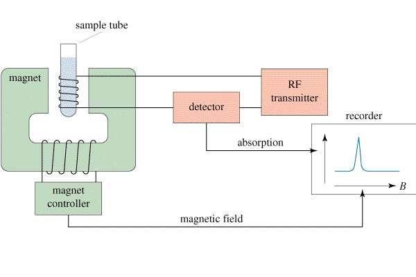
20 20 The NMR Spectrometer
21 The sample holder The sample holder is situated in a sample probe located at the intersection of the planes of the applied magnetic field, the applied radio-frequency field, and the detector radio-frequency field, all three of which are mutually perpendicular. It is a small tube about 5 mm O.D., is composed of glass or some other chemically inert material, and is transparent to RF radiation. It is about 3 in. long and contains approximately 0. 5 ml of the liquid. 21
22 The Permanent Magnet The magnet must be of a large capacity (approximately 14,000 G) and must be capable of producing and maintaining a strong homogeneous field for an extend period of time. Since the homogeneous field (1 part in 10 8 ) requirement is critical that any slight temperature variation would change the physical dimesion of the magnet and thus the field homogeneity, thermostatically control systems are utilized. 22
23 The Radio-frequency Generator A radio-frequency oscillator is used to produce the RF radiation. The RF coil is wound around the sample probe in such a manner that the field plane is perpendicular to the plane of the applied magnetic field and the plane of the detector RF field. The oscillator must yield the same degree of precision as the magnet, i.e., about part in 10 8 for high resolution applications. 23
24 The Sweep System The resonance energy may be obtained by either changing the applied magnet field (field sweeping) and holding the RF field constant, or by changing the applied RF field (frequency sweeping) and holding the applied magnetic field constant. The many difficulties which may be encountered when attempting to vary a magnetic field of a large permanent magnet are avoided by superimposing a small variable magnetic field (usually furnished by a set of Helmholtz coils) on top of the permanent magnetic field. 24
25 The Detector and Recorder The detector is an RF coil, the field of which is perpendicular to both the applied RF and magnetic fields. It detects the radiation which is absorbed and subsequently reemitted by the sample. From the detector the signal passes to a pre-calibrated recorder. 25
26 Sample handling A dilute solution (about 2 to 10%) is normally used If we want to study protons in the sample, the ideal solvent should contain no additional protons. Carbon tetrachloride meets this requirement Deuterated chloroform, CDCl 3, or deuterated benzene are preferably used Deuterated solvents may give a small additional peak for a residual proton impurity Deuterium oxide (deuterated water) is also available for samples soluble only in aqueous solutions A 5-mm OD glass tube serves as a sample container. It is held by a propeller (rigid) arrangement so that it can be spun by a jet of compressed air 26
27 The absorption Spectrum As previously stated, the proton, 11 H, nuclei may possess only one of two possible energy states. The transition from the low to high energy corresponds to the absorption process, while that from the high to the low to an emission. If an equal population of the nuclei existed at the two energy levels, the energy lost by emission would equal the energy gained by absorption and nothing could be measured. This is not the case. A very slight (approximately %) excess of nuclei exists in the lower energy spin state. It is this slight excess upon which nuclear magnetic resonance depends. 27
28 IN THE CLASSICAL NMR EXPERIMENT THE INSTRUMENT SCANS FROM LOW FIELD TO HIGH FIELD LOW FIELD NMR CHART HIGH FIELD DOWNFIELD scan UPFIELD

29 NMR Spectrum of Phenylacetone O CH 2 C CH 3 Each different type of proton comes at a different place. You can tell how many different types of hydrogen there are in the molecule.
30 Peaks integration Not only does each different type of hydrogen give a distinct peak in the NMR spectrum, but we can also tell the relative numbers of each type of hydrogen by a process called integration. Integration = determination of the area under a peak The area under a peak is proportional to the number of hydrogens that generate the peak.
31 Benzyl acetate The integral line rises an amount proportional to the number of H in each peak integral line METHOD 1 integral line 55 : 22 : 33 = 5 : 2 : 3 simplest ratio of the heights
32 Benzyl acetate (FT-NMR) Actually : / 11.3 = / 11.3 = / 11.3 = 3.00 CH 2 O O C CH 3 METHOD 2 digital integration assume CH / 3 = 11.3 Integrals are good to about 10% accuracy. Modern instruments report the integral as a number.
33 Magnetic Shielding If all protons absorbed the same amount of energy in a given magnetic field, not much information could be obtained. But protons are surrounded by electrons that shield them from the external field. Circulating electrons create an induced magnetic field that opposes the external magnetic field.
34 Shielding by valence electrons (Diamagnetic anisotropy) The applied field induces circulation of the valence electrons - this generates a magnetic field that opposes the applied field. valence electrons shield the nucleus from the full effect of the applied field magnetic field lines B o applied B induced (opposes B o ) fields subtract at nucleus

35 Shielded Protons Magnetic field strength must be increased for a shielded proton to flip at the same frequency.
36 Protons differ in their shielding All different types of protons in a molecule have a different amounts of shielding. They all respond differently to the applied magnetic field and appear at different places in the spectrum. This is why an NMR spectrum contains useful information (different types of protons appear in predictable places). DOWNFIELD Less shielded protons appear here. SPECTRUM UPFIELD Highly shielded protons appear here. It takes a higher field to cause resonance.
37 H 3 C CH 3 Si CH 3 CH 3 Tetramethylsilane, TMS The chemical shifts of nuclei are measured (and defined) relative to a standard nucleus. A popular standard for proton NMR is tetramethylsilane (TMS), which has the chemical formula Si(CH 3 ) 4. In this compound all 12 hydrogen nuclei are chemically equivalent; that is, they are all exposed to the same shielding and give a single absorption peak. The chemical shift for other hydrogen nuclei is represented as follows:
38 Delta Scale Chapter =>
39 Herz equivalence of 1 ppm 1 H Operating Frequency Hz Equivalent of 1 ppm 60 Mhz 60 Hz 100 MHz 100 Hz 300 MHz 300 Hz 1 part per million of n MHz is n Hz n MHz 1 ( ) 10 6 = n Hz ppm Each ppm unit represents either a 1 ppm change in B o (magnetic field strength, Tesla) or a 1 ppm change in the precessional frequency (MHz).
40 Shielding and deshielding of the nuclei In terms of NMR, within a given molecule, the electronic and steric environment of each nucleus produces a shielding effect against the external field Bo. As the screening effect increases, the nuclei are said to be shielded On a continuous wave instrument operating at fixed frequency, the intensity of the field Bo has to be increased in order to obtain resonance. Signals to the right of the spectrum are said to be resonant at high field. Signals observed to the left of the spectrum correspond to deshielded nuclei and are said to be resonant at low field
41 Peaks are measured relevant to TMS Rather than measure the exact resonance position of a peak, we measure how far downfield it is shifted from TMS. CH 3 CH 3 Si CH 3 CH 3 reference compound tetramethylsilane TMS Highly shielded protons appear way upfield. n shift in Hz downfield TMS 0 Chemists originally thought no other compound would come at a higher field than TMS.

42 Protons in a Molecule Depending on their chemical environment, protons in a molecule are shielded by different amounts.
43 Higher frequencies give larger shifts The shift observed for a given proton in Hz also depends on the frequency of the instrument used. Higher frequencies = larger shifts in Hz. n shift in Hz downfield TMS 0
44 The chemical shift The shifts from TMS in Hz are bigger in higher field instruments (300 MHz, 500 MHz) than they are in the lower field instruments (100 MHz, 60 MHz). We can adjust the shift to a field-independent value, the chemical shift in the following way: parts per million chemical shift = d = shift in Hz spectrometer frequency in MHz = ppm This division gives a number independent of the instrument used. A particular proton in a given molecule will always come at the same chemical shift (constant value).
45 The Chemical Shift (Also Called d) Scale Here s how it works. We decide on a sample we ll use to standardize our instruments. We take an NMR of that standard and measure its absorbance frequency. We then measure the frequency of our sample and subtract its frequency from that of the standard. We then then divide by the frequency of the standard. This gives a number called the chemical shift, which does not depend on the magnetic field strength. Why not? Let s look at two examples.
46 Imagine that we have a magnet where our standard absorbs at 300,000,000 Hz (300 megahertz), and our sample absorbs at 300,000,300 Hz. The difference is 300 Hz, so we take 300/300,000,000 = 1/1,000,000 and call that 1 part per million (or 1ppm)
47 Now lets examine the same sample in a stronger magnetic field where the reference comes at 500,000,000 Hz, or 500 megahertz. The frequency of our sample will increase proportionally, and will come at 500,000,500 Hz. The difference is now 500 Hz, but we divide by 500,000,000 (500/500,000,000 = 1/1,000,000, = 1 ppm).
48 If the sample nucleus is upfield it means that d is positive and that the sample is more highly shielded than the reference. When downfield, d is negative and the reference is more highly shielded than the sample. The unit of the chemical shift is ppm. This is not a concentration unit but is the ratio of numerator and denominator in the above equation, i.e., it is the difference in the magnetic field of the sample, H S, and the magnetic field of the reference, H r, divided by the magnetic field of the reference 48
49 The chemical shift If NMR were suitable only for detecting and measuring the presence of hydrogen or carbon in organic compounds, it would be a technique with very limited usefulness. There are a number of fast, inexpensive methods for detecting and measuring hydrogen and carbon in organic compounds. According to the NMR experiment, protons in different chemical environments within a molecule absorb at slightly different frequencies. This variation in absorption frequency is caused by a slight difference in the electronic environment of the proton as a result of different chemical bonds and adjacent atoms. The absorption frequency for a given proton depends on the chemical structure of the molecule.
50 This variation in absorption frequency is called the chemical shift. The same type of chemical shift occurs for carbon in different chemical environments within a molecule. NMR were suitable only for detecting and measuring the presence of hydrogen or carbon in organic compounds, it would be a technique with very limited usefulness. There are a number of fast, inexpensive methods for detecting and measuring hydrogen and carbon in organic compounds. The absorption frequency for a given proton depends on the chemical structure of the molecule. This variation in absorption frequency is called the chemical shift. The same type of chemical shift occurs for carbon in different chemical environments within a molecule.
51 Suppose that we take a molecule with several different "types" of hydrogen atoms, such as the molecule ethanol, CH 3 CH 2 OH. This molecule has hydrogen atoms in three different chemical environments: the three hydrogen atoms in the terminal CH 3, The two hydrogen atoms in the CH 2 group, and the one in the OH group. Consider the nuclei of the different types of hydrogen. Each one is surrounded by orbiting electrons, but the orbitals may vary in shape and the bonds vary in electron density distribution. This changes the length of time the electrons spend near a given type of hydrogen nucleus. Let us suppose that we place this molecule in a strong magnetic field Bo. The electrons associated with the nuclei will be rotated by the applied magnetic field Bo.
52 This rotation, or drift, generates a small magnetic field which opposes the much larger applied magnetic field Bo. The nuclei are shielded slightly from the applied magnetic field by the orbiting electrons. The extent of the shielding depends on the movement of the electrons caused by the magnetic field (not by the simple orbiting of the electrons). A shielded nucleus resonates or absorbs at a lower frequency than an unshielded nucleus. This change in frequency of absorption because of shielding is the chemical shift. Instead of one absorption signal for the protons in ethanol, we would predict three absorption signals at slightly different frequencies.
53 Absorption of CH 3 CH 2 OH
54 The chemical shifts of nuclei are measured (and defined) relative to a standard nucleus. A popular standard for proton NMR is tetramethylsilane (TMS), which has the chemical formula Si(CH 3 ) 4. In this compound all 12 hydrogen nuclei are chemically equivalent; that is, they are all exposed to the same shielding and give a single absorption peak. The chemical shift for other hydrogen nuclei is represented as follows:

55 The NMR Spectrum
56 Number of Major Absorption Peaks The number of major absorption peaks in an nmr spectrum equals the number of different types of protons which exist in the molecular compound. The protons within the molecule which possess the same magnetic environment are said to be equivalent and will absorb at the same applied field strength. Protons with different magnetic environments will absorb at different applied field strengths and are non-equivalent. The equivalency or non-equivalency of protons in any given molecule is determined by the type and character of its bonding to adjacent atoms and in some cases to a group of atoms. 56
57 Hydrogens bonded to different atoms are non-equivalent. For example, in methyl alcohol, CH 3 OH, the hydrogens bonded to the carbon are not equivalent to that one which is bonded to the oxygen and two major absorption peaks will be obtained. Because of the tetrahedral nature of the carbon atom, the three hydrogens in the methyl group are equivalent to each other and appear as a single major absorption peak in an nmr spectrum. The other major absorption peak arises from the OH bonding
58
59 The two hydrogens in a CH 2 group are equivalent to each other. Three major absorption peaks would be expected from CH 3 -CH 2 -OH; One from the CH 3 group, one from the CH 2, group, and one from the OH group.
60
61 At times it is necessary to consider not only the atom to which the hydrogen is bonded but also any bonds which are once or twice removed from this primary bond. For example, the bonding of the CH 3 hydrogens in a CH 3 -CH 2 - group will not be equivalent to the bonding of the CH 3 hydrogens in a CH 3 -O- group. The compound methyl ethyl ether, CH 3 -O-CH 2 -CH 3 will show three major absorption bands-one for the hydrogens of the methyl group with the CH 3 - O bond, one for hydrogens of the methyl group with the CH 3 -CH 2 - bond, and one from the hydrogens of the CH 2 group. On the other hand, diethyl ether, CH 3 CH 2 -O-CH 2 -CH 3, shows only two major bands.
62 Bonding to atoms which are twice removed must also be considered. For example, all of the groups of hydrogen atoms in CH 3 -CH 2 -CH 2 OH are non-equivalent and four major absorption peaks will be observed. In the benzene molecule all six hydrogens are equivalent since the bonding of all six to the carbon atoms is identical. Only one peak will be observed. A substitution on one or more of the positions of the benzene ring will upset this equivalency -the extent of change depends upon the number and/or nature of the substituted group(s). The presence of double or triple bonds may cause a non-equivalency
63 Areas Under Absorption Peaks Area under an nmr absorption peak is directly proportional to the number of,equivalent protons which are causing the peak. The instruments are equipped with an electronic integrator. The integral curve is recorded on the vertical axis of the recorder chart simultaneously with the absorption curves. The relative areas of the peaks are determined by the heights of the integral curves at the respective frequencies. These data are then reduced to a whole number ratio and may be used for quantitative determination or for the elucidation of structure
64
65 65
66 Spin-spin coupling (Splitting) The signals in the NMR spectra don t appear as single lines, sometimes they appear as multiple lines. This is due to 1 H - 1 H coupling (also called spin-spin splitting or J-coupling). Imagine we have a molecule which contains a proton (let s call it H A ) attached to a carbon, and that this carbon is attached to another carbon which also contains a proton (let s call it H B ). It turns out that H A feels the presence of H B. Since these protons are tiny little magnets, they can be oriented either with or against the magnetic field of the NMR machine.
67 When the field created by H B reinforces the magnetic field of the NMR machine (B0 ) HA feels a slightly stronger field, but when the field created by HB opposes B0, HA feels a slightly weaker field. So, we see two signals for HA depending on the alignment of HB. The same is true for HB, it can feel either a slightly stronger or weaker field due to HA s presence. So, rather than see a single line for each of these protons, we see two lines for each. For this line, H B is lined up with the magnetic field (adds to the overall magnetic field, so the line comes at higher frequency) For this line, H B is lined up against the magnetic field (subtracts from the overall magnetic field, so the line comes at lower frequency) H A H B H A H A is split into two lines because it feels the magnetic field of H B. H B H B is split into two lines because it feels the magnetic field of H A. C C
68 Each hydrogen nucleus of an organic molecule is spinning and is magnetic. Since the axis of rotation of the nucleus may be with or against the applied magnetic field, the magnetic field of the nucleus may be parallel with or opposed to the applied magnetic field. With reference to a molecule of ethyl alcohol, The spinning of the hydrogen on the OH group causes a small magnetic field which may be with or against the applied magnetic field.
69 This spinning interferes with the spinning of the adjacent CH, group causing the effective field of the CH, group to change slightly. The CH, hydrogens which are adjacent to the hydrogen spinning with the field absorb at a slightly different frequency from that of the CH, hydrogens which are spinning against the field. The number of possible spin combinations, and the relative numbers of each combination, and thus the fine absorption spectra are predictable.
70 A. For a single functional group the number of bands = (n + 1) where n is the number of equivalent hydrogens on the adjacent group. See methyl alcohol or methyl ethyl ketone examples in the Table B. f the protons of the two adjacent groups are not equivalent to each other, the multiplicity of bands will be (n 1 + 1)(n 2 + 1) where n 1 and n 2 are the number of equivalent protons on the first and second groups, respectively. See the ethyl alcohol and 1-chloropropane examples in the Table
71 C.If all the carbon atoms of a group such as CH 3 or a compound such as in H 3 C-CH 3 are equivalent, splitting will not occur. See ethane in the Table D.The protons of the aromatic hydrogens on the substituted benzene ring are often non-equivalent. Complex splitting patterns may occur.
72 72
73 73
74 The number of peaks which will actually be seen depends upon the resolution of the instrument. The splitting pattern of isobutyl alcohol and normal butyl alcohol can be seen in the Figures below.
75 NMR absorption spectrum of normal butyl alcohol
76 NMR absorption spectra of ethyl alcohol. The numbers designate the relative areas of the groups in the low resolution spectrum and of the peaks within a functional group in the high resolution spectrum
77 The absorption position of a particular functional group will vary with the applied magnetic field strength but the magnitude of separation which is caused by spin-spin coupling is independent of the applied field strength. This spacing is called the spin- spin coupling constant, is denoted by the symbol, J, and has the units of frequency in cycles per second, cps.
78 There is no coupling between hydrogens on the same carbon. The constant, J, is from 6-8 cps between the hydrogens on adjacent carbons and decreases rapidly as the distance between the carbons increases. The areas of the peaks which arise from spin - spin splitting are in direct ratio to the number of possibilities of each of these peaks being observed. For example, the relative areas of the - CH, - CH 2, and - OH groups in ethyl alcohol should be in the ratio of 3:2:1, respectively. Upon higher resolution where splitting occurs, the three bands of the - CH 3 triplet would exist in an area ratio of 1:2:1 and the quartet of the - CH 2 group would have a ratio of 1: 3: 3:1. See last Figure above.
79 Analytical applications The procedures are limited to structure determinations involving nuclei which possess either an odd mass number such as the proton or an even mass number with an odd atomic number such as the deuterium The nmr spectroscopy techniques are applied primarily to liquid samples. The absorption bands for solids are generally too wide and instruments are not sensitive enough for gaseous samples withou instrumental modification. Liquid sample sizes range from 0.1 to 0.4 ml. If the sample itself is a liquid it may be determined directly; if not, it may be dissolved in a suitable solvent. The minimum concentration which may be detected in a liquid is approximately 1%. The choice of a liquid solvent is made on the basis of its purity, chemical stability and volatility.
80 CARBON-13 NMR
81 13 C NMR 13 C signals are 6000 times weaker than 1 H because: 1. Natural abundance of 13 C is small (1.08% of all C) 2. Magnetic moment of 13 C is small 3. The chemical shift range is larger than for protons ppm
82 Because of its low natural abundance (0.0108) there is a low probability of finding two 13 C atoms next to each other in a single molecule. 13 C - 13 C Coupling not probable Spectra are determined by many molecules contributing to the spectrum, each having only one 13 C atom. However, 13 C does couple to hydrogen atoms (I = 1/2) 13 C - 1 H coupling YES! very common
83 COUPLING TO ATTACHED PROTONS
84 COUPLING TO ATTACHED PROTONS 3 protons 2 protons 1 proton 0 protons H H 13 C H 13 C H 13 C H 13 C H n+1 = 4 n+1 = 3 n+1 = 2 n+1 = 1 Methyl carbon Methylene carbon Methine carbon Quaternary carbon The effect of attached protons on 13 C resonances ( n+1 rule applies ) (J s are large ~ Hz)
85 ETHYL PHENYLACETATE 13 C coupled to the hydrogens
86 Qualitative Analysis nmr would offer little information which is not available by other less expensive methods. The nmr is primarily applied in organic chemistry and is used in the elucidation of the structure of and identification of organic molecules. As we have seen, the chemical shift yields information as to the types of hydrogens which are present, whereas the spin-spin splitting yields information concerning the nature of the neighboring groups. These two features, combined with the fact that the relative areas of absorption peaks are in direct proportion to the number of equivalent hydrogens in each group, make nmr a powerful structure identification tool.
87 Quantitative Analysis The fact that the areas of nmr absorption peaks are in direct proportion to the types of hydrogens which are causing the respective peaks is the basis for the nmr quantitative analysis of organic liquids. These areas may be rapidly determined with the integration recorders. In order for quantitative procedures to be applied, the components must be known. In addition, each component of the mixture must provide at least one absorption band which is isolated from the absorption bands of any other components. Impure samples may be determined by the addition of a known pure compound as an internal standard. Quantitative applications have been used to elucidate chain length or the percentage of hydrogen in a pure compound. The degree of saturation in a compound of known chain length and known percentage composition may be calculated.
88 The Hard Part - Interpreting Spectra Learning how an NMR machine works is straightforward. What is less straightforward is learning how to use the data we get from an NMR machine (the spectrum of ethyl acetate is shown below). That s because each NMR spectrum is a puzzle, and there s no single fact that you simply have to memorize to solve these spectra. You have to consider lots of pieces of data and come up with a structure that fits all the data. What kinds of data do we get from NMR spectra? For 1 H NMR, there are three kinds each of which we will consider each of these separately: 1) Chemical shift data - tells us what kinds of protons we have. 2) Integrals - tells us the ratio of each kind of proton in our sample. 3) 1 H - 1 H coupling - tells us about protons that are near other protons.
Nuclear Magnetic Resonance
 Nuclear Magnetic Resonance PRINCIPLES OF NMR SPECTROSCOPY Contents Principles of nuclear magnetic resonance The nmr spectrometer Basic principles in nmr application NMR tools used to obtain information
Chapter 15 Lecture Outline
 Organic Chemistry, First Edition Janice Gorzynski Smith University of Hawaii Chapter 5 Lecture Outline Introduction to NMR Two common types of NMR spectroscopy are used to characterize organic structure:
Nuclear Magnetic Resonance (NMR) Spectroscopy Introduction:
 Spectroscopy Introduction:") Nuclear Magnetic Resonance (NMR) Spectroscopy Introduction: Nuclear magnetic resonance spectroscopy (NMR) is the most powerful tool available for organic structure determination. Like IR spectroscopy,
Nuclear Magnetic Resonance (NMR) Spectroscopy Introduction: Nuclear magnetic resonance spectroscopy (NMR) is the most powerful tool available for organic structure determination. Like IR spectroscopy,
16.1 Introduction to NMR Spectroscopy. Spectroscopy. Spectroscopy. Spectroscopy. Spectroscopy. Spectroscopy 4/11/2013
 What is spectroscopy? NUCLEAR MAGNETIC RESONANCE (NMR) spectroscopy may be the most powerful method of gaining structural information about organic compounds. NMR involves an interaction between electromagnetic
Chapter 14. Nuclear Magnetic Resonance Spectroscopy
 Organic Chemistry, Second Edition Janice Gorzynski Smith University of Hawai i Chapter 14 Nuclear Magnetic Resonance Spectroscopy Prepared by Rabi Ann Musah State University of New York at Albany Copyright
Module 13: Chemical Shift and Its Measurement
 Subject Chemistry Paper No and Title Module No and Title Module Tag Paper 12: Organic Spectroscopy CHE_P12_M13_e-Text TABLE OF CONTENTS 1. Learning Outcomes 2. Introduction 3. Shielding and deshielding
NMR Spectroscopy. for 1 st B.Tech INTRODUCTION Lecture -1 Indian Institute of Technology, Dhanbad
 NMR Spectroscopy for 1 st B.Tech Lecture -1 Indian Institute of Technology, Dhanbad by Dr. R P John & Dr. C. Halder INTRODUCTION Nucleus of any atom has protons and neutrons Both Proton and Neutron has
7a. Structure Elucidation: IR and 13 C-NMR Spectroscopies (text , , 12.10)
") 2009, Department of Chemistry, The University of Western Ontario 7a.1 7a. Structure Elucidation: IR and 13 C-NMR Spectroscopies (text 11.1 11.5, 12.1 12.5, 12.10) A. Electromagnetic Radiation Energy is
2009, Department of Chemistry, The University of Western Ontario 7a.1 7a. Structure Elucidation: IR and 13 C-NMR Spectroscopies (text 11.1 11.5, 12.1 12.5, 12.10) A. Electromagnetic Radiation Energy is
Chapter 7. Nuclear Magnetic Resonance Spectroscopy
 Chapter 7 Nuclear Magnetic Resonance Spectroscopy I. Introduction 1924, W. Pauli proposed that certain atomic nuclei have spin and magnetic moment and exposure to magnetic field would lead to energy level
Chem 325 NMR Intro. The Electromagnetic Spectrum. Physical properties, chemical properties, formulas Shedding real light on molecular structure:
 Physical properties, chemical properties, formulas Shedding real light on molecular structure: Wavelength Frequency ν Wavelength λ Frequency ν Velocity c = 2.998 10 8 m s -1 The Electromagnetic Spectrum
MOLECULAR SPECTROSCOPY AND PHOTOCHEMISTRY
 20 CHAPTER MOLECULAR SPECTROSCOPY AND PHOTOCHEMISTRY 20.1 Introduction to Molecular Spectroscopy 20.2 Experimental Methods in Molecular Spectroscopy 20.3 Rotational and Vibrational Spectroscopy 20.4 Nuclear
Chapter 13 Structure t Determination: Nuclear Magnetic Resonance Spectroscopy
 John E. McMurry www.cengage.com/chemistry/mcmurry Chapter 13 Structure t Determination: ti Nuclear Magnetic Resonance Spectroscopy Revisions by Dr. Daniel Holmes MSU Paul D. Adams University of Arkansas
NMR = Nuclear Magnetic Resonance
 NMR = Nuclear Magnetic Resonance NMR spectroscopy is the most powerful technique available to organic chemists for determining molecular structures. Looks at nuclei with odd mass numbers or odd number
NMR Spectroscopy. Chapter 19
 NMR Spectroscopy Chapter 19 Nuclear Magnetic Resonance spectroscopy is a powerful analytical technique used to characterize organic molecules by identifying carbon-hydrogen frameworks within molecules.
NUCLEAR MAGNETIC RESONANCE SPECTROSCOPY
 NMR Spectroscopy 1 NULEAR MAGNETI RESONANE SPETROSOPY Involves interaction of materials with the low-energy radiowave region of the electromagnetic spectrum Origin of Spectra Theory All nuclei possess
16.1 Introduction to NMR. Spectroscopy
 16.1 Introduction to NMR What is spectroscopy? Spectroscopy NUCLEAR MAGNETIC RESNANCE (NMR) spectroscopy may be the most powerful method of gaining structural information about organic compounds. NMR involves
William H. Brown & Christopher S. Foote
 Requests for permission to make copies of any part of the work should be mailed to:permissions Department, Harcourt Brace & Company, 6277 Sea Harbor Drive, Orlando, Florida 32887-6777 William H. Brown
Nuclear Magnetic Resonance Spectroscopy: Tools for Structure Determination
 Nuclear Magnetic Resonance Spectroscopy: Tools for Structure Determination Chung-Ming Sun Department of Applied Chemistry National Chiao Tung University Hualien 300, Taiwan Introduction NMR (Nuclear Magnetic
Chapter 13 Nuclear Magnetic Resonance Spectroscopy
 Organic Chemistry, 6 th Edition L. G. Wade, Jr. Chapter 13 Nuclear Magnetic Resonance Spectroscopy Jo Blackburn Richland College, Dallas, TX Dallas County Community College District 2006, Prentice Hall
Química Orgânica I. Nuclear Magnetic Resonance Spectroscopy (I) Ciências Farmacêuticas Bioquímica Química AFB QO I 2007/08 1 AFB QO I 2007/08 2
 Ciências Farmacêuticas Bioquímica Química AFB QO I 2007/08 1 AFB QO I 2007/08 2") Química Orgânica I Ciências Farmacêuticas Bioquímica Química AFB QO I 2007/08 1 Nuclear Magnetic Resonance Spectroscopy (I) AFB QO I 2007/08 2 1 Adaptado de: Organic Chemistry, 6th Edition; L. G. Wade,
Química Orgânica I Ciências Farmacêuticas Bioquímica Química AFB QO I 2007/08 1 Nuclear Magnetic Resonance Spectroscopy (I) AFB QO I 2007/08 2 1 Adaptado de: Organic Chemistry, 6th Edition; L. G. Wade,
Principles of Molecular Spectroscopy: Electromagnetic Radiation and Molecular structure. Nuclear Magnetic Resonance (NMR)
") Principles of Molecular Spectroscopy: Electromagnetic Radiation and Molecular structure Nuclear Magnetic Resonance (NMR) !E = h" Electromagnetic radiation is absorbed when the energy of photon corresponds
Principles of Molecular Spectroscopy: Electromagnetic Radiation and Molecular structure Nuclear Magnetic Resonance (NMR) !E = h" Electromagnetic radiation is absorbed when the energy of photon corresponds
Chapter 9. Nuclear Magnetic Resonance. Ch. 9-1
 Chapter 9 Nuclear Magnetic Resonance Ch. 9-1 1. Introduction Classic methods for organic structure determination Boiling point Refractive index Solubility tests Functional group tests Derivative preparation
- 1/2. = kb o = hνν + 1/2. B o increasing magnetic field strength. degenerate at B o = 0
 NMR EXPERIMENT When magnetically active nuclei are placed into an external magnetic field, the magnetic fields align themselves with the external field into two orientations. During the experiment, electromagnetic
1. neopentyl benzene. 4 of 6
 I. 1 H NMR spectroscopy A. Theory 1. The protons and neutrons in atomic nuclei spin, as does the nucleus itself 2. The circulation of nuclear charge can generate a nuclear magnetic moment, u, along the
CHEM Chapter 13. Nuclear Magnetic Spectroscopy (Homework) W
 W") CHEM 2423. Chapter 13. Nuclear Magnetic Spectroscopy (Homework) W Short Answer 1. For a nucleus to exhibit the nuclear magnetic resonance phenomenon, it must be magnetic. Magnetic nuclei include: a. all
CHEM 2423. Chapter 13. Nuclear Magnetic Spectroscopy (Homework) W Short Answer 1. For a nucleus to exhibit the nuclear magnetic resonance phenomenon, it must be magnetic. Magnetic nuclei include: a. all
Structure Determination: Nuclear Magnetic Resonance Spectroscopy
 Structure Determination: Nuclear Magnetic Resonance Spectroscopy Why This Chapter? NMR is the most valuable spectroscopic technique used for structure determination More advanced NMR techniques are used
Chapter 16 Nuclear Magnetic Resonance Spectroscopy
 hapter 16 Nuclear Magnetic Resonance Spectroscopy The Spinning Proton A spinning proton generates a magnetic field, resembling that of a small bar magnet. An odd number of protons in the nucleus creates
4) protons experience a net magnetic field strength that is smaller than the applied magnetic field.
 protons experience a net magnetic field strength that is smaller than the applied magnetic field.") 1) Which of the following CANNOT be probed by an NMR spectrometer? See sect 15.1 Chapter 15: 1 A) nucleus with odd number of protons & odd number of neutrons B) nucleus with odd number of protons &even
1) Which of the following CANNOT be probed by an NMR spectrometer? See sect 15.1 Chapter 15: 1 A) nucleus with odd number of protons & odd number of neutrons B) nucleus with odd number of protons &even
Nuclear spin and the splitting of energy levels in a magnetic field
 Nuclear spin and the splitting of energy levels in a magnetic field Top 3 list for 13 C NMR Interpretation 1. Symmetry 2. Chemical Shifts 3. Multiplicity 13 C NMR of C 3 O 1 NMR of C 3 O 13 C NMR of C
Chapter 13: Molecular Spectroscopy
 Chapter 13: Molecular Spectroscopy Electromagnetic Radiation E = hν h = Planck s Constant (6.63 x 10⁻³⁴ J. s) ν = frequency (s -1 ) c = νλ λ = wavelength (nm) Energy is proportional to frequency Spectrum
The Use of NMR Spectroscopy
 Spektroskopi Molekul Organik (SMO): Nuclear Magnetic Resonance (NMR) Spectroscopy All is adopted from McMurry s Organic Chemistry The Use of NMR Spectroscopy Used to determine relative location of atoms
NMR Nuclear Magnetic Resonance Spectroscopy p. 83. a hydrogen nucleus (a proton) has a charge, spread over the surface
 has a charge, spread over the surface") NMR Nuclear Magnetic Resonance Spectroscopy p. 83 a hydrogen nucleus (a proton) has a charge, spread over the surface a spinning charge produces a magnetic moment (a vector = direction + magnitude) along
NMR Nuclear Magnetic Resonance Spectroscopy p. 83 a hydrogen nucleus (a proton) has a charge, spread over the surface a spinning charge produces a magnetic moment (a vector = direction + magnitude) along
Nuclear Magnetic Resonance H-NMR Part 1 Introduction to NMR, Instrumentation, Sample Prep, Chemical Shift. Dr. Sapna Gupta
 Nuclear Magnetic Resonance H-NMR Part 1 Introduction to NMR, Instrumentation, Sample Prep, Chemical Shift Dr. Sapna Gupta Introduction NMR is the most powerful tool available for organic structure determination.
3.15 Nuclear Magnetic Resonance Spectroscopy, NMR
 3.15 Nuclear Magnetic Resonance Spectroscopy, NMR What is Nuclear Magnetic Resonance - NMR Developed by chemists and physicists together it works by the interaction of magnetic properties of certain nuclei
Nuclear Magnetic Resonance Spectroscopy
 Nuclear Magnetic Resonance Spectroscopy Structural Elucidation Nuclear magnetic resonance spectroscopy is the name given to the technique which exploits the magnetic properties of nuclei and measures their
Instrumental Chemical Analysis
 L15 Page1 Instrumental Chemical Analysis Nuclear Magnetic Resonance Dr. Ahmad Najjar Philadelphia University Faculty of Pharmacy Department of Pharmaceutical Sciences 1 st semester, 2017/2018 Nuclear Magnetic
Nuclear magnetic resonance spectroscopy
 nuclear spin transitions O Nuclear magnetic resonance spectroscopy 1 H, 13 C, 2-dimensional which transitions? wavelength and intensity; ppm what happens if we change the environment of the nucleus? substituent
In a solution, there are thousands of atoms generating magnetic fields, all in random directions.
 Nuclear Magnetic Resonance Spectroscopy: Purpose: onnectivity, Map of - framework Process: In nuclear magnetic resonance spectroscopy, we are studying nuclei. onsider this circle to represent a nucleus
Chapter 13: Nuclear Magnetic Resonance (NMR) Spectroscopy direct observation of the H s and C s of a molecules
 Spectroscopy direct observation of the H s and C s of a molecules") hapter 13: Nuclear Magnetic Resonance (NMR) Spectroscopy direct observation of the s and s of a molecules Nuclei are positively charged and spin on an axis; they create a tiny magnetic field + + Not all
hapter 13: Nuclear Magnetic Resonance (NMR) Spectroscopy direct observation of the s and s of a molecules Nuclei are positively charged and spin on an axis; they create a tiny magnetic field + + Not all
UNIT 12 NMR SPECTROSCOPY
 UIT 12 MR SPECTROSCOPY MR Spectroscopy Structure 12.1 Introduction 12.2 Theory of MR Spectroscopy Types of uclei Magnetic Moment Quantisation Population of Energy Levels Larmor Precession Mechanism of
In a solution, there are thousands of atoms generating magnetic fields, all in random directions.
 Nuclear Magnetic Resonance Spectroscopy: Purpose: onnectivity, Map of - framework Process: In nuclear magnetic resonance spectroscopy, we are studying nuclei. onsider this circle to represent a nucleus
Nuclear Magnetic Resonance (NMR)
") Nuclear Magnetic Resonance (NMR) E E increases with increasing magnetic field strength Boltzmann distribution at thermal equilibrium: N (m=-1/2) /N (m=+1/2) = e ( E/kT) with E = γ(h/2π)b o NMR Physical
Nuclear Magnetic Resonance (NMR) E E increases with increasing magnetic field strength Boltzmann distribution at thermal equilibrium: N (m=-1/2) /N (m=+1/2) = e ( E/kT) with E = γ(h/2π)b o NMR Physical
Introduction of Key Concepts of Nuclear Magnetic Resonance
 I have not yet lost that sense of wonder, and delight, that this delicate motion should reside in all ordinary things around us, revealing itself only to those who looks for it. E. M. Purcell, Nobel Lecture.
Experiment 2 - NMR Spectroscopy
 Experiment 2 - NMR Spectroscopy OBJECTIVE to understand the important role of nuclear magnetic resonance spectroscopy in the study of the structures of organic compounds to develop an understanding of
Chapter 18: NMR Spectroscopy
 The most important tool of the chemist for the determination of molecular structure is Nuclear Magnetic Resonance Spectroscopy, or NMR spectroscopy. NMR spectra are acquired on a special instrument called
Lecture 02 Nuclear Magnetic Resonance Spectroscopy Principle and Application in Structure Elucidation
 Application of Spectroscopic Methods in Molecular Structure Determination Prof. S. Sankararaman Department of Chemistry Indian Institution of Technology Madras Lecture 02 Nuclear Magnetic Resonance Spectroscopy
Lecture Notes Chem 51A S. King
 Lecture Notes hem 51A S. King hapter 14 Nuclear Magnetic Resonance Spectroscopy Nuclear Magnetic Resonance (NMR) spectroscopy uses energy in the radiowave portion of the electromagnetic spectrum. The nuclei
Ferdowsi University of Mashhad
 Spectroscopy in Inorganic Chemistry Nuclear Magnetic Resonance Spectroscopy spin deuterium 2 helium 3 The neutron has 2 quarks with a -e/3 charge and one quark with a +2e/3 charge resulting in a total
Nuclear Magnetic Resonance Spectroscopy (NMR)
") OCR Chemistry A 432 Spectroscopy (NMR) What is it? An instrumental method that gives very detailed structural information about molecules. It can tell us - how many of certain types of atom a molecule
OCR Chemistry A 432 Spectroscopy (NMR) What is it? An instrumental method that gives very detailed structural information about molecules. It can tell us - how many of certain types of atom a molecule
4) protons experience a net magnetic field strength that is smaller than the applied magnetic field.
 protons experience a net magnetic field strength that is smaller than the applied magnetic field.") 1) Which of the following CANNOT be probed by an spectrometer? See sect 15.1 Chapter 15: 1 A) nucleus with odd number of protons & odd number of neutrons B) nucleus with odd number of protons &even number
1) Which of the following CANNOT be probed by an spectrometer? See sect 15.1 Chapter 15: 1 A) nucleus with odd number of protons & odd number of neutrons B) nucleus with odd number of protons &even number
Fundamental MRI Principles Module 2 N. Nuclear Magnetic Resonance. X-ray. MRI Hydrogen Protons. Page 1. Electrons
 Fundamental MRI Principles Module 2 N S 1 Nuclear Magnetic Resonance There are three main subatomic particles: protons positively charged neutrons no significant charge electrons negatively charged Protons
NMRis the most valuable spectroscopic technique for organic chemists because it maps the carbon-hydrogen framework of a molecule.
 Chapter 13: Nuclear magnetic resonance spectroscopy NMRis the most valuable spectroscopic technique for organic chemists because it maps the carbon-hydrogen framework of a molecule. 13.2 The nature of
Spectroscopy and Chromatography
 Spectroscopy and Chromatography Introduction Visible light is one very small part of the electromagnetic spectrum. The different properties of the various types of radiation depend upon their wavelength.
10.4 Continuous Wave NMR Instrumentation
 10.4 Continuous Wave NMR Instrumentation coherent detection bulk magnetization the rotating frame, and effective magnetic field generating a rotating frame, and precession in the laboratory frame spin-lattice
Electron Spin Resonance, Basic principle of NMR, Application of NMR in the study of Biomolecules, NMR imaging and in vivo NMR spectromicroscopy
 Electron Spin Resonance, Basic principle of NMR, Application of NMR in the study of Biomolecules, NMR imaging and in vivo NMR spectromicroscopy Mitesh Shrestha Electron Spin Resonance Electron paramagnetic
Experiment 11: NUCLEAR MAGNETIC RESONANCE SPECTROSCOPY
 Experiment 11: NUCLEAR MAGNETIC RESONANCE SPECTROSCOPY Purpose: This is an exercise to introduce the use of nuclear magnetic resonance spectroscopy, in conjunction with infrared spectroscopy, to determine
11. Proton NMR (text , 12.11, 12.12)
") 2009, Department of Chemistry, The University of Western Ontario 11.1 11. Proton NMR (text 12.6 12.9, 12.11, 12.12) A. Proton Signals Like 13 C, 1 H atoms have spins of ±½, and when they are placed in
2009, Department of Chemistry, The University of Western Ontario 11.1 11. Proton NMR (text 12.6 12.9, 12.11, 12.12) A. Proton Signals Like 13 C, 1 H atoms have spins of ±½, and when they are placed in
With that first concept in mind, it is seen that a spinning nucleus creates a magnetic field, like a bar magnet
 NMR SPECTROSCOPY This section will discuss the basics of NMR (nuclear magnetic resonance) spectroscopy. Most of the section will discuss mainly 1H or proton spectroscopy but the most popular nuclei in
Tuesday, January 13, NMR Spectroscopy
 NMR Spectroscopy NMR Phenomenon Nuclear Magnetic Resonance µ A spinning charged particle generates a magnetic field. A nucleus with a spin angular momentum will generate a magnetic moment (μ). If these
NMR Spectroscopy. This handout is intended to give you a practical understanding of NMR Spectroscopy.
 NMR Spectroscopy This handout is intended to give you a practical understanding of NMR Spectroscopy. 1. Quantum theory allows us to consider each nucleus as a spinning charge. Note: we are only considering
Chapter 14 Spectroscopy
 hapter 14 Spectroscopy There are four major analytical techniques used for identifying the structure of organic molecules 1. Nuclear Magnetic Resonance or NMR is the single most important technique for
Spectroscopy in Organic Chemistry. Types of Spectroscopy in Organic
 Spectroscopy in Organic Chemistry Spectroscopy Spectrum dealing with light, or more specifically, radiation Scope to see Organic Spectroscopy therefore deals with examining how organic molecules interact
Introduction to Nuclear Magnetic Resonance Spectroscopy
 Introduction to Nuclear Magnetic Resonance Spectroscopy Dr. Dean L. Olson, NMR Lab Director School of Chemical Sciences University of Illinois Called figures, equations, and tables are from Principles
Fundamental MRI Principles Module Two
 Fundamental MRI Principles Module Two 1 Nuclear Magnetic Resonance There are three main subatomic particles: protons neutrons electrons positively charged no significant charge negatively charged Protons
Can you differentiate A from B using 1 H NMR in each pair?
 Can you differentiate A from B using 1 H NMR in each pair? To be NMR active any nucleus must have a spin quantum number, different from zero (I 0) As in 1 H, the spin quantum number (I) of 13 C is 1/2
Nuclear Magnetic Resonance Spectroscopy
 Nuclear Magnetic Resonance Spectroscopy Features: Used to identify products of reactions Also gives information about chemical environment, connectivity and bonding of nuclei Requirements: Pure or mostly
Magnetic Resonance Spectroscopy EPR and NMR
 Magnetic Resonance Spectroscopy EPR and NMR A brief review of the relevant bits of quantum mechanics 1. Electrons have spin, - rotation of the charge about its axis generates a magnetic field at each electron.
Chapter 13 Spectroscopy
 hapter 13 Spectroscopy Infrared spectroscopy Ultraviolet-Visible spectroscopy Nuclear magnetic resonance spectroscopy Mass Spectrometry 13.1 Principles of Molecular Spectroscopy: Electromagnetic Radiation
Lecture 2 nmr Spectroscopy
 Lecture 2 nmr Spectroscopy Pages 427 430 and Chapter 13 Molecular Spectroscopy Molecular spectroscopy: the study of the frequencies of electromagnetic radiation that are absorbed or emitted by substances
Biochemistry 530 NMR Theory and Practice
 Biochemistry 530 NMR Theory and Practice Gabriele Varani Department of Biochemistry and Department of Chemistry University of Washington Lecturer: Gabriele Varani Biochemistry and Chemistry Room J479 and
Nuclear Magnetic Resonance Spectroscopy Thomas Wenzel Department of Chemistry Bates College, Lewiston ME
 Nuclear Magnetic Resonance Spectroscopy Thomas Wenzel Department of Chemistry Bates College, Lewiston ME 04240 twenzel@bates.edu The following textual material is designed to accompany a series of in-class
OAT Organic Chemistry - Problem Drill 19: NMR Spectroscopy and Mass Spectrometry
 OAT Organic Chemistry - Problem Drill 19: NMR Spectroscopy and Mass Spectrometry Question No. 1 of 10 Question 1. Which statement concerning NMR spectroscopy is incorrect? Question #01 (A) Only nuclei
Module 20: Applications of PMR in Structural Elucidation of Simple and Complex Compounds and 2-D NMR spectroscopy
 Subject Chemistry Paper No and Title Module No and Title Module Tag Paper 12: Organic Spectroscopy Module 20: Applications of PMR in Structural Elucidation of Simple and Complex Compounds and 2-D NMR spectroscopy
Unit 11 Instrumentation. Mass, Infrared and NMR Spectroscopy
 Unit 11 Instrumentation Mass, Infrared and NMR Spectroscopy Spectroscopic identification of organic compounds Qualitative analysis: presence but not quantity (i.e. PEDs) Quantitative analysis: quantity
4) protons experience a net magnetic field strength that is smaller than the applied magnetic field.
 protons experience a net magnetic field strength that is smaller than the applied magnetic field.") 1) Which of the following CANNOT be probed by an spectrometer? See sect 16.1 Chapter 16: 1 A) nucleus with odd number of protons & odd number of neutrons B) nucleus with odd number of protons &even number
1) Which of the following CANNOT be probed by an spectrometer? See sect 16.1 Chapter 16: 1 A) nucleus with odd number of protons & odd number of neutrons B) nucleus with odd number of protons &even number
Nuclear Spin States. NMR Phenomenon. NMR Instrumentation. NMR Active Nuclei. Nuclear Magnetic Resonance
 Nuclear Magnetic Resonance NMR Phenomenon µ A spinning charged particle generates a magnetic field. A nucleus with a spin angular momentum will generate a magnetic moment (!). E Nuclear Spin States aligned
Ala-Arg-Pro-Tyr-Asn-Phe-Cpa-Leu-NH 2
 Applied Spectroscop Ala-Arg-Pro-Tr-Asn-Phe-Cpa-Leu-NH 2 Cpa Ala Pro Guillermo Mona What is Spectroscop? Without going into latin or greek, spectroscop is the stud of the interactions between light and
January 30, 2018 Chemistry 328N
 Lecture 4 Some More nmr January 30, 2018 Tricks for solving unknowns Review. Empirical formula is lowest common denominator ratio of atomic composition From Homework: unknown has an empirical formula of
NUCLEAR MAGNETIC RESONANCE. The phenomenon of nuclear magnetic resonance will be used to study magnetic moments of nuclei.
 14 Sep 11 NMR.1 NUCLEAR MAGNETIC RESONANCE The phenomenon of nuclear magnetic resonance will be used to study magnetic moments of nuclei. Theory: In addition to its well-known properties of mass, charge,
4) protons experience a net magnetic field strength that is smaller than the applied magnetic field.
 protons experience a net magnetic field strength that is smaller than the applied magnetic field.") 1) Which of the following CANNOT be probed by an spectrometer? See sect 16.1 Chapter 16: 1 A) nucleus with odd number of protons & odd number of neutrons B) nucleus with odd number of protons &even number
1) Which of the following CANNOT be probed by an spectrometer? See sect 16.1 Chapter 16: 1 A) nucleus with odd number of protons & odd number of neutrons B) nucleus with odd number of protons &even number
Laboratory and Rotating frames
 Laborator and Rotating frames The coordinate sstem that we used for the previous eample (laborator frame) is reall pathetic. The whole sstem is spinning at ω o, which makes an kind of analsis impossible.
To Do s. Answer Keys are available in CHB204H
 To Do s Read Chapters 2, 3 & 4. Complete the end-of-chapter problems, 2-1, 2-2, 2-3 and 2-4 Complete the end-of-chapter problems, 3-1, 3-3, 3-4, 3-6 and 3-7 Complete the end-of-chapter problems, 4-1, 4-2,
Physical Background Of Nuclear Magnetic Resonance Spectroscopy
 Physical Background Of Nuclear Magnetic Resonance Spectroscopy Michael McClellan Spring 2009 Department of Physics and Physical Oceanography University of North Carolina Wilmington What is Spectroscopy?
ORGANIC - EGE 5E CH NUCLEAR MAGNETIC RESONANCE SPECTROSCOPY
 !! www.clutchprep.com CONCEPT: PURPOSE OF ANALYTICAL TECHNIQUES Classical Methods (Wet Chemistry): Chemists needed to run dozens of chemical reactions to determine the type of molecules in a compound.
Chapter 13 Nuclear Magnetic Resonance Spectroscopy
 William. Brown Christopher S. Foote Brent L. Iverson Eric Anslyn http://academic.cengage.com/chemistry/brown Chapter 13 Nuclear Magnetic Resonance Spectroscopy William. Brown Beloit College Two Nobel Prizes
Other problems to work: 3-Chloropentane (diastereotopic H s), 1- chloropentane.
, 1- chloropentane.") Let s look at some specific examples. Dichloroacetaldehyde, l 2 HHO, has two inequivalent toms, H1 and H2. We expect to see two resonances, one at around δ 10.5 ppm and one around δ 5.5 ppm. (The H2 resonance
Let s look at some specific examples. Dichloroacetaldehyde, l 2 HHO, has two inequivalent toms, H1 and H2. We expect to see two resonances, one at around δ 10.5 ppm and one around δ 5.5 ppm. (The H2 resonance
NMR Spectroscopy Laboratory Experiment Introduction. 2. Theory
 1. Introduction 64-311 Laboratory Experiment 11 NMR Spectroscopy Nuclear Magnetic Resonance (NMR) spectroscopy is a powerful and theoretically complex analytical tool. This experiment will introduce to
To Do s. Answer Keys are available in CHB204H
 To Do s Read Chapters 2, 3 & 4. Complete the end-of-chapter problems, 2-1, 2-2, 2-3 and 2-4 Complete the end-of-chapter problems, 3-1, 3-3, 3-4, 3-6 and 3-7 Complete the end-of-chapter problems, 4-1, 4-2,
C h a p t e r S i x t e e n: Nuclear Magnetic Resonance Spectroscopy. An 1 H NMR FID of ethanol
 0.2 0.4 0.6 0.8 1.0 1.2 1.4 1.6 1.8 2.0 2.2 2.4 2.6 2.8 3.0 3.2 3.4 3.6 C h a p t e r S i x t e e n: Nuclear Magnetic Resonance Spectroscopy An 1 NMR FID of ethanol Note: Problems with italicized numbers
E35 SPECTROSCOPIC TECHNIQUES IN ORGANIC CHEMISTRY
 E35 SPECTRSCPIC TECNIQUES IN RGANIC CEMISTRY Introductory Comments. These notes are designed to introduce you to the basic spectroscopic techniques which are used for the determination of the structure
Magnetic Nuclei other than 1 H
 Magnetic Nuclei other than 1 H 2 H (Deuterium): I = 1 H,D-Exchange might be used to simplify 1 H-NMR spectra since H-D couplings are generally small; - - - -O- - - -D 2 -O- triplet of triplets slightly
Spectroscopy. Empirical Formula: Chemical Formula: Index of Hydrogen Deficiency (IHD)
") Spectroscopy Empirical Formula: Chemical Formula: Index of Hydrogen Deficiency (IHD) A)From a structure: B)From a molecular formula, C c H h N n O o X x, Formula for saturated hydrocarbons: Subtract the
Spectroscopy Empirical Formula: Chemical Formula: Index of Hydrogen Deficiency (IHD) A)From a structure: B)From a molecular formula, C c H h N n O o X x, Formula for saturated hydrocarbons: Subtract the
Lecture 3 NMR Spectroscopy. January 26, 2016 Chemistry 328N
 Lecture 3 NMR Spectroscopy January 26, 2016 Please see me after class Enrollment issue Hugo Nicolas Eichner Nurbol Kaliyev E = hn Energy per photon Frequency Wave Length Electromagnetic Radiation Important
12-June-2016 Chemsheets A Page 1
 www.chemsheets.co.uk 12-June-2016 Chemsheets A2 1070 Page 1 SECTION 1 1 H NMR Why compounds absorb radiowaves (background information beyond specifications) NMR (nuclear magnetic resonance) is a very powerful
Structure Determination
 There are more than 5 million organic compounds, the great majority of which are colourless liquids or white solids. Identifying or at least characterising determining some of its properties and features
Magnetic Resonance Spectroscopy ( )
") Magnetic Resonance Spectroscopy In our discussion of spectroscopy, we have shown that absorption of E.M. radiation occurs on resonance: When the frequency of applied E.M. field matches the energy splitting
Magnetic Resonance Spectroscopy In our discussion of spectroscopy, we have shown that absorption of E.M. radiation occurs on resonance: When the frequency of applied E.M. field matches the energy splitting
Magnetic Resonance Imaging (MRI)
") Magnetic Resonance Imaging Introduction The Components The Technology (MRI) Physics behind MR Most slides taken from http:// www.slideworld.org/ viewslides.aspx/magnetic- Resonance-Imaging- %28MRI%29-MR-Imaging-
Magnetic Resonance Imaging Introduction The Components The Technology (MRI) Physics behind MR Most slides taken from http:// www.slideworld.org/ viewslides.aspx/magnetic- Resonance-Imaging- %28MRI%29-MR-Imaging-
10.3 NMR Fundamentals
 10.3 NMR Fundamentals nuclear spin calculations and examples NMR properties of selected nuclei the nuclear magnetic moment and precession around a magnetic field the spin quantum number and the NMR transition
Química Orgânica I. Nuclear Magnetic Resonance Spectroscopy (II) Ciências Farmacêuticas Bioquímica Química AFB QO I 2007/08 1 AFB QO I 2007/08 2
 Ciências Farmacêuticas Bioquímica Química AFB QO I 2007/08 1 AFB QO I 2007/08 2") Química Orgânica I Ciências Farmacêuticas Bioquímica Química AFB QO I 2007/08 1 Nuclear Magnetic Resonance Spectroscopy (II) AFB QO I 2007/08 2 1 Adaptado de Organic Chemistry, 6th Edition; L.G. Wade,
Química Orgânica I Ciências Farmacêuticas Bioquímica Química AFB QO I 2007/08 1 Nuclear Magnetic Resonance Spectroscopy (II) AFB QO I 2007/08 2 1 Adaptado de Organic Chemistry, 6th Edition; L.G. Wade,
COPYRIGHTED MATERIAL. Production of Net Magnetization. Chapter 1
 Chapter 1 Production of Net Magnetization Magnetic resonance (MR) is a measurement technique used to examine atoms and molecules. It is based on the interaction between an applied magnetic field and a