William B. Sanders 2,4 and Asunción de los Ríos 3. Spain. Sanders and de los Ríos Structure and development of microlichen 1
|
|
|
- Sophia Owen
- 5 years ago
- Views:
Transcription
1 Sanders and de los Ríos Structure and development of microlichen 1 Structure and in situ development of the microlichen Gyalectidium paolae (Gomphillaceae, Ascomycota), an overlooked colonist on palm leaves in southwest Florida 1 William B. Sanders 2,4 and Asunción de los Ríos 3 2 Department of Biological Sciences, Florida Gulf Coast University, Ft. Myers, FL USA; 3Museo Nacional de Ciencias Naturales (MNCN), CSIC, Serrano 115 bis, Madrid, Spain.
2 Sanders and de los Ríos Structure and development of microlichen 2 1Manuscript received ; revision accepted. Electron microscopy work was funded by the grant CTM C The authors thank Virginia Souza-Egipsy (ICA microscopy Service) for technical assistance. 4 Author for correspondence ( wsanders@fgcu.edu)
3 Sanders and de los Ríos Structure and development of microlichen 3 Premise of the study: The rarely reported lichen Gyalectidium paolae can be locally abundant on palm leaves in southwest Florida, where it may reproduce when as small as 0.15 mm diameter. We examined structural and developmental features to better understand the lifestyle of this extreme ephemeral. Methods: Blocks containing resin-embedded thalli were sectioned and examined with TEM and SEM-BSE. Propagule development was studied with light microscopy applied to inoculated and naturally colonized plastic coverslips placed in the field. Key Results: Thallus areolae showed a heterogeneous covering that varied from cellular cortex to a simpler structure derived from fungal wall materials and sparse fungal cells of reduced diameter. Plates of crystalline deposits seemed to interrupt thallus structure, elevating the surface layer. No organized algal layer was present. Symbiont interactions were limited to appositional wall contacts with no haustorial penetration observed. Symbiotic propagules germinated promptly, but relative growth of fungal versus algal components varied considerably. Smaller photobiont cells released from sporangia were present at the periphery of the thallus, or escaped to some distance. Fully formed hyphophores with abundant propagules appeared within five months, although there was evidence that propagule formation in Gyalectidium might occur much sooner. Conclusions: Gyalectidium paolae builds relatively simple thalli with limited fungal structure, prioritizing rapid formation of asexual propagules. Co-dispersal of algal symbionts permitted propagules to develop directly into thalli, but microenvironmental conditions may strongly influence survival and developmental equilibrium between the two symbionts necessary for success as a lichen. INTRODUCTION Lichen-forming fungi associate with specific algal symbionts to produce a wide variety of collaborative constructions. Their composite thalli include some of the most complex vegetative structures produced by fungi, often with well-differentiated tissue layers and distinct organs (Honegger, 2012). Many lichens are conspicuous, long-lived components of their communities; some fruticose species, such as the lace lichen (Ramalina menziesii) or Methuselah s beard (Usnea longissima), may reach several meters in length (Herre, 1904; Esseen and Renhorn, 1998). At the other end of the spectrum, there are a number of diminutive, much simpler lichens that are relatively short-lived and commonly overlooked. As weaker competitors, they tend to make use of transient, unstable substrata, and reproduce in relatively short time (Poelt and Vězda, 1990; Scheidegger, 1995). In their itinerant existence they resemble many non-symbiotic fungi that disperse from one
4 Sanders and de los Ríos Structure and development of microlichen 4 ephemeral food source to the next. For these lichen fungi, however, it is the physical substratum rather than the organic carbon source that is of limited duration. Perhaps the greatest diversity of ephemeral lichens is found among the specialized colonizers of non-deciduous leaf surfaces in tropical and subtropical environments. The foliicolous (epiphyllous) lichen fungi have been the subject of two extensive monographs, and currently encompass over 800 known species of phylogenetically diverse ascomycetes (Santesson, 1952; Lücking, 2008). Showing relatively little preference for particular host plant species (Lücking, 1998), these lichens share similar adaptations for completion of their life cycle within the time span of their leaf substratum (Lücking, 2001), which most typically lasts for about two to three years (Coley, 1988; Lücking, 1998; Sanders, 2014b). Most foliicolous lichens build crustose thalli that proceed to asexual or sexual reproduction while still quite small. Although available anatomical details are meager, the thallus appears to be more simply constructed than in many other lichens. Those that utilize the discoid, multicellular Phycopeltis as photobiont often consist of little more than a network of fungal hyphae that overrun and penetrate beneath one to several adjacent plates of radiating algal filaments (Grube and Lücking, 2002; Sanders, 2002). However, a large percentage of foliicolous lichens house unicellular green algal populations, and in such cases it is the lichen fungus that must organize and structure the thallus. In one such group (Asterothyriaceae), a plate of radiating, tightly branched fungal filaments resembling the alga Phycopeltis comprises a cortex overlying the unicellular algal symbiont (Henssen and Lücking, 2002), but this arrangement appears to be exceptional. More commonly reported is a simpler corticiform layer (Lücking, 2008), although further information about this structure is lacking. Because the minute foliicolous thalli are difficult to section effectively and fresh material may not be readily available to researchers in temperature regions, their anatomical organization and the interactions between the symbionts have been little studied. And while early stages of lichen establishment have been observed in a foliicolous community (Sanders and Lücking, 2002) and the life cycles of two members documented (Sanders 2002, 2014a), for most of these diverse associations the patterns of thallus formation and development remain unknown. During the course of field studies in southwestern Florida, the presence of the foliicolous lichen Gyalectidium paolae was briefly noted (Sanders, 2014a,b). This distinctive taxon was previously known only from a few sites in the rain forests of east-central Mexico (Herrera- Campos and Lücking, 2003); its occurrence in Florida, where it can be locally abundant, suggests a considerably broader distribution. Most likely, G. paolae is not rare but overlooked. Its tiny thalli are barely visible to the unaided eye and not easily distinguished with an ordinary hand lens. Under the higher magnifications of the dissecting microscope, however, their characteristic asexual reproductive structures (hyphophores) are readily recognized. Despite its size, often miniscule even by foliicolous standards, this lichen forms discrete thalli of one to several areolae mottled with large, irregular, raised patches of crystalline material. At their margins, the thallus areolae bear striking hyphophores that resemble eyelids fringed with black lashes (Figs. 1-4). Developing beneath these structures
5 Sanders and de los Ríos Structure and development of microlichen 5 are the propagules known as diahyphae: bunches of sausage-like chains of conidia that are dispersed as a unit with associated photobiont cells (Fig. 30). As a distinctive example of an ephemeral lichen reproductive at extremely small size, Gyalectidium paolae provides an intriguing subject for study of the structural and developmental adaptations associated with its specialized niche. The present work applies light and electron microscopy to investigate thallus structure, type of symbiont interactions, and early stages of propagule establishment and development in this remarkable lichen. MATERIALS AND METHODS Electron microscopy Hand-sections of lichen and leaf substratum collected from the FGCU campus were fixed in 3% glutaraldehyde in phosphate buffer, post-fixed with osmium tetroxide, dehydrated in a graded ethanol series, and embedded in Spurr s lowviscosity resin (de los Ríos & Ascaso, 2002). Ultrathin sections were stained with lead citrate (Reynolds, 1963) and examined in a Zeiss Leo 910 transmission electron microscope. After ultrathin sectioning, the sectioned surfaces of specimen blocks were stained with lead citrate and coated with carbon. The blocks were then affixed to SEM stubs and the cut surfaces examined with a FEI INSPECT scanning electron microscope using backscattered electron imaging mode. In situ development from naturally dispersed propagules Plastic netting was cut into strips of approximately 3 x 25 cm; vinyl microscope cover slips were then attached by fitting their corners into diagonal slits made in the strips. The strips were tied with synthetic cord onto the surfaces of Sabal palmetto leaves on the FGCU campus where foliicolous lichen colonization was evident. At varying intervals, some cover slips were removed, the bottom surface wiped clean, then placed onto a drop of water on a glass microscope slide. Another drop of water was placed on the upper surface of the colonized cover slip, a fresh glass cover slip was fitted over it, and the surface microorganisms were examined under an Olympus BX-51 compound microscope. In situ development of inoculated propagules A simplified version of Larsen s propagule-sowing approach (2010) was adopted. Leaves of Sabal palmetto and Serenoa repens (saw palmetto) bearing foliicolous lichens were collected from the FGCU campus, and leaf segments bearing thalli of G. paolae were removed with scissors. The thalli were covered with a drop of water and allowed to soak for about one minute. Masses of diahyphal propagules were then transferred with a fine forceps to single drops of water placed at the center of vinyl cover slips fitted into mesh strips as described above. The strips were allowed to dry for 24 h, then placed over sabal palm leaves within foliicolous communities near to where the source lichens had been collected. At intervals, individual cover slips were removed and examined microscopically as described above. RESULTS
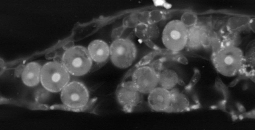
6 Sanders and de los Ríos Structure and development of microlichen 6 Electron microscopy Examination of the thallus of G. paolae with electron microscopy revealed an outer covering layer of fungal origin that was not continuously cellular (Fig. 5). In some areas, particularly where the thallus was thicker and algal cells proliferated in abundance, a fungal cortex 1-2 (-3) cells deep was present (Figs. 6-7). These cells were roundish with only modestly thickened walls (Fig. 7). In other areas, the covering layer appeared to consist of cell wall material extending from and continuous with sparsely distributed fungal cells whose lumina were much smaller than elsewhere in thallus (Fig. 12). Below the covering layer, larger fungal cells were interspersed with scattered algal cells; no discrete algal layer could be discerned. Significant spaces occurred within the thallus (S in Fig. 5), likely facilitating gas exchange, although such spaces were considerably reduced in places where algal cell division was abundant (Figs. 6-7). Larger cavities of jagged outline were also observed, particularly where the overlying surface layer appeared elevated (asterisks in Fig. 8); these spaces appeared to correspond to sites of mineral crystal deposition (compare Figs. 3-4), although the crystals themselves were not evident in the electron micrographs. Toward the periphery of the areolae, the thickness of the thallus decreased considerably (Fig. 9), except where hyphophores developed. Photobionts divided into packets of spores (Fig. 10). Fungal cells made close wall-to-wall contact with one to several algal cells (Figs. 13, 17); they often showed some degree of appressoria-like flattening against the algal wall that increased the surface area of contact, and a fine outer layer uniting the symbionts could sometimes be distinguished (arrows in Fig. 13). However, no evidence of algal wall penetration or initiation of haustorial outgrowths from fungal cells was visible in any of the symbiont contact zones examined. Near to hyphophores, the thallus became thicker, with cell division and cell density notably greater. The hyphophores appeared as raised margins with a profusion of fungal and algal cells beneath (Fig. 11). The darkly pigmented hyphophore scale was composed of elongate cells with extremely thick, electron-dense cell walls (Fig. 14). These specialized cells arose directly from fungal cells of normal appearance (Fig. 15); their cytoplasm appeared to senesce with maturity, although poor penetration of fixative through their massive walls might be responsible for this impression. Conidia emerging from below the hyphophore scale showed budding at both ends (Fig. 16), and frequently made wall-to-wall contact with nearby algal cells, often partially encircling them (Fig. 17). The fungal cells of the conidial chains were readily distinguished cytologically from vegetative cells of the thallus by their rich content of oil droplets (Figs ). In situ development Propagules of G. paolae sown onto plastic cover slips showed sequential stages of development into organized thalli, although mortality was high. Germination of many propagules was observed within 5 d after placement in the field. Hyphae emerged from the tips of the terminal conidial segments and extended radially outward (Figs ). Many of the sown propagules, however, failed to germinate (Fig. 20); some showed loss of pigment in the co-dispersed algal symbiont (Fig. 21). Among those propagules that successfully germinated, subsequent development was highly variable. In a number of cases, algal symbionts at the periphery of the propagule
7 Sanders and de los Ríos Structure and development of microlichen 7 proliferated abundantly while extension of fungal hyphae was still relatively limited (Figs ). In other cases, vigorous growth and branching of fungal hyphae took place before any multiplication of photobiont cells was apparent (Fig. 24). Some of the recent products of algal cell division visible as cells of considerably smaller size (Fig. 22) were clearly not held firmly by the lichen fungus and could be observed free of the developing thallus (arrows in Fig. 23). Some propagules appeared to have developed vigorously toward an organized thallus, but were moribund when observed after several weeks (Fig. 25). Successful organization of the thallus seemed to involve the spreading of algal cells more or less uniformly over the periphery of the primordium, presumably assisted by oriented growth of the fungal hyphae beneath (Figs ). The alga-containing primordia developed into thallus areolae, with fungal hyphae extending well beyond these lichenized portions to form a prothallus (Figs ). Reproductive stages were not seen on the inoculated cover slips, but did occur on some of those colonized naturally. Formation of the characteristic vegetative propagule of Gyalectidium was observed on an incipient hyphophore developing on colonizing thalli only 36 d after substrate placement (Fig. 28). Fully developed hyphophores characteristic of G. paolae, bearing abundant propagules, occurred on cover slips retrieved about five months after placement (Figs ). The smallest areolae observed with mature hyphophores extruding propagules were less than 150 μm in diameter. Granules of crystalline material developed with an initially scattered distribution, appearing to accrete gradually into larger continuous patches (Fig. 31). DISCUSSION Construction of the thallus Although not present in all lichens, the cortex is often the most differentiated and characteristic layer of the thallus; it may be represented by any of a diverse range of tissue types (Henssen and Jahns, 1974; Hale, 1983; Büdel and Scheidegger, 2008). According to Ferraro et al. (2001), a cortex is characteristic of the foliicolous genus Gyalectidium. In G. paolae, it is evident as an overlying layer of approximately isodiametric fungal cells (Figs. 6-7), but only some portions of the thallus areolae are corticated. Elsewhere one finds a sparsely cellular covering of fungal wall material and cell lumina of reduced diameter, a simpler structure that may better correspond to a corticiform layer (Lücking, 2008). Irregular patches of white crystalline material can also occupy a significant proportion of the upper region of the thallus (Figs. 1-4). The crystals were not directly visible in the electron microscope images, suggesting that these components may be removed in processing the specimens for electron microscopy. The localized elevations of the corticiform layer and irregularly jagged spaces below these regions (Fig. 8) indicate that crystal deposition may be particularly concentrated in places where the cellular cortex is not developed, perhaps substituting for the protective function of the cortex. Although not investigated chemically, the white crystals are likely to be calcium oxalate hydrates, as observed in diverse species of Gyalectidium (Ferraro et al., 2001) and other foliicolous members of the Gomphillaceae (de Oliveira et al., 2002), as well as many other lichens (Wadsten and Moberg, 1985; Giordani
8 Sanders and de los Ríos Structure and development of microlichen 8 et al., 2003). Various possible biological roles have been proposed for such crystals in lichens, including chemical storage of water (Wadsten and Moberg, 1985), photoprotective reflection of excessive radiation (Lücking, 1999), or concentrative reflection of suboptimal light within the algal layer (Modenesi et al., 2000). Calcium oxalate depositions are also known to offer plants protection from herbivory (Franceschi and Nakata, 2005), although invertebrates that feed on foliicolous lichens are not necessarily deterred (Lücking and Bernecker-Lücking, 2000). Within an economically constructed foliicolous thallus, calcium oxalate, which is metabolically inexpensive to produce (Giordani et al., 2003), may represent an alternative shielding material that saves on the energetic costs required to maintain a complete cortex of respiring fungal cells. However, the highly irregular distribution of the crystalline plates in G. paolae is not so easily reconciled with any of the functional roles proposed for them. In situ development of propagules Co-dispersal of lichen algae, particularly in the form of vegetative propagules, is widely held to be a common strategy among lichens (Bowler and Rundel, 1975). Co-dispersal may be especially important in facilitating colonization of newly exposed leaf surfaces, which are unlikely to harbor a diverse pool of potential photobionts (Sanders, 2014a). The propagules of G. paolae were very similar in morphology and mode of germination to those of another species of Gyalectidium that colonized cover slips placed in a neotropical lowland forest (Sanders and Lücking, 2002). The subsequent developmental stages reported here provide further insight into the functionality of co-dispersal as an establishment strategy in this lichen. Because development was not studied under controlled conditions, other organisms, including additional lichen propagules, were also able to colonize the inoculated coverslips. However, the cohort of G. paolae propagules, sown within a restricted area of the coverslip, could be distinguished with a fair degree of confidence, at least in the first several weeks after placement in the field. Most notable was the range of variability in development, with many propagules showing photobiont degeneration before germination (Fig. 21) or after substantial development (Fig. 25). Conceivably, some of the propagules may have been immature when taken for inoculation, although it is not clear what sort of additional maturation process might be required after propagules are fully formed morphologically. While genetic differences among propagules cannot be entirely ruled out (the source thalli were not necessarily a single clone), the highly variable results suggest that microenvironmental heterogeneities in light, moisture, and/or nutrient availability may strongly impact development at a very fine scale. The remarkable variability observed in the relative rates of fungal/algal development in the early stages of growth (Figs vs. 24) complements the findings of laboratory culture and resynthesis studies, where different regimes of moisture, nutrients and light often favored growth of one symbiont over the other, impeding formation or maintenance of the lichen symbiosis (Thomas, 1939; Scott, 1960; Ahmadjian, 1962; Bertsch and Butin, 1967; Pearson 1970). It would be interesting to follow the eventual fates of developmental stages in each case, but unfortunately the destructive sampling applied in the present study did not allow sequential observation of the same propagules over time. The appearance of mature
9 Sanders and de los Ríos Structure and development of microlichen 9 hyphophores characteristic of G. paolae on the uninoculated cover slips by 5 months after placement is consistent with the observations of Larsen (2010), who noted hyphophore formation in an unidentified Gyalectidium species within a comparable time frame in a Costa Rican forest. Another foliicolous lichen, Calopadia puiggarii, developed thalli with mature asexual reproductive structures in slightly less time, about 3-4 months (Sanders, 2014a). With Gyalectidium, however, there was evidence in the present study that asexual propagules could form on thalli as young as five weeks old, before a recognizable hyphophore scale has even developed (Fig. 28). With asexual cycles completed in less than 5 months, these lichens might, with favorable timing and conditions, conceivably grow and reproduce on deciduous leaves. However, perpetuation would only be ensured if some propagules managed to disperse to a more enduring substratum before leaf fall. On sabal palm leaves, which last for about 3 years at the field site (Sanders, 2014b), G. paolae could complete numerous asexual generations on the same leaf under favorable conditions. Sexual stages (apothecia) have not yet been reported in this species. Contacts between symbionts The variety of fungal-algal contacts in numerous, mainly crustose lichens was first discerned with light microscopy in the remarkable studies of Tschermak (1941) and Plessl (1963). The general pattern of their findings was that simpler crustose thalli without distinct tissue differentiation tended to show fine, peg-like fungal structures penetrating the algal cell wall and protruding into the lumen, whereas anatomically more complex lichens with distinct tissue layers exhibited more limited penetration pegs that entered the algal cell wall without fully traversing it (intraparietal); intermediates were also observed. Subsequent application of electron microscopy largely confirmed the light microscopic observations of those pioneering studies (Galun et al., 1970; Honegger, 1984, 1986). In the present study of Gyalectidium paolae, we saw only wall-to-wall apposition between symbionts without haustorial penetration, a situation more commonly reported in anatomically more complex lichens (Honegger, 1988). By contrast, in Strigula, a foliicolous lichen examined in previous TEM studies, well-developed haustoria commonly penetrate the cells of its ulvophycean photobiont (Chapman, 1976; Matthews et al., 1989). The varying composition and structural properties of the cell wall in different photobiont taxa may influence the type of symbiont contacts that develop. Sporopollenin-like polymers highly resistant to degradation have been identified in the walls of lichen algae such as Coccomyxa and Elliptochloris, and correlated with the absence of haustorial penetration by the lichen fungi that house them (Honegger, 1984). On the other hand, the photobionts Myrmecia and Dictyochloropsis lack such wall polymers but are usually not penetrated by mycobiont haustoria (Brunner and Honegger, 1985). Whether resistant polymers occur in the photobiont of G. paolae is not known. The Trebouxia-like algae associated with the numerous foliicolous species of Pilocarpaceae and Gomphillaceae including G. paolae have not yet been identified, and are currently under study. Although the penetrative contacts between lichen symbionts are known as haustoria, they are not directly involved in transfer of metabolites, which appear to simply leak from the symplast of the alga in symbiosis (Collins and Farrar, 1978). The lichen
10 Sanders and de los Ríos Structure and development of microlichen 10 haustorium, most notably the intraparietal type, seems to have evolved into a more strictly physical means of symbiont attachment that might play a role in coordinating symbiont distribution to produce anatomically complex thalli (Honegger, 1996). An attachment function for these penetrative contacts is particularly evident in the marine lichen Wahlenbergiella (=Verrucaria) tavaresiae, where haustoria expand laterally to form an anchoring flange inserted between wall layers of its phaeophycean photobiont (Sanders et al., 2004). Similarly, the intraparietal haustorial complexes formed by Mastodia tessellata have been interpreted as a possible mechanism for coordinating mycobiont growth with that of its multicellular green photobiont (Pérez-Ortega et al., 2010). In Gyalectidium paolae, algal distribution within the developing areolae appears to be achieved without penetrative attachments between symbionts, although it should be noted that a relatively low level of anatomical organization is achieved. Studies of symbiont contacts in other lichens have suggested that no photobionts are free within the lichen thallus (Honegger, 1996); fungal hyphae penetrate the spore packets of dividing trebouxioid algal symbionts, establishing attachments to the stillcontained daughter cells that will facilitate their distribution by subsequent fungal growth (Greenhalgh and Anglesea, 1979; Honegger, 1987). By contrast, examination of wetmounted, living foliicolous thalli growing on cover slips suggests that the nascent unicellular photobionts of these lichens are not initially attached to any fungal hyphae. One can occasionally observe the release of small algal cells from the autosporangia at the periphery of the thallus; the exiting spores immediately position themselves alongside the larger neighboring algal cells (Figs ). Presumably they are held there by surface interactions until extending mycobiont cells establish appositional contact, sealing an apoplastic conduit between symbionts with secreted wall materials (Fig. 13) as elucidated by Honegger (1984, 1986, 1988, 1996). The released algal cells can also escape the thallus entirely (Fig. 23), where they might be subsequently recaptured by prothallic hyphae, or give rise to (perhaps transient) free-living populations available to other lichen fungi of the foliicolous community (Sanders, 2014a). On an ephemeral substratum with limited time and capacity for a diverse pool of photobionts to develop, these thallus fugitives may be important sources of symbiotic partners for the many fungal propagules dispersed by foliicolous lichens without accompanying algae. LITERATURE CITED Ahmadjian, V Investigations on lichen synthesis. American Journal of Botany 49: Bertsch, A., and H. Butin Die Kultur der Erdflechte Endocarpon pusillum im Labor. Planta 72: Bowler, P. A., and P. W. Rundel Reproductive strategies in lichens. Botanical Journal of the Linnean Society 70:
11 Sanders and de los Ríos Structure and development of microlichen 11 Brunner, U., and R. Honegger Chemical and ultratructural studies on the distribution of sporopolleninlike biopolymers in six genera of lichen phycobionts. Canadian Journal of Botany 63: Büdel, B., and C. Scheidegger Thallus morphology and anatomy. In T. H. Nash III [ed.], Lichen Biology, Second Edition, Cambridge University Press, New York, USA. Chapman., R. L Ultrastructural investigation on the foliicolous pyrenocarpous lichen Strigula elegans (Fée) Müll. Arg. Phycologia 15: Coley, P. D Effects of plant growth rate and leaf lifetime on the amount and type of anti-herbivore defense. Oecologia (Berlin) 74: Collins, C. R., and J. F. Farrar Structural resistances to mass transfer in the lichen Xanthoria parietina. New Phytologist 81: de los Ríos, A., and C. Ascaso Preparative Techniques for transmission electron microscopy and confocal laser scanning microscopy. In I. C. Kranner, R. P. Beckett, and A. K. Varma [eds.], Protocols in Lichenology, Springer-Verlag, Berlin and Heidelberg, Germany. de Oliveira, L. F. C., H. G. M. Edwards, J. C. Feo-Manga, M. R. D. Seaward, and R. Lücking FT-Raman spectroscopy of three foliicolous lichens from Costa Rican rainforests. Lichenologist 34: Esseen, P.-A., and K.-E. Renhorn Edge effects on a epiphytic lichen in fragmented forests. Conservation Biology 12: Franceschi, V. R., and P. A. Nakata Calcium oxalate in plants: formation and function. Annual Review of Plant Biology 56: Ferraro, L., R. Lücking, and E. Sérusiaux A world monograph of the lichen genus Gyalectidium (Gomphillaceae). Botanical Journal of the Linnean Society 137: Galun, M., N. Paran, and Y. Ben-Shaul The fungus-alga association in the Lecanoraceae: an ultrastructural study. New Phytologist 69: Giordani, P., P. Modenesi, and M. Tretiach Determinant factors for the formation of the calcium oxalate minerals, weddelite and whewellite, on the surface of foliose lichens. Lichenologist 35: Greenhalgh, G. N., and D. Anglesea The distribution of algal cells in lichen thalli. Lichenologist 11: Grube, M., and R. Lücking Fine structure of foliicolous lichens and their lichenicolous fungi studied by epifluorescence. Symbiosis 32:
12 Sanders and de los Ríos Structure and development of microlichen 12 Hale. M. E Biology of Lichens, Third Edition. Edward Arnold, London, UK. Henssen, A., and H.-M. Jahns Lichenes: Eine Einführung in die Flechtenkunde. Georg Thieme Verlag, Stuttgart, Germany. Henssen, A., and R. Lücking Morphology, anatomy and ontogeny in the Asterothyriaceae (Ascomycota: Ostropales), a misunderstood group of lichenized fungi. Annales Botanici Fennici 39: Herre. A. C The growth of Ramalina menziesii. Botanical Gazette 38: Herrera-Campos, M. A., and R. Lücking The foliicolous lichen flora of Mexico II. New species from the montane forest in Oaxaca and Puebla. Bryologist 106: 1-8. Honegger, R Cytological aspects of the mycobiont-phycobiont relationship in lichens. Lichenologist 16: Honegger, R Ultrastructural studies in lichens. I. Haustorial types and their frequencies in a range of lichens with trebouxioid photobionts. New Phytologist 103: Honegger, R Questions about pattern formation in the algal layer of lichens with stratified (heteromerous) thalli. In E. Peveling (ed.), Progress and problems in lichenology in the Eighties. Bibliotheca Lichenologica 25: Honegger, R The functional morphology of cell-to-cell interactions in lichens. In S. Scannerini, DC Smith, P. Bonfante-Fasolo, V. Gianinazzi-Pearson [eds.], Cell to cell signals in plant, animal and microbial symbiosis, Springer-Verlag, Berlin and Heidelberg, Germany. Honegger, R Structural and functional aspects of mycobiont-photobiont relationships in lichens compared with mycorrhizae and plant pathogenic interactions. In M. Nicole and Gianinazzi-Pearson [eds.], Histology, ultrastructure and molecular cytology of plant-microorganism interactions, Kluwer Academic Publishers, Netherlands. Honegger, R The symbiotic phenotype of lichen-forming ascomycetes and their endo- and epibionts. In B. Hock [ed.], Fungal associations, 2nd edition, The Mycota IX Springer-Verlag, Berlin and Heidelberg, Germany. Larsen, E Progress in culturing foliicolous lichens on cover slips. In T. H. Nash III, L. Geiser, B. McCune, D. Triebel, A. M. F. Tomescu, and W. B. Sanders [eds.], Biology of lichens symbiosis, ecology, environmental monitoring, systematics and cyber applications, Bibliotheca Lichenologica 105: J. Cramer in der Gebrüder Borntraeger Verlagsbuchhandlung, Berlin Stuttgart, Germany. Lücking, R Ecology of foliicollous liches at the Botarrama trail (Costa Rica), a neotropical rain forest site. Part II. Patterns of diversity and area cover, and their dependence on microclimate and phorophyte species. Ecotropica 4: 1-24.
13 Sanders and de los Ríos Structure and development of microlichen 13 Lücking, R Ecology of foliicolous lichens at the Botarrama trail (Costa Rica), a neotropical rainforest. IV. Species associations, their salient features and their dependence on environmental variables. Lichenologist 31: Lücking, R Lichens on leaves in tropical rainforests: life in a permanently ephemerous environment. In G. Gottsberger and S. Liede [eds.], Life forms and dynamics in tropical forests, Dissertationes Botanicae 346: J. Cramer in der Gebrüder Borntraeger Verlagsbuchhandlung, Berlin Stuttgart, Germany. Lücking, R Foliicolous Lichenized Fungi. Flora Neotropica 103: New York Botanical Garden Press, New York, USA. Lücking, R., and A. Bernecker-Lücking Lichen feeders and lichenicolous fungi: do they affect dispersal and diversity in tropical foliicolous lichen communities? Ecotropica 6: Matthews, S. W., S. C. Tucker, and R. L. Chapman Ultrastructural features of mycobionts and trentepohliaceous phycobionts in selected subtropical crustose lichens. Botanical Gazette 150: Modenesi, P., M. Piana, P. Giordani, A. Tafanelli, and A. Bartoli Calcium oxalate and medullary architecture in Xanthomaculina convoluta. Lichenologist 32: Pearson, L. C Varying environmental factors in order to grow intact lichens under laboratory conditions. American Journal of Botany 57: Pérez-Ortega, S., A. de los Ríos, A. Crespo, and L. G. Sancho. Symbiotic lifestyle and phylogenetic relationships of the bionts of Mastodia tessellata (Ascomycota, incertae sedis). American Journal of Botany 97: Formatted: English (United States) Plessl, A Über die Beziehungen von Haustorientypus und Organizationshöhe bei Flechten. Österreichische Botanische Zeitschrift 110: Poelt, J., and A. Vězda Über kurzlebige Flechten. In H.-M. Jahns [ed.], Contributions to Lichenology in honor of A. Henssen. Bibliotheca Lichenologica 38: Reynolds, S The use of lead citrate at high ph as an electron-opaque stain in electron microscopy. Journal of Cell Biology 17: Sanders, W. B In situ development of the foliicollous lichen Phyllophiale (Trichotheliaceae) from propagule germination to propagule production. American Journal of Botany 89: Sanders, W. B. 2014a. Complete life cycle of the lichen fungus Calopadia puiggarii (Pilocarpaceae, Ascomycetes) documented in situ: propagule dispersal, establishment of symbiosis, thallus development, and formation of sexual and asexual reproductive structures. American Journal of Botany 101:
14 Sanders and de los Ríos Structure and development of microlichen 14 Sanders, W. B. 2014b. Duration of sabal palm leaves and their lichen colonists in southwest Florida. Bulletin of the British Lichen Society 115: Sanders, W. B., and R. Lücking Reproductive strategies, relichenization and thallus development observed in situ in leaf dwelling lichen communities. New Phytologist 155: Sanders, W. B., R. L. Moe, and C. Ascaso The intertidal marine lichen formed by the pyrenomycete fungus Verrucaria tavaresiae (Ascomycotina) and the brown alga Pertroderma maculiforme (Phaeophyceae): thallus organization and symbiont interaction. American Journal of Botany 91: Scheidegger, C Reproductive strategies in Vezdaea (Lecanorales, lichenized Ascomycetes): A low-temperature scanning electron microscope study of a ruderal species. Cryptogamic Botany 5: Scott, G. D Studies of the lichen symbiosis. I. The relationship between nutrition and moisture content in the maintenance of the symbiotic state. New Phytologist 59: Santesson, R Foliicolous Lichens I. Symbolae Botanicae Upsaliensis 12: Thomas, E. A Über die Biologie von Flechtenbildnern. Beiträge zur Kryptogamenflora der Schweiz 9 (1): (+6 tab.). Tschermak, E Untersuchungen über die Beziehungen von Pilz und Alge im Flechtenthallus. Österreichische Botanische Zeitschrift 90: Wadsten, T, and R. Moberg Calcium oxalate on the surface of lichens. Lichenologist 17:
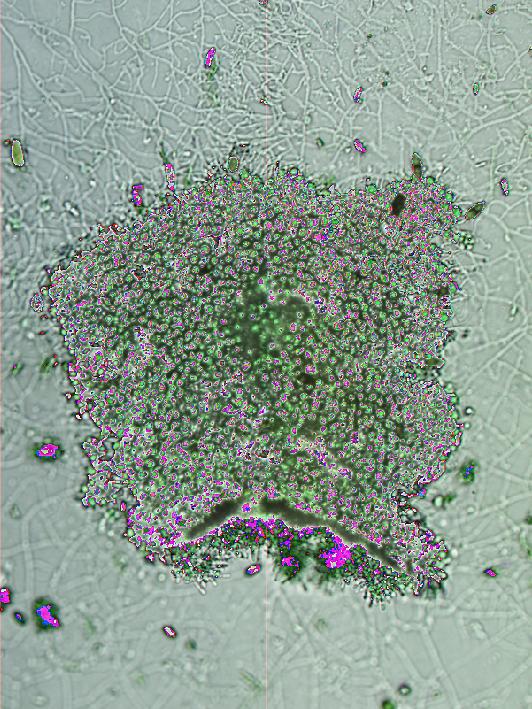
15 Sanders and de los Ríos Structure and development of microlichen 15 FIGURE LEGENDS 1-4. Light microscope images of G. paolae colonizing sabal palm leaves on the FGCU campus. 1. Thallus of several areolae. 2. Tiny areolae (brackets) barely distinguishable against green and white leaf background; hyphophores (arrows) already developed at this stage. 3. Single thallus areola. 4. Larger, continuous thallus with several marginal hyphophores and scattered crystalline deposits. Scale bars = (Figs. 1, 2, 3, 4) 200 μm SEM-backscattering images showing cut surfaces of resin blocks within which G. paolae has been embedded. 5. Thallus with only localized development of cellular cortex (arrowheads); fungal wall material without distinguishable cell lumina forms much of uppermost layer (arrow). Many large spaces (S) are present among fungal and algal cells. 6. Thallus showing much more extensive development of a cellular fungal cortex (c). Below, symbiont cells are densely packed with fewer spaces between them. This image might represent a thallus zone adjacent to a hyphophore. 7. Detail of 6 showing fungal cortex (c). 8. Thallus with large jagged spaces (asterisks) possibly corresponding to mineral crystals. 9. Edge of thallus areola showing gradually declining thickness. Scale bars = (Figs. 5, 7) 10 μm; (Figs. 6, 8, 9) 20 μm SEM-backscattering images showing cut surfaces of resin blocks within which G. paolae has been embedded. 10. Interior of thallus with dividing packet of algal cells (arrows). 11. Edge of thallus with hyphophore (h) and masses of propagules (p) composed of conidial chains (electron-bright due to osmiophilic lipid content) and photobiont cells. Scale bars = (Fig. 10) 5 μm; (11) 10 μm TEM images of G. paolae. 12. Upper layer of thallus composed of wall materials (arrows) continuous with sparse fungal cells of reduced diameter (Compare fungal cells within covering layer at upper left of figure with those below layer at lower right). 13. Fungal cell making intimate wall contact with three algal cells. 14. Thick-walled fungal cells composing hyphophore scale; less specialized fungal cell nearby. 15. Thick-walled cell of hyphophore scale arising from less specialized fungal cell. 16. Conidial cell of diahyphal propagule, with cells budding off at both ends (arrows); oil droplets occupy much of cytoplasm. 17. Conidial cell with budded proliferations (arrows), making close contacts with adjacent algal cell. Abbreviations: a, algal cell; c, conidium; f, fungal cell; h, fungal cell of hyphophore.; o, oil globule. Scale bar = (Figs. 12, 13, 14, 15, 16, 17) 1 μm Light microscope images of lichen development in situ on cover slips inoculated with G. paolae propagules. 18. Germinating propagule after 5 d with hyphae emerging from tips of terminal conidia (arrows). 19. Developing propagule (13 d) with radiating fungal hyphae and proliferating algal cells. 20. Propagule showing no signs of germination after 5 d. 21. Ungerminated propagule (5 d) showing loss of pigment in algal cells (arrows). 22. Germinated propagule with algal cells (arrows) proliferating at margin but relatively limited extension of fungal hyphae (13 d). 23. Extensive proliferation of algal symbionts relative to modest development of fungal prothallus; some algal cells (arrows) apparently liberated from thallus (13 d). 24. Germination of a group of propagules, showing extensive
16 Sanders and de los Ríos Structure and development of microlichen 16 development of fungal hyphae but no proliferation of algal cells (arrow) at this stage (13 d). 25. Young thallus moribund after abundant proliferation of algal cells (arrows), now discolored, and extensive fungal prothallus development (arrowhead). 26. Developing thallus showing relatively uniform distribution of algal cells elevated above substratum (55 d). 27. Detail of developing thallus showing algal cells at periphery, some dividing (arrow) into autospores; 55 d. Scale bars = (Figs. 18, 20, 21) 10 μm; (19, 22, 25, 27) 20 μm; (23, 24, 26) 50 μm Development from naturally dispersed propagules on uninoculated cover slips. 28. Margin of young areola with developing hyphophore (arrowheads) that is already producing a propagule (arrow) 36 d after cover slip exposure. 29. Areola with fully formed hyphophore (arrowheads) and extensive surrounding prothallus (p); 151 d after exposure. 30. Detail of hyphophore with several propagules (arrows); 151 d after exposure. 31. Areola with hyphophore, showing accretion of crystalline deposits (arrows); 151 d after exposure. Scale bars = (Figs. 28, 30) 20 μm; (Figs. 29, 31) 50 μm.


17 2 { 3 4 { 1


18 5 f S a S 6 c 7 8 c * 9 *


19 10 11 h p


20 f a f f f a h h a a o f o a h o c


21


22 28 29 p p 30 31
AJB Advance Article published on August 20, 2015, as /ajb
 AJB Advance Article published on August 20, 2015, as 10.3732/ajb.1500202. The latest version is at http://www.amjbot.org/cgi/doi/10.3732/ajb.1500202 RESEARCH ARTICLE AMERICAN JOURNAL OF BOTANY Structure
Kingdom Fungi. Learning Objectives. Introduction. Activity1: Zygomycota. Revised Fall 2017
 Kingdom Fungi Revised Fall 2017 ** You will require your text book Biological Science during this lab ** Learning Objectives Building on the learning objectives from your lab syllabus, you will be expected
Kingdom Fungi. 1. Student will be able to describe the characteristic features in the kingdom Fungi.
 Kingdom Fungi Molds, Sac Fungi, Mushrooms, and Lichens Essential Question(s): What makes fungi have their own kingdom? Objectives: 1. Student will be able to describe the characteristic features in the
Fungi are absorptive heterotrophs that secrete digestive enzymes and are major decomposers of dead organic material
 Fungi 1 2002 Prentice Hall, Inc The scarlet hood (Hygrocybe coccinea) Fungi are absorptive heterotrophs that secrete digestive enzymes and are major decomposers of dead organic material 2 Animals 3 Myxozoa
3/22/2011. Review. Review. Mitosis: division of cells that results in two identical daughter cells with same genetic information as the first cell
 Review Review Mitosis: division of cells that results in two identical daughter cells with same genetic information as the first cell Meiosis: division of cells that results in daughter cells with one-half
Fungi What are they? Diverse group of eukaryotic organisms 100,000 to 1,000,000 species
 Kingdom Fungi Fungi What are they? Diverse group of eukaryotic organisms 100,000 to 1,000,000 species Fungi Characteristics Kingdom includes Molds, mushrooms & yeasts Characteristically: Most are multicellular
Nadia Langha Biology 106 Honors Project
 Nadia Langha Biology 106 Honors Project Cyanobacteria Domain Bacteria Division Cyanophyta Cyanobacteria also known as BlueGreen Algae -Cyano=blue Bacteria are more closely related to prokaryotic bacteria
THE FREQUENCY OF HETEROCYSTS IN THE NOSTOC PHYCOBIONT OF THE LICHEN PELTIGERA CANINA WILLD.
 New Phytol. (1972) 71, 11-13. THE FREQUENCY OF HETEROCYSTS IN THE NOSTOC PHYCOBIONT OF THE LICHEN PELTIGERA CANINA WILLD. BY H. BRONWEN GRIFFITHS, A. D. GREENWOOD AND J. W. MILLBANK Department of Botany,
Plant Structure and Organization - 1
 Plant Structure and Organization - 1 In our first unit of Biology 203 we will focus on the structure and function of the higher plants, in particular the angiosperms, or flowering plants. We will look
Chapter 4 Ecosystems and Living Organisms
 Chapter 4 Ecosystems and Living Organisms I. Evolution A. The cumulative genetic changes that occur in a population of organisms over time 1. Current theories proposed by Charles Darwin, a 19 th century
Chapter 9. Fungi and Aquatic Plants. Introduction: The Big Step: DIVISION OF LABOUR
 Chapter 9. Fungi and Aquatic Plants Introduction: The Big Step: DIVISION OF LABOUR In single cell organisms (protists) all life functions are performed by specialized organelles within one cell (a.k.a.
Phaeocalicium populneum
 Phaeocalicium populneum markpowell222@btinternet.com After conducting a survey of the RHS garden at Wisley on 18 th August 2018, Fay Newbery kindly showed me the colony of P. populneum at Esher Common.
THE WORLD OF BIOLOGY SECTION 1-1 REVIEW. VOCABULARY REVIEW Define the following terms. MULTIPLE CHOICE Write the correct letter in the blank.
 SECTION 1-1 REVIEW THE WORLD OF BIOLOGY VOCABULARY REVIEW Define the following terms. 1. development 2. reproduction 3. organ 4. tissue MULTIPLE CHOICE Write the correct letter in the blank. 1. Biology
Lesson Overview. Niches and Community Interactions. Lesson Overview. 4.2 Niches and Community Interactions
 Lesson Overview 4.2 Niches and Community Interactions The Niche What is a niche? A niche is the range of physical and biological conditions in which a species lives and the way the species obtains what
Useful Propagation Terms. Propagation The application of specific biological principles and concepts in the multiplication of plants.
 Useful Propagation Terms Propagation The application of specific biological principles and concepts in the multiplication of plants. Adventitious Typically describes new organs such as roots that develop
THE BEHAVIOUR OF CHLOROPLASTS DURING CELL DIVISION OF ISOETES LACUSTRIS L.
 New Phytol (1974) 73, 139-142. THE BEHAVIOUR OF CHLOROPLASTS DURING CELL DIVISION OF ISOETES LACUSTRIS L. BY JEAN M. WHATLEY Botany School, University of Oxford (Received 2 July 1973) SUMMARY Cells in
LOW-POWER ELECTRON MICROSCOPY OF THE ROOT CAP REGION OF EUCALYPT MYCORRHIZAS
 New Phytol. (1968) 67, 663-665. LOW-POWER ELECTRON MICROSCOPY OF THE ROOT CAP REGION OF EUCALYPT MYCORRHIZAS BY G. A. CHILVERS Botany Department, School of General Studies, Australian National University,
Plant Structure. Lab Exercise 24. Objectives. Introduction
 Lab Exercise Plant Structure Objectives - Be able to identify plant organs and give their functions. - Learn distinguishing characteristics between monocot and dicot plants. - Understand the anatomy of
The World of Lichens Part of: Joint Science Education Project at Dartmouth Developed by: Ruth Heindel, Earth Sciences Department, Dartmouth College
 The World of Lichens Part of: Joint Science Education Project at Dartmouth Developed by: Ruth Heindel, Earth Sciences Department, Dartmouth College Overview Lichens are incredible symbiotic organisms that
Characterizing and Classifying Eukaryotes. Fungi. Chemoheterotrophic. Have cell walls typically composed of chitin. Do not perform photosynthesis
 PowerPoint Lecture Presentations prepared by Mindy Miller-Kittrell, North Carolina State University C H A P T E R 12 Characterizing and Classifying Eukaryotes Chemoheterotrophic Have cell walls typically
more than 380,000 species, of which more than two-thirds
 The plant world contains more than 380,000 species, of which more than two-thirds are green plants. From the most complex flowering plants to single-cell sea algae, plants present a surprising diversity
CHAPTER 1 BIOLOGY THE SCIENCE OF LIFE
 CHAPTER 1 BIOLOGY THE SCIENCE OF LIFE BIOLOGICAL THEMES 1. Cell Structure & Function cell is the basic unit of life all organisms are composed of at least one cell Unicellular single celled ; bacteria,
INTRODUCTION prokaryotic eukaryotic pigments
 INTRODUCTION This exercise is intended for you to get familiar and comfortable with using a microscope as well as identifying common microbial groups. Thus, we will observe representatives of all microbes
Eukaryotes Most are saprobes (live on dead organisms) Grow best in warm, moist environments Mycology is the study of fungi
 Grow best in warm, moist environments Mycology is the study of fungi") KINGDOM FUNGI 1 Characteristics 2 THE CHARACTERISTICS OF FUNGI Eukaryotes Most are saprobes (live on dead organisms) Grow best in warm, moist environments Mycology is the study of fungi 3 THE CHARACTERISTICS
KINGDOM FUNGI 1 Characteristics 2 THE CHARACTERISTICS OF FUNGI Eukaryotes Most are saprobes (live on dead organisms) Grow best in warm, moist environments Mycology is the study of fungi 3 THE CHARACTERISTICS
Lab Exercise 4: Primary Growth and Tissues in Stems
 Lab Exercise 4: Primary Growth and Tissues in Stems Tissues of the plant body can be classified in a variety of ways: functionally (based on the tissue function, e.g. vascular tissue ), morphologically
INTRODUCTION budding, binary fission hyphae mycelium Figure 1.
 INTRODUCTION Although most of our work in this lab is done on bacteria, fungi are nonetheless an important aspect in microbiology. Besides being important food providers, fungi play central roles in recycling
Characterizing and Classifying Eukaryotes. Fungi. Chemoheterotrophic. Have cell walls typically composed of chitin. Do not perform photosynthesis
 PowerPoint Lecture Presentations prepared by Mindy Miller-Kittrell, North Carolina State University C H A P T E R 12 Characterizing and Classifying Eukaryotes Chemoheterotrophic Have cell walls typically
Transmission Electron Microscope Technique for Risk Assessment of Manufactured Nanomaterials
 Transmission Electron Microscope Technique for Risk Assessment of Manufactured Nanomaterials Kazuhiro Yamamoto and Miyabi Makino National Institute of Advanced Industrial Science and Technology (AIST),
4 Marine Biology Notes. Multi-cellular Primary Producers: Seaweeds and Plants
 4 Marine Biology Notes Multi-cellular Primary Producers: Seaweeds and Plants Marine Algae Marine algae are important primary producers (photosynthetic) These algae are called by a generic term seaweeds
VIII. Kingdom Protista- (protists) A. General characteristics of protists:
 A. General characteristics of protists:") VIII. Kingdom Protista- (protists) A. General characteristics of protists: 1. Protists are unicellular organisms that have a nucleus to organize their hereditary material. 2. Some protists help their host
VIII. Kingdom Protista- (protists) A. General characteristics of protists: 1. Protists are unicellular organisms that have a nucleus to organize their hereditary material. 2. Some protists help their host
Lab 1: Using the Microscope & Cell Biology
 Name Lab 1: Using the Microscope & Cell Biology The anatomy of the compound microscope Review or learn the following parts of the compound microscope and their functions. Eyepieces Objectives Arm Stage
Abiotic Stress in Crop Plants
 1 Abiotic Stress in Crop Plants Mirza Hasanuzzaman, PhD Professor Department of Agronomy Sher-e-Bangla Agricultural University E-mail: mhzsauag@yahoo.com Stress Stress is usually defined as an external
Domain: Eukarya Kingdom: FUNGI
 Domain: Eukarya Kingdom: FUNGI Fungi are eukaryotic heterotrophs that have cell walls. They are part of the nature s recycling system. They break down organic compounds. Fungi are used in wine, beer, cheese,
The Microbial World. Chapter 5
 The Microbial World Chapter 5 Viruses Non-cellular infectious agents that have two basic characteristics: Not capable of reproduction without a host cell Structure: Nucleic acid core- can be DNA or RNA
Protists: Algae Lecture 5 Spring 2014
 Protists: Algae Lecture 5 Spring 2014 Meet the algae 1 Protist Phylogeny Algae - Not monophyletic What unites them as a group? Range from unicellular to multicellular From phytoplankton to kelp forests
Introduction to Biology
 Introduction to Biology Biology The Study of Life Life arose more than 3.5 billion years ago First organisms (living things) were single celled Only life on Earth for millions of years Organisms changed
Introduction to Biology
 1 Introduction to Biology 2 Biology The Study of Life Life arose more than 3.5 billion years ago First organisms (living things) were single celled Only life on Earth for millions of years Organisms changed
Protists: Algae Lecture 5 Spring Protist Phylogeny. Meet the algae. Primary & Secondary Endosymbiosis. Endosymbiosis. Secondary Endosymbiosis
 Meet the algae Protists: Algae Lecture 5 Spring 2014 Protist Phylogeny 1 Primary & Secondary Endosymbiosis 2 Algae - Not monophyletic What unites them as a group? Range from unicellular to multicellular
-Each asexual organs. -Anchors the plant -Absorbs water and minerals -Stores sugars and starches
 Plants are made up of: -organs, tissues, and cells The three major plant organs are: -Roots, stems, and leaves -Each asexual organs Plants have a Root System beneath the ground that us a multicellular
CYTOLOGICAL ASPECTS OF THE MYCOBIONT- PHYCOBIONT RELATIONSHIP IN LICHENS
 Lichenologist 16(2): 111-127 (1984) CYTOLOGICAL ASPECTS OF THE MYCOBIONT- PHYCOBIONT RELATIONSHIP IN LICHENS Haustorial types, phycobiont cell wall types, and the ultrastructure of the cell surface layers
Weather is the day-to-day condition of Earth s atmosphere.
 4.1 Climate Weather and Climate Weather is the day-to-day condition of Earth s atmosphere. Climate refers to average conditions over long periods and is defined by year-after-year patterns of temperature
CELL STRUCTURE & FUNCTION
 CELL STRUCTURE & FUNCTION CELL TYPES Living cells can be classified into 2 different types on the basis of their internal structure: 4. Prokaryotic Cells 5. Eukaryotic Cells 1. Prokaryotic Cells Are the
Characteristics of Living Things
 Characteristics of Living Things All Living Things Are made up of units called cells A cell is the smallest unit of an organism that can be considered alive Types of Cellular Organisms Unicellular Uni
Primary Plant Body: Embryogenesis and the Seedling
 BIOL 221 Concepts of Botany Primary Plant Body: Embryogenesis and the Seedling (Photo Atlas: Figures 1.29, 9.147, 9.148, 9.149, 9.150, 9.1, 9.2) A. Introduction Plants are composed of fewer cell types,
Grade Seven Science Focus on Life Sciences. Main Ideas in the Study of Cells
 Grade Seven Science Focus on Life Sciences Main Ideas in the Study of Cells Research is an effective way to develop a deeper understanding of challenging content. The following fill-in-the-blanks activity
ARE YOU familiar with the sayings Get to
 Root Anatomy ARE YOU familiar with the sayings Get to the root of the problem or the root of all evil? Both these sayings suggest that the root is an essential part of something. With plants, the essential
The mode of development in animals and plants is different
 The mode of development in animals and plants is different Outcome of animal embryogenesis is a mini edition of the adult Outcome of plant embryogenesis is a simple structure with -root apical meristem
National Cell structure Pupil notes. Cell Biology. Sub-topic (1.1) Cell Structure. On completion of this topic I will be able to state that:
 Cell Structure. On completion of this topic I will be able to state that:") Cell Biology Sub-topic (1.1) Cell Structure On completion of this topic I will be able to state that: Cells differ in structure as to whether they are animal, plant, fungi or bacterial cells. The detail
Cell Biology Sub-topic (1.1) Cell Structure On completion of this topic I will be able to state that: Cells differ in structure as to whether they are animal, plant, fungi or bacterial cells. The detail
CASE STUDY WATER ABSORPTION AND TRANSPORT IN PLANTS
 CASE STUDY WATER ABSORPTION AND TRANSPORT IN PLANTS Presentation of the problem: We need a pump to uplift water to a tank. The requirement of a pump is to pull water against the gravity. Look at the human
Copyright 2009 Pearson Education, Inc. FUNGI
 Copyright 2009 Pearson Education, Inc. FUNGI FUNGI Fungi are absorptive heterotrophic eukaryotes that digest their food externally and absorb the nutrients Most fungi consist of a mass of threadlike hyphae
Assessment Schedule 2016 Biology: Demonstrate understanding of biological ideas relating to micro-organisms (90927)
") NCEA Level 1 Biology (90927) 2016 page 1 of 5 Assessment Schedule 2016 Biology: Demonstrate understanding of biological ideas relating to micro-organisms (90927) Evidence Statement Question One No response
NCEA Level 1 Biology (90927) 2016 page 1 of 5 Assessment Schedule 2016 Biology: Demonstrate understanding of biological ideas relating to micro-organisms (90927) Evidence Statement Question One No response
Groups of Fungi. Section 2
 Groups of Fungi Section 2 Chytrid Fungi Key Idea: The chytrids are a group of aquatic fungi that provide clues about the evolution of fungi. Chytrid Fungi Chytrids were once classified with protists because
SG 9.2 notes Ideas about targets and terms: 9.2 In the past, all living things were classified in either the kingdom of animals or plants
 Ideas about targets and terms: 9.2 In the past, all living things were classified in either the kingdom of animals or plants Euglena are singled celled organisms in pond water They are green, so contain,
Microscopy and the Diversity of Microorganisms
 Microscopy and the Diversity of Microorganisms Today we will learn how to use one of the most important tools a biologist has, the microscope. We will use the microscope to study organisms throughout the
FUNGI are very successful and widespread
 because fungi have cell walls, and show a superficial resemblance, Fungi were long allied with PLANTS in fact they differ greatly from plants and are now considered to be more closely related to ANIMALS
Unit 10: The simplest living beings
 Unit 10: The simplest living beings 1. Fungi 2. Protoctists 2.1. Protozoa 2.2. Algae 3. Bacteria 4. Viruses Think and answer? a. What type of organism can you see in the photograph? b. What type of cells
13. The diagram below shows two different kinds of substances, A and B, entering a cell.
 Name 1. In the binomial system of nomenclature, which two classification groups provide the scientific name of an organism? A) kingdom and phylum B) phylum and species C) kingdom and genus D) genus and
6 th Grade Life Science Strand 3: Characteristics and Interactions of Living Organisms
 Middle School Life Science Standards There are 15 standards that encompass the proposed middle school life science standards. The new standards are listed 4 times to match the four times life science is
Plant Structure and Function Extension
 Plant Structure and Function Extension NGSSS: SC.912.L.14.7 Relate the structure of each of the major plant organs and tissues to physiological processes. (AA) Part 1A: Leaves The leaf of a plant serves
CH 11 PROTISTS AND FUNGI
 CH 11 PROTISTS AND FUNGI Name Day M T W Th F Weekly Lifeline Period B_ Check Question What is a parasite? KICK-OFF LEARNING LOG KICK-OFF Response (1) A parasite is an organism that feeds off of another
Trophic and community ecology
 Trophic and community ecology Top carnivore Trophic levels Carnivore Herbivore Plant Trophic ecology Trophic related to feeding Autotrophs: synthesize their food Heterotrophs: eat other organisms Trophic
Effect of host plant, cultivation media and inoculants sources on propagation of mycorrhizal fungus Glomus Mossae
 EUROPEAN ACADEMIC RESEARCH Vol. V, Issue 12/ March 2018 ISSN 2286-4822 www.euacademic.org Impact Factor: 3.4546 (UIF) DRJI Value: 5.9 (B+) Effect of host plant, cultivation and inoculants sources on propagation
NUTRITION: A) Saprophytes = break down material extracellularly with secreted enzymes : eg) mushrooms, molds
 Saprophytes = break down material extracellularly with secreted enzymes : eg) mushrooms, molds") KINGDOM FUNGI (MYCOPHYTA) Mycology = the study of fungi fossil record dates to 900 million years ago at one time classified in the Plantae Kingdom Recent molecular evidence suggests that fungi are probably
KINGDOM FUNGI (MYCOPHYTA) Mycology = the study of fungi fossil record dates to 900 million years ago at one time classified in the Plantae Kingdom Recent molecular evidence suggests that fungi are probably
Dr. Sarita Srivastava Assistant Professor Botany Department
 Dr. Sarita Srivastava Assistant Professor Botany Department Dr. Sarita Srivastava Assistant Professor, Botany CMP College, University of Allahabad Theophrastus first gave the term Lichen in 371-284 BC
General Fungus Anatomy: Yeast: single cell fungi that reproduces by fission or budding
 Make-Up Assignment: Using the notes below as a guide, look up the organisms you are required to draw on the internet or in a book. Draw the organism in the circles provided and write a description of the
Porifera. Thomas M. Frost Trout Lake Station Center for Limnology University of Wisconsin Madison, Wisconsin '"'. , ' I.
 , ' Porifera Thomas M. Frost Trout Lake Station Center for Limnology University of Wisconsin Madison, Wisconsin 53706 4 '"'. Chapter Outline I. INTRODUCTION II. ANATOMY AND PHYSIOLOGY A. External Morphology
14.1 Habitat And Niche
 14.1 Habitat And Niche A habitat differs from a niche. Habitat physical area in which an organism lives Niche each species plays a specific role in an ecosystem niche includes the species habitat, feeding
Working with Mycorrhizas in Forestry and Agriculture
 Working with Mycorrhizas in Forestry and Agriculture SUB Gdttingen 206 384661 Mark Brundrett, Neale Bougher, Bernie Dell, Tim Grove and Nick Malajczuk CONTENTS Chapter I. INTRODUCTION 1.1. MYCORRHIZAL
Plant Tissues and Organs. Topic 13 Plant Science Subtopics , ,
 Plant Tissues and Organs Topic 13 Plant Science Subtopics 13.1.2, 13.1.3, 13.1.4 Objectives: List and describe the major plant organs their structure and function List and describe the major types of plant
UNIT XI. Kingdom Fungi
 UNIT XI Kingdom Fungi Kingdom Fungi The Study of Fungi is called Mycology What is probably the largest living organism on earth has been discovered in the Malheur National Forest in eastern Oregon. A fungus
Keywords. Podosphaera leucotricha, scanning electron microscopy. Summary
 Scanning electron microscopy of apple powdery mildew (Podosphaera leucotricha, Ell and Ev.) fungi infecting susceptible Jonathan apple cultivar leaf mesophyll ZS. JAKAB-ILYEFALVI 1 1 Fruit Research and
The occurrence and diversity of mycorrhizal fungi found in blueberry. Susan McCallum
 The occurrence and diversity of mycorrhizal fungi found in blueberry Susan McCallum Blueberry root system Shallow rooting system mainly concentrated near the soil surface Roots that are larger than 1mm
Compatibility and thigmotropism in the lichen symbiosis: A reappraisal
 SYMBIOSIS (2009) 47, 109 115 2009 Balaban, Philadelphia/Rehovot ISSN 0334-5114 Compatibility and thigmotropism in the lichen symbiosis: A reappraisal Suzanne Joneson * and François Lutzoni Duke University,
Agronomy 485/585 Test #1 October 2, 2014
 Agronomy 485/585 Test #1 October 2, 2014 Name Part I. Circle the one best answer (2 points each). 1. The most important microbial group in promoting soil structure likely is the. a) actinomycetes b) algae
DEPARTMENT OF LIFE AND CONSUMER SCIENCES. Plant Structure BOT1501. Semester I: Assignment no. 2 Memorandum
 University Examinations DEPARTMENT OF LIFE AND CONSUMER SCIENCES Plant Structure BOT1501 Semester I: Assignment no. 2 Memorandum 2018 QUESTION 1 1.1 Primary growth is the production of new primary tissues
Today s materials: Cell Structure and Function. 1. Prokaryote and Eukaryote 2. DNA as a blue print of life Prokaryote and Eukaryote. What is a cell?
 Today s materials: 1. Prokaryote and Eukaryote 2. DNA as a blue print of life Prokaryote and Eukaryote Achadiah Rachmawati What is a cell? Cell Structure and Function All living things are made of cells
Plant Anatomy: roots, stems and leaves
 Plant Anatomy: roots, stems and leaves The plant body has a hierarchy of organs, tissues and cells Plants, like animals, have organs composed of different tissues, which are composed of cells. Tissue is
THE WORLD OF BIOLOGY SECTION 1-1 REVIEW. VOCABULARY REVIEW Define the following terms. MULTIPLE CHOICE Write the correct letter in the blank.
 SECTION 1-1 REVIEW THE WORLD OF BIOLOGY Define the following terms. 1. development 2. reproduction 3. organ 4. tissue Write the correct letter in the blank. 1. Biology is the study of a. animals. b. plants
LABORATORY 7 ENDOSPORE STAIN AND BACTERIAL MOTILITY
 LABORATORY 7 ENDOSPORE STAIN AND BACTERIAL MOTILITY A. Endospore Stain B. Bacterial Motility A. ENDOSPORE STAIN DISCUSSION A few genera of bacteria, such as Bacillus and Clostridium have the ability to
Types of intertidal communities
 Between the tides Marine ecosystems 1 Intertidal Delimited by the highest high tide and the lowest low tides marks The best studied and best-understood by humans Relatively easy to sample compared to other
Plant Pathology Fact Sheet
 Plant Pathology Fact Sheet PP-22 Selerotinia Diseases of Vegetable and Field Crops in Florida Ken Pernezny and L. H. Purdy, Professor, Everglades Research and Education Center, Belle Glade; and Professor,
Ch.5 Evolution and Community Ecology How do organisms become so well suited to their environment? Evolution and Natural Selection
 Ch.5 Evolution and Community Ecology How do organisms become so well suited to their environment? Evolution and Natural Selection Gene: A sequence of DNA that codes for a particular trait Gene pool: All
INDEPENDENT STUDY: KINGDOM FUNGI
 INDEPENDENT STUDY: KINGDOM FUNGI Please complete the questions using your textbook as a reference. As you will see, this is not as detailed as your usual Guided Readings and you will have to focus on some
Chapter 31: Fungi. Student:
 Chapter 31: Fungi Student: 1. Specialized symbiotic associations between the roots of plants and fungi are called A) lichens. B) hyphal associations. C) heterokaryotic junctions. D) mycorrhizae. E) a mycelium
Topic 14. The Root System. II. Anatomy of an Actively Growing Root Tip
 Topic 14. The Root System Introduction. This is the first of two lab topics that focus on the three plant organs (root, stem, leaf). In these labs we want you to recognize how tissues are organized in
Department of Botany, University of Dhaka, Dhaka 1000, Bangladesh. Key words: Seaweeds, Marine algae, Kallymenia spp., St. Martin's Is.
 Bangladesh J. Bot. 37(2): 173-178, 2008 (December) MARINE ALGAE OF THE ST. MARTIN S ISLAND, BANGLADESH. VI. NEW RECORDS OF SPECIES OF THE GENUS KALLYMENIA J. AG. (RHODOPHYTA) ABDUL AZIZ, A.K.M. NURUL ISLAM
Plants. Tissues, Organs, and Systems
 Plants Tissues, Organs, and Systems Meristematic cells Specialized cells that are responsible for producing specialized cells, they produce three types of tissue in the body of a plant. Meristematic Cells
CAMBIUM, meristem, heartwood, and lenticel are
 Examining the Structures of a Tree CAMBIUM, meristem, heartwood, and lenticel are some terms that may be new to you. These terms are used to describe various tree structures. Not surprisingly, many terms
Chapter 8. Biogeographic Processes. Upon completion of this chapter the student will be able to:
 Chapter 8 Biogeographic Processes Chapter Objectives Upon completion of this chapter the student will be able to: 1. Define the terms ecosystem, habitat, ecological niche, and community. 2. Outline how
ENVE203 Environmental Engineering Ecology (Nov 19, 2012)
") ENVE203 Environmental Engineering Ecology (Nov 19, 2012) Elif Soyer Biological Communities COMPETITION Occurs when 2 or more individuals attempt to use an essential common resource such as food, water,
ENVE203 Environmental Engineering Ecology (Nov 19, 2012) Elif Soyer Biological Communities COMPETITION Occurs when 2 or more individuals attempt to use an essential common resource such as food, water,
Biology the study of life. Lecture 15
 Biology the study of life Lecture 15 Life (a life form: an organism ) can be defined as an organized genetic unit capable of metabolism, reproduction, & evolution (Purves et al., 2003) There is order to
-plant bodies composed of tissues produced by an apical meristem. -spores with tough walls. -life history of alternation of generations
 Chapter 21-Seedless Plants Major modern plant groups All groups of land-adapted plants have a common set of characteristics: -plant bodies composed of tissues produced by an apical meristem -spores with
Anatomy of Plants Student Notes
 Directions: Fill in the blanks. Anatomy of Plants Student Notes Plant Cell Biology Segment 1. Plants Plants are organisms are incapable of movement produce food through 2. Animals Animals are multicellular
How we study diversity: phylogenetic tree. Fungi vs. Animals. Fungi vs. Plants 3/8/18
 Ya Yang yangya@umn.edu How we study diversity: phylogenetic tree Office Hours: Monday 10-12 AM 714 Biological Sciences Center Fungi are eukaryotic organisms that are more closely related to animals than
Locating water in the dehydrated thallus of lichens from extreme microhabitats (Antárctica)
") Lichenological Contributions in Honour ofg.b. Feige. M. Jensen (ed.): Bibliotheca Lichenologica 86: 215-223. J. Cramer in der Gebrüder Borntraeger Veríagsbuchhandlung, Berlín Stuttgart, 2003 Locating water
Lichenological Contributions in Honour ofg.b. Feige. M. Jensen (ed.): Bibliotheca Lichenologica 86: 215-223. J. Cramer in der Gebrüder Borntraeger Veríagsbuchhandlung, Berlín Stuttgart, 2003 Locating water
Kingdom Fungi. Announcements
 Kingdom Fungi Announcements Friday lab: Fungi & Lichen Bring a Lichen to ID! Do prelab Quiz #4 Friday Study Prokaryotes & Protists Mushroom Fest extra credit due Fri Email me or bring to lab Endosymbiosis
B.Sc. PLANT BIOLOGY & PLANT BIOTECHNOLGY. ALGOLOGY AND MYCOLOGY B. Sc. va (Candidates admitted from the academic year )
") B.Sc. PLANT BIOLOGY & PLANT BIOTECHNOLGY ALGOLOGY AND MYCOLOGY Paper 1a ALGOLOGY AND MYCOLOGY Algology CORE THEORY UNIT I Introduction to Algae: Definition; Distribution of algae: Freshwater algae, Brackish
B.Sc. PLANT BIOLOGY & PLANT BIOTECHNOLGY ALGOLOGY AND MYCOLOGY Paper 1a ALGOLOGY AND MYCOLOGY Algology CORE THEORY UNIT I Introduction to Algae: Definition; Distribution of algae: Freshwater algae, Brackish
1 Towards Ecological Relevance Progress and Pitfalls in the Path Towards an Understanding of Mycorrhizal Functions in Nature... 3 D.J.
 Contents Section A: Introduction 1 Towards Ecological Relevance Progress and Pitfalls in the Path Towards an Understanding of Mycorrhizal Functions in Nature... 3 D.J. Read 1.1 Summary.............................
VARIATION IN THE SIZE OF RAY PITS OF CONIFERS.*
 VARIATION IN THE SIZE OF RAY PITS OF CONIFERS.* FOREST B. H. BROWN. Since Haeckel proposed the word Ecology in 88, there has been an ever growing interest in the influence which environmental factors may
Bacillus anthracis. Clostridium botulinum Clostridium perfringens and other, but never Gram-negative microbes
 SPORES (endospores) the spore is formed inside the parent vegetative cell hence the name endospores The spore is a dehydrated, multishelled structure that protects and allows the bacteria to exist in suspended