SEAWEEDS. OF MU KO THA LAE TAI (SE Thailand) Methodologies and field guide to the dominant species
|
|
|
- Shonda Elfrieda Griffith
- 6 years ago
- Views:
Transcription
 Methodologies and field")


1 SEAWEEDS OF MU KO THA LAE TAI (SE Thailand) Methodologies and field guide to the dominant species Eric Coppejans Anchana Prathep Frederik Leliaert Khanjanapaj Lewmanomont Olivier De Clerck โครงการพ ฒนาองค ความร และศ กษานโยบาย การจ ดการทร พยากรช วภาพในประเทศไทย Biodiversity Research and Training Program
2 BRT Book Series, Area-Based No. 11. Seaweeds of Mu Ko Tha Lae Tai (SE Thailand): Methodologies and field guide to the dominant species by Eric Coppejans Anchana Prathep Frederik Leliaert Khanjanapaj Lewmanomont Olivier De Clerck Published by Biodiversity Research and Training Program (BRT Program) Thailand Research Fund (TRF) National Center for Genetic Engineering and Biotechnology (BIOTEC) National Science and Technology Development Agency (NSTDA) TOTAL Foundation, TOTAL E&P Thailand Prince of Songkhla University
3 Seaweeds of Mu Ko Tha Lae Tai (SE Thailand) Methodologies and field guide to the dominant species Eric Coppejans Universiteit Gent Krijgslaan, 281 (S8), B-9000 Gent, België Anchana Prathep Prince of Songkhla University Hatyai, Songkhla 90112, Thailand Khanjanapaj Lewmanomont Kasetsart University Bangkok 10900, Thailland First Published in Thailand, July 2010 Biodiversity Research and Training Program (BRT) 5th Floor NSTDA Building, 73/1 Rama VI Road, Rajthevee Bangkok 10400, Thailand Tel ext 552 fax Editors : Visut Baimai and Rungsima Tanthalakha ru@biotec.or.th and anchana.p@psu.ac.th Frederik Leliaert Universiteit Gent Krijgslaan, 281 (S8), B-9000 Gent, België frederik.leliaert@ugent.be Olivier De Clerck Universiteit Gent Krijgslaan, 281 (S8), B-9000 Gent, België olivier.declerck@ugent.be No parts of this publication may be reproduced or transmitted in any form or by any means, electronic or mechanical including photocopying, recording or any information storage and retrieval system without the prior permission of the copyright owner. ISBN : Printed in Thailand Suggested citation : Coppejans E., A. Prathep, F. Leliaert, K. Lewmanomont, O. De Clerck Seaweeds of Mu Ko Tha Lae Tai (SE Thailand): Methodologies and field guide to the dominant species. Biodiversity Research and Training Program (BRT), Bangkok 274 pp. BRT Book Series, Area-Based 1. ค ม อตรวจว ดค ณภาพน ำด วยส ตว หน าด น โดย บ ญเสฐ ยร บ ญส ง และ นฤมล แสงประด บ (2547), in Thai 2. การว จ ยความหลากหลายเช งพ นท (area-based): กรณ ศ กษาช ดโครงการว จ ยทองผาภ ม ตะว นตก โดย สมโภชน ศร โกสามาตร และ ร งส มา ต ณฑเลขา (2547), in Thai 3. สามส เร องของฉ นท เก ยวพ นก บพ โดย ถาวร สาร มานนท (2548), in Thai 4. ส ตว สะเท นน ำสะเท นบกท ทองผาภ ม ตะว นตก โดย ว เชฏฐ คนซ อ และคณะ (2549), in Thai 5. มวนน ำท ทองผาภ ม ตะว นตก โดย จร ยา เล กประย ร และคณะ (2549), in Thai 6. พรรณไม ในป าพ ท ทองผาภ ม ตะว นตก โดย ปร ญญน ช ดร มาศ และคณะ (2549), in Thai 7. เขาน น ป าเมฆ : ธรรมชาต ก บภาวะโลกร อน (2550), in Thai 8. ลมหายใจหม เกาะทะเลใต (2550), in Thai 9. หอยทากบกในอ ทยานแห งชาต เขาน น โดย จ รศ กด ส จร ต และ สมศ กด ป ญหา (2551), in Thai 10. มะเด อและไทรในอ ทยานแห งชาต เขาน น โดย ภาน มาศ จ นทร ส วรรณ (2552), in Thai 11. Seaweeds of Mu Ko Tha Lae Tai (SE Thailand): Methodologies and field guide to the dominant species. Eric Coppejans et al. 2010
4 Table of contents Preface p. 4 Foreword p Purpose of this book p Ko Samui - Mu Ko Tha Lae Tai p Main communities containing seaweeds p Seasonality p Zonation p Accessibility and threats p History of phycological research in Thailand p Marine plants versus seaweeds p Seaweeds - What are they? p Seaweed colour and classification p Morphology p Life histories and reproduction p Biodiversity of seaweeds p Nomenclature, taxonomy and classification of seaweeds p Identification of seaweeds p Survey methods for seaweeds p Divisions of Algae from Thailand and general remarks p Chlorophyta - Green algae p Phaeophyceae - Brown algae p Rhodophyta - Red algae p Glossary p Bibliography p Index p. 269
5 Preface Thailand s rich marine algal resources have attracted much interest from both the industries as well as the scientists. Compared to its neighbouring countries, Thailand has a high number of phycologists, especially from amongst the younger generation. It is therefore timely that this field guide be published so as to facilitate the identification and therefore sustainable management of the rich marine algal resources of Thailand. This is the third field guide on tropical seaweeds produced by the Ghent Phycology Research Group, this time in collaboration with Seaweed and Seagrass Research Unit, Prince of Songkla University and a local, well-respected senior algae taxonomist, Professor Khanjanapaj Lewmanomont. This fi eld guide provides a comprehensive account of the seaweeds from Ko Samui Mu Ko Tha Lae Tai, Thailand and includes a description of this group of islands, starting from the geography and geology, to the physical attributes and the seaweed communities with notes on seasonality, zonation and threats to the ecosystem. This field guide also gives a historical perspective of phycological research in Thailand. Additional chapters on the general biology of seaweeds, classification and taxonomy, life-histories and reproduction, provide good introductions to those who wish to learn about seaweeds in general. Practical guidance to seaweed collection and research is provided for by the chapter on survey methods for seaweeds, including collection and analysis of ecological and environmental data. A total of 77 taxa with 10 new records for Thailand and one new species to science, are reported in this field guide. This represents one third of the reported seaweed species of Thailand, indicating the importance of the Ko Samui group of islands in the Gulf of Thailand. This checklist is also an important contribution to the overall inventory of seaweeds in the Indo-west Pacific region which has one of the world s highest biodiversity. I am indeed honoured to write the preface for this book, contributed by my old friend Prof. Khan who holds my great respect as an everlasting and evergreen phycologist who continuously strives to excel with each new endeavour and Dr. Anchana, the young phycologist with exceptional passion, stamina and foresight into the needs of Thai phycology. This field guide also represents a successful collaboration between Thai and Belgian phycologists. My heartiest congratulations on an excellent publication, one that will enlighten both the aspiring young phycologists as well as the well seasoned phycologists. 4 Professor Dr. Siew-Moi Phang F.A.Sc. President, Asian-Pacific Phycological Association Director, Institute of Ocean and Earth Sciences, University of Malaya
6 Foreword This guidebook Seaweeds of Mu Ko Tha Lae Tai (SE Thailand): Methodologies and fi eld guide to the dominant species presents many unseen beautiful seaweeds of the Gulf of Thailand and of the region. The total of 77 recorded taxa with 10 new records for Thailand and one new species is the result of work accomplished through close collaboration between phycologists from The University of Ghent in Belgium and from the Prince of Songkla University in Thailand together with Professor Khanjanapaj Lewmanomont, the pioneer of seaweed research in Thailand. This is one of only a few books from the region that provides a comprehensive knowledge on the biology and ecology of seaweeds. It is also a good source of information for students, researchers and marine biologists including naturalists who would like to know more about this important provider of food, shelter and homes for other marine life. The book covers only part of the marine life found during the The Khanom-South Sea Islands (Mu Ko Tha Lae Tai) Initiative Project, a 3-year collaboration ( ) between the BRT, Total E&P Thailand and the Total Foundation of France. A total of 719 species of marine organisms have been discovered in this little paradise. The BRT has arranged training workshops on seaweed and seagrass biodiversity for capacity building for students and young scientists and this guidebook is an output from these workshops and the collaborative work by both universities. This field guide should encourage more people at every level to understand and to be aware of such beautiful life on the coast. The BRT has promoted the basic research for better understanding of this ecosystem so that knowledge-based and integrated conservation and development can be achieved. I am pleased to see this book published. I appreciate the hard work and collaborative efforts of everyone who have been working together at all levels. I am sure that this beautiful and informative book will be useful for seaweed study in Thailand as well as for those who are interested in the flora of this region. This book will inspire young biologists and nature lovers to fall in love with this beautiful underwater forest, a hidden gem. Visut Baimai BRT Director 5
7 Acknowledgements BRT (Biodiversity Research and Training Program, Thailand), TOTAL and TOTAL Foundation subsidized the workshops in 2007 and 2008, which immensely built up the specimen collections and this field guide. The Department of Biology, Excellence Center for Biodiversity of Peninsular Thailand, and Princess Maha Chakri Sirindhorn Natural History Museum are acknowledged for all the support. The first author gratefully acknowledges the Ghent University and the Research Foundation - Flanders for subsidizing some of the collecting trips. Prince of Songkla University and the Faculty of Science subsidized AP to work at Ghent University under the Prince of Songkla University MOU Foreign Collaboration Grant SCI520110S. Our sincere gratitude is extended to the following students (in alphabetical order) for their help in organizing some of the field trips and for helping preparing some of the specimens respectively or getting general information on the area: Anuchit Darakrai, Bongkot Wichachucherd, Chalermpon Bunsom, Ekkaluk Rattanachot, Jaruwan Mayakun, Pimchanok Buapet, Pimonrat Thongroy, Piyalap Tantiprapas, Sutinee Sinutok, Supattra Pongparadon, Wilawan Chaitham, Wongkot Phuphumerat, Yingyod Lapwong and the students who attended the workshop in 2007 and Thidarat Noiraksa helped with Sargassum identifications. Pictures were mainly taken by Nat Sumanatemeya and Piyalap Tantiprapas. 6

8 Fig. 1. World positioning of Thailand; Position of Ko Samui along the SE coast of Thailand; The islands of Mu Ko Tha Lae Tai. 7
.")
9 INTRODUCTION 1 Purpose of this book In the first place, this book is meant to provide a summary of field and laboratory techniques in phycology (the study of algae), and their ecosystems. It also gives a glance to the dominant species of marine algae present along the coast of the Nature Reserve of Mu Ko Tha Lae Tai (SE. Thailand, Fig. 1). In addition, it should, therefore, be clear that the species described and illustrated here are only part of the marine flora of Mu Ko Tha Lae Tai, not at all a comprehensive Flora. It is an easy-to-use guide to the identification of the most frequent seaweeds, intended to be used by biologists, students, amateur naturalists, and others interested in the marine life of this area. The taxonomic part covers the different groups of marine macroalgae (Chlorophyta or green algae, Phaeophyceae or brown algae and Rhodophyta or red algae). Numerous smaller seaweeds (mostly epiphytic ones) and turf algae (low, dense mats of grazed algae) are omitted although they can locally be a very important component of tropical and subtropical marine ecosystems, especially Fig. 2. The first settlers replaced the original coastal vegetation of Ko Samui by coconut plantations. 8
10 INTRODUCTION in heavily grazed areas. Encrusting coralline algae are not included as they are not well studied in the region and are mostly not easily identified by simple observation and descriptions. The prokaryotic blue-green algae (= Cyanophyta or Cyanobacteria) are not covered either. Although the Thai seaweed flora has been relatively poorly studied, it seems to be relatively rich, with about 326 taxa (excluding blue-greens), of which 98 belong to the Chlorophyta, 56 to the Phaeophyceae and 172 to the Rhodophyta currently recorded along a coastline of km. The species included in this book therefore only represent a fraction of the total seaweed flora of the country. The correct identification of seaweeds mostly requires the study of microscopic structures (see chapter 8.7). Therefore, this guide represents a compromise between ease of use and technical detail. The photographs of the macroalgae in their natural environment, sometimes combined with herbarium and/or microscope pictures as well as the relatively detailed descriptions should enable anybody to identify these most frequent seaweeds in the field. We do hope that this book may lead to an increased interest of local scientists and enhance the study of these beautiful and intriguing marine organisms. Although they generally go unnoticed, they are extremely important as primary producers along the shores as well as providing food, shelter, spawning areas and living biotopes for numerous animals. 2 Ko Samui - Mu Ko Tha Lae Tai 2.1. Introduction Apparently, the first settlers on the volcanic island of Ko Samui were ethnic Malay fishermen and seafarers from Hainan (S. China) at a time that the surrounding waters were teemed with fish. At that time (1687) the island was called Pulo Cornam. The name Ko Samui would either be derived from samui, the name of a native tree, or from Saboey, a Chinese word for safe haven, an apt description of the island s largely protected waters. These settlers replaced the original coastal vegetation by coconut plantations (Fig. 2) on the coastal plains and by rubber plantations on the lower hill slopes. 9

11 INTRODUCTION Fig. 3. On Ko Samui, building is progressing inland, along the hill slopes, with even higher buildings and more deforestation. Fig. 4. A view on Ko Taen from the south coast of Ko Samui; mainland at the horizon. 10
12 INTRODUCTION The island remained peaceful for a long time, without roads, except for some sandy trails, without electricity or tap water. It then took a whole day to trek from one side of the island to the other, through the mountainous central jungle. The first roads around the island were constructed as late as in the 1970 s and now the concrete coastal road covers the circumference of the island and the whole tour around Ko Samui (about 80 km) by car would now take about 1h1/2 (that is, without traffic jams!). The old capital is Nathon, which remains the major port for fishing and inter-island transportation. The old Chinese shop houses along the middle street whisper of an exotic history. It is not until the 1980 s that tourists discovered one of the last world s paradises, resulting in about 1 million visitors per year nowadays. Chaweng is the major tourist location. Originally, all visitors arrived by boat, but since 1996 there is an international airport on Ko Samui. A beautiful, new terminal, was opened in 2007 along the same airstrip. On its turn, this touristic explosion attracted more locals (from continental Thailand) for construction works and other businesses, resulting in about full time inhabitants in Reflecting Samui s growth as a tourist destination, the Cunard ship MS Queen Victoria (a 2000-plus passenger ship) docked at Samui during its 2008 world cruise. This touristic revolution induced a dramatic change in the development of the island, the coastal road now being almost a continuous construction of guest houses, hotels, tourist and other shops, restaurants and bars, private houses only seasonally inhabited by foreigners. As the coastline has almost been completely constructed, building is progressing inland, along the hill slopes, with even higher buildings (Fig. 3). As a result, the road network is becoming more extensive. This huge population increase also results in problems in water and electricity supply (presence of numerous autonomous generators), huge amounts of garbage and wastewater, the latter being directly dumped at sea. In the peak season (Christmas and New Year), the beaches just get overcrowded. If all this might be the joy for many backpackers and other tourists loving jet-skiing along the island and visiting noisy disco-bars as well as for tourism associated business people, for the nature-lover and biologist it is definitely paradise lost. 11


13 INTRODUCTION A B C Fig. 5. A. One of the rock outcrops in the Mu Ko Tha Lae Tai area; B. Ko Taen as seen from the coast of Ko Samui; C. Most islands still have a very dense, original vegetation down to high tide level. 12
14 INTRODUCTION 2.2. Location Ko Samui, the third largest island of Thailand, is situated in the Gulf of Thailand, a shallow semi-enclosed bay in the Pacific Ocean. The gulf roughly has a rectangular shape with a width of 400 km and a length of 720 km. It is directed in a NW-SE direction. Ko Samui is more or less polygonal, 15 km across, with a huge bay in the north, has a total surface of 229 km 2 and is peaking at 635 m (Khao Pom). It lays at 16 km east of continental Thailand, about 500 km south of Bangkok and 275 km north of Hat Yai (Fig. 1). Ko Samui is surrounded by about 60 islands, of which Ko Phangan is the largest one, situated 16 km north of the main island. The islands of the Angthong National Marine Park in the West are at about 25 km from Ko Samui, and the islands of Mu Ko Tha Lae Tai ( Islands of the South Sea ) are close by on the south. The last ones are spread along the SW of Ko Samui and East of Ao Thong Nean along the continental shore. They are composed of 6 small islands (Figs 1, 4), of which 5 have been studied: Ko Taen (Fig. 4), Ko Mat Sum, Ko Wang Nai ( Inner Island ), Ko Wang Nok ( Outer Island ) and Ko Rap ( Flat Island ), as well as 5 small rock outcrops (Fig. 5A). It should be emphasized that we exclude Ko Tha Rai from this field guide as it is far more west and extremely close to the continental coast than the other islands which are more clustered. The surface of the individual islands varies between 7.14 km 2 and 0.32 km 2 whereas the total seasurface on which they are spread is approximately 316 km 2. The total coastline of the studied islands is approximately 42 km. The islands are situated between 9 17 and 9 23 N latitude and and E longitude. The highest island (Ko Taen) culminates at 214 m, whereas the lowest one (Ko Rap) only reaches 43 m of height. Ko Taen is only 2 km away from Ko Samui (Fig. 5B), whereas Ko Wang Nai, the furthermost one, is at 13 km from it and only 6 km from the continental coast. The middle island, Ko Rap is on equidistance (12 km) between the mainland and Ko Samui. They are all completely and densely covered with the original vegetation down to the high tide level (Fig. 5C), except for limited surfaces around the rare habitations of fisherman. The area of Mu Ko Tha Lae Tai has been proposed to be protected as a Natural Park by the Department of National Parks, Wildlife and Plant Conservation. 13


 completely surrounded")
 or rock boulders (D-F); D.")
15 INTRODUCTION A B Fig. 6. A, B. Extensive, subhorizontal intertidal flats at low tide, covered by sand and coral rubble between the south coast of Ko Samui and the islands of Mu Ko Tha Lae Tai (B. The continent in the far background). A B C D E F Fig. 7. A-F. Most islands of Mu Ko Tha Lae Tai are (almost) completely surrounded by an intertidal zone of solid rocks (A-C) or rock boulders (D-F); D. The continent in the background. 14
16 INTRODUCTION 2.3. Geography and geology The coast of Khanom Mu Ko Tha Lae Tai is situated in the Gulf of Thailand. It is composed of a huge plain and connected to the open water of the South China Sea. The huge plain consists of ridges and basins, and is known as several large thick basins which provide several gas fields and petroleum for the country. The Gulf of Thailand is known to emerge since Oligocene, having a thick rock layer of over meters. Geophysicists believe that there was rifting in the Mesozoic Era and quickly lifting in the Quaternary Period. They formed the rock floor during late Cretatious - Tertiary Period, resulting in large tertiary basins. The area of Khanom Mu Ko Tha Lae Tai, Nakorn Si Thammarat contains limestone and crystalline limestone. They are rich in cobalt and barite and are known as a source for cement The coastline At low tide the south coast of Ko Samui (towards Mu Ko Tha Lae Tai) shows an extensive intertidal, covered by sand and coral rubble (Figs 6A, B). Most islands of Mu Ko Tha Lae Tai area (almost) completely surrounded by an intertidal zone of solid rocks or rock boulders with a limited cover of intertidal macroalgae (Figs 7A-F), especially in the dry season. The intertidal slope (Fig. 8A) is followed by a subtidal rocky platform, either covered by extensive and dense coral communities (Fig. 8B) or by an alternation of coral heads and narrow sand-covered canals and depressions (Fig. 8C). At the seaward rim of these platforms, there mostly is a steep drop off, quite often forming a vertical wall, down to 3-4 m at low tide (Fig. 8D). At most places, a very soft, gluey, silty, subhorizontal substrate follows at their basis, devoid of macroalgae but locally provided with patches of benthic micro-organisms (blue-greens or diatoms). The whole subtidal of the Mu Ko Tha Lae Tai area is very shallow (the major part is less than 20 m deep, only limited areas being slightly deeper). 15


17 INTRODUCTION A B C D Fig. 8. The intertidal slope is followed by a subtidal rocky platform (A), either covered by extensive and dense coral communities (B) or by an alternation of coral heads and narrow sand-covered canals and depressions (C). At the seaward rim of these platforms, there mostly is a steep drop off (D). A B Fig. 9. A, B. Most islands show at least locally a narrow intertidal sandy beach, followed by rock boulders that are air-exposed with good low tides. 16
18 INTRODUCTION Most islands show at least locally a narrow intertidal sandy beach, followed by rock boulders that are air-exposed with good low tides (Figs 8C, 9A, B). Locally, a narrow lagoon can be present (Fig. 9A). Some islands have bays with larger sandy beaches, partly covered by pebbles and coral debris, bordered by rocks at both ends (Figs 10A-D). These beaches gradually slope in the subtidal, where they form shallow sandy lagoons that can reach a width of 350 m as in a bay of Ko Taen (Figs 11A-E). The soft substrate of these lagoons is locally densely covered by seagrasses (Figs 11A, B), sand-dwelling seaweeds (Fig. 11C) or loose-lying seaweed balls (Fig. 11D). Isolated coral heads or at least coral rubble are mostly present, on which diverse epilithic macroalgae develop (Fig. 11E). A small seaward fringing reef (Fig. 11F) generally protects the lagoon from wave action. Except for July and August, most places are characterized by milky, rather murky water (Fig. 12A) resulting in a high sedimentation rate which is readily visible by the large amount of flocculose particles that are on the surface of or intricated to the macroalgae (Figs 12B, C). This could be the result of the important erosion on Ko Samui, because of large scale deforestation and touristic activities. One exception though is the coral garden (Fig. 13) on the NE-coast of Ko Wang Nok with crystal-clear water all year around, and an impressive diversity of corals and reef fishes. The substrate around this coral garden is composed of sand and not of the gluey silt that occurs in most other places. This might be due to the situation of the island, relatively isolated from the sediment plumes from Ko Samui and the continent Climate and seasons The climate around Ko Samui is tropical. The average air temperature (in the shade) varies from 25.5 C in January to 29 C (but up to 35 C) in April. The average rainfall varies from 8 mm in February to 302 mm in November, totaling a yearly average of mm. 17


,")
 or")
;")


19 INTRODUCTION A B C D Fig. 10. A-D. Some islands locally have sandy beaches, partly covered by pebbles and coral debris (D). A B C D E F Fig. 11. The wide lagoon of Ko Taen with patches of the seagrass Enhalus acoroides (A, B), sand-dwelling seaweeds, Halimeda macroloba (C) or loose-lying seaweed balls, Laurencia nidifica (D); diverse epilithic macroalgae develop on isolated coral heads or on rubble (E); a small seaward fringing reef protects the lagoon from wave action (F). 18


20 INTRODUCTION A C B Fig. 12. Water turbidity and siltation. A. Large specimens of Turbinaria conoides in the deeper parts of a lagoon with turbid water; B. A Canistrocarpus cervicornis plant (prostrate growth form), covered by silt; C. Lobophora variegata covered by silt agglomerations. Fig. 13. The coral garden on the NE-coast of Ko Wang Nok, with crystal-clear water all year around. Fig. 14 In May, heavy thunderstorms occur. 19


21 INTRODUCTION A B C D E F Fig. 15. Seaweed vegetation types. A. Intertidal rock boulders at Ko Rap at extreme low tide, almost devoid of macroalgae; B. Monospecific vegetation of Asparagopsis taxiformis; C. Monospecific vegetation of Turbinaria decurrens; D. Mixed vegetation of mainly Padina, Dictyota, Caulerpa; E, F. Intricated seaweeds: E. Hypnea pannosa between Turbinaria decurrens; F. Boodlea composita between Laurencia nidifica. A B Fig. 16. A. Intertidal, shaded rock pools can be densely covered by crustose corallines and Hildenbrandia ; B. Most macroalgae grow epilithic in the subtidal (Sargassum swartzii and Turbinaria decurrens). 20
22 INTRODUCTION From May to September a south-west monsoon prevails, resulting in a dry season, although even then thunderstorms can occur (Fig. 14). From November to February a north-east monsoon dominates, resulting in a wet season with a minor rain peak in May and a major one in November Currents, seawater temperature, salinity, tides and wave action The Gulf of Thailand is slightly under the influence of a current from the South China Sea. The seawater surface circulation is mainly influenced by the monsoons. The north-east monsoon, from November to February, directly hits the Gulf of Thailand, causing a strong current and large waves. The south-west monsoon, from May to September, directly hits the Andaman coast and the continental mountain ranges before reaching the Gulf, thus less influencing the islands. The sea is calm and there are no strong currents in most months. However, strong wave action can appear in the intermonsoon months, March- April and October. The sea surface water temperature varies between C, the lowest temperature being in the months of the rain season, whereas the highest are in the dry season. In the shallow intertidal reef flat area, where there are dikes along the boat canals, obstructing the water circulation, the water temperature can raise to 44 C. The salinity varies between ppt, being rather stable throughout the year as there is no important freshwater run off nor large estuaries from the islands or nearby mainland. The maximum tidal range for the Ko Samui coast is about 3 m, the minimum being 0.8 m. The tides are semi-diurnal (1 tidal cycle per day). Both the swell and the waves are highest between October and December, this is during the NE-monsoon. 21

 on")


; B.")

23 INTRODUCTION A B C D E F Fig. 17. Epiphytic algae. A. Ectocarpaceae on Sargassum; B. Laurencia nidifica (left) on Sargassum polycystum; C, D. Jania on Sargassum and Turbinaria respectively; E. Crustose corallines on Udotea flabellum; F. Centroceras on Halimeda macroloba. A B C Fig. 18. Seaweeds in sheltered, shallow lagoons. A. Some seaweeds are attached to shell or coral fragments (here mainly Padina); B. Laurencia nidifica growing on coral fragments and epiphytic on Enhalus acoroides; C. A loose-lying ball of L. nidifica. 22
24 INTRODUCTION 3 Main communities containing seaweeds Seaweeds occur in three major marine communities: seaweed vegetations sensu stricto, seagrass beds and mangrove forests. Seaweed vegetations sensu stricto. They are best developed on rocky substrate; most benthic marine macroalgae are thus epilithic. In the Mu Ko Tha Lae Tai area, the intertidal zone is almost devoid of seaweeds (Fig. 15A). Their presence depends on the season and the surf (see chapters 4 and 5). Monospecific vegetations can occur, e.g. Asparagopsis taxiformis (Fig. 15B), Turbinaria decurrens (Fig. 15C), but mostly tufts of different species are mixed or contiguous (Fig. 15D), or different genera/species can really be intricated (Figs 15E-F). Intertidal rock pools are rare on the islands. High intertidal pools warm up too much and mostly contain coloured water (blooms of specific phytoplankters) with an elevated salinity (as a result of evaporation). Shaded ones can be covered by crustose corallines and Hildenbrandia (Fig. 16A). In low intertidal pools a few seaweeds have been observed. Most seaweeds are growing in the subtidal, attached to rocks or dead parts of the coral heads (Fig. 16B) or epiphytic on other seaweeds (Figs. 17A-F). In sheltered lagoons with sandy substrate, some seaweeds are attached to shell or coral fragments (e.g. Boergesenia forbesii, Dictyota spp., Padina spp., Acanthophora spicifera, Gracilaria spp. and Hypnea spp.) (Fig. 18A). Laurencia nidifica originally grows epilithic or epiphytic in these shallow lagoons (Figs. 17B, 18B), then breaks off from the substrate and goes on growing in the lagoon as loose-lying balls (Fig. 11D), mostly 10 but up to 20 cm in diameter (Fig. 18C). In April they can be extremely numerous and cover large surfaces of the soft substrate. Other macroalgae are sand-dwelling (Halimeda macroloba, Udotea flabellum) and grow in discrete patches (Fig. 19). Seagrass ecosystems develop in surf-sheltered, subtidal biotopes. They thrive best in shallow lagoons. The sand-dwelling green alga Avrainvillea erecta mainly grows between the seagrass plants; smaller algae grow as epiphytes on 23


25 INTRODUCTION Fig. 19. Udotea flabellum and Halimeda macroloba are sand-dwelling macroalgae growing in sheltered, shallow lagoons. Fig. 20. Crustose corallines are the most frequent epiphytes on seagrass leaves. 24
26 INTRODUCTION the seagrass stipes and leaves (e.g. species of Dictyota, Ceramium, Polysiphonia, small encrusting corallines, Fig. 20), but their number and biomass is limited in the area studied. Mangrove forests are not included in this field guide as they have been included in previous publications (Aksornkaew, 1989; Aksornkaew et al., 1992). Some macroalgae develop in the mangrove tide channels (e.g. Caulerpa species), others in the silty pools in the mangrove vegetation (e.g. filamentous Chaetomorpha spp., tubular and blade-like Ulva species), others again on the aerial roots (mainly rhizophores and pneumatophores) and the basis of the tree trunks (e.g. species of Laurencia, Caloglossa, Catenella and Bostrychia). As these macroalgae are rather small and largely covered by sediments, they often go unnoticed. Fouling. Some seaweeds grow very well on floating hard substrata which are submerged or at least continuously wave-swept, such as boats and ropes (Figs 21A, B). Other macroalgae live in symbiosis with animals: Ceratodictyon spongiosum (Fig. 21C) is a symbiosis between a red alga and a sponge. 4 Seasonality As a result of the seasonal monsoons, the macroalgae of the intertidal zone show a well-marked seasonality. In April, most intertidal species have disappeared due to overheating and desiccation. As a matter of fact, most of them are still there, but reduced to their crustose basis or present as a short algal turf. Some small specimens can still be found under rock overhangs, in crevices or in shaded intertidal pools. Because of this pronounced seasonality of seaweed development, it is absolutely necessary to visit study sites in different seasons to get a complete view of the alpha-diversity of the area. The seaweeds from the subtidal biotopes are less sensitive to seasonality as they are continuously submerged and the seawater temperature does not vary as much as the air temperature. On the other hand, presence of reproductive 25


27 INTRODUCTION A C B Fig. 21. Fouling: seaweeds growing on submerged ropes: A. Gracilaria sp.; B. mainly Padina. Symbiosis: C. Ceratodictyon spongiosum represents a symbiosis between a red alga and a sponge. A B C Fig. 22. Seaweeds from the supralittoral fringe. A. The blue-green Brachytrichia quoyi; B. Bostrychia tenella on vertical, shaded rock walls in the supralittoral fringe; C. Chnoospora minima in the upper part of the intertidal, together with barnacles, limpets, oysters and a crustose ralfsioid. 26
28 INTRODUCTION structures (frequently needed for identification) is mostly seasonal, even in the subtidal. 5 Zonation The marine phytal zone (where photosynthetic organisms occur) can be subdivided in two fundamentally different ecosystems: the intertidal that undergoes the tides once a day and the subtidal that is continuously submerged. The seaweeds occurring in the intertidal are subject to variable periods of emersion and submersion from high tide to low tide level. As a result there is a strong variation of ecophysiological factors such as temperature, salinity, surf, light, desiccation in the intertidal. Moreover, competition between different organisms (both plants and animals) also influences the distribution of algae along a shore. The combination of all these factors results in the presence of superposed zones, parallel with the tides, each with a characteristic species composition of seaweeds and animals. The species from the upper zones are more tolerant to variation of the ecophysiological factors (they are eurytherm, euryhaline and euryionic). Those from the lower zones are less tolerant (they are stenotherm, stenohaline and stenoionic). It is clear that along wave-swept coasts the sprayand splash-zones will be much higher than along sheltered coasts, proving that zonation is not exclusively dictated by tidal levels. So for example, along a harbour wall, the same species will be present in a higher zone along the surfexposed seaward side, than on the sheltered, harbour side. In the subtidal and circalittoral zones light, hydrodynamics and siltation are the main factors defining the presence and the distribution of marine plants. In the description of the ecological distribution of the taxa included in this book the following zonation terminology is used: * The supralittoral, corresponding with the spray-zone, is dominated by crustose lichens and some blue-greens; it is never submerged by seawater, even at extreme high water. 27

 Brachytrichia quoyi; can develop in this zone (Fig.")


29 INTRODUCTION * The supralittoral fringe (the lowermost part of the supralittoral), corresponding with the splash-zone is a relatively arid zone transitional between land and sea; it is only submerged at spring high tides. Relatively few seaweed species occupy this zone (and only during the season with rough sea, as they completely dry out once the sea is getting calmer. In the area studied, the bluegreen (Cyanobacteria) Brachytrichia quoyi; can develop in this zone (Fig. 22A). Bostrychia tenella is locally present on the shaded, vertical or overhanging rock walls (Fig. 22B). A B Fig. 23. Seaweeds from the intertidal and infralittoral fringe. A. During the dry season, rocks of the intertidal are only covered by a short algal turf; B. In the infralittoral fringe, some populations of Rhipidosiphon javensis become air-exposed at spring low tides. A B Fig. 24. A. In shallow, sheltered sandy lagoons, seagrass patches can develop, mainly of Enhalus acoroides; B. At low tide, its leaf tips float on the water surface. 28
30 INTRODUCTION * The intertidal, frequently called eulittoral in Anglo-Saxon literature, roughly corresponds with the zone between mean high water and mean low water level. On surf-exposed rock outcrops the intertidal is densely covered by barnacles, limpets and oysters. Macroalgae are scarce in April: Chnoospora minima has been observed in this zone, only around Eric s Cave on Ko Taen (Fig. 22C). In the lower intertidal, a crustose ralfsioid brown alga has been frequently observed. * The infralittoral fringe is uncovered only during spring tides when the sea is smooth, but generally this zone is continuously wave-washed, even at low tide. In the Mu Ko Tha Lae Tai area, this zone is characterized by the presence of algal turf (Fig. 23A), a densely intricated cushion of numerous dwarf growth forms of algae (e.g. Hypnea spp. and Laurencia spp. ). It is impossible to recognize them in the field. Locally, Hydroclathrus clathratus can be present on horizontal substrate, especially in the wet season. At spring low tide, populations of Rhipidosiphon sp. can be seen air-exposed on vertical to overhanging walls (Fig. 23B). * The infralittoral or subtidal is continuously covered by seawater. Depending on the substrate, the vegetation can be very varied. In sandy lagoons, seagrass patches can develop, mainly of Enhalus acoroides (Fig. 11A). At low tide, its leaf tips float on the water surface (Figs 24A, B). On these shallow sand surfaces, mixed to coral debris, sand-dwelling green algae can be abundant: Halimeda macroloba is most frequent (Fig. 25A), either in continuous open vegetations or in discrete patches, sometimes alternating with patches of Udotea flabellum(fig. 25B) or open stands of Avrainvillea erecta (Fig. 25C). On small coral heads or fragments, Boergesenia forbesii, Caulerpa verticillata (Fig. 26A), Padina australis (Fig. 26B), Ceratodictyon spongiosum (Figs 21C, 26C), Canistrocarpus cervicornis (Fig. 26D), Amphiroa fragilis (Fig. 26E), Hypnea spinella (Fig. 27A), Gracilaria salicornia (Fig. 27B), Chondrophycus cartilagineus (Fig. 27C), Laurencia nidifica (Fig. 27D), Boodlea composita (Fig. 27E) and large specimens of Acanthophora spicifera (Fig. 27F), can locally be abundant. The larger coral heads in deeper parts of these lagoons are frequently completely covered by large specimens of Sargassum polycystum, S. binderi, 29


31 INTRODUCTION A B C Fig. 25. Sand dwelling macroalgae from shallow, sheltered sandy lagoons. A. Halimeda macroloba; B. Udotea flabellum; C. Avrainvillea erecta. 30


32 INTRODUCTION A C B D E Fig. 26. Macroalgae on coral rubble and small coral heads in lagoons. A. Caulerpa verticillata; B. Padina australis; C. Ceratodictyon spongiosum; D. Canistrocarpus cervicornis (prostrate growth form); E. Amphiroa fragilis. 31


33 INTRODUCTION A B C D E F Fig. 27. Macroalgae on coral rubble and small coral heads in shallow lagoons. A. Hypnea spinella; B. Gracilaria salicornia; C. Chondrophycus cartilagineus; D. Laurencia nidifica; E. Boodlea composita; F. Acanthophora spicifera. 32
34 INTRODUCTION Hormophysa cuneiformis, Turbinaria conoides and T. ornata f. ecoronata (Figs 28A-D), forming dense vegetations as well as a large biomass. On the horizontal, dead, upper parts of these coral heads, just under low water mark, Caulerpa serrulata (Fig. 29A) and C. taxifolia (Fig. 29B) can develop, together with Actinotrichia fragilis (Fig. 29C), Acanthophora spicifera (Fig. 29A) and Gelidiopsis acerosa (Fig. 29D). Along rocky shores, the subtidal is dominated by corals. Rocks and dead coral surfaces (mostly vertical and overhanging parts of them) show a diverse seaweed flora. Along surf-exposed coasts, horizontal substrate just under low water mark is covered by dense, isolated carpets of Dictyota ceylanica (Fig. 30A). Lower down dense stands of Turbinaria ornata f. ecoronata (Fig. 30B) and T. decurrens (Fig. 30C) are developed, locally together with Sargassum crassifolium (Fig. 30D) and S. swartzii (Fig. 30E). On vertical walls close to low water mark, as well as on horizontal substrate under overhangs, Rhipidosiphon javensis (Fig. 30F) and Rhipidosiphon sp. (Fig. 30G) form numerous, extensive, dense vegetations. Locally, tufts of Bryopsis pennata can be abundant (Fig. 30H), in different growth forms, but always with an obviously greenish iridescent main axis. Asparagopsis taxiformis (Fig. 31A) and its tetrasporophyte Falkenbergia hildenbrandii (Fig. 31B) are well developed in a similar biotope at a single site. On shallow, horizontal rock surfaces, protected between rock boulders, Canistrocarpus cervicornis (Fig. 31C) and Padina australis (Fig. 31D) can be frequent and an isolated patch of Caulerpa racemosa var. racemosa f. macrophysa (Fig. 31E) can be observed. As a matter of fact, large surfaces are completely covered by carpets of Cladophora herpestica (Fig. 31F), but generally it is overlooked as it is completely covered by a thin layer of soft sediments. Somewhat deeper, the bluish iridescent Hypnea pannosa (Fig. 32A) develops in large quantities between the (vertical) branches of corals, whereas the eye-catching, orangy-coloured Lobophora variegata (Fig. 32B) is dominant on the vertical walls. In some areas, numerous specimens of Avrainvillea amadelpha (Fig. 32C) grow in coral crevices. Gelidiopsis intricata (Fig. 32D) can be present on horizontal substrate, whereas Gelidiopsis repens (Fig. 32E), unidentified peltate Rhodymeniaceae (Fig. 32F), Champia compressa (Fig. 33A) and 33


35 INTRODUCTION A B C D Fig. 28. Macroalgae on hard substrate in somewhat deeper lagoons. A. Sargassum oligocystum; B. Sargassum oligocystum; C. Turbinaria conoides and Sargassum oligocystum; D. Hormophysa cuneiformis. A B C D Fig. 29. Macroalgae on the horizontal, dead, upper parts of coral heads in the lagoons, just under low water mark. A. Caulerpa serrulata and Acanthophora spicifera; B. Caulerpa taxifolia; C. Actinotrichia fragilis; D. Gelidiella acerosa. 34
36 INTRODUCTION Peyssonnelia sp. (Fig. 33B) mostly develop on vertical, surf- or current-exposed walls. On the seaward margin of the coral boulders (just before the drop-off), large populations of Turbinaria decurrens (Fig. 33C) and patches of Caulerpa racemosa (Fig. 33D) develop on horizontal surfaces. The total absence of Colpomenia sinuosa and Halimeda opuntia and any species of Galaxaura sp. in the Mu Ko Tha Lae Tai area, characteristic components of the seaweed flora in other tropical regions, is remarkable. 6 Accessibility and threats Except for stormy weather in October-November, the area is fully accessible. As opposed to Ko Samui, no blade-like Ulva-species has yet been observed on any of the islands, possibly indicating that the progressing eutrophication around the main island (where Ulva is locally dominant in the intertidal) has not yet reached the smaller ones. Another possible ecological drawback might be the siltation of the area. The first settlers already felled the dense, natural coastal vegetation to replace it by open plantations of shallow-rooted palm trees. As a result, the original protective zone, functioning as a sponge during the wet season, was removed. The agricultural surfaces also increased gradually, resulting in larger erosion sensitive, bare areas (at least during some parts of the year). The recent clear-cutting for building purposes is also increasing the eroding surfaces, whereas the buildings themselves together with the tarred roads limit the possible water infiltration zones, resulting in flooding in the coastal zone. Drainage canals have been dug, not only on the terrestrial parts (Fig. 34A), but also in the coastal intertidal and subtidal coral (rubble) platform to allow the long-tail boats to get close to the beaches for the tourists, even at low tide (Fig. 34B). All this results in a high speed at which the muddy rainwater is drained to the sea instead of being recycled locally by the terrestrial vegetation. In contact with the seawater, this detritus-rich freshwater, combined with the wastewater being directly discharged in the drainage channels and in the sea, flocculates, 35


; D.")


37 INTRODUCTION A B C D E F G H Fig. 30. Macroalgae along surf-exposed coasts. A. Dictyota ceylanica on horizontal substrate just under low water mark; B. Populations of Turbinaria ornata f. ecoronata; C. Lower down, dense stands of Turbinaria decurrens, locally together with Sargassum swartzii (E); D. Sargassum crassifolium, just under low water level; F. Vertical walls close to low water mark, as well as on horizontal substrate under overhangs covered by Rhipidosiphon javensis; G. Dense stands of Rhipidosiphon sp. H. Locally, tufts of Bryopsis pennata can be abundant. 36


38 INTRODUCTION A B C D E F Fig. 31. Macroalgae along surf-exposed coasts. A, B. Asparagopsis taxiformis and its tetrasporophyte Falkenbergia hildenbrandii; C. Shallow, horizontal rock surfaces, protected between rock boulders, with the erect growth form of Canistrocarpus cervicornis, Padina australis (D) and Caulerpa racemosa var. racemosa f. macrophysa (E); F. Sediment-cleaned carpets of Cladophora herpestica. 37


39 INTRODUCTION A B C D E F Fig. 32. Macroalgae deeper along surf-exposed coasts. A. Hypnea pannosa between coral branches; B. Lobophora variegata, dominant on the vertical walls; C. Avrainvillea amadelpha in coral crevices; D. Gelidiella intricata on horizontal substrate; E. Gelidiopsis repens and F. peltate iridescent Rhodymeniaceae on vertical, surf- or current-exposed walls. 38


, but also in the")
 platform to allow the long-tail")
40 INTRODUCTION A B C D Fig. 33. Macroalgae deeper along surf-exposed coasts. A, B. Champia compressa and Peyssonnelia sp. on vertical, surf- or current-exposed walls; C. Turbinaria decurrens on the seaward margin of the coral boulders (just before the drop-off); D. Caulerpa racemosa at the drop-off. A B Fig. 34. Drainage canals, not only on the terrestrial parts (A), but also in the coastal intertidal and subtidal coral (rubble) platform to allow the long-tail boats to get close to the beaches for the tourists, even at low tide (B). 39
41 INTRODUCTION resulting in murky seawater most of the year. As there is no major current in the Mu Ko Tha Lae Tai-area, this flocculation gradually sedimentates, resulting in the soft, gluey silt covering increasing surfaces. This might become a critical factor for the survival of the corals fringing all the islands. 7 History of phycological research in Thailand The first collections of seaweeds in Thailand were carried out by the German botanist Georg von Martens (1868) during an expedition between 1864 and 1866 in the South China Sea including the Gulf of Thailand. His collection included two new species to science, Acetabularia major Martens and Polysiphonia siamensis Martens. A second publication containing Thai seaweed taxa is of Johannes Schmidt ( ) on Ko Chang and adjacent islands, on the East coast of the Gulf of Thailand. In his Flora of Ko Chang, flowering plants, ferns, mosses, fungi, lichens and algae (both freshwater and marine) are included. Two new seaweed species to science were described: Boodlea siamensis Reinbold and Rhabdonia schmidtii Reinbold, next to the blue-greens Brachytrichia maculans Gomont and Scytonema schmidtii Gomont. Other reports by foreign researchers were by Dawson (1954), Egerod (1971, 1974, 1975), Abbott (1988) and Umezaki & Lewmanomont (1991). They reported numerous new seaweed taxa from Thai waters. As these investigations were mostly carried out without Thai counterparts, most of the specimens are deposited in museums outside Thailand. The first publication by a Thai phycologist was by Boonnag (1935) on agar from Gracilaria from Songkhla Lake. This was followed by papers from Suwatti (1947, 1951) and Boonnag (1954) on useful seaweeds and their applications, and Thiemmedh (1960) with a first report of Porphyra in Thailand. In 1967 a phycology course was initiated at Chulalongkorn University and Kasetsart University, starting up algal research. But it was not until the visit of Gregorio T. Velasquez from the University of the Philippines in 1974 that research on marine algae became more active (Velasquez & Lewmanomont, 1975). It started with surveys on seaweeds in Thailand and their utilization 40
42 INTRODUCTION (Suwatti, 1951; Egerod, 1974, 1975), followed by studies on life histories of some economically important species of Gracilaria and Porphyra (Lewmanomont & Ogawa, 1978; Prommanon & Techanarawong, 1989; Lewmanomont & Chitpoolkusol, 1993; Techanarawong, 1995; Ruangchuay & Notoya, 2007). New species to science were described from Thailand (Abbott, 1988). More recently, phycological research focused on algal biotechnology (Biopolymer Research Unit, 1989; Chetsumon et al., 1995; Powtongsook et al., 2000) and applied phycology such as algal cultivation (Prommanoon & Techanarawong, 1991; Chaiyakam & Tunvilai, 1992; Lewmanomont & Kaewsuralikhit, 1993), agar extraction (Tam & Edwards, 1982; Chandkrachang, 1996), utilisation of marine algae as abalone feed (Kunavongdate et al., 1995; Thongrod et al., 2002) and for the improvement of water quality (Daroonchoo, 1991; Sriviriyachai et al., 2005). Since shrimp farming is becoming more popular, the excess of nutrients has increased rapidly. To reduce these excesses, seaweed farming has been introduced to protect the environment (Chaiyakam & Tunvilai, 1992; Musikasung & Songsangjinda, 2004). The effect of the 2004 tsunami on the community structure of macroalgae has also been studied (Prathep & Tantiprapas, 2006; Prathep et al., 2007) as well as the variation in density and thallus morphology of Turbinaria ornata (Prathep et al., 2008). A major drawback in marine phycological research was the absence of local identification books. Lewmanomont and Ogawa s (1995) fieldguide was the first one of its kind for the region. Since 1995, when the Biodiversity Research and Training Program (BRT) was initiated, research funding was finally focused on biodiversity studies, but then again mainly on microalgae. In 1999, a group of phycologists created the Algal and Plankton Society of Thailand, supported by BRT, aiming to encourage young scientists and students to participate in phycological research. Conferences and training courses are being organized every alternate year. At present more than 500 members are registered. Recently, there has been a series of taxonomical studies on, or additional records of red algae such as Gracilaria in Thailand (Lewmanomont, 1994, 1995; Lewmanomont & Chirapart, 2004), Aspragopsis taxiformis (Chirapart & Lewmanomont, 2003: ) the cultivation of Gracilaria (Chirapart & Lewmanomont, 2004) and on seasonal and temporal variations in macroalgae 41
43 INTRODUCTION at Sirinart Marine National Park of Phuket and Ko Samui (Prathep, 2005; Mayakun & Prathep, 2005; Thongroy et al., 2007). Some seaweeds from Mu Ko Tha Lae Tai are reported in Baimai & Tantalakha (2007) and Prathep et al., (2007) in a review study of seaweed biodiversity in Thailand. Recent studies were focused on population ecology of common macroalgae found in Thailand such as Halimeda macroloba (Sinutok, 2008; Sinutok et al., 2008), Padina boryana (Wichachucherd, 2008; Wichachucherd et al., 2010), Gelidium pusillum (Prathep et al., 2009). The bloom-forming green tide species Ulva reticulata was also reported (Buapet et al., 2008) and monographs on Ulva (Pongparadon et al., 2008), Caulerpa (Lewmanomont, 2008) and Gracilaria in Thailand were produced (Chirapart, 2008). There is also a study on succession and recruitment of macroalgae in the intertidal reef habitat (Mayakun, 2006; Mayakun et al., 2010). In the framework of the Plant Genetic Project as the Royal Initiative of Her Royal Highness Princess Maha Chakri Sirindhorn, started in 1999, terrestrial plants as well as marine organisms are studied around Thai islands. Previously, seaweeds were only collected by wading or snorkeling, but now specimens could be collected from deeper waters by SCUBA-diving with the support of the Royal Thai Navy. This resulted in numerous new records of marine algae for Thailand. Up to now 326 taxa of marine algae (excluding blue-greens) have been reported for Thailand (e.g. Aungtonya & Liao, 2002). However, numerous specimens remain unidentified and many groups have not been studied in detail yet, such as corallines, epiphytic and turf algae. Young phycologists are being trained now in different universities and they will hopefully fill up the gap of seaweed taxonomy in Thailand. This is absolutely needed, as the basic knowledge of the seaweed resources is a prerequisite to evaluate the economical potentials in terms of food, medicine and sources of bioactive compounds. On the other hand, correct and complete species lists of marine organisms, including seaweeds, are the basic information for decision makers for defining marine nature reserves or protected areas. 42 Field work (collecting and photographing) for this field guide was carried
44 INTRODUCTION out in April, August and December 2007, January and May Reference specimens are deposited in the herbarium of Princess Maha Chakri Sirindhorn Natural History Museum, Prince of Songkhla University (PSU) and some in Ghent University (GENT). 8 Marine plants versus seaweeds Marine plants are photosynthetic organisms in different evolutionary lineages (only macroscopic ones are included here): they are represented by the seaweeds, the seagrasses and the mangroves. Only the seaweeds are treated in the present book. Marine microalgae, prokaryotic blue-greens (Cyanobacteria), seagrasses and mangroves are not included in this guide. For more information we refer to the more general work on Marine Botany by Dawes (1998) and more specialized, recent books such as Graham & Wilcox (2000) on seaweeds, Larkum et al. (1989, 2006) on seagrasses and Tomlinson (1986), De Lasserda (2002) on mangroves. The website offers a concise but highly informative introduction on seaweeds and their uses Seaweeds - What are they? Seaweeds are marine macroscopic (mostly visible with the naked eye), photosynthetic (carrying out oxygen-producing photosynthesis) eukaryotic organisms. They are non-vascular, which means no vascular bundles present as in higher plants, the uptake of nutrients from the surrounding seawater succeeding through diffusion through the whole plant surface. Their primitive plant body, called a thallus, is not composed of roots, stems and leaves (like in terrestrial plants and seagrasses), although some structures can look like them (Fig. 35A). They do not produce flowers nor seeds but reproduce by spores (Figs 35B-D). The Chlorophyta (green algae), Phaeophyceae (brown algae) and Rhodophyta (red algae) originated separately, spaced in time. The seaweeds therefore are not a natural group as they have different ancestors: evolutionary they are polyphyletic. This is also reflected in the different pigments, cell wall compo- 43

.")


45 INTRODUCTION nents and storage products of the three groups of seaweeds. The Chlorophyta are more closely related to the land plants than to the other two groups of seaweeds (they also contain chlorophyll a and b, their main cell wall component is also cellulose and their storage product is also starch). Seaweeds therefore refers to an ecological grouping of organisms which look similar because these forms occur in the same environment in which they live, and have similar roles in the coastal ecosystem (equivalent to groupings as herbs, shrubs, trees or succulents on land). A B C D Fig. 35. General characters of seaweeds. A. Some seaweeds look similar to higher plants, with stems (stipes), leaves (blades) and inflorescences (receptacles) (Sargassum swartzii); B. Tetrasporangia in the red alga Leveillea jungermannioides; C. Carpospores in a gonimoblast of Skeletonella nelsoniae; D. Carpospores in a cystocarp of Polysiphonia sp. 44
46 INTRODUCTION 8.2. Seaweed colour and classification Although seaweeds are classified into green, brown and red algae, it is not always easy to determine in the field to which of these groups a certain specimen belongs. They all contain chlorophyll a (the primary photosynthetic pigment) and therefore can all be green(ish) if this pigment is dominant. Brown algae contain additional brown coloured compounds (accessory pigments) which are called xanthophylls. Depending on the amount of xanthophylls, brown algae can vary from yellowish orange to blackish brown. Red algae have accessory pigments belonging to the phycobilins. The most important ones are phycoerythrin (red) and phycocyanin (blue). Depending on the balance of the chlorophyll and the various phycobilins, red algae vary from pink to purplish red. In specimens growing in sun-lit sites, chlorophyll can become dominant and the red alga then can become greenish (Fig. 36A). Looking at the specimen in transparency (holding it against the sunlight) sometimes more clearly reveals the real colour of the seaweed. Some seaweeds show the phenomenon of iridescence. As a result of layered cell walls or cell inclusions, some of the light reaching these algae is diffracted, certainly when they are submerged (or wet). These specimens then iridesce, either completely or only the branch tips, or in a banded or spotted pattern, in shiny greenish as in (mainly the main axes of) Bryopsis pennata (Fig. 36B), bluish as in Hypnea pannosa (Fig. 36C), creamy banded as in Champia compressa (Fig. 36D), greenish-bluish as in Dictyota ceylanica (Fig. 36E) or banded and spotted as in Dictyota friabilis (Fig. 36F). Iridescence generally disappears as soon as the specimen is out of the water or dries out. It definitely cannot be observed on herbarium specimens anymore and it therefore is important to mention this iridescence on the herbarium label. Other species are spotted, as for example Caulerpa racemosa f. macrophysa (Fig. 37A), and unidentified peltate Rhodymeniaceae (Fig. 37B), or their pigmentation can be striped as in the prostrate growth form of Lobophora variegata (Figs 37C, D). In some cases the original colour of the seaweed can dramatically change upon drying: bright orange Lobophora specimens in situ (Fig. 37E) become black upon drying (Fig. 37F), but many other species change colour (mostly become darker) upon drying. 45


47 INTRODUCTION A B C D E F Fig. 36. Seaweed colours. A. Red algae, exposed to strong sunlight can become greenish because of the dominance of chlorophyll, rather than purplish red as a result of the phycobilins (Acanthophora spicifera); B. Blue-green iridescence mainly in the main axes of Bryopsis pennata; C. Blue iridescence of Hypnea pannosa; D. Banded creamy iridescence in Champia compressa; E. Banded, creamy-bluish iridescence in Dictyota ceylanica; F. Banded and patchy iridescence in Dictyota friabilis. 46


48 INTRODUCTION A B C D E F Fig. 37. Seaweed colours. A, B. Spotted pigmentation in Caulerpa racemosa f. macrophysa (A), and peltate, iridescent Rhodymeniaceae (B); C, D. Striped pigmentation in the prostrate growth form of Lobophora variegata; E, F. The orangy colour of resupinate Lobophora variegata in situ becomes blackish brown upon drying. 47

; B.")

 as a result of intracellular calcification. The brown alga Padina can also be whitish, mostly on the upper surface (Fig.")
49 INTRODUCTION A B C D Fig. 38. Seaweed colours. A. Whitish segments of the green alga Halimeda macroloba as a result of intracellular calcification, best visible on fertile plants (the dark green, grape-like structures are gametangia); B. Whitish upper surface of the blades of the brown alga Padina, as a result of calcification on the upper surface; C. The brittle thallus of the coralline red alga Amphiroa rigida is pinkish white as a result of heavy calcification; D. Crustose coralline red algae are also pinkish because of the heavy calcification. Some green algae (e.g. Acetabularia, Neomeris, Halimeda) can be completely or partly (towards the basis) white (Fig. 38A) as a result of intracellular calcification. The brown alga Padina can also be whitish, mostly on the upper surface (Fig. 38B) by extracellular calcification. Articulated corallines (red algae) such as Amphiroa (Fig. 38C) and Jania as well as plate-like (Fig. 38D) and crustose corallines (Fig. 16A) also become whitish pink by calcification, especially when they get older and grow in sun-lit biotopes. 48
50 INTRODUCTION 8.3. Morphology The form of seaweeds is extremely diverse: from filamentous and only a few mm high to complex fronds of up to more than 60 m long in colder water (in the tropics they rarely reach 1 m). They can be supple, stiff to even brittle or stone-like. Filamentous algae are mostly composed of a single row of cells (= uniseriate). They can be unbranched (Chaetomorpha crassa, Fig. 39A) or branched (Ectocarpaceae, Fig. 39B). In some species the filaments become stiff, intertwined and ascendant on the substrate, resulting in crispy, spongy cushions (Cladophora herpestica, Fig. 39C). Sometimes the branches anastomose and form a two- or three-dimensional reticulum (Boodlea montagnei, Fig. 39D, Boodlea composita, Fig. 39E) or blades (Anadyomene wrightii, Fig. 39F). Sometimes the filamentous species are composed of branched, unicellular, siphonal, coenocytic structures (Bryopsis pennata, Fig. 40A). Such siphons can become adjacent and form single-layered flabella, as in Rhipidosiphon javensis (Fig. 40B) or they can become three-dimensionally intricated and form spongy flabella, as in Avrainvillea sp. (Fig. 40C). Complex seaweeds as Codium (Fig. 40D), Halimeda (Fig. 40E) and Udotea (Fig. 40F) are also composed of such intricated siphons. Even a large Caulerpa-plant represents a single, branched siphonal, coenocytic cell (Fig. 41A). More rarely, filamentous thalli are composed of a few cell rows (Falkenbergia hildenbrandii, Fig. 41B, Lophocladia cf. minima, Fig. 41C, Tolypiocladia, Fig. 42F. The branching of these filamentous representatives can be very diverse: from irregular (Falkenbergia, Fig. 42A) over dichotomous (Chlorodesmis spp. Fig. 42B), pseudodichotomous (Gayliella flaccida., Fig. 42C), unilateral (Euptilota fergusonii, Fig. 42D), alternate (Bostrychia tenella, Fig. 42E), spiralized (Tolypiocladia glomerulata, Fig. 42F), opposite (Boodlea montagnei, Fig. 39D, Boodlea, Fig. 39E, Bryopsis, Fig. 43A), to whorled (= verticillate, Caulerpa verticillata, Fig. 43B). In some taxa, the filaments can be covered by a rhizoidal cortex (Euptilota fergusonii, Fig. 43C). 49


; D.")
; E.")

51 INTRODUCTION A B C D E F Fig. 39. Seaweed morphology: filaments. A. Unbranched filaments in Chaetomorpha crassa; B. Branched filaments in Ectocarpaceae; C. Stiff, intertwined, branched filaments, resulting in crispy, spongy cushion-like structures (Cladophora herpestica, with some Champia compressa); D. Branches anastomosing and forming a reticulum in a single plane (Boodlea montagnei); E. Branches anastomosing and forming a three-dimensional reticulum (Boodlea composita); F. Branches anastomosing and forming blades (Anadyomene wrightii). 50


; C.")

,")
52 INTRODUCTION A B C D E F Fig. 40. Seaweed morphology: siphons. A. Filamentous species can be composed of branched, tubular, unicellular siphons (Bryopsis pennata); B. Such siphons can become adjacent and form single-layered flabella (Rhipidosiphon javensis); C. The siphons can become intricated and form spongy flabella (Avrainvillea erecta); D-F. Complex seaweeds as Codium (D), Halimeda (E), and Udotea (F) are also composed of such intricated siphons. 51
.")
, composed of an inner medulla and an outer cortex, gelatinous or spongy in texture.")
 or branched and being composed of compressed to flattened axes A")
53 INTRODUCTION Blade-like species can be very thin, membranous and supple (Porphyra spp., Fig. 44A: a single cell layer, Ulva sp.: 2 cell layers, Fig. 44B, Padina spp.: 3 to several cell layers depending on the species, Fig. 44C). Others are somewhat thicker, becoming fleshy, cartilaginous (Lobophora variegata, Fig. 44D, unidentified peltate Rhodymeniaceae, Fig. 44E), composed of an inner medulla and an outer cortex, gelatinous or spongy in texture. Some are entire (some species of Porphyra), others are lobed (unidentified peltate Rhodymeniaceae, Peyssonnelia sp., Fig. 44F) or branched and being composed of compressed to flattened axes A B C Fig. 41. Seaweed morphology: coenocytic-polysiphonous structures. A. A Caulerpa-plant represents a single, branched tubular cell without a single transverse wall (a coenocyte); B, C. Filamentous thalli can be composed of a few cell rows (B. Falkenbergia hildenbrandii, C. Lophocladia cf. minima). 52


 of the")


54 INTRODUCTION A B C D E F Fig. 42. Seaweed morphology: branching pattern. A. Irregular branching in Falkenbergia hildenbrandii; B. Dichotomous branching (with supradichotomal constrictions) of the tubular filaments of Chlorodesmis; C. Pseudodichotomous branching in Gayliella flaccida; D. Unilateral branching in terminal branches of Euptilota fergusonii; E. Alternate branching in subterminal branches of Bostrychia tenella; F. Spiral branching in Tolypiocladia glomerulata. 53
.")
. The fronds may have a marked, thickened central portion (= midrib) as in the genus Dictyopteris (Fig. 46E). Some seaweeds look like brains (cerebriform): Colpomenia sinuosa, Fig.")
 or are A C B Fig. 43. Seaweed morphology: branching pattern; cortication. A. Bryopsis: thallus composed of tubular, unicellular, pinnately branched structures; B.")
55 INTRODUCTION (straps) (Ulva fasciata, Fig. 45A; Polyopes ligulatus, Fig. 45B, Halymenia durvillei, Fig. 45C; Canistrocarpus cervicornis, Fig. 45D and Dictyota spp.). Their branching type can be as diversified as in the filamentous type. The flattened axes can also anastomose and form a two-dimensional reticulum (Claudea multifida, Fig. 46A; Martensia fragilis, Fig. 46B). Some blades are somewhat (Ulva pertusa) or (locally) profusely perforated (Ulva reticulata, Fig. 46C). They can be smooth or show smaller or larger surface proliferations (Halymenia durvillei, Fig. 46D). The fronds may have a marked, thickened central portion (= midrib) as in the genus Dictyopteris (Fig. 46E). Some seaweeds look like brains (cerebriform): Colpomenia sinuosa, Fig. 47A; Dictyosphaeria cavernosa (young specimens, Fig. 47B), Hydroclathrus clathratus (which is profusely perforated). Others again are composed by large, inflated cells (Boergesenia forbesii, Fig. 47C; Valonia utricularis, Fig. 47D) or are A C B Fig. 43. Seaweed morphology: branching pattern; cortication. A. Bryopsis: thallus composed of tubular, unicellular, pinnately branched structures; B. Whorled (verticillate) branching in Caulerpa verticillata; C. In some taxa (Euptilota fergusonii) the main axes can be covered by a rhizoidal cortex. 54


56 INTRODUCTION A B C D E F Fig. 44. Seaweed morphology: blades. A. Blade-like species can be very thin and membranous, a single cell-layer thick (Porphyra); B. Blade-like Ulva sp. are two cell layers thick; C. Funnel-shaped blades of Padina, 3 to 4 cell layers thick; D-F. Cartilaginous blades are composed of an internal medulla and an outer cortex: D. Lobophora variegata; E, F. Lobed blades of peltate iridescent Rhodymeniaceae and Peyssonnelia sp. respectively. 55


57 INTRODUCTION A B C D Fig. 45. Seaweed morphology: strap-like blades. A. Ulva fasciata; B. Polyopes ligulatus; C. Halymenia durvillei; D. Canistrocarpus cervicornis. 56

; C. The blades can be (profusely) perforated (Ulva reticulata); D.")
. crustose (like crusts) (Ralfsia, Fig.")
, stipe(s) and frond(s).")

58 INTRODUCTION A B C D E Fig. 46. Seaweed morphology: anastomosing blades; perforations; proliferations; midvein. A, B. Thallus straps can anastomose and form a two-dimensional reticulum (A: Claudea multifida, B: Martensia fragilis); C. The blades can be (profusely) perforated (Ulva reticulata); D. The blades can present surface proliferations (Halymenia durvillei); E. Strap-like thallus with midvein (Dictyopteris delicatula). crustose (like crusts) (Ralfsia, Fig. 47E; crustose reds, Fig. 47F). The most complex seaweeds are composed by holdfast(s), stipe(s) and frond(s). A typical example of this morphology is the genus Sargassum (Fig. 48A). The function of the holdfast is solely attachment (as opposed to roots in higher plants which also play a role in extracting water and nutrients from the soil). It can be rhizoids (thin filamentous structures: Caulerpa, Fig. 48C). In Avrainvillea erecta (Fig. 48B) and Halimeda macroloba, these filamentous structures get intricated and hold large amounts of sand, resulting in a bulbous 57


59 INTRODUCTION A B C D E F Fig. 47. Seaweed morphology: inflated and crustose. A. Some seaweeds look like brains (cerebriform): Colpomenia sinuosa; B. Cerebriform thalli of young Dictyosphaeria cavernosa; C, D. Thalli composed of large inflated cells: C. Boergesenia forbesii; D. Valonia utricularis; E, F. Crustose algae: E. Ralfsia sp., (between crustose corallines); F. Crustose red algae. 58
 of the thallus can be cylindrical or compressed, unbranched or branched, supple or rigid. It bears one or several blades (Sargassum, Fig.")
, spreading across the substrate, possibly attaching to the substrate again and giving rise to new uprights. In some species (Sargassum) the uprights bear air bladders (Fig.")
60 INTRODUCTION holdfast which is completely sunken in the soft substrate. Attachment can also be performed by a disc (most Sargassum spp., most red algae, Fig. 48D). The stem-like portion (stipe) of the thallus can be cylindrical or compressed, unbranched or branched, supple or rigid. It bears one or several blades (Sargassum, Fig. 48E) which are wider than the stipe and are the main photosynthetic part of the seaweed. At the basis of the stipe, horizontally spread branches can be present (stolons or rhizomes, Fig. 48F), spreading across the substrate, possibly attaching to the substrate again and giving rise to new uprights. In some species (Sargassum) the uprights bear air bladders (Fig. 48G) as floaters, to keep the plant upright and optimalize the surface for photosynthesis. A B Fig. 48. Seaweed morphology: holdfasts, leaf-like structures, air bladders. A. Sargassum swartzii: a thallus with holdfast, stipes and blades; B. Avrainvillea erecta with a bulbous holdfast composed of interwined filaments. 59


, bearing erect assimilators; G.")
61 INTRODUCTION C D F E F G Fig. 48. Seaweed morphology: holdfasts, leaf-like structures, air bladders. C. Caulerpa taxifolia with prostrate rhizomes attached by numerous rhizoids; D. Halymenia durvillei: discoid holdfast; E. Sargassum swartzii with leaf-like blades on the stipes; F. Horizontally spread rhizomes (Caulerpa racemosa), bearing erect assimilators; G. Sargassum swartzii with some air bladders (aerocysts). 60


62 INTRODUCTION A B C D E F G Fig. 49. Growth forms. A. Erect: Chondrophycus cartilagineus; B. Prostrate: Valonia utricularis; C. Ascending: Halimeda gracilis; D. Arched branches of Gracilaria canaliculata; E, F. Resupinate: Peyssonnelia sp. (E), Lobophora variegata (F); G. Pendulous: Halimeda sp. hanging down from an overhang. 61


63 INTRODUCTION A B C D E F G H I Fig. 50. Cell division. A. Acropetal organization: the side branchlets gradually become more developed proximally; B. Non acropetal organization, with short side branchlets alternating with longer ones; C. Postponed transverse cell wall formation after the formation of a side branch (Cladophoropsis sundanensis); D-G. Segregative cell division in Siphonocladus; H. Final stage of segregative cell division: numerous side branchlets growing out of the parental cell; I. Formation of apical lenticular cells from where new cells grow out in Ernodesmis. 62
64 INTRODUCTION The growth direction of seaweeds can vary: most are erect (Fig. 49A), at least when they are submerged. Others grow horizontally and mostly have numerous attachment points to the substrate (Valonia utricularis, Fig. 49B): they are prostrate. Some are horizontally spread in the basal portion, but upwardly curved towards their apices (Halimeda gracilis, Fig. 49C): they are ascending, or downwardly curved: they are arcuate (Gracilaria canaliculata, Fig. 49D). Others again are horizontally spread from a vertical wall (Peyssonnelia sp., Fig. 49E; Lobophora variegata, Fig. 49F): they are resupinate. Finally some seaweeds hang down from vertical or overhanging walls (some Halimeda sp., Fig. 49G): they are pendulous. Another vegetative character that can be used in some groups of seaweeds is the way of cell division. In most cases, the apical cell undergoes a transverse division, the daughter cells grow longitudinally, elongating the main axes. A successive inclined division at the apical pole results in a lateral branch. If this cell division process is repeated, the result is an acropetal organization of the thallus: the side branches are progressively longer from the apex to the basis (Fig. 50A). In other taxa, intercalary cell divisions occur: older cells undergo cell divisions. This results in a non acropetal organization of the thallus: longer alternate with shorter ones (Fig. 50B). In other green algae, the formation of a transverse wall at the basis of the side branch is delayed (Fig. 50C). In some green algae a special kind of cell division occurs, called segregative cell division. A multinucleate protoplast divides into several, rounded daughter protoplasts within the mother cell (Figs 50D, E), which subsequently become surrounded by a wall (Fig. 50F). The newly formed cells are either released after rupture of the mother cell (Valonia ventricosa), remain in situ and form parenchymatic thalli (Dictyosphaeria spp.), or rupture old parental walls and form branches (species of Struvea, Siphonocladus, Figs 50G-H). In the genera Ernodesmis and Valonia, small, lens-like cells are formed at the apex of the mature cells (Fig. 50I), growing out to new cells. 63


65 INTRODUCTION A B C D E Fig. 51. Reproduction structures in brown algae. A. Sori of female gametangia (oogonia) on the haploid gametophyte of Dictyota; B. Detail of a sorus of oogonia of Dictyota; C. Sori of male gametangia (spermatangia) on the haploid gametophyte of Dictyota; D. Detail of a sorus of spermatangia of Dictyota; E. Receptacles, containing the gametangia in Turbinaria conoides. 64
66 INTRODUCTION A major problem in identifying seaweeds is their morphological plasticity. Depending on the ecological conditions, the same species can become larger (in a sheltered lagoon) or smaller (on the seaward, surf-exposed rock wall), less or more densely branched, plane or spirally twisted, without or with hook-like branches. An extreme example is the Caulerpa racemosa-complex, where on the same stolon (thus, the same individual) the erect branches can have a different morphology from the proximal to the distal part of that stolon. Sometimes the side branchlets of a single upright can be different from the basis to the tip, being cylindrical at the basis, club-shaped higher up, becoming turbinate or even peltate at the tip. As the morphology of these side branchlets has been used in the past to describe taxa (species, varieties or forms), the presence of a mixture of morphologies creates major identification problems. Other seaweeds change their morphology by ageing or show sexual dimorphism (e.g. the genus Sargassum, Boodlea composita-phyllodictyon anastomosans complex). On the other hand, molecular research frequently points out to cryptic diversity : seaweeds with a similar morphology appear to belong to different taxa, based on the DNA-information. As a result, new species will have to be described, preferably with (at least) a distinguishing morphological or anatomical character or a different geographical distribution (the different taxa being present in different oceans) Life histories and reproduction Life histories in seaweeds are complex; moreover they vary among and even within groups. Therefore only a general scheme can be given here, although characters of the reproductive structures can be critical for the identification on species or even on genus level. In most green and brown algae there is an alternation of two generations: the haploid gametophytes and the diploid sporophytes. The gametophytes produce gametes in gametangia, specialized structures which, in general, can only be observed by microscope. In several brown algae, where reproductive structures are often grouped in sori (Dictyota sp. Figs 51A-D) or in receptacles with gametangia (genera Sargassum and Turbinaria, Fig. 51E) which can be observed with the naked eye. The male and female gametangia are mostly produced on different plants, but in some cases they are both present on the same plant. The gametes will fuse and produce a diploid zygote which germinates into a diploid sporophyte. 65
, the life cycle is reduced to a single, diplont generation, the only haploid stages being the gametes.")
.")
, produced in a spermatangium (Fig.")

67 INTRODUCTION On the sporophyte, meiosis takes place and haploid spores are produced (Dictyota sp., Figs 52A, B), developing into new gametophytes. In some rare cases (Codium, Caulerpa), the life cycle is reduced to a single, diplont generation, the only haploid stages being the gametes. Moreover, in the genera Halimeda, Caulerpa and other green algae, the whole cytoplasmic content of the thallus is being transformed to gametes (= holocarpy, Figs 52C, D). In red algae, the life history is generally even more complex by the addition of a third generation: fertilisation of the female gamete (carpogonium) attached on a carpogonial branch, is performed by a male gamete (spermatium), produced in a spermatangium (Fig. 53A); spermatangia can be grouped in sori (Fig. 53B). The diploid zygote remains attached to the haploid female gametophyte and develops in a diploid carposporophyte. This part of the life A B C D Fig. 52. Reproduction structures in brown and green algae. A. Tetrasporangia on the diploid sporophyte of Dictyota; B. Detail of a cruciately divided tetrasporangium of Dictyota; C. Fertile Halimeda-plant with grape-like gametangia; D. Detail of the gametangia of Halimeda macroloba. 66
 or as lateral, ball-like structures, called cystocarps (Figs 53D-E).")

 which in some cases can be grouped in stichidia (Figs 54B-C) or in sori (Fig. 54D).")


68 INTRODUCTION history (a generation) usually has only small dimensions, but generally they are visible with the naked eye, as globular structures, called gonimoblasts (Fig. 53C) or as lateral, ball-like structures, called cystocarps (Figs 53D-E). In some cases, the cystocarps are embedded in the thallus and therefore more difficult to see in the field. The carposporophytes produce diploid carpospores which germinate after liberation into tetrasporophytes in which meiosis takes place with the production of haploid tetraspores (Fig. 54A) which in some cases can be grouped in stichidia (Figs 54B-C) or in sori (Fig. 54D). The tetraspores germinate into haploid gametophytes. In most of the red algae, the life cycle thus consists of carposporophyte generations, of which the gametophyte and the tetrasporophyte are often (almost) identical (isomorphic generations). A B C D E Fig. 53. Reproduction structures in red algae. A. Spermatangia arranged in grape-like structures in Lophocladia cf. minima; B. A sorus of spermatangia in Platysiphonia delicata; C. Gonimoblasts (groups of diploid carpospores) in Sciurothamnion stegengae; D. Cystocarps on Polysiphonia sp.; E. Cystocarps as wart-like protrusions on Gracilaria salicornia. 67


69 INTRODUCTION A B C D E F G Fig. 54. Reproduction structures in red algae. A. Tetraspores in Sciurothamnion stegengae, produced after meiosis on the diploid sporophyte; B. Tetrasporangia in transformed branchlets in Leveillea jungermannioides; C. Stichidia of tetrasporangia in Lophocladia cf. minima; D. Sori of tetrasporangia in Augophyllum marginifructum; E. Asparagopsis taxiformis: the large gametophyte; F. The filamentous tetrasporophyte (Falkenbergia hildenbrandii) of Asparagopsis taxiformis. G. Propagules in Sphacelaria novae-hollandiae. 68
70 INTRODUCTION In some cases (Asparagopsis taxiformis), the tetrasporophyte (Falkenbergia hildenbrandii) is markedly different from the gametophyte (heteromorphic generations; Figs. 54E-F). In the past, both generations of the seaweed have been described as different algae, placed in different genera, as phycologists then were unaware of the fact that they represent two phases of the same seaweed. It is only after culture experiments in aquaria that this was discovered. In some brown (Laminaria) and red algal genera (Liagora) one of the phases can be microscopic or crustose. Reproductive structures, or even the presence of a particular life history phase, are generally seasonal. It is therefore imperative to carry out collecting in different seasons as reproductive characters are mostly needed for correct identification (as flowers are in higher plants). Many seaweeds also reproduce asexually (without formation of gametes), by the production of asexual spores. Some even multiply vegetatively, by fragmentation (some branches break off, stay alive, attach to the substrate and go on growing to new plants) or by production of propagules (Sphacelaria spp.: branchlets with a special morphology, detaching from the mother plant and each of them producing a new juvenile; Fig. 54G). Others again, growing in soft substrate, can produce underground, horizontally growing bundles of rhizoids from which new erect plants develop (genera like Udotea, Halimeda) Biodiversity of seaweeds In most biodiversity studies the algae are omitted, probably because they are invisible as a result of their submersed habitat. The total number of species of algae is difficult to assess: the important environmentally induced morphological plasticity and variability results in major identification problems: some entities are classified on different taxonomic levels, depending on the author (species, variety, form). The total number of algae [including (freshwater) microalgae] would be approximately spp. (Brodie & Lewis, 2007). Some areas in the world are more species rich than others. In the Pacific Ocean, species-rich areas are the Philippines and Japan; in the Atlantic Ocean: 69
71 INTRODUCTION Europe (N-Spain, France, United Kingdom), the Caribbean Sea. The Red Sea and the Indian Ocean are still understudied, but South Africa and southern Australia seem to have a high seaweed diversity. In South Africa this could be the result of the presence of different climate zones. Maximum seaweed endemism is present in Antarctica, southern Australia and New Zealand. Based on data from the literature, 326 taxa (excluding blue-greens) of seaweeds have been recorded from Thailand, of which 98 belong to the Chlorophyta, 56 to the Phaeophyceae and 172 to the Rhodophyta. However, as already mentioned in the chapter on the history of Thai seaweed research, this geographic area is absolutely understudied. Historical collections from sublittoral biotopes are sporadic, and recent ones are still under study. Even though the alpha-biodiversity (number of species) of the larger macroalgae of Mu Ko Tha Lae Tai is rather low, the seaweed cover and biomass (especially where representatives of the larger brown algae Sargassum, Hormophysa and Turbinaria are growing) can be quite impressive. Smaller species (such as turf-forming algae and epiphytes) are more numerous, adding to the alpha-diversity, but they are not readily observable/identifiable and therefore most of these are not included in this field guide Nomenclature, taxonomy and classification of seaweeds The nomenclature of algae (giving scientific names to organisms and groups to which they belong), similarly to higher plants, follows the International Code of Botanical Nomenclature (ICBN, 2006). Macroscopic seaweeds belong to 4 Divisions (or Phyla) if the blue-greens are included: Cyanophyta (Cyanobacteria) - Blue-green algae (prokaryotic), Chlorophyta - Green algae, Phaeophyceae - Brown algae, Rhodophyta - Red algae. The divisions are subdivided in classes, which names end on -phyceae (exclusively for Algae). The classes contain orders, ending on -ales, subdivided in families, ending on -aceae. The nomenclature in botany (including flowering plants, ferns, mosses, algae and fungi) is binomial, meaning that the name is composed of two words: the 70
72 INTRODUCTION genus name (e.g. Fucus), with a capital initial followed by the species epithet (e.g. Fucus taxifolius) written in lower case. The genus and species names are usually written in italics. They are followed by the name of the author(s) who described the species (e.g. Fucus taxifolius Vahl). Sometimes, further research shows that the original author(s) placed the species in a wrong genus. In this case, the name of the first author(s) is placed between brackets and the name of the author who makes the new combination (putting the species in the correct genus) is added (e.g. Caulerpa taxifolia (Vahl) C.Agardh. In some species, subentities (infraspecific taxa) are distinguished which are called varieties (var.) or forms (f.), the names of which are again usually written in italics. When proposing a new species, a type specimen is designated after which the species is described. For seaweeds this is generally a herbarium specimen, which is then deposited in a registered herbarium. Quite frequently isotype specimens are deposited in other important herbaria. These specimens were collected at the same type locality (place where the type specimen is coming from), on the same day as the type specimen and were regarded as duplicates by the original author (= form part of a single collection). Type specimens are extremely important for subsequent studies of the species (checking for new characters, for DNA-analysis, etc). Preferably, several specimens should be mounted on a type sheet, with the indication of the real type specimen, the holotype, as to show the morphological variation of the species (sometimes gametophytes and sporophytes are (slightly) different, or different ecological situations induce a morphological change). The description of a new species of seaweed has to include the reference to the type specimen as well as a diagnosis in Latin (what are the characters of this species, distinguishing it from other species of the same genus). Illustrations also have to be added. Subsequent analysis sometimes indicate that two species, each with their own name, described from different areas (even from different oceans) are identical. Only a single name can be applied for that species, and the name from the oldest description has to be chosen; the other name then becoming a synonym. Opposite to this, molecular analysis sometimes proves that a species, present in 71
73 INTRODUCTION different oceans (with a similar morphology at the different locations), belongs to different species according to the locality (cryptic species). The specimens from the type locality then keep the original name, whereas the other ones have to be described as new species. A thorough study of morphological and anatomical characters then hopefully leads to discriminating characters for each species. All this means that names of seaweeds change in time and that the same taxon can have different names in different books, depending on the time of publication. If one is compiling a species list from a region, he should be aware of these synonymies for not including the same species several times under different names. We here follow the Index Nominum Algarum ( ucjeps.berkeley.edu/ina.html) and AlgaeBase ( which are both excellent sources for keeping up with recent taxonomic revisions as they are continuously updated (see remark at introduction of Rhodophyta). As a result of ongoing molecular research, the higher rank classification of seaweeds also changes on a regular basis. Silva et al. (1996, berkeley.edu/rlmoe/tioc/ioctoc.html) and AlgaeBase ( are also excellent bases to find synonymies, taxonomic remarks, and a systematic classification of the seaweeds of the Indian Ocean. Be aware, however, that since 1996 a surprisingly large number of names have changed already. As opposed to terrestrial plants, seaweeds rarely have common (vernacular) names. Moreover, they sometimes induce confusion, such as Ceylon Moss which is not a moss at all, but a red alga, Hydropuntia edulis Identification of seaweeds If possible, one should start by following a training course where specialists can introduce you to the most common genera and species of the area. If this is not possible, field guides on the area (such as this book) or from the same ocean or from an adjacent tropical region should be used. They are becoming more numerous nowadays. Useful recent guides for Mu Ko Tha Lae Tai are: Lewmanomont & Ogawa (1995) and Lewmanomont et al. (2007) on seaweeds from Thailand, Huisman (2000) on Marine Plants of Australia, Littler & Littler (2003) on South Pacific reef plants, De Clerck et al. (2005) on the seaweeds of Kwazulu-Natal, Oliveira et al. (2005) on Marine Plants of Tanzania, Huisman
74 INTRODUCTION et al. (2007) on Hawaiian Reef Plants, Ohba et al. (2007) on Marine Plants of Palau, Skelton & South (2007) on Samoan Benthic Marine Algae, Coppejans et al. (2009) on Sri Lankan seaweeds. For the identification of red turf algae, Price & Scott (1992) is very useful. Anyway, one should remain cautious with identifying organisms solely based on field guides: as opposed to a real Flora they only contain the dominant species! The possibility that a different, closely related species was collected cannot be excluded. Therefore, the next step is the use of (preferably recent) monographs of a group (e.g. De Clerck, 2003 on the genus Dictyota in the Indian Ocean, or Leliaert & Coppejans, 2006 on the genus Cladophoropsis) or detailed regional publications (e.g. Van den Heede & Coppejans, 1995 on the genus Codium from Kenya, Tanzania and the Seychelles; Kraft, 2007 on the marine green algae of the Lord Howe Island area), as well as comparison with specimens from existing herbaria with trustworthy identifications. Anyway, for the identification of macroalgae on species-level, morphological and anatomical characters are needed (e.g. in the genus Codium, measurements of utricles have to be made; in Ulva spp., the number of pyrenoids per cell have to be counted, etc.). In brown and red algae, quite often the analysis of reproductive structures is important for identification on genus and/or species level (just like flowers in higher plants!). The analysis of these characters can only be carried out in a laboratory, with the use of a microscope with a calibration plate. Sterile specimens therefore frequently remain unidentified because critical characters for species (or even genus) distinction are absent Seaweed resources from Thailand Since ever, Caulerpa racemosa var. corynephora (Montagne) Weber-van Bosse, collected in the mangrove channel of Kohng Yang, is being consumed in the area of Krabi (Figs 55A,B). Porphyra vietnamensis T. Tanaka & Pham-Hoàng Ho is collected at Ko Nuu-Ko Meaw, Suan Song Thalae and Kao Seang, Songkhla Province for food (Fig. 55C). 73


75 INTRODUCTION A B C E D Fig. 55. Seaweed resources in Thailand. A, B. Caulerpa racemosa var. corynephora being sold on the market in the area of Krabi; C. Porphyra vietnamensis being collected at different locations in Songkhla Province for food; D, E. Hydropuntia fisheri harvested in Songkhla and Pattani. 74
76 INTRODUCTION For more than 50 years, natural populations of the red alga Hydropuntia fisheri (B.M. Xia et I.A. Abbott) M.J. Wynne (Figs 55 D, E) and Gracilaria tenuistipitata Chang et Xia were harvested in Songkhla and Pattani. They have been used as a spicy salad, a typical dish to the area around Ko Yor, Songkhla Lake. Originally the dried material was exported to Japan and other countries for extraction of agar from its cell walls. In the last ten years Thailand has a small agar factory using them as principal raw material. Unfortunately it was recently closed, as a result of the unstable supply of raw material and problems with the water quality both in Songkhla Lake and Pattani Bay. 9 Survey methods for seaweeds For this chapter we also refer to Leliaert & Coppejans (2004: ; Qualitative assessment of the macroalgal flora of an area Qualitative assessment of the marine flora of a coastal area implies general collecting in a specific area, resulting in a more or less complete list of species. Depending on the study, the coastal area can vary from a small area (e.g. a coastal strip of 10 m, a rock outcrop, etc.) to a large area (e.g. one to several km of coastline, a small offshore island, etc.). When comparing species numbers or biodiversity indices of different coastal areas, these areas should be of comparable size. The resulting species list is important for calculating biodiversity indices of an area. A major disadvantage of qualitative collection data is that species abundance is not taken into account. This can partially be corrected by making the sampling method semi-quantitative. This implies that each species is ranked based on its abundance, evaluated by visual observations. An example of such a ranking is the Tansley scale (Table 1 in Appendix). The growth form (sociability) of the seaweeds can also be taken into account; here the Braun-Blanquet s sociability scale can be used for each species. As a matter of fact, Braun-Blanquet s cover-abundance scale is most used. These data can be added on the herbarium labels. 75


; C.")

77 INTRODUCTION A B C D E Fig. 56. Field work. A. Using a plexiglass plate and a pencil for taking notes in the water; B. Collecting by wading at low tide (Eric s Cave at Ko Taen); C. Putting specimens in collecting bags; D. Collecting Canistrocarpus cervicornis; E. Teaching in the field. 76
78 INTRODUCTION Getting ready for fieldwork It is evident that adapted clothing (protection against the sun/rain) is needed. In this respect good footwear is extremely important. The use of booties (tight, ankle-high, rubber boots with a thick sole and a zipper) is advisable, as well on rocky as on sandy or even muddy substrate, because they completely protect the feet against sharp obstacles (barnacles, oysters, coral fragments, ). If snorkeling is planned, a (thin) rubber wetsuit is useful for protection against sharp walls or irritating animals (jelly fishes, siphonophores, etc.), or at least knee pads. The availability of a towel also comes out handy. The value of a report/publication on the biodiversity of an area largely depends on the presence of reference (voucher) specimens which allow ulterior control of the identifications. On its turn, the value of these specimens depends on the field data which are added to them. Therefore, a notebook (intertidal work) or a white plexiglass plate (in the subtidal and in intertidal pools) and a pencil are indispensable (Fig. 56A). Collecting gear includes a bucket, plastic vials, plastic bags, prenumbered labels on hard paper. Many algae and some seagrasses can be removed by hand, but a scraper or a stout knife may be handy or even necessary. Some thick encrusting algae can be removed with a knife, but many (especially the crustose coralline algae) must be collected along with the substrate. This can only be done by use of a heavy instrument such as a hammer and a chisel. If available, a camera, a map, and a Global Positioning System (GPS) can be extremely useful. Be careful in this wet environment: put them in a watertight camerabox or (ziplock) bags! Intertidal habitats can be sampled by wading (Fig. 56B) during (extreme) low tide or by snorkeling at high tide. Therefore, check the time of low tide as to get organized for the sampling. If snorkeling is planned (deep intertidal pools or subtidal) mask, snorkel, fins, mesh bag, plastic collecting bags and labels should not be forgotten (Figs 56C-D). For extensive collecting or observation in the subtidal, SCUBA diving is 77

. These generally check the diving license, so don t forget to bring it.")


79 INTRODUCTION advisable. Next to the snorkeling gear, a rubber suit, a belt, weights, the full air-cylinder, regulator, diving watch, depth gauge, inflatable backpack (= BCD, buoyancy control device), and a buoy should be brought or hired at a diving center (Figs 57A,B). These generally check the diving license, so don t forget to bring it. Underwater photography results in in situ impressions of seaweeds for non-snorkelers/divers (Figs 57C,D). Freshly collected specimens should be processed as soon as possible to minimize decay. If the way back to the laboratory is long, the specimens might decay under way. It then is preferable to prepare the collected specimens in the field or to store them in a cool box. If specimens are sorted at the collecting site, A B C D Fig. 57. SCUBA-diving. A. Fully equipped for SCUBA-diving and underwater photography; B. Putting specimens in zip-lock bags during SCUBA-diving; C, D. Underwater photography. 78


80 INTRODUCTION A B C D E F Fig. 58. Describing the coast morphology. A. Peninsula; B. Straight coastline; C. Coast with a beachrock platform; D. Wide bay; E. Enclosed bay; F. Isolated rock boulders. 79


81 INTRODUCTION A B C D Fig. 59. Describing the coast morphology. A. Island; B. Lagoon; C. Solid rock; D. Rock boulders. A B C D Fig. 60. Biotopes. A. Seaweed vegetation; B. Seagrass vegetation; C. Mangrove; D. Coral reef. 80
82 INTRODUCTION bring sorting and preparation trays, a floater, herbarium paper, a plant press with straps, card board, newspaper and fleeces, jars (Eppendorfs) and silicagel, formalin, zip-lock bags and hermetically closed jars (e.g. ice cream boxes) Arriving in the field Note the date, the locality (name of the closest town or village + eventually local name of the collecting site). If you have a GPS-system: add the GPS and longitude-latitude coordinates. Make a general description of the site: is it a peninsula (Fig. 58A), a straight coastline (Fig. 58B), eventually with a beachrock platform (Fig. 58C), a wide bay (Fig. 58D), an enclosed bay (Fig. 58E), isolated rock outcrops (Fig. 58F), an island (Fig. 59A), a lagoon (Fig. 59B). Describe the substrate: solid rock (Fig. 59C), boulders (Fig. 59D), sand, mud. Rate the general coast inclination: overhanging, vertical, sloping, subhorizontal. Give a general description of the biotope(s): seaweed vegetation (Fig. 60A), seagrasses (Fig. 60B), mangrove (Fig. 60C), coral reef (Fig. 60D). Eventually add pictures Field collecting Extensive and well-prepared collections are the basis of diversity based studies of (marine) organisms. The importance of good collections for taxonomic studies is evident, but it is equally important that representative collections - often referred to as voucher specimens - be kept of each species recorded in ecological surveys. Without such specimens, there is little or no possibility of later checking on the basis of names used in publications. Such specimens should be numbered, labeled and be deposited in a recognized herbarium (Womersley, 1984). Take the time of low tide into account, certainly if you want to collect by wading. If it is already low tide upon arrival, go to the lowermost part first and come up with the tide. Take care not to get encircled by water (Figs 61A-C). If the tide is still going down, go down with the tide and do the uppermost parts on your way back. Collecting can be done by species (a single species and label per bag: numerous bags will be needed, but sorting out will be much easier); note the field 81


83 INTRODUCTION A B C D E F Fig. 61. Dangers on the field Sorting out on the field. A-C. Don t get encircled by the incoming tide!; D. Even with a seemingly smooth sea, a sudden big wave can emerge; E, F. Sorting out specimens on the field. 82
84 INTRODUCTION identification of each number. Sometimes preference is given to collecting by biotope (a pool, a rock wall, a phorophyte: several species in a single bag, with a single label). In species-rich areas or time shortage the latter method is being used. Always add ample seawater in the bags as to avoid decay by temperature rise or desiccation. Also add a label, which corresponds with a number in your note book (plexiglass plate) where you add: the detailed ecology of the collecting site (air-exposed/submerged at low tide; pool: vertical/overhanging/sloping wall/(sand-covered) bottom; epilithic/epiphytic (on what?); the level relative to the tides [above high tide level (supralittoral); between high and low tide [intertidal (high -, mid -, low -)]; under low water mark (subtidal)]. Make notes on morphological characters which will be lost after processing the specimens, such as growth form (isolated plants, individual tufts, gregarious, forming intricated cushions); growth direction (erect, ascendant, prostrate, pendulous); in situ colour: some seaweeds are iridescent when alive; some seaweeds change colour upon drying; consistency: membranous, gelatinous, cartilaginous, stiff, brittle; eventually, presence of reproductive structures. ALWAYS collect several specimens as to illustrate morphological variability and to be able to look for fertile specimens. ALWAYS collect complete specimens, including the holdfast as this might be a character needed for identification: presence of a disc, haptera, rhizoids, a bulbous structure. While collecting, be aware of possible danger: even with a seemingly smooth sea, a sudden big wave can emerge (Fig. 61D) Coming back from the field If the laboratory is far from the sea and not provided with seawater, collect a (plastic) drum with seawater for sorting out the specimens, or sort out in the field! (Figs 61E-F) Sorting out the specimens If the species have been collected individually, put them in separate trays (vials) and add the field number. If the collecting was made by collecting site, put the collection of one bag and its label in a large tray and sort out the different species in smaller trays (vials) giving them each a subnumber (e.g. collection from site 3: species 3a, 3b, 3c, 3d) (Fig. 62A). 83


 in another seawater filled tray; C.")


85 INTRODUCTION A D DB C E F Fig. 62. Preparing herbarium specimens A. Sorting out the different taxa in a tray filled with seawater; B. Putting a floater (or inclined smooth surface) in another seawater filled tray; C. Writing the herbarium number on the bristol card; D. Putting the bristol paper on the floater in the tray; E. Arranging the specimen(s) in an optimal way, by pushing the floater under water; F. Taking the floater, bristol card and specimen slightly inclined out of the water to let a maximum of water run off. 84
86 INTRODUCTION Finally numbering and labelling the species Copy the data from your field notebook or from the plexiglass plate on the computer or in the final notebook: date, place, general description of the site. Each species gets a final serial number, preferably preceded by the collector s initials [e.g. HEC = Herbarium Eric Coppejans). Start with 0001 and go on all of your life: e.g. day 1: HEC , day 2 HEC and so on). Add the detailed ecological data from the field as well as the morphological data (eventually add observations carried out in the laboratory). A HERBARIUM SPECIMEN WITHOUT A (complete) LABEL IS SCIENTIFICALLY (almost) USELESS! The final label Number: HEC (eventually +F, +S, +L; see further) Name: Udotea flabellum (Ellis et Solander) Howe Locality: Thailand, Mu Ko Tha Lae Tai, Wang Nai Collection date: 02 May 2008 Ecology: On the sandy substrate of the lagoon, -0.5 m at low tide. Morphology: Thallus attached by a well-developed bulbous holdfast, blade shortly stipitate, blade erect, fan-shaped, extremely stiff and tough with concentric stripes, dark bluish green, entire in juvenile specimens, becoming lobed and incised in young plants, repeatedly and irregularly proliferous from the margin in older ones; in very old specimens the proliferations become superposed and plicate, resulting in more bushy plants reaching 20 cm in height. Collector: Eric Coppejans Identification: Eric Coppejans (+ date of identification) Individual labels are printed out and added to the herbarium specimens. All these label data are introduced in a data base. This way data can be retrieved by: collector, place, period, genus or species level (over different regions, oceans); herbarium, formalin preserved, silicagel, culture specimens. 85

; - take a bristol card (or strong drawing paper) of the size adapted to that of")

; - choose (a) nice, complete specimen(s) (with holdfast; eventually fertile); -")
; filamentous, supple specimens can be spread by a small brush; A B C D E F Fig.")

87 INTRODUCTION Preparation of a herbarium specimen - Take a tray and fill it with clean SEAwater; - put a (cork)floater in the water (or an inclined smooth surface; Fig. 62B); - take a bristol card (or strong drawing paper) of the size adapted to that of the specimen that you want to prepare; - write the serial number IN PENCIL in the right down corner (Fig. 62C); - put the bristol paper and the seaweed in the tray, on the floater (Fig. 62D); - choose (a) nice, complete specimen(s) (with holdfast; eventually fertile); - arrange the specimen(s) in an optimal way, by pushing the floater under water (Fig. 62 E); filamentous, supple specimens can be spread by a small brush; A B C D E F Fig. 63. Preparing herbarium specimens. A. Putting the bristol card + specimen on a newspaper on a horizontal surface to have it somewhat air-dried; B-E. Putting the air-dried specimens between newspapers, covered by a fliece, preferably regularly alternated by corrugated cardboard (for aeration); F. Closing the plantpress. 86


88 INTRODUCTION A B C D E F Fig. 64. Preparing herbarium specimens. A. Closing the plantpress with belts; B. Finished plant press; C. Putting the plant press in the sunshine; D. Mounted specimens of Chnoospora minima, with the full label; E. Cupboard with the large herbarium specimens at the GENT herbarium; F. Cupboard with smaller herbarium specimens classified in ring binders at the GENT herbarium. 88
89 INTRODUCTION - take the floater, bristol card and specimen slightly inclined out of the water and let the surplus of water run off (Fig. 62F); - put the bristol card + specimen on a newspaper on a horizontal surface and let it air-dry somewhat (Fig. 63A); don t leave it in the sun and don t wait too long: the specimens should not shrivel! - put the air-dried specimens between newspapers, covered by a fliece, preferably regularly alternated by corrugated cardboard (for aeration) (Figs 63B-E); - close the plantpress with belts or put weights on them (Fig. 63F, 64A,B); - keeping the plant press in the sun (Fig. 64C) or adding a ventilator directed on the press increases the drying speed, avoiding molding of the specimens; NEVER put the plant press in an oven (unless it is a drying oven with ventilation)! - change the newspapers daily until the specimens are dry; - mount the herbarium specimen on standard dimension sheets (eventually stick the loose plants with glued paper strips, never directly with glue; certainly don t plastify them!!!) together with the label (Figs 64D,G); - store in a dry room, sheltered from direct sunlight (Figs 64E,F) Formalin-preserved specimens Most herbarium specimens can be resoaked for anatomical analysis, but most of the time cells remain shrivelled and cytological details (e.g. plasts) difficult to observe. Therefore it is better to keep (part of) a specimen in 4% formaldehyde (pure formalin = 40%, so 1 part of formalin + 9 parts of SEAwater; the concentration is not critical and even half the above will usually give good preservation). Add the same label (number) as the serial number of the herbarium specimen and add +F on the herbarium label and in your data set as to indicate that there is a formalin-preserved specimens. 87


90 INTRODUCTION A B Fig. 66. Silicagel-dried specimens. A,. Eppendorf with silicagel, seaweed fragment and label; B. Indicating that the eppendorf has been used. A B C Fig. 67. Field lab observations. A, B. Observation and photographing living specimens if microscopic infrastructure is available; C. In the evening an overview of the activities of the day, underwater and microscopic pictures can be showed. 90
. G.")


 the")
91 INTRODUCTION G Fig. 64 (continued). G. Example of seaweed herbarium specimen (Rhipidosiphon sp.) and label (with field identification as Rhipidosiphon javensis) in the ring binder at the GENT herbarium. A B C D E F Fig. 65. Silicagel-dried specimens. A. Eppendorfs with silicagel balls; B. Taking an apical part of a thallus; C. Drying (and cleaning) the fragment; D. Putting a specimen in a Eppendorf; E. Cutting a specimen number; F. Adding the label in the Eppendorf. 89
92 INTRODUCTION Formalin is a strong irritant and carcinogenic and therefore should be handled with care, avoiding inhalation or direct contact with the skin. Store the formalin preserved specimens in hermetically closed vials, out of the light, in a (preferable cool) room with ventilation and NEVER in a room where persons are working on a regular basis (separate store room)!!! Silica-preserved specimens Fragments of most herbarium specimens can be used for molecular analysis (in as far as they have not been previously stored in formalin), but most of the time results are (much) better when fragments are immediately dried in silicagel. Therefore, Eppendorfs are being used, (almost) filled with fine-grained silicagel (Fig. 65A). The Eppendorfs should be kept closed at all times, otherwise the silicagel would attract air humidity. Only a small fragment (a few mm only) of an apical part of the specimen should be cut off (Fig. 65B) and cleaned and dried with a paper tissue (Fig. 65C). The young apices are less epiphytized but still have to be cleaned as to remove the eventual single-celled epiphytes (e.g. diatoms). The fragment is put in the Eppendorf (Fig. 65D) and a tiny label with the same serial number as the herbarium specimen is added (Figs 65E,F, 66A). The Eppendorf should be closed immediately and somewhat shaked as to completely surround the fragment by silicagel: the quicker the drying process, the better the molecular extraction will proceed. Some scientists prefer to dry two fragments for the case that the DNA-extraction on the first fragment didn t succeed. It is useful to indicate on the top of the Eppendorf that it has been used (Fig. 66B). On the herbarium label and in the data set +S should be added as to indicate that there is a silicagel-preserved portion. Of course this should be deleted from the data set as soon as the fragment(s) have been used. Molecular techniques are outside the scope of this field guide. For details we refer to Hillis & Moritz (1996). If optical material (stereoscopic and plain microscopes) is available in the field, observation and photographing living specimens is possible (Fig. 67A). In the evening an overview of the activities of the day, underwater and microscopic pictures can be viewed (Fig. 67B). 91
93 INTRODUCTION Living specimens Sometimes, research is carried out on living specimens. They are put into culture for the study of life cycles, cell division, morphological variation in response to temperature, light, salinity, eutrophication, etc. Therefore, small fragments (e.g. an apical branch) are isolated, delicately cleaned (to take away most of the epiphytes) and put in a large amount of seawater (+ same serial number as the herbarium specimen). In the laboratory, the fragments are brought in special vials (depending on the size of the seaweed), with enriched seawater. Depending on the research to be carried out they are brought into culture rooms with controlled light intensity, light cycle, temperature, etc. This is a very intensive and time-consuming work as contamination by Bacteria or microalgae has to be checked continuously. On the herbarium label and in the data set +L should be added as to indicate that there is a living portion in culture. Of course this should be deleted from the data set as soon as the fragment died or is not kept in culture anymore. For further information on algal culturing techniques we refer to Andersen R. (2005) Important remark Although seaweeds are not included on the CITES-list of protected organisms, most countries require official authorisations for the export of specimens. The administration of an export permit can take up to several months. Furthermore, one should pay attention to import regulations specific to certain countries which may prohibit bringing living as well as dead plant material into the country (e.g. Australia, New Zealand). Therefore, inform yourself well and take care of starting up the necessary procedures well in advance. 92
94 INTRODUCTION 9.2. Quantitative assessment of the macroalgal and seagrass flora of an area Mapping vegetations/communities (phytosurvey) Survey techniques include creation of landscape and vegetational maps through remote sensing (aerial photography or scanning systems) and groundtruth observations. Surveys are particularly useful for the study of large areas (e.g. kilometres of coastline). The procedures are simple and yield repeatable results in studies of seaweed and seagrass communities. Groundtruth observations can be carried out by qualitative or quantitative assessment of the marine flora of the area. Quantitative assessment is carried out using sample plots that are selected along the coast based on the visual observations. The combination of remote sensing and groundtruth observations offers the information for the creation of vegetation maps. A concrete example is given in Dahdouh-Guebas et al. (1999) Remote sensing Remote sensing uses sensors to identify or measure parameters of an object according to variations in the electromagnetic radiation (EMR) reflected or emitted by the object. EMR can be natural; either reflected radiation from the sun or emitted heat from the earth. It can also be man-made such as a radar system. Electric radiation spans many orders of wavelength magnitude and is conveniently divided into several arbitrary regions (e.g. ultra violet, visible, near infrared, infrared, etc.). The amount and type of radiation reflected or emitted depends upon incident energy (e.g. thermal radiation) and the nature of the earth s surface. Remote sensing can be carried out by aerial photography or scanning systems (airborne spectral scanners or satellite sensors). A discussion of the spectral properties of seaweeds in their natural habitat and a critical review of sensors and data processing for remote sensing of seaweed communities is given by Guillaumont et al. (1997). Methods for distribution and mapping of seagrass communities using remote sensing and groundtruth observations are dealt with by Kirkman (1990: 19-25). 93
95 INTRODUCTION Aerial photography. Aerial photography can be carried out on fixed wing aircrafts (light or medium altitude aircrafts), or helicopters. Photography is done by using several types of photographic emulsions simultaneously. Films are chosen according to their respective performances: colour and infrared in intertidal, colour in submerged areas, colour and false colour film in floating algae. Photographs have little spectral capacities (infrared and visible field). However they provide high spatial resolution, allowing texture analysis and good geometric quality. Airborne spectral scanners. Image spectrometers have a good to excellent radiometric and spectral resolution but are much more expensive than photographic systems. They are also more expensive and complex to use over large regions than satellite data. Satellite sensors. Satellite imagery provides reliable synoptic information reaching the user cheaply at regular intervals. It is a consistent and repeatable method. Historical data are available since the 1970 s. Radiometric calibration can be produced in good conditions. However, they have limited performance in seaweed studies because of their low spatial and spectral resolution, frequency and sensitivity. Moreover, bands are not optimal for underwater studies (Vanderstraete, 2007) Data acquisition Qualitative images obtained from the methods discussed above need to be transformed to quantitative information. This requires measurement of the areas covered by the various identified populations. Different techniques are developed. Classical methods such as manual measurement of the areas covered by the various identified populations are time consuming. Other methods include planimeter methods, grid count methods and scannerization. Data processing has been reviewed by Guillaumont et al. (1997). The most significant advances in the use of remote sensing data is in the area of Geographical Information System (G.I.S.). 94
96 INTRODUCTION Groundtruth observations and creation of vegetation maps Once the aerial photographs have been examined, some form of groundtruth must be carried out. Groundtruth observations can be carried out by qualitative or quantitative assessment (or a combination of the two) of the marine flora of the area. Qualitative assessment implies general collection of a large area: several metres to kilometres of coastline. Qualitative assessment implies selecting sample plots (1-10 m 2 ) along the coast (Dawes, 1998: 372). The choice of location of the sample plots is determined by the data from the remote sensing. In these sample plots each dominant species is ranked for abundance, cover, and growth form (see next chapter, quadrat sampling). The combination of data acquired from remote sensing and groundtruth observations can then be used to draw up vegetation maps Investigation of spatial community variation Transect sampling. Transects are used in plant zonation studies of intertidal communities (seaweeds) or where line quadrats are used (across seagrass beds). Stakes are aligned from the highest to lowest zones and a metric tape stretched between them. Samples for identification can be taken along the transect in each zone or every unit of measurement (every centimetre to every few meters, depending on the slope and detail required). Percent species is determined by dividing the number of individuals within a zone by the total present along the entire transect. Percent species cover is calculated by dividing the length (cm, m) of the transect (or zone) species cover by the total length of the transect (or zone). Quadrat sampling. Unit-area measurements can be done using quadrats ranging in size from 25 cm 2 to 1 m 2 squares; larger or smaller areas can be used according to the community structure and the accuracy required. Determination of the quadrat size is crucial. The frame size is a reflection of the size of the patches in the population. For instance, if seagrass shoots or seaweeds are clumped in 1 dm 2 patches, frames considerably larger (e.g. 1 m 2 ) should be used to ensure the inclusion of several patches. Quadrat frames can be easily and 95
97 INTRODUCTION inexpensively constructed from plastic pipe (PVC works well). Quadrats may be subdivided if detailed sampling is required. Quadrat samplers are useful to determine changes in species composition in areas with major shifts in abiotic factors (e.g. temperature gradient along a stretch of coastline). They also can be used in zonation studies to develop a more accurate determination of percent cover, frequency and abundance. To avoid bias in sampling, random or haphazard methods can be used for quadrat placement. Fig. 145 shows a fictive example of a sample strategy to determine changes in species composition along a stretch of coastline. Quadrats can also be placed at regular intervals along a transect (Fig. 146). Species abundance in each quadrat can be determined by a number of ways: 1) counting individuals of each species, 2) estimating cover of each species, or 3) determining biomass (standing crop). Other vegetation parameters that can be recorded for the species in a quadrat are sociability and phenology. For the sociability the Braun-Blanquet s sociability scale can be used. The phenology of a species can be indicated as: g = germling, v = vegetative, f = fertile (if possible with indication of the life stage), dis = old thallus parts remain, dth = thallus almost vanished. Counting the number of individuals of each species can be problematic: in many seaweed species individuals cannot be distinguished, they rather grow in a diffuse manner forming algal tufts. Counting the number of individuals in seaweed communities should only be considered with large distinct species, e.g. large browns. Instead of absolute numbers, a scale, e.g. the Tansley scale (already mentioned in chapter 9.1) can be used (Table 1 in appendix). In seagrass communities, species abundance is often determined by estimating the number of seagrass shoots in a quadrat. Shoot density refers only to the aboveground leafy portions of the plant. The density of roots is correlated to the density of shoots, but due to difficulty in measurement, seldomly quantified. Both destructive and non-destructive means of estimating shoot density can be used. A destructive technique commonly used is clipping a quadrat of shoots at the sediment surface and measuring leaf surface in the laboratory. The advantage of using destructive sampling is that samples can be processed in the laboratory 96
98 INTRODUCTION and leaf area and biomass (see below) can be conducted on the same sample. Non-destructive estimates of shoot density allow for minimal perturbation of the meadow, useful for repeated sampling (see below: investigation of temporal community variation, permanent quadrat method). Counting shoots within a quadrat can be accomplished at low tide in intertidal meadows and with SCUBA in subtidal meadows (Dennison, 1990: 61-63). Percent cover can be estimated using broad categories (e.g. the Braun-Blanquet scale). Seaweed communities are often characterized by different layers of algal growth forms: e.g. crustose species, an algal turf layer overgrown by a layer of larger foliose or filamentous algae, overgrown by large fucoid algae or kelp. In such a case the percent cover of species are somewhat more complicate to estimate; moreover the total cover (i.e. the sum of all species covers in a quadrat) can exceed 100%. Cover estimates can also be applied in seagrass beds but the estimation of shoot density is more widely used (see above). Species abundance can also be determined by biomass or standing crop measurements. There are a number of ways of expressing biomass or standing crop: wet weight, dry weight, weight of organic carbon or inorganic nitrogen. The most widely used unit is dry weight in g/m 2. Dry weight of seaweed and seagrass species can be determined by oven-drying the specimens at 70 C for 72 hours. To allow comparisons, this unit should be given whenever possible, specifying whether it applies to pure stands or to a larger area including bare patches. In the latter case, percentage-cover of the seaweed or seagrass bed in the area considered should be noted. If only wet weight can be determined routinely, at least one series of wet weight/dry weight (wwt/dwt) correlations per dominant species should be made, since this ratio may vary considerably between different seaweed and seagrass species according to the texture of the plant tissue. For more specific techniques for the investigation of seagrass communities we refer to Leliaert & Coppejans (2004). 97
99 INTRODUCTION Investigation of temporal community variation In order to investigate the temporal variation of seaweed and seagrass communities, permanent quadrats (P.Q.) can be used. The methods used in this sample technique are explained by Polderman (1980: ). In principle, the procedures for a general survey (e.g. quadrat sampling explained above) and for the monitoring of one particular station (permanent quadrat sampling) are the same, the difference being that the latter procedure is repeated at regular time intervals. The choice of the time interval depends on what we want to investigate. If, for example, we want to study seasonal changes in species composition, then the permanent quadrats should be examined at least once a month. Quadrats are placed in a homogeneous vegetation and measure 1 m 2 to 25 cm 2. The different measurements for species abundance used in the quadrat sampling can also be used here Primary productivity In any study of seaweed or seagrass ecology or physiology, a measurement of fundamental interest is the primary production of the seaweed or seagrass population or community. Carbon fixed in photosynthesis and organic matter accumulated with plant growth constitute the very basis for the seaweed or seagrass community, its physical structure, its food supply and its mineral cycle. Numerous techniques are available for measuring primary production. For the description of the different elaborate techniques to measure primary production we refer to Kemp et al. (1990: ), Arnold & Littler (1985: ) and Kennish (1989: ) Measurement of environmental variables Water temperature. It has been shown that individual seaweed species distributions over a biogeographic scale are overwhelmingly limited by seawater temperature regimes. In the ideal situation, temperature should be recorded continuously daily or several times a day in order to calculate minimum, maximum and average temperatures per day, month and year. If daily temperature recording is not possible, it should be recorded on regular time intervals (e.g.
100 INTRODUCTION once a month) over several years. Temperature varies with water depth, currents and waves, the amount of seaweeds or seagrasses to retard water motion as well as local insolation. It is recommended to measure the temperature near the substrate at different depths. Temperature should be recorded using the Celsius scale in degrees centigrade. A glass thermometer, protected in a steel case, can be used for these measurements. For many studies, combination sensors recording temperature as well as salinity or conductivity simplify in situ measurements. Light. The measurement of the sun s energy for photosynthesis is complex under any circumstances and the complexity is compounded when the light is filtered through the water. Different measurement techniques can be used in combination with one another: Secchi disc measurements, hours of daylight, total solar radiation and total irradiance under water. The simple long-standing Secchi disc method measures the depth at which light, reflected by the Secchi disc, disappears from the view of a human observer as the disc is lowered into the water. Measurement of the hours of daylight are useful when making comparisons, over many years, of overall light conditions that may have contributed to the presence, growth, or disappearance of a seaweed or seagrass bed. Total solar radiation is measured on land, usually with a pyrhelliometer. Daily measurements may be available from a nearby installation, such as a marine station. To relate these data to underwater measurements, conversion factors may be derived by taking a number of simultaneous readings above and under water. Total irradiance under water is measured by immersing a radiometer or quantum meter at the appropriate depth. Shore height above the low water mark (mean or extreme low tide) is measured using a level meter (or theodolite) and a surveyor s rod. Height is measured relatively to the low water mark of a specific day and time. These relative measurements have to be transformed to absolute measurements by using tide tables and curves. Depth under low water mark can be measured using a depth sounder on board or a depth meter while SCUBA diving. 99
101 INTRODUCTION Sand inundation can be determined by estimating the percentage of sand cover in a quadrat or by removing all sand in a quadrat and measuring the wet or dry weight. Substrate. Seaweeds grow on different types of substrate: different rock types, fossil coral, or artificial substrates such as plastic buoys or wooden constructions. Type as well as texture of the substrate should be determined. Seagrasses grow on sand or mud flats. Here the substrate type is determined by measuring particle size. This is done by taking a core of the sediment, drying the sediment, then sieving it using a set of standard screens (2 mm to mm pore size). Particle-size distribution is obtained by dividing the dry weights of each size class by the total dry weight of the sample. Slope of the substrate should be measured in degrees: 0 (horizontal) - 90 (vertical) (overhanging substrate). Instead of numerical values, broad categories can be used (e.g. horizontal, sub-vertical, vertical, overhang). Water movement, including waves, tides and currents can be an important factor structuring a seaweed or seagrass community. Different types of measurements include cumulative water motion, maximum force and continuous measurement of water velocity. An overview of techniques is presented by Denny (1985: 7-32). Salinity can be measured using a refractometer. Refractometers are small and portable and give reliable readings. Measurements of salinity do not generally suffer from the diel variations experienced with measurements of temperature except for smaller intertidal pools (evaporation versus rain). Rather, seasonal variation and changes associated with storm events are among the primary sources of variation to consider in designing a sampling protocol for a given site. Nutrients. Four primary elements necessary for plant growth are oxygen, carbon, nitrogen and phosphorus. Nitrogen and phosphorus can be limiting nutrients to marine plants. Sample and analytic methods for determining nutrient concentration are elaborate and will not be discussed here. A review of techniques is given by Wheeler (1985: 53-64). 100
102 INTRODUCTION 9.4. DATA ANALYSIS Calculation of species richness Biodiversity indices are an overall measure of diversity that usually combine aspects of species richness and evenness. Species richness is the number of species in a given area. Evenness or equitability is the uniformity of abundance in an assemblage of species. Equitability is greatest when species are equally abundant. Two commonly used indices used to express biodiversity are the Simpson s index and the Shannon-Weaver (Weiner) index. Simpson s index assumes that the proportion of individuals in an area adequately weighs their importance to diversity. The equation for this index is simply: D = 1/(Sum (p i2 )) where D is the diversity and p i is the proportion of the i th species in the total sample. This index goes from one to the total number of species (S). An index of one indicates that all of the individuals in the area belong to a single species. When D = S then every individual belongs to a different species and species are equally abundant. The Shannon-Weaver index is very similar to the Simpson s index except for the underlying distribution. The Simpson index assumes that the probability of observing an individual is proportional to its frequency in the habitat while the Shannon-Weaver index assumes that the habitat contains an infinite number of individuals. The equation for this index is: H = -sum(p i ln(p i )) H is high when species number and equitability are high. The terms alpha, beta and gamma diversity are used to refer to biodiversity on different spatial levels. Alpha diversity or local diversity is the diversity within a site, or quadrat. Beta diversity or species turnover is the change in species composition from site to site. Gamma diversity or regional diversity is the diversity of a landscape, or of all sites combined. 101
103 INTRODUCTION Different types of curves are used to visualise species diversity. A speciesindividual curve is a plot of the cumulative number of species encountered versus the cumulative number of individuals captured. A species-area curve is a plot of the (cumulative) number of species encountered as a function of area. A species-area curve can be used to compare different regions Analysis of ecological data For the analysis of ecological data, the different sample methods require different analysis techniques. This topic is rather theoretical and we therefore refer to Leliaert & Coppejans (2004) where the following subjects are treated with reference to specialized literature. Sample data input: spreadsheets and databases Preliminary data analysis: exploratory data analysis by means of descriptive statistics Multivariate statistics Transformation of data Ordination Numerical classification Computer software 102
104 INTRODUCTION 10 Divisions of Algae from Thailand and general remarks Voucher specimens of the taxa included in this field guide, collected by Eric Coppejans have been deposited in the herbarium of the Ghent University, Belgium (GENT); many more specimens, of taxa not included in this book, are also deposited in GENT. A large number of specimens is also deposited in the herbarium of Princess Maha Chakri Sirindhorn Natural History Museum, Prince of Songkla University, Thailand. We do not provide identification keys as they may give the false impression that all taxa occurring along the coasts of Mu Ko Tha Lae Tai are included. This is definitely not the case: only the dominant species are presented. Although the seaweed flora of the Mu Ko Tha Lae Tai area seems rather species-poor, 10 new taxa for Thailand are described and illustrated in this field guide. Several genera are under monographic study in different research institutes all over the world. Molecular data indicate that the actual species concept in some of these genera (Sargassum, Laurencia/Chondrophycus, Gelidium, Jania, etc.) have been superseded. In those cases we include some species without final identification but eventually with indication of their traditionally used names. 103
105 INTRODUCTION Green algae Chlorophyta Ulvophyceae Chlorophyta, Ulvophyceae - Green algae Taxonomic overview of the species included in this guide ULVALES Ulvaceae Ulva compressa Linnaeus p. 104 CLADOPHORALES Cladophoraceae Chaetomorpha crassa (C. Agardh) Kützing p. 106 Cladophora coelothrix Kützing p. 108 Cladophora herpestica (Montagne) Kützing p. 110 Siphonocladaceae Boergesenia forbesii (Harvey) J. Feldmann p. 112 Boodlea composita (Harvey) Brand p. 114 Dictyosphaeria cavernosa (Forsskål) Børgesen p. 116 Dictyosphaeria versluysii Weber-van Bosse p
106 INTRODUCTION Valoniaceae Valonia utricularis (Roth) C. Agardh p. 119 BRYOPSIDALES Bryopsidaceae Bryopsis pennata Lamouroux p. 120 Codiaceae Codium arabicum Kützing p. 122 Codium geppiorum O.C. Schmidt p. 124 Caulerpaceae Caulerpa racemosa var. racemosa (Forsskål) J. Agardh p. 126 Caulerpa racemosa var. racemosa f. macrophysa (Sonder ex Kützing) Svedelius p. 128 Caulerpa racemosa var. racemosa f. remota (Svedelius) Coppejans p. 129 Caulerpa serrulata (Forsskål) J. Agardh p. 130 Caulerpa taxifolia (Vahl) C. Agardh p. 132 Caulerpa verticillata J. Agardh p. 134 Halimedaceae Halimeda macroloba Decaisne p. 136 Udoteaceae Avrainvillea amadelpha (Montagne) A. Gepp et E. Gepp p. 138 Avrainvillea erecta (Berkeley) A. Gepp et E. Gepp p. 140 Avrainvillea obscura (C. Agardh) J. Agardh p. 142 Rhipidosiphon javensis Montagne p. 143 Rhipidosiphon sp. p. 144 Udotea flabellum (Ellis et Solander) Howe p. 146 DASYCLADALES Polyphysaceae Parvocaulis clavatus (Yamada) S. Berger et al. p. 148 Parvocaulis parvulus (Solms-Laubach) S. Berger et al. p
107 ULVALES Ulvaceae Ulva Linnaeus 1753: 1163 Ulva compressa Linnaeus 1753: 1163 Figs 68A,B References: Tseng (1984: 254, pl. 126, fig. 1, as Enteromorpha), Huisman (2000: 230, + figs, as Enteromorpha), Abbott & Huisman (2004: 48, fig. 5D, as Enteromorpha), Huisman et al. (2007: 162, + figs), Kraft (2007: 35, fig. 13), N Yeurt & Payri (2007: 8, figs 4,5), Coppejans et al. (2009: 76, fig. 51). Type locality: Probably Bognor, Sussex, England, according to Hayden et al. (2003: 289). Description: Plants gregarious, mostly in extensive (frequently monospecific) populations; thallus tubular to slightly compressed towards the apices, erect, 2-3 cm long, with some side branches or at least smaller proliferations at the (extreme) basis, generally unbranched in the upper part, monostromatic, light green to almost transparent (bleached); in surface view, the polygonal cells with rounded corners are not systematically arranged in longitudinal rows; they are about μm in diameter; 1 (-2) pyrenoid(s) per cell. Ecology: Epilithic on pebbles and dead coral fragments on sand, along the beach, just under low water level. Distribution: Reported to occur globally. Notes: Ulva was circumscribed to consist of green seaweeds with distromatic blades, and Enteromorpha was established later for tubular forms. The taxonomy of both genera in Europe has been studied by Bliding (1963, 1969) and more recently by Maggs et al. (2007). Molecular phylogenetic studies have demonstrated that Ulva and Enteromorpha are not distinct evolutionary entities and therefore a single genus, Ulva, is presently recognized (Hayden et al. 2003). 104


108 A B Figs 68. Ulva compressa. A. General view of a monospecific population; B. Detail. 105
109 CLADOPHORALES Cladophoraceae Chaetomorpha Kützing 1845: 203 Chaetomorpha crassa (C. Agardh) Kützing 1845: 204 Figs 69A,B References: Jaasund (1976: 5, fig. 10), Lawson & John (1987: 66), Moorjani & Simpson (1988: 14, pl. 19), Sartoni (1992: 299, fig. 4E), Lewmanomont & Ogawa (1995: 42, + fig.), Calumpong & Meñez (1997: 110, fig. p. 111), Trono (1997: 18, fig. 8), Littler et al. (1998: 32, fig. 2, p. 33), De Clerck & Coppejans (2002: 82, fig. p. 83), Oliveira et al. (2005: 194, figs p. 194), Lewmanomont et al. (2007: 32, + figs), Coppejans et al. (2009: 84, figs 21A, 60). Syntype localities: Trieste and Venezia, Italy; England. Description: Plants forming loose-lying clumps or entangled tufts with other algae of intricated, thick, tough, curly, unbranched, mostly dark green filaments (sometimes with a bluish hue). Cells visible with the naked eye, μm in diameter, mostly as long as broad, but up to twice as long (just before cell division even sometimes up to 1 mm long). Some specimens with marked constrictions at the transverse walls (resulting in barrel-shaped cells), others more cylindrical. Ecology: Entangled to several macroalgae, e.g. Turbinaria conoides, shallow subtidal. Distribution: Pantropical. Note: DNA sequence data has shown that C. crassa from Europe is conspecific with C. linum and that the tropical C. crassa constitutes a new species (Leliaert & Boedeker 2007). The latter can be easily distinguished from other unattached Chaetomorpha species by the coarse filaments. 106


110 A B Fig. 69. Chaetomorpha crassa. A. In situ; B. Under microscope. 107
111 CLADOPHORALES Cladophoraceae Cladophora Kützing 1843: 262 Cladophora coelothrix Kützing 1843: 272 Fig. 70 References: van den Hoek (1963: 40-43, pl. 5, figs 55-67, pl. 6, figs 68-71, pl. 7, figs 72-77, pl. 8, fig. 78; 1982: 47-52, figs 11-29); De Clerck & Coppejans (1996: 214, fig. 18), van den Hoek & Chihara (2000: 36-40, fig. 14); Leliaert & Coppejans (2003: 50, fig. 2); Leliaert & Boedeker (2007: , fig. 71). Type locality: Golfo di Genova, Italy. Description: Thallus dark green, forming mats or cushions, composed of interwoven, branched filaments; attached to the substrate by rhizoids developing from the basal poles of cells in any part of the thallus, but rare. Growth mainly by division of conspicuous apical cells, and by intercalary cell division lower down. Cells each producing a single lateral branch at their apical pole; newly formed branches often without basal cross-walls; in older branches cross-walls steeply inclined to the parent cell. Apical cells cylindrical with rounded tip, (-145) μm in diameter, up to 2 mm long. Ecology: Epilithic, or epiphytic on algal turf (mainly on Hypnea spp.) or on stolons and stipes of Sargassum polycystum in shallow lagoons. Distribution: Cladophora coelothrix has a worldwide distribution in tropical and subtropical seas, where it penetrates into the warm-temperate margins in the southern as well as the northern hemispheres (Leliaert & Boedeker, 2007). Notes: C. coelothrix was traditionally thought to be most closely related to C. socialis from which it was distinguished by its larger cell diameter (van den Hoek, 1982). Molecular phylogenetic studies revealed that C. coelothrix is not a monophyletic taxon, with representatives being distributed in different siphonocladalean clades along with a number of other Cladophora species, including C. aokii, C. prolifera, C. sibogae and C. socialis (Leliaert et al. 2007). 108

112 Fig. 70. Cladophora coelothrix. 109
113 CLADOPHORALES Cladophoraceae Cladophora Kützing 1843: 262 Cladophora herpestica (Montagne) Kützing 1849: 415 Figs 31F; 71A,B References: Cribb (1996: 25, middle fig. p. 24, as Cladophoropsis), Huisman (2000: 239, + figs, as Cladophoropsis), Coppejans et al. (2005: 54, fig. 22, as Cladophoropsis), Kraft (2007: 85-89, fig. 36), N Yeurt & Payri (2007: 20, fig. 24), Skelton & South (2007: 245, figs 676,677); Coppejans et al. (2009: 86, Fig. 62). Type locality: Bay of Islands, New Zealand. Description: Plants mostly forming a horizontally spread, rather open network, 0.5 to 1 cm high to compact, rigid, hemispherical cushions, about 2 cm thick, firmly attached to the substrate; the prostrate growth form composed of very well attached, arcuate branches, the dense cushions composed of densely set, rather stiff, straight, erect, radially arranged, strongly entangled branchlets; prostrate plants and upper part of the cushions light green, inner part of the cushions dark green; attachment to the substrate by basal hapteroid rhizoids and by rhizoids sprouting from the proximal pole of cells in any part of the thallus; rhizoids in upper parts of the cushions horizontally directed and hereby consolidating the structure. Cells of the filaments μm in diameter, generally each producing a single lateral at their apical pole, mostly unilaterally placed; at increasing distance from the apex a cell may give off a second branch; cross wall formation at the base of the laterals markedly delayed; older laterals eventually displacing the main axes, which then appear as lateral appendages. Filaments generally branching to the 1 st or 2 nd order. Apical cells and cells of the terminal branch systems subcylindrical, generally curved or sinuous, μm in diameter in the prostrate growth form, μm in the cushion-like growth form, up to 10 mm long. Cell walls up to 90 μm thick in the basal cells, coarsely striated longitudinally. Tetrahedral protein crystals present in most cells. Ecology: Epilithic, mostly on horizontal substrate between rock boulders, from just under low water mark to 0.5 m depth; the prostrate growth form abundant, but mostly covered by flocculose sediments and overlooked; the cushion-like growth form more rare and growing at wave-exposed sites. Distribution: Widely distributed in the tropical to subtropical Indo-Pacific. Notes: This species was placed in the genus Cladophoropsis, based on the typical branching pattern but has been returned to its original genus by Leliaert et al. (2007) based on DNA sequence data. Cladophora is a large and common green algal genus with a worldwide distribution in marine as well as freshwater habitats. The taxonomy of the genus has been studied by van den Hoek (1963, 1982), van den Hoek & Chihara (2000), Leliaert & Coppejans (2003) and Leliaert & Boedeker (2007). Molecular phylogenetic studies have demonstrated that the genus is polyphyletic (Leliaert et al. 2003, 2007). 110


114 A B Fig. 71. Cladophora herpestica. A. A hemispherical tuft; B Detail of open prostrate growth form. 111
115 CLADOPHORALES Siphonocladaceae Boergesenia J. Feldmann 1938: 206 Boergesenia forbesii (Harvey) J. Feldmann 1938: Figs 47C, 72 References: Jaasund (1976: 15, fig. 31), Tseng (1984: 272, pl. 135, fig. 3), Sartoni (1992: 306, fig. 7b), Lewmanomont & Ogawa (1995: 27, + fig.), Cribb (1996: 11, bottom fig. p. 10), Calumpong & Meñez (1997: 92, fig. p. 93), Trono (1997: 21, fig. 10), Leliaert et al. (1998: 184, fig. 13), Huisman (2000: 237, + fig.), De Clerck & Coppejans (2002: 84, fig. p. 85), Littler & Littler (2003: 202, middle fig. p. 203), Oliveira et al. (2005: 198, + fig.), Lewmanomont et al. (2007: 25 + fig.), Ohba et al. (2007: 18, + figs), Skelton & South (2007: 249, fig. 669), Coppejans et al. (2009: 92, fig. 68). Syntype localities: Ryukyu-retto, Japan; Sri Lanka. Description: Plants mostly gregarious, radially arranged, more rarely solitary; thalli composed of a single, inflated, club-shaped and curved cell (at least at the basis), cm long, 5-10 mm in diameter at the widest part, bright light green; presence of basal annular constrictions differentiating them from Valoniaspecies; attachment by small pad-like structures but clustered cells connected with each other by a septate stoloniferous rhizoidal system. Ecology: Epilithic on coral rubble on sand, lagoon, -0.5 m at low tide. Not frequently observed in the study area. Distribution: Widespread in the tropical Indo-Pacific. 112

116 Fig. 72. Boergesenia forbesii. 113
117 CLADOPHORALES Siphonocladaceae Boodlea (Dickie) G. Murray et De Toni in G.Murray1889: 243 Boodlea composita (Harvey) Brand 1904: Figs 15F; 27E; 39E; 73A,B References: Jaasund (1976: 11, fig. 23), Magruder & Hunt (1979: 17, top fig. p. 16), Tseng (1984: 276, pl. 137, fig. 1), Lewmanomont & Ogawa (1995: 26, + fig.), Cribb (1996: 13, top fig. p. 10), Calumpong & Meñez (1997: 110, + fig.), Trono (1997: 22, fig. 11), Huisman (2000: 238, + figs), De Clerck & Coppejans (2002: 84, fig. p. 85), Littler & Littler (2003: 200, top fig. p. 201), Abbott & Huisman (2004: 85, figs 26A,B), Coppejans et al. (2005: 52, fig. 21), Oliveira et al. (2005: 198, figs. p. 199), Kraft (2007: 94, pl. 3C, fig. 39), Leliaert & Coppejans (2007), Lewmanomont et al. (2007: 26, + figs), N Yeurt & Payri (2007: 23, figs 31-34), Ohba et al. (2007: 19, +figs), Skelton & South (2007: 249, figs ), Coppejans et al. (2009: 94, figs 35G, 69). Type locality: Mauritius. Description: Plants forming light green, spongy cushions, 4-18 cm across, composed of tightly interwoven filaments, forming a three-dimensional network; attachment by rhizoids and tenacular cells, produced in any part of the thallus; branching rather sparse in the basal portions, more abundant above; side branches originally in one plane, opposite, resulting in fanshaped structures; later side branchlets are formed in planes, perpendicular on the original netlike blade; reinforcement of the thallus by tightly interweaving curved branch systems and attachment of adjacent branches by tenacular cells, borne singly on the tips of the apical cells. Main axes up to 340 μm in diameter, terminal branchlets μm. Plants breaking up in small fractions after squeezing, possibly representing a mode of vegetative reproduction. Ecology: Originally attached on coral fragments on sand or at the basis of other algae (e.g. Sargassum polycystum) in the shallow lagoon, becoming loose-lying balls when ageing; locally rather abundant. Distribution: Widespread in all tropical to subtropical waters. Note: This taxon consists of at least 13 cryptic species Leliaert & Coppejans (2007) and Leliaert et al. (2009). 114


118 A B Fig. 73. Boodlea composita. A. Young specimen, mainly branched in a single plane; B. Older plant with three-dimensional branching. 115
119 CLADOPHORALES Siphonocladaceae Dictyosphaeria Decaisne 1842: 328 Dictyosphaeria cavernosa (Forsskål) Børgesen 1932: 2 Figs 47B, 74A,B References: Egerod (1952: , figs 1b-f, 2f,g), Jaasund (1976: 15, fig. 32), Magruder & Hunt (1979: 27, fig. 1, p. 26), Tseng (1984: 268, pl. 133, fig. 5), Moorjani & Simpson (1988: 15, pl. 24), Sartoni (1992: 319, fig. 13A), Lewmanomont & Ogawa (1995: 48, + fig.), Cribb (1996: 29, top fig. p. 28), De Clerck & Coppejans (1996: 220, fig. 28), Calumpong & Meñez (1997: 98, fig. p. 99), Leliaert et al. (1998: 188, figs 30-33), Huisman (2000: 240, + fig.), Kraft (2000: 578, figs 27A,B), Littler & Littler (2000: 332, bottom fig. p. 333), Payri et al. (2000: 76, fig. p. 77), Littler & Littler (2003: 202, bottom fig. p. 203), Abbott & Huisman (2004: 89, fig. 29A), Coppejans et al. (2005: 54, fig. 23), Oliveira et al. (2005: 202, figs p. 203), Huisman et al. (2007: 173, + figs), Kraft (2007: 118, pl. 4E, fig. 49), Lewmanomont et al. (2007: 35, + fig.), N Yeurt & Payri (2007: 25, fig. 35), Ohba et al. (2007: 21, + figs), Skelton & South (2007: 253, figs 737, 792), Coppejans et al. (2009: 96, figs 20E, 40B, 71). Syntype localities: Red Sea (Saudi Arabia, Yemen). Description: Thalli forming stiff-brittle, hollow structures composed of large, polygonal cells (visible with the naked eye), arranged in a monostromatic layer, dark green; young specimens spherical, 1-2 cm across, later becoming convoluted and ruptured when the roofs disappear, looking like bowls, up to 5 cm across; in old specimens large clumps of several hollow and bowl-like structures are combined. Attachment by numerous, minute hapteroidal cells produced by the basal cells in contact with the substrate. Cells isodiametric, polygonal in surface view, mm in diameter; margins of the cells joined by rows of contiguous, minute, tenacular cells arising alternately from the two opposing cells; inner cell walls without trabecular spines. Ecology: Epilithic in the shallow subtidal. Distribution: Pantropical. Note: Leliaert et al. (2007) showed that the pantropical D. cavernosa consists of several cryptic species, with D. versluysii (which differs from D. cavernosa by the formation of solid thalli) being more closely allied to Indian Ocean representatives of D. cavernosa than to D. cavernosa from the Pacific Ocean. 116


120 A B Fig. 74. Dictyosphaeria cavernosa. A. Juvenile stages; B. Fully grown specimen. 117
, Jaasund (1976: 15, fig. 33), Magruder & Hunt (1979: 27, fig. 2, p. 26), Tseng (1984: 270, pl. 134, fig. 2), Moorjani & Simpson (1988: 15, pl.")
121 CLADOPHORALES Siphonocladaceae Dictyosphaeria Decaisne 1842: 328 Dictyosphaeria versluysii Weber-van Bosse 1905: 144 Fig. 75 References: Egerod (1952: 351, 354,355, figs 1a, 2h-k), Jaasund (1976: 15, fig. 33), Magruder & Hunt (1979: 27, fig. 2, p. 26), Tseng (1984: 270, pl. 134, fig. 2), Moorjani & Simpson (1988: 15, pl. 24), Sartoni (1992: , figs 13 B, 14 A,B), Cribb (1996: 29, middle fig. p. 28), Leliaert et al. (1998: 188, 190, figs 37-39), Littler & Littler (2000: 334, middle fig. p. 335), Payri et al. (2000: 76, bottom fig. p. 77), Skelton & South (2002: 162, figs 23C,D), Littler & Littler (2003: 200, bottom fig. p. 201), Abbott & Huisman (2004: 89, fig. 29B), Coppejans et al. (2005: 56, fig. 24), Oliveira et al. (2005: 202, fig. p. 203), Huisman et al. (2007: 173, + fig.), Kraft (2007: 117, pl. 4F, fig. 48), Lewmanomont et al. (2007: 36, + figs), N Yeurt & Payri (2007: 26, fig. 36), Ohba et al. (2007: 22, + figs), Skelton & South (2007: 254, figs 738, 791), Coppejans et al. (2009: 96, figs 19B, 72). Syntype localities: Plusieurs récifs dans l Archipel Malaisien. Description: Thallus, forming isolated or clustered, solid, tough, button-shaped cushions; individual plants 5-10 mm across, composed of polygonal cells; when several specimens are clustered, they sometimes have a jigsaw morphology, 2-3 cm in diameter; light grey-green. Attachment by basal, rhizoidal cells in contact with the substrate; the thallus tissue is formed by diffuse segregative cell division with daughter cells maturing in many planes; adjacent cells held together by rows of contiguous, minute, bi- or trifurcate tenacular cells arising alternately from the two opposing cells. Inner cell walls producing simple or bifurcate trabecular spines; cells μm in diameter. Ecology: Epilithic in the low intertidal; air-exposed at low tide but continuously waveswept. Distribution: Widespread in the Indo-Pacific. Also reported from the Caribbean Sea. Fig. 75. Dictyosphaeria versluysii. 118
, Littler & Littler (2003: 208, top fig. p. 209), Skelton & South (2007: 260, fig. 689), Coppejans et al. (2009: 98, fig. 74). Type locality: Mediterranean Sea.")
122 CLADOPHORALES Valoniaceae Valonia C. Agardh 1823: 428 Valonia utricularis (Roth) C. Agardh 1823: 431 Figs 47D; 49B; 76 References: Tseng (1984: 270, pl. 134, fig. 4), Leliaert et al. (1998: 192, fig. 45), Littler & Littler (2003: 208, top fig. p. 209), Skelton & South (2007: 260, fig. 689), Coppejans et al. (2009: 98, fig. 74). Type locality: Mediterranean Sea. Description: Thallus forming succulent, stiff-brittle, mostly diffuse (more rarely compact), repent structures, 7-14 cm across, composed of vesicular cells, dark green with a bluish sheen. Branching irregular, di- polychotomous, distal or lateral. Cells horizontally arranged, mostly clavate or irregularly shaped (3-) 5-20 (-30) mm long, diameter (3-) 4-6 (-8) mm. Adjacent cells laterally cohering by tenacula which are randomly arranged along the cell walls. Ecology: Mostly on horizontal walls in the shallow subtidal. Distribution: Widespread in tropical to warm-temperate regions. Fig. 76. Valonia utricularis. 119
123 BRYOPSIDALES Bryopsidaceae Bryopsis J.V. Lamouroux 1809a: 333 Bryopsis pennata J.V. Lamouroux 1809a: 333 Figs 30H; 36B; 40A; 77A,B,C References: Dawson (1954: 393, fig. 11b), Lawson & John, (1987: 92, pl. 10, fig. 5), Lewmanomont & Ogawa (1995: 28, + fig.), Coppejans & Van den Heede (1996: 52-54, figs 8, 9, 12, 16, 20), Littler & Littler (2000: 342, fig. p. 343), Payri et al. (2000: 82, top fig. p. 83), De Clerck & Coppejans (2002: 84, fig. p. 85), Skelton & South (2002: 163, fig. 24E), Littler & Littler (2003: 208, middle fig. p. 209), Abbott & Huisman (2004: 98, figs 33B-C), Coppejans et al. (2004: 2976, figs 3-5), Baimai & Tantalakha (2007: 47, + fig.), Huisman et al. (2007: 179, + figs), Lewmanomont et al. (2007: 27, + figs), N Yeurt & Payri (2007: 31), Skelton & South (2007: 263, figs 690,691), Coppejans et al. (2009: 100, figs 32B, 36G, 76). Type locality: Antilles, West Indies. Description: In the study area, this taxon is abundant and exhibits different growth forms (especially at Lham Lie). Thalli gregarious, frequently in dense tufts, (2-) 3-10 (-12) cm high; main axis generally unbranched, length of the naked part ( stipe ) variable; plumule linear-lanceolate to narrowly triangular, with an acute apex, distichous, (-3) cm long, 2-5 (-7) mm broad; dark green, sometimes with a bluish iridescence, especially in the main axes. Diameter of the main axis increasing towards the base, μm; pinnules acropetally directed, with a rather constant length (1-3 mm), resulting in the linear aspect of the plumule, and a diameter of (90-) (-295) μm, constricted at their base (38-75 μm) and with a truncated apex. Plumule distichous, but position of the ramuli either on 2 opposite, single, straight rows, or on a single and a double row or on 2 opposite double rows. Plastids rounded, oval or irregular, μm long, each one with a single pyrenoid. Ecology: Epilithic, from about spring low water level, to 0.5 m depth, mostly on vertical walls or under overhangs; the numerous, large specimens from Lham Tie grow in surf channels between rock boulders. Distribution: Widespread in tropical to warm-temperate regions. Notes: Bryopsis is a large genus with a worldwide distribution in tropical to temperate marine waters. More than 50 species are currently accepted. The genus has been studied in the Indian Ocean by Coppejans & Van den Heede (1996). Some tufts from the study area are exclusively composed of B. pennata var. pennata with typical, flat plumules; others exclusively contain specimens with unilaterally bent branchlets, corresponding with B. pennata var. secunda (Harvey) Collins et Hervey, or exclusively plants with bare parts along the rachis (interrupted plumules), corresponding to B. pennata var. leprieurii (Kützing) Collins et Hervey (Fig. 77C). Other tufts again are dominated by one variety, but also contain the others or even branches with a combination of unilaterally bent and interrupted branchlets. At the small island 120


. B.")
124 of Lham Tie, plants are becoming tall (up to 12 cm) and the main axes are richly branched, resulting in extremely intricated tufts. We therefore follow Skelton & South (2007: 264) in not distinguishing varieties as these growth forms can occur together in the same tuft. A B C Fig. 77. Bryopsis pennata. A. Whole tuft with densely, unilaterally directed side branchlets (var. secunda). B. Detail of typically pinnately branched axes in a single plane (var. pennata); C. Herbarium specimen of interrupted plumules (var. lepieurii). 121
125 BRYOPSIDALES Codiaceae Codium arabicum Kützing Codium Stackhouse 1797: xvi, xxiv 1856: 35, pl. 100, fig. II Fig. 78 References: Jaasund (1976: 33, fig. 66), Magruder & Hunt (1979: 25, fig. 1, p. 24), Tseng (1984: 296, pl. 147, fig. 2), Lewmanomont & Ogawa (1995: 46, + fig.), Calumpong & Meñez (1997: 118, fig. p. 119), Trono (1997: 45, fig. 28), Payri et al. (2000: 102, top fig. p. 103), Littler & Littler (2003: 210, bottom fig. p. 211), Abbott & Huisman (2004: 102, figs 35A,B), Oliveira et al. (2005: 217, figs p. 217), Huisman et al. (2007: 186, + fig.), Kraft (2007: 146, pl. 5B, fig. 54), N Yeurt & Payri (2007: 34, fig. 42), Skelton & South (2007: 270, figs 700, ), Coppejans et al. (2009: 102, figs 18D, 25C, 77). Type locality: Tor, Sinai Peninsula, Egypt. Description: Thallus crustose, firm, with superficial knobs when young, developing irregular, contorted, upright lobes with age and thus becoming convoluted; up to 10 cm long, tightly adherent to the rocky substrate; olive to dark green. Thallus dissecting out into large clusters of utricles varying greatly in size among plants and from the margin to the center of individual specimens; large primary utricles (sub) cylindrical to clavate (75-) (-350) μm in diameter, (400-) (-1100) μm long; secondary utricles arising as buds from the lower part of primary utricles, (sub)cylindrical to capitate, markedly more elegant than the primary utricles. Utricular wall slightly (6 μm) to markedly (15 μm) thickened at the rounded to truncate apices, pitted, at least in the central portions of the plants. Hairs or hair scars common on older utricles (max. 20 per utricle), borne in the zone μm below the apex. Gametangia fusiform, elliptical to oval, shortly pedicellate, on both primary and secondary utricles. Ecology: Epilithic, on vertical walls just under low water level; rare. Distribution: Common in the Indian Ocean and western Pacific Ocean; also mentioned from Chile. Note: The genus Codium is distributed throughout the world s seas, and contains about 150 species. Codium thalli can spread out over hard surfaces as mats, form spheres or grow upright, either unbranched and finger-like, or branched, with cylindrical or flattened branches. The taxonomy of the genus has been studied by Silva (1959, 1960) and Silva & Womersley (1956). Species boundaries and phylogenetic relationships within the genus have been studied by Verbruggen et al. (2007). 122

126 Fig. 78. Codium arabicum. 123
127 BRYOPSIDALES Codiaceae Codium geppiorum O.C. Schmidt Codium Stackhouse 1797: xvi, xxiv 1923: 50, fig. 33 ( geppii ) Fig. 79 References: Jaasund (1976: 33, fig. 67), Tseng (1984: 300, pl. 149, fig. 1), Lewmanomont & Ogawa (1995: 47, + fig., as C. geppei), Cribb (1996: 27, top fig. p. 26, as C. geppii), Calumpong & Meñez (1997: 119, fig. p. 120, as C. geppii), Payri et al. (2000: 102, bottom fig. p. 103), De Clerck & Coppejans (2002: 86, fig. p. 87), Oliveira et al. (2005: 218, fig. p. 218), Kraft (2007: 153, pl. 5G, fig. 57), Lewmanomont et al. (2007: 34, + figs), N Yeurt & Payri (2007: 34, figs 43,44), Skelton & South (2007: 273, figs 701, ), Coppejans et al. (2009: 102, fig. 78). Syntype localities: Kai Islands and Celebes, Indonesia. Description: Thallus repent, frequently with downwardly directed apices but other plants ascendant or even erect; branching dense, irregularly divaricately (sub) dichotomous (sometimes trichotomous); branches cylindrical, anastomosing, about 3 mm in diameter, repeatedly attached to the substrate by means of indiscriminately placed rhizoids; olive- to dark green. Thallus dissecting out into individual utricles; these clavate, elongate pyriform or (sub)cylindrical; mature utricles (50-) (-320) μm diameter and (300-) (-900) μm long; apices rounded or more rarely truncate; utricular wall 2 μm thick all over, without any ornamentation. Hairs or hair scars in small numbers, borne in the zone just below the apex. Gametangia fusiform (with or without a nozzle) to ellipsoidal, μm in diameter, μm long, generally 1 per utricle, each borne on a short pedicel (4 μm). Ecology: On a vertical wall just under low water level; only observed once. Distribution: Reported worldwide in tropical to warm-temperate seas (but see note). Note: Verbruggen et al. (2007) showed that C. geppiorum consists of at least five cryptic species. Slender growth forms of C. repens (Crouan) Vickers are morphologically very similar to C. geppiorum; they can be distinguished from the latter by the presence of inflated pyriform utricles, being slender pyriform in C. geppiorum. Several other species of cylindrical Codium species have been mentioned along the Thai coasts. 124

128 Fig. 79. Codium geppiorum. 125
129 BRYOPSIDALES Caulerpaceae Caulerpa J.V. Lamouroux 1809a: 332 Caulerpa racemosa var. racemosa (Forsskål) J. Agardh 1873: Figs 33D; 48F; 80 References: Magruder & Hunt (1979: 19, fig. 1, p. 18), Tseng (1984: 282, pl. 140, fig. 4, as var. clavifera), Coppejans & Meinesz (1988: 191, fig. 23; as var. clavifera), Coppejans & Prud homme van Reine (1992: 698, figs 18A, B), Coppejans (1992: 401, figs 4C, D), Lewmanomont & Ogawa (1995: 35, + fig.); Cribb (1996: 17, bottom fig. p. 16), Calumpong & Meñez (1997: 115, + fig.), Littler & Littler (2003: 226, middle fig. p. 227), Payri et al. (2000: 94, top fig. p. 95), Oliveira et al. (2005: 212, fig. p. 213, left under), N Yeurt & Payri (2007: 42, figs 58,59), Skelton & South (2007: 267, figs , 790), Coppejans et al. (2009: 112, figs 10D,F, 12D, 44I, 87). Type locality: Suez, Egypt. Description: Thallus forming intricated coverings because of the richly ramified, 2 mm thick stolons, very well fixed to the substrate by numerous, well developed rhizoidal holdfasts; erect parts densely grape-like: rachis short: up to 2 cm, bearing irregularly, closely packed, pearshaped to subspherical, shortly stipitate branchlets with a diameter of (2-) 3 mm and rounded apex, generally resulting in a single layer of contiguous round branchlets, completely hiding the rachis; stalks of the branchlets shorter than the spherical part; bright yellowish green in strongly insolated pools, darker green in subtidal biotopes, where the rachis becomes slightly longer and the number of vesicular branchlets on them are more numerous, but are still very densely packed; young thallus parts becoming brownish after drying. Ecology: Epilithic on horizontal substrate, along surf-exposed coasts, in the shallow subtidal (0.5-1 m depth), where it frequently develops between coral branches. Distribution: Pantropical. Note: Caulerpa is a common genus of (sub)-tropical coastal waters throughout the world. The Caulerpa plant body shows a complex external morphology, differentiated into creeping stolons, rhizophores with rhizoid clusters, and erect assimilators. The assimilators usually bear numerous branchlets termed ramuli. The genus includes about 75 morphospecies worldwide, with numerous varieties, forms or ecads, which are primarily defined on the basis of their assimilator morphology (Weber-van Bosse 1898, Coppejans & Meinesz 1988, Coppejans 1992, Coppejans & Prud homme van Reine 1992). Taxon boundaries and phylogenetic relationships within Caulerpa have recently been studied by Famà et al. (2002), de Senerpont Domis et al. (2003) and Stam et al. (2006). 126

130 Fig. 80. Caulerpa racemosa var. racemosa. 127
 Webervan Bosse (1898) f.")
131 BRYOPSIDALES Caulerpaceae Caulerpa J.V. Lamouroux 1809a: 332 Caulerpa racemosa var. racemosa f. macrophysa (Sonder ex Kützing) Svedelius 1906: , fig. 13 Figs 31E; 37A; 81 References: Coppejans & Beeckman (1989: 384; fig. 4, as var. clavifera (Turner) Webervan Bosse (1898) f. macrophysa (Kützing) Weber-van Bosse), Littler & Littler (2000: 362, bottom fig. p. 363, as C. macrophysa (Sonder ex Kützing) G. Murray), Payri et al. (2000: top fig. p. 95 as C. racemosa), Littler & Littler (2003: 220, bottom fig. p. 221, as C. macrophysa), Abbott & Huisman (2004: 120, fig. 43E, as C. macrophysa), Baimai & Tantalakha (2007: 48, + fig., as C. racemosa), Coppejans et al. (2009: 112, fig. 88). Type locality: Central America. Description: Stolons thick and fleshy, spreading, very well attached to the substrate by numerous tufts of rhizoids on the rhizoidal branchlets; uprights composed of either single, very shortly stipitate, vesiculate structures up to 5 mm in diameter or of small, almost sessile, clustered groups of such vesicles; creamy to light green, branchlets frequently mottled (according to Littler & Littler, 2000: 486), prior to going reproductive. Ecology: Epilithic (or on short algal turf) just under low water level along surfexposed coasts. Distribution: Indian Ocean, tropical Pacific and tropical western Atlantic Ocean. 128 Fig. 81. Caulerpa racemosa var. racemosa f. macrophysa.
, Coppejans et al. (2009: 114, fig. 89). Type locality: Galle, Sri Lanka. Description: Very similar to var.")
132 BRYOPSIDALES Caulerpaceae Caulerpa J.V. Lamouroux 1809a: 332 Caulerpa racemosa var. racemosa f. remota (Svedelius) Coppejans in Coppejans et al. (2009: 114) Fig. 82 References: Svedelius (1906: , fig. 14), Coppejans et al. (2009: 114, fig. 89). Type locality: Galle, Sri Lanka. Description: Very similar to var. racemosa, but the upright branches are separated by long intervals on the stolons, the rachis are longer (3-4) cm or even more (up to 10 cm) in deeper populations (-2 m), the inflated branchlets are more separated (not completely hiding the rachis) and the stalks of the spherical branchlets are also longer (as long as or even slightly longer than the diameter of the spheres), resulting in a less dense, more slender aspect than the typical var. racemosa. Ecology: Epilithic on horizontal substrate in the subtidal, at about 2 m depth at low tide, just before the drop-off of the coral heads; the best developed specimens on a sponge on the top of a coral head (-2 m). Distribution: Sri Lanka, Thailand. Pantropical when including var. occidentalis. Note: This taxon is morphologically similar to what some authors are calling C. racemosa var. occidentalis (J. Agardh) Børgesen (Børgesen 1907: 379, figs 28,29; Coppejans & Meinesz (1988: fig 23), Coppejans & Beeckman (1989: p. 384, pl. 2, figs 5-6), Coppejans (1992: 399, fig. 4B), Skelton & South (2007: 267, figs 694, 696, 790). Fig. 82. Caulerpa racemosa var. racemosa f. remota. 129
, Tseng (1984: 284, pl. 141, fig. 1), Coppejans & Beeckman (1989: 120; figs 24,25), Coppejans & Meinesz (1988: 191, figs 25,26), Moorjani & Simpson (1988: 13, pl.")
133 BRYOPSIDALES Caulerpaceae Caulerpa J.V. Lamouroux 1809a: 332 Caulerpa serrulata (Forsskål) J. Agardh 1837: 174 Figs 29A; 83A,B References: Jaasund (1976: 23, fig. 48), Magruder & Hunt (1979: 19, fig. 2, p. 18), Tseng (1984: 284, pl. 141, fig. 1), Coppejans & Beeckman (1989: 120; figs 24,25), Coppejans & Meinesz (1988: 191, figs 25,26), Moorjani & Simpson (1988: 13, pl. 16), Coppejans & Prud homme van Reine (1992: 701, fig. 20 B), Lewmanomont & Ogawa (1995: 37, + fig.), Cribb (1996: 19, bottom fig. p. 18), Calumpong & Meñez (1997: 116, + fig.), Trono (1997: 39, fig. 23), Huisman (2000: 257, + fig.), Littler & Littler (2000: 372, figs p. 373), Payri et al. (2000: 94, bottom fig. p. 95), De Clerck & Coppejans (2002: 86, fig. p. 87), Littler & Littler (2003: 230, figs p. 231), Abbott & Huisman (2004: 123, fig. 45A), Coppejans et al. (2005: 70, fig. 41), Oliveira et al. (2005: 214, figs p. 215), Huisman et al. (2007: 182, + fig.), Kraft (2007: 177, pl. 6F, figs 65E-G), N Yeurt & Payri (2007: 45, figs 64,65), Ohba et al. (2007: 38, + figs), Skelton & South (2007: 268, figs 697,698, 776, 788), Coppejans et al. (2009: 116, fig. 91). Type locality: Mokha (Yemen). Description: Stolons mm thick, mostly richly branched, with numerous well developed rhizoid-bearing branchlets. Assimilators, 2-4 cm high, shortly stipitate (2-5 mm), stipe terete, densely placed, resulting in contiguous to even intricated assimilators; blades narrow straplike, dichotomous, strongly coiled and spirally twisted, fleshy-stiff, with markedly serrate margins, dark green. Ecology: Epilithic, mostly on the upper (dead) parts of coral heads, just under low water mark, in shallow lagoons. Distribution: Tropical Indo-Pacific. A Fig. 83A. Caulerpa serrulata 130

134 B Fig. 83B. Caulerpa serrulata with markedly spirally twisted straps. 131
135 BRYOPSIDALES Caulerpaceae Caulerpa J.V. Lamouroux 1809a: 332 Caulerpa taxifolia (Vahl) C. Agardh 1817: XXII Figs 41A; 84 References: Jaasund (1976: 23, fig. 46), Magruder & Hunt (1979: 21, fig. 1, p. 20), Tseng (1984: 284, pl. 141, fig. 3), Coppejans & Beeckman (1990: 122; figs 36-39); Lawson & John (1987: 90, pl. 8, fig. 3), Coppejans & Prud homme van Reine (1992: 706, figs 6B, 22B), Coppejans (1992: 406, figs 8A,B), Lewmanomont & Ogawa (1995: 39, + fig.), Cribb (1996: 21, middle fig. p. 20), Huisman (2000: 258, + fig. p. 259), Littler & Littler (2000: 376, top fig. p. 377), Payri et al. (2000: 98, top fig. p. 99), Littler & Littler (2003: 234, top fig. p. 235), Abbott & Huisman (2004: 124, figs 46A,B), Baimai & Tantalakha (2007: 49, + fig.), Huisman et al. (2007: 183, + figs), Kraft (2007: 177, pls 6D, 7C,D, figs 66A-C), N Yeurt & Payri (2007: 47, figs 71,72), Ohba et al. (2007: 41, + figs), Coppejans et al. (2009: 118, figs 22E, 93). Type locality: St. Croix, Virgin Islands. Description: Stolons thin, richly branched and intricated, uprights feather-like with markedly compressed ramuli, dark green. Stolons densely branched, bearing numerous downward growing branchlets with terminal groups of rhizoids and mostly closely packed, contiguous, pinnate erect fronds, 2-4 cm high, 4-8 mm wide; rachis slightly compressed, mm wide, naked at the base (2-3 mm) resulting in a stipitate aspect; pinnae 2-4 mm long, closely placed on 2 opposite rows in a single plane, almost perpendicular on the rachis, dorsoventrally compressed, upwardly curved in their upper part, slightly constricted at the base, with parallel sides and gradually tapering to the acuminate apex; pinnae very densely set, but not overlapping. Ecology: Mostly developing on horizontal substrate between rock boulders, in the lagoon, from just under low water level down to 1 m depth, from the beach-side to the drop-off, but also observed on vertical walls of coral heads. Distribution: Pantropical (and Mediterranean Sea, introduced). 132


136 Fig. 84. Caulerpa taxifolia. 133
137 BRYOPSIDALES Caulerpaceae Caulerpa J.V. Lamouroux 1809a: 332 Caulerpa verticillata J. Agardh 1847: 6 Figs 26A; 43B; 85 References: Tseng (1984: 284, pl. 141, fig. 4), Coppejans & Beeckman (1990: 124; figs 28-32), Coppejans & Prud homme van Reine (1992: 708, fig. 21B), Lewmanomont & Ogawa (1995: 40, + fig.), Trono (1997: 44, fig. 27), Littler & Littler (2000: 376, middle figure p. 377), Littler & Littler (2003: 234, bottom fig. p. 235), Abbott & Huisman (2004: 125, fig. 46C), Oliveira et al. (2005: 214, + fig), N Yeurt & Payri (2007: 50, fig. 76), Ohba et al. (2007: 43, + figs), Coppejans et al. (2009: 118, figs 22D, 36H, 94). Type locality: Not specified (West Indies). Description: Growing in dense, extremely soft and slender, light green tufts. Stolons thin (up to 250 μm in diameter), very densely branched, well fixed by very numerous groups of rhizoids; upright branches densely set, cm high, with a naked base and conspicuous whorls of determinate branchlets higher up; 2-4 (5) superposed whorls, 2-3 mm in diameter, 2-3 mm apart; ramelli tubular, branching dichotomously 4-6 times, not constricted at the dichotomies, approximately 80 μm in diameter at the base, tapering to 25 μm at the rounded apices. Ecology: On sand-covered rock substrate in shallow, heated lagoons, just under low water level. Distribution: Indian Ocean, tropical Pacific Ocean and Caribbean Sea. 134


138 Fig. 85. Caulerpa verticillata. 135
139 BRYOPSIDALES Halimedaceae Halimeda J.V. Lamouroux 1812: Halimeda macroloba Decaisne 1841: 118 Figs 25A; 38A; 40E; 52C,D; 86A-I References: Jaasund (1976: 31, fig. 63), Hillis-Colinvaux (1980: , fig. 28), Tseng (1984: 288, pl. 143, fig. 4), Moorjani & Simpson (1988: 15, pl. 26, left), Lewmanomont & Ogawa (1995: 51, + fig.), Cribb (1996: 31, bottom fig. p. 30), Calumpong & Meñez (1997: 105, + fig.), Trono (1997: 57, fig. 36), Payri et al. (2000: 112, top fig. p. 113), De Clerck & Coppejans (2002: 86, fig. p. 87), Littler & Littler (2003: 248, middle fig. p. 249), Abbott & Huisman (2004: 135, fig. 50B), Oliveira et al. (2005: 220, + fig. p. 221), Baimai & Tantalakha (2007: 52, + fig.), Kraft (2007: 192, pl. 8E, fig. 69F-L), Lewmanomont et al. (2007: 37, + fig.), N Yeurt & Payri (2007: 59, figs 94,95), Ohba et al. (2007: 50, + figs), Skelton & South (2007: 279, figs 718,719, 725, 730, 782). Type locality: Red Sea. Description: Thallus erect, very stiff, mostly expanded in a single plane, resulting in a fan-shaped appearance, more rarely somewhat bushy, up to 20 cm high excluding the generally well developed bulbous holdfast (up to 5 cm long); light green, brownish to dark green; branching polychotomous at the base, rather sparse and dichotomous higher up; segments thick, fleshy, calcification moderate to rather light, best visible during reproduction, when the cytoplasm is concentrated in the gametangia; basal segment unbranched, compressed-cylindrical or cuneate, resulting in a stipitate aspect, supporting numerous (sometimes undulated) segments which may consolidate laterally, upper segments either smooth or slightly ribbed, rounded to (sub-)reniform, the upper margin of which is entire, undulate or even somewhat lobed, mm long, mm broad, approximately 1 mm thick. Cortex generally composed of 3-4 (more rarely only 2) layers of utricles; peripheral utricles separating after decalcification, resulting in a round surface view, (23-) μm in diameter, (-115) μm long on a section, (2-) 4 supported by each secondary utricle; these (sub-)globose, μm in diameter and (28-) (-115) μm long. Nodal medullary filaments united as a single group for a distance of μm, the adjacent filaments communicating by pores through the thickened and pigmented walls. Fertile specimens frequently observed, with numerous, dark green, grape-like gametangia in the upper part of the thallus, mainly along the upper rim of segments but also on the segment surface. Ecology: Mostly on sandy substrate, but also on coral rubble or sand-covered rock in the shallow subtidal in lagoons; frequently growing in discrete open populations in which all the fan-shaped plants are in the same direction (parallel with the incoming waves). Distribution: Tropical Indo-Pacific.


140 A B C D E F G H I Fig. 86. Halimeda macroloba. A-F. Different growth stages; G. Fertile gametophyte; H-I. Details of gametangia. 137
141 BRYOPSIDALES Udoteaceae Avrainvillea Decaisne 1842: 108 Avrainvillea amadelpha (Montagne) A. Gepp et E. Gepp 1908: , pl.23: fig. 20, pl. 24: figs 21, 22 Figs 32C; 87 References: Olson-Stojkovich (1985: 36-38, fig. 19, pl. 7a), Coppejans & Prud homme van Reine (1989: 121, pl. 1, figs 1-17), De Clerck & Coppejans (1996: 209, figs 5,6, 13-15), Littler & Littler (2003: 236, bottom fig. p. 237), Abbott & Huisman (2004: 137, fig. 51A), Huisman et al. (2007: 192, + fig.), Ohba et al. (2007: 57, + figs), Coppejans et al. (2009: 122, figs 25B, 41C, 98). Type locality: Agalega Islands. Description: Plants gregarious, in dense clusters, arising from an extensive felted holdfast; stipes cylindrical in the basal part, becoming compressed towards the blades; in most collections stipes 0.5 to 1 cm long, more rarely up to 2 cm widening up to the fan- (to wedge-) shaped blades, 2-4 cm long and wide, soft and spongy, with a smoothly rounded margin, more rarely ragged, dark green; some specimens with faint concentric lines on the blades; intertwined filaments of the blades dichotomous, markedly constricted at the dichotomies, μm in diameter and with rounded apices. Ecology: In rock crevices and between coral branches just under low water mark; locally abundant. Distribution: Indian and tropical Pacific Ocean. Notes: Avrainvillea includes about 25 species, which are distributed in tropical waters. The genus has been monographed by Olson-Stojkovich (1985) and Littler & Littler (1992) (tropical western Atlantic). Avrainvillea amadelpha is morphologically similar to A. lacerata J. Agardh, but in the latter, blades are papery thin (instead of soft and spongy), blade filaments are markedly thinner (12-9 (-6) μm in surface regions) and have pointed apices (instead of rounded). 138


142 Fig. 87. Avrainvillea amadelpha. 139
143 BRYOPSIDALES Udoteaceae Avrainvillea Decaisne 1842: 108 Avrainvillea erecta (Berkeley) A. Gepp et E. Gepp 1911: 29-32, pl. X: fig.89 Figs 25C; 40C; 48B; 88 References: Tseng (1984: 286, pl. 142, fig. 1), Olsen- Stojkovich (1985: 22, fig. 11, pl. 3a), Coppejans & Prud homme van Reine (1989: 123, pl. 2, figs 18-37, as A. erecta-a. obscura), Lewmanomont & Ogawa (1995: 23, + fig.), Trono (1997: 66, fig. 43), Payri et al. (2000: 118, top fig. p. 119), De Clerck & Coppejans (2002: 86, fig. p. 87, as A. obscura), Littler & Littler (2003: 238, top fig. p. 239), Oliveira et al. (2005: 222, figs p. 222), Lewmanomont et al. (2007: 22, + figs), N Yeurt & Payri (2007: 67, figs 106,107), Ohba et al. (2007: 58, + figs), Coppejans et al. (2009: 124, fig. 100). Type locality: Philippines. Description: Plants generally solitary, but mostly growing in open populations; in some cases a few, closely gathered specimens anastomose laterally; fully grown thalli consisting of a flabellate to reniform, spongy-felted blade that can be longitudinally undulated, up to 3 cm high and 4.5 cm wide, dirty dark green, supported by a very short, stout, unbranched stalk (5-10 mm long, 5-8 mm in diameter); plants attached by a well-developed more or less cylindrical, bulbous holdfast, up to 9 cm long and 15 mm in diameter; blade siphons loosely intricated, from greenish orange to yellowish brown (under microscope), μm in diameter, cylindrical with deeply constricted equal dichotomies and rounded apices; olive-green to orangy coloured. Ecology: Sand dwelling, mostly around (and partly in) seagrass patches (Enhalus acoroides) in shallow (0.5 m at low tide) lagoons; locally rather abundant. Distribution: Indian and tropical Pacific Ocean. 140

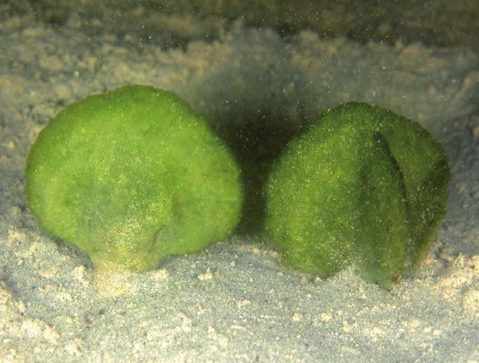
144 Fig. 88. Avrainvillea erecta. 141
, Lewmanomont & Ogawa (1995: 25, + fig.), Trono (1997: 70, fig. 47), Lewmanomont et al. (2007: 24, + figs), N Yeurt & Payri (2007: 68, figs 113, 114). Type locality: Guam, Mariana Is.")
145 BRYOPSIDALES Udoteaceae Avrainvillea Decaisne 1842: 108 Avrainvillea obscura (C. Agardh) J. Agardh 1887: 53 Fig. 89 References: Olsen- Stojkovich (1985: 19, figs 9, 10, pl. 2), Lewmanomont & Ogawa (1995: 25, + fig.), Trono (1997: 70, fig. 47), Lewmanomont et al. (2007: 24, + figs), N Yeurt & Payri (2007: 68, figs 113, 114). Type locality: Guam, Mariana Is. Description: Juvenile specimens only forming a small, hemispherical tuft of radially arranged loose filaments on top of a very short stipe, attached by a welldeveloped, long bulbous holdfast; fully developed specimens mostly forming shave-brush-like thalli (looking like a Penicillus), but some plants becoming flabellate, the flabellum remaining rather small and cuneate (as opposed to the broad, fanshaped to reniform flabellum of A. erecta); filaments of rather constant diameter, about 75 μm, cylindrical with deeply constricted equal dichotomies and round apices, orangy coloured. Ecology: Sand dwelling, in shallow lagoons, but also observed on sand between coral boulders (mostly on the sheltered, landward side); rare, compared to A. erecta. Distribution: Tropical Indian Ocean and western Pacific Ocean. Notes: We did not observe clavate siphons as mentioned in Olsen-Stojkovich (1985: 19). Fig. 89. Avrainvillea obscura. 142
146 BRYOPSIDALES Udoteaceae Rhipidosiphon Montagne 1842a: 15 Rhipidosiphon sp. Figs 30G; 64G; 91A-D Description: Plants gregarious, erect, in extremely dense tufts of supple, cuneate blades, 3-4 (-5) cm long; no visible stipes, the numerous fronds originating from a felted mat of rhizoids from where the blades gradually broaden. Young specimens still with a gradually tapering monosiphonous basis, 1-2 mm long that becomes surrounded by thin, downwardly growing, branched rhizoids in a very early stage; the monosiphonous basis becoming invisible in older plants where it is covered by an intricated rhizoidal pseudocortex; rhizoids even arising from the basal part of the flabellum (up to the 3rd or 4th dichotomy of the blade filaments) in fully grown plants; the blade thin, cuneate to narrowly flabellate, frequently spatulate, 4-6 (-8) cm long, cm wide in the subapical, widest part, with a rounded or lobed apical margin, unistratose all over; fully grown blades markedly and deeply longitudinally lacerate, greyish dark green, composed of parallel, straight and smooth filaments of about µm diameter at the blade basis, 58 µm at the margin, without papillae or appendages (even in the basal part), with rounded apices, dichotomously branching, with asymmetrical supradichotomous constrictions; the lowermost constrictions mostly more pronounced and frequently directed to the same side of the blade; intercalary constrictions also present and locally numerous and pronounced. Filaments loosely attached to each other by a thin layer of calcification that remains limited to the ridges between the siphons. Stacks of needle-like crystals are present in the siphons. Ecology: Mostly on horizontal, rocky substrate, best developed under small overhangs, close under low water level; some tufts on vertical walls, air-exposed at low tide. Discussion: The genus Rhipidosiphon now contains two species, R. javensis in the Indo-Pacific and R. floridensis Littler & Littler, only known from the Caribbean Sea. The genus has been studied by Gepp & Gepp (1904), Littler & Littler (1990b) and Vroom et al. (2001). Both species are characterized by a well-marked and visible (naked) monosiphonous stipe (well illustrated by Abbott & Huisman, 2004: figs 52C,D, and Kraft, 2007: figs 83B,C). R. javensis was collected in the Mu Ko Tha Lae Tai area. The here described taxon greatly resembles R. javensis but mainly differs in its larger size, the gregarious growth and the rhizoids also being produced at the basis of the flabellum, resulting in a rhizoidal pseudocortex at the basis of the plants, secluding the monosiphonous stipe. This material may therefore be a new species to science and it is actually under study. 144
, Tseng (1984: 294, pl. 146, fig. 2, as Udotea), Coppejans & Prud homme van Reine (1989: 139, pl. 10, figs 3-9, as Udotea), Trono (1997: 77, fig. 52 as Udotea), Payri et al.")
147 BRYOPSIDALES Udoteaceae Rhipidosiphon Montagne 1842a: 15 Rhipidosiphon javensis Montagne 1842a: 15 Figs 23B; 30F; 40B; 90 References: Jaasund (1976: 29, fig. 60, as Udotea javensis), Tseng (1984: 294, pl. 146, fig. 2, as Udotea), Coppejans & Prud homme van Reine (1989: 139, pl. 10, figs 3-9, as Udotea), Trono (1997: 77, fig. 52 as Udotea), Payri et al. (2000: 120, bottom fig. p. 121), Littler & Littler (2003: 254, middle fig. p. 255), Oliveira et al. (2005: 224, fig. p. 224), Huisman et al. (2007: 191, + figs), Kraft (2007: 233, fig. 83), Lewmanomont et al. (2007: 40, + figs), N Yeurt & Payri (2007: 71, figs 116,117), Skelton & South (2007: 289, figs 739,740, 795), Coppejans et al. (2009: 126, fig. 102). Type locality: Leiden Island (Nyamuk besar), Java, Indonesia. Description: Thalli erect, composed of stipitate, fan-shaped flabella, mostly 5-10 mm high, growing in open populations, dark green to greyish green (depending on the degree of calcification); attachment by hyaline rhizoids, sprouting from the basis of a slightly calcified, monosiphonous, unbranched, smooth, naked and clearly visible stipe, 1-4 mm long, 100 μm diameter; flabellum mostly flabellate to cuneate, 2-6 mm wide, 4-6 mm long, unistratose, composed of parallel, straight, contiguous, dichotomous (rarely trichotomous) siphons, radiating from the stipe to the margin, μm diameter, with asymmetrical constrictions above the dichotomies, without lateral appendages, kept together by the calcification between the siphons; crystals sometimes present in the blade siphons. Ecology: On vertical and overhanging rock walls, -2 m. Distribution: Indian Ocean, tropical Pacific Ocean. Fig.90. Rhipidosiphon javensis. 143


148 A B C D Fig. 91. Rhipidosiphon sp. A. Morphology in situ; B. Detail of a young specimen under stereoscopic microscope: the monosiphonous stipe is completely covered by a rhizoidal pseudocortex; C. Detail of the basis of a juvenile plant with developing rhizoids, under stereoscopic microscope, after decalcification; D. Detail of some asymmetrical supradichotomic constrictions under microscope. 145
149 BRYOPSIDALES Udoteaceae Udotea J.V. Lamouroux 1812: 186 Udotea flabellum (Ellis et Solander) Howe 1842a: 15 Figs 17E; 19; 25B; 40F; 92A,B References: Gepp & Gepp (1911: 131, pl. 3, figs 26-28), Tseng (1984: 292, pl. 145, fig. 3), Coppejans & Prud homme van Reine (1989: 137, pl. 9, figs 7-10), Littler & Littler (1989: 74, bottom fig. p. 75), Littler & Littler (1990: 226, fig. 12), Lewmanomont & Ogawa (1995: 58, + fig.), Huisman (2000: 269, + fig.), Littler & Littler (2000: 428, top fig. p. 429), Kraft (2007: 229, pl. 9C, fig. 81), Littler et al. (2008: 230, + figs). Type locality: West Indies. Description: Thallus erect, fan-shaped, extremely stiff and tough as a result of the strong calcification, dark bluish green; fixation by a well-developed bulbous holdfast; cylindrical stipe short (up to 1 cm), becoming compressed toward the flabellum; blade cuneate at the basis, flabellate, entire in juvenile specimens, becoming lobed and incised in young plants, repeatedly and irregularly proliferous from the margin in older ones; in very old specimens the proliferations become superposed and plicate, resulting in more bushy plants reaching 20 cm in height; surface markedly concentrically zonated and longitudinally striated; outer medullary frond siphons bearing lateral branchlets of unequal length, at irregular intervals, more or less perpendicularly placed, densely and fasciculately branched at their apex; these apices contiguous and forming a dense cortex which is very difficult to disentangle, even after decalcification. Ecology: On sandy substrate in the shallow subtidal (-0.5 to -1 m) in lagoons; mostly growing in discrete, open, monospecific populations in which all the fan-shaped plants are in the same direction (parallel to the incoming waves). Distribution: Pantropical. Note: Older specimens are densely covered by crustose corallines. 146


150 A B Fig. 92. Udotea flabellum. A. Juvenile specimens; B. A fully grown plant. 147
, Berger & Kaever (1992: 172, figs 3.83,3.")
, Lewmanomont et al. (2007: 19, + figs as Acetabularia), N Yeurt & Payri (2007: 73, fig. 121). Type locality: Tonga.")
151 DASYCLADALES Polyphysaceae Parvocaulis S. Berger et al. 2003: 559 Parvocaulis clavatus (Solms-Laubach) S. Berger et al. 2003: 559 Figs 93A,B References: Valet (1969: 620, pl. 12, figs 1, 3-6, pl. 19, fig. 1, pl. 20, figs 3, 8, pl. 22, fig. 3, pl. 23, fig.1, pl. 30, fig. 7, pl. 38, fig. 2, pl. 45, figs 1-4, as Acetabularia), Berger & Kaever (1992: 172, figs 3.83,3.84, as Polyphysa), Trono (1997: 87, fig. 60 as Acetabularia), Berger et al. (2003: 537, figs 13, 27), Abbott & Huisman (2004: 146, fig. 56A, as Acetabularia), Oliveira et al. (2005: 227, + fig.), Lewmanomont et al. (2007: 19, + figs as Acetabularia), N Yeurt & Payri (2007: 73, fig. 121). Type locality: Tonga. Description: Plants solitary, small, like an umbrella with only spokes; dark green as a result of the limited calcification along the lateral margins; holdfast a series of blunt digitate lobes, stalk 2-4 mm long; cap 1-2 mm in diameter, composed of 5-8 (-10), completely free, clavate rays that do not vary in diameter along their length up to the subapical part which is slightly inflated with smooth, rounded apices, but the rays are slightly constricted at their basis; each ray bearing a round, button-shaped corona superior segment with 2 or (more rarely) 3 hairs; corona inferior absent. Ecology: Epilithic, on dead coral surfaces and coral rubble, just under extreme low water mark, in sheltered habitats (lagoons); mostly overlooked because of the tiny dimensions. Distribution: Pantropical. Note: Berger et al. (2003: 559) propose a new genus (Parvocaulis) to include species, previously placed in the genera Polyphysa and Acetabularia. Frequently growing together with P. parvulus. A B 148 Fig. 93. Parvocaulis clavatus. A. Single plant; B. With P. parvulus (left).
, Tseng (1984: 268, pl. 133, fig.")
, Berger et al. (2003: 533, figs 11, 25), Abbott & Huisman (2004: 147, fig. 56C, as Acetabularia), Oliveira et al. (2005: 227 + fig., as Acetabularia), Huisman et al.")
152 DASYCLADALES Polyphysaceae Parvocaulis S. Berger et al. 2003: 559 Parvocaulis parvulus (Solms-Laubach) S. Berger et al. 2003: 559 Figs 93B; 94A,B. References: Valet (1969: 621, pl. 11, figs 1-7, pl. 12, fig. 7, pl. 19, figs 2-4, pl. 20, figs 5-8, pl. 22, figs 1, 4, 7, pl. 29, pl. 38, figs 1, 4,5, pl. 45, figs 5-7, as Acetabularia), Tseng (1984: 268, pl. 133, fig. 4, as Acetabularia), Berger & Kaever (1992: 166, figs , as Polyphysa), Trono (1997: 91, fig. 64 as Acetabularia moebii Solms-Laubach), Payri et al. (2000: 122, bottom fig. p. 123, as Polyphysa), Berger et al. (2003: 533, figs 11, 25), Abbott & Huisman (2004: 147, fig. 56C, as Acetabularia), Oliveira et al. (2005: fig., as Acetabularia), Huisman et al. (2007: 197, + fig.), Kraft (2007: 286, fig. 105), Lewmanomont et al. (2007: 21, + figs as Acetabularia), Skelton & South (2007: 295, figs ). Syntype localities: Tropical India, Macassar, Sulawesi, Indonesia. Description: Plants solitary or in small groups, small, like a flat umbrella; dark green to slightly whitish depending on the degree of calcification which is limited between the rays; holdfast a series of blunt digitate lobes, stalk 2-5 mm long; cap 2-3 mm in diameter, composed of 12-15, wedge-shaped, closely apposed rays with smooth, obtusely rounded apices, but the rays are slightly constricted at their basis; each ray bearing a round, button-shaped corona superior segment with 3-5 hairs; corona inferior absent. Ecology: Epilithic, on dead coral surfaces and coral rubble, just under extreme low water mark, in sheltered habitats (lagoons); mostly overlooked because of the tiny dimensions. Distribution: Tropical Indo-Pacific and Atlantic Ocean, Mediterranean. Note: Frequently growing together with P. clavatus. A B Fig. 94. Parvocaulis parvulus. 149
153 Brown algae Phaeophyceae Phaeophyceae - Brown algae Taxonomic overview of the species included in this guide DICTYOTALES Dictyotaceae Canistrocarpus cervicornis (Kützing) De Paula et De Clerck p. 152 Dictyota ceylanica Kützing p. 154 Dictyota ciliolata Sonder ex Kützing p. 156 Dictyota friabilis Setchell p. 158 Lobophora variegata (J.V. Lamouroux) Womersley ex Oliveira p. 160 Padina australis Hauck p. 162 Padina minor Yamada p. 164 SCYTOSIPHONALES Chnoosporaceae Chnoospora minima (Hering) Papenfuss p. 166 Scytosiphonaceae Hydroclathrus clathratus (C. Agardh) Howe p
154 FUCALES Cystoseiraceae Hormophysa cuneiformis (J. Gmelin) P. Silva p. 168 Sargassaceae Sargassum crassifolium C. Agardh p. 170 Sargassum oligocystum Montagne p. 172 Sargassum polycystum C. Agardh p. 174 Sargassum swartzii C. Agardh p. 176 Turbinaria conoides (J. Agardh) Kützing p. 178 Turbinaria decurrens Bory de Saint-Vincent p. 180 Turbinaria ornata (Turner) J. Agardh f. ecoronata W.R. Taylor p
155 DICTYOTALES Dictyotaceae Canistrocarpus De Paula et De Clerck in De Clerck et al. 2006: 1285 Canistrocarpus cervicornis (Kützing) De Paula et De Clerck in De Clerck et al Figs 12B; 26D; 31C; 45D; 56D; 95A,B References: Tseng (1984: 192, pl. 97, fig. 2, as Dictyota), Lewmanomont & Ogawa (1995: 70, + fig., as Dictyota), Calumpong & Meñez (1997: 129, fig. p. 130, as Dictyota), Trono (1997: 106, fig. 73, as Dictyota), De Clerck & Coppejans (2002: 88, figs p. 89), De Clerck (2003: 42-50, figs 11-14, as Dictyota), Tronchin & De Clerck (2005: 102, fig. 73, as Dictyota), Oliveira et al. (2005: 159, + fig., as Dictyota), Lewmanomont et al. (2007: 49, + figs, as Dictyota), Littler et al. (2008: 133, + figs). Type locality: Key West, Florida, USA. Description: Plants ligulate, those just under low water level along surf-exposed coasts, forming low mats, 3-5 cm tall, composed of rather stiff, intertwined, undulated and frequently longitudinally coiled straps of uniform width (3-5 mm), medium brown; plants from medium sheltered habitats, typically ascending with a well developed base, consisting of broad, thick straps and more slender (2-3 mm wide), supple erect straps; subtidal and epiphytic plants from sheltered lagoons lacking conspicuous basal parts, are more erect slender and supple; the prostrate parts attached at various points by marginal rhizoids; branching dichotomous in the middle and lower thallus, typically cervicorn in the apical parts; branch angle of (40-)60-70(-90), wider in apical parts (up to 120 ); interdichotomies 2-3 mm wide, mm long; apices rounded to truncate; margins smooth, locally sinoidally curved; hair tufts common on the strap surfaces. Internal structure composed of a single-layered medulla and upper and lower cortex; medullary cells μm long, μm wide, μm high, conspicuously narrow and long, cell wall thickenings absent; cortical cells μm long, μm wide, μm high. Sporangia mostly single and scattered over both surfaces in middle and subapical thallus parts, never in apical segments, not placed on transverse lines, surrounded by an involucrum, μm wide; gametangia not observed. Ecology: Dense, prostrate growth form close under low water level along surf-exposed coasts; prostrate growth form with some short, erect axes somewhat deeper and specimens without the basal, prostrate parts in sheltered lagoons, frequently epiphytic (on old Sargassum stipes). Distribution: Widespread in tropical regions. 152


156 A B Fig. 95. Canistrocarpus cervicornis. A. Prostrate growth form just under low water level; B. Erect growth form in sheltered, shallow lagoons. 153
157 DICTYOTALES Dictyotaceae Dictyota J.V. Lamouroux 1809b: 331 Dictyota ceylanica Kützing 1859: 11, pl. 25: fig. 1 Figs 36E; 96 References: Jaasund (1976: 41, fig. 83, as D. divaricata, fig. 84), Abbott & Huisman (2004: 202, fig. 77B), Payri et al. (2000: 134, top fig. p. 135, as D. divaricata), De Clerck (2003: 52-57, figs 15,16), Littler & Littler (2003: 168, top fig. p. 169), Oliveira et al. (2005: 159, fig. p. 161, as D. divaricata), N Yeurt & Payri (2006: 124, fig. 20), Huisman et al. (2007: 219, + fig.), Coppejans et al. (2009: 134, fig. 23A, 33A, 38D, 108). Type locality: Sri Lanka. Description: Thalli within a single tuft relatively heterogenous, but generally with a rather slender appearance and supple in the typical filiform growth form, relatively crisp in the specimens with broader straps; composed of relatively small (3-4 cm) ascending plants, without a conspicuous base, basal straps procumbent to repent, becoming erect higher up; erect straps frequently filiform, resulting in an intricate appearance; sometimes forming dense, low mats; often bluish or greenish iridescent, sometimes with marked yellowish axils; repent straps attached at various points by marginal rhizoids but rhizoid patches also present higher up along the erect straps; branching isotomous dichotomous all over the thallus, but the possible presence of numerous marginal proliferations can obscure the original branching system; branch angle broadly divaricate, of (60-) (-100) ; straps of variable width, the basal ones up to 2 mm wide, tapering gradually or abruptly to the filiform apical straps, some specimens without filiform parts other without the broad basal parts; apices of the broad straps rounded, those of the filiform branchlets acute; margins smooth; surface proliferations absent, hair tufts common; marginal proliferations common. Whole plant tristromatic, internal structure composed of a single-layered medulla and cortex; sporangia with a single stalk cell, not surrounded by an involucrum, ca 100 μm wide; gametangia not observed. Ecology: Epilithic, as well as epiphytic (on turf algae) just under low water level. Distribution: Indian Ocean, tropical Pacific Ocean. Note: Indian Ocean specimens traditionally attributed to D. divaricata J.V. Lamouroux were referred to Dictyota ceylanica by De Clerck (2003). Most likely, however, the latter does not represent a natural species. Dictyota ceylanica probably may contain several cryptic species characterized by irregular sprawling tufts composed of narrow, divaricate axes. 154

158 Fig. 96. Dictyota ceylanica (in situ) 155
159 DICTYOTALES Dictyotaceae Dictyota J.V. Lamouroux 1809b: 331 Dictyota ciliolata Sonder ex Kützing 1859: 12, pl. 27: fig. 1 Figs 97A,B References: Jaasund (1976: 41, fig. 82), Huisman (2000: 189, + fig.), Littler & Littler (2000: 262, top fig. p. 263), De Clerck (2003: 58-65, figs 17-19), Abbott & Huisman (2004: 204, fig. 77C), Tronchin & De Clerck (2005: 102, fig. 74), Oliveira et al. (2005: 160, fig. p. 160), Coppejans et al. (2009: 136, fig. 109). Type locality: La Guaira, Venezuela. Description: Plants erect, 4-8 cm long, attached by means of a single stupose holdfast giving rise to a crisp, brown frond, slightly iridescent in situ and often with transverse bands; branching dichotomous, never alternate, generally fairly regular and evenly distributed over the thallus; branch angle (30-) (-60) ; straps 5-9 mm wide, width generally similar throughout thallus but sometimes widening towards apices; apices rounded, rarely truncate; margins dentate (sometimes smooth but then frequently sinoidal), degree of dentation variable but generally less conspicuous near the base, with teeth slightly to prominently directed towards the apices; teeth may give rise to marginal proliferations which grow into new straps of similar aspect and obscure the original branching pattern; hair tufts common. Internal structure composed of single-layered medulla and cortex. Sporangia scattered on both surfaces, occurring singly, most frequently in middle and subapical parts, absent from apical segments, leaving a conspicuous sterile zone near the margin when occurring in high densities, not surrounded by an involucrum, about 100 μm wide. Gametangia not observed. Ecology: On coral rubble on the lagoon bottom, 0.5 to 1 m deep as well as epiphytic (e.g. on Sargassum stipes). Distribution: Pantropical and subtropical Atlantic Ocean (Europe). Note: Diagnostic features of Dictyota ciliolata include the stupose base, the regular dichotomous outline of the thallus and the dentate margins. The margins, however, are not always dentate. Individuals with smooth margins are fairly common in the area. 156


160 A B Fig. 97. Dictyota ciliolata. A. In situ; B. Herbarium specimens. 157
161 DICTYOTALES Dictyotaceae Dictyota J.V. Lamouroux 1809b: 331 Dictyota friabilis Setchell 1926: 91,92, pl. 13: figs 4-7, pl. 20: fig. 1 Fig. 98 References: Jaasund (1976: 39, fig. 79); Tseng (1984: 194, pl. 98, fig. 1), De Clerck & Coppejans (1996: 227, figs 40, 46), De Clerck (2003: 89-93, figs 28,29), Payri et al. (2000: 132, bottom fig. p. 133), Littler & Littler (2003: 168, middle fig. p. 169), Abbott & Huisman (2004: 205, fig. 77E), Tronchin & De Clerck (2005: 104, fig. 75), N Yeurt & Payri (2006: 125, fig. 21), Lewmanomont et al. (2007: 50, + figs), Skelton & South (2007: 210, figs ), Coppejans et al. (2009: 136, figs 32D, 33C, 110). Type locality: Tafaa Point, Tahiti. Description: Plants completely procumbent, forming dense imbricate mats composed of several layers of somewhat brittle (friable) straps, resulting in a typical jigsaw aspect, medium brown with varying iridescence (yellow-green to blue and often with small non-iridescent stripes or dots, or broader transverse non-iridescent bands); size of individual thalli ca. 3-5 cm in diameter, mats cm in diameter; attached by marginal patches of rhizoids; branching dichotomous to somewhat irregular, evenly branched throughout with broad branching angle (60-) (110), axils rounded; interdichotomies typically short and broad, on average mm wide, mm long, entire thallus of the same width; apices rounded to obtuse (rarely acute); margins smooth, rarely with proliferations; hair tufts common. Internal structure composed of a single-layered medulla and cortex. Sporangia scattered on the upper surface, occurring singly, absent from the apical segments, not surrounded by an involucrum, μm wide. Ecology: On horizontal rock substrate between coral heads, at about -4 m; rarely observed in the study area. Distribution: Indian Ocean, tropical Pacific Ocean. 158

162 Fig. 98. Dictyota friabilis. 159
163 DICTYOTALES Dictyotaceae Lobophora J. Agardh 1894: 21 Lobophora variegata (J.V. Lamouroux) Womersley ex Oliveira 1977: 217 Figs 12C; 32B; 37C-F; 44D; 49F; 99A,B References: Magruder & Hunt (1979: 47, middle fig. p. 46), Tseng (1984: 196, pl. 99, fig. 2), Lewmanomont & Ogawa (1995: 74, + fig.), Cribb (1996: 49, bottom fig. p. 48), De Clerck & Coppejans (1996: 228, figs 43, 47), Calumpong & Meñez (1997: 128, + fig.), Trono (1997: 111, fig. 75), Huisman (2000: 193, + figs), Payri et al. (2000: 136, figs p. 137), Littler & Littler (2000: 268, 269, bottom fig. p. 269, figs p. 271), De Clerck & Coppejans (2002: 90, fig. p. 91), Littler & Littler (2003: 172, bottom fig. p. 173), Abbott & Huisman (2004: 209, figs 80A-C), Tronchin & De Clerck (2005: 110, fig. 82), Oliveira et al. (2005: 162, figs p. 163), N Yeurt & Payri (2006: 126, figs 24-26), Baimai & Tantalakha (2007: 53, + fig.), Huisman et al. (2007: 221, + figs), Lewmanomont et al. (2007: 54, + figs), Ohba et al. (2007: 77, + figs), Skelton & South (2007: 212, figs ), Coppejans et al. (2009: 138, figs 24E, 27F, 37E, 111). Type locality: Antilles, West Indies. Description: Plants ranging from prostrate to resupinate, irregularly lobed to fan-shaped blades; thalli up to 7 cm in diameter and μm thick, orangy to dark brown, the prostrate ones frequently radially striped and greyish brown; prostrate thalli longish and irregularly lobed, thick, stiff, attached by moniliform rhizoids arising from the whole ventral surface; resupinate blades semicircular, thin and supple, attached by discrete holdfasts and rhizoids mainly at the basal parts of the blades; margins not inrolled; growth from a marginal meristem of apical cells, sometimes incised and longitudinally split in places; hairs usually arranged in concentric zones. Internal structure composed of a single-layered central medulla of large, rectangular cells with 2-4 layers of subcortical cells on either side with a single-layered cortex. Sporangia grouped in ovate to irregular and confluent sori, covered by an indusium, clavate, up to 110 μm long and 40 μm wide, lacking a stalk cell, containing 8 spores each. Ecology: Prostrate plants on horizontal substrate between coral heads or rock boulders, subtidal, from 1 to 5 m deep, rather rare; resupinate specimens in large, cascade-like populations on vertical or steeply inclined walls, from just under low water level down to 4 m depth, extremely abundant in the study area. Distribution: Pantropical. Note: There is molecular evidence that the different growth forms are in fact different taxa. 160


164 A B Fig. 99. Lobophora variegata A. Resupinate growth form; B. Prostrate specimen. 161
165 DICTYOTALES Dictyotaceae Padina Adanson 1763: 13, 586 Padina australis Hauck 1887: 44 Fig. 100A,B References: Magruder & Hunt (1979: 47, bottom fig. p. 46), Tseng (1984: 198, pl. 100, fig. 1), Lewmanomont & Ogawa (1995: 75, + fig.), Calumpong & Meñez (1997: 126, + fig. p. 127), Trono (1997: 112, fig. 77), Abbott & Huisman (2004: 212, figs 81A-C), Baimai & Tantalakha (2007: 54, + fig.), Huisman et al. (2007: 224, + figs), Lewmanomont et al. (2007: 56, + figs), Ohba et al. (2007: 78, + figs). Type locality: Cape York, Queensland, Australia. Description: Plants erect, up to 15 cm tall, with numerous fan-shaped blades, funnel-like arranged, usually lightly calcified and of rather soft texture, slightly transparent in situ to light greyish white on both surfaces; holdfast conical, composed of a felt of rhizoids; absence of basal Vaughaniella-stage. As all Padina species, growth from a marginal meristem at the inrolled margin; the blade side towards the inrolled margin is here called the inner (= upper) surface; blades 2 cells thick throughout; hairs in concentric rows on both surfaces of the frond, usually alternating between inner and outer (= lower) surfaces, producing a pattern of broad and narrow smooth zones between the hair bands; gametophytes not observed but mentioned to be monoecious in literature; when reproductive, sporangia exclusively formed on the outer surface, closely distal to a hair band; such pairs of sporangial line and hair line alternating with hair lines on the other (inner) surface; on a transverse section a hair line on the inner surface is relatively close to a pair (sporangial line and hair line) on the outer surface, but more distant than the hair row and sporangial line to each other. Sporangia non-indusiate. Ecology: Subtidal, as well on rock substrate as on coral fragments on sand, mostly in deeper parts of lagoons. Distribution: Scattered reports from central and western tropical and subtropical Pacific and Indian Ocean. Notes: Padina australis differs from P. boryana Thivy by the rows of hairs being produced on both sides of the blade in the former (only on the outer surface in the latter), and from P. sanctae-crucis Børgesen by its lighter calcification and the absence of the Vaughaniella-stage. In P. boergesenii Allender & Kraft, the flabellum is thicker (3-4 layers in the middle part, but up to 8 near the base). P. boryana also differs from P. boergesenii by the presence of an indusium on young sporangial sori in the former, whereas they are non-indusiate in the latter. 162


166 A B Fig Padina australis. A. Habit in situ; B. Detail of a sporophyte with concentric bands of spores, alternately on both blade surfaces, and light superficial calcification (herbarium specimen). 163
167 DICTYOTALES Dictyotaceae Padina Adanson 1763: 13, 586 Padina minor Yamada 1925: , fig. V Fig. 101A-C References: Lewmanomont & Ogawa (1995: 78, + fig.), De Clerck & Coppejans (1996: 230, figs 49, 52), Calumpong & Meñez (1997: 126, + fig.), Trono (1997: 115, fig. 79), Coppejans et al. (2009: 140, fig. 114). Type locality: Taiwan. Description: Plants generally up to 6 cm tall, composed of erect, fan-shaped blades, markedly orangy brown on the lower, uncalcified face, with a white, strongly calcified layer on the upper surface; stipe very short (up to 0.5 cm long), usually surrounded by a very well developed, conspicuous Vaughaniella-stage; this superficially looks like a prostrate to ascendant Dictyota species with narrow, rather stiff straps, a main axis and alternately placed side branches, all with a single apical, lens-like cell; blades somewhat funnel-shaped, young ones simple, older ones often longitudinally split, but not as compound as e.g. in P. australis, with a distinctive inrolled distal margin; blade 6 (-8) cm broad, 50 μm thick apically, up to 80 μm proximally. Internal structure invariably 2 layers thick. Tetrasporangial sori produced on the ventral (lower) surface in concentric transverse bands just distal to the hair bands, not covered by an indusium; gametophytes not observed. Ecology: A common species in the shallow subtidal, up to just under low water level. Distribution: Indian Ocean, tropical west Pacific Ocean. Note: The species is characterized by its 2-layered thallus, with hairs restricted to the outer (lower) surface and the well-developed Vaughaniella- stage. It does resemble P. boryana Thivy somewhat, which is also mentioned from Thailand, but the latter is more variably calcified and is composed of 2-3 layers of cells rather than 2 throughout. 164


168 A B C Fig Padina minor. A,B Habit in situ with well-developed Vaughaniella-stage; C. Detail of a sporophyte with concentric bands of spores on the lower, uncalcified surface and heavy calcification on the upper surface (= folded part; herbarium specimen). 165
, Lewmanomont & Ogawa (1995: 66, + fig.), Cribb (1996: 39, middle fig. p. 38), Trono (1997: 119), Littler et al. (1998: 120, bottom fig. p. 121), Littler & Littler (2000: 246, bottom fig. p. 247), Payri et al.")
169 SCYTOSIPHONALES Chnoosporaceae Chnoospora J. Agardh 1847: 7 Chnoospora minima (Hering) Papenfuss 1956: Figs 22C; 102 References: Tseng (1984: 184, pl. 93, fig. 2), Lewmanomont & Ogawa (1995: 66, + fig.), Cribb (1996: 39, middle fig. p. 38), Trono (1997: 119), Littler et al. (1998: 120, bottom fig. p. 121), Littler & Littler (2000: 246, bottom fig. p. 247), Payri et al. (2000: 142, bottom fig. p. 143, Littler & Littler (2003: 176, bottom fig. p. 177), Abbott & Huisman (2004: 177, figs 66C,D), Tronchin & De Clerck (2005: 118, fig. 89), N Yeurt & Payri (2006: 133, fig. 39), Huisman et al. (2007: 233, + figs), Skelton & South (2007: 217, figs ), Coppejans et al. (2009: 142, figs 11A, 16A, 116). Type locality: Port Natal (Durban), South Africa. Description: Plants erect in the basal part, pendulous in the upper parts, up to 7(-11) cm long, gregarious, forming isolated patches, reddish brown; tightly adhering to the substrate by a discoid holdfast; erect axes cylindrical to compressed, 1-2 mm in diameter, 2-4 times dichotomously to irregularly branched; branching angle narrow, ; apices acute and often bifurcate; hair tufts distinctive, scattered across the entire thallus surface. Plurilocular reproductive structures clavate, up to 55 μm long, grouped in sori, often in association with hair tufts. Ecology: Confined to extremely wave-exposed habitats in the supralittoral fringe and high intertidal; showing a marked seasonality, (almost) disappearing in the dry season; only observed at Eric s Cave, Ko Taen. Distribution: Widespread in the Indian Ocean, tropical Pacific Ocean and tropical Atlantic Ocean. Fig Chnoospora minima, in the intertidal, air-exposed at low tide. 166
, Tseng (1984: 186, pl. 94, fig. 2), Littler et al. (1998: 118, bottom fig. p. 119), Lewmanomont & Ogawa (1995: 73, + fig.), Cribb (1996: 49, middle fig. p. 48), De Clerck & Coppejans (1996: 245, fig.")
170 SCYTOSIPHONALES Scytosiphonaceae Hydroclathrus Bory de Saint-Vincent 1825: 419 Hydroclathrus clathratus (C. Agardh) M.A. Howe 1920: 590 Fig. 103 References: Magruder & Hunt (1979: 47, top fig. p. 46), Tseng (1984: 186, pl. 94, fig. 2), Littler et al. (1998: 118, bottom fig. p. 119), Lewmanomont & Ogawa (1995: 73, + fig.), Cribb (1996: 49, middle fig. p. 48), De Clerck & Coppejans (1996: 245, fig. 76), Calumpong & Meñez (1997: 133, + fig. p. 132), Trono (1997: 121, fig. 82), Huisman (2000: fig.), Littler & Littler (2000: 248, middle fig. p. 249), Payri et al. (2000: 144, bottom fig. p. 145), De Clerck & Coppejans (2002: 90, fig. p. 91), Littler & Littler (2003: 178, top fig. p. 179), Abbott & Huisman (2004: 183, figs 68C,D), Oliveira et al. (2005: 170, middle fig. p. 171), Tronchin & De Clerck (2005: 120, fig. 92), N Yeurt & Payri (2006: 135, figs 42-43), Huisman et al. (2007: 230, + fig.), Lewmanomont et al. (2007: 53, + fig.), Ohba et al. (2007: 82, + figs), Skelton & South (2007: 221, figs 615, 784). Type locality: Uncertain. Description: Plants forming irregularly lobed, three-dimensional net-like structures, up to 7 cm in diameter, relatively stiff but slippery to the touch, yellowish brown; young specimens initially spherical and hollow, becoming irregularly convoluted with numerous irregularly shaped perforations of variable size when older, margins of perforations mostly inrolled; attachment by groups of rhizoids developing from the whole lower surface. Internal structure consisting of a well-defined cortex surrounding a cellular medulla; cortex 2 cell-layers thick, composed of small, pigmented, isodiametric cells, 7-10 μm in diameter; medulla composed of inwardly progressively large, hyaline, thin-walled cells, up to 70 μm in diameter arranged around a hollow center; hair tufts formed from depressions in the cortex. Ecology: Epilithic and on short algal turf in shallow intertidal rock pools. Distribution: Widespread in tropical and warm temperate regions. Note: This taxon has only rarely been observed in the study area; along other coasts of Thailand it is (seasonally) abundant and washes ashore in large quantities. Fig Hydroclathrus clathratus (herbarium specimen). 167
171 FUCALES Cystoseiraceae Hormophysa Kützing 1843: 359 Hormophysa cuneiformis (J. Gmelin) P.C. Silva in P.C. Silva, Meñez & Moe 1987: 81, 131 Figs 28D; 104A,B References: Tseng (1984: 210, pl. 106, fig. 1, as H. articulata Kützing), Lewmanomont & Ogawa (1995: 72, + fig. as H.triquetra (C. Agardh) Kützing), Cribb (1996: 49, top fig. p. 48, as H. triquetra, De Clerck & Coppejans (1996: 236, fig. 62), Calumpong & Meñez (1997: 134, + fig.), Trono (1997: 125, fig. 85), Huisman (2000: 219, + fig.), De Clerck & Coppejans (2002: 92, fig. p. 93), Littler & Littler (2003: 180, middle fig. p. 181), Oliveira et al. (2005: 174, fig. p. 175), Tronchin & De Clerck (2005: 124, fig. 95), Ohba et al. (2007: 83, + figs). Type locality: Suez, Egypt. Description: Plants erect, cm high, rather stiff, apical parts yellowish brown, darker towards the basis; discoid holdfast with a short, conoidal perennial basis from which several primary axes arise; branching irregular; axes irregularly triquetrous, more or less segmented, because the wings are constricted between two consecutive aerocysts; wings 1-5 mm wide, with markedly dentate margins; locally only 2 opposite wings, resulting in a plane thallus; oblong aerocysts developing in the upper parts of the axes, intercalary, centrally in the medulla of the axes, giving the thallus an articulated appearance. Ecology: Epilithic in shallow lagoons. Distribution: Widespread in the tropical Indo-Pacific. Note: The basis of old specimens are densely covered by crustose corallines and other epiphytes such as Leveillea jungermannioides. 168


172 A B Fig Hormophysa cuneiformis. A. Habit in situ; B. Detail of a branch with intercalary air vesicles (arrows). 169
173 FUCALES Sargassaceae Sargassum C. Agardh 1820: 1 Sargassum crassifolium J. Agardh 1848: Figs 30D; 105 References: Tseng (1984: 226, pl. 114, fig. 2), Tsuda (1988: 60, fig. 1), Yoshida (1988: 12, fig. 6), Trono (1992: 50, figs 9-11, 112), Lewmanomont & Ogawa (1995: 82, + fig.), Cribb (1996: 55, bottom fig. p. 54), Ajisaka et al. (1997: 34, fig. 12), Trono (1997: 131, figs 88A, B), Tseng & Lu (1997: 23, figs 7, 14), Tronchin & De Clerck (2005: 126, fig. 97), Skelton & South (2007: 223, figs , 767), Coppejans et al. (2009: 144, figs 19A,E, 41G,H, 118). Syntype localities: Cape of Good Hope, South Africa; New Ireland; New Zealand. Description: Plants gregarious, erect, tough, leathery, up to 25 cm high, brown. Holdfast disc-like; main stipes one to several, smooth, bearing radially placed side branches becoming gradually shorter towards the thallus apex; leaves fleshy and stiff, frequently upwardly curved, numerous, often crowded, to 1 cm wide, 1 cm long, with midvein; tips rounded, with thickened, two-edged margin; margins with coarse, irregular teeth; air bladders spherical, to 6 mm diameter, often with apical spines forming a crest. Cryptostomata (pits with hairs) as scattered dark dots on blades and air bladders. Receptacles forked, densely branching, spinose, to 5 mm long. Ecology: Epilithic on horizontal rock surfaces, along the surf-exposed coasts, just under low water level; rather rare. Distribution: Widespread in the tropical Indo-Pacific. Note: In literature, confusion exists between S. crassifolium and S. cristaefolium C. Agardh (= S. duplicatum J. Agardh). According to Trono (1997, , fig. 88Ba), the leaves of S. crassifolium are coarsely dentate and the vesicles are smooth or with an apical spine (fig. 88Bb) whereas in S. cristaefolium the leaves are finely dentate (fig. 89Ba) and the vesicles have 2 horn-like or lateral spines or dentate wings (fig. 89Bb). Representatives of the genus Sargassum are notoriously difficult to identify on species level. The characters, generally used for identification exhibit an extreme variability on several scales: temporal, intraindividual (morphology of the basal leaves different from the apical ones), interindividual, environmental and geographical next to sexual dimorphism and random phenotypic expression (Kilar et al., 1992). Moreover, according to Guiry & Guiry (2009), 584 species names are currently accepted worldwide (845 including the synonyms). For Thailand, 12 species have been recorded in literature. 170


174 A B Fig Sargassum crassifolium. A. Some specimens in situ; B. Detail of an apex with fleshy leaves with duplicated, coarsely dentate margin. 171
175 FUCALES Sargassaceae Sargassum C. Agardh 1820: 1 Sargassum oligocystum Montagne 1845: Figs 28A,B; 106A,B References: Yoshida (1988: 10), Trono (1992: 46, figs 1-4, 109, as S. binderi, 60, figs 35-38, 115, as S. oligocystum), Noro et al. (1994: 27, figs 3, 8), Lewmanomont & Ogawa (1995: 83, + fig.), Calumpong & Meñez (1997: 135, + top fig. p. 136), Trono (1997: 143, figs 94A, B), Ajisaka et al. (1999: 28, figs 3a-c, 7e-g, as S. binderi, 34, figs 3e,f, 8g, as S. oligocystum), De Clerck & Coppejans (2002: 92, figs p. 93), Littler & Littler (2003: 182, bottom fig. p. 183), Oliveira et al. (2005: figs), Lewmanomont et al. (2007: 59, + figs). Type locality: Lampung Bay, Sumatra, Indonesia. Description: Plants erect as a result of the numerous, large air vesicles, very supple, up to 1 m long, forming dense, bushy tufts, medium brown; holdfast discoid, perennial stem terete to slightly compressed, warty, cm long; primary branches numerous, flattened or compressed, up to 1 m long and 3 mm wide, smooth, producing distichously and alternately arranged secondary branches. Lower leaves large, lanceolate, simple, up to 3 cm long and 9 mm wide, with very short stalks, rounded apices, margin dentate with small teeth, midrib indistinct, vanishing below upper parts of the leaves or otherwise completely evanescent, small cryptostomata scattered. Upper leaves of fully grown secondary branches and branchlets broadly lanceolate, markedly undulated, simple, up to 1.7 cm long and 5 mm wide, with sharp apices, margin dentate with sharp teeth, midrib vanishing near apices, small cryptostomata scattered. Vesicles spherical to elliptical, up to 8 mm long, smooth at the apex or slightly mucronate; stalks flattened, usually longer than the vesicles. Receptacles flattened, often twisted, up to 5 mm long, sharply dentate at the margin, simple to furcated once, racemosely arranged, clustered. Ecology: Epilithic in shallow lagoons. Distribution: Tropical Indo-West Pacific. Note: The synonymy of Sargassum binderi Sonder ex J.G. Agardh with S. oligocystum has been proposed by Womersley and Bailey (1970). 172


176 A B Fig Sargassum oligocystum. A. In situ tuft of fully grown plants; B. Detail of an apical part with numerous air bladders. 173
177 FUCALES Sargassaceae Sargassum C. Agardh 1820: 1 Sargassum polycystum C. Agardh 1824: 304 Fig. 107 A,B References: Tseng (1984: 236, pl.119, fig. 1), Tseng & Lu (1988: 47, figs 13, 26, 27), Tsuda (1988: 62, fig. 4), Yoshida (1988: 17, fig. 14), Trono (1992: 63), Ajisaka et al. (1995: 34, figs 19, 20), Lewmanomont & Ogawa (1995: 84, + fig.), Cribb (1996: 59, middle fig. p. 58), Calumpong & Meñez (1997: 136, + fig.), Trono (1997: 147, figs 96A,B), Ajisaka et al. (1999: 36, figs 6b), Littler & Littler (2003: 184, top fig. p. 185), Oliveira et al. (2005: 178, + fig.), Lewmanomont et al. (2007: 60, + figs), Ohba et al. (2007: 84, + figs), Skelton & South (2007: 226, figs , 768), Coppejans et al. (2009: 146, figs 41A, 119). Type locality: Sunda Strait, Indonesia. Description: Plants erect, as a result of the numerous, small air vesicles, very supple, up to 80 cm long, dark brown; attachment by a disc, bearing a short (1 cm) perennial stem, producing several annual erect branches, but also presence of basal stolonoidal branches, sprawling over the substrate and bearing alternately, pinnately placed small and narrow cauline leaves and no air vesicles; main axes terete, bearing numerous short (3-4 cm, occasionally longer), radially arranged, upwardly directed side branches; main axes and side branches muricate with Y- shaped or simple, spiny processes; leaves crowded and larger on young annual branchlets, more scattered, smaller and narrower on older main branches; fertile specimens frequently with naked basal parts and densely placed leaves and receptacles on the upper side branches; leaves ovate-oblong to narrow lanceolate, to 2-3 cm long and 1 cm wide in basal parts, 1 cm long and 0.5 cm wide in upper parts, dentate; midrib generally extending the entire length of the blade. Air bladders often extremely abundant, small (1.5 to 2.5 mm in diameter), spherical, apiculate, borne on a short pedicel; cryptostomata scattered on the blades, branches and air bladders. Receptacles crowded, filiform, forked, 6-12 mm long, clustered in the axils of the leaves; some receptacles can be foliaceous towards the apices, resembling linear leaves. Ecology: Epilithic, in lagoons. Distribution: Widespread in the Indian Ocean, tropical Pacific Ocean, tropical eastern Atlantic Ocean. 174

. 175")
178 A B Fig Sargassum polycystum. A. Whole thallus with basal stolons; B. Detail of the numerous, small air bladders (herbarium specimens). 175
179 FUCALES Sargassaceae Sargassum C. Agardh 1820: 1 Sargassum swartzii C. Agardh 1820: 11 Figs 16B; 30E; 35A; 48E,G; 108 References: Tseng (1984: 238, pl. 120, fig. 1), Yoshida (1988: 19, fig. 18), Noro et al. (1994: 27, figs 4, 9), Tseng & Lu (1995a: figs 2-5), Ajisaka (2002: 82, figs 12-15), Noiraksa & Ajisaka (2008:193, fig.10). Type locality: Not known. Description: Plants erect, up to 60 cm high, rather bushy; holdfast discoid, perennial stem terete, up to 10 mm long, with a smooth surface, issuing annual main branches radially; these branches markedly compressed, mm wide, mm thick, issuing secondary branches distichously: secondary branches also compressed, similarly branched distichously resulting in a dense aspect. Leaves of the basal parts of main branches elongated lanceolate, up to 4 cm long and 5 mm wide, asymmetrical at the base, acute at the apex, margin entire or sinoidal, midrib obscure and immersed at the apex, cryptostomata small and scattered; leaves of the median parts of the thallus up to 4 cm long and 3 mm wide, linear lanceolate to linear with smooth to sinoidal margins in some plants, 8 cm long and 10 mm wide with markedly dentate margin, midrib obscure and vanishing near the apex, cryptostomata arranged on each side of the midrib, vesicles rare in numerous specimens, somewhat more frequent in others, elliptical, up to 6 mm long and 5 mm wide, pointed at the apex or mucronate with a 1-4 mm long spine; stipes of vesicles cuneate, about 5 mm long, sometimes longer than the vesicles. Ecology: Epilithic in the shallow subtidal along surf-exposed coasts (sometimes mixed with Turbinaria decurrens); rather rare. Distribution: Tropical Indo-Pacific. 176


180 Fig Sargassum swartzii. 177
181 FUCALES Sargassaceae Turbinaria J.V. Lamouroux 1825: 71 Turbinaria conoides (J. Agardh) Kützing 1860: 24, pl. 66: fig. IIe, f Figs 12A; 51E; 109A,B References: Tseng (1984: 240, pl. 121, fig. 3), Lewmanomont & Ogawa (1995: 85, + fig.), Calumpong & Meñez (1997: 138, + fig.), Trono (1997: 152, fig. 98), Littler & Littler (2003: 184, bottom fig. p. 185), Oliveira et al. (2005: 180, + fig.), Lewmanomont et al. (2007: 64, + fig.), Ohba et al. (2007: 85, + figs). Syntype localities: ad oras Hindostaniae ; China; Sri Lanka; West Australia. Description: Plants erect, supple, up to 50 cm high, orangy to dark brown, attached by a disc, arising from a rhizomatous base; the main axis richly branched, the side branches possibly rebranching, resulting in large bushy plants with several parallel, erect axes; the horizontally placed, radially arranged leaves spaced, especially in fertile specimens, trumpet-shaped, with a slender, cylindrical stipe, about 10 mm long, becoming obconoidal by the presence of a marked air bladder, bearing a relatively thin, horizontally spread (peltate) to funnel-shaped wing with acutely dentate margin; blades heart-shaped (markedly emarginated) to irregularly triangular in surface view, about 5-8 mm in diameter, upper surface from flat (peltate) branchlets to markedly convex with a central, sunken, inflated aerocyst in the funnel-shaped ones. Reproductive receptacles racemose, originally borne in dense clusters on the adaxial side of the stipes of the leaves; fertile specimens frequently losing the upper part of the branchlets (air vesicle and peltate part) acropetally, resulting in axes covered by receptacles (on the remaining basal part of the blade stipes) in the basal parts of the plants and apical tufts of peltate branchlets. Ecology: Epilithic in medium deep lagoons (0.5 to 1 m at low tide). Old plants frequently covered by Jania spp. Distribution: Tropical to subtropical Indo-Pacific. Note: Our specimens correspond very well with Tseng s (1984) pl.121, fig. 3 and Trono s (1997) fig


182 A B A C Fig Turbinaria conoides. A, B. Habit in situ. C. Detail of peltate branchlets and receptacles. 179
, Trono (1997: 154, fig. 99), Littler & Littler (2003: 178, top fig. p. 179), Oliveira et al. (2005: 180, + figs p. 181), Lewmanomont et al. (2007: 65, + figs), Ohba et al. (2007: 86, + figs).")
183 FUCALES Sargassaceae Turbinaria J.V. Lamouroux 1825: 71 Turbinaria decurrens Bory de Saint-Vincent 1828: 119 Figs 8D; 15C,E; 16B; 30B,D; 33C; 110 References: Lewmanomont & Ogawa (1995: 86, + fig.), Trono (1997: 154, fig. 99), Littler & Littler (2003: 178, top fig. p. 179), Oliveira et al. (2005: 180, + figs p. 181), Lewmanomont et al. (2007: 65, + figs), Ohba et al. (2007: 86, + figs). Type locality: In the sea between Tahiti and New Guinea. Description: Plants mostly gregarious, erect and extremely stiff, (-20) cm high, brown (young specimens more orangy); attachment by a disc, hidden by the dense stolons; main axes unbranched, densely covered by stiff, fleshy leaves that are spirally placed on the axis, resulting in 5 well-separated vertical rows; leaves mm long, obpyramidal in lateral view, obtriangular in surface view (sometimes slightly emarginated along the upper margin), mm in diameter, the lower angle of a leaf contiguous or even slightly overlapping the horizontal margin of the proximal leaf of the same row; stipe triangular in transverse section; the ribs of the stipe as well as the margin of the obtriangular surface finely toothed; small air vesicle completely embedded in the leaf; receptacles at the basis of and on the adaxial side of the leaf stipe. Old specimens loosing the basal leaves, resulting in a bare stipe with warty scars of the lost leaves. Ecology: On horizontal rock substrate (or dead upper part of coral heads), from 0.5 m under low water level down to the drop off at about 2 m depth, best developed and abundant along surf-exposed coasts. Distribution: Tropical Indo-Pacific. Fig Turbinaria decurrens. 180
. Type locality: Strawn Island, Palmyra Atoll, Line Islands.")
184 FUCALES Sargassaceae Turbinaria J.V. Lamouroux 1825: 71 Turbinaria ornata (Turner) J. Agardh f. ecoronata W.R. Taylor 1964: 485, pl. 3: figs. 7-9 Figs 28C; 30B; 111 Reference: Skelton & South (2007: 228, figs 628,629). Type locality: Strawn Island, Palmyra Atoll, Line Islands. Description: Plants erect, clustered, rather stiff but pliable, arising from a rhizomatous base, cm tall, only sparsely branched at the basis; the horizontally placed, radially arranged leaves densely placed, almost contiguous, peltate, with a slender, cylindrical stipe, about 10 mm long, abruptly distended in the distal blade; blades round to irregularly triangular in surface view, about 10 mm in diameter, upper surface markedly convex with a central, sunken, inflated aerocyst and coarsely dentate margin; the upwardly directed margin of the blades sometimes slightly emarginated; the typical intramarginal crown-teeth of T. ornata are absent. Reproductive receptacles borne in dense clusters on the adaxial side of the stipes of the leaves. Ecology: Epilithic, shallow subtidal down to 0.5 m depth. Rather rare. Distribution: Scattered locations in the tropical Indo-Pacific. Notes: Taylor (1964: 485, pl. 3: figs 7-9) described specimens without or with only a few intramarginal teeth as Turbinaria ornata f. ecoronata W.R. Taylor. Tsuda (1972) and Skelton & South (2007: 228) on the other hand, state that f. ecoronata might be an environmentally induced growth form. In the study area, however, we never found the typical, stiff T. ornata with numerous intramarginal teeth on the peltate branchlets. On the other hand, these plants are similar to young specimens of Turbinaria conoides, but are coarser, more stiff and the peltate leaves are somewhat larger. Fig Turbinaria ornata f. ecoronata 181
185 Red algae Rhodophyta Rhodophyta - Red algae Taxonomic overview of the species included in this guide For the classification of the red algae we follow Schneider & Wynne (2007), except for the Ceramiaceae (sensu lato) where we follow the more recent paper by Cho et al. (2008) who propose a splitting of the family. NEMALIALES Galaxauraceae Actinotrichia fragilis (Forsskål) Børgesen p. 184 GELIDIALES Gelidiaceae Gelidium sp. p. 185 Gelidiellaceae Gelidiella acerosa (Forsskål) J. Feldmann et G. Hamel p. 186 GRACILARIALES Gracilariaceae Congracilaria babae Yamamoto p. 188 Gracilaria salicornia (C. Agardh) Dawson p. 189 BONNEMAISONIALES Bonnemaisoniaceae Asparagopsis taxiformis (Delile) Trevisan p. 190 CORALLINALES Corallinaceae Amphiroa fragilissima (Linnaeus) J.V. Lamouroux p
186 Amphiroa rigida J.V. Lamouroux p. 193 Jania adhaerens J.V. Lamouroux p. 194 Jania ungulata (Yendo) Yendo p. 195 GIGARTINALES Cystocloniaceae Hypnea pannosa J. Agardh p. 196 Hypnea spinella (C. Agardh) Kützing p. 198 Peyssonneliaceae Peyssonnelia sp. p. 200 RHODYMENIALES Champiaceae Champia compressa Harvey p. 202 Lomentariaceae Gelidiopsis intricata (C. Agardh) Vickers p. 204 Gelidiopsis repens (Kützing) Weber-van Bosse p. 205 Gelidiopsis variabilis (J. Agardh) Schmitz p. 206 Rhodymeniaceae Botryocladia sp. p. 207 Ceratodictyon spongiosum Zanardini p. 208 Peltate, iridescent Rhodymeniaceae p. 210 CERAMIALES Ceramiaceae Centroceras clavulatum (C. Agardh) Montagne p. 212 Gayliella flaccida (Harvey ex Kützing) T.O. Cho et L.J. McIvor p. 213 Delesseriaceae Taenioma perpusillum (J. Agardh) J. Agardh p. 214 Rhodomelaceae Acanthophora spicifera (Vahl) Børgesen p. 215 Bostrychia tenella (J.V. Lamouroux) J. Agardh p. 218 Chondrophycus cartilagineus (Yamada) Garbary et Harper p. 220 Laurencia nidifica J. Agardh p. 222 Leveillea jungermannioides (Hering et G. Martens) Harvey p. 224 Lophocladia cf. minima Itono p. 226 Palisada papillosa (C. Agardh) K.W. Nam p. 228 Polysiphonia/Neosiphonia sp. p. 230 Tolypiocladia glomerulata (C. Agardh) Schmitz p. 231 Wrangeliaceae Wrangelia argus (Montagne) Montagne p
; Cribb (1983: 25, pl. 8, fig. 1), Tseng (1984: 58, pl. 32, fig. 1); Verheij & Prud homme van Reine (1993: 439, pl. 14, fig. 1), Lewmanomont & Ogawa (1995: 91, + fig.")
, Littler & Littler (2003: 62, top fig. p. 63), Oliveira et al. (2005: 47, + fig), Huisman et al. (2007: 69, + figs), Ohba et al.")
187 NEMALIALES Galaxauraceae Actinotrichia Decaisne 1842: 118 Actinotrichia fragilis (Forsskål) Børgesen 1932: 6 Figs 29C; 112A,B References: Jaasund (1976: 65, fig. 131); Magruder & Hunt (1979: 57, fig. 2, p. 56); Cribb (1983: 25, pl. 8, fig. 1), Tseng (1984: 58, pl. 32, fig. 1); Verheij & Prud homme van Reine (1993: 439, pl. 14, fig. 1), Lewmanomont & Ogawa (1995: 91, + fig.), Calumpong & Meñez (1997: 150, bottom fig. p. 93, top fig. p. 151), Trono (1997: 171, fig. 172), Abbott (1999: 64, figs 7A-C), Huisman (2000: 37, + figs), Payri et al. (2000: 160, bottom fig. p. 161), De Clerck & Coppejans (2002: 94, figs p. 95), Littler & Littler (2003: 62, top fig. p. 63), Oliveira et al. (2005: 47, + fig), Huisman et al. (2007: 69, + figs), Ohba et al. (2007: 90, + figs), Skelton & South (2007: 16, figs 14,15, 780), Coppejans et al. (2009: 158, fig. 127). Type locality: Mokha, Yemen. Description: Plants forming rigid, pinkish red to orangy, hemispherical plants of up to 5 cm in diameter, composed of radially arranged, intricated, calcified, cylindrical, stiff axes, repeatedly dichotomously branched in different planes, with wide axils; a single, centrally placed holdfast from where the branches start more or less radially; interdichotomies cm long, diameter less than 1 mm, blunt apices; presence of typical verticils of tough, unbranched, pigmented hairs, perpendicularly placed on the branches, 0.5 mm long and verticils 0.5 mm apart. Ecology: Epilithic, on horizontal rocks, close under low water level; locally abundant (fringing reef of Ko Taen). Distribution: Indian Ocean, tropical Pacific Ocean. A B Fig Actinotrichia fragilis. A. Habit in situ; B. Detail with verticillate hairs. 184
, but the infrequency of reproductive plants means that most specimens can only")
188 GELIDIALES Gelidiaceae Gelidium J.V. Lamouroux 1813 Gelidium sp. Fig. 113 Over 130 taxa are currently recognized worldwide, although many are in need of re-assessment. The genus is characterised by bilocular cystocarps with evenly developed locules (on both surfaces of the blades), but the infrequency of reproductive plants means that most specimens can only provisionally be identified, based on morphological characters. On the other hand, it is known that vegetative characters vary greatly with ecological parameters. Their variability is so big that it is even difficult to separate Gelidium from related genera. In the absence of reproductive structures in type material, this genus might still be one of the most confused ones in the red algae. Freshwater et al. (1995) and Shimada et al. (1999) have shown a clustering of species into several monophyletic clades, many of which correspond to specific geographical regions. Based on data from literature, three Gelidium species are mentioned from Thailand: Gelidium amansii (J.V. Lamouroux) J.V. Lamouroux, G. crinale (Hare ex Turner) Gaillon, G. pusillum (Stackhouse) Le Jolis. Representatives of Gelidium in Thailand are generally small to tiny, gregarious plants (rarely up to 2 cm high) with cylindrical prostrate axes, very well attached to the substrate by groups of rhizoids; erect plants cartilaginous, supple but tough, partly cylindrical (towards the basis) or completely compressed, variously branched, mostly distichously in one plane; a single, lens-shaped apical cell. In transverse section the axes show small, thick-walled rhizines among the pseudoparenchymatous cells of the medulla. Cruciately divided terasporangia in sori occupying the central part of lateral branchlets. Fig Gelidium sp. In situ view of a population. 185
, Lewmanomont & Ogawa (1995: 105, + fig.), Cribb (1996: 83, bottom fig. p. 82), Calumpong & Meñez (1997: 170, + fig.), Trono (1997: 179, fig.")
189 GELIDIALES Gelidiellaceae Gelidiella J. Feldmann et G. Hamel 1934:529 Gelidiella acerosa (Forsskål) J. Feldmann et G. Hamel 1934: 533 Figs 29D, 114A-C References: Tseng (1984: 64, pl. 35, fig. 4), Lewmanomont & Ogawa (1995: 105, + fig.), Cribb (1996: 83, bottom fig. p. 82), Calumpong & Meñez (1997: 170, + fig.), Trono (1997: 179, fig. 113), Abbott (1999: 202, figs 53A-C), Huisman (2000: 42, + figs), Payri et al. (2000: 170, top fig. p. 171), De Clerck & Coppejans (2002: 94, figs p. 95), Littler & Littler (2003: 56, top fig. p. 57), De Clerck et al. (2005: 156, fig. 121), Oliveira et al. (2005: 59, + fig.), Huisman et al. (2007: 107, + figs), Lewmanomont et al. (2007: 87, + figs), Coppejans et al. (2009: 166, figs 10C, 133). Type locality: Mokha, Yemen. Description: Plants growing from a stoloniferous holdfast, uprights 3-4 cm long, forming tufts or clumps of tough and wiry, flexible axes, brownish-black in shaded places to greenish-straw-coloured when directly exposed to sunlight; attached by peg-like haptera or terminating in multicellular rhizoids; erect axes cylindrical, certainly in the basal parts to compressed in the upper parts; to 1 mm in diameter, most often pinnately and distichously branched, more rarely radially or unilaterally branched; main axes frequently recurved, arcuate; pinnae short, 2-6 mm long, perpendicularly placed on the rachis and gradually shorter towards the branch apices; branches sometimes anastomosing, predominantly in the prostrate parts; apices acute, hairs often present. Internal structure consisting of a cortex of small, ovoid, darkly-pigmented cells, centrally grading into larger, irregularly-shaped, thin-walled medullary cells; rhizines consistently absent. Tetrasporangial sori yellowish, borne in expanded apices of lateral branchlets. Ecology: Epilithic on horizontal rock substrate or dead upper surfaces of coral heads just under low water level; rather rare in the area. Distribution: Pantropical. A 186


190 B C Fig Gelidiella acerosa. A, B. Whole plants; C. Detail of a single branch with stichidia (arrows). 187
 Coppejans & Millar (2000: 318). Type locality: Japan. Description: Young specimens have a typical mushroom-shape (like a bolete), with a short stipe (up to 0.")

191 GRACILARIALES Gracilariaceae Congracilaria Yamamoto 1986: 287, figs 1-12 Congracilaria babae Yamamoto 1986: 287, figs 1-12 Figs 115A,B References: Yamamoto & Phang (1997: 91, figs 1-15), Terada et al. (1999: ,+fig.) Coppejans & Millar (2000: 318). Type locality: Japan. Description: Young specimens have a typical mushroom-shape (like a bolete), with a short stipe (up to 0.2 mm) or sessile, the cap being 3 mm high and a diameter of 3 mm, the colour is orangy-yellow, just like the phorophyte; the parasite has no rhizoids penetrating in the host tissue, but the border between host and parasite tissue is distinguishable to some degree by a zone of comparatively small cells. Mature tetrasporophytes have a smooth surface; the tetrasporangia are cruciately divided, μm high, μm wide. Spermatangia are formed in a deep pot-shaped conceptacle, up to 70 μm deep and occur in a sorus on the same frond as the cystocarps, thus showing the monoecious nature of the species. A number of projecting cystocarps are formed on a single frond, appearing as small tubercles, μm high and μm wide. Ecology: On cystocarpic Gracilaria salicornia, on horizontal substrate of a very shallow lagoon, just under low water level; heated water at low tide and noon time. Distribution: Japan, Philippines, Papua New Guinea, Thailand, Malaysia. Note: This taxon is an adelphoparasite of Gracilaria salicornia. A B 188 Fig Congracilaria babae. A. Epiphytic (arrows) on Gracilaria salicornia; B. Detail.
, Jaasund (1976: 85, fig. 171), Tseng (1984: 108, pl. 57, fig. 1), Lewmanomont & Ogawa (1995: 116, + fig.), Cribb (1996: 87, middle fig. p. 86), Calumpong & Meñez (1997: 177, + fig. p. 178), Trono (1997: 217, fig.")
, Huisman et al. (2007: 114, + figs), Ohba et al. (2007: 93, + figs), Coppejans et al. (2009: 172, fig. 139). Type locality: Probably Manila, Philippines.")
192 GRACILARIALES Gracilariaceae Gracilaria Greville 1830: 121 Gracilaria salicornia (C. Agardh) Dawson A 1954: 4 Fig. 27B, 116A,B References: Magruder & Hunt (1974: 73, middle fig. p. 72), Jaasund (1976: 85, fig. 171), Tseng (1984: 108, pl. 57, fig. 1), Lewmanomont & Ogawa (1995: 116, + fig.), Cribb (1996: 87, middle fig. p. 86), Calumpong & Meñez (1997: 177, + fig. p. 178), Trono (1997: 217, fig. 139), Abbott (1999: 214, figs 59D-H), De Clerck & Coppejans (2002: 98, fig. p. 99), Littler & Littler (2003: 100, middle fig. p. 101), De Clerck et al. (2005: 162, fig. 126), Oliveira et al. (2005: 62, + fig. p. 63), Huisman et al. (2007: 114, + figs), Ohba et al. (2007: 93, + figs), Coppejans et al. (2009: 172, fig. 139). Type locality: Probably Manila, Philippines. Description: Plants mainly prostrate, up to 5 cm long but gregarious and forming larger coverings of intricate individuals; succulent, stiff cartilaginous and brittle, orangy to yellowish green; attachment by numerous, small discoid holdfasts; axes cylindrical, mm in diameter, with a few, irregularly placed constrictions; apices straight or downwardly bent, rounded; branching (sub)dichotomous to irregular with wide branching angles. Many of the collected specimens were gametophytes with obvious cystocarps. Ecology: Epilithic on horizontal substrate just under low water level in a shallow lagoon with heated water at low tide. Distribution: Widespread in the tropical Indo-Pacific. Note: Abbott (1999: 216) mentions that this species is used as food in Hawai i, when there is shortage of cultured Gracilaria species. B Fig Gracilaria salicornia. A. Cystocarpic plants (mixed to some Sargassum); B. Detail of cystocarps. 189
193 BONNEMAISONIALES Bonnemaisoniaceae Asparagopsis taxiformis (Delile) Trevisan Asparagopsis Montagne in Barker-Webb et Berthelot 1840: xv 1845: 45 Figs 15B, 31A,B, 41B, 54E,F, 117A,B References: Tseng (1984: 64, pl. 35, fig. 3), Cribb (1996: 69, bottom fig. p. 68), De Clerck & Coppejans (1996: 251, fig. 88), Calumpong & Meñez (1997: 161, + fig. p. 162), Trono (1997: 169, fig. 106), Abbott (1999: 174, figs 43A-D), Huisman (2000: 47, + figs), Payri et al. (2000: 160, top fig. p. 161), De Clerck & Coppejans (2002: 94, fig. p. 95), Littler & Littler (2003: 68, middle fig. p. 69), De Clerck et al. (2005: 164, fig. 128, 129), Oliveira et al. (2005: 64, + figs p. 65), Huisman et al. (2007: 83, + figs), Coppejans et al. (2009: 172, figs 23B, 45J, 140). Type locality: Alexandria, Egypt. Description: Gametophytic plants erect, gregarious, forming extensive greyish pink patches, mostly 2-4 cm high; attached to the substrate by cylindrical, irregularly branched stolonoidal branches, mm in diameter; uprights supple, three-dimensionally plumose with a single percurrent axis forming densely branched determinate branches in all directions becoming shorter towards the thallus apex, resulting in an attenuated outline; in older specimens the basal part often denuded and stalk-like; branchlets divided to several orders, the divisions becoming progressively finer so plants are soft and silky. Tetrasporangial plants ( Falkenbergia -stage) forming isolated pinkish-red tufts, 1-3 cm in diameter, consisting of radially placed, finely branched polysiphonous filaments, μm in diameter, composed of a central axial filament surrounded by 3 pericentral cells; each of these cells containing a single, markedly refringent, iodine containing cell inclusion; segments at maturity μm long. Tetrasporangia one per segment in short series, μm in diameter, tetrahedrally divided. Ecology: Gametophyte: Mostly epilithic in the subtidal, from low water level down to 1 m depth, where it locally forms extensive vegetations. Tetrasporophyte: often entangled or attached to algal turf in the shallow subtidal. Distribution: Pantropical and warm temperate regions. Notes: When taken out of the water, the plants have a very strong iodine smell. Abbott (1999: 175) mentions that this species probably is the most favoured (and expensive) seaweed food for Hawaiians. 190


194 A B Fig Asparagopsis taxiformis. A. Gametophytes; B. Tetrasporophytes (Falkenbergiaphase). 191
, Lewmanomont & Ogawa (1995: 92, + fig.), Payri et al. (2000: 182, top fig. p. 183), De Clerck & Coppejans (2002: 96, figs p. 97), Littler & Littler (2003: 26, middle fig. p. 27), Lewmanomont et al.")
195 CORALLINALES Corallinaceae Amphiroa J.V. Lamouroux 1812: 185 Amphiroa fragilissima (Linnaeus) J.V. Lamouroux 1816: 298 Figs 26E, 118 References: Magruder & Hunt (1979: 59, middle fig. p. 58), Lewmanomont & Ogawa (1995: 92, + fig.), Payri et al. (2000: 182, top fig. p. 183), De Clerck & Coppejans (2002: 96, figs p. 97), Littler & Littler (2003: 26, middle fig. p. 27), Lewmanomont et al. (2007: 75, + figs), Ohba et al. (2007: 105, + figs), Coppejans et al. (2009: 174, fig. 142). Type locality: Jamaica. Description: Plants forming hemispherical tufts to more elongated cushions, composed of radially arranged, calcified and brittle branches, pinkish grey to yellowish white; all branches cylindrical, intergenicula very thin ( μm), up to 1 cm long, branching divaricately dichotomous (Y-shaped, rarely trichotomous) at the genicula, more lax in sheltered, subtidal habitats between seagrasses, more dense in surf-exposed sites. Conceptacles small but conspicuous, as raised hemispherical projections, lateral on the intergenicula. Ecology: Best developed in the subtidal of lagoons, frequent between seagrasses but also present in medium surf-exposed habitats. Distribution: Pantropical and Mediterranean Sea. Fig Amphiroa fragilissima. 192
, Littler & Littler (2000: 22, bottom fig. p. 23), Huisman et al. (2007: 74, + fig.), Lewmanomont et al. (2007: 76, + figs).")
196 CORALLINALES Corallinaceae Amphiroa J.V. Lamouroux 1812: 185 Amphiroa rigida J.V. Lamouroux 1816: 297, pl. XI: fig. 1 Figs 38C, 119 References: Abbott (1999: 178, figs 44D, 45C), Littler & Littler (2000: 22, bottom fig. p. 23), Huisman et al. (2007: 74, + fig.), Lewmanomont et al. (2007: 76, + figs). Type locality: Mediterranean Sea. Description: Plants forming prostrate coverings to open, stiff-brittle hemispherical tufts, several cm in diameter, purplish pink with more whitish apices; attachment not observed, but according to literature (Cabioch, 1969), by peglike endophytic holdfasts embedded in epilithic crustose corallines; thalli up to 3 cm long, composed of terete axes, mm in diameter, articulated, with strongly calcified intergenicula and very narrow genicula which are barely visible; branching irregular to pseudodichotomous in different planes, at wide angles and mostly well below the genicula, resulting in typical Y-shaped intergenicula. Ecology: Epilithic, on horizontal substrate in the shallow subtidal; regularly growing among algal turf. Distribution: Widespread in tropical and subtropical seas. Fig Amphiroa rigida. Loosely entangled specimens, mixed to Gelidiella acerosa. 193
, Calumpong & Meñez (1997: 149, + fig.), Abbott (1999: 187, fig. 48A), De Clerck & Coppejans (2002: 96, figs p. 97), Littler & Littler (2003: 32, bottom fig. p. 33), De Clerck et al.")
197 CORALLINALES Corallinaceae Jania J.V. Lamouroux 1812: 186 Jania adhaerens J.V. Lamouroux 1816: 270 Figs 17C, 120A, 121 p.p. References: Tseng (1984: 90, pl. 48, fig. 2), Cribb (1996: 97, bottom fig. p. 96), Calumpong & Meñez (1997: 149, + fig.), Abbott (1999: 187, fig. 48A), De Clerck & Coppejans (2002: 96, figs p. 97), Littler & Littler (2003: 32, bottom fig. p. 33), De Clerck et al. (2005: 176, figs 147, 148), Oliveira et al. (2005: 79, + figs), Skelton & South (2007: 45, figs 66-69), Coppejans et al. (2009: 176, figs 14C, 144). Type locality: Mediterranean Sea. Description: Plants forming lax, delicate, irregularly lobed (sub)spherical tufts; individual plants 2-4 cm in diameter, greyish pink with more whitish tips, attached by basal discs; all branches very thin ( μm, somewhat thicker at the basis), branching divaricate (>45 ), dichotomous to irregular in various planes, resulting in a very intricated aspect; some intergenicula not branching (number of unbranched ones larger in the basal parts, but also variable among specimens); intergenicula cylindrical, μm long; secondary adhesion discs present. Conceptacles in amphora-shaped intergenicula which are markedly broader than vegetative intergenicula and mostly bearing two short branchlets at the upper angles; pore in the middle of the upper margin of the conceptacle. Ecology: Mostly epiphytic on other seaweeds (as Sargassum and Turbinaria), or on stolons of seagrasses, in the subtidal of lagoons. Distribution: Pantropical and warm temperate; widespread. Note: Skelton & South (2007: 46) state that specimens from Pacific islands, previously identified as J. capillacea Harvey and J. micrarthrodia (Lamouroux) Reinbold are indistinguishable from J. adhaerens, and therefore most probably are referable to that taxon. 194 Fig Jania adhaerens. In situ habit, epiphytic on Turbinaria ornata.
, Oliveira et al. (2005: 80, + fig.), Coppejans et al. (2009: 180, fig. 147). Syntype localities: Japan: Wakasa Province (Fukui Prefecture) and Misaki, Kanagawa Prefecture.")
198 CORALLINALES Corallinaceae Jania J.V. Lamouroux 1812: 186 Jania ungulata (Yendo) Yendo 1905: 38 Fig. 17D, 121 References: Trono (1997: 198, fig. 127), Littler & Littler (2003: 36, top fig. p. 37), Oliveira et al. (2005: 80, + fig.), Coppejans et al. (2009: 180, fig. 147). Syntype localities: Japan: Wakasa Province (Fukui Prefecture) and Misaki, Kanagawa Prefecture. Description: Plants forming small, spherical tufts, cm in diameter, whitish pink; branching divaricately dichotomous, some intergenicula not branching; branches composed of an alternation of calcified, cylindrical intergenicula, μm in diameter, and uncalcified genicula. The most typical character of this species is the presence of compressed, wedge- to hoof-shaped terminal intergenicula. Ecology: Epiphytic on submerged seaweeds (peltate branches of Turbinaria conoides. Distribution: Tropical and subtropical Indian and Pacific Ocean. Fig Jania ungulata (arrows), next to J. adhaerens, epiphytic on a peltate branch of Turbinaria ornata. 195
199 GIGARTINALES Cystocloniaceae Hypnea J.V. Lamouroux 1813: 131 Hypnea pannosa J. Agardh 1847: 14 Figs 15E, 32A, 36C, 122A,B. References: Tseng (1984: 100, pl. 53, fig. 1), Lewmanomont & Ogawa (1995: 125, + fig.), Cribb (1996: 95, middle fig. p. 94), Trono (1997: 238, fig. 150), Abbott (1999: 117, fig. 25A), Huisman (2000: 78, + fig.), Payri et al. (2000: 222, bottom fig. p. 223), De Clerck & Coppejans (2002: 100, figs p. 101), Littler & Littler (2003: 76, top fig. p. 77), Oliveira et al. (2005: 90, + figs p. 91), Lewmanomont et al. (2007: 94, + fig.), Skelton & South (2007: 57, figs , 769,770), Coppejans et al. (2009: 182, figs 10C, 20A, 32F, 149). Type locality: San Agustín, Oaxaca, Mexico. Description: Plants forming stiff-brittle prostrate clumps, 5-10 cm in diameter, 2-3 cm thick, composed of densely entangled and frequently anastomosing branches, greenish to purplish red, but mostly with a pronounced bluish iridescence; all axes subcylindrical to compressed, up to 1.5 mm wide at their basal part, gradually tapering to acute tips; branching irregular; presence of numerous short, tapering side branchlets, resulting in a spiny aspect. Anatomy parenchymatous, with prominent axial cells, μm in diameter, surrounded by medullary cells, μm in diameter and cortical cells, μm in diameter; no lenticular cell wall thickenings. Tetrasporangial sori in mid to lower parts of fertile branchlets, initially on the abaxial side, later sometimes encircling the branchlet; tetrasporangia zonate, μm in diameter, μm long. Ecology: Locally extremely abundant in the shallow subtidal, frequent between coral (Acropora) branches. Some specimens have been heavily grazed on. Distribution: Indian Ocean and tropical Pacific Ocean. Note: Molecular research indicates that different entities are comprised in H. pannosa s.l. 196


200 A B Fig Hypnea pannosa. A. General view; B. Detail of a strongly iridescent specimen that has been partly grazed. 197
201 GIGARTINALES Cystocloniaceae Hypnea J.V. Lamouroux 1813: 131 Hypnea spinella (C. Agardh) Kützing 1847: 23 Figs 27A, 123 References: Magruder & Hunt (1979: 79, top fig. p. 78, as H. cervicornis), Ogawa & Lewmanomont (1981: fig. as H. cervicornis), Tseng (1984: 98, pl. 52, fig. 2), Cribb (1996: 97, top fig. p. 96), De Clerck & Coppejans (1996: 274, figs , as H. cervicornis), Abbott (1999: 117, figs 25B-E and 113, fig. 24A, as H. cervicornis), Huisman (2000: 79, + figs), Payri et al. (2000: 224, bottom fig. p. 225), Littler & Littler (2003: 76, middle fig. p. 77), Huisman et al. (2007: 86, + fig., as H. cervicornis), Coppejans et al. (2009: 182, fig. 150). Syntype localities: Brazil, Mauritius, Mexico, West Indies. Description: Plants growing just under low water level and being continuously wave-swept form rather stiff, red to yellowish green caespitose clumps of intricated branches, up to 3 cm high from where a few erect branches are poking out; specimens from a rope in a lagoon forming laxly branched, supple, yellowish, entangled erect plants, up to 20 cm long. In both growth forms secondary attachments points are possible, all axes are cylindrical, branching (sub)dichotomous, without main axes, widely divaricate, the two divergent branches sometimes horizontally spread before bending upward or curling downward, resulting in the characteristic entangled plants; in the spinella ecomorph, the diameter at the basis is markedly larger than in the upper parts whereas in the cervicornis ecomorph the diameter is more homogeneous in a single plant, but can be rather different between populations, from 0.5 mm in slender specimens up to 1.5 mm in coarser ones; mostly presence of numerous short (1-2 mm) branchlets, perpendicularly placed, mostly simple, bifurcate or more rarely branched 2 to 3 times; branchlets more frequent in the basal parts of the plants. Ecology: The spinella ecomorph just under low water level and; the cervicornis ecomorph in deeper parts of the lagoon. Distribution: Pantropical. Note: This species is edible, boiled in coconut milk, being eaten in several parts of the Pacific Ocean. 198


202 A B Fig Hypnea spinella cervicornis ecomorph. A. In stiu. B. Herbarium specimen. 199

203 GIGARTINALES Peyssonneliaceae Peyssonnelia Decaisne 1841: 168 Peyssonnelia sp. Figs 33B, 44F,124A-C. Description: Plants resupinate, forming cascading, partly overlapping, loosely attached, fan-shaped thalli, 5-8 cm in diameter, brittle as a result of the heavy hypobasal calcification, uniformly bright to rather dark red, smooth, without concentric zones or radially arranged stripes on the upper surface; attachment by extremely numerous, unicellular, cylindrical rhizoids, up to 200 μm long, extruding from the thick hypobasal calcification that is up to 190 μm thick; thallus μm thick; basal layer of the thallus (hypothallus) composed of radially aligned, contiguous filaments (in surface view), producing the unicellular rhizoids downwardly from the distal poles of hypothallial cells, and upcurved, contiguous, dichotomously branched filaments (the perithallus) becoming vertical towards the upper surface, (7-)8(-9) cells long, with increasingly smaller cells towards the blade surface; cell wall between the hypothallial cells and the calcification very thick (17-18 μm); cystoliths absent. Male sori originally isolated, later confluent and forming whitish concentric bands; other reproductive structures not observed. Ecology: Mostly in quite large populations on vertical or steep rock walls, mostly under overhangs, 1 to 3 m depth. Notes: Worldwide, over 70 species are currently recognized, but many of them have to be critically studied to determine the variability of characters. Recently, some species were reassessed (Guimarães & Fujii, 1999; Ballantine & Ruiz, 2005; Kato et al., 2009). Only P. rubra (Greville) J. Agardh has been mentioned from Thailand so far, a crustose species, well characterized by the presence of cystoliths. In the absence of female gametophytes and tetraspophytes, we refrain to identify this taxon on species level. B 200


204 A C Fig Peyssonnelia sp. A. Habit; B. Radial section of a decalcified specimen (stained); C. Detail of B. 201
205 RHODYMENIALES Champiaceae Champia Desvaux 1809: 245 Champia compressa Harvey 1838: 402 Figs 33A, 36D, 125. References: Stegenga et al. (1997: 368, pl. 135), Coppejans & Millar (2000: 324, figs 19-20), Huisman (2000: 108, + figs), Littler & Littler (2003: 102, top fig. p. 103), De Clerck et al. (2005: 210, fig. 184), Skelton & South (2007: 68, figs ). Type locality: Muizenberg, False Bay, Cape Province, South Africa. Description: Thalli procumbent, to 3 cm long, compressed throughout except for the subterete branch bases; μm in diameter (broadest in the middle parts); branching irregular but all originating from the sides; all axes hollow (filled with jelly), partitioned by regularly spaced unistratose septa (diaphragms) and lined peripherally by longitudinally running medullary filaments that bear gland cells; diaphragms 4-9 cells across from dorsal to ventral surfaces, separated from each other by μm, associated with slight constrictions resulting in a segmented appearance, but never barrel-shaped, dark red, frequently with well marked creamy coloured, iridescent transverse bands; wall of the thallus virtually unistratose, with scattered cortical cells 19 μm wide, 32 μm high; surface cells ovate to obovoid, to 27 μm wide and 67 μm long; tetrasporangia scattered over the whole thallus surface, projecting into the central cavity; cystocarps spherical to urceolate, protruding from the surface; spermatangia in irregular sori. Ecology: Mostly on and mixed to algal turf close under low water level, most frequent in channels between boulders, with marked currents. Distribution: Mostly pantropical, but the type from cold temperate. Note: Stegenga et al. (1997: 368) mention anastomosing axes, a character that was not observed in the material from the study area. Confusion with Champia parvula (C. Agardh) Harvey, also mentioned from Thailand, is possible. The latter has terete branches and barrel-shaped segments. In C. vieillardii Kützing the segments are even more compressed and wider, with only 2-3 cell layers in the diaphragms between ventral and dorsal side. 202


206 A B B Fig Champia compressa with marked banded iridescence. 203
, Littler & Littler (2000: 124, middle fig. p. 125), Payri et al. (2000: 232, top fig. p. 233), Oliveira et al. (2005: 105, + figs), Lewmanomont et al.")
207 RHODYMENIALES Lomentariaceae Gelidiopsis Schmitz 1895: 148 Gelidiopsis intricata (C. Agardh) Vickers 1905: 61 Figs 32D, 126 References: Tseng (1984: 100, pl. 53, fig. 4), Abbott (1999: 221, fig. 61A), Littler & Littler (2000: 124, middle fig. p. 125), Payri et al. (2000: 232, top fig. p. 233), Oliveira et al. (2005: 105, + figs), Lewmanomont et al. (2007: 88, + figs), Skelton & South (2007: 79, figs ). Syntype localities: Mauritius; Hawaiian Islands; Waigeo Island, Moluccas, Indonesia. Description: Plants gregarious, tough and wiry, forming hemispherical tufts with radially arranged branches; basal, prostrate system densely entangled; upright axes up to 5 cm tall, cylindrical, up to 0.5 mm in diameter, tapering distally; branching sparse, irregularly dichotomous, characteristically anastomosing; cortex composed of 2-3 layers, 7-15 μm in diameter; medullary cells spherical, up to μm in diameter. Tetrasporangia grouped in spatulate apices. Gametophytes not observed. Ecology: Epilithic on horizontal substrate between low water level and 0.5 m depth. Distribution: Widely distributed in warm temperate and tropical seas. Note: The hemispherical tufts with radially placed, terete, anastomosing axes markedly distinguishes this taxon from both others found in the study area. Shell fragments are frequently held in the intricated tufts. 204 Fig Gelidiopsis intricata.
, Littler & Littler (2003: 104, bottom fig. p. 105), De Clerck et al. (2005: 216, fig. 190), Oliveira et al. (2005: 105, + fig.), Huisman et al. (2007: 117, + fig., as G.")

208 RHODYMENIALES Lomentariaceae Gelidiopsis Schmitz 1895: 148 Gelidiopsis repens (Kützing) Weber-van Bosse 1928: Figs 32E; 127A,B References: Payri et al. (2000: 232, bottom fig. p. 233), Littler & Littler (2003: 104, bottom fig. p. 105), De Clerck et al. (2005: 216, fig. 190), Oliveira et al. (2005: 105, + fig.), Huisman et al. (2007: 117, + fig., as G. scoparia), Lewmanomont et al. (2007: 89, + figs, as G. scoparia), Skelton & South (2007: 79, figs ), Coppejans et al. (2009: 196, fig. 164). Type locality: Wagap, New Caledonia. Description: Plants gregarious, forming extremely dense and intricate, wiry tufts, very well attached by terete, creeping stolons; erect fronds, 2-4 cm high, dark red, tough and stiff but flexible, with a basal, cylindrical, unbranched portion giving rise to markedly compressed to flattened branches, mm wide and ca 120 μm thick, with 2-5 dichotomies in a single plane; branching often strongly condensed, giving the axes a typical pseudopalmate appearance. Internal structure consisting of a darkly pigmented outer cortex composed of up to 4 layers of small cells grading into larger, angular inner cortical cells; medulla composed of hyaline cells of mixed size; rhizines lacking. Ecology: Small groups on vertical walls, frequently under rock overhangs, in the lower intertidal, frequently in surf-exposed gulleys; larger populations in the fish gardens in the shallow subtidal down to -1 m. Distribution: Indian Ocean, tropical Pacific Ocean. Note: According to Skelton & South (2007: 81), the confusion between G. repens and G. scoparia (Montagne et Millardet) De Toni is unlikely to be resolved until the presently unlocated type materials are studied. A B Fig Gelidiopsis repens. A. Whole tuft; B. Detail of the upper, compressed branches. 205
, Abbott (1999: 222, figs 61C-D), Littler & Littler (2000: 126, middle fig. p. 127), De Clerck & Coppejans (2002: 98, fig. p. 99), Oliveira et al. (2005: 105, + fig.), Lewmanomont et al.")
209 RHODYMENIALES Lomentariaceae Gelidiopsis Schmitz 1895: 148 Gelidiopsis variabilis (J. Agardh) Schmitz 1895: 148 Fig. 128 References: Cribb (1996: 85, top fig. p. 84), Abbott (1999: 222, figs 61C-D), Littler & Littler (2000: 126, middle fig. p. 127), De Clerck & Coppejans (2002: 98, fig. p. 99), Oliveira et al. (2005: 105, + fig.), Lewmanomont et al. (2007: 90, + figs), Coppejans et al. (2009: 196, fig. 165). Type locality: Madras, India. Description: Plants gregarious, very well attached by densely branched, thin, pinkish stolons; erect fronds radially arranged, 5-8 cm high, dark (blackish) red, tough and stiff but flexible, all axes cylindrical and very thin, sometimes slightly compressed in the upper portions; branching lax, irregular to dichotomous in basal parts, irregular or opposite in the upper parts, resulting in typical crosslike apices. Anatomy similar to that of G. repens. Ecology: Epilithic, mostly on horizontal rock substrate in the shallow subtidal, in surf-exposed areas. Distribution: Pantropical. Note: Some smaller specimens are difficult to distinguish from G. repens without palmate apical branching, but the latter is characterized by more compressed apical parts. Fig Gelidiopsis variabilis (herbarium specimen). 206

210 RHODYMENIALES Rhodymeniaceae Botryocladia (J. Agardh) Kylin 1931: 17 Botryocladia sp. Fig. 129 Description: Plants isolated, but growing in open populations; thallus 5 mm high, composed of a short, unbranched, solid stipe and a single, obovate, terminal vesiculate part; the vesiculate part hollow and filled with mucilage, the wall composed of an inner layer of large medullary cells which is covered by one or several layers of small cortical cells. Discussion: As reference specimens are not available, crucial identification characters cannot be analyzed, such as the presence and number of gland cells on (some of) the medullary cells, projecting in the central cavity, the number of cortical cell layers and the arrangement of these cells (especially of the outer layer). We therefore refrain to identify this material on species level, solely based on the observation of the in situ pictures. Ecology: Epilithic, in the shallow subtidal; rare. Note: Up to now, only Botryocladia skottsbergii (Børgesen) Levring has been mentioned from Thailand, but the genus contains over 30 species, some of which will have to be transferred to the recently erected genus Irvinea Guiry in Saunders et al. (1999). Fig Botryocladia sp. 207
, Lewmanomont & Ogawa (1995: 99, + fig.), Cribb (1996: 73, middle fig. p. 72), Calumpong & Meñez (1997: 163, + bottom fig. p. 162), Trono (1997: 205, fig.")
211 RHODYMENIALES Rhodymeniaceae Ceratodictyon Zanardini 1878: 36 Ceratodictyon spongiosum Zanardini 1878: 37 Figs 21C; 26C; 130 References: Tseng (1984: 100, pl. 53, fig. 3), Lewmanomont & Ogawa (1995: 99, + fig.), Cribb (1996: 73, middle fig. p. 72), Calumpong & Meñez (1997: 163, + bottom fig. p. 162), Trono (1997: 205, fig. 132), Huisman (2000: 115, + figs), De Clerck & Coppejans (2002: 98, figs p. 99), Oliveira et al. (2005: 102, + figs p. 103), Baimai & Tantalakha (2007: 50, + fig.), Ohba et al. (2007: 116, + figs). Type locality: Wokam, Aru Islands, Indonesia. Description: Plants stiff spongy, forming irregular, blue-green to brownish humps, up to 30 cm diameter and high; locally, several specimens anastomosing and covering large surfaces; humps composed of rather cylindrical branches or of more compressed axes, irregularly dichotomously branching in different planes, densely placed and frequently anastomosing; small holes (a few mm to 1 cm in diameter), are scattered (mostly) on the upwardly directed surfaces of the branches, resembling ostioles of true sponges. As this organism is a symbiosis between a red alga and a sponge, the algal filaments ( μm in diameter) from where numerous, short, attenuating and intertwining branchlets arise, are interspersed with sponge spicules; the filaments are most easily observable at the thallus apices, where they are extruding from the spicules. Fertile specimens form tetrasporangial stichidia on these apical algal filaments. Ecology: Subtidal, mostly in lagoons where it can be extremely abundant. Distribution: Tropical and subtropical Indo-Pacific. A 208 Fig Ceratodictyon spongiosum.

212 B 209
213 RHODYMENIALES Rhodymeniaceae Peltate, iridescent Rhodymeniaceae Peltate, iridescent Rhodymeniaceae Figs 32F; 37B; 44E; 131 Reference: Saunders et al. (2006) Description: Individual plants extremely well attached, composed of a short (few mm), vertical, cylindrical, solid stipe, bearing a horizontally placed, cartilaginous, stiff but flexible, lubricous, irregularly roundly lobed frond, 2-3 cm in diameter; several of the gregariously growing plants laterally anastomosing, resulting in a complex, lobed and irregularly perforated, horizontally spread frond, up to 10 cm in diameter with numerous stipes; maculate bright red with irregular iridescent patches. Blade thickness about 520 μm; hyaline medulla compact-parenchymatous, composed of about 10 cell layers, the largest cells being in the central part, becoming gradually smaller towards both surfaces; pigmented cortex composed of 3-4 cell layers of gradually smaller diameter; no crystals observed. One collection is cystocarpic. Ecology: Epilithic, mostly on horizontal substrate under overhangs, at about 1 m depth along a surf-exposed coast. Only observed on the seaward side of the fringing reef of Ko Taen. Note: In the field, the first author identified these plants as Asteromenia peltata (W.R. Taylor) Huisman et A. Millar (1996), based on their general morphology, texture and striking iridescence, similar to material collected in N. Papua New Guinea (Coppejans & Millar, 2000: 325, figs 22-24). In the recent paper of Saunders et al. (2006), the taxon A. peltata sensu lato is split up in 5 species, based on molecular evidence, as well as on anatomical (reproductive) characters. Moreover these authors stress that the genera Drouetia and Halichrysis can only be distinguished from Asteromenia (mainly) on reproductive characters. In the absence of male and tetrasporic reproductive material from the study area, we refrain from identifying these collections, even on genus level. 210


214 A B Fig Peltate, iridescent Rhodymeniaceae. 211
, Lewmanomont & Ogawa (1995: 98, + fig.), De Clerck & Coppejans (1996: 254, figs 90, 95), Abbott (1999: 261, figs 73A-F), Huisman (2000: 134, + fig.), Littler & Littler (2000: 144, bottom figs p.")
, South & Skelton (2007: 89, figs 192-197), Coppejans et al. (2009: 198, fig. 167). Type locality: Callao, Peru.")
215 CERAMIALES Ceramiaceae Centroceras Kützing 1841: 731 Centroceras clavulatum (C. Agardh) Montagne 1846: 2 Figs 17F; 132A,B References: Tseng (1984: 126, pl. 66, fig. 2), Lewmanomont & Ogawa (1995: 98, + fig.), De Clerck & Coppejans (1996: 254, figs 90, 95), Abbott (1999: 261, figs 73A-F), Huisman (2000: 134, + fig.), Littler & Littler (2000: 144, bottom figs p. 145), Payri et al. (2000: 240, bottom figs p. 241), De Clerck & Coppejans (2002: 100, figs p. 101), Oliveira et al. (2005: 108, + figs), Huisman et al. (2007: 123, + figs), Lewmanomont et al. (2007: 80, + figs), South & Skelton (2007: 89, figs ), Coppejans et al. (2009: 198, fig. 167). Type locality: Callao, Peru. Description: Thalli gregarious, forming isolated tufts or intricated mats, 1 cm high along surf-exposed shores, up to 4 cm long in more sheltered pools, composed of supple filaments; pinkish red to creamy; attachment by clustered rhizoids arising from periaxial cells and ending in a multicellular pad; axes cylindrical, up to 300 μm in diameter, sub-dichotomously branched, successive branches 8-12 segments apart, apices mostly forcipate, more rarely unbranched and slightly incurved; adventitious branches often present; axial cells cylindrical to barrel-shaped, up to 750 μm long; nodes with 14 periaxial cells, each giving rise to 3 corticating filaments: 2 acropetal and 1 longer basipetal; nodes provided with whorled, 1-3-celled spines; internodes covered by 28 major basipetal cortical filaments composed of square cells which are arranged on longitudinal as well as on transverse rows. Tetrasporangia emergent, loosely enclosed by 3-4-celled colourless involucral filaments, surrounding the nodes, mainly of the main axes (occasionally also of lateral branches). Ecology: Mixed in algal turf just under low water level, but also observed as epiphyte on Halimeda macroloba in shallow lagoons, about 1 m depth at low tide. Distribution: Pantropical and warm temperate. Note: Molecular findings (Won et al. 2004) have shown Centroceras clavulatum as a complex of many species. A B Fig Centroceras clavulatum. A. Apex with numerous spines, of a male gametophyte; B. Detail of the pericentral cells and pluricellular spines at the nodes, spermatangia and cortication. 212
, Huisman (2000: 135, + fig.), Littler & Littler (2000: 150, middle fig. p. 151), Payri et al. (2000: 248, top fig. p. 271), De Clerck et al.")
, Skelton & South (2007: 95, figs 216-221), Littler et al. (2008: 46, + figs). Type locality: Kilkee, Co. Clare, Ireland.")
216 CERAMIALES Ceramiaceae Gayliella T.O. Cho, L.J. McIvor & S.M. Boo 2008 Gayliella flaccida (Harvey ex Kützing) T.O. Cho & L.J. McIvor Cho et al. 2008: 723, figs 1, a-r Figs. 133A,B References (all as Ceramium flaccidum: Abbott (1999: 274, figs 76D-H), Huisman (2000: 135, + fig.), Littler & Littler (2000: 150, middle fig. p. 151), Payri et al. (2000: 248, top fig. p. 271), De Clerck et al. (2005b: 222, fig ), Oliveira et al. (2005: 110, + figs p. 111), Huisman et al. (2007: 126, + fig.), Lewmanomont et al. (2007: 83, + figs), Skelton & South (2007: 95, figs ), Littler et al. (2008: 46, + figs). Type locality: Kilkee, Co. Clare, Ireland. Description: Small, isolated plants with basal, prostrate axes and ascendant axes, less than 5-10 mm high, dark red; attachment by unicellular rhizoids; branching alternately pseudodichotomous, at intervals of 5 (-6) segments; apices forcipate; filaments about 80 μm in diameter, alternately composed of naked axial cells, in the middle and apical parts markedly elongated (up to twice the height of the nodes), and markedly shorter nodes; nodes composed of 6 pericentral cells, each producing 2 acropetally and a single basipetally directed cortical filaments; basipetal filaments composed of markedly transversely elongate cells, resulting in a clear separation between the upper and lower part of the nodes. Reproductive specimens not observed. Ecology: Epiphytic on diverse phorophytes in the subtidal; also present in algal turf. Distribution: Widespread in tropical and temperate regions. Notes: Until recently this taxon was known as Ceramium flaccidum (Harvey ex Kützing) Ardissone. Molecular research by Cho et al. (2008) showed that this species belongs to a different genus. A B Fig Gayella flaccida. A,B. Microscopic details. 213
, Skelton & South (2007: 165, figs 445,446), Coppejans et al. (2009: 212, fig. 181). Type locality: San Agustín, Oaxaca, Mexico.")
217 CERAMIALES Delesseriaceae Taenioma J. Agardh 1863: 1256 Taenioma perpusillum (J. Agardh) J. Agardh 1863: 1257 Figs. 134A-C References: Abbott (1999: 348, figs 101A-E), Lewmanomont et al. (2007: 100, + figs), Skelton & South (2007: 165, figs 445,446), Coppejans et al. (2009: 212, fig. 181). Type locality: San Agustín, Oaxaca, Mexico. Description: Plants forming a short turf, a few mm high, composed of prostrate and erect parts with a central axis and 4 pericentral cells, dark red; prostrate axes μm in diameter, repeatedly branched, anchored by unicellular rhizoids, up to 1 mm long, with rounded apex or terminal pad-like structure; erect axes about 80 μm diameter, issuing short, determinate branches which are flattened, two opposed periaxial cells of every segment each producing two isodiametric flanking cells, each again producing two tangentially elongated marginal cells, resulting in bladelets 7 cells ( μm) wide; apex of fully grown determinate branchlets bearing (2-) 3 (-4) cylindrical, non-pigmented, unbranched, hairs. Ecology: Epiphytic on larger subtidal seaweeds as well as in algal turf.. Distribution: Pantropical to subtropical. Note: According to Abbott (1999: 350) T. dotyi Hollenberg is a synonym of T. perpusillum, as the number of terminal monosiphonous filaments, which is used as a main distinguishing character (3 in T. perpusillum and 4-5 (occasionally 3) in T. dotyi), seems to be more variable than previously recognized. T. nanum (Kützing) Papenfuss and T. macrourum Thuret, both with 2-haired apices were merged by Papenfuss (1952). Hollenberg (1967) has argued that even the grounds for separating T. nanum and T. perpusillum are insufficient. A 214

. A.")


218 B C D E Fig Taenioma perpusillum. Microscopic details (stained). A. Prostrate part with adhesion pads; B. Whole specimens; C. Detail of uprights; D, E. Detail of apical parts. 215
219 CERAMIALES Rhodomelaceae Acanthophora J.V. Lamouroux 1813: 132 Acanthophora spicifera (Vahl) Børgesen 1910: 44 Figs 27F; 36A; 135 References: Tseng (1984: 142, pl. 74, fig. 2), Lewmanomont & Ogawa (1995: 90, + fig.), Cribb (1996: 65, middle fig. p. 64), De Clerck & Coppejans (1996: 259, fig. 105), Calumpong & Meñez (1997: 167, + fig.), Trono (1997: 255, fig. 159), Abbott (1999: 355, figs 102D,E), Huisman (2000: 154, + fig.), Payri et al. (2000: 270, fig. p. 271), De Clerck & Coppejans (2002: 104, figs p. 105), Littler & Littler (2003: 142, bottom fig. p. 143), Oliveira et al. (2005: 126, + figs), Huisman et al. (2007: 139, + figs), Lewmanomont et al. (2007: 73, + figs), Ohba et al. (2007: 120, + figs), Skelton & South (2007: 170, figs ), Coppejans et al. (2009: 214, figs 26A, 182). Type locality: St Croix, Virgin Islands. Description: Plants gregarious, erect, about 10 cm high, stiff-brittle, greenish to purplish red; thalli attached by digitate holdfasts that may produce rhizomatous branches; main branching irregular, from sparse to dense, axes cylindrical, 2-3 mm in diameter; determinate branches spirally arranged, bearing spirally arranged spine-like laterals; branch apices pyramidal with branched trichoblasts which are fugacious. Structure seemingly pseudoparenchymatous but in fact polysiphonous, with 5 pericentral cells covered by a thick cortex; outer cortical cells rectangular and longitudinally oriented. Lenticular thickenings in walls of pericentral and inner cortical cells may occur. Reproductive structures borne on the short, spine-like branchlets; tetrasporangial branchlets swollen, very spiny, without trichoblasts; 1 tetrasporangium per fertile segment, developing in tiers in the stichidium. Ecology: Best developed in the subtidal, on coral fragments on sand, in lagoons; smaller, tougher and more densely branched specimens on the dead upper surface of coral heads just under low water level. Distribution: Pantropical. 216

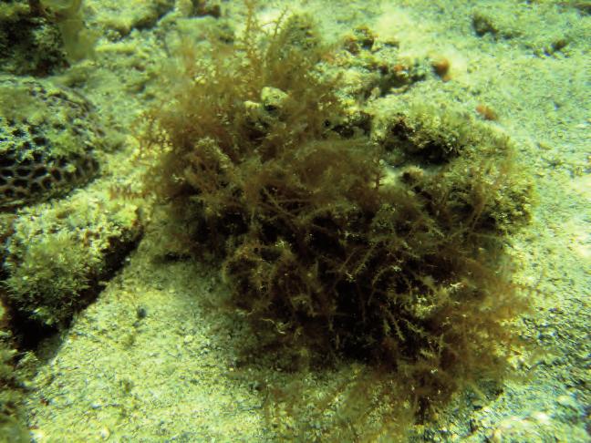
220 A B Fig Acanthophora spicifera. 217
221 CERAMIALES Rhodomelaceae Bostrychia Montagne in Ramon de la Sagra 1842b: 39 Bostrychia tenella (J.V. Lamouroux) J. Agardh 1863: Figs 22B; 42E; 136 References: Tseng (1984: 144, pl. 75, fig. 3), Lewmanomont & Ogawa (1995: 96, + fig., as B. binderi), Cribb (1996: 71, top fig. p. 70), Trono (1997: 259, fig. 161, as B. binderi), Calumpong & Meñez (1997: 159, 160, + figs), Huisman (2000: 156, + fig.), De Clerck & Coppejans (2002: 102, figs p. 103), De Clerck et al. (2005b: 248, fig. 250), Oliveira et al. (2005: 128, + figs p. 129), Skelton & South (2007: 169, figs ), Coppejans et al. (2009: 214, figs 16E, 183). Type locality: St. Croix, Virgin Islands. Description: Plants prostrate, forming mats up to 40 cm in diameter, dark purple to brown; attached by rhizoids developing from the ventral side of the prostrate axes, associated with bifurcations of the axes; axes cylindrical, to 500 μm in diameter; up to 3-4 orders alternately branched, from every to every fourth axial cell. Internal structure polysiphonous except for ultimate and penultimate branches; composed of an axial filament surrounded by 5-7 pericentral cells; major axes covered by a cortex 2-3 cells thick; pericentral cells dividing transversely, resulting in 2 tiers of pericentral cells per axial cell. Tetrasporangia formed in series in inflated ultimate branchlets (stichidia), produced in whorls of 4 and covered by (2-)3 cover cells, tetrahedrally divided, μm in diameter. Ecology: On shaded vertical and overhanging walls, upper intertidal zone (supralittoral fringe); rare. Distribution: Pantropical. 218

222 A B Fig Bostrychia tenella. A. Habit In situ. B. Microscopic detail. 219
 Garbary et J.T.")
223 CERAMIALES Rhodomelaceae Chondrophycus (J. Tokida et Y. Saito) Garbary et J.T. Harper 1998: 194 Chondrophycus cartilagineus (Yamada) Garbary & Harper 1998: 194 Figs 27C; 49A; 137A-C References: Lewmanomont & Ogawa (1995: 126, + fig, as Laurencia), Trono (1997: 263, fig. 164, as Laurencia), Abbott (1999: 384, figs 111D,E, as Laurencia), Huisman et al. (2007: 144, + fig.), Lewmanomont et al. (2007: 86, + figs). Syntype localities: Mogi, Fukuoka Prefecture, Japan; Kyushu I., Southern Japan; Chikuzen and Iyo Provinces (Fukuoka and Ehime Prefectures), Japan. Description: Plants erect, cm tall, bushy and rather stiff cartilaginous, but pliable, dark red; several axes originating from an entangled basis, less densely branched towards the basis, very densely so in upper parts; all axes and branches terete, branching radially, branches directed upwardly; main axes up to 2 mm in diameter, side branches about 1 mm; branchlets of last order short, warty or even knobby with truncate apices. Cortical cells subquadrate in cross section; medullary cells with uniformly thickened cell walls (no lenticular thickenings); secondary pit connections absent. Ecology: Epilithic in the shallow subtidal, mostly along the coasts with some wave action, but also observed in more sheltered, deeper lagoons (1 m depth), where the largest specimens develop. Locally in rather dense, extensive populations. Distribution: Pacific Ocean, from Japan over the Philippines to Queensland, Indonesia and Thailand, but also mentioned from Eritrea (Indian Ocean). Note: According to Trono (1997: 263), this species is commonly prepared into salad in the Philippines. A 220


224 B C Fig Chondropycus cartilagineus. A. Population just under low water level; B. Whole plant; C. Detail of ultimate branchlets. 221
225 CERAMIALES Rhodomelaceae Laurencia J.V. Lamouroux 1813: 130 Laurencia nidifica J. Agardh 1852: Figs 11D; 15F; 17B; 18B,C; 27D; 138A-C References: Magruder & Hunt (1979: 81, top fig. p. 80), Trono (1997: 264, fig. 165), Abbott (1999: 390, figs 113F,G). Type locality: O ahu, Hawaiian Islands. Description: Plants erect, originally forming hemispherical, densely intricated tufts, about 10 cm in diameter, when still attached to the substrate, becoming more or less isodiametrical balls, up to 20 cm in diameter when becoming loose-lying; structure supple cartilaginous; all axes terete, with a small diameter (0.5-1 mm); most plants typically with greenish main axes and pinkish red last order branchlets, but other specimens lacking the green axes; Branching irregular (from subdichotomous in main axes, to alternate, opposite or even whorled) and extremely dense, resulting in intricate structures in which individual specimens are difficult to isolate; last order branches warty. Cortical cells subquadrate in transverse section; according to Abbott (1999: 391) the number of lenticular cell wall thickenings of medullary cells is extremely variable among specimens; they can be so rare that they can be missed if only a limited number of transverse sections is observed. Ecology: Subtidal, in shallow lagoons, originally on coral rubble on sand or epiphytic on the basis of Sargassum spp. and Enhalus. Larger specimens become detached and grow as loose-lying balls on the lagoon bottom, locally in huge quantities. Distribution: Widely reported in the South Pacific and Indian Ocean but also mentioned from the Atlantic Ocean. Notes: According to Abbott (1999: 391), L. mcdermidiae Abbott is morphologically very similar to L. nidifica, the former completely lacking lenticular cell wall thickenings. She also states that L. nidifica is favoured by Hawaiians as a fresh condiment because of its peppery taste. 222


226 A B C Fig Laurencia nidifica. A. Whole plant; B. Detail of ultimate branchlets; remark the green sub-ultimate branchlets; C. Microscopic detail. 223
227 CERAMIALES Rhodomelaceae Leveillea Decaisne 1839: 375 Leveillea jungermannioides (Hering et G. Martens) Harvey 1855: 539 Figs 35B; 54B; 139A,B References: Tseng (1984: 156, pl. 81, fig. 3), Lewmanomont & Ogawa (1995: 129, + figs), De Clerck & Coppejans (1996: 265, figs 127,128), Calumpong & Meñez (1997: 165, + fig.), Abbott (1999: 396, figs 116A-D), Huisman (2000: 173, + fig.), De Clerck & Coppejans (2002: 104, figs p. 105), Oliveira et al. (2005: 140, + fig. p. 141), Lewmanomont et al. (2007: 95, + figs), Skelton & South (2007: 181, figs ), Coppejans et al. (2009: 220, fig. 188). Type locality: Tor, Sinai Peninsula, Egypt. Description: Plants prostrate, up to 5 cm long, blackish red, composed of branched axes bearing 2 rows of erect, broadly ovate, membranous branches; thallus decumbent, with cylindrical main axes with inrolled apices cutting off alternate indeterminate side axes with similar morphology; these axes with 4 pericentral cells in the juvenile parts and 7 pericentral cells in mature parts; attachment at intervals by groups of multicellular rhizoids with well-adhering terminal pad-like structures; prostrate axes bearing bilaterally arranged, erect, asymmetrical, broadly ovate, monostromatic (except midrib) bladelets, μm broad and μm high, arranged in an alternate sequence, partly overlapping one another and bearing colourless, deciduous trichoblasts at the apices; cells of these bladelets more or less arranged in vertical and horizontal rows. Four to eight tetrasporangia formed in short, curved, stichidia-like branchlets that replace indeterminate branches; each tetrasporangium in own chamber demarcated by vertical elongate cells and surrounded distally by 2-3 cover cells: only 4-6 tetrasporangia mature. Ecology: Epiphytic, mostly on Sargassum and Hormophysa in lagoons. Frequently overlooked as a result of its small size and similar colour as the phorophyte. Distribution: Widespread in the Indian Ocean and western tropical Pacific Ocean. 224


228 A B Fig Leveillea jungermannioides. A,B. Microscopic details (stained). 225
229 CERAMIALES Rhodomelaceae Lophocladia (J. Agardh) Schmitz 1893: 222 Lophocladia cf. minima Itono 1973: , figs Figs 41C; 53A; 54C; 140A-C Type locality: Hateruma-jima, Ryukyu-retto, Japan. Description: Thalli gregarious, forming very elegant and extremely supple tufts, composed of short, prostrate axes producing several, sporadically branched erect axes cm long, pink; attachment by rhizoids with discoid ends, produced by the ventral periaxial cells of the prostrate axes; all axes polysiphonous, with 4 periaxials; cortication limited to the basal, older parts of the axes, by means of descending rhizoids originating at the proximal end of the periaxial cells; diameter of the erect axes 250 μm at the basis, with the segments only slightly longer than broad, gradually tapering towards the apices where they are 75 μm in diameter and 180 μm long; each segment with a persistent, monosiphonous pigmented trichoblast radially arranged in 1/4 spiral on the axis, basal-most cell of the trichoblasts slightly embedded between periaxial cells, markedly smaller than the subsequent cells; trichoblasts several times alternately branched, gradually decreasing in diameter. Tetrasporangia in a spiral series in torulose stichidia on the proximal branch of a trichoblast, one sporangium per segment and no hairs on the stichidium. Male capitula replacing a branch of the second dichotomy of trichoblasts, very long and elegant, without apical or lateral trichoblastic branches. Female gametophytes not observed. Ecology: Epilithic, on corals, shallow subtidal. Distribution: Japan, Micronesia, South Africa. Notes: The genus Lophocladia differs from the very similar genus Spirocladia by the absence of unbranched trichoblasts protruding from fertile segments of the spermatangial heads in the former. Species of Lophocladia can be grouped, on the basis of their size, which is a poor character (4-15 cm in L. japonica Yamada, L. kuetzingii (Kuntze) P.C. Silva, L. lallemandii (Montagne) Schmitz and L. trichoclados (J. Agardh) Schmitz), smaller (1.5-3 cm) in L. minima and L. kipukaia Schlech. The latter is distinguished from the former by the staggered pericentral cells. Another differentiating character on species level is the placement and morphology of cystocarps. As female gametophytes are lacking in our collections, we refrain from an absolute identification. 226


230 A B C Fig Lophocladia cf. minima. A,B. Microscopic details of tetrasporophytes with stichidia; C. Spermatangia in grape-like structures. 227
231 CERAMIALES Rhodomelaceae Palisada K.W. Nam 2007 Palisada papillosa (C. Agardh) K.W. Nam 2007: 54 Fig. 141A,B References: Lewmanomont & Ogawa (1995: fig, as Laurencia), Littler et al. (1998: 182, bottom fig. p. 183, as Laurencia), Cribb (1996: 101, bottom fig. p. 100, as Laurencia), De Clerck & Coppejans (1996: 263, figs 113,114, as Laurencia), Calumpong & Meñez (1997: 172, + fig., as Laurencia), Trono (1997: 266, fig. 166, as Laurencia), Huisman (2000: 159, + figs, as Chondrophycus), Littler & Littler (2000: 218, top figs p. 219, as Laurencia), De Clerck & Coppejans (2002: 104, figs p. 105, as Chondrophycus), Littler & Littler (2003: 148, top fig. 149, as Chondrophycus), Coppejans et al. (2009: 218, fig. 186F). Type locality: Mokha, Yemen, Red Sea. Description: Plants prostrate to ascendant, very stiff cartilaginous, up to 5 cm long, forming intricate stiff cushions, attached by a basal disc bearing several axes; orangy red to cream-coloured; all axes terete, without a marked main axis, branching irregular; basal parts of the main axes (almost) naked; median and upper parts and side branches densely covered by radially placed (sometimes in spirally longitudinal lines), short, papillose, unbranched branchlets with invaginated apex. In transverse section, no lenticular cell wall thickenings of medullary cells, the cortical cells are radially elongate, resulting in a palisade-like appearance, not projecting. Ecology: Epilithic on dead upper surface of coral heads in a shallow lagoon with heated water, just under low water level. Distribution: Widespread in tropical to warm-temperate seas. Note: The specimens from the study area morphologically best correspond with Huisman (2000: fig. p. 159). Chondrophycus tronoi (E. Ganzon-Fortes) K.W. Nam, also mentioned from Thailand, morphologically differs from Palisada papillosa by the ultimate branchlets becoming bi-, trilobed or paniculate in subapical parts of the plants. 228


232 A B Fig Palisada papillosa (covered by numerours epiphytes). 229
 and placement of")
233 CERAMIALES Rhodomelaceae Polysiphonia Greville 1823: pl. 90 Neosiphonia M.-S. Kim & Lee 1999: Polysiphonia/Neosiphonia sp. Figs 35D; 53D; 142 Description: Plants slender and supple, erect, up to 10 mm high; basal parts not visible on the pictures and therefore the formation (pit-connected or not) and placement of rhizoids are unknown; erect axes polysiphonous, composed of a central axis surrounded by 4 periaxial cells; cortication absent; segments up to 1.5 longer than broad in basal and middle parts, isodiametric in upper parts of the thallus; colourless trichoblasts caducous and very small; mature cystocarps spherical without enlarged cells around the ostiolum. Male gametophytes and tetrasporophytes not observed. Ecology: Epiphytic on larger, subtidal seaweeds. Note: Kim & Lee (1999) separated the genus Neosiphonia from Polysiphonia on the basis of several morphological and reproductive characters. As only microscopic pictures of cystocarpic material are available of collections from the study area we refrain from more precise identification, even on genus level. Fig Polysiphonia/Neosiphonia sp. Microscopic detail of a specimen with cystocarps. 230
234 CERAMIALES Rhodomelaceae Tolypiocladia F. Schmitz in F.Schmitz et Falkenberg 1897: 441 Tolypiocladia glomerulata (C. Agardh) Schmitz in Schmitz et Falkenberg 1897: Figs 42F, 143A-D References: Magruder & Hunt (1979: 95, middle fig. p. 94), Tseng (1984: 160, pl. 83, fig. 4), Abbott (1999: 442, figs 132F-H), Coppejans & Millar (2000: 338, fig. 32), Huisman (2000: 179, + fig.), Littler & Littler (2003: 158, middle fig. p. 159), Oliveira et al. (2005: 147, + figs), Huisman et al. (2007: 154, + figs), Lewmanomont et al. (2007: 101, + figs), Skelton & South (2007: 191, figs ). Type locality: Shark Bay, Western Australia. Description: Plants prostrate, only 5-10 mm long, mm in diameter, gradually tapering towards the pointed apices; sparsely, irregularly branched spongy axes; dark red, becoming black upon drying; attachment by numerous unicellular rhizoids, produced by periaxial cells, terminating in digitate discs; all axes polysiphonous, with 4 pericentral cells, devoid of any cortication; segments shorter than wide; axes and indeterminate branches densely clothed with numerous determinate, branchlets, perpendicularly placed on the axes; branchlets exogenously formed on most segments in a 1/4 spiral sequence, dichotomously branching at wide angles, the distal ends being divided 1-2 (or more) times into 1-several short spine-like branchlets; unbranched trichoblast borne on branchlet in early stages, young cells of trichoblast pigmented, later becoming colourless and deciduous; determinate branchlets not mutually anastomosing with those of the previous and following determinate branchlet. Ecology: Epiphytic on larger seaweeds (Sargassum, Turbinaria) and seagrass leaves from the shallow subtidal; also observed in algal turf. Distribution: Tropical Indo-Pacific. Note: Differing from T. calodictyon (Harvey ex Kützing) Silva by the absence of anastomoses between the determinate branchlets. Skelton and South (2007: 191) state that the features used to distinguish the four species of Tolypiocladia are yet to be properly tested. Falkenberg (1901) already suggested they may all be conspecific, belonging to T. glomerulata. 231

 bearing")


235 A B C D Fig Tolypiocladia glomerulata A. Determinate branchlets (on indeterminate axis) bearing unbranched trichoblasts; B. Apical part; C. Subapical part; D. Detail of short, spine-like last order branchlets (having lost the trichoblasts). 232
, Oliveira et al. (2005: 118, + figs), Coppejans et al. (2009: 204, fig. 173), Littler et al. (2009: 60, + figs). Type locality: Roque del Gando, Islas Canarias.")
236 CERAMIALES Wrangeliaceae Wrangelia C. Agardh 1828: 136 Wrangelia argus (Montagne) Montagne 1856: 444 Fig. 144 References: Lewmanomont & Ogawa (1995: 140, + fig.), De Clerck et al. (2005b: 232, figs ), Oliveira et al. (2005: 118, + figs), Coppejans et al. (2009: 204, fig. 173), Littler et al. (2009: 60, + figs). Type locality: Roque del Gando, Islas Canarias. Description: Plants decumbent, soft and fluffy, penicillate, entirely uniseriate, composed of prostrate and erect axes, up to 5-8 mm high, dark purplish red; prostrate axes composed of cells μm in diameter and up to 700 μm long; erect axes with determinate branchlets produced in whorls of 4 from every axial cell; determinate laterals unequally developed with one lateral being much smaller than the remaining three, 1-3 times dichotomously branched, up to 400 μm long, proximally 30 μm in diameter, tapering gradually toward the apices, with acute apical cells; cortication loose and rag-like. Tetrasporangia produced on the proximal cells of whorl-branchlets, enclosed by two involucral filaments of 2 cells each. Sexual reproductive structures not observed. Ecology: Epilithic in the shallow subtidal. Distribution: Widespread in tropical and warm temperate regions. Fig Wrangelia argus. Under dissecting microscope. 233
Diversity and distribution of seaweed at Khanom-Mu Ko Thale Tai National Park, Nakhon Si Thammarat Province, Thailand
 Songklanakarin J. Sci. Technol. 33 (6), 633-640, Nov. - Dec. 2011 http://www.sjst.psu.ac.th Original Article Diversity and distribution of seaweed at Khanom-Mu Ko Thale Tai National Park, Nakhon Si Thammarat
The Effects of the 2004 Tsunami on the Seaweed and Seagrass Community at. Talibong Island and Nearby Trang province, Thailand
 The Effects of the 2004 Tsunami on the Seaweed and Seagrass Community at Talibong Island and Nearby Trang province, Thailand Anchana Prathep 1 *, Piyalarp Tantiprapas 2, Bongkot Wichachucherd 1 Ajchara
Environmental Science
 Environmental Science A Study of Interrelationships Cui Jiansheng Hebei University of Science and Technology CH06 Kinds of Ecosystems and Communities Chapter Objectives After reading this chapter, you
CHAPTER 6 & 7 VOCABULARY
 CHAPTER 6 & 7 VOCABULARY 1. Biome 2. Climate 3. Latitude 4. Altitude 5. Emergent layer 6. Epiphyte 7. Understory 8. Permafrost 9. Wetland 10.Plankton 11.Nekton 12.Benthos 13.Littoral zone 14.Benthic zone
Relatively little hard substrate occurs naturally in the
 CHAPTER FIVE Rock Habitats Relatively little hard substrate occurs naturally in the estuary, owing mainly to the vast quantities of fine sediment that have been deposited by the rivers. Rock habitat is
Types of intertidal communities
 Between the tides Marine ecosystems 1 Intertidal Delimited by the highest high tide and the lowest low tides marks The best studied and best-understood by humans Relatively easy to sample compared to other
Science 8 - Water Systems Test - Chapters 1-2
 Science 8 - Water Systems Test - Chapters 1-2 Multiple Choice (30 marks) Identify the choice that best completes the statement or answers the question. 1. What percentage represents the amount of fresh
200 Meters Down Topic: Ocean Zones (5 th 8 th grade)
") 200 Meters Down Topic: Ocean Zones (5 th 8 th grade) by Lodge 200 Meters Down Have you people heard about the plankton? Just tiny algae and it s floating, right What about the nekton like jellyfish? They
200 Meters Down Topic: Ocean Zones (5 th 8 th grade) by Lodge 200 Meters Down Have you people heard about the plankton? Just tiny algae and it s floating, right What about the nekton like jellyfish? They
What creates a coral reef? Why are corals able to form huge reefs?
 Marine ecosystems 5: Coral Reefs Unique features The foundation of the ecosystem is produced by living things Reef-building corals Similarities with tropical rain forests Richness and complexity 3-dimensional
Chapter 5: Marine Algae & Plants
 Chapter 5: Marine Algae & Plants Marine Algae Belong to the kingdom Protista Seaweeds are multicellular algae. We will learn about 3 types: green, red, and brown. Algae are Nonvascular Vascular Plants:
Time allowed: 1 hour 30 minutes
 SPECIMEN ASSESSMENT MATERIAL: SET 2 GCSE GEOGRAPHY Paper 1 Living with the physical environment Specimen Materials For this paper you must have: a pencil a ruler. Time allowed: 1 hour 30 minutes Instructions
The Marine Environment
 The Marine Environment SECTION 16.1 Shoreline Features In your textbook, read about erosional landforms, beaches, estuaries, longshore currents, and rip currents. For each statement below, write or. 1.
Small area of the ocean that is partially surrounded by land. The Ocean Basins. Three Major Oceans. Three Major Oceans. What is a SEA?
 The Ocean Basins How Deep is the Ocean? 1 2 Three Major Oceans Three Major Oceans Pacific Atlantic the shallowest ocean (3.3km average depth) Indian second shallowest ocean (3.8km average depth) Pacific
Brief report on Belize carbonate environments field trip Feb. 2012
 Brief report on Belize carbonate environments field trip Feb. 2012 In february 2012 I travelled to San Pedro (Ambergris Caye) Belize (Figure 1) to examine and study depositional environments of carbonate
THE INTERTIDAL ZONE AND BENTHIC ORGANISMS
 THE INTERTIDAL ZONE AND BENTHIC ORGANISMS EPSS 15 Lab #8 OUTLINE I. Intertidal zonation Tides Biotic zonation Physical conditions & biotic interactions II. Intertidal organisms & adaptations Snails Mussels
Changes to Land 5.7B. landforms: features on the surface of Earth such as mountains, hills, dunes, oceans and rivers
 All the landforms on Earth have changed over time and continue to change. Many of the changes were caused by wind, moving water, and moving ice. Mountains have grown and shrunk. Rivers have cut away land
Earth Science S5E1b (EarthScienceS5E1b)
") Name: Date: 1. The flattest part of the ocean floor is the A. trench. B. abyssal plain. C. continental shelf. D. mid-ocean rift valley. 2. Which causes some parts of the ocean to be saltier than other
Name: Date: 1. The flattest part of the ocean floor is the A. trench. B. abyssal plain. C. continental shelf. D. mid-ocean rift valley. 2. Which causes some parts of the ocean to be saltier than other
Think about the landforms where you live. How do you think they have changed over time? How do you think they will change in the future?
 reflect All the landforms on Earth have changed over time and continue to change. Many of the changes were caused by wind, moving water, and moving ice. Mountains have grown and shrunk. Rivers have cut
Unit 4 - Water. Earth s Interior. Earth s Interior. Continental Drift. Continental Drift. Continental Drift. Crust. Mantle. Core.
 Unit 4 - Water How did the oceans form? What special adaptations do saltwater organisms have? Where does our water come from? How do humans affect the Earth s water? Crust Rigid outer shell of Earth Oceanic
Shoreline and Climate Change Adaptation Alternatives for The Letter Parcel, Bolinas Lagoon
 Shoreline and Climate Change Adaptation Alternatives for The Letter Parcel, Bolinas Lagoon Scenic shoreline vista of Bolinas Lagoon and ridges, from Letter Parcel, March 2016 Peter Baye, Coastal Ecologist
Understanding Oceans, Gulfs & Tides
 SCS-2005-13 Water and Me Series Understanding s, Gulfs & Tides Monty C. Dozier Assistant Professor and Extension Water Resources Specialist The dictionary defines the ocean as the whole body of saltwater
ARE YOU READY TO THINK? Look at the first slide THINK PAIR SHARE!
 ARE YOU READY TO THINK? Look at the first slide THINK PAIR SHARE! WHAT PROMINENT FEATURE CAN YOU IDENTIFY IN THIS PICTURE? What do you think the different colors represent? Who might find such a picture
Marine biologists have identified over 250,000 marine species. This number is constantly increasing as new organisms are discovered.
 A wide variety of organisms inhabit the marine environment. These organisms range in size from microscopic bacteria and algae to the largest organisms alive today blue whales, which are as long as three
Biosphere. All living things, plants, animals, (even you!) are part of the zone of the earth called the biosphere.
 are part of the zone of the earth called the biosphere.") Unit 1 Study Guide Earth s Spheres Biosphere All living things, plants, animals, (even you!) are part of the zone of the earth called the biosphere. Hydrosphere Water covers ¾ of the earth, made up mostly
Unit 1 Study Guide Earth s Spheres Biosphere All living things, plants, animals, (even you!) are part of the zone of the earth called the biosphere. Hydrosphere Water covers ¾ of the earth, made up mostly
Your web browser (Safari 7) is out of date. For more security, comfort and the best experience on this site: Update your browser Ignore
 is out of date. For more security, comfort and the best experience on this site: Update your browser Ignore") Your web browser (Safari 7) is out of date. For more security, comfort and the best experience on this site: Update your browser Ignore CO NTINENTAL SHEL F For the complete encyclopedic entry with media
Your web browser (Safari 7) is out of date. For more security, comfort and the best experience on this site: Update your browser Ignore CO NTINENTAL SHEL F For the complete encyclopedic entry with media
Treasure Coast Science Scope and Sequence
 Course: Marine Science I Honors Course Code: 2002510 Quarter: 3 Topic(s) of Study: Marine Organisms and Ecosystems Bodies of Knowledge: Nature of Science and Life Science Standard(s): 1: The Practice of
UNIT 5: ECOLOGY Chapter 15: The Biosphere
 CORNELL NOTES Directions: You must create a minimum of 5 questions in this column per page (average). Use these to study your notes and prepare for tests and quizzes. Notes will be stamped after each assigned
4 Marine Biology Notes. Multi-cellular Primary Producers: Seaweeds and Plants
 4 Marine Biology Notes Multi-cellular Primary Producers: Seaweeds and Plants Marine Algae Marine algae are important primary producers (photosynthetic) These algae are called by a generic term seaweeds
DIVERSITY ASSESSMENT ON MALAYSIAN BENTHIC MACROALGAE IN TIOMAN ISLAND AND TULAI CANDANG ISLAND, PAHANG
 DIVERSITY ASSESSMENT ON MALAYSIAN BENTHIC MACROALGAE IN TIOMAN ISLAND AND TULAI CANDANG ISLAND, PAHANG 1 NUR FARAH AIN ZAINEE, 2 MOHAMED EFFENDI TAIP School of Environmental and Natural Resource Sciences,
The Coast: Beaches and Shoreline Processes
 1 2 3 4 5 6 7 8 9 The Coast: es and Shoreline Processes Trujillo & Thurman, Chapter 10 Oceanography 101 Chapter Objectives Recognize the various landforms characteristic of beaches and coastal regions.
The Marine Environment
 The Marine Environment SECTION 16.1 Shoreline Features In your textbook, read about erosional landforms, beaches, estuaries, longshore currents, and rip currents. For each statement below, write true or
The Coast: Beaches and Shoreline Processes Trujillo & Thurman, Chapter 10
 The Coast: es and Shoreline Processes Trujillo & Thurman, Chapter 10 Oceanography 101 Chapter Objectives Recognize the various landforms characteristic of beaches and coastal regions. Identify seasonal
Geoduck Floating Nursery Monitoring Plan, Quarterly Reporting
 December 23, 2014 Mason County Department of Community Development Attn: Grace Miller, Senior Planner 411 N. Fifth Street PO Box 279 Shelton, WA 98584 Re: Geoduck Floating Nursery Monitoring Plan, Quarterly
"The Relationship Between Seagrass Cover and Species- richness of Invertebrates"
 "The Relationship Between Seagrass Cover and Species- richness of Invertebrates" SCIE 2204: Marine Systems The Cottesloe Marine Ecosystem Research Project 2014 By Baronie Shaw, K., Bortoloso, T., Cargill,
The surface of the ocean floor is as varied as the land. The five major oceans, from largest to smallest, are
 11.1 Ocean Basins The surface of the ocean floor is as varied as the land. The five major oceans, from largest to smallest, are w the Pacific w the Atlantic w the Indian w the Southern w the Arctic The
Earth / Environmental Science. Ch. 14 THE OCEAN FLOOR
 Earth / Environmental Science Ch. 14 THE OCEAN FLOOR The Blue Planet Nearly 70% of the Earth s surface is covered by the global ocean It was not until the 1800s that the ocean became an important focus
Lesson 2. Antarctic Oceanography: Component I - Ice/Glaciers Component II - Marine Snow
 Lesson 2. Antarctic Oceanography: Component I - Ice/Glaciers Component II - Marine Snow Lesson Objectives: Introduces students to the different kinds of ice found in Antarctica, Students will become familiar
Chapter 11. Rivers: Shaping our landscape
 Chapter 11 Rivers: Shaping our landscape Learning outcomes In this presentation you will learn: Common terms associated with rivers About the three stages of a river About the processes of river erosion
Unit 1: Water Systems on Earth Chapter 2
 Unit 1: Water Systems on Earth Chapter 2 Create a mind map with the driving question, Why are Oceans Important? Remember: Why are oceans so important? Why are oceans so important? Primary water source
Laboratory Exercise #4 Geologic Surface Processes in Dry Lands
 Page - 1 Laboratory Exercise #4 Geologic Surface Processes in Dry Lands Section A Overview of Lands with Dry Climates The definition of a dry climate is tied to an understanding of the hydrologic cycle
Oceanography. Oceanography is the study of the deep sea and shallow coastal oceans.
 Oceanography Oceanography is the study of the deep sea and shallow coastal oceans. Studying the Ocean Floor To determine the shape and composition of the ocean floor, scientists use techniques such as
Module 10: Resources and Virginia Geology Topic 4 Content: Virginia Geology Notes
 Virginia is composed of a very diverse landscape that extends from the beaches and barrier islands all of the way to the highly elevated Appalachian Plateau. Geologists have discovered ancient shallow
> True/False Indicate whether the statement is true or false.
 Class: Date: Biology Chapter 3 Test: Communities, Bionics, and Ecosystems > True/False Indicate whether the statement is true or false. 1. The main abiotic distinction between temperate grassland and tropical
Shorelines and Diesel
 Shorelines and Diesel Rock Platforms (bedrock): A common shoreline type with a rocky environment that is both exposed and submerged from tide cycles. Platforms are generally horizontal and collect sediments
Chapter 6, Part Colonizers arriving in North America found extremely landscapes. It looked different to region showing great.
 Social Studies 9 Unit 1 Worksheet Chapter 6, Part 1. 1. Colonizers arriving in North America found extremely landscapes. It looked different to region showing great. 2. The Earth is years old and is composed
UNIT 1: WATER SYSTEMS ON EARTH CHAPTER 2: OCEANS CONTROL THE WATER CYCLE
 UNIT 1: WATER SYSTEMS ON EARTH CHAPTER 2: OCEANS CONTROL THE WATER CYCLE ORIGINS OF OCEAN WATER OCEANS HAVE FILLED OVER HUNDREDS OF MILLIONS OF YEARS SCIENTISTS BELIEVE THE OCEANS ARE MORE THAN 3 BILLION
Significant Ecological Marine Area Assessment Sheet
 Significant Ecological arine Area Assessment Sheet Name: Eastern Bay of Island Biogenic Soft Sediment Complex Summary: The semi-sheltered areas between the central islands of the Eastern Bay of Islands
Name Hour. Section 4-1 The Role of Climate (pages 87-89) What Is Climate? (page 87) 1. How is weather different from climate?
 What Is Climate? (page 87) 1. How is weather different from climate?") Name Hour Section 4-1 The Role of Climate (pages 87-89) What Is Climate? (page 87) 1. How is weather different from climate? 2. What factors cause climate? The Greenhouse Effect (page 87) 3. Circle the
Name Hour Section 4-1 The Role of Climate (pages 87-89) What Is Climate? (page 87) 1. How is weather different from climate? 2. What factors cause climate? The Greenhouse Effect (page 87) 3. Circle the
Rising Sea Levels: Time for Proactive Action in Florida and the Caribbean?
 Rising Sea Levels: Time for Proactive Action in Florida and the Caribbean? Dr. Alan Fyall and Dr. Thomas Wahl National Center for Integrated Coastal Research, University of Central Florida Dr. Roberta
Name: Period: Date: ID: A. Circle the choice that best completes the statement or answers the question and write the letter on the blank.
 Name: Period: _ Date: _ ID: A Unit 7 Practice Circle the choice that best completes the statement or answers the question and write the letter on the blank. 1. What term describes the movement of rock
TASMANIAN SEAGRASS COMMUNITIES
 TASMANIAN SEAGRASS COMMUNITIES by Christopher Grant Rees, B.Ed (}Ions.) Submitted in partial fulfilment of the requirement for the degree of Master of Environmental Studies (By Coursework) Centre for Environmental
Objectives: Describe the structure of the ocean floor. Describe light intensity and temperature characteristics at different ocean depths.
 Ocean Structure Virtual Lab What are some characteristics of the ocean and the ocean floor? Earths highest mountains, deepest valleys, and flattest plains are found not on land but under the ocean. Beyond
Question: What is the primary reason for the great abundance of fish along the Peruvian coast?
 Buzzer Question # 1 Question Type: toss-up Question Format: Multiple Choice Category: Biology What is the primary reason for the great abundance of fish along the Peruvian coast? Answer W: upwelling Answer
Oceanography is the scientific study of oceans Oceans make up over 70% of the Earth s surface
 Oceanography Oceanography is the scientific study of oceans Oceans make up over 70% of the Earth s surface An ocean must be large and have features which set it apart from other oceans (currents, water
Dawood Public School Secondary Section Class VII
 Dawood Public School Secondary Section Class VII Introduction: Geography is the study of the Earth and its lands, features, inhabitants, and phenomena relating to the sciences of aforementioned. This subject
by B.A. Foster THE EFFECT OF ASPECT ON POPULATION COMPOSITION
 TANE (1966) 12 : 37-44 37 THE EFFECTS OF WAVE EXPOSURE AND ASPECT ON THE VERTICAL SHORE DISTRIBUTION AND POPULATION COMPOSITION OF MELARHAPHA OLIVERI. by B.A. Foster INTRODUCTION The periwinkle Melarhapha
Marine Ecology Pacing Guide
 Marine Ecology Pacing Guide Course Description: The focus of the course is the interrelationships among marine organisms and the physical, chemical, geological and biological factors. North Carolina coastal
Objectives: Define Relative Age, Absolute Age
 S6E5. Students will investigate the scientific view of how the earth s surface is formed. c. Classify rocks by their process of formation. g. Describe how fossils show evidence of the changing surface
Changes in Texas Ecoregions Copy the questions and answers
 Changes in Texas Ecoregions Copy the questions and answers 1. What are some kinds of damage that hurricanes cause? Roads and bridges might be washed away. Trees and power lines can be knocked down. Area
e Southeast Region of the United Statesg
 e Southeast of the United Statesg - relative location: the location of a place in relation to another place (i.e. south, near, bordering, next to) - barrier islands: located off the southeast coast of
Evolution and Life in the Ocean
 Characteristics of All Living Things Contain matter in a highly organized state Capture, store and transmit energy; all organisms require energy Capable of reproduction Change through time and adapt to
Term Knowledge Using and applying Grade Criteria Autumn 1 Assessment window October Natural hazards pose major risks to people and property.
 Assessment Schedule Year 9 2015/6 Term Knowledge Using and applying Grade Criteria Autumn 1 Assessment window October 2015 3.1.1.2 Tectonic Natural hazards pose major risks to people and property. Definition
Overview of Chapter 6
 Overview of Chapter 6 Earth s Major Biomes Aquatic Ecosystems Freshwater ecosystems Estuaries Marine Ecosystems Wildfires and Ecosystems Wildfire unexpected fire in grass, shrub, or forests (a significant
Which map shows the stream drainage pattern that most likely formed on the surface of this volcano? A) B)
 B)") 1. When snow cover on the land melts, the water will most likely become surface runoff if the land surface is A) frozen B) porous C) grass covered D) unconsolidated gravel Base your answers to questions
1. When snow cover on the land melts, the water will most likely become surface runoff if the land surface is A) frozen B) porous C) grass covered D) unconsolidated gravel Base your answers to questions
GY 111 Lecture Note Series Sedimentary Environments 2: Rivers and Deltas
 GY 111 Lecture Notes D. Haywick (2008-09) 1 GY 111 Lecture Note Series Sedimentary Environments 2: Rivers and Deltas Lecture Goals A) Rivers and Deltas (types) B) Water Flow and sedimentation in river/deltas
Sea Level Rise in Miami-Dade County Florida Implications for Management of Coastal Wetlands and the Everglades
 Sea Level Rise in Miami-Dade County Florida Implications for Management of Coastal Wetlands and the Everglades Peter W. Harlem Dr. John F. Meeder Florida International University Southeast Environmental
MARINE GEOLOGY & GEOGRAPHY
 MARINE GEOLOGY & GEOGRAPHY Bathymetry BATHYMETRY BATHYMETRY THE UNDERWATER EQUIVALENT TO TOPOGRAPHY THE STUDY OF WATER DEPTH A BATHYMETRIC MAP SHOWS FLOOR RELIEF OR TERRAIN AS CONTOUR LINES Bathymetry
netw rks Guided Reading Activity Essential Question: How does geography influence the way people live? Earth's Physical Geography
 Guided Reading Activity Lesson 1 Earth and the Sun Essential Question: How does geography influence the way people live? Looking at Earth Directions: What are the layers that make up Earth? Use your textbook
Abiotic Dominant Dominant Factors Plants Animals
 TERRESTRIAL BIOMES Abiotic Dominant Dominant Factors Plants Animals Tropical Rain Forest Hot & wet year round Ferns Woody vines Broad leaved evergreen trees Sloths Jaguars Monkeys Toucans Tropical Dry
Appendix E: Cowardin Classification Coding System
 Appendix E: Cowardin Classification Coding System The following summarizes the Cowardin classification coding system and the letters and numbers used to define the USFWS NWI wetland types and subtypes:
Subtidal permanently flooded with tidal water. Irregularly exposed surface exposed by tides less often than daily
 Types of Wetlands Tidal Systems COASTAL WETLAND ECOSYSTEMS Tidal Salt Marshes Tidal Freshwater Marshes Mangrove Wetlands Tidal Estuarine Wetland 1 Definition and Formation of Estuaries u Estuary : partially
Types of Wetlands. Tidal Systems
 Types of Wetlands Tidal Systems 1 COASTAL WETLAND ECOSYSTEMS Tidal Salt Marshes Tidal Freshwater Marshes Mangrove Wetlands 2 Tidal Estuarine Wetland 3 Definition and Formation of Estuaries Estuary: : partially
Science EOG Review: Landforms
 Mathematician Science EOG Review: Landforms Vocabulary Definition Term canyon deep, large, V- shaped valley formed by a river over millions of years of erosion; sometimes called gorges (example: Linville
Name Date Class. well as the inland, found near the Tropics. 4. In the, or the regions near the Equator, you may find a lush
 WATER, CLIMATE, AND VEGETATION Vocabulary Activity DIRECTIONS: Fill in the Blanks Select a term from below to complete each of the following sentences. CHAPTER 1. The constant movement of water, a process
Grade 8 Science. Unit 1: Water Systems on Earth Chapter 2
 Grade 8 Science Unit 1: Water Systems on Earth Chapter 2 Oceans are important... 1. Primary water source for the water cycle 2. Control weather 3. Support diverse life 4. Provides humans with food, minerals,
N05/3/GEOGR/HP2/ENG/TZ0/XX/Q GEOGRAPHY HIGHER LEVEL PAPER 2. Monday 7 November 2005 (morning) 2 hours 30 minutes INSTRUCTIONS TO CANDIDATES
 2 hours 30 minutes INSTRUCTIONS TO CANDIDATES") IB GEOGRAPHY HIGHER LEVEL PAPER 2 DIPLOMA PROGRAMME PROGRAMME DU DIPLÔME DU BI PROGRAMA DEL DIPLOMA DEL BI 88055202 Monday 7 November 2005 (morning) 2 hours 30 minutes INSTRUCTIONS TO CANDIDATES Do not
IB GEOGRAPHY HIGHER LEVEL PAPER 2 DIPLOMA PROGRAMME PROGRAMME DU DIPLÔME DU BI PROGRAMA DEL DIPLOMA DEL BI 88055202 Monday 7 November 2005 (morning) 2 hours 30 minutes INSTRUCTIONS TO CANDIDATES Do not
Natural Disasters. in Florida. Severe Thunderstorms
 Natural Disasters in Florida SC.6.E.7.7 Investigate how natural disasters have affected human life in Florida. Severe Thunderstorms While the typical afternoon thunderstorm in Florida may not appear to
OCEAN ZONES. 1. Intertidal Zone 2. Near-Shore Zone 3. Open-Ocean Zone
 OCEAN ZONES 1. Intertidal Zone 2. Near-Shore Zone 3. Open-Ocean Zone Where the Ocean Meets the Land (Place) Intertidal Zone The intertidal zone is the area between the high- and low-tide lines. At high
OCEAN ZONES. 1. Intertidal Zone 2. Near-Shore Zone 3. Open-Ocean Zone
 OCEAN ZONES 1. Intertidal Zone 2. Near-Shore Zone 3. Open-Ocean Zone Where the Ocean Meets the Land (Place) Intertidal Zone The intertidal zone is the area between the high- and low-tide lines. At high
About places and/or important events Landmarks Maps How the land is, hills or flat or mountain range Connected to maps World Different countries
 What do you think you know about geography? About places and/or important events Landmarks Maps How the land is, hills or flat or mountain range Connected to maps World Different countries What do you
Ecological mapping using satellite imagery: an Abu Dhabi case study Middle East Geospatial Forum 16 th February 2015
 Ecological mapping using satellite imagery: an Abu Dhabi case study Middle East Geospatial Forum 16 th February 2015 Richard Flemmings rf@proteusgeo.com About Proteus Formed in UAE 2011 We specialise in
Changes in Texas Ecoregions
 Comment On Lesson Changes in Texas Ecoregions The state of Texas can be divided into 10 distinct areas based on unique combinations of vegetation, topography, landforms, wildlife, soil, rock, climate,
Florida s Changing Shape
 Florida s Changing Shape Background: For much of its history, Florida was underwater. At first, Florida consisted solely of the Florida Platform, a limestone base formed from the calcium carbonate remains
Unit 7.2 W.E.D. & Topography Test
 Name: Score: Unit 7.2 W.E.D. & Topography Test 1. 2. 3. 4. 5. 6. 7. 8. 9. 10. 11. 12. 13. 14. 15. 16. 17. 18. 19. 20. 21. 22. 23. 24. 25. 1. The formation of mountains is due mainly to while the destruction
Period: Date: Marine Science Midyear Exam Study Guide & Review This packet will be collected on the day of the exam for 2 HOMEWORK GRADES.
 Marine Science Midyear Exam Study Guide & Review This packet will be collected on the day of the exam for 2 HOMEWORK GRADES. Topics: Intro: the water planet; scientific method Properties of Water Tides,
THE DEPOSITS OF TSUNAMIS WESLEY PESANTEZ, CATHERINE NIELD, COLIN WINTER
 THE DEPOSITS OF TSUNAMIS WESLEY PESANTEZ, CATHERINE NIELD, COLIN WINTER AN OVERVIEW OF OUR SEMINAR WHAT IS A TSUNAMI WHY STUDY TSUNAMIS PROPERTIES OF TSUNAMIS TSUNAMI HYDRODYNAMICS IDEALIZED DEPOSITS SEDIMENT
9693 MARINE SCIENCE. Mark schemes should be read in conjunction with the question paper and the Principal Examiner Report for Teachers.
 CAMBRIDGE INTERNATIONAL EXAMINATIONS GCE Advanced Subsidiary Level and GCE Advanced Level www.xtremepapers.com MARK SCHEME for the May/June 01 series 969 MARINE SCIENCE 969/01 Paper 1 (AS Structured Questions),
Coastal issues: Beaches and Estuaries
 Coastal issues: Beaches and Estuaries Definitions: Coast = broad zone where both marine & terrestrial processes act It includes the beach, lagoons, marshes, bars etc. Coastal processes act to shape the
Coastal Oceanography. Coastal Oceanography. Coastal Waters
 Coastal Oceanography Coastal Oceanography 95% of ocean life is in coastal waters (320 km from shore) Estuaries and wetlands are among most productive ecosystems on Earth Major shipping routes, oil and
LECTURE #14: Extreme Heat & Desertification
 GEOL 0820 Ramsey Natural Disasters Spring, 2018 LECTURE #14: Extreme Heat & Desertification Date: 27 February 2018 (lecturer: Dr. Shawn Wright) I. Start of Part 2 of the Course weather-related disasters
TEST NAME: Biome Test 10/7 TEST ID: GRADE:05 - Fifth Grade SUBJECT:Life and Physical Sciences TEST CATEGORY: My Classroom
 TEST NAME: Biome Test 10/7 TEST ID:1239163 GRADE:05 - Fifth Grade SUBJECT:Life and Physical Sciences TEST CATEGORY: My Classroom Biome Test 10/7 Page 1 of 8 10/07/16, Biome Test 10/7 Student: Class: Date:
TAKE HOME EXAM 8R - Geology
 Name Period Date TAKE HOME EXAM 8R - Geology PART 1 - Multiple Choice 1. A volcanic cone made up of alternating layers of lava and rock particles is a cone. a. cinder b. lava c. shield d. composite 2.
MAINTENANCE DREDGE BENTHIC ASSESSMENT SUNSET POINT FARM LLC LONG POINT KEY MONROE COUNTY, FLORIDA. Prepared by:
 MAINTENANCE DREDGE BENTHIC ASSESSMENT SUNSET POINT FARM LLC LONG POINT KEY MONROE COUNTY, FLORIDA Prepared by: December 11, 2016 INTRODUCTION The owners of the Sunset Point Farms LLC, located on the northern
General Information about Tampa Bay
 General Information about Tampa Bay 400 square miles Watershed is 6 times that size Over 100 + tributaries Phytoplankton is the primary producer More than 200 fish species Five counties: Pasco, Polk, Pinellas,
Geoduck Floating Nursery Monitoring Plan, Quarterly Reporting
 September 27, 2013 Mason County Department of Community Development Attn: Grace Miller, Senior Planner 411 N. Fifth Street PO Box 279 Shelton, WA 98584 Re: Geoduck Floating Nursery Monitoring Plan, Quarterly
APPENDIX E-4. Field Report: Intertidal Habitat and Marine Biota Survey
 APPENDIX E-4 Field Report: Intertidal Habitat and Marine Biota Survey Field Report Intertidal Habitat and Marine Biota Survey of the Vallejo Marine Terminal Site, Vallejo, California April 18, 2014 Prepared
Coastal and Marine Ecological Classification Standard (CMECS)
") Coastal and Marine Ecological Classification Standard (CMECS) Mark Finkbeiner NOAA Coastal Services Center Presented at the International Workshop on Seabed Mapping Methods and Technology 18 October 2012
Coastal and Marine Ecological Classification Standard (CMECS) Mark Finkbeiner NOAA Coastal Services Center Presented at the International Workshop on Seabed Mapping Methods and Technology 18 October 2012
4. In areas where tectonic plates collide, the seafloor has deep. 5. In areas where tectonic plates separate, the seafloor has mid- ocean
 Name Date Hour Table Chapter 14 Lesson One- General Directions: Use the word bank below to complete each statement. NOT all terms are used. abyssal plains brackish water condensation energy freshwater
UNIT 4. ECOSYSTEMS ACTIVITIES
 UNIT 4. ECOSYSTEMS ACTIVITIES Tania Ostolaza Fernández sharpandsavvy.es CHECK YOUR LEARNING Answer the following questions. Use full sentences and be careful with your handwriting. ELEMENTS OF AN ECOSYSTEM
The Ocean Floor Chapter 14. Essentials of Geology, 8e. Stan Hatfield and Ken Pinzke Southwestern Illinois College
 The Ocean Floor Chapter 14 Essentials of Geology, 8e Stan Hatfield and Ken Pinzke Southwestern Illinois College The vast world ocean Earth is often referred to as the water planet 71% of Earth s surface