Verlag Ferdinand Berger & Söhne Ges.m.b.H., Horn, Austria, download unter
|
|
|
- Lesley Pierce
- 5 years ago
- Views:
Transcription
1 Phyton (Horn, Austria) Vol. 35 Fasc Contribution from the Department of Botany, University of Lucknow, Lucknow (India), New Series (Bryophyta) No. 242 Morphology and Sporeling Development of Fossombronia wondraczekii var. loitlesbergeri (Hepaticae) Suresh Chandra SRIVASTAVA and Deepak SHARMA *) With 55 Figures Received December 23, 1993 Key words: Codoniaceae, Fossombronia, Fossombronia wondraczekii var. loitlesbergeri, Hepaticae. - Morphology, sporeling development. Summary SRIVASTAVA S. C. & SHARMA D Morphology and sporeling development of Fossombronia wondraczekii var. loitlesbergeri (Hepaticae). - Phyton (Horn, Austria) 35 (1): 63-77, 55 figures. - English with German summary. Fossombronia wondraczekii (CORDA) DUM. var. loitlesbergeri (SCHIFFN.) K. MÜLLER previously found in southern Europe, Japan and Algeria is being reported from Nandi Hills, Karnataka (South India) for the first time in India's bryoflora. The morphology is described and the characteristics are compared with other representative species of the genus in India. The sporoderm (under LM and SEM) is reticulate with 5-9 meshes across the diameter, with usually 1-2 wart-like projections in the lumen of almost each mesh. Elaters are narrowly elongate, slender, 2-3 spirate and blunt (at ends). Three ways of sporeling development are observed; filamentous, septate germ-tubes were formed predominantly. Zusammenfassung SRIVASTAVA S. C. & SHARMA D Morphologie und Sporlingsentwicklung von Fossombronia wondraczekii var. loitlesbergeri (Hepaticae). - Phyton (Horn, Austria) 35 (1): 63-77, 55 Abbildungen. - Englisch mit deutscher Zusammenfassung. Die bisher aus Süd-Europa, Japan und Algerien bekannte Fossombronia wondraczekii (CORDA) DUM. var. loitlesbergeri (SCHIFFN.) K. MÜLLER wurde erstmals für *) Suresh Chandra SRIVASTAVA M. SC, Ph. D. and Deepak SHARMA M. SC, Ph. D., Department of Botany, University of Lucknow, Lucknow, India
 ist retikulat mit 5-9 Netzmaschen entlang des Sporendurchmessers und mit meist 1-2 warzenförmigen Vorwölbungen pro Maschenlumen.")
2 64 Indien aus den Nandi Hills, Karnataka (Süd-Indien) nachgewiesen. Die Morphologie wird beschrieben und die Merkmale werden mit anderen Arten der Gattung in Indien verglichen. Das Sporoderm (LM- und REM-Beobachtungen) ist retikulat mit 5-9 Netzmaschen entlang des Sporendurchmessers und mit meist 1-2 warzenförmigen Vorwölbungen pro Maschenlumen. Die Elateren sind lang und schmal, haben 2-3 Schraubenverdickungen und sind an den Enden stumpf. Drei Wege der Sporlingsentwicklung wurden beobachtet, meist werden fadenförmige, septierte Keimschläuche gebildet. 1. Introduction SRIVASTAVA & UDAR 1975a in a detailed study of the genus Fossombronia reported 7 species from various parts of India and grouped them into three broad categories constructed on the basis of spore morphology and elater characteristics viz. 1. F. cristula-type (including F. cristula AUST., F. foreaui SRIVASTAVA & UDAR), 2. F. wondraczekii-type (including F. wondraczekii (CORDA) DUM., F. himalayensis KASH., F. pusilla DUM., F. kashyapii SRIVASTAVA & UDAR), and 3. F. indica-type (including F. indica STEPH). It is noteworthy that the sporoderm ornamentation on the distal face provides tangible features in species recognition while those on the proximal faces show some ornamentation but are of no taxonomic value (SRIVASTAVA 1984). Earlier KNOX 1939 also used the same criteria as a diagnostic marker and categorised the species of Fossombronia into three groups. Recently during a plant collection trip to south India, a territory hosting nearly all the Indian species of Fossombronia (except F. kashyapii), some fruiting plants of Fossombronia with ripe sporogonia were collected from nearly exposed (but partly shaded by the tree canopy) rocky soil at Nandi Hills (53 kms from Bangalore), Karnataka. The plants on detailed investigation revealed characteristic reticulate spores with 5-9 meshes across the distal face of the spore, 1-2 wart-like projections in almost each mesh lumina, and slender, well-developed, 2-3 spirate blunt (at ends) elaters convincingly answering to F wondraczekii var. loitlesbergeri, a taxon known so far only from southern Europe, Japan and Algeria. This variety falls under F. wondraczekii-type too. Nomenclatural history: The relevant taxon was recognised for the first time by CORBIERE 1903 from France under the specific epithet F. crozalsii CORB. (see also BONNER 1965). SCHIFFNER 1909: described it as F. loitlesbergeri from Dalmatia and regarded it as closely related to F. wondraczekii (CORDA) DUM. MACVICAR 1926 reported it (as F crozalsii) from England and suggested its similarity with F. wondraczekii too (see also MÜLLER 1954: 538). According to MACVICAR, F crozalsii was more closely connected with F. dumortieri-gvoup rather than F. caespitiformis-group as mentioned by CORBIERE. MÜLLER 1909: 391 admits F. crozalsii and presumed affinities to the F. caespitiformis group and
3 to F. dumortieri group; 1916: he added F loitlesbergeri. CHALAUD 1930: 569 made the combination F. cristata LINDB. var. loitlesbergeri (SCHIFFN.) CHALAUD and he also included as a variety the older taxon F. wondraczekii into this species in an illegitime mode. He put F. cristata together with F. pusilla and his 'Section III. - Sporae cristatae'. CHALAUD 1937: revised a sample labeled F. crozalsii as F. cristata var. loitlesbergeri, maintained the position in the group 'Cristatae' and remarked that MACVICAR 1926 and MÜLLER 1916 omitted to represent the projections visible on the sporoderm between the lamellae. MÜLLER 1954: 538, retained F. crozalsii, synonymized F cristata LINDB. with F wondraczekii correctly and has combined F wondraczekii var. loitlesbergeri (SCHIFFNER) K. MÜLLER but he ascribed this erroneously to CHALAUD. Recently INOUE & HIBINO 1984 while describing spore morphology of Metzgeriales have treated F. crozalsii as F wondraczekii var. loitlesbergeri. BOROS 1968: 224 mentioned the occurence of the latter in S Hungary. STEPHANI 1900 reported F. wondraczekii var. wondraczekii under the misspelled epithet F crispata STEPH. (= F cristata STEPH.) from Himalayas. As our plants were collected in perfect fruiting stage it was thought to provide various details of the generations in the haplophase and in the diplophase. Besides, morphotaxonomic account of the sporoderm pattern (under SEM) and sporeling development has also been provided Materials and Methods Fossombronia wondraczekii var. loitlesbergeri was collected by one of us (DS) from the western ghats in Nandi hills (53 kms N from Bangalore) Lat ' N, Long 'E, Alt. ca m, Karnataka (south India) during the last week of August, 1991 when the plants normally complete their life-cycle showing ripe or dehisced sporogonia. Fresh as well as specimens preserved in 90% ethyl alcohol were utilised in morphotaxonomic study. For the study of spore germination and sporeling pattern, blackish or dark-brown fully mature capsules were dissected out from the parent plant and repeatedly washed in distilled water. Spores were sown on October 14, 1991 in different grades of Knop's solution on sterilized cavity slides as well as on corrugated plain slides kept in covered pyrex glass petridishes containing water to maintain humidity. The following constitutents (as formulated by INOUE 1960) were used and the culture medium was prepared in 1000 ml of distilled water: KNO 3 (0.25 gm), KH 2 PO 4 (0.25 gm), Ca(NO 3 ) 2 (1.00 gm), MgSO 4 (0.25 gm), Fe 3 (PO 4 ) 2 (0.20 gm) and FeCl 3 solution (1 drop). Four sets of the above experiment containing sterilized 100% Knop's, 50% Knop's, 25% Knop's and distilled water with spores in covered petridishes containing water at the bottom were placed near the North facing window panes of the laboratory. The incidence of diffused light (avoiding a direct exposure to the sun) was available for approximately 10 hrs daily and at a temperature mean range of 33.7, 27.7, 26.9, 23.9, 24.4 and 31.4 C (maximum) and mean minimum of 17.0, 12.2, 9.5, 8.0, 9.3 and 13.4 C respectively from October to March..,
, installed at Birbal Sahni Institute of Palaeobotany,")
4 66 The glass wares and the medium used in the present investigation were autoclaved at lb pressure before use. For the study of sporoderm architecture the spores were investigated under Scanning Electron Microscope (Philips model 505 SEM, Made in Holland), installed at Birbal Sahni Institute of Palaeobotany, Lucknow. Thoroughly cleaned capsules were taken in small tubes and kept in a hot-water bath for 24 hrs, then dehydrated through usual ethanol series, just after the washing, two ultrasonic treatments (each of 5 minutes) at an interval of 15 minutes were given to the material for ultimate cleaning of the sample. The dehydrated capsules were incised with microneedle and non-acetolysed spores were dusted on the glass stubs affixed by double sided adhesive tape to the aluminium stubs. The samples were then glow discharged and coated with thin layer of Gold Palladium in a PS-2 coating unit equipped with vacuum chamber and pump for about seconds. Immediately after coating, the mounted samples were stereoscanned at an accelerating potential of KV for different sample and tilt difference of 25. Specimens examined: *Hepaticae Selectae et Criticae, edidit F. VERDOORN, Series X (1937). cf. Ann. Bryologici, Fossombronia crozalsii CORB., Vol. X Britannia, Cornwall Occ, in Loc. d. The Lizard, terricola in rupibus ad mare vergentibus; leg. et det. W. E. NICHOLSON, V LWU 10123/91, Bryophytes from South India. Nandi Hills (ca. 53 km N from Bangalore), Karnataka. Lat. ca ' N and Long. ca 'E; Alt. ca m; leg. D. SHARMA, R. DIXIT & A. SRIVASTAVA; ; det. S. C. SRIVASTAVA & D. SHARMA. - LWU 5005/81, 5006/81 and 5007/81, Panchgani, Pune, Maharastra. Lat. ca 'N and Long. ca 'E; Alt. ca. 170 m; leg. R. UDAR & PARTY, det. S. C. SRIVASTAVA & D. SHARMA. 3. Morphology of Fossombronia wondraczekii (CORDA) DUM. var. loitlesbergeri (SCHIFFN.) K. MÜLLER (Figures 1-21, 45-50). Thallus: Plants are small, green to yellowish-green, differentiated into stem and leaves, usually prostrate or somewhat ascending at apex, growing exposed or under shade on moist soil or rock surface, usually in small patches. Stem is 4-9 mm long, mm wide laterally and mm wide vertically across diameter (in transverse-section), dorsiventrally flattened, dichotomously branched sometimes with tuberous apex. Rhizoids dense, hyaline or pale-yellow. Stem in transverse section (Fig. 1) is undifferentiated. Internal cells parenchymatous, thin-walled, larger (41-56x23-45 um) towards periphery and smaller (15-34x15-26 urn) in the centre containing mycorrhiza. During winter the plants perrenate by means of apical tubers like all other members of Fossombroniaceae. At the advent of rains the tubers germinate to give rise to a new thallus. Leaves and leaf-cells: Leaves are simple, succubous and imbricate, closely arranged at apex with cauliflower-like phyllotaxis, oblong to subquadrate (Figs. 2-4), mm long and mm wide, always wider than long, with highly undulate and irregularly angled or lobed margins, with mucilage papillae (26-30x19-26 urn). They are internally
. - Fig. 10. A mature capsule with short seta. - Fig. 11.")
5 67 Fig Fossombronia wondraczekii var. loitlesbergeri. - Fig. 1. Transverse section of stem. - Fig Lateral leaves. - Fig. 5. Apical and subapical cells of the leaf. - Fig. 6. Marginal cells of the leaf. - Fig. 7. Median cells of the leaf. - Fig. 8. Basal cells of the leaf. - Fig. 9. Archegonium (fertilized). - Fig. 10. A mature capsule with short seta. - Fig. 11. Outer layer cells of the capsule wall. - Fig. 12. Inner layer cells of the capsule wall (middle region). - Fig. 13. Inner layer cells of the capsule wall (basal region). - Fig. 14. Transverse section of the capsule wall. - Fig. 15. Spore (equatorial view). - Figs Spore (distal view). - Figs. 19, 20. Spore (proximal view). - Fig. 21. Elaters.
 are thin-walled, quadrate to subquadrate or polygonal, occluded with chloroplasts; apical to subapical marginal cells (23-)26-30(-38) x (23-)30-34(-45) im, basal marginal to submarginal cells")
6 68 unistratose throughout except at the base where more than one (usually 2-3) cell layer thick. Leaf cells (Figs. 5-8) are thin-walled, quadrate to subquadrate or polygonal, occluded with chloroplasts; apical to subapical marginal cells (23-)26-30(-38) x (23-)30-34(-45) im, basal marginal to submarginal cells broadly subquadrate to polygonal (23-)30-38 (-41)x(41-)45-60(-98) um, median cells polygonal (30-)41-53(-64)x(30-) 34-41(-53) (am; mid-basal cells large, rectangular or polygonal, (79-) 86-94(-135) x (45-)56-64(-68) um. Sexuality and Pseudoperianth: This taxon is stated to be monoecious (heteroecious) but Antheridia were not found in the specimen examined which possibly indicate that they are protandrous and the antheridia at the time of sporogonia formation might have already dehisced and disintegrated. Archegonia (Fig. 9) naked and purple, scattered over the flattened dorsal stem near the apex at the proximity of the leaves, up to 186 p.m long and (im wide, bracts absent. Pseudoperianth campanulate or inverted bell-shaped, margins highly undulate or irregularly lobed, open on one side by means of a longitudinal incision up to the base, calyptra delicate and thin, 2-3 cell layers thick. Sporophyte: The Sporophyte (Fig. 10) is differentiated into a foot, seta and capsule. The foot is multicellular, small and triangular. The seta is short, and massive type, 7-8 cells across diameter with thin-walled and angular cells. The capsule is spherical, up to 908 (im wide, brownish-black at maturity, exserted, dehiscence irregular, capsule wall 2-3 stratose (Figs. 13, 46), with outer layer of cells x \xm, thin-walled, hyaline and without any secondary thickenings (Fig. 11) and inner layer of cells with incomplete (complete) to tangentially dilated subnodular (Fig. 12) or sometimes continuous and sheet-like (Figs. 13, 45) deep-brown thickenings on both longitudinal and transverse walls, in optical section. The secondary thickenings are usually restricted to the radial walls and feebly connected to the tangential walls (Figs. 14, 46). Spores: Spores are tetrahedral, 38-45(-60) (im in diameter, darkbrown, distal face with thick and high (up to 5 im) lamellae which anastomose to form 5-8 reticulations (or meshes) across the diameter of the spore (Figs. 18, 22, 39, 50). Meshes are sometimes few when the lamellae are somewhat parallel or irregularly forked (Figs. 16, 17) with usually 1-2 wart-like projections in the centre of the mesh-lumina (Figs , 22, 39, 50). The lamellae extend up to periphery of the spore (perispore) and project out as narrow, nearly truncate, 19-28, up to 5 jim high spines (Figs. 15, 40). The proximal faces of the spores have papillate to vermiform ornamentation consisting of thick and low lamellae scattered throughout in the form of small broken pieces (Figs. 19, 20, 23, 40, 47-49), sometimes they anastomose and form a broken triradiate mark (Figs. 19, 48).

7 36 22,23 Fig Fossombronia wondraczekii var. loitlesbergeri. - Fig. 22, 23. Distal and proximal views of the spore respectively. - Fig. 24. Spore with emerging germ tube. - Fig. 25. Formation of first transverse spetum. - Fig Formation of several successive transverse septa and filament. - Fig. 31-^33. Formation of germdisc. - Fig. 34. Unusually long and three-celled filament. - Fig. 35. A portion of the same with bulbous and swollen apex (after one week). - Fig. 36. Branched germ filament. - Fig. 37. Germ filament with four - celled disc formed at apex. - Fig. 38. Juvenile gametophyte differentiated into young leaves and stem with rhizoid.
. Spores under SEM: The SEM studies of the spore also reveal similar sporoderm pattern as observed under LM (Fig. 39).")
8 70 Elaters: The Elaters are jam long, 5-8 im wide, reddishbrown, narrowly elongated, slender, well developed, rarely branched, 2-3 spirate, spirals less pigmented and loosely to compactly twisted with obtuse ends (Fig. 21). Spores under SEM: The SEM studies of the spore also reveal similar sporoderm pattern as observed under LM (Fig. 39). The basic architectural components of the sporoderm on distal face of the spore are the lamellae (or ridges) which usually anastomose to form angulate meshes or the lamellae become forked and form parallel ridges. The lamellae are usually broad and gradually taper towards the distal end with 1-2(-3) wart-like blunt projections in the centre of the mesh-lumina or, at times in between the two parallel ridges (Fig. 41). The proximal faces of the spore bears a conspicuous tri-radiate ridge (Fig. 42) which is inconspicuous under LM (Fig. 40). The spines at the periphery of the spore (projections of the lamellae at the spore margin) which are distinct under the LM (Figs. 39, 40) were difficult to locate under SEM (Figs. 41, 42) Discussion The chief characteristics of F. wondraczekii var. loitlesbergeri include monoecious sexuality, tuberous stem apices, presence of mycorrhiza in the central cells of the axis, highly convoluted and irregularly lobed leaves aggregated at the stem apices, campanulate and irregularly lobed pseudoperianth, 2-3 stratose capsule wall with nodular to continuous and sheetlike thickening in the inner layer cells, reticulate spores of (im in diameter with 5-9 meshes across the distal face having 1-2 wart-like projections in the mesh-lumina, well developed perispore having spines, slender and well developed 2-3 spirate elaters with less pigmented and loosely twisted spirals. Of the three groups of Indian species of Fossombronia categorized by SRIVASTAVA & UDAR 1975a F. wondraczekii var. loitlesbergeri closely approaches F. wondraczekii-type which includes F. kashyapii, F. wondraczekii var. wondraczekii, F. pusilla and F. himalayensis. The plant size (4-10 mm long), hyaline to pale yellow rhizoids, and tuberous apex are by and large identical in all these species. MACVICAR 1926 suggested its similarity with F. w. var. wondraczekii except for the violet coloured rhizoids in the latter one. F. w. var. wondraczekii differs in the absence of mycorrhiza which need reinvestigation, while F. kashyapii can be easily separated in having more longer than wide ( x (am) leaf cells and plicate pseudoperianth (SRIVASTAVA & UDAR 1975a). F. wondraczekii var. loitlesbergeri has more wider than long (23-64 x im) leaf cells and aplicate pseudoperianth.
 and F. indica-type also resemble F. wondraczekii var. loitlesbergeri in the above features but the species under F.")
9 Another very common species, F. himalayensis also shows some affinity with F. wondraczekii var. loitlesbergeri and both can be easily distinguished. The other two Indian species, belonging to F. cristula-type {F. foreaui and F. cristula) and F. indica-type also resemble F. wondraczekii var. loitlesbergeri in the above features but the species under F. cristula-type differ from the latter in having large leaves, plicate pseudoperianths and reduced elaters. The stem in F. cristula and F. indica is not tuberous at apex. F. indica however differs in having vinous-purple rhizoids. All the species of F. cristula-type are monoecious and F indica-type is dioecious while those of F. wondraczekii-type show both monoecious as well as dioecious sexuality. Thus the present variety approaches F. wondraczekii-type so far as monoecious/heteroecious sexuality is concerned. Apart from the vegetative characteristics and nature of sexuality the present variety shows some sporophytic features including the sporoderm morphology which are highly stable and diagnostic. The capsule wall is 2-3 cell layers thick as compared to generally 2-layered in other Indian species (SRIVASTAVA & UDAR 1975a), F. longiseta has however the same thickness of capsule wall as F. wondraczekii var. loitlesbergeri (HUMPHREY 1906). In reproductive structures the present variety shows affinities with F. cristula and F. foreaui in monoecious sexuality, spore size, and reticulate distal face of the spore. The latter two, however, differ from the former in having greatly reduced elaters,which are usually arched in F. cristula and 1-2 celled in F. foreaui having l(-2) spirals or annular rings and also in lacking wart-like projections in the mesh lumina of the sporoderm in contrast to F. wondraczekii var. loitlesbergeri which possesses narrowly elongate, well-developed, 2-3 spirate elaters and wart-like projections on the sporoderm. They further differ in the number of spines (projections of the lamellae) at spore margin. F. cristula reportedly has (-2 5) spines (UDAR & SRIVASTAVA 1969) which are inconspicuous or absent in F. foreaui (UDAR & SRIVASTAVA 1973). There are no regularly reticulate spores in F. wondraczekii type but the elater characteristics in this as well as in F. indica-type correspond with F. wondraczekii var. loitlesbergeri but it is slightly narrower (5-8 jim wide) in the latter and more broader (8-19 im) in the former two types. The number of spines at the periphery of the spore in optical section is reported to be in F. w. var. wondraczekii, in F. pusilla, and in F. wondraczekii var. loitlesbergeri. F. indica shows similarities with F. wondraczekii var. loitlesbergeri in spore size and lamellae forming reticulations (on distal face) and much developed elaters. However, the former differs from the latter in dioecious 71
10 72
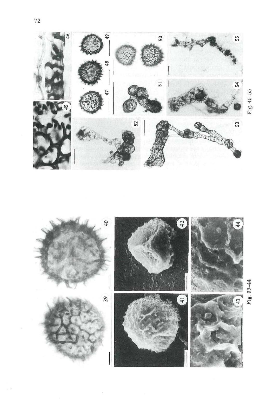
11 sexuality, thin and low lamellae and absence of wart-like projections on the distal face of the spore. 4. Sporeling development (Figures 22-38, 51-55) The spores cultured on October 14, 1991 showed initial stages of germination in all the four sets containing different concentrations of the culture medium. The spores after a few days of sowing became pale green and showed an increase in size by absorption of moisture which results into the rupture of the spore coat through the proximal face. The spores started sprouting on 12th day (i. e. on October 26, 1991) in 100% Knop's medium while in 50% and 25% Knop's media the spores germinated only after 17th day (i. e. on October 31, 1991). However, in distilled water it germinated after about 3 weeks (19 days). The lowest germination percentage (ca. 40%) was observed in spores cultured in distilled water. The highest germination percentage (ca. 86.4%) was observed in 50% knop's medium, however, ca. 83.3% spores germinated in 100% Knop's solution (see also Table I). Table 1 S.No. Media used (concn. Time of Number of spores % of Knop's soln.) Germination Observed Germinated Germination 1 s $ 4 100% 50% ii% 0% (distilled water) 12 days 17 days 17 days 19 days 90± ± ±10 ca. 83.3% ca. 86.4% ca. 71.4% ca. 40% In majority of the spores the rupturing of sporecoat is followed by emergence of a germ-papilla (as in most Marchantiales) from the proximal face, the papillae at this stage contains numerous chloroplasts and oil globules (Fig. 24), and soon these richly supplied chloroplasts migrate towards the tip as the germ-papilla elongates into a germ-tube. The first wall 73 Fig Fossornbronia wondraczekii var. loitlesbergeri. - Fig. 39. Spore (distal view). - Fig. 40. Spore (proximal view). - Fig. 41. Spore (distal view) under SEM. - Fig. 42. Spore (proximal view) under SEM. - Fig A portion of under SEM. - Scale bars equal 10 im. Fig Fossornbronia wondraczekiivav. loitlesbergeri. -Fig. 45. Inner layer cells of the capsule wall. -Fig. 46. Transverse section of the capsule wall. - Figs Spores. - Fig. 51. Young sporeling. - Figs Advanced stages of the sporeling development. - Fig. 55. Juvenile gametophyte differentiated into young leaves and stem. - In Fig scale bars equal 25 (am, in Fig equal 100 (am.

12 74 is laid down transversely dividing the short germ-tube into a basal cell and an upper cell, the latter being packed with dense chloroplasts. The upper cell further divides either by a transverse, or a vertical wall. In sporelings where the germ-tube is predominently formed, there occurs repeated transverse divisions to form a 5-6 celled filamentous sporeling with exospore attached to the basal cell (Figs ). This type of filamentous sporeling was also observed in other species of the genus viz., F. pusilla (LEITGEB 1877, CHALAUD 1926), F. longiseta (HUMPHREY 1906), F. japonica (INOUE 1959, NEHIRA 1966) and F. kashyapii (SRIVASTAVA & UDAR 1975b). In some sporelings the first wall is laid down at a very early stage (Fig. 25) and subsequent transverse and vertical divisions in the upper most cell results in the formation of a cell mass or a multicellular germ-disc (Figs. 31, 33). Ultimately an apical cell with two cutting faces is organised (Figs. 31, 33) which initiates further growth in the sporeling and thus well organised sporelings are formed (Figs. 52, 54) which subsequently become differentiated into a juvenile gametophyte with an axis and leaves. The juvenile leaves develop laterally, and the first leaf is always transversely inserted. The rhizoids are formed as an elongation of the ventral superficial cells of the juvenile axis and maintain the hyaline pale colour even at maturity. Some sporelings after nearly 3 weeks of sowing in 50% Knop's medium show a fairly long germ-tube up to 373 jam without any septum and ceases to grow after 4 weeks except for the chloroplasts which aggregate towards terminal end forming a swollen and bulbous apex. Some of the chloroplasts, however, remain aggregated in the middle and the sporeling develop a branch thus forming a branched and aseptate germtube (Figs ). Occasionally transverse and vertical walls are laid down, forming 4 to several celled germ-disc (Figs. 37, 52, 53) somewhat similar to that of Dumortiera hirsuta and Ricciocarpus natans (see also YANG & Hsu 1967). The stages illustrated (Figs , 51-55) have been drawn from 142 days old cultures. 5. Conclusions The present study reveals that Fossombronia wondraczekii var. loitlesbergeri is different from all other taxa of Fossombronia known so far from India and it constitutes a new record for the country. Like other species of the genus sporoderm ornamentation is stabilized in this taxon also. The sporeling development follows three patterns (i) where the germ-papilla develops into a long unsegmented germ-tube bearing germ-disc at the distal end (Figs , 53), (ii) where the germ-papilla grows and becomes segmented by several transverse division forming a filamentous germ-tube

13 (Figs. 29, 30, 51), and (iii) where germ-papilla first divides by a transverse division but later divides by a transverse or vertical divisions forming a several-celled germ-disc (Fig. 33). There is no prominent germ-tube in the last pattern. The overall pattern of the sporeling development nearly corresponds to those already described in F pusilla (LEITGEB 1877 and CHALAUD 1926, 1929, 1930), F. longiseta (HUMPHREY 1906), F. japonica (INOUE 1959, NEHIRA 1966), F. cristula (UDAR & SRIVASTAVA 1972) and F. kashyapii (SRIVASTAVA & UDAR 1975b). All the above species show a cell mass formation with the stray occurrence of filamentous stages which are considered to have developed because of environmental effect, or overcrowding of the spores (INOUE 1959). In F wondraczekii var. loitlesbergeri, however the germ-tube formation and filamentous sporeling are predominent and only a few sporelings show cell-mass formation. The earlier stages of the sporeling pattern of F wondraczekii var. loitlesbergeri and other species of the genus previously described seem rather similar to those of the Marchantialean taxa (MEHRA & KACHROO 1951, 1952, INOUE 1960, UDAR 1957a, 1957b, 1958a, 1958b, 1976, UDAR & CHANDRA 1965, UDAR & KUMAR 1972, UDAR & SRIVASTAVA 1968, SRIVASTAVA & UDAR 1975b, UDAR & SINGH 1978). Like F. kashyapii the predominance of germ-tube formation in this taxon further suggests its affinity with Marchantiales (SRIVASTAVA & UDAR 1975b) than those with predominance of cell mass formation as observed in F. cristula (UDAR & SRIVASTAVA 1972). The filamentous sporeling is generally formed in the Jungermanniales (NEHIRA 1966). However, the exospore does not remain attached to the basal cell of the sporeling at the later stage, and the differentiation of the axis is usually direct from the sporeling. While in Fossombronia the sporeling pattern corresponds with Marchantiales in which the germ-disc itself develops into a thallus (INOUE 1960, YANG & Hsu 1967). Among the thalloid anacrogynous taxa filamentous sporelings are formed somewhat like Jungermanniales as reported in Riccardia and Metzgeria (INOUE 1959), but the thallus is formed in direct continuation of the sporeling which is never so in Fossombronia. The sporeling development pattern in this taxon further strengthens theory of derivation of Marchantialean thallus from foliose ancestors (MEHRA 1957) as has also been noted earlier (see also INOUE 1959, UDAR & SRIVASTAVA 1972, SRIVASTAVA & UDAR 1975b). 6. Acknowledgements Facility for SEM 'from the Director Birbal Sahni Institute of Palaeobotany and Dr. K. AMBWANI of the same Institute is gratefully acknowledged. Financial assistance from the Council of Scientific and Industrial Research, New Delhi is also acknowledged. Thanks to Prof. Dr. H. TEPPNER (Graz) for his advice for the nomenclature chapter. 75
14 76 7. References BONNER C. E. B Index Hepaticarum. Pars. V: Delavayella to Geothallus. - Weinheim. BOROS Ä Bryogeographie und Bryoflora Ungarns. - Budapest. CHALAUD M. G Le stade protonemique et stade de premiere jeunesse de Fossombroniapusilla. -Bull. Soc. Hist, natur. Toulouse 54: , Le cycle evolutif de Fossombronia pusilla DUM. - Rev. gen er. Bot. 41: 24-34, , , , , , , , , , ; 42:31-37, , , , , , , , Fossombronia cristata var. loitlesbergieri (SCHIFFN.), G. CHALAUD In: VERDOORN F. (Ed.), Hepaticae selectae et criticae series IX (1936) et series X (1937) and Musci selecti et critici Series III (1936) et Series IV (1937), p In: Ann. bryol. 10: CORBIERE L Fossombronia Crozalsii sp. nov. - Rev. bryol. 30 (1): HUMPHREY H. B The development of Fossombronia longiseta AUST. - Ann. Bot. 20: INOUE H Studies on Spore Germination of Hepaticae 5. Fossombronia japonica SCHIFFN. -Bot. Mag. Tokyo, 72 (850): Studies in spore germination and earlier stages of gametophyte development in Marchantiales. - J. Hattori bot. Lab. 23: & HIBINO R Studies on spore morphology of Hepatics (1). Metzgeriales. - Bull, nation. Sei. Mus., Tokyo, Ser. B, 10 (4): KNOX E. M The spores of Bryophyta compared with those of Carboniferous Age. - Trans. Proc. bot. Soc. Edinburgh 32: LEITGEB H Untersuchungen über die Lebermoose. III. Die frondosen Jungermannieen. - Leipzig. MACVICAR C. M Student's Handbook of British Hepatics. - Eastbourne. MEHRA P. N A new suggestion on the origin of thallus in the Marchantiales. II. The Theory. - Amer. J. Bot. 44: & KACHROO P Sporeling germination studies in Marchantiales. I. Rebouliaceae. - The Bryologist 54: & Sporeling germination studies in Marchantiales. II. Stephenoniella brevipedunculata KASH. - The Bryologist 55: MÜLLER K , Die Lebermoose Deutschlands, Österreichs u. d. Schweiz. - In: RABENHORST'S Kryptogamen-Flora... 6 (1, 2), 2. Aufl. - Leipzig Die Lebermoose Europas. - In: RABENHORST'S Kryptogamen-Flora... 6 (1), 3. Aufl.-Leipzig. NEHIRA K Sporelings in the Jungermanniales. - J. Sei. Hiroshima Univ., Ser. B, Div. 2, 11: SCHIFFNER V Über Lebermoose aus Dalmatien und Istrien. - Hedwigia 48: SRIVASTAVA S. C Observations on spore morphology in Indian species of Fossombronia. - Yushania 1 (3): 1-4. & UDAR R. 1975a. The genus Fossombronia RADDI in India. - With a note on the Indian taxa of the family Fossombroniaceae. - Nova Hedwigia 26:
15 77 & 1975b. Sporeling development in Fossombronia kashyapii SRIVASTAVA & UDAR. - Geophytology 5 (1): STEPHANI F Species Hepaticarum I. - Geneve. UDAR R. 1957a. Culture studies in the genus Riccia (MICH.) L. I. Sporeling germination in Riccia billardieri Mont, et N.-J. Indian bot. Soc, 36: b. Culture studies in the genus Riccia (MICH.) L. II. - Sporeling germination in Riccia crystallina L. - J. Indian bot. Soc. 36: a. Culture studies in the genus Riccia (MICH.) L. III. - Sporeling germination in R. trichocarpa Howe. - A reinvestigation. - J. Indian bot. Soc. 37: b. Studies in Indian Sauteriaceae I. Sporeling patterns in Athalamia pinguis FALC. - J. Indian bot. Soc. 37: Bryology in India. -Ann. Cryptog. Phytopathol. 4: i-vii, & CHANDRA V Morphology and ife history of Plagiochasma intermedium L. et G. - J. Hattori bot. Lab 28: & KUMAR D Sporeling development in Athalamia pusilla. - Phyton (Horn, Austria) 14 (3-4): & SINGH D. K In vitro studies on the spore germination of Cryptomitrium himalayense KASH. - New Botanist: & SRIVASTAVA S. C Sporeling development in the genus Exormotheca - I. Exormotheca ceylonensis. - Canad. J. Bot. 46: & Fossombronia cristula Ausx A taxon new to Indian flora. - Curr. Sei. 38: & Sporeling development in the genus Fossombronia RADDI - I. F. cristula AUST. - J. Palynol. 8: 1-7. & A new species of Fossombronia RADDI, F. foreaui UDAR et SRI- VASTAVA from Kodaikanal (Palni Hills), South India. - Nova Hedwigia 47: YANG B. Y. & Hsu F. M Studies on spore germination and gemmae development of Riccardia multifida, Dumortiera hirsuta, Xenochila integrifolia and Marchantia polymorpha. - Taiwania 13:
A New Locality of Fossombronia mylioides (Fossombroniaceae, Marchantiophyta)
") Bull. Natl. Mus. Nat. Sci., Ser. B, 42(1), pp. 19 23, February 22, 2016 A New Locality of Fossombronia mylioides (Fossombroniaceae, Marchantiophyta) Masanobu Higuchi Department of Botany, National Museum
Bull. Natl. Mus. Nat. Sci., Ser. B, 42(1), pp. 19 23, February 22, 2016 A New Locality of Fossombronia mylioides (Fossombroniaceae, Marchantiophyta) Masanobu Higuchi Department of Botany, National Museum
Introduction to Bryophyta
 Introduction to Bryophyta Botany Department, Brahmanand PG College, Bryophyta (Greek Bryon = Moss, phyton = plants) is a group of simplest and primitive plants of the class Embryophyta. The group is represented
The differentiation of sterile thalli of Aneura and Pellia and the problem of Pellia species with unistratose margins
 Differentiation of sterile Aneura and Pellia 1 The differentiation of sterile thalli of Aneura and Pellia and the problem of Pellia species with unistratose margins Jan-Peter Frahm Zusammenfassung: Thallöse
Nonvascular Plants. Believed to have evolved from green-algae. Major adaptations in going from water to land. Chlorophylls a & b and cartenoids
 Nonvascular Plants Believed to have evolved from green-algae Chlorophylls a & b and cartenoids Store starch within chloroplasts Cell wall made up mostly of cellulose Major adaptations in going from water
Scanning Electron Microscopy of some selected south Indian taxa of Marchantiales (Bryophyta: Hepaticae)
") Scanning Electron Microscopy of some selected taxa South India 1 Scanning Electron Microscopy of some selected south Indian taxa of Marchantiales (Bryophyta: Hepaticae) Afroz Alam 1*, Vinay Sharma, Praveen
Scanning Electron Microscopy of some selected taxa South India 1 Scanning Electron Microscopy of some selected south Indian taxa of Marchantiales (Bryophyta: Hepaticae) Afroz Alam 1*, Vinay Sharma, Praveen
Type studies on Pycnolejeunea (Lejeuneaceae, Hepaticae), III. Two Asiatic species described by Hoffmann
, III. Two Asiatic species described by Hoffmann") Ann. Bot. Fennici 33: 59 64 ISSN 0003-3847 Helsinki 6 May 1996 Finnish Zoological and Botanical Publishing Board 1996 Type studies on Pycnolejeunea (Lejeuneaceae, Hepaticae), III. Two Asiatic species described
Ann. Bot. Fennici 33: 59 64 ISSN 0003-3847 Helsinki 6 May 1996 Finnish Zoological and Botanical Publishing Board 1996 Type studies on Pycnolejeunea (Lejeuneaceae, Hepaticae), III. Two Asiatic species described
-plant bodies composed of tissues produced by an apical meristem. -spores with tough walls. -life history of alternation of generations
 Chapter 21-Seedless Plants Major modern plant groups All groups of land-adapted plants have a common set of characteristics: -plant bodies composed of tissues produced by an apical meristem -spores with
Department of Botany, University of Dhaka, Dhaka 1000, Bangladesh. Key words: Seaweeds, Marine algae, Kallymenia spp., St. Martin's Is.
 Bangladesh J. Bot. 37(2): 173-178, 2008 (December) MARINE ALGAE OF THE ST. MARTIN S ISLAND, BANGLADESH. VI. NEW RECORDS OF SPECIES OF THE GENUS KALLYMENIA J. AG. (RHODOPHYTA) ABDUL AZIZ, A.K.M. NURUL ISLAM
Overview. Revised through 30 June Initial Groups ("naked-eye" characters)
") Overview Revised through 30 June 2010 Initial Groups ("naked-eye" characters) Plants essentially leafless, consisting of strongly inclined, highly asymmetric capsules on a stout papillose seta; the "bug-on-a-stick"
Overview Revised through 30 June 2010 Initial Groups ("naked-eye" characters) Plants essentially leafless, consisting of strongly inclined, highly asymmetric capsules on a stout papillose seta; the "bug-on-a-stick"
I. Lycopodiales: The Vegetative Features of the Sporophyte Phase
 Lab II. Lycopodiales: the Clubmosses I. Lycopodiales: The Vegetative Features of the Sporophyte Phase The clubmosses (traditionally classified as species of the genus Lycopodium) are low, evergreen plants
[279] A NOTE ON THE ORIGIN OF LATERAL ROOTS AND THE STRUCTURE OF THE ROOT-APEX OF LYGINOPTERIS OLDHAMIA
![[279] A NOTE ON THE ORIGIN OF LATERAL ROOTS AND THE STRUCTURE OF THE ROOT-APEX OF LYGINOPTERIS OLDHAMIA](/thumbs/83/87080534.jpg "[279] A NOTE ON THE ORIGIN OF LATERAL ROOTS AND THE STRUCTURE OF THE ROOT-APEX OF LYGINOPTERIS OLDHAMIA") [279] A NOTE ON THE ORIGIN OF LATERAL ROOTS AND THE STRUCTURE OF THE ROOT-APEX OF LYGINOPTERIS OLDHAMIA BY A. C. HALKET (With Plate XI and i figure in the text) E 'GlNOPTERis oi.dh.imi.i, a plant of the
[279] A NOTE ON THE ORIGIN OF LATERAL ROOTS AND THE STRUCTURE OF THE ROOT-APEX OF LYGINOPTERIS OLDHAMIA BY A. C. HALKET (With Plate XI and i figure in the text) E 'GlNOPTERis oi.dh.imi.i, a plant of the
S. SANDHYA RANI, M. SOWGHANDIKA,
 S. SANDHYA RANI, M. SOWGHANDIKA, 76 Habitat: Rare, grows on red rocks over soil. Specimen examined: Coffee plantations in Galikonda (VSKP), MS 33959. India : Darjeeling, Sikkim, Khasi hills. World : East
CHAPTER 2-4 BRYOPHYTA TAKAKIOPSIDA
 Glime, J. M. 2017. Bryophyta - Takakiopsida. Chapt. 2-4. In: Glime, J. M. Bryophyte Ecology. Volume 1. Physiological Ecology. 2-4-1 Ebook sponsored by Michigan Technological University and the International
PTERIS REPTANS (PTERIDACEAE) - A NEW RECORD FOR INDIA
 - A NEW RECORD FOR INDIA") FERN GAZ. 19(1):25-29. 2012 25 PTERIS REPTANS (PTERIDACEAE) - A NEW RECORD FOR INDIA V.K. SREENIVAS 1 & P.V. MADHUSOODANAN 2 1 Department of Botany, University of Calicut, Kerala, India - 673635 (Email:
FERN GAZ. 19(1):25-29. 2012 25 PTERIS REPTANS (PTERIDACEAE) - A NEW RECORD FOR INDIA V.K. SREENIVAS 1 & P.V. MADHUSOODANAN 2 1 Department of Botany, University of Calicut, Kerala, India - 673635 (Email:
A handful of primary features are useful for distinguishing water primrose (Ludwigia) from other plants. Understand what to look for, such as leaf
 from other plants. Understand what to look for, such as leaf") A handful of primary features are useful for distinguishing water primrose (Ludwigia) from other plants. Understand what to look for, such as leaf arrangement and number of petals. Pairing morphological
A handful of primary features are useful for distinguishing water primrose (Ludwigia) from other plants. Understand what to look for, such as leaf arrangement and number of petals. Pairing morphological
Observations on Chytridiaceous Parasites of Phanerogams
 Arch. Mikrobiol. 70, 104--109 (1970) Observations on Chytridiaceous Parasites of Phanerogams XYIII. A Physoderma on Juncus pelocarpus Mey. F. K. SPArrow * University of Michigan, Biological Station and
Cytological studies of some Indian liverworts
 Lindbergia 39: 7 11, 2016 ISSN 2001-5909 Accepted 11 March 2016 Cytological studies of some Indian liverworts Sunita Kapila S. Kapila (s_kapila0802@yahoo.co.in), Dept of Botany, Panjab University, Chandigarh,
Chapter 20 Nonvascular Plants: Mosses, Liverworts, and Hornworts
 Chapter 20 Nonvascular Plants: Mosses, Liverworts, and Hornworts Major plant groups Topics Bryophyte adaptations synapomorphies Alternation of generation in Bryophytes Phylum Hepaticophyta Phylum Bryophyta
Mosquito Systematics Vol. 6(Z) June 1974
 June 1974") Mosquito Systematics Vol. 6(Z) June 1974 93 Research on the Mosquitoes of Angola. VII - Redescription of the Larva of Aedes durbanensis durbanensis (Theo., 1903) and Description of Aedes durbanensis angozae
Mosquito Systematics Vol. 6(Z) June 1974 93 Research on the Mosquitoes of Angola. VII - Redescription of the Larva of Aedes durbanensis durbanensis (Theo., 1903) and Description of Aedes durbanensis angozae
EPIDERMAL STRUCTURE AND DEVELOPMENT OF STOMATA IN EPHEDRA FOLIATA BOISS.
 EPIDERMAL STRUCTURE AND DEVELOPMENT OF STOMATA IN EPHEDRA FOLIATA BOISS. BY D. D. PANT AND BHARATI MEHRA Department of Botany, The University, Allahabad, India {Received z August 1963) SUMMARY The epidermal
SPECIES FACT SHEET. Taxonomic Note: None.
 SPECIES FACT SHEET Common Name: Granite moss, Lantern moss Scientific Name: Andreaea nivalis Hook. Recent synonyms: Andreaea baileyi Holz. A. macounii Kindb. in Mac. Division: Bryophyta Class: Bryopsida
Acrobotrys tritubus Riedel
 151 Acrobotrys tritubus Riedel Acrobotrys tritubus Riedel, 1957, p.80, pl.1, fig.5 DESCRIPTION Cephalis trilobate, with large subglobular [antecephalic] lobe, smaller globular [cephalic] lobe, and inflated-conical
Plant Structure. Lab Exercise 24. Objectives. Introduction
 Lab Exercise Plant Structure Objectives - Be able to identify plant organs and give their functions. - Learn distinguishing characteristics between monocot and dicot plants. - Understand the anatomy of
Literature. Morphology. Morphology of the mycorrhizal system. Morphology of the unramified ends
 Literature references Müller WR, Rauscher T, Agerer R, Chevalier G (1996) Tuber aestivum Vitt. + Corylus avellana L.Descr Ectomyc 1: 167-172. Rauscher T, Müller WR, Chevalier G, Agerer R (1996) Tuber aestivum.
SPORE-FORMS IN SPOROPHORES OF GANODERMA LUCIDUM (LEYSS.) KARST.
 KARST.") SPORE-FORMS IN SPOROPHORES OF GANODERMA LUCIDUM (LEYSS.) KARST. BY SACHINDRANATH BANERJEE AND ANJALI SARKAR (Department of Botany, University o[ Calcutta) Received September 12, 1958 (Communicated by Dr.
SPORE-FORMS IN SPOROPHORES OF GANODERMA LUCIDUM (LEYSS.) KARST. BY SACHINDRANATH BANERJEE AND ANJALI SARKAR (Department of Botany, University o[ Calcutta) Received September 12, 1958 (Communicated by Dr.
Useful Propagation Terms. Propagation The application of specific biological principles and concepts in the multiplication of plants.
 Useful Propagation Terms Propagation The application of specific biological principles and concepts in the multiplication of plants. Adventitious Typically describes new organs such as roots that develop
Phaeocalicium populneum
 Phaeocalicium populneum markpowell222@btinternet.com After conducting a survey of the RHS garden at Wisley on 18 th August 2018, Fay Newbery kindly showed me the colony of P. populneum at Esher Common.
The Petiolar Structure of Christella dentata (Forssk.) Brownsey & Jermy (Thelypteridaceae, Pteridophyta)
 Brownsey & Jermy (Thelypteridaceae, Pteridophyta)") Ethnobotanical Leaflets 12: 96-102. 2008. The Petiolar Structure of Christella dentata (Forssk.) Brownsey & Jermy (Thelypteridaceae, Pteridophyta) KAMINI SRIVASTAVA, M.Sc, D.Phil Department of Botany,
Ethnobotanical Leaflets 12: 96-102. 2008. The Petiolar Structure of Christella dentata (Forssk.) Brownsey & Jermy (Thelypteridaceae, Pteridophyta) KAMINI SRIVASTAVA, M.Sc, D.Phil Department of Botany,
Plant Crib VERONICA. 1. Veronica serpyllifolia
 VERONICA 1. Veronica serpyllifolia Illustrations reproduced, with permission, from M. McC. Webster (1978). Flora of Moray, Nairn & East Inverness. Aberdeen. Subsp. humifusa (Dicks.) Syme Subsp. serpyllifolia
ARE PLAGIOTHECIUM CAVIFOLIUM, P. NEMORALE AND P. SUCCULENTUM INDEED VARIABLE SPECIES?
 Pak. J. Bot., 50(4): 1579-1589, 2018. ARE PLAGIOTHECIUM CAVIFOLIUM, P. NEMORALE AND P. SUCCULENTUM INDEED VARIABLE SPECIES? Department of Geobotany and Plant Ecology, Faculty of Biology and Environmental
IX. PRIMARY STEM STRUCTURE AND DEVELOPMENT Bot 404 Fall 2004
 IX. PRIMARY STEM STRUCTURE AND DEVELOPMENT Bot 404 Fall 2004 A. Shoot apex -plants have an open system of growth, therefore the ability (at least potentially) to continue growth because there is a meristem
March, 1906.] Life Cycle 0/ a Homosporous Pteridophyte. 483 THE LIFE CYCLE OF A HOMOSPOROUS PTERIDOPHYTE. JOHN H. SCHAFFNER.
![March, 1906.] Life Cycle 0/ a Homosporous Pteridophyte. 483 THE LIFE CYCLE OF A HOMOSPOROUS PTERIDOPHYTE. JOHN H. SCHAFFNER.](/thumbs/89/100118323.jpg "March, 1906.] Life Cycle 0/ a Homosporous Pteridophyte. 483 THE LIFE CYCLE OF A HOMOSPOROUS PTERIDOPHYTE. JOHN H. SCHAFFNER.") March, 1906.] Life Cycle 0/ a Homosporous Pteridophyte. 483 THE LIFE CYCLE OF A HOMOSPOROUS PTERIDOPHYTE. JOHN H. SCHAFFNER. The Homosporous Pteridophytes constitute the lowest subkingdom of vascular plants.
March, 1906.] Life Cycle 0/ a Homosporous Pteridophyte. 483 THE LIFE CYCLE OF A HOMOSPOROUS PTERIDOPHYTE. JOHN H. SCHAFFNER. The Homosporous Pteridophytes constitute the lowest subkingdom of vascular plants.
Anthoceros Habitat and distribution Systematic position Habitat and distribution
 Anthoceros L. Habitat and distribution The name anthoceros means flower horn, and refers to the characteristic horn-shaped sporophytes that all hornworts produce. The genus Anthoceros was first established
Two representatives of the genus Mindarus (Homoptera, Aphidoidea, Mindaridae) in Baltic amber
 in Baltic amber") Ann. Naturhist. Mus. Wien 92 A 73-77 Wien, April 1991 Two representatives of the genus Mindarus (Homoptera, Aphidoidea, Mindaridae) in Baltic amber By ANDRZEJ CZYLOK 1 ) (With 4 textfigures) Manuscript
Ann. Naturhist. Mus. Wien 92 A 73-77 Wien, April 1991 Two representatives of the genus Mindarus (Homoptera, Aphidoidea, Mindaridae) in Baltic amber By ANDRZEJ CZYLOK 1 ) (With 4 textfigures) Manuscript
THE BEHAVIOUR OF CHLOROPLASTS DURING CELL DIVISION OF ISOETES LACUSTRIS L.
 New Phytol (1974) 73, 139-142. THE BEHAVIOUR OF CHLOROPLASTS DURING CELL DIVISION OF ISOETES LACUSTRIS L. BY JEAN M. WHATLEY Botany School, University of Oxford (Received 2 July 1973) SUMMARY Cells in
Microthyriales of Tierra del Fuego I: The Genus Parasterinella SPEGAZZINI
 Verlag Ferdinand Berger & Söhne Ges.m.b.H., Horn, Austria, download unter www.biologiezentrum. Sydowia, Annales Mycologici Ser. II. Vol. 38: 1-5 (1985) Verlag Ferdinand Berger & Söhne Gesellschaft m.b.h.,
Phylum Bryophyta : (Page 169)
") Kingdom Plantae : Plants... - nonmotile eukaryotic, multicellular, autotrophic organisms - rigid cell walls built of cellulose - life cycles show alternation of generations...two distinct phases called
Kingdom Plantae : Plants... - nonmotile eukaryotic, multicellular, autotrophic organisms - rigid cell walls built of cellulose - life cycles show alternation of generations...two distinct phases called
Kingdom Plantae. Biology : A Brief Survey of Plants. Jun 22 7:09 PM
 Kingdom Plantae Biology 2201 6.1 6.2 : A Brief Survey of Plants The study of plants is called botany. Plants are believed to have evolved from green algae. The main plant (land) characteristics are as
Topic 22. Introduction to Vascular Plants: The Lycophytes
 Topic 22. Introduction to Vascular Plants: The Lycophytes Introduction to Vascular Plants Other than liverworts, hornworts, and mosses, all plants have vascular tissues. As discussed earlier, the mosses
Master Gardener Program. Utah State University Cooperative Extension
 Master Gardener Program Utah State University Cooperative Extension Plant Parts and Functions Overview Plant Classification Stems Buds Leaves Flowers Fruits Roots Plant Classifications Woody vs. Herbaceous
ANATOMY OF PLANTS Introduction: The study of gross internal structure of plant organs by the technique of section cutting is called plant anatomy.
 ANATOMY OF PLANTS Introduction: The study of gross internal structure of plant organs by the technique of section cutting is called plant anatomy. (Pandey, 2002). Various plant organ viz. root, stem, leaves,
DIFFERENTIATION OF AVOCADO BLOSSOM BUDS IN FLORIDA
 Reprinted for private circulation from the Botanical Gazette, Vol. 104, No. 2, December, 1942. DIFFERENTIATION OF AVOCADO BLOSSOM BUDS IN FLORIDA PHILIP C. REECE 1 (WITH THIRTEEN FIGURES) Subtropical Fruit
ANTHER TYPES OF THE MONOCOTS WITHIN FLORA OF KARACHI, PAKISTAN
 Pak. J. Bot., 40(5): 1839-1849, 2008. ANTHER TYPES OF THE MONOCOTS WITHIN FLORA OF KARACHI, PAKISTAN ROOHI BANO, RUBINA ABID AND M. QAISER * Department of Botany, University of Karachi, Karachi, Pakistan
Two new species of the genus Cleaveius Subrahmanian, 1927 (Aeanthocephala Micracanthorhynchinidae Yamaguti, 1963)
") Proc. Indian.&cad. Sci., Vol. 88 B, Part I, Number 4, August 1979, pp. 305-3100 @ printed in India. Two new species of the genus Cleaveius Subrahmanian, 1927 (Aeanthocephala Micracanthorhynchinidae Yamaguti,
Proc. Indian.&cad. Sci., Vol. 88 B, Part I, Number 4, August 1979, pp. 305-3100 @ printed in India. Two new species of the genus Cleaveius Subrahmanian, 1927 (Aeanthocephala Micracanthorhynchinidae Yamaguti,
CALLISPHENUS GRACILIS, N. GEN., N. SP. A FOSSIL ALGA FROM THE WENLOCK OF THE OSLO REGION
 CALLISPHENUS GRACILIS, N. GEN., N. SP. A FOSSIL ALGA FROM THE WENLOCK OF THE OSLO REGION BY OVE HØEG WITH 2 PLATES The specimen described below was found in August, 191 O, on the small island of Kommersøy
Primary Plant Body: Embryogenesis and the Seedling
 BIOL 221 Concepts of Botany Primary Plant Body: Embryogenesis and the Seedling (Photo Atlas: Figures 1.29, 9.147, 9.148, 9.149, 9.150, 9.1, 9.2) A. Introduction Plants are composed of fewer cell types,
LENTIBULARIACEAE BLADDERWORT FAMILY
 LENTIBULARIACEAE BLADDERWORT FAMILY Barry Rice Center for Plant Diversity, Department of Plant Sciences, University of California, 1 Shields Avenue, Davis CA 95616 Perennial and annual herbs, carnivorous,
Plant Evolution & Diversity
 Plant Evolution & Diversity Ancestors of plants were probably charophytes (green algae) Chlorophyll a and b, beta carotene Similar thylakoid arrangements Identical cell walls Starch as a storage carbohydrate
The Japanese Fissidens neomagofukui (Bryophyta: Fissidentaceae) new to India from the Western Ghats
 new to India from the Western Ghats") The Japanese Fissidens neomagofukui (Bryophyta: Fissidentaceae) new to India from the Western Ghats Author(s): Albert Ebenezer Dulip Daniels, Ratheesh Sreebha and Kochumani Chinnapilla Kariyappa Source:
The Japanese Fissidens neomagofukui (Bryophyta: Fissidentaceae) new to India from the Western Ghats Author(s): Albert Ebenezer Dulip Daniels, Ratheesh Sreebha and Kochumani Chinnapilla Kariyappa Source:
NOTES: CH 35 - Plant Structure & Growth
 NOTES: CH 35 - Plant Structure & Growth In their evolutionary journey, plants adapted to the problems of a terrestrial existence as they moved from water to land ANGIOSPERMS (flowering plants) -most diverse
Aquatic Bryophytes. What is a bryophyte? I. Big picture. II. Biology -Life Life Cycle -Major Groups -Adaptations. III. Aquatic Bryophyte Ecology
 Aquatic Bryophytes What is a bryophyte? -Life Life Cycle -Major Groups -Adaptations Heather Lintz Botany and Plant Pathology Oregon State University Bryophytes are here All land plants likely to have freshwater
 Question 1: State the location and function of different types of meristem. Meristems are specialised regions of plant growth. The meristems mark the regions where active cell division and rapid division
A Strange New Genus and Species of Mesosini from North Thailand (Coleoptera, Cerambycidae, Lamiinae) [Studies on Asian Mesosini, VII]
![A Strange New Genus and Species of Mesosini from North Thailand (Coleoptera, Cerambycidae, Lamiinae) [Studies on Asian Mesosini, VII]](/thumbs/94/118924602.jpg "A Strange New Genus and Species of Mesosini from North Thailand (Coleoptera, Cerambycidae, Lamiinae) [Studies on Asian Mesosini, VII]") Elytra, Tokyo, New Series, 1(1): 119 123 July 31, 2011 A Strange New Genus and Species of Mesosini from North Thailand (Coleoptera, Cerambycidae, Lamiinae) [Studies on Asian Mesosini, VII] Junsuke YAMASAKO
Elytra, Tokyo, New Series, 1(1): 119 123 July 31, 2011 A Strange New Genus and Species of Mesosini from North Thailand (Coleoptera, Cerambycidae, Lamiinae) [Studies on Asian Mesosini, VII] Junsuke YAMASAKO
BOTANY LAB #1 MITOSIS AND PLANT TISSUES
 Mitosis and cytokinesis in plants BOTANY LAB #1 MITOSIS AND PLANT TISSUES In plants the formation of new cells takes place in specialized regions of meristematic tissue. Meristematic tissues contain immature,
APPLICATIONS UNDER EXAMINATION. MAGNOLIA (Magnolia) Proposed denomination: Cleopatra Application number: Application date: 2011/02/25
 Proposed denomination: Cleopatra Application number: Application date: 2011/02/25") (Magnolia) Proposed denomination: Cleopatra Application number: 11-7201 Application date: 2011/02/25 Applicant: Agent in Canada: BioFlora Inc., St. Thomas, Ontario Breeder: Description: PLANT: weak vigour,
(Magnolia) Proposed denomination: Cleopatra Application number: 11-7201 Application date: 2011/02/25 Applicant: Agent in Canada: BioFlora Inc., St. Thomas, Ontario Breeder: Description: PLANT: weak vigour,
OF THE LEMNA FROND MORPHOLOGY
 MORPHOLOGY OF THE LEMNA FROND FREDERICK H. BLODGETT (WITH PLATE XIV AND ONE FIGURE) In the case of structure simplified by reduction, it is sometimes necessary to trace the development of the parts through
The taxonomic status of Fontinalis mollis and F. antipyretica var. cymbifolia with the description of Fontinalis antipyretica var.
 1 The taxonomic status of Fontinalis mollis and F. antipyretica var. cymbifolia with the description of Fontinalis antipyretica var. rotundifolia Jan-Peter Frahm Abstract: During the past years, Fontinalis
CHAPTER 4-3 ADAPTIVE STRATEGIES: PHENOLOGY, A SPHAGNUM CASE STUDY
 Glime, J. M. 2017. Adaptive Strategies: Phenology, A Sphagnum Case Study. Chapt. 4-3. In: Glime, J. M. Bryophyte Ecology. Volume 4-3-1 1. Physiological Ecology. Ebook sponsored by Michigan Technological
(Photo Atlas: Figures 9.147, 9.148, 9.150, 9.1, 9.2, )
") BIOL 221 Concepts of Botany Fall 2007 Topic 07: Primary Plant Body: The Root System (Photo Atlas: Figures 9.147, 9.148, 9.150, 9.1, 9.2, 9.5 9.23) A. Introduction The root has the primary functions of
BIOL 221 Concepts of Botany Fall 2007 Topic 07: Primary Plant Body: The Root System (Photo Atlas: Figures 9.147, 9.148, 9.150, 9.1, 9.2, 9.5 9.23) A. Introduction The root has the primary functions of
Class XI Chapter 6 Anatomy of Flowering Plants Biology
 Class XI Chapter 6 Anatomy of Flowering Plants Biology Question 1: State the location and function of different types of meristem. Meristems are specialised regions of plant growth. The meristems mark
Plants Review 1. List the 6 general characteristics of plants. 2. What did plants probably evolve from? 3. What are some advantages for life on land
 Plants Review 1. List the 6 general characteristics of plants. 2. What did plants probably evolve from? 3. What are some advantages for life on land for a plant? 4. What are the 3 main groups of plants?
A review of main morphological features in European taxa of the genus Chiloscyphus Corda (Marchantiophyta, Geocalycaceae)
") A review of main morphological features in European taxa of the genus Chiloscyphus Corda (Marchantiophyta, Geocalycaceae) Anna Salachna Department of Ecology and Nature Conservation, Institute of Environmental
A review of main morphological features in European taxa of the genus Chiloscyphus Corda (Marchantiophyta, Geocalycaceae) Anna Salachna Department of Ecology and Nature Conservation, Institute of Environmental
BIODIVERSITY OF PLANTS 12 FEBRUARY 2014
 BIODIVERSITY OF PLANTS 12 FEBRUARY 2014 In this lesson we: Lesson Description Look at how plants are classified Define Alternation of generations Summarise the main characteristics of four groupings of
TARGET STUDY MATERIAL
 TARGET STUDY MATERIAL Plus-1 Botany VOL I TARGET EDUCATIONAL INSTITUTION Target Educational institution is the one and only Entrance coaching and CBSE 10 th coaching centre at Mukkam with advanced technologies
2.5 : Cells are grouped into tissue
 2.5 : Cells are grouped into tissue 1 CELL STRUCTURE AND FUNCTIONS Prokaryotic and eukaryotic cells Structures & functions: Cell membrane and organelles Animal Cells are grouped into tissue Plant Cell
Plants. Tissues, Organs, and Systems
 Plants Tissues, Organs, and Systems Meristematic cells Specialized cells that are responsible for producing specialized cells, they produce three types of tissue in the body of a plant. Meristematic Cells
Non Permanent Tissues - Meristematic Tissue
 PLANT TISSUES Non Permanent Tissues - Meristematic Tissue Undifferentiated plant cells that are continually dividing by mitosis Large thin walled cells No vacuole Dense cytoplasm Large nucleus Found at
Topic 23. The Ferns and Their Relatives
 Topic 23. The Ferns and Their Relatives Domain: Eukarya Kingdom: Plantae Ferns Leptosporangiate Ferns Psilophytes Genus: Psilotum Horsetails Genus: Equisetum In this treatment we lump the Psilophytes and
LOOKING AT PLANT STEMS
 Activity 4.17 Student Sheet LOOKING AT PLANT STEMS Purpose To look at the structure of xylem vessels, phloem sieve tubes and sclerenchyma fibres. To locate the position of these tissues within the stem.
The Living World. AIIMS,CBSE,AIPMT, AFMC,Bio.Tech & PMT, Contact : , Mail at :- by AKB
 The Living World Very Short Answer Questions 1. What does ICBN stand for? A: International Code for Botanical Nomenclature. 2. What is flora? A: It is a publication containing actual account of habitat,
THE OHIO JOURNAL OF SCIENCE
 THE OHIO JOURNAL OF SCIENCE VOL. XXIV JULY, 1924 No.. 4 THE VASCULAR ANATOMY OF CALAMOVILFA LONGIFOLIA.* ERNEST LINCOLN STOVER Eastern Illinois State Teachers' College The present study of the anatomy
Other Commonly Used Names: wintergreen quillwort, evergreen quillwort
 Common Name: WINTER QUILLWORT Scientific Name: Isoetes hyemalis D.F. Brunton Other Commonly Used Names: wintergreen quillwort, evergreen quillwort Previously Used Scientific Names: none Family: Isoetaceae
with others and thus regenerate a functioning conductive system. Regeneration
 388 BOTANY: SINNOTT AND BLOCH PROC. N. A. S. VISIBLE EXPRESSION OF CYTOPLASMIC PA TTERN IN THE DIFFERENTIATION OF XYLEM STRANDS BY EDMUND W. SINOTT AND ROBERT BLOCH OsBORN BOTANCAL LABORATORY, YALE UNIVERSITY
NOTES ON GINKGO BILOBA'
 NOTES ON GINKGO BILOBA' WALTER WV. TUPPER (WITH PLATE xx) Among the gymnosperms, one of the groups most interesting from a morphological standpoint is the Ginkgoales, the only living representative of
Downloaded from
 POINTS TO REMEMBER : 6. Anatomy of Flowering Plants Study of internal structure of plant is called anatomy. In plants cells are the basic unit. Cells organized into tissues and tissues organized into organs.
INEA HYBRIDISATION PROTOCOLS 2011
 INEA HYBRIDISATION PROTOCOLS 2011 Anton Ivancic Hybridisation of taro (Colocasia esculenta) Floral characteristics of taro Colocasia esculenta is an allogamous, protogynous species, for which the main
SPECTROSCOPIC STUDY OF LUMINESCENCE PATTERNS IN DIAMOND BY ANNA MANI. Received August 21, 1944 (Communicated by Sir C. V. Raman, Kt., F.R.S., N.L.
 SPECTROSCOPIC STUDY OF LUMINESCENCE PATTERNS IN DIAMOND BY ANNA MANI (From the Department of Physics, Indian Institute of Science, Bangalore) Received August 21, 1944 (Communicated by Sir C. V. Raman,
A MEGASECOPTERON FROM UPPER CARBONIFEROUS BY F. M. CARPENTER. In I962 Professor F. Stockmans, of the Institut Royal des Sciences STRATA IN SPAIN
 A MEGASECOPTERON FROM UPPER CARBONIFEROUS STRATA IN SPAIN BY F. M. CARPENTER Harvard University In I962 Professor F. Stockmans, of the Institut Royal des Sciences Naturelles de. Belgique, kindly sent me
Exercise 12. Procedure. Aim: To study anatomy of stem and root of monocots and dicots.
 Aim: To study anatomy of stem and root of monocots and dicots. Principle: The study of internal morphology, i.e., cells of various tissues in an organ of a living body is called Anatomy. Tissue, which
2. SPHAGNACEAE Dumortier
 2. SPHAGNACEAE Dumortier Cyrus B. McQueen Richard E. Andrus Plants with branches in fascicles, branches usually of spreading and pendent types but rarely spreading only. Protonemata thallose. Leaves usually
ON OCCURRENCE OF THE GENUS PORPHYRIDIUM NAGELI: NEW TO INDIA
 J. Algal Biomass Utln. 2009, 1 (1): 102 106 Abstract ON OCCURRENCE OF THE GENUS PORPHYRIDIUM NAGELI: NEW TO INDIA M. S. Gaikwad, B. G. Meshram and B. B. Chaugule * Department of Botany, University of Pune,
Plant Structure. Objectives At the end of this sub section students should be able to:
 Name: 3.2 Organisation and the Vascular Structures 3.2.1 Flowering plant structure and root structure Objectives At the end of this sub section students should be able to: 1. Label a diagram of the external
Liverwort Control in Nurseries
 Liverwort Control in Nurseries Richard Smith, Steve Tjosvold and Tiffany Bensen University of California Cooperative Extension Monterey and Santa Cruz Counties, California Liverworts natural habitat is
Polytrichum psilocorys 153 A NOTE ON THE PERIODICITY OF LEAF- FORM IN TARAXACUM OFFICINALE
 Polytrichum psilocorys 153 sterilized by boiling and kept in a glass box. They germinated abundantly and the culture remained pure. The young moss-plants appeared on the protonema, but they showed an extraordinarily
NOTES ON FOUR NEW AND NOTEWORTHY RECORDS OF LEJEUNEA FROM MEGHALAYA, INDIA
 Acta Botanica Hungarica 57(3 4), pp. 407 418, 2015 DOI: 10.1556/034.57.2015.3-4.10 NOTES ON FOUR NEW AND NOTEWORTHY RECORDS OF LEJEUNEA FROM MEGHALAYA, INDIA S. K. Singh 1 *, T. PÓcs 2 and S. Kumar 1 1
CAMBIUM, meristem, heartwood, and lenticel are
 Examining the Structures of a Tree CAMBIUM, meristem, heartwood, and lenticel are some terms that may be new to you. These terms are used to describe various tree structures. Not surprisingly, many terms
PSEUDOCHLORELLA ENCAPSULATA SP. NOV. (CHLOROPHYCEAE) FROM MAHAGONI BARK, DHAKA, BANGLADESH
 FROM MAHAGONI BARK, DHAKA, BANGLADESH") Bangladesh J. Bot. 46(4): 1407-1414, 2017 (December) PSEUDOCHLORELLA ENCAPSULATA SP. NOV. (CHLOROPHYCEAE) FROM MAHAGONI BARK, DHAKA, BANGLADESH MAHIN MOHID AND ABDUL AZIZ* Department of Botany, University
Bangladesh J. Bot. 46(4): 1407-1414, 2017 (December) PSEUDOCHLORELLA ENCAPSULATA SP. NOV. (CHLOROPHYCEAE) FROM MAHAGONI BARK, DHAKA, BANGLADESH MAHIN MOHID AND ABDUL AZIZ* Department of Botany, University
The Classification of Plants, II.
 The Ohio State University Knowledge Bank kb.osu.edu Ohio Journal of Science (Ohio Academy of Science) Ohio Journal of Science: Volume 6, Issue 1 (November, 1905) 1905-11 The Classification of Plants, II.
Topic 14. The Root System. II. Anatomy of an Actively Growing Root Tip
 Topic 14. The Root System Introduction. This is the first of two lab topics that focus on the three plant organs (root, stem, leaf). In these labs we want you to recognize how tissues are organized in
162. Protosequoia (n, g.) in Taxodiaceae from Pinus tri f olia Beds in Central Honshu, Japan
 in Taxodiaceae from Pinus tri f olia Beds in Central Honshu, Japan") No. 8] Proc. Japan Acad., 45 (1969) 727 162. Protosequoia (n, g.) in Taxodiaceae from Pinus tri f olia Beds in Central Honshu, Japan By Shigeru MIKI Mukogawa Women's Univ., Nishinomiya City, Hyogo (Comm.
No. 8] Proc. Japan Acad., 45 (1969) 727 162. Protosequoia (n, g.) in Taxodiaceae from Pinus tri f olia Beds in Central Honshu, Japan By Shigeru MIKI Mukogawa Women's Univ., Nishinomiya City, Hyogo (Comm.
SPECIES FACT SHEET Common Name: Scientific Name: Technical Description: Distinctive characters: Similar species:
 SPECIES FACT SHEET Common Name: Platyhypnidium moss Scientific Name: Platyhypnidium riparioides Recent synonym: Eurhynchium riparioides, Rhynchostegium riparioides Phylum: Bryophyta Class: Bryopsida Order:
Dendrology FOR 219. Instructor: Dr. Jeremy Stovall. Lecture 3: Anatomy I: Vegetative Morphology
 Dendrology FOR 219 Instructor: Dr. Jeremy Stovall Lecture 3: 01.24.2018 Anatomy I: Vegetative Morphology Tree Life Cycle Seed Seed Production Germination THIS CLASS Reproductive Tree Seedling Flowering
Topic 2: Plants Ch. 16,28
 Topic 2: Plants Ch. 16,28 Characteristics of Plants p. 316 1. Multicellular eukaryotic organisms 2. Composed of tissues, organs and organ systems. 3. Cell walls made of cellulose. 4. Store energy as starch.
Notes concerning the development of Nemalion inultifidum.
 Notes concerning the development of Nemalion inultifidum. GRACE D. CHESTER. WITH PLATES XXV AND XXVI. The resemblance between the structure of the frond and the development of the cystocarp in the genera
Bryophyte Gametophytes. Bryophyte Gametophytes. A spore germinates into a gametophyte. composed of a protonema and gamete producing gametophore
 A spore germinates into a composed of a protonema and gamete producing gametophore Rhizoids Anchor s to substrate Lack of vascular :ssues Bryophyte Gametophytes Restricts the height of s Mature s produce
Conocephalum conicum (L.) Dumort. (Snake Liverwort) Threatened in Bhaderwah (J & K) due to Environmental Shock
 Dumort. (Snake Liverwort) Threatened in Bhaderwah (J & K) due to Environmental Shock") American Journal of Plant Sciences, 2011, 2, 554-560 doi:10.4236/ajps.2011.24066 Published Online October 2011 (http://www.scirp.org/journal/ajps) Conocephalum conicum (L.) Dumort. (Snake Liverwort) Threatened
American Journal of Plant Sciences, 2011, 2, 554-560 doi:10.4236/ajps.2011.24066 Published Online October 2011 (http://www.scirp.org/journal/ajps) Conocephalum conicum (L.) Dumort. (Snake Liverwort) Threatened
Visit For All NCERT solutions, CBSE sample papers, Question papers, Notes for Class 6 to 12. Chapter-6 ANATOMY OF FLOWERING PLANTS
 Chapter-6 ANATOMY OF FLOWERING PLANTS POINTS TO REMEMBER Anatomy : Anatomy is the study of internal structure of organisms. Plant anatomy includes organisation and structure of tissues. Tissue : A group
A TEST OF SACHS'S THEORY OF THE PLAGIOTROPISM OF LAMINAE
 [25] A TEST OF SACHS'S THEORY OF THE PLAGOTROPSM OF LAMNAE BY R. SNOW Fellozv of Magdalen College, Oxford (With 5 figures in the text) According to a suggestion of Sachs (179, pp. 246 and 254) the movements
4 Marine Biology Notes. Multi-cellular Primary Producers: Seaweeds and Plants
 4 Marine Biology Notes Multi-cellular Primary Producers: Seaweeds and Plants Marine Algae Marine algae are important primary producers (photosynthetic) These algae are called by a generic term seaweeds
13.4 Roots Figure 2 primary root: primary root secondary root: secondary root taproots fibrous taproots: roots. fibrous roots: adventitious roots
 10. Why is it not surprising that many hydrophytes have little or no tissue? 11. The leaves of many underwater plants are finely divided, dramatically increasing the surface area that is in contact with