Heteropolymer. Mostly in regular secondary structure
|
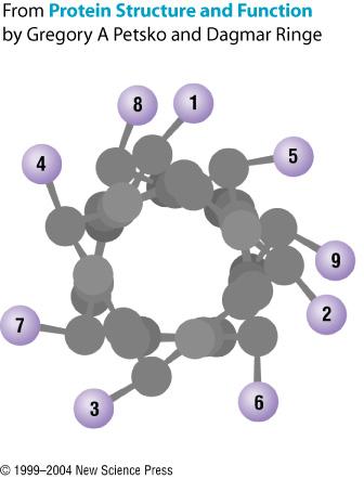
|
|
- Richard Leonard
- 5 years ago
- Views:
Transcription
1 Heteropolymer Mostly in regular secondary structure
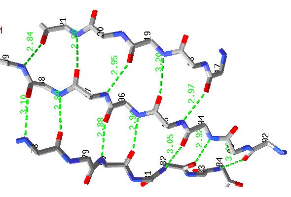
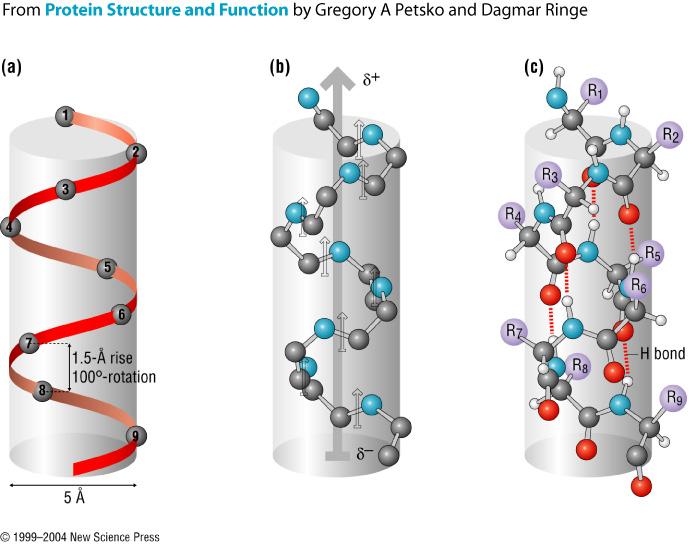
*(+2!3/'!4#5'!1/,#64!#6!,6! 7/'8#9!40#1:!;%0!0;!0/'!4#5'<!,4!=#'>'5!#6!,! &#??;6!5#,$&,-!4/;>6!;6!0/'!8'@0*!!A6!0/'!&#$/0!")


2 C >N trace how you go around the helix C >N C2 >N6 C1 >N5 What s the pattern? Ci>Ni+? 5 6 move around not quite 120 "#$%&'!()*(+2!3/'!4#5'!1/,#64!#6!,6! 7/'8#9!40#1:!;%0!0;!0/'!4#5'<!,4!=#'>'5!#6!,! &#??;6!5#,$&,-!4/;>6!;6!0/'!8'@0*!!A6!0/'!&#$/0!#4!,!/'8#1,8!>/''8!=#'>!;@!,6! 7/'8#9! >#0/!0/'!(B!&'4#5%'4!4/;>6!,?;='*!!3/#4!'9,-.8'!4/;>4!,6!,-./#.,0/#1!/'8#9<!>#0/!.;8,&!,65!6;67.;8,&!&'4#5%'4!;6!;..;4#0'!4#5'4*!!3/'!/'8#1,8!>/''8!#4!0,:'6!@&;-!,! C,=,!D..8'0!>�'6!?E!F5>,&5!G*!AHI'#8!,65!J/,&8'4!K*!L/,-!MN6#='&4#0E!;@! O#&$#6#,!#6!J/,&8;00'4=#88'<!O#&$#6#,P2! /00.2QQ10#*#01*O#&$#6#,*FRNQS1-$QR'-;Q>/''8Q>/''8D..*/0-8*!, Each side can have different properties All of the amino acids are on the outside Gennis 1f3c
3 notice up-down-up-down the boxes show amino acids

 (A) (1/3)-b-glucanase (Varghese et al., 1994) represents the basic (Trans) glycosidases superfamily (c.1.8).")
4 Motifs Scop: Mruzin et at JMB 1995 (Janet Thornton) Cath: Orengo et al Structure 1997 (Cyrus Chothia) Each starts with domains 13 Proteins are made of domains. A domain is a structural and an evolutionary unit. They have residues. Domains that are families or superfamilies come from a common ancestor. similar sequence - family diverged sequence but similar fold and function - superfamily Chothia and Gough (Biochem J (2009) 419, SCIENCE VOL JUNE 2003 Figure 3. Glycosyl Hydrolases (A C) (A) (1/3)-b-glucanase (Varghese et al., 1994) represents the basic (Trans) glycosidases superfamily (c.1.8). Homologous catalytic domains are found in (B) b-glucuronidase and (C) b-galactosidase. (B) In b-glucuronidase (Jain et al., 1996), the catalytic domain is 3 (in red) and is joined by two other domains: 1 restricts the binding site, and 2 links 1 to 3. (C) b-galactosidase.the first three domains have the same structure as b-glucuronidase (Jacobson et al., 1994). Domain 4 links domain 3 to 5, which contributes to the active site. Bashton and Chothia: structure 15: (2007) 15 16
5 Dominant mechanisms that produce new proteins are Duplication of the genes of old proteins divergence of these sequences to produce modified functions Some superfamilies have many protein domains found (9 take up 20% of the human genome) and others have few. There are superfamilies in animals; bacterial combination of genes to further modify properties Many superfamilies are found in all kingdoms of life. Chothia and Gough (Biochem J (2009) 419, Chothia and Gough (Biochem J (2009) 419, Classification: based on structure and sequence Class (C-level): secondary structure composition and contacts. The first, most general level of the classification, class, describes the relative content of! helices and " sheets in a similar way to that described by Levitt and Chothia [29], except that we only define three major classes mainly!, mainly " and! ". Although the latter class can be sub- divided into alternating!/" and!+", in CATH, this information is considered at a lower level describing topology. Architecture (A-level): description of the gross arrangement of secondary structures, independent of connectivity This level distinguishes structures in the same class with different architectures, but does not distinguish between different topologies (connectivities). The architectural groupings can sometimes be rather broad as they describe general features of protein-fold shape, for example, the number of layers in an!-" sandwich. A given architecture will contain structures with diverse connectivities (see Figure 2) which will be distinguished at the next level down (topology). For example, in the!-" class (C = 3), there are two common architectures each containing a large number of different fold families. One is the barrel- like architecture (A = 20) adopted, (egtim-barrel folds). These have an inner " barrel and an outer layer of! helices (Figure 2). Alternatively, the three-layer!-" sandwich architecture (A = 40) consists of a central " sheet which is covered by a layer of! helices on both sides of the sheet (Figure 2). Topology (T-level): fold families Structures which are grouped at the T-level have the same overall fold, which means that they have a similar number and arrangement of secondary structures and that the connectivity linking their secondary structure elements is the same. In this paper, the words fold and topology have the same meaning. Proteins with the same CAT numbers have the same class, architecture and topology but do not necessarily belong to the same homologous superfamily.within a given topology level, the structures are similar, but may have diverse functions. Homologous superfamily (H-level): highly similar structures and functional similarity At the H-level, structures are grouped by their high structural similarity and similar functions, which suggest that they may have evolved from a common ancestor, particularly, where there are resemblances in core packing or putative active sites. Using the example of the mainly!.non-bundle. globin-like folds the erythrocruorins, colicins, phycocya- nins and domain 1 of diptheria toxin all have the same CAT number ( ), but are differentiated by their H numbers 10, 20, 30 and 40, respectively (see Figure 3). Sequence family (S-level): significant sequence similarity and thus a high probability of having similar structure/function Members which are clustered at this level (having the same CATHS number) have sequence identities >35% and as such are presumed to have extremely similar structures and functions they may be slightly different examples of the same protein from different species belonging to the same sequence superfamily. 19 CATH Class: α,β,αβ Architecture: gross arrangement of 2 structure independent of connectivity Topology: Fold family linking of 2 structure Fold=Topology Homologous superfamily structure similar function similar Sequence family >35% identity Scop Class: α,β,αβ,α+β Fold same 2 structure elements same topology not related Superfamily Common evolutionary origin low seq identity Family >30% identical or >15% with same function 20

 and the jelly-roll fold family (1tnfA) ure 1997, Vol 5 No 8 through overlap")

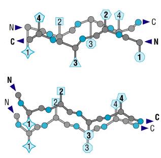
6 Illustration of motif overlaps in the mainly! sandwich architecture. Each structure shown can be related to the central tenascin structure by a motif containing at56-60 least four! strands (although these are not sequential in the transthyretin structure) up to seven! strands in plastocyanin and the immunoglobulin variable domain structures. It can be seen that this results in the possible merging of the immunoglobulin fold family (2rhe) and the jelly-roll fold family (1tnfA) ure 1997, Vol 5 No 8 through overlap of a large motif containing five! strands. This is not currently done in C ATH, as both families are commonly referred to as separate folds in the literature. 1TTF.pdb SHEET 1 SHEET 2 SHEET 3 SHEET 1 SHEET 2 SHEET 3 SHEET GLU A 9 THR A SER A 17 ASP A 23-1 O SER A 21 N GLU A THR A 56 SER A 60-1 N ALA A 57 O ILE A GLN A 46 PRO A TYR A 31 GLU A 38-1 N TYR A 36 O GLN A VAL A 66 THR A 76-1 N VAL A 75 O TYR A ILE A 88 THR A 94-1 N ILE A 88 O VAL A 72 Brandon and Tooze 23 24
. 110800 Domains.")
 Class Number of folds Number of superfamilies Number of")
 147 244 803 Alpha and beta")


 101 146 201 Multi-domain proteins 20 20 25 Membrane and cell")

7 25 26 SCOP: Structural Classification of Proteins release PDB Entries (23 Feb 2009) Domains. 1 Literature Reference (excluding nucleic acids and theoretical models) Class Number of folds Number of superfamilies Number of families All alpha proteins All beta proteins Alpha and beta proteins (a/b) Alpha and beta proteins (a+b) Multi-domain proteins Membrane and cell surface proteins Small proteins Total SCOP: Structural Classification of Proteins release 6497 PDB Entries (20 Oct 1997) Domains. 101 Literature References (including nucleic acids and theoretical models) Class Number of folds Number of superfamilies Number of families All alpha proteins All beta proteins Alpha and beta proteins (a/b) Alpha and beta proteins (a+b) Multi-domain proteins Membrane and cell surface proteins Small proteins Total


8 CATH numbering scheme for representative structures from the globin-like fold family in the mainly " class. Four of the seven levels within the CATH database are shown, associated with Class, Architecture, Topology, and Homology. Each level is associated with a unique number. The (A), (T) and (H) levels are numbered in bins of ten to allow expansion of the database. Class Architecture Topology Homology 1 Mainly " 2 Mainly! 10 Non-bundle 3 "! 20 Bundle Few SS 30 Few SS 470 Variant surface glycoprotein Glucoamylase, domain Globin-like!lactamase, domain 2 Casein kinase # hlm 20 1cpc chain A col chain A 40 1ddt domain Mainly ".Non-bundle.Globin-like.1cpc chain A 2BMF-Bovine ATPase F Table 1 The numbers of families identified at different levels in the CATH hierarchy is shown for the mainly ", mainly! and "$! classes. A T H S N I Domains Class Number % Number % Number % Number % Number % Number % Number % Mainly " Mainly! "! Few SS* Total *The number of families for proteins having few secondary structure (SS) elements is also shown at each level in the hierarchy. Chain A all beta Left handed superhelix P-loop containing nucleoside trip hydrolase Vogal et al: Current Opinion in Structural Biology 2004: 14:
remembering Secondary Structures Does everyone know what the backbone and residue/side chains are? Clear about 1, 2 3 structures?
 remembering Secondary Structures add blast Does everyone know what the backbone residue/side chains are? Clear about 1, 2 3 structures? Heteropolymer - + Mostly in regular secondary structure + - Secondary
CMPS 6630: Introduction to Computational Biology and Bioinformatics. Structure Comparison
 CMPS 6630: Introduction to Computational Biology and Bioinformatics Structure Comparison Protein Structure Comparison Motivation Understand sequence and structure variability Understand Domain architecture
Packing of Secondary Structures
 7.88 Lecture Notes - 4 7.24/7.88J/5.48J The Protein Folding and Human Disease Professor Gossard Retrieving, Viewing Protein Structures from the Protein Data Base Helix helix packing Packing of Secondary
Protein Structure: Data Bases and Classification Ingo Ruczinski
 Protein Structure: Data Bases and Classification Ingo Ruczinski Department of Biostatistics, Johns Hopkins University Reference Bourne and Weissig Structural Bioinformatics Wiley, 2003 More References
Procheck output. Bond angles (Procheck) Structure verification and validation Bond lengths (Procheck) Introduction to Bioinformatics.
 Structure verification and validation Bond lengths (Procheck) Introduction to Bioinformatics.") Structure verification and validation Bond lengths (Procheck) Introduction to Bioinformatics Iosif Vaisman Email: ivaisman@gmu.edu ----------------------------------------------------------------- Bond
Structure verification and validation Bond lengths (Procheck) Introduction to Bioinformatics Iosif Vaisman Email: ivaisman@gmu.edu ----------------------------------------------------------------- Bond
Secondary Structure. Bioch/BIMS 503 Lecture 2. Structure and Function of Proteins. Further Reading. Φ, Ψ angles alone determine protein structure
 Bioch/BIMS 503 Lecture 2 Structure and Function of Proteins August 28, 2008 Robert Nakamoto rkn3c@virginia.edu 2-0279 Secondary Structure Φ Ψ angles determine protein structure Φ Ψ angles are restricted
2MHR. Protein structure classification is important because it organizes the protein structure universe that is independent of sequence similarity.
 Protein structure classification is important because it organizes the protein structure universe that is independent of sequence similarity. A global picture of the protein universe will help us to understand
Supersecondary Structures (structural motifs)
") Supersecondary Structures (structural motifs) Various Sources Slide 1 Supersecondary Structures (Motifs) Supersecondary Structures (Motifs): : Combinations of secondary structures in specific geometric
Supersecondary Structures (structural motifs) Various Sources Slide 1 Supersecondary Structures (Motifs) Supersecondary Structures (Motifs): : Combinations of secondary structures in specific geometric
Introduction to Comparative Protein Modeling. Chapter 4 Part I
 Introduction to Comparative Protein Modeling Chapter 4 Part I 1 Information on Proteins Each modeling study depends on the quality of the known experimental data. Basis of the model Search in the literature
1. Protein Data Bank (PDB) 1. Protein Data Bank (PDB)
 1. Protein Data Bank (PDB)") Protein structure databases; visualization; and classifications 1. Introduction to Protein Data Bank (PDB) 2. Free graphic software for 3D structure visualization 3. Hierarchical classification of protein
Protein structure databases; visualization; and classifications 1. Introduction to Protein Data Bank (PDB) 2. Free graphic software for 3D structure visualization 3. Hierarchical classification of protein
Protein structure alignments
 Protein structure alignments Proteins that fold in the same way, i.e. have the same fold are often homologs. Structure evolves slower than sequence Sequence is less conserved than structure If BLAST gives
Physiochemical Properties of Residues
 Physiochemical Properties of Residues Various Sources C N Cα R Slide 1 Conformational Propensities Conformational Propensity is the frequency in which a residue adopts a given conformation (in a polypeptide)
BIRKBECK COLLEGE (University of London)
") BIRKBECK COLLEGE (University of London) SCHOOL OF BIOLOGICAL SCIENCES M.Sc. EXAMINATION FOR INTERNAL STUDENTS ON: Postgraduate Certificate in Principles of Protein Structure MSc Structural Molecular Biology
BIRKBECK COLLEGE (University of London) SCHOOL OF BIOLOGICAL SCIENCES M.Sc. EXAMINATION FOR INTERNAL STUDENTS ON: Postgraduate Certificate in Principles of Protein Structure MSc Structural Molecular Biology
Advanced Certificate in Principles in Protein Structure. You will be given a start time with your exam instructions
 BIRKBECK COLLEGE (University of London) Advanced Certificate in Principles in Protein Structure MSc Structural Molecular Biology Date: Thursday, 1st September 2011 Time: 3 hours You will be given a start
Objective: Students will be able identify peptide bonds in proteins and describe the overall reaction between amino acids that create peptide bonds.
 Scott Seiple AP Biology Lesson Plan Lesson: Primary and Secondary Structure of Proteins Purpose:. To understand how amino acids can react to form peptides through peptide bonds.. Students will be able
Model Mélange. Physical Models of Peptides and Proteins
 Model Mélange Physical Models of Peptides and Proteins In the Model Mélange activity, you will visit four different stations each featuring a variety of different physical models of peptides or proteins.
Major Types of Association of Proteins with Cell Membranes. From Alberts et al
 Major Types of Association of Proteins with Cell Membranes From Alberts et al Proteins Are Polymers of Amino Acids Peptide Bond Formation Amino Acid central carbon atom to which are attached amino group
Understanding Sequence, Structure and Function Relationships and the Resulting Redundancy
 Understanding Sequence, Structure and Function Relationships and the Resulting Redundancy many slides by Philip E. Bourne Department of Pharmacology, UCSD Agenda Understand the relationship between sequence,
The CATH Database provides insights into protein structure/function relationships
 1999 Oxford University Press Nucleic Acids Research, 1999, Vol. 27, No. 1 275 279 The CATH Database provides insights into protein structure/function relationships C. A. Orengo, F. M. G. Pearl, J. E. Bray,
CS612 - Algorithms in Bioinformatics
 Fall 2017 Databases and Protein Structure Representation October 2, 2017 Molecular Biology as Information Science > 12, 000 genomes sequenced, mostly bacterial (2013) > 5x10 6 unique sequences available
A General Model for Amino Acid Interaction Networks
 Author manuscript, published in "N/P" A General Model for Amino Acid Interaction Networks Omar GACI and Stefan BALEV hal-43269, version - Nov 29 Abstract In this paper we introduce the notion of protein
Supporting information to: Time-resolved observation of protein allosteric communication. Sebastian Buchenberg, Florian Sittel and Gerhard Stock 1
 Supporting information to: Time-resolved observation of protein allosteric communication Sebastian Buchenberg, Florian Sittel and Gerhard Stock Biomolecular Dynamics, Institute of Physics, Albert Ludwigs
Basic structures of proteins
 Basic structures of proteins Structural Hierarchy of Protein Primary structure Functional elements : α-helix, strands, β-sheet, loops.. - Structure, affinity, activity, specificity, stability etc. Secondary
D Dobbs ISU - BCB 444/544X 1
 11/7/05 Protein Structure: Classification, Databases, Visualization Announcements BCB 544 Projects - Important Dates: Nov 2 Wed noon - Project proposals due to David/Drena Nov 4 Fri PM - Approvals/responses
Protein Structure. Role of (bio)informatics in drug discovery. Bioinformatics
informatics in drug discovery. Bioinformatics") Bioinformatics Protein Structure Principles & Architecture Marjolein Thunnissen Dep. of Biochemistry & Structural Biology Lund University September 2011 Homology, pattern and 3D structure searches need
Bioinformatics Protein Structure Principles & Architecture Marjolein Thunnissen Dep. of Biochemistry & Structural Biology Lund University September 2011 Homology, pattern and 3D structure searches need
Giri Narasimhan. CAP 5510: Introduction to Bioinformatics. ECS 254; Phone: x3748
 CAP 5510: Introduction to Bioinformatics Giri Narasimhan ECS 254; Phone: x3748 giri@cis.fiu.edu www.cis.fiu.edu/~giri/teach/bioinfs07.html 2/15/07 CAP5510 1 EM Algorithm Goal: Find θ, Z that maximize Pr
HMM applications. Applications of HMMs. Gene finding with HMMs. Using the gene finder
 HMM applications Applications of HMMs Gene finding Pairwise alignment (pair HMMs) Characterizing protein families (profile HMMs) Predicting membrane proteins, and membrane protein topology Gene finding
SCOP. all-β class. all-α class, 3 different folds. T4 endonuclease V. 4-helical cytokines. Globin-like
 SCOP all-β class 4-helical cytokines T4 endonuclease V all-α class, 3 different folds Globin-like TIM-barrel fold α/β class Profilin-like fold α+β class http://scop.mrc-lmb.cam.ac.uk/scop CATH Class, Architecture,
Analysis and Prediction of Protein Structure (I)
") Analysis and Prediction of Protein Structure (I) Jianlin Cheng, PhD School of Electrical Engineering and Computer Science University of Central Florida 2006 Free for academic use. Copyright @ Jianlin Cheng
Analysis and Prediction of Protein Structure (I) Jianlin Cheng, PhD School of Electrical Engineering and Computer Science University of Central Florida 2006 Free for academic use. Copyright @ Jianlin Cheng
09/06/25. Computergestützte Strukturbiologie (Strukturelle Bioinformatik) Non-uniform distribution of folds. Scheme of protein structure predicition
 Non-uniform distribution of folds. Scheme of protein structure predicition") Sequence identity Structural similarity Computergestützte Strukturbiologie (Strukturelle Bioinformatik) Fold recognition Sommersemester 2009 Peter Güntert Structural similarity X Sequence identity Non-uniform
Sequence identity Structural similarity Computergestützte Strukturbiologie (Strukturelle Bioinformatik) Fold recognition Sommersemester 2009 Peter Güntert Structural similarity X Sequence identity Non-uniform
Structural Alignment of Proteins
 Goal Align protein structures Structural Alignment of Proteins 1 2 3 4 5 6 7 8 9 10 11 12 13 14 PHE ASP ILE CYS ARG LEU PRO GLY SER ALA GLU ALA VAL CYS PHE ASN VAL CYS ARG THR PRO --- --- --- GLU ALA ILE
Study of Mining Protein Structural Properties and its Application
 Study of Mining Protein Structural Properties and its Application A Dissertation Proposal Presented to the Department of Computer Science and Information Engineering College of Electrical Engineering and
Protein Structure & Motifs
 & Motifs Biochemistry 201 Molecular Biology January 12, 2000 Doug Brutlag Introduction Proteins are more flexible than nucleic acids in structure because of both the larger number of types of residues
Protein Structure Prediction and Display
 Protein Structure Prediction and Display Goal Take primary structure (sequence) and, using rules derived from known structures, predict the secondary structure that is most likely to be adopted by each
CAP 5510: Introduction to Bioinformatics CGS 5166: Bioinformatics Tools. Giri Narasimhan
 CAP 5510: Introduction to Bioinformatics CGS 5166: Bioinformatics Tools Giri Narasimhan ECS 254; Phone: x3748 giri@cis.fiu.edu www.cis.fiu.edu/~giri/teach/bioinff18.html Proteins and Protein Structure
Protein structure analysis. Risto Laakso 10th January 2005
 Protein structure analysis Risto Laakso risto.laakso@hut.fi 10th January 2005 1 1 Summary Various methods of protein structure analysis were examined. Two proteins, 1HLB (Sea cucumber hemoglobin) and 1HLM
Bahnson Biochemistry Cume, April 8, 2006 The Structural Biology of Signal Transduction
 Name page 1 of 6 Bahnson Biochemistry Cume, April 8, 2006 The Structural Biology of Signal Transduction Part I. The ion Ca 2+ can function as a 2 nd messenger. Pick a specific signal transduction pathway
Homology and Information Gathering and Domain Annotation for Proteins
 Homology and Information Gathering and Domain Annotation for Proteins Outline Homology Information Gathering for Proteins Domain Annotation for Proteins Examples and exercises The concept of homology The
Any protein that can be labelled by both procedures must be a transmembrane protein.
 1. What kind of experimental evidence would indicate that a protein crosses from one side of the membrane to the other? Regions of polypeptide part exposed on the outside of the membrane can be probed
ALL LECTURES IN SB Introduction
 1. Introduction 2. Molecular Architecture I 3. Molecular Architecture II 4. Molecular Simulation I 5. Molecular Simulation II 6. Bioinformatics I 7. Bioinformatics II 8. Prediction I 9. Prediction II ALL
Protein structure. Protein structure. Amino acid residue. Cell communication channel. Bioinformatics Methods
 Cell communication channel Bioinformatics Methods Iosif Vaisman Email: ivaisman@gmu.edu SEQUENCE STRUCTURE DNA Sequence Protein Sequence Protein Structure Protein structure ATGAAATTTGGAAACTTCCTTCTCACTTATCAGCCACCT...
Structure to Function. Molecular Bioinformatics, X3, 2006
 Structure to Function Molecular Bioinformatics, X3, 2006 Structural GeNOMICS Structural Genomics project aims at determination of 3D structures of all proteins: - organize known proteins into families
DATE A DAtabase of TIM Barrel Enzymes
 DATE A DAtabase of TIM Barrel Enzymes 2 2.1 Introduction.. 2.2 Objective and salient features of the database 2.2.1 Choice of the dataset.. 2.3 Statistical information on the database.. 2.4 Features....
Protein Data Bank Contents Guide: Atomic Coordinate Entry Format Description. Version Document Published by the wwpdb
 Protein Data Bank Contents Guide: Atomic Coordinate Entry Format Description Version 3.30 Document Published by the wwpdb This format complies with the PDB Exchange Dictionary (PDBx) http://mmcif.pdb.org/dictionaries/mmcif_pdbx.dic/index/index.html.
Lecture 10 (10/4/17) Lecture 10 (10/4/17)
 Lecture 10 (10/4/17)") Lecture 10 (10/4/17) Reading: Ch4; 125, 138-141, 141-142 Problems: Ch4 (text); 7, 9, 11 Ch4 (study guide); 1, 2 NEXT Reading: Ch4; 125, 132-136 (structure determination) Ch4; 12-130 (Collagen) Problems:
Lecture 10 (10/4/17) Reading: Ch4; 125, 138-141, 141-142 Problems: Ch4 (text); 7, 9, 11 Ch4 (study guide); 1, 2 NEXT Reading: Ch4; 125, 132-136 (structure determination) Ch4; 12-130 (Collagen) Problems:
Protein Structure Prediction
 Page 1 Protein Structure Prediction Russ B. Altman BMI 214 CS 274 Protein Folding is different from structure prediction --Folding is concerned with the process of taking the 3D shape, usually based on
Number sequence representation of protein structures based on the second derivative of a folded tetrahedron sequence
 Number sequence representation of protein structures based on the second derivative of a folded tetrahedron sequence Naoto Morikawa (nmorika@genocript.com) October 7, 2006. Abstract A protein is a sequence
Motif Prediction in Amino Acid Interaction Networks
 Motif Prediction in Amino Acid Interaction Networks Omar GACI and Stefan BALEV Abstract In this paper we represent a protein as a graph where the vertices are amino acids and the edges are interactions
114 Grundlagen der Bioinformatik, SS 09, D. Huson, July 6, 2009
 114 Grundlagen der Bioinformatik, SS 09, D. Huson, July 6, 2009 9 Protein tertiary structure Sources for this chapter, which are all recommended reading: D.W. Mount. Bioinformatics: Sequences and Genome
Properties of amino acids in proteins
 Properties of amino acids in proteins one of the primary roles of DNA (but not the only one!) is to code for proteins A typical bacterium builds thousands types of proteins, all from ~20 amino acids repeated
Protein Science (1997), 6: Cambridge University Press. Printed in the USA. Copyright 1997 The Protein Society
, 6: Cambridge University Press. Printed in the USA. Copyright 1997 The Protein Society") 1 of 5 1/30/00 8:08 PM Protein Science (1997), 6: 246-248. Cambridge University Press. Printed in the USA. Copyright 1997 The Protein Society FOR THE RECORD LPFC: An Internet library of protein family
1 of 5 1/30/00 8:08 PM Protein Science (1997), 6: 246-248. Cambridge University Press. Printed in the USA. Copyright 1997 The Protein Society FOR THE RECORD LPFC: An Internet library of protein family
COMP 598 Advanced Computational Biology Methods & Research. Introduction. Jérôme Waldispühl School of Computer Science McGill University
 COMP 598 Advanced Computational Biology Methods & Research Introduction Jérôme Waldispühl School of Computer Science McGill University General informations (1) Office hours: by appointment Office: TR3018
Basics of protein structure
 Today: 1. Projects a. Requirements: i. Critical review of one paper ii. At least one computational result b. Noon, Dec. 3 rd written report and oral presentation are due; submit via email to bphys101@fas.harvard.edu
Chapter 2 Structures. 2.1 Introduction Storing Protein Structures The PDB File Format
 Chapter 2 Structures 2.1 Introduction The three-dimensional (3D) structure of a protein contains a lot of information on its function, and can be used for devising ways of modifying it (propose mutants,
Sequential resonance assignments in (small) proteins: homonuclear method 2º structure determination
 proteins: homonuclear method 2º structure determination") Lecture 9 M230 Feigon Sequential resonance assignments in (small) proteins: homonuclear method 2º structure determination Reading resources v Roberts NMR of Macromolecules, Chap 4 by Christina Redfield
Lecture 9 M230 Feigon Sequential resonance assignments in (small) proteins: homonuclear method 2º structure determination Reading resources v Roberts NMR of Macromolecules, Chap 4 by Christina Redfield
Peptides And Proteins
 Kevin Burgess, May 3, 2017 1 Peptides And Proteins from chapter(s) in the recommended text A. Introduction B. omenclature And Conventions by amide bonds. on the left, right. 2 -terminal C-terminal triglycine
Central Dogma. modifications genome transcriptome proteome
 entral Dogma DA ma protein post-translational modifications genome transcriptome proteome 83 ierarchy of Protein Structure 20 Amino Acids There are 20 n possible sequences for a protein of n residues!
Basic Principles of Protein Structures
 Basic Principles of Protein Structures Proteins Proteins: The Molecule of Life Proteins: Building Blocks Proteins: Secondary Structures Proteins: Tertiary and Quartenary Structure Proteins: Geometry Proteins
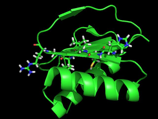
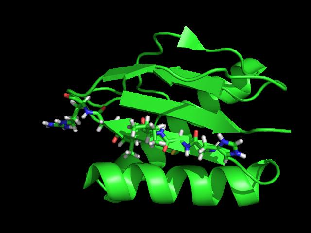
Supplementary Figure 3 a. Structural comparison between the two determined structures for the IL 23:MA12 complex. The overall RMSD between the two
 Supplementary Figure 1. Biopanningg and clone enrichment of Alphabody binders against human IL 23. Positive clones in i phage ELISA with optical density (OD) 3 times higher than background are shown for
Supplementary Figure 1. Aligned sequences of yeast IDH1 (top) and IDH2 (bottom) with isocitrate
 and IDH2 (bottom) with isocitrate") SUPPLEMENTARY FIGURE LEGENDS Supplementary Figure 1. Aligned sequences of yeast IDH1 (top) and IDH2 (bottom) with isocitrate dehydrogenase from Escherichia coli [ICD, pdb 1PB1, Mesecar, A. D., and Koshland,
SUPPLEMENTARY FIGURE LEGENDS Supplementary Figure 1. Aligned sequences of yeast IDH1 (top) and IDH2 (bottom) with isocitrate dehydrogenase from Escherichia coli [ICD, pdb 1PB1, Mesecar, A. D., and Koshland,
Bioinformatics Practical for Biochemists
 Bioinformatics Practical for Biochemists Andrei Lupas, Birte Höcker, Steffen Schmidt WS 2013/14 03. Sequence Features Targeting proteins signal peptide targets proteins to the secretory pathway N-terminal
NUCLEOTIDE BINDING ENZYMES
 NUCLEOTIDE BINDING ENZYMES The Rossmann fold Relationship between sequence, structure and function. Anna Casas, Júlia Gasull and Nerea Vega Index 1. Introduction: Adenine nucleotides 1. The most commonly
B O C 4 H 2 O O. NOTE: The reaction proceeds with a carbonium ion stabilized on the C 1 of sugar A.
 hbcse 33 rd International Page 101 hemistry lympiad Preparatory 05/02/01 Problems d. In the hydrolysis of the glycosidic bond, the glycosidic bridge oxygen goes with 4 of the sugar B. n cleavage, 18 from
Homology. and. Information Gathering and Domain Annotation for Proteins
 Homology and Information Gathering and Domain Annotation for Proteins Outline WHAT IS HOMOLOGY? HOW TO GATHER KNOWN PROTEIN INFORMATION? HOW TO ANNOTATE PROTEIN DOMAINS? EXAMPLES AND EXERCISES Homology
Analysis on sliding helices and strands in protein structural comparisons: A case study with protein kinases
 Sliding helices and strands in structural comparisons 921 Analysis on sliding helices and strands in protein structural comparisons: A case study with protein kinases V S GOWRI, K ANAMIKA, S GORE 1 and
Details of Protein Structure
 Details of Protein Structure Function, evolution & experimental methods Thomas Blicher, Center for Biological Sequence Analysis Anne Mølgaard, Kemisk Institut, Københavns Universitet Learning Objectives
Hidden symmetries in primary sequences of small α proteins
 Hidden symmetries in primary sequences of small α proteins Ruizhen Xu, Yanzhao Huang, Mingfen Li, Hanlin Chen, and Yi Xiao * Biomolecular Physics and Modeling Group, Department of Physics, Huazhong University
1. What is an ångstrom unit, and why is it used to describe molecular structures?
 1. What is an ångstrom unit, and why is it used to describe molecular structures? The ångstrom unit is a unit of distance suitable for measuring atomic scale objects. 1 ångstrom (Å) = 1 10-10 m. The diameter
Protein Structure. Hierarchy of Protein Structure. Tertiary structure. independently stable structural unit. includes disulfide bonds
 Protein Structure Hierarchy of Protein Structure 2 3 Structural element Primary structure Secondary structure Super-secondary structure Domain Tertiary structure Quaternary structure Description amino
Supplemental Materials for. Structural Diversity of Protein Segments Follows a Power-law Distribution
 Supplemental Materials for Structural Diversity of Protein Segments Follows a Power-law Distribution Yoshito SAWADA and Shinya HONDA* National Institute of Advanced Industrial Science and Technology (AIST),
MSAT a Multiple Sequence Alignment tool based on TOPS
 MSAT a Multiple Sequence Alignment tool based on TOPS Te Ren, Mallika Veeramalai, Aik Choon Tan and David Gilbert Bioinformatics Research Centre Department of Computer Science University of Glasgow Glasgow,
Getting To Know Your Protein
 Getting To Know Your Protein Comparative Protein Analysis: Part III. Protein Structure Prediction and Comparison Robert Latek, PhD Sr. Bioinformatics Scientist Whitehead Institute for Biomedical Research
Today. Last time. Secondary structure Transmembrane proteins. Domains Hidden Markov Models. Structure prediction. Secondary structure
 Last time Today Domains Hidden Markov Models Structure prediction NAD-specific glutamate dehydrogenase Hard Easy >P24295 DHE2_CLOSY MSKYVDRVIAEVEKKYADEPEFVQTVEEVL SSLGPVVDAHPEYEEVALLERMVIPERVIE FRVPWEDDNGKVHVNTGYRVQFNGAIGPYK
Intro Secondary structure Transmembrane proteins Function End. Last time. Domains Hidden Markov Models
 Last time Domains Hidden Markov Models Today Secondary structure Transmembrane proteins Structure prediction NAD-specific glutamate dehydrogenase Hard Easy >P24295 DHE2_CLOSY MSKYVDRVIAEVEKKYADEPEFVQTVEEVL
What makes a good graphene-binding peptide? Adsorption of amino acids and peptides at aqueous graphene interfaces: Electronic Supplementary
 Electronic Supplementary Material (ESI) for Journal of Materials Chemistry B. This journal is The Royal Society of Chemistry 21 What makes a good graphene-binding peptide? Adsorption of amino acids and
Ranjit P. Bahadur Assistant Professor Department of Biotechnology Indian Institute of Technology Kharagpur, India. 1 st November, 2013
 Hydration of protein-rna recognition sites Ranjit P. Bahadur Assistant Professor Department of Biotechnology Indian Institute of Technology Kharagpur, India 1 st November, 2013 Central Dogma of life DNA
PROTEIN SECONDARY STRUCTURE PREDICTION: AN APPLICATION OF CHOU-FASMAN ALGORITHM IN A HYPOTHETICAL PROTEIN OF SARS VIRUS
 Int. J. LifeSc. Bt & Pharm. Res. 2012 Kaladhar, 2012 Research Paper ISSN 2250-3137 www.ijlbpr.com Vol.1, Issue. 1, January 2012 2012 IJLBPR. All Rights Reserved PROTEIN SECONDARY STRUCTURE PREDICTION:
Review. Membrane proteins. Membrane transport
 Quiz 1 For problem set 11 Q1, you need the equation for the average lateral distance transversed (s) of a molecule in the membrane with respect to the diffusion constant (D) and time (t). s = (4 D t) 1/2
Orientational degeneracy in the presence of one alignment tensor.
 Orientational degeneracy in the presence of one alignment tensor. Rotation about the x, y and z axes can be performed in the aligned mode of the program to examine the four degenerate orientations of two
Membrane proteins Porins: FadL. Oriol Solà, Dimitri Ivancic, Daniel Folch, Marc Olivella
 Membrane proteins Porins: FadL Oriol Solà, Dimitri Ivancic, Daniel Folch, Marc Olivella INDEX 1. INTRODUCTION TO MEMBRANE PROTEINS 2. FADL: OUTER MEMBRANE TRANSPORT PROTEIN 3. MAIN FEATURES OF FADL STRUCTURE
EBI web resources II: Ensembl and InterPro
 EBI web resources II: Ensembl and InterPro Yanbin Yin http://www.ebi.ac.uk/training/online/course/ 1 Homework 3 Go to http://www.ebi.ac.uk/interpro/training.htmland finish the second online training course
Large-Scale Genomic Surveys
 Bioinformatics Subtopics Fold Recognition Secondary Structure Prediction Docking & Drug Design Protein Geometry Structural Informatics Homology Modeling Sequence Alignment Structure Classification Gene
Structure and evolution of the spliceosomal peptidyl-prolyl cistrans isomerase Cwc27
 Acta Cryst. (2014). D70, doi:10.1107/s1399004714021695 Supporting information Volume 70 (2014) Supporting information for article: Structure and evolution of the spliceosomal peptidyl-prolyl cistrans isomerase
Translation. A ribosome, mrna, and trna.
 Translation The basic processes of translation are conserved among prokaryotes and eukaryotes. Prokaryotic Translation A ribosome, mrna, and trna. In the initiation of translation in prokaryotes, the Shine-Dalgarno
Identification of Representative Protein Sequence and Secondary Structure Prediction Using SVM Approach
 Identification of Representative Protein Sequence and Secondary Structure Prediction Using SVM Approach Prof. Dr. M. A. Mottalib, Md. Rahat Hossain Department of Computer Science and Information Technology
Protein Structure Bioinformatics Introduction
 1 Swiss Institute of Bioinformatics Protein Structure Bioinformatics Introduction Basel, 27. September 2004 Torsten Schwede Biozentrum - Universität Basel Swiss Institute of Bioinformatics Klingelbergstr
Protein Structure Prediction II Lecturer: Serafim Batzoglou Scribe: Samy Hamdouche
 Protein Structure Prediction II Lecturer: Serafim Batzoglou Scribe: Samy Hamdouche The molecular structure of a protein can be broken down hierarchically. The primary structure of a protein is simply its
Amino Acid Structures from Klug & Cummings. 10/7/2003 CAP/CGS 5991: Lecture 7 1
 Amino Acid Structures from Klug & Cummings 10/7/2003 CAP/CGS 5991: Lecture 7 1 Amino Acid Structures from Klug & Cummings 10/7/2003 CAP/CGS 5991: Lecture 7 2 Amino Acid Structures from Klug & Cummings
Massachusetts Institute of Technology Computational Evolutionary Biology, Fall, 2005 Notes for November 7: Molecular evolution
 Massachusetts Institute of Technology 6.877 Computational Evolutionary Biology, Fall, 2005 Notes for November 7: Molecular evolution 1. Rates of amino acid replacement The initial motivation for the neutral
SUPPLEMENTARY INFORMATION
 doi:10.1038/nature17991 Supplementary Discussion Structural comparison with E. coli EmrE The DMT superfamily includes a wide variety of transporters with 4-10 TM segments 1. Since the subfamilies of the
1-D Predictions. Prediction of local features: Secondary structure & surface exposure
 1-D Predictions Prediction of local features: Secondary structure & surface exposure 1 Learning Objectives After today s session you should be able to: Explain the meaning and usage of the following local
Outline. Levels of Protein Structure. Primary (1 ) Structure. Lecture 6:Protein Architecture II: Secondary Structure or From peptides to proteins
 Structure. Lecture 6:Protein Architecture II: Secondary Structure or From peptides to proteins") Lecture 6:Protein Architecture II: Secondary Structure or From peptides to proteins Margaret Daugherty Fall 2003 Outline Four levels of structure are used to describe proteins; Alpha helices and beta sheets
Lecture 6:Protein Architecture II: Secondary Structure or From peptides to proteins Margaret Daugherty Fall 2003 Outline Four levels of structure are used to describe proteins; Alpha helices and beta sheets
Genome Databases The CATH database
 Genome Databases The CATH database Michael Knudsen 1 and Carsten Wiuf 1,2* 1 Bioinformatics Research Centre, Aarhus University, DK-8000 Aarhus C, Denmark 2 Centre for Membrane Pumps in Cells and Disease
Bioinformatics. Macromolecular structure
 Bioinformatics Macromolecular structure Contents Determination of protein structure Structure databases Secondary structure elements (SSE) Tertiary structure Structure analysis Structure alignment Domain
Sequence analysis and comparison
 The aim with sequence identification: Sequence analysis and comparison Marjolein Thunnissen Lund September 2012 Is there any known protein sequence that is homologous to mine? Are there any other species
Amino Acids: General Properties
 Lehninger: Principles of Biochemistry, 4th Ed., D. Nelson and M. Cox, 2005, p. 83. Amino Acids: General Properties R R pk a of the α-carboxylic acid groups are near 2.2.* H 2 N C α H CO 2 H + H 3 N C α
CAP 5510 Lecture 3 Protein Structures
 CAP 5510 Lecture 3 Protein Structures Su-Shing Chen Bioinformatics CISE 8/19/2005 Su-Shing Chen, CISE 1 Protein Conformation 8/19/2005 Su-Shing Chen, CISE 2 Protein Conformational Structures Hydrophobicity
Lecture 10: Cyclins, cyclin kinases and cell division
 Chem*3560 Lecture 10: Cyclins, cyclin kinases and cell division The eukaryotic cell cycle Actively growing mammalian cells divide roughly every 24 hours, and follow a precise sequence of events know as
Computer simulations of protein folding with a small number of distance restraints
 Vol. 49 No. 3/2002 683 692 QUARTERLY Computer simulations of protein folding with a small number of distance restraints Andrzej Sikorski 1, Andrzej Kolinski 1,2 and Jeffrey Skolnick 2 1 Department of Chemistry,
Supporting Online Material for
 www.sciencemag.org/cgi/content/full/309/5742/1868/dc1 Supporting Online Material for Toward High-Resolution de Novo Structure Prediction for Small Proteins Philip Bradley, Kira M. S. Misura, David Baker*