Introduction to Comparative Protein Modeling. Chapter 4 Part I
|
|
|
- Penelope Newman
- 5 years ago
- Views:
Transcription
1 Introduction to Comparative Protein Modeling Chapter 4 Part I 1
2 Information on Proteins Each modeling study depends on the quality of the known experimental data. Basis of the model Search in the literature May find 3D structure on the protein of interest When you have a sequence Compare it to other to find similarities or differences Several algorithms have been developed Comparison within a few minutes Differences between a healthy and a diseased individual. 2
3 Databases Nucleotide and Protein Sequences: EMBL (European Molecular Biology Laboratory) Nucleotide Sequence Database. Universal Protein Resource (UniPort) Database. 3D structural Information: Protein Data Bank, PDB. Search on author's name, journal name or a part of a sequence. 3
4 PDB file The format of the data files are similar. PDB files are widely used I will describe the standard format of a protein file Header General information about the protein Includes official name, references, resolution of the crystal structure and other useful remarks Atomic coordinates Atoms belonging to standard amino acids are labeled ATOM To distinguish between individual peptide chains ATOMS are separated by TER Non standard amino acids are labeled HETATMS 4
5 PDB file When the file is read in a modeling program bonds are built between ATOMS but not between HETATMS An additional connectivity table is at the end of the data file Atom type of HETATMS is often incorrect when reading into a modeling program necessary to check all atom types PDB do not include hydrogen atoms. Keep in mind: The resolution of the crystal structure should be least between 2.5 and 1.5 Å As NMR measurements are performed in solution the results are highly dependent on the solvent 5
6 Protein Structure The 3D structure of proteins is characterized by 4 levels of structural organization: The primary structure represents the linear arrangement of the amino acids in the protein sequence The secondary structure describes the local architecture of the linear fragments of the chain (α-helix, β-sheet). The supersecondary structure is a new level, which describes the association of the secondary elements through the side chain interactions. Also called a motif (hairpins, Greek key..) The tertiary structure shows the overall topology of the folded peptide chain The quaternary structure describes the arrangement of separate subunits in the functional protein 6
7 Conformational Properties of Proteins 20 different amino acids found in nature Physicochemical properties of their side chains (size, shape, hydrophobicity, charge and hydrogen bonding) span a considerable range They have strongly restricted degrees of freedom The dominant influences on protein conformation is: Hydrogen bonding capabilities Chirality of the amino acids. All 19 chiral amino acids (glycine are not chiral) possess a L-configuration Linear connectivity Steric volume 7
8 Conformational Properties of Proteins Central carbon, C α Backbone: amino N, C α and the carbonyl C. i is the number of residue, starting from the amino end of the chain Main chain torsion angles: Φ: N-C α Ψ: C α -C ω: C -N (peptide bond) χ: side chain 8
9 Conformational Properties of Proteins The peptide bon is planar. Nearly always trans configuration (ω = 180 ) which is more energetically favorable than cis (ω = 0 ) The imino Proline is sometimes found in cis 9

10 Conformational Properties of Proteins Rotation about ϕ and ψ: Makes the peptide chain flexible Constrained geometrically due to steric hindrance between neighboring atoms Conformations of ϕ and ψ Ramachandran plot White: Disallowed region Red: Favored region Yellow: Allowed region Sub-regions: α and β 10

11 Types of Secondary Structural α-helix: Best known and most easily recognized structure Repetitive structure: C α -atoms in identical relative positions Thus the ϕ and ψ angles are the same for each residue in the helix Repeats itself every 5.4 Å 3.6 amino acids per turn Hydrogen-bonds between carbonyl of residue n and NH of residue n+4 regular and favored state Right-handed helix due to L-amino acids Side chains point outwards Length: residues Elements 11
12 Types of Secondary Structural β-sheet: Second most regular and recognizable structure Periodic element formed from β- strands Hydrogen-bonds are intermolecular therefore are β-sheets less favorable Parallel: All strands run in the same direction Anti-parallel: Run in the opposite direction. Most common Side chains are perpendicular to the plane of hydrogen-bonds Length of stand: 3-10 residues Elements 12

13 Turns: Types of Secondary Structural Elements 1/3 of all residues of glubular peoteins are involved in turns General function: Reverse the direction of the peptide chain Located on the protein surface charged and polar amino acids Turns often connect anti-parallel β-strands named β-turns or hairpin bends. They often only contain 2 residues Schematic form: Very helpful tool to se and understand the overall structure of a protein Side chains are often omitted to give a more clear picture 13

14 Homologous Proteins Proteins that have evolved from a common ancestor are said to be homologous. The 3D structure for homologous proteins are more preserved than the identity The structure is crucial for the function Trypsin and α-chymotrypsin belongs to the serine protease family. Has only 44 % identity but they are very homolog Dissimilarities: Loop regions. The core is more preserved Homologous proteins appear to be highly conserved during evolution basis for comparative protein modeling 14
15 Comparative Protein Modeling Known sequences vs. known 3D structures: Protein sequence determination is much faster than determining the 3D structure from X-ray or NMR theoretical procedures for predicting the 3D structure on the basis of the sequence is needed No general rule for folding of a protein base structural predictions on the conformation of available homologous reference proteins Use Comparative Protein Modeling approach when: A sequence is found homologous to another with a known 3D structure, then this method is used to predict the structure for the unknown protein Also called Homology Modeling Approach 15
16 Process: Comparative Protein Modeling 1. Determination of proteins which are related to the protein being studied Sequence alignment 2. Identification of structurally conserved regions (SCRs) and structurally variable regions (SVRs) 3. Alignment of the sequence of the unknown protein with those of the reference protein(s) within the SCRs 4. Construction of SCRs of the target protein using coordinates from the template structure(s) 5. Construction of the SVRs 6. Side chain modeling 7. Structural refinement using energy minimization methods and molecular dynamics 16
17 Sequence Alignment Sequence alignment by data base search: Major methods: FASTA and BLAST Used in many available software: HOMOLOGY, MODELLER Sequence alignment important because: Find related sequences Identify conserved regions Find amino acids of the known reference protein that correspond with those of the protein to be modeled basis for transferring the coordinates of the reference protein to the new protein. Need more sensitive and selective alignment procedures Needleman and Wunsch algorithm (align two sequences) 17
18 Sequence Alignment Optimal local alignment: Best local identity between two sequences Only consider relatively conserved subsequences Important tool for comparing sequences 18
19 Sequence Alignment Scoring Scheme: Indicates the weight for substituting one amino acid with another matrices High no: Substitution is likely Low no: Substitution is unlikely Different kind of Matrices: Identity matix: Most simple, gives 1 to identical pairs and 0 to all others Codon substitution matix: Scoring values are derives from codons. Identical amino acids get 9, íf one mutation is required the score is 3 and if two is required the score is 1 Mutation or Dayhoff matrix: Obtained by counting the number of substitutions from one amino acid by others observed in related proteins, across species. Larges scores are given to substitutions that are frequently, and low scores to substitutions which are not observed. 19

20 Dayhoff matrix: Larger scores for some non-identical mutations than for some identical one Statistic method Sequence Alignment 20
21 Gabs: Sequence Alignment If there is differences in the sequence length or variations in the location of conserved regions it complicate the alignment gabs are introduced An additional factor is introduced in the alignment algorithms gap penalty function The balance between the number of aligned amino acids and the smallest number of required gaps leads to an optimal alignment Combination of an alignment algorithm, a scoring matrix and a gab function: Optimal alignment of two or more sequences The quality of the alignment is described by an alignment score The derived alignment can only be used as a basis for a protein model if it agrees with all known structural data 21
22 Problems: Sequence Alignment Sequence similarity is lost more quickly during evolution than the structural similarities This makes it difficult to makes some simple rules Investigations to solve problems: Doolittle et al.: Rules of thumb. Sequences are longer than 100 residues and are found to be more than 25% identical very likely to be related. If the identity is 15-25% the sequences may still be related and if the identity is less than 15% they are probably not related Chothis and Lesk: To have success in modeling the structure of a protein from its sequence, using the 3D structure of a homologous protein as template depends very much on the sequence identity above 50% 22
23 Determination and Generation of Structurally Conserved Regions (SCRs) When building a model protein using the homology approach, it is based on the fact that there are regions in all proteins that belong to the same family, that are nearly identical in there 3D structures These regions tend to be located at the inner core of the protein SCRs: These regions in strongly related proteins have the same relative orientations of their secondary structural units in space (α-helices and β-sheets) throughout the whole family Used as a natural framework for the atomic coordinates for another protein in the family 23
24 Determination and Generation of Structurally Conserved Regions (SCRs) Find SCRs within a family: Depends on the number of available crystal structures of related proteins If more than one crystal structure is available superimpose them relative to each other. Done by a least-square fitting method Problem to selection the fitting atoms Fit by the C α -atoms. This method can then be optimized by using only matching points located in the secondary structure The resulting superimposed 3D structures tend to show that large parts of the two proteins are very similar and they appear to be the SCRs, while other regions differ vary much Keep in mind: The definition of SCRs is that a SCR must be terminated at the end of a secondary structural unit, so therefore the secondary structural elements must be assigned for the protein Crystal files Programs like DSSP or STRIDE (based on the H-bonding pattern or the main chain dihedral angle) 24
25 25 Determination and Generation of Structurally Conserved Regions (SCRs) Find SCRs within a family: If only one crystal structure is available detect SCRs manually using both sequence and structural information of the proteins Residues in the core are more conserved than residues on the surface Amino acids involved in hydrogen bonds and disulfide bridges are most likely to be conserved within the protein family. Also the residues in the active site tend to be conserved If the SCRs of the reference protein are known: Find the regions on the model protein that corresponds to the SCRs Done by an alignment of the target sequence with the sequences of the SCRs No gabs are allowed in the SCRs, so different programs are needed After the alignment the coordinates for the SCRs can be assigned, by use of the coordinates of the reference protein as a basis Segments with identical side chains: All coordinates are used Segments with non identical side chains: Only backbone coordinates
26 SVRs: Construction of Structurally Variable Occur normally in loop regions Construction of these are more difficult Regions (SVRs) Insertions and deletions make the modeling procedure complicated due to variations in the number of amino acids A good guide for modeling a missing region: Use a segment of similar length in a homologous protein Studies have showed that when loops has the same length and amino acid character, their conformation will be the same The coordinates can then be transferred to the model protein 26
27 Construction of Structurally Variable Regions (SVRs) If no comparable loops exits in the protein family The coordinates for the SVRs can be retrieved from: Loop search method: A peptide segment which are found in other proteins and fit into the model s spatial environment are used De novo generation: Generation a loop segment de novo. A peptide chain is built between two conserved segments using randomly generated values for all backbone dihedral angles. Rather complex so can only be used when the loop is smaller than seven residues All loops should be refines by an energy minimization in order to remove steric hindrance and relax the loop conformations 27
28 Side-Chain Modeling Backbone is constructed, the next step is to add the side chains: The predictions of the side chain conformations is a much more complex process Many of the side chains have one or more degrees of freedom can adopt many energetically allowed conformations It has been generally assumed that identical residues in homologous proteins adopt similar conformations Side chain with amino acids that shows high similarity (Isoleucine and Leucine or Valine) are also assumed to adopt the same orientation in the protein Difficult when the substituted amino acids are not related Showed that side chains usually adopt only a small number of the many possible conformations Statistical rotamer libraries Still difficult due to the conformations depends on the local environment 28
29 Final Model A refinement of the final model is often desirable due to: Regions where SCRs and SVRs are connected often have a lot of steric strain and need to be minimized Several side chains also adopt positions which has a bad van derwaals contact A stepwise refinement is needed, because an approach on all the residues at once will destroy important internal hydrogen bonds 29
30 Secondary Structure Prediction In the case where a homologous protein does not exist, methods for predictions the secondary structure have been developed 90% of the residues are either in α-helices, β-sheets or reverse turns If these are predicted it seems possible to combine the segments complete protein structure Not as reliable as homology modeling Three different methods: Statistical Stereochemical Neutral network-based 30
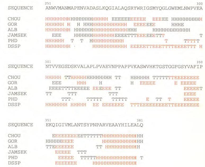
31 Secondary Structure Prediction Statistical method: First to be developed Idea: Many of the 20 amino acids have preferred secondary structures Ala, Arg, Gln, Glu, Met, Leu and Lys: α-helix Cys, Ile, Phe, Thr, Tyr and Val: β-sheets The most simple method is proposed by Chou and Fasman Calculating the probability of which secondary structure an amino acids is in by its frequency in the different structures found in the PDB Limitations: Below 56% accurate in predicting helix, sheets and loops 31
32 Secondary Structure Prediction Stereochemical: Based on the hydrophobic, hydrophilic and electrostatic properties of the side chains The method of Lim: Takes into account the interactions between side chains separated with up to 3 residues in the sequence, in view of their packing behavior If a sequence have alternating hydrophobic and hydrophilic side chains likely to be found in a β-sheet hydrophilic residues exposed to the solvent and the hydrophobic residues buried in the interior of the protein Neutral-based: Uses neutral networks, which can be trained rules are not need in advance but they are formed by the network itself, based on known facts More than 70% accuracy in the prediction of three classes of secondary structures on the basis of just one known homologous sequence 32
33 33 H
34 Fold recognition/threading Methods Use when: The structural similarity is limited to only the part of the structure having a common structural motif, and the rest is completely different First methods: Recognize folds in the absence of sequence similarity. Now: Comparative modeling and threading approaches are done simultaneously Close related to ab initio methods, but are limited to search for conformations of known structures Thus, threading methods fail for any protein that adopts a new fold 34
Physiochemical Properties of Residues
 Physiochemical Properties of Residues Various Sources C N Cα R Slide 1 Conformational Propensities Conformational Propensity is the frequency in which a residue adopts a given conformation (in a polypeptide)
Secondary Structure. Bioch/BIMS 503 Lecture 2. Structure and Function of Proteins. Further Reading. Φ, Ψ angles alone determine protein structure
 Bioch/BIMS 503 Lecture 2 Structure and Function of Proteins August 28, 2008 Robert Nakamoto rkn3c@virginia.edu 2-0279 Secondary Structure Φ Ψ angles determine protein structure Φ Ψ angles are restricted
Protein structure. Protein structure. Amino acid residue. Cell communication channel. Bioinformatics Methods
 Cell communication channel Bioinformatics Methods Iosif Vaisman Email: ivaisman@gmu.edu SEQUENCE STRUCTURE DNA Sequence Protein Sequence Protein Structure Protein structure ATGAAATTTGGAAACTTCCTTCTCACTTATCAGCCACCT...
CAP 5510 Lecture 3 Protein Structures
 CAP 5510 Lecture 3 Protein Structures Su-Shing Chen Bioinformatics CISE 8/19/2005 Su-Shing Chen, CISE 1 Protein Conformation 8/19/2005 Su-Shing Chen, CISE 2 Protein Conformational Structures Hydrophobicity
Properties of amino acids in proteins
 Properties of amino acids in proteins one of the primary roles of DNA (but not the only one!) is to code for proteins A typical bacterium builds thousands types of proteins, all from ~20 amino acids repeated
Section Week 3. Junaid Malek, M.D.
 Section Week 3 Junaid Malek, M.D. Biological Polymers DA 4 monomers (building blocks), limited structure (double-helix) RA 4 monomers, greater flexibility, multiple structures Proteins 20 Amino Acids,
Peptides And Proteins
 Kevin Burgess, May 3, 2017 1 Peptides And Proteins from chapter(s) in the recommended text A. Introduction B. omenclature And Conventions by amide bonds. on the left, right. 2 -terminal C-terminal triglycine
HOMOLOGY MODELING. The sequence alignment and template structure are then used to produce a structural model of the target.
 HOMOLOGY MODELING Homology modeling, also known as comparative modeling of protein refers to constructing an atomic-resolution model of the "target" protein from its amino acid sequence and an experimental
Secondary and sidechain structures
 Lecture 2 Secondary and sidechain structures James Chou BCMP201 Spring 2008 Images from Petsko & Ringe, Protein Structure and Function. Branden & Tooze, Introduction to Protein Structure. Richardson, J.
Read more about Pauling and more scientists at: Profiles in Science, The National Library of Medicine, profiles.nlm.nih.gov
 2018 Biochemistry 110 California Institute of Technology Lecture 2: Principles of Protein Structure Linus Pauling (1901-1994) began his studies at Caltech in 1922 and was directed by Arthur Amos oyes to
Supersecondary Structures (structural motifs)
") Supersecondary Structures (structural motifs) Various Sources Slide 1 Supersecondary Structures (Motifs) Supersecondary Structures (Motifs): : Combinations of secondary structures in specific geometric
Supersecondary Structures (structural motifs) Various Sources Slide 1 Supersecondary Structures (Motifs) Supersecondary Structures (Motifs): : Combinations of secondary structures in specific geometric
Basics of protein structure
 Today: 1. Projects a. Requirements: i. Critical review of one paper ii. At least one computational result b. Noon, Dec. 3 rd written report and oral presentation are due; submit via email to bphys101@fas.harvard.edu
Protein Structure Bioinformatics Introduction
 1 Swiss Institute of Bioinformatics Protein Structure Bioinformatics Introduction Basel, 27. September 2004 Torsten Schwede Biozentrum - Universität Basel Swiss Institute of Bioinformatics Klingelbergstr
B O C 4 H 2 O O. NOTE: The reaction proceeds with a carbonium ion stabilized on the C 1 of sugar A.
 hbcse 33 rd International Page 101 hemistry lympiad Preparatory 05/02/01 Problems d. In the hydrolysis of the glycosidic bond, the glycosidic bridge oxygen goes with 4 of the sugar B. n cleavage, 18 from
Major Types of Association of Proteins with Cell Membranes. From Alberts et al
 Major Types of Association of Proteins with Cell Membranes From Alberts et al Proteins Are Polymers of Amino Acids Peptide Bond Formation Amino Acid central carbon atom to which are attached amino group
Examples of Protein Modeling. Protein Modeling. Primary Structure. Protein Structure Description. Protein Sequence Sources. Importing Sequences to MOE
 Examples of Protein Modeling Protein Modeling Visualization Examination of an experimental structure to gain insight about a research question Dynamics To examine the dynamics of protein structures To
Protein Structures: Experiments and Modeling. Patrice Koehl
 Protein Structures: Experiments and Modeling Patrice Koehl Structural Bioinformatics: Proteins Proteins: Sources of Structure Information Proteins: Homology Modeling Proteins: Ab initio prediction Proteins:
Using Higher Calculus to Study Biologically Important Molecules Julie C. Mitchell
 Using Higher Calculus to Study Biologically Important Molecules Julie C. Mitchell Mathematics and Biochemistry University of Wisconsin - Madison 0 There Are Many Kinds Of Proteins The word protein comes
Design of a Novel Globular Protein Fold with Atomic-Level Accuracy
 Design of a Novel Globular Protein Fold with Atomic-Level Accuracy Brian Kuhlman, Gautam Dantas, Gregory C. Ireton, Gabriele Varani, Barry L. Stoddard, David Baker Presented by Kate Stafford 4 May 05 Protein
Homology Modeling (Comparative Structure Modeling) GBCB 5874: Problem Solving in GBCB
 GBCB 5874: Problem Solving in GBCB") Homology Modeling (Comparative Structure Modeling) Aims of Structural Genomics High-throughput 3D structure determination and analysis To determine or predict the 3D structures of all the proteins encoded
Homology Modeling (Comparative Structure Modeling) Aims of Structural Genomics High-throughput 3D structure determination and analysis To determine or predict the 3D structures of all the proteins encoded
Central Dogma. modifications genome transcriptome proteome
 entral Dogma DA ma protein post-translational modifications genome transcriptome proteome 83 ierarchy of Protein Structure 20 Amino Acids There are 20 n possible sequences for a protein of n residues!
Outline. Levels of Protein Structure. Primary (1 ) Structure. Lecture 6:Protein Architecture II: Secondary Structure or From peptides to proteins
 Structure. Lecture 6:Protein Architecture II: Secondary Structure or From peptides to proteins") Lecture 6:Protein Architecture II: Secondary Structure or From peptides to proteins Margaret Daugherty Fall 2004 Outline Four levels of structure are used to describe proteins; Alpha helices and beta sheets
Lecture 6:Protein Architecture II: Secondary Structure or From peptides to proteins Margaret Daugherty Fall 2004 Outline Four levels of structure are used to describe proteins; Alpha helices and beta sheets
Details of Protein Structure
 Details of Protein Structure Function, evolution & experimental methods Thomas Blicher, Center for Biological Sequence Analysis Anne Mølgaard, Kemisk Institut, Københavns Universitet Learning Objectives
CS612 - Algorithms in Bioinformatics
 Fall 2017 Protein Structure Detection Methods October 30, 2017 Comparative Modeling Comparative modeling is modeling of the unknown based on comparison to what is known In the context of modeling or computing
Protein Structure Prediction II Lecturer: Serafim Batzoglou Scribe: Samy Hamdouche
 Protein Structure Prediction II Lecturer: Serafim Batzoglou Scribe: Samy Hamdouche The molecular structure of a protein can be broken down hierarchically. The primary structure of a protein is simply its
Modeling for 3D structure prediction
 Modeling for 3D structure prediction What is a predicted structure? A structure that is constructed using as the sole source of information data obtained from computer based data-mining. However, mixing
Introduction to" Protein Structure
 Introduction to" Protein Structure Function, evolution & experimental methods Thomas Blicher, Center for Biological Sequence Analysis Learning Objectives Outline the basic levels of protein structure.
Molecular Modeling. Prediction of Protein 3D Structure from Sequence. Vimalkumar Velayudhan. May 21, 2007
 Molecular Modeling Prediction of Protein 3D Structure from Sequence Vimalkumar Velayudhan Jain Institute of Vocational and Advanced Studies May 21, 2007 Vimalkumar Velayudhan Molecular Modeling 1/23 Outline
1. Amino Acids and Peptides Structures and Properties
 1. Amino Acids and Peptides Structures and Properties Chemical nature of amino acids The!-amino acids in peptides and proteins (excluding proline) consist of a carboxylic acid ( COOH) and an amino ( NH
Model Mélange. Physical Models of Peptides and Proteins
 Model Mélange Physical Models of Peptides and Proteins In the Model Mélange activity, you will visit four different stations each featuring a variety of different physical models of peptides or proteins.
Packing of Secondary Structures
 7.88 Lecture Notes - 4 7.24/7.88J/5.48J The Protein Folding and Human Disease Professor Gossard Retrieving, Viewing Protein Structures from the Protein Data Base Helix helix packing Packing of Secondary
Problem Set 1
 2006 7.012 Problem Set 1 Due before 5 PM on FRIDAY, September 15, 2006. Turn answers in to the box outside of 68-120. PLEASE WRITE YOUR ANSWERS ON THIS PRINTOUT. 1. For each of the following parts, pick
Can protein model accuracy be. identified? NO! CBS, BioCentrum, Morten Nielsen, DTU
 Can protein model accuracy be identified? Morten Nielsen, CBS, BioCentrum, DTU NO! Identification of Protein-model accuracy Why is it important? What is accuracy RMSD, fraction correct, Protein model correctness/quality
Building 3D models of proteins
 Building 3D models of proteins Why make a structural model for your protein? The structure can provide clues to the function through structural similarity with other proteins With a structure it is easier
Protein structure prediction. CS/CME/BioE/Biophys/BMI 279 Oct. 10 and 12, 2017 Ron Dror
 Protein structure prediction CS/CME/BioE/Biophys/BMI 279 Oct. 10 and 12, 2017 Ron Dror 1 Outline Why predict protein structure? Can we use (pure) physics-based methods? Knowledge-based methods Two major
Outline. Levels of Protein Structure. Primary (1 ) Structure. Lecture 6:Protein Architecture II: Secondary Structure or From peptides to proteins
 Structure. Lecture 6:Protein Architecture II: Secondary Structure or From peptides to proteins") Lecture 6:Protein Architecture II: Secondary Structure or From peptides to proteins Margaret Daugherty Fall 2003 Outline Four levels of structure are used to describe proteins; Alpha helices and beta sheets
Lecture 6:Protein Architecture II: Secondary Structure or From peptides to proteins Margaret Daugherty Fall 2003 Outline Four levels of structure are used to describe proteins; Alpha helices and beta sheets
Viewing and Analyzing Proteins, Ligands and their Complexes 2
 2 Viewing and Analyzing Proteins, Ligands and their Complexes 2 Overview Viewing the accessible surface Analyzing the properties of proteins containing thousands of atoms is best accomplished by representing
Sequence analysis and comparison
 The aim with sequence identification: Sequence analysis and comparison Marjolein Thunnissen Lund September 2012 Is there any known protein sequence that is homologous to mine? Are there any other species
ALL LECTURES IN SB Introduction
 1. Introduction 2. Molecular Architecture I 3. Molecular Architecture II 4. Molecular Simulation I 5. Molecular Simulation II 6. Bioinformatics I 7. Bioinformatics II 8. Prediction I 9. Prediction II ALL
Protein Structure Basics
 Protein Structure Basics Presented by Alison Fraser, Christine Lee, Pradhuman Jhala, Corban Rivera Importance of Proteins Muscle structure depends on protein-protein interactions Transport across membranes
Overview. The peptide bond. Page 1
 Overview Secondary structure: the conformation of the peptide backbone The peptide bond, steric implications Steric hindrance and sterically allowed conformations. Ramachandran diagrams Side chain conformations
Solutions In each case, the chirality center has the R configuration
 CAPTER 25 669 Solutions 25.1. In each case, the chirality center has the R configuration. C C 2 2 C 3 C(C 3 ) 2 D-Alanine D-Valine 25.2. 2 2 S 2 d) 2 25.3. Pro,, Trp, Tyr, and is, Trp, Tyr, and is Arg,
Protein Structure. Role of (bio)informatics in drug discovery. Bioinformatics
informatics in drug discovery. Bioinformatics") Bioinformatics Protein Structure Principles & Architecture Marjolein Thunnissen Dep. of Biochemistry & Structural Biology Lund University September 2011 Homology, pattern and 3D structure searches need
Bioinformatics Protein Structure Principles & Architecture Marjolein Thunnissen Dep. of Biochemistry & Structural Biology Lund University September 2011 Homology, pattern and 3D structure searches need
The Structure and Functions of Proteins
 Wright State University CORE Scholar Computer Science and Engineering Faculty Publications Computer Science and Engineering 2003 The Structure and Functions of Proteins Dan E. Krane Wright State University
Protein Struktur (optional, flexible)
") Protein Struktur (optional, flexible) 22/10/2009 [ 1 ] Andrew Torda, Wintersemester 2009 / 2010, AST nur für Informatiker, Mathematiker,.. 26 kt, 3 ov 2009 Proteins - who cares? 22/10/2009 [ 2 ] Most important
Protein Struktur (optional, flexible) 22/10/2009 [ 1 ] Andrew Torda, Wintersemester 2009 / 2010, AST nur für Informatiker, Mathematiker,.. 26 kt, 3 ov 2009 Proteins - who cares? 22/10/2009 [ 2 ] Most important
Announcements. Primary (1 ) Structure. Lecture 7 & 8: PROTEIN ARCHITECTURE IV: Tertiary and Quaternary Structure
 Structure. Lecture 7 & 8: PROTEIN ARCHITECTURE IV: Tertiary and Quaternary Structure") Announcements TA Office Hours: Brian Eckenroth Monday 3-4 pm Thursday 11 am-12 pm Lecture 7 & 8: PROTEIN ARCHITECTURE IV: Tertiary and Quaternary Structure Margaret Daugherty Fall 2003 Homework II posted
Announcements TA Office Hours: Brian Eckenroth Monday 3-4 pm Thursday 11 am-12 pm Lecture 7 & 8: PROTEIN ARCHITECTURE IV: Tertiary and Quaternary Structure Margaret Daugherty Fall 2003 Homework II posted
Conformational Analysis
 Conformational Analysis C01 3 C C 3 is the most stable by 0.9 kcal/mole C02 K eq = K 1-1 * K 2 = 0.45-1 * 0.048 = 0.11 C04 The intermediate in the reaction of 2 has an unfavorable syn-pentane interaction,
Homology Modeling. Roberto Lins EPFL - summer semester 2005
 Homology Modeling Roberto Lins EPFL - summer semester 2005 Disclaimer: course material is mainly taken from: P.E. Bourne & H Weissig, Structural Bioinformatics; C.A. Orengo, D.T. Jones & J.M. Thornton,
Lecture 2 and 3: Review of forces (ctd.) and elementary statistical mechanics. Contributions to protein stability
 and elementary statistical mechanics. Contributions to protein stability") Lecture 2 and 3: Review of forces (ctd.) and elementary statistical mechanics. Contributions to protein stability Part I. Review of forces Covalent bonds Non-covalent Interactions: Van der Waals Interactions
Lecture 2 and 3: Review of forces (ctd.) and elementary statistical mechanics. Contributions to protein stability Part I. Review of forces Covalent bonds Non-covalent Interactions: Van der Waals Interactions
Objective: Students will be able identify peptide bonds in proteins and describe the overall reaction between amino acids that create peptide bonds.
 Scott Seiple AP Biology Lesson Plan Lesson: Primary and Secondary Structure of Proteins Purpose:. To understand how amino acids can react to form peptides through peptide bonds.. Students will be able
Bioinformatics III Structural Bioinformatics and Genome Analysis Part Protein Secondary Structure Prediction. Sepp Hochreiter
 Bioinformatics III Structural Bioinformatics and Genome Analysis Part Protein Secondary Structure Prediction Institute of Bioinformatics Johannes Kepler University, Linz, Austria Chapter 4 Protein Secondary
Procheck output. Bond angles (Procheck) Structure verification and validation Bond lengths (Procheck) Introduction to Bioinformatics.
 Structure verification and validation Bond lengths (Procheck) Introduction to Bioinformatics.") Structure verification and validation Bond lengths (Procheck) Introduction to Bioinformatics Iosif Vaisman Email: ivaisman@gmu.edu ----------------------------------------------------------------- Bond
Structure verification and validation Bond lengths (Procheck) Introduction to Bioinformatics Iosif Vaisman Email: ivaisman@gmu.edu ----------------------------------------------------------------- Bond
Orientational degeneracy in the presence of one alignment tensor.
 Orientational degeneracy in the presence of one alignment tensor. Rotation about the x, y and z axes can be performed in the aligned mode of the program to examine the four degenerate orientations of two
Protein Structure Prediction, Engineering & Design CHEM 430
 Protein Structure Prediction, Engineering & Design CHEM 430 Eero Saarinen The free energy surface of a protein Protein Structure Prediction & Design Full Protein Structure from Sequence - High Alignment
Supplementary Figure 3 a. Structural comparison between the two determined structures for the IL 23:MA12 complex. The overall RMSD between the two
 Supplementary Figure 1. Biopanningg and clone enrichment of Alphabody binders against human IL 23. Positive clones in i phage ELISA with optical density (OD) 3 times higher than background are shown for
Programme Last week s quiz results + Summary Fold recognition Break Exercise: Modelling remote homologues
 Programme 8.00-8.20 Last week s quiz results + Summary 8.20-9.00 Fold recognition 9.00-9.15 Break 9.15-11.20 Exercise: Modelling remote homologues 11.20-11.40 Summary & discussion 11.40-12.00 Quiz 1 Feedback
Bulk behaviour. Alanine. FIG. 1. Chemical structure of the RKLPDA peptide. Numbers on the left mark alpha carbons.
 Bulk behaviour To characterise the conformational behaviour of the peptide, first we looked at the statistics of alpha carbons and the torsion angles. Then they were correlated with positions of positively
PROTEIN STRUCTURE AMINO ACIDS H R. Zwitterion (dipolar ion) CO 2 H. PEPTIDES Formal reactions showing formation of peptide bond by dehydration:
 CO 2 H. PEPTIDES Formal reactions showing formation of peptide bond by dehydration:") PTEI STUTUE ydrolysis of proteins with aqueous acid or base yields a mixture of free amino acids. Each type of protein yields a characteristic mixture of the ~ 20 amino acids. AMI AIDS Zwitterion (dipolar
PTEI STUTUE ydrolysis of proteins with aqueous acid or base yields a mixture of free amino acids. Each type of protein yields a characteristic mixture of the ~ 20 amino acids. AMI AIDS Zwitterion (dipolar
D Dobbs ISU - BCB 444/544X 1
 11/7/05 Protein Structure: Classification, Databases, Visualization Announcements BCB 544 Projects - Important Dates: Nov 2 Wed noon - Project proposals due to David/Drena Nov 4 Fri PM - Approvals/responses
Computational Protein Design
 11 Computational Protein Design This chapter introduces the automated protein design and experimental validation of a novel designed sequence, as described in Dahiyat and Mayo [1]. 11.1 Introduction Given
Biomolecules: lecture 10
 Biomolecules: lecture 10 - understanding in detail how protein 3D structures form - realize that protein molecules are not static wire models but instead dynamic, where in principle every atom moves (yet
Protein Structure Marianne Øksnes Dalheim, PhD candidate Biopolymers, TBT4135, Autumn 2013
 Protein Structure Marianne Øksnes Dalheim, PhD candidate Biopolymers, TBT4135, Autumn 2013 The presentation is based on the presentation by Professor Alexander Dikiy, which is given in the course compedium:
Protein Structure. W. M. Grogan, Ph.D. OBJECTIVES
 Protein Structure W. M. Grogan, Ph.D. OBJECTIVES 1. Describe the structure and characteristic properties of typical proteins. 2. List and describe the four levels of structure found in proteins. 3. Relate
PROTEIN SECONDARY STRUCTURE PREDICTION: AN APPLICATION OF CHOU-FASMAN ALGORITHM IN A HYPOTHETICAL PROTEIN OF SARS VIRUS
 Int. J. LifeSc. Bt & Pharm. Res. 2012 Kaladhar, 2012 Research Paper ISSN 2250-3137 www.ijlbpr.com Vol.1, Issue. 1, January 2012 2012 IJLBPR. All Rights Reserved PROTEIN SECONDARY STRUCTURE PREDICTION:
1. What is an ångstrom unit, and why is it used to describe molecular structures?
 1. What is an ångstrom unit, and why is it used to describe molecular structures? The ångstrom unit is a unit of distance suitable for measuring atomic scale objects. 1 ångstrom (Å) = 1 10-10 m. The diameter
Protein structure analysis. Risto Laakso 10th January 2005
 Protein structure analysis Risto Laakso risto.laakso@hut.fi 10th January 2005 1 1 Summary Various methods of protein structure analysis were examined. Two proteins, 1HLB (Sea cucumber hemoglobin) and 1HLM
Dihedral Angles. Homayoun Valafar. Department of Computer Science and Engineering, USC 02/03/10 CSCE 769
 Dihedral Angles Homayoun Valafar Department of Computer Science and Engineering, USC The precise definition of a dihedral or torsion angle can be found in spatial geometry Angle between to planes Dihedral
From Amino Acids to Proteins - in 4 Easy Steps
 From Amino Acids to Proteins - in 4 Easy Steps Although protein structure appears to be overwhelmingly complex, you can provide your students with a basic understanding of how proteins fold by focusing
Protein Structure Prediction
 Page 1 Protein Structure Prediction Russ B. Altman BMI 214 CS 274 Protein Folding is different from structure prediction --Folding is concerned with the process of taking the 3D shape, usually based on
Protein Secondary Structure Prediction
 part of Bioinformatik von RNA- und Proteinstrukturen Computational EvoDevo University Leipzig Leipzig, SS 2011 the goal is the prediction of the secondary structure conformation which is local each amino
Structure and evolution of the spliceosomal peptidyl-prolyl cistrans isomerase Cwc27
 Acta Cryst. (2014). D70, doi:10.1107/s1399004714021695 Supporting information Volume 70 (2014) Supporting information for article: Structure and evolution of the spliceosomal peptidyl-prolyl cistrans isomerase
CMPS 6630: Introduction to Computational Biology and Bioinformatics. Tertiary Structure Prediction
 CMPS 6630: Introduction to Computational Biology and Bioinformatics Tertiary Structure Prediction Tertiary Structure Prediction Why Should Tertiary Structure Prediction Be Possible? Molecules obey the
Neural Networks for Protein Structure Prediction Brown, JMB CS 466 Saurabh Sinha
 Neural Networks for Protein Structure Prediction Brown, JMB 1999 CS 466 Saurabh Sinha Outline Goal is to predict secondary structure of a protein from its sequence Artificial Neural Network used for this
Week 10: Homology Modelling (II) - HHpred
 - HHpred") Week 10: Homology Modelling (II) - HHpred Course: Tools for Structural Biology Fabian Glaser BKU - Technion 1 2 Identify and align related structures by sequence methods is not an easy task All comparative
Week 10: Homology Modelling (II) - HHpred Course: Tools for Structural Biology Fabian Glaser BKU - Technion 1 2 Identify and align related structures by sequence methods is not an easy task All comparative
Analysis and Prediction of Protein Structure (I)
") Analysis and Prediction of Protein Structure (I) Jianlin Cheng, PhD School of Electrical Engineering and Computer Science University of Central Florida 2006 Free for academic use. Copyright @ Jianlin Cheng
Analysis and Prediction of Protein Structure (I) Jianlin Cheng, PhD School of Electrical Engineering and Computer Science University of Central Florida 2006 Free for academic use. Copyright @ Jianlin Cheng
titin, has 35,213 amino acid residues (the human version of titin is smaller, with only 34,350 residues in the full length protein).
.") Introduction to Protein Structure Proteins are large heteropolymers usually comprised of 50 2500 monomer units, although larger proteins are observed 8. The monomer units of proteins are amino acids. Proteins
Introduction to Protein Structure Proteins are large heteropolymers usually comprised of 50 2500 monomer units, although larger proteins are observed 8. The monomer units of proteins are amino acids. Proteins
Advanced Certificate in Principles in Protein Structure. You will be given a start time with your exam instructions
 BIRKBECK COLLEGE (University of London) Advanced Certificate in Principles in Protein Structure MSc Structural Molecular Biology Date: Thursday, 1st September 2011 Time: 3 hours You will be given a start
114 Grundlagen der Bioinformatik, SS 09, D. Huson, July 6, 2009
 114 Grundlagen der Bioinformatik, SS 09, D. Huson, July 6, 2009 9 Protein tertiary structure Sources for this chapter, which are all recommended reading: D.W. Mount. Bioinformatics: Sequences and Genome
Resonance assignments in proteins. Christina Redfield
 Resonance assignments in proteins Christina Redfield 1. Introduction The assignment of resonances in the complex NMR spectrum of a protein is the first step in any study of protein structure, function
LS1a Fall 2014 Problem Set #2 Due Monday 10/6 at 6 pm in the drop boxes on the Science Center 2 nd Floor
 LS1a Fall 2014 Problem Set #2 Due Monday 10/6 at 6 pm in the drop boxes on the Science Center 2 nd Floor Note: Adequate space is given for each answer. Questions that require a brief explanation should
Useful background reading
 Overview of lecture * General comment on peptide bond * Discussion of backbone dihedral angles * Discussion of Ramachandran plots * Description of helix types. * Description of structures * NMR patterns
Protein Dynamics. The space-filling structures of myoglobin and hemoglobin show that there are no pathways for O 2 to reach the heme iron.
 Protein Dynamics The space-filling structures of myoglobin and hemoglobin show that there are no pathways for O 2 to reach the heme iron. Below is myoglobin hydrated with 350 water molecules. Only a small
CMPS 3110: Bioinformatics. Tertiary Structure Prediction
 CMPS 3110: Bioinformatics Tertiary Structure Prediction Tertiary Structure Prediction Why Should Tertiary Structure Prediction Be Possible? Molecules obey the laws of physics! Conformation space is finite
Basic Principles of Protein Structures
 Basic Principles of Protein Structures Proteins Proteins: The Molecule of Life Proteins: Building Blocks Proteins: Secondary Structures Proteins: Tertiary and Quartenary Structure Proteins: Geometry Proteins
BCH 4053 Spring 2003 Chapter 6 Lecture Notes
 BCH 4053 Spring 2003 Chapter 6 Lecture Notes 1 CHAPTER 6 Proteins: Secondary, Tertiary, and Quaternary Structure 2 Levels of Protein Structure Primary (sequence) Secondary (ordered structure along peptide
Molecular Modeling lecture 2
 Molecular Modeling 2018 -- lecture 2 Topics 1. Secondary structure 3. Sequence similarity and homology 2. Secondary structure prediction 4. Where do protein structures come from? X-ray crystallography
COMP 598 Advanced Computational Biology Methods & Research. Introduction. Jérôme Waldispühl School of Computer Science McGill University
 COMP 598 Advanced Computational Biology Methods & Research Introduction Jérôme Waldispühl School of Computer Science McGill University General informations (1) Office hours: by appointment Office: TR3018
Translation. A ribosome, mrna, and trna.
 Translation The basic processes of translation are conserved among prokaryotes and eukaryotes. Prokaryotic Translation A ribosome, mrna, and trna. In the initiation of translation in prokaryotes, the Shine-Dalgarno
Chemistry Chapter 22
 hemistry 2100 hapter 22 Proteins Proteins serve many functions, including the following. 1. Structure: ollagen and keratin are the chief constituents of skin, bone, hair, and nails. 2. atalysts: Virtually
Lecture 15: Realities of Genome Assembly Protein Sequencing
 Lecture 15: Realities of Genome Assembly Protein Sequencing Study Chapter 8.10-8.15 1 Euler s Theorems A graph is balanced if for every vertex the number of incoming edges equals to the number of outgoing
Figure 1. Molecules geometries of 5021 and Each neutral group in CHARMM topology was grouped in dash circle.
 Project I Chemistry 8021, Spring 2005/2/23 This document was turned in by a student as a homework paper. 1. Methods First, the cartesian coordinates of 5021 and 8021 molecules (Fig. 1) are generated, in
Supplemental Materials for. Structural Diversity of Protein Segments Follows a Power-law Distribution
 Supplemental Materials for Structural Diversity of Protein Segments Follows a Power-law Distribution Yoshito SAWADA and Shinya HONDA* National Institute of Advanced Industrial Science and Technology (AIST),
Dana Alsulaibi. Jaleel G.Sweis. Mamoon Ahram
 15 Dana Alsulaibi Jaleel G.Sweis Mamoon Ahram Revision of last lectures: Proteins have four levels of structures. Primary,secondary, tertiary and quaternary. Primary structure is the order of amino acids
Basic structures of proteins
 Basic structures of proteins Structural Hierarchy of Protein Primary structure Functional elements : α-helix, strands, β-sheet, loops.. - Structure, affinity, activity, specificity, stability etc. Secondary
Course Notes: Topics in Computational. Structural Biology.
 Course Notes: Topics in Computational Structural Biology. Bruce R. Donald June, 2010 Copyright c 2012 Contents 11 Computational Protein Design 1 11.1 Introduction.........................................
Molecular Modelling. part of Bioinformatik von RNA- und Proteinstrukturen. Sonja Prohaska. Leipzig, SS Computational EvoDevo University Leipzig
 part of Bioinformatik von RNA- und Proteinstrukturen Computational EvoDevo University Leipzig Leipzig, SS 2011 Protein Structure levels or organization Primary structure: sequence of amino acids (from
Protein Structure Prediction and Display
 Protein Structure Prediction and Display Goal Take primary structure (sequence) and, using rules derived from known structures, predict the secondary structure that is most likely to be adopted by each
Protein Structure Prediction
 Protein Structure Prediction Michael Feig MMTSB/CTBP 2006 Summer Workshop From Sequence to Structure SEALGDTIVKNA Ab initio Structure Prediction Protocol Amino Acid Sequence Conformational Sampling to
Molecular Modeling Lecture 7. Homology modeling insertions/deletions manual realignment
 Molecular Modeling 2018-- Lecture 7 Homology modeling insertions/deletions manual realignment Homology modeling also called comparative modeling Sequences that have similar sequence have similar structure.
Protein Struktur. Biologen und Chemiker dürfen mit Handys spielen (leise) go home, go to sleep. wake up at slide 39
 go home, go to sleep. wake up at slide 39") Protein Struktur Biologen und Chemiker dürfen mit Handys spielen (leise) go home, go to sleep wake up at slide 39 Andrew Torda, Wintersemester 2016/ 2017 Andrew Torda 17.10.2016 [ 1 ] Proteins - who cares?
Protein Struktur Biologen und Chemiker dürfen mit Handys spielen (leise) go home, go to sleep wake up at slide 39 Andrew Torda, Wintersemester 2016/ 2017 Andrew Torda 17.10.2016 [ 1 ] Proteins - who cares?