Mössbauer Spectroscopy. Carsten Krebs Department of Chemistry Department of Biochemistry and Molecular Biology The Pennsylvania State University
|
|
|
- Thomasina Benson
- 5 years ago
- Views:
Transcription
1 Mössbauer Spectroscopy Carsten Krebs Department of Chemistry Department of Biochemistry and Molecular Biology The Pennsylvania State University


2 Recommended Literature P. Gütlich, E. Bill, A. X. Trautwein Mössbauer Spectroscopy and Transition Metal Chemistry Springer, 2011 E. Münck Aspects of 57 Fe Mössbauer Spectroscopy Chapter 6 in Physical Methods in Bioinorganic Chemistry L. Que, Jr. (editor) University Science Books, 2000
3 General remarks Outline Quadrupole doublet spectra (isomer shift, quadrupole splitting) Magnetically split spectra (spin expectation value, hyperfine tensor) The correlation between EPR and Mössbauer spectroscopies (effective g-values and spin expectation values) How is the internal field oriented relative to the external field? How does the fluctuation rate of the electronic states affect the Mössbauer spectrum? Example 1: EPR and Mössbauer of the high-spin Fe(III) center in transferrin Example 2: EPR and Mössbauer of the Fe(II)/Fe(III) cluster in myo-inositol oxygenase (incl. magnetic Mössbauer of dinuclear clusters) Example 3: Mössbauer studies of the Fe(III)/Fe(III) cluster E. coli RNR Example 4: The high-spin Fe(IV)-oxo intermediate in TauD Example 5: A mononuclear Fe-dinitrosyl complex with S = 1/2 Considerations for sample preparation
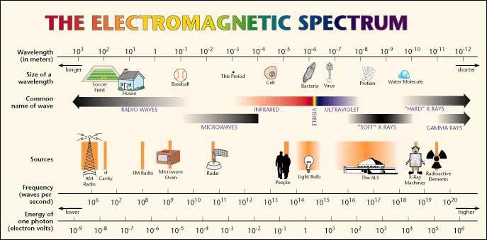
4 The Electromagnetic Spectrum


5 Recoil Effect in Free Atoms nucleus recoil energy: E R = E 2 0 / 2Mc 2 M γ-photon E γ = E nuc - E R E R 5-6 orders of magnitude greater than natural linewidth no resonance possible => Nuclear γ-resonance cannot be observed with gases and liquids!
6 Recoil Effect in Free Atoms.. too short! 7

7 Rudolf L. Mössbauer 1958: Discovers the recoilless nuclear resonance absorption of γ- radiation emitting and absorbing nuclei must be embedded in solid lattice there is recoil-less emission and absorption of -photons (ffactor) 1961: Receives the Nobel Prize in Physics

8 Mössbauer periodic table

9 Periodic table of life
10 Mössbauer spectroscopy The light source : Decay scheme of 57 Co Nuclear Spin I = 5/2 Electron capture 57 Co kev 3,300 times the energy of a 285-nm UV photon recoil imparts significant change of energy of the photon 9% 91% emitting and absorbing nuclei must be embedded in solid lattice I = 3/ kev there is recoil-less emission and absorption of -photons (f-factor) I = 1/2 57 Fe at low temperatures, all Fe species have same f-factor fraction of Fe species in sample is proportional to area of Mössbauer subspectrum
11 Mössbauer spectroscopy The light source : Decay scheme of 57 Co Nuclear Spin Electron capture 57 Co I = 5/ kev I = 3/2 9% 91% 14.4 kev v DE = c E v = source velocity c = speed of light I = 1/2 57 Fe Doppler effect allows the energy of the photon to be varied slightly
12 Mössbauer spectroscopy The light source : Decay scheme of 57 Co absorption Nuclear Spin Electron capture 57 Co I = 5/ kev 9% 91% 0 Doppler velocity I = 3/ kev I = 3/2 I = 1/2 57 Fe I = 1/2 57 Fe (sample) Photon can be absorbed by a 57 Fe nucleus in the sample
13 Experimental setup (transmission geometry) v = 1 mm/s => DE = ev = 11.6 MHz = cm -1 = 5.6 K

14 Low-Field Mössbauer spectrometer Velocity Transducer 57 Co source Sample Detector

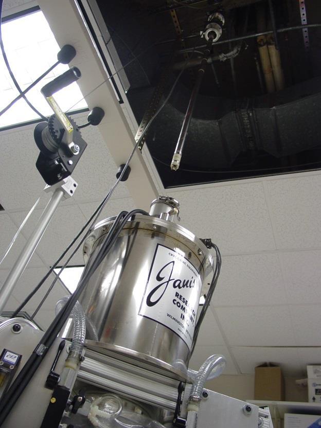
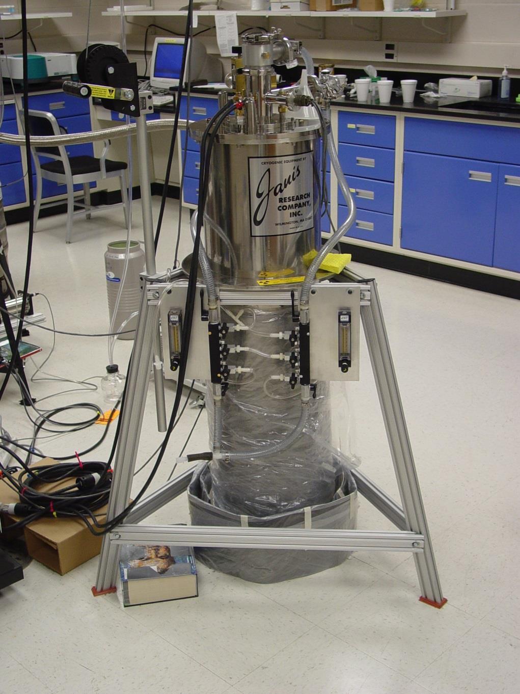
15 High-Field Mössbauer spectrometer

16 High-Field Mössbauer spectrometer Magnetic field -beam
17 General remarks Outline Quadrupole doublet spectra (isomer shift, quadrupole splitting) Magnetically split spectra (spin expectation value, hyperfine tensor) The correlation between EPR and Mössbauer spectroscopies (effective g-values and spin expectation values) How is the internal field oriented relative to the external field? How does the fluctuation rate of the electronic states affect the Mössbauer spectrum? Example 1: EPR and Mössbauer of the high-spin Fe(III) center in transferrin Example 2: EPR and Mössbauer of the Fe(II)/Fe(III) cluster in myo-inositol oxygenase (incl. magnetic Mössbauer of dinuclear clusters) Example 3: Mössbauer studies of the Fe(III)/Fe(III) cluster E. coli RNR Example 4: The high-spin Fe(IV)-oxo intermediate in TauD Example 5: A mononuclear Fe-dinitrosyl complex with S = 1/2 Considerations for sample preparation
18 absorption [%] Types of Mössbauer spectra: 1) Quadrupole Doublet Source Absorber M I = 3/2 I = 3/2 DE Q M I = 1/2 E S E A I = 1/2 Isomer shift (δ) M I = 1/2 Quadrupole Splitting (ΔE Q ) 0 6 δ DE Q velocity [mm/s] 2 4
19 absorption [%] Types of Mössbauer spectra: 1) Quadrupole Doublet M I = 3/2 How long are the black arrows if the red double arrow is 1 m? DE Q M I = 1/2 M I = 1/2 Quadrupole Splitting (ΔE Q ) 0 6 δ DE Q velocity [mm/s] 2 4
20 absorption [%] Types of Mössbauer spectra: 1) Quadrupole Doublet M I = 3/2 How long are the black arrows if the red double arrow is 1 m? DE Q M I = 1/2 ΔE = 2 mm/s = ev E = 14.4 kev ΔE/E = M I = 1/2 Quadrupole Splitting (ΔE Q ) 150,000,000,000 m (distance earth to sun) 0 6 δ DE Q velocity [mm/s] 2 4
.")
21 Isomer shift electron density at nucleus properties of 57 Fe nucleus δ = ( sample (0) 2 - source (0) 2 ) 4/5 π Ze 2 R 2 (ΔR/R) ΔR/R is the change of radius in ground and excited state (negative for 57 Fe). (0) 2 is the probability to find an electron at the 57 Fe nucleus only s-electrons have non-zero probability to be at nucleus d-electrons affect s-electron density by shielding nucleus atom 3d - electrons shield the nuclear potential for s - orbitals r(bohr)
 S=0 Fe(V) S=3/2 (adapted from Gütlich, Bill, Trautwein Mössbauer Spectroscopy and Transition Metal Chemistry, Springer 2011) Fe(V) S=1/2 Fe(VI) S=1 (relative to -iron at 300 K ) Fe(VI) S=0")
22 Typical isomer shift values for various spin- and oxidation states of iron Fe(I) S=3/2 Fe(I) S=1/2 Fe(II) S=2 Fe(II) S=1 Fe(II) S=0 Fe(III) S=5/2 Fe(III) S=3/2 Fe(III) S=1/2 Fe(IV) S=2 Fe(IV) S=1 Fe(IV) S=0 Fe(V) S=3/2 (adapted from Gütlich, Bill, Trautwein Mössbauer Spectroscopy and Transition Metal Chemistry, Springer 2011) Fe(V) S=1/2 Fe(VI) S=1 (relative to -iron at 300 K ) Fe(VI) S=0
23 Isomer shift correlations Oxidation state: (Fe IV ) < (Fe III ) < (Fe II ) number of d-electrons increases shielding of s-electrons increases (0) 2 decreases increases (because ΔR/R is negative) Spin state: Ligands: (low-spin) < (high-spin) low-spin complexes have shorter, more covalent M-L bonds less d-electron density shielding of s-electrons decreases (0) 2 increases decreases (because ΔR/R is negative) (S-ligands) < (N,O-ligands) (4-coordinate) < (6-coordinate)
24 Quadrupole splitting Nuclei with I > 1/2 have an electric quadrupole moment Q, which has different energies in an electric field gradient E (efg). E E electric field lines Q
 unfavorable orientation favorable")
25 Quadrupole splitting - theoretical '2D' model: the four model charges ±q generate an inhomogeneous field with an electric field gradient (efg) unfavorable orientation favorable orientation
 + (I x 2 I y2 )] DE Q = eqv zz /2 [ 1 + 2 /3] 1/2 = (V xx V yy ) /")
26 Electric charge distribution and EFG tensor between 0 and 1 only two independent comp. Ĥ q = I Q I = eqv zz /12 [ 3 I z 2 I (I + 1) + (I x 2 I y2 )] DE Q = eqv zz /2 [ /3] 1/2 = (V xx V yy ) / V zz (asymmetry parameter)
27 Expectation values of the efg tensor elements (V ii ) val / e<r -3 > for d-electrons orbital V xx V yy V zz d x2-y2-2/7-2/7 4/7 0 d z2 +2/7 +2/7-4/7 0 d xy -2/7-2/7 +4/7 0 d xz -2/7 +4/7-2/7 +3 d yz +4/7-2/7-2/7-3 to convert V ii in ΔE Q multiply by 4.2 mms -1 / 4/7 e <r-3> (for <r -3 >=5a 0-3, Q=0.15b) (Gütlich, Bill, Trautwein, Mössbauer Spectroscopy and Transition Metal Chemistry, Springer 2011 ) for a general 3d n valence electron configuration: add up the individual contributions for all d-electrons
28 Typical values of and DE Q for biological samples Oxidation state Spin state Ligands (mm/s) DE Q (mm/s) Fe(II) S = 2 heme Fe-(O/N) Fe/S S = 0 heme < 1.5 Fe(III) S = 5/2 heme Fe-(O/N) Fe/S < 1.0 S = 3/2 heme S = 1/2 heme Fe-(O/N) Fe(IV) S = 2 Fe-(O/N) S = 1 heme Fe-(O/N) Adapted from E. Münck, Physical Methods in Bioinorganic Chemistry, L. Que, Jr. (ed) 2000

29 Calculation of and DE Q using DFT Recent important advances by Neese and co-workers showed that Mössbauer parameters can be predicted well computationally using DFT methods Isomer shifts and quadrupole splittings are predicted to within 0.1 mm/s and 0.5 mm/s, respectively One can evaluate hypothetical structures and compare them to the experimentally determined Mössbauer parameters e.g. F. Neese, (2002) Inorg. Chim. Acta 337C, 181.
30 General remarks Outline Quadrupole doublet spectra (isomer shift, quadrupole splitting) Magnetically split spectra (spin expectation value, hyperfine tensor) The correlation between EPR and Mössbauer spectroscopies (effective g-values and spin expectation values) How is the internal field oriented relative to the external field? How does the fluctuation rate of the electronic states affect the Mössbauer spectrum? Example 1: EPR and Mössbauer of the high-spin Fe(III) center in transferrin Example 2: EPR and Mössbauer of the Fe(II)/Fe(III) cluster in myo-inositol oxygenase (incl. magnetic Mössbauer of dinuclear clusters) Example 3: Mössbauer studies of the Fe(III)/Fe(III) cluster E. coli RNR Example 4: The high-spin Fe(IV)-oxo intermediate in TauD Example 5: A mononuclear Fe-dinitrosyl complex with S = 1/2 Considerations for sample preparation
31 Types of Mössbauer spectra: 2) Magnetic Spectra I=3/2 Dm /2 +1/2-1/2-3/2 I=1/2-1/2 +1/2 Splitting of the six lines increases as the magnetic field experienced by the 57 Fe nucleus (the effective magnetic field) increases
32 Types of Mössbauer spectra: 2) Magnetic Spectra I=3/2 Dm /2 +1/2-1/2-3/2 I=1/2-1/2 +1/2 Splitting of the six lines increases as the magnetic field experienced by the 57 Fe nucleus (the effective magnetic field) increases
33 Types of Mössbauer spectra: 2) Magnetic Spectra Intensity ratio of the six lines depends on the orientation of the effective magnetic field to the propagation direction of the beam. I=3/2 Selection rule Δm = 0, 1 Intensity of Δm = 1 lines (1 +cos 2 ) Intensity of Δm = 0 lines sin 2 +3/2 +1/2-1/2-3/2 B effective -beam 3:4:1:1:4:3 B effective -beam 3:0:1:1:0:3 Powder spectrum 3:2:1:1:2:3 I=1/2-1/2 +1/2 Dm
34 Types of Mössbauer spectra: 2) Magnetic Spectra I=3/2 +3/2 +1/2-1/2-3/2 I=1/2-1/2 +1/2 inner four lines are shifted relative to the outer two lines
35 What causes the large field sensed by the 57 Fe nucleus? The paramagnetism of the Fe ions! High-spin Fe 2+ High-spin Fe 3+ S = 2 S = 5/2 High-spin Fe 4+ S = 2 Low-spin Fe 2+ Low-spin Fe 3+ S = 0 S = 1/2 Low-spin Fe 4+ S = 1
36 General remarks Outline Quadrupole doublet spectra (isomer shift, quadrupole splitting) Magnetically split spectra (spin expectation value, hyperfine tensor) The correlation between EPR and Mössbauer spectroscopies (effective g-values and spin expectation values) How is the internal field oriented relative to the external field? How does the fluctuation rate of the electronic states affect the Mössbauer spectrum? Example 1: EPR and Mössbauer of the high-spin Fe(III) center in transferrin Example 2: EPR and Mössbauer of the Fe(II)/Fe(III) cluster in myo-inositol oxygenase (incl. magnetic Mössbauer of dinuclear clusters) Example 3: Mössbauer studies of the Fe(III)/Fe(III) cluster E. coli RNR Example 4: The high-spin Fe(IV)-oxo intermediate in TauD Example 5: A mononuclear Fe-dinitrosyl complex with S = 1/2 Considerations for sample preparation
37 Spin Hamiltonian for EPR Spectroscopy Ĥ = μ B S g B + S D S + S A I electron Zeeman zero field splitting (ZFS) hyperfine coupling = μ B S g B + D (S z 2 S(S+1)/3) + E (S x 2 S y2 ) + S A I ZFS removes the (2S + 1)-fold degeneracy of the spin Only observed for systems with S 1 D and E are axial and rhombic ZFS parameters E/D also known as rhombicity E/D can take values between 0 and 1/3
38 EPR and Mössbauer spectroscopy are complementary Electron Spin Method EPR Half-Integer Spin S = 1/2, 3/2, 5/2, EPR-active Integer Spin S = 0, 1, 2, 3, EPR-silent (in most cases)
39 Energy (cm -1 ) Energy (cm -1 ) EPR and Mössbauer spectroscopy are complementary Electron Spin Method EPR Half-Integer Spin S = 1/2, 3/2, 5/2, EPR-active Integer Spin S = 0, 1, 2, 3, EPR-silent (in most cases) B B
40 Energy (cm -1 ) Effective g-values for an S = 5/2 spin system with ZFS z B x B y B z Calculated with D = 2 cm -1 and E/D = 0 g eff g eff g eff y z x E/D x y z x y
41 Energy (cm -1 ) Effective g-values for an S = 5/2 spin system with ZFS B x B y B z Calculated with D = 2 cm -1 and E/D = 1/ g eff g eff g eff y z x E/D z x y z x y
42 Powder EPR spectra of species with anisotropic g-values g = [GHz] / B [G] Taken from G. Palmer, Physical Methods in Bioinorganic Chemistry, L. Que (ed) 2000
43 Effective g-values for an S = 5/2 spin system with ZFS rhombic 4.3-signal 4.3 protocatechuate-3,4-dioxygenase 200 B (mt) Adapted from G. Palmer, Physical Methods in Bioinorganic Chemistry, L. Que (ed) 2000 g eff g eff g eff y z x E/D z x y z x y
44 Spin Hamiltonian for Mössbauer Spectroscopy electron spin hyperfine coupling nuclear spin Ĥ = μ B S g B + S D S + S A I - g N μ N B I + I Q I electron Zeeman zero field splitting hyperfine 57 Fe nuclear Zeeman quadrupole splitting In small external magnetic fields (e.g. 10 mt) the first two term are much larger than hyperfine coupling
45 Spin Hamiltonian for Mössbauer Spectroscopy electron spin hyperfine coupling nuclear spin Ĥ = μ B S g B + S D S + S A I - g N μ N B I + I Q I electron Zeeman zero field splitting hyperfine 57 Fe nuclear Zeeman quadrupole splitting = S A I - g N μ N B I + I Q I S is spin expectation value; it contains information of electronic structure
46 Spin Hamiltonian for Mössbauer Spectroscopy electron spin hyperfine coupling nuclear spin Ĥ = μ B S g B + S D S + S A I - g N μ N B I + I Q I electron Zeeman zero field splitting hyperfine 57 Fe nuclear Zeeman quadrupole splitting = S A I - g N μ N B I + I Q I S is spin expectation value; it contains information of electronic structure = - g N μ N [ - S A/ g N μ N + B ] I + I Q I B int B ext B eff
47 Spin Hamiltonian for Mössbauer Spectroscopy electron spin hyperfine coupling nuclear spin Ĥ = μ B S g B + S D S + S A I - g N μ N B I + I Q I electron Zeeman zero field splitting hyperfine 57 Fe nuclear Zeeman quadrupole splitting = S A I - g N μ N B I + I Q I S is spin expectation value; it contains information of electronic structure = - g N μ N [ - S A/ g N μ N + B ] I + I Q I B int B ext B eff The internal magnetic field, B int, depends on the spin expectation value, S, and the hyperfine coupling tensor, A.
48 A( 57 Fe) Hyperfine Coupling Tensor A = A Fermi-contact + A dipole + A orbit a.) Fermi - Contact Contribution Exchange interaction affords polarization of the filled inner s-shells. (different radial distribution of spin-up and spin-down electrons) - in general the largest contribution to A - isotropic, negative sign (-20 to -22 T)
49 A( 57 Fe) Hyperfine Coupling Tensor A = A Fermi-contact + A dipole + A orbit Dipole - Contribution, Adipole Arises from non-spherical distribution of the electronic spin density. Orbital - Contribution, Aorbit Arises from non-quenched orbital momentum of the electronic state due to spin-orbit coupling (SOC).
50 Spin expectation values for half-integer spin systems S ~ de/db Have the full expectation even in small external fields S g eff /4 Correlation between EPR and Mössbauer!
51 Spin expectation values for half-integer spin systems S ~ de/db Have the full expectation even in small external fields S g eff /4 Correlation between EPR and Mössbauer! Each electronic state has a S associated with it First we look at properties of S first (magnitude, anisotropy, orientation relative to external field); Next, we take into consideration that more than one state is populated
52 Energy (cm -1 ) S Spin expectation values for S = 1/2 3 S = 1/ B B S is (nearly) isotropic [i.e. the same in the x, y, and z-direction
53 S S Energy (cm -1 ) Spin expectation values for S = 5/2 Calculated with D = 2 cm -1 and E/D = 1/ B (T) ground doublet B (T) B (T) z x 0-1 middle doublet x, y, z B (T) y B (T)
54 S Spin expectation values for integer spin systems Calculated for S = 2 with D = 10 cm -1 and E/D = 1/3 x y z B (T) B (T) B (T) Have in most cases S 0 for B ext = 0 0 z Small B ext may result in small S [depends on ZFS parameters] -1 x Large B ext results in sizeable S B (T) y
55 EPR and Mössbauer spectroscopy are complementary Electron Spin Method EPR Half-Integer Spin S = 1/2, 3/2, 5/2, EPR-active Integer Spin S = 0, 1, 2, 3, EPR-silent (in most cases) Mössbauer Sizeable B int in small B ext Magnetically split spectra Small B int in small B ext Quadrupole doublets (analysis complex, but facilitated using results from EPR) (in most cases, but not always*) * (there are exceptions, such as high-spin Fe(III)-superoxo complexes or the [3Fe-4S] 0 cluster, see Eckard Münck s PSU workshop talk in 2014and Mike Hendrich s section in Palmer chapter in Que book)
56 General remarks Outline Quadrupole doublet spectra (isomer shift, quadrupole splitting) Magnetically split spectra (spin expectation value, hyperfine tensor) The correlation between EPR and Mössbauer spectroscopies (effective g-values and spin expectation values) How is the internal field oriented relative to the external field? How does the fluctuation rate of the electronic states affect the Mössbauer spectrum? Example 1: EPR and Mössbauer of the high-spin Fe(III) center in transferrin Example 2: EPR and Mössbauer of the Fe(II)/Fe(III) cluster in myo-inositol oxygenase (incl. magnetic Mössbauer of dinuclear clusters) Example 3: Mössbauer studies of the Fe(III)/Fe(III) cluster E. coli RNR Example 4: The high-spin Fe(IV)-oxo intermediate in TauD Example 5: A mononuclear Fe-dinitrosyl complex with S = 1/2 Considerations for sample preparation

57 Orientation of B int relative to B ext Fe Representation of a Fe-containing protein Representation of an isotropic S of an electronic state of the Fe-containing protein (e.g. middle Kramers doublet of a mononuclear rhombic ferric site Representation of an anisotropic S of an electronic state of the Fe-containing protein (e.g. ground Kramers doublet of a mononuclear rhombic ferric site
58 Orientation of B int relative to B ext ray B external ray B external The internal field is aligned antiparallel to the external field
59 Orientation of B int relative to B ext ray or B external The internal field is oriented along the axis with the greatest component of S The orientation of S depends on molecular frame; thus, because molecules are frozen randomly, the internal fields are oriented randomly (powder averaged spectrum)
60 General remarks Outline Quadrupole doublet spectra (isomer shift, quadrupole splitting) Magnetically split spectra (spin expectation value, hyperfine tensor) The correlation between EPR and Mössbauer spectroscopies (effective g-values and spin expectation values) How is the internal field oriented relative to the external field? How does the fluctuation rate of the electronic states affect the Mössbauer spectrum? Example 1: EPR and Mössbauer of the high-spin Fe(III) center in transferrin Example 2: EPR and Mössbauer of the Fe(II)/Fe(III) cluster in myo-inositol oxygenase (incl. magnetic Mössbauer of dinuclear clusters) Example 3: Mössbauer studies of the Fe(III)/Fe(III) cluster E. coli RNR Example 4: The high-spin Fe(IV)-oxo intermediate in TauD Example 5: A mononuclear Fe-dinitrosyl complex with S = 1/2 Considerations for sample preparation
61 Relaxation of the electronic states and their effect on the Mössbauer spectrum Paramagnetic Fe-sites have more than one electronic state; fluctuation rate between electronic states needs to be considered for such systems. Three cases are possible: The relaxation between electronic states is slow compared to the time scale of Mössbauer spectroscopy (10-7 s). (typically encountered for metalloproteins at 4.2 K) The relaxation between electronic states is fast compared to the time scale of Mössbauer spectroscopy. (encountered at high temperatures; depends on system under consideration) The relaxation between electronic states is comparable to the time scale of Mössbauer spectroscopy. This case is more difficult to treat and one tries to avoid it by choosing different experimental conditions (temperature, external field).
62 Energy (cm -1 ) Calculate S for each electronic state Slow relaxation limit Calculate Mössbauer spectrum for each electronic state Add the subspectra of all electronic states according to their Boltzmann population factors [~exp(-e/kt)] The resulting spectrum contains multiple subspectra (one for every electronic state) The subspectra are magnetically split B
63 Energy (cm -1 ) Fast relaxation limit Calculate S for each electronic state Calculate the average spin expectation value, S av, from the individual S values according to their Boltzmann factors Calculate Mössbauer spectrum using S av. There is only one subspectrum associated with all electronic states In small magnetic fields S av 0, therefore no hyperfine interactions, i.e. spectrum is a quadrupole doublet B
64 Cases when S is zero S = 0 B int =0 quadrupole doublet for small B ext 1. Diamagnetic compounds 2. Paramagnetic compounds with integer spin ground state for B ext = 0 (or B ext small) 3. Compound in fast relaxation limit in small magnetic field (then S av 0, therefore no hyperfine interactions, i.e. spectrum is a quadrupole doublet
65 EPR and Mössbauer spectroscopy are complementary Electron Spin Method EPR Half-Integer Spin S = 1/2, 3/2, 5/2, EPR-active Integer Spin S = 0, 1, 2, 3, EPR-silent (in most cases) Mössbauer Magnetically Split Spectra (at low T) (analysis complex, but facilitated using results from EPR) Quadrupole doublets at high temperatures Quadrupole doublets in small B ext (in most cases) (analysis straightforward) Magnetically Split Spectra for large B ext
66 General remarks Outline Quadrupole doublet spectra (isomer shift, quadrupole splitting) Magnetically split spectra (spin expectation value, hyperfine tensor) The correlation between EPR and Mössbauer spectroscopies (effective g-values and spin expectation values) How is the internal field oriented relative to the external field? How does the fluctuation rate of the electronic states affect the Mössbauer spectrum? Example 1: EPR and Mössbauer of the high-spin Fe(III) center in transferrin Example 2: EPR and Mössbauer of the Fe(II)/Fe(III) cluster in myo-inositol oxygenase (incl. magnetic Mössbauer of dinuclear clusters) Example 3: Mössbauer studies of the Fe(III)/Fe(III) cluster E. coli RNR Example 4: The high-spin Fe(IV)-oxo intermediate in TauD Example 5: A mononuclear Fe-dinitrosyl complex with S = 1/2 Considerations for sample preparation
 site in transferrin (S")
67 Example 1 The high-spin Fe(III) site in transferrin (S = 5/2)
68 The high-spin Fe(III) site in transferrin (S = 5/2) g eff doublet 1 doublet 2 doublet E/D g eff E/D g eff E/D
69 The high-spin Fe(III) site in transferrin (S = 5/2) D = 0.25 cm mt 50 mt perp 0.5 T 2 T E/D = 0.3 g = 2.0 δ = 0.54 mm/s ΔE Q = 0.30 mm/s η = 1.0 A/g n n = (-22.3, -21.9, -22.3) T 6 T Kretchmar, et al. Biol. Metals 1988 (1) 26
70 The high-spin Fe(III) site in transferrin (S = 5/2) 50 mt 50 mt perp 0.5 T 2 T 6 T Kretchmar, et al. Biol. Metals 1988 (1) 26
71 General remarks Outline Quadrupole doublet spectra (isomer shift, quadrupole splitting) Magnetically split spectra (spin expectation value, hyperfine tensor) The correlation between EPR and Mössbauer spectroscopies (effective g-values and spin expectation values) How is the internal field oriented relative to the external field? How does the fluctuation rate of the electronic states affect the Mössbauer spectrum? Example 1: EPR and Mössbauer of the high-spin Fe(III) center in transferrin Example 2: EPR and Mössbauer of the Fe(II)/Fe(III) cluster in myo-inositol oxygenase (incl. magnetic Mössbauer of dinuclear clusters) Example 3: Mössbauer studies of the Fe(III)/Fe(III) cluster E. coli RNR Example 4: The high-spin Fe(IV)-oxo intermediate in TauD Example 5: A mononuclear Fe-dinitrosyl complex with S = 1/2 Considerations for sample preparation
72 Example 2 The exchange-coupled high-spin Fe 2 (II/III) cofactor of myo-inositol oxygenase
73 Energy The spin-coupled Fe 2 II/III cluster in myo-inositol oxygenase The active form of myo-inositol oxygenase harbors an antiferromagnetically coupled dinuclear site with a high-spin Fe 3+ ion (S 1 = 5/2) and a high-spin Fe 2+ ion (S 2 = 2). It has an EPR-active S = 1/2 ground state. S = 9/2 S = S 1 + S 2 g = (1.95, 1.81, 1.81) 4.5 J Ĥ HDvV = J S 1 S 2 S = 7/2 E(S) = J/2 S (S + 1) 3.5 J 2.5 J 1.5 J S = 5/2 S = 3/2 S = 1/2 EPR-spectroscopy probes the total ground spin state of a coupled cluster.
74 The spin-coupled Fe 2 II/III cluster in myo-inositol oxygenase = 1.09 mm/s DE Q = 2.86 mm/s high-spin Fe(II) = 0.48 mm/s DE Q = 1.10 mm/s high-spin Fe(III) Mössbauer-spectroscopy probes the local spin/oxidation state of each 57 Fe-labeled site of a coupled cluster. At 120 K in zero field: fast-relaxation limit quadrupole doublets intrinsic Fe oxidation state
75 The spin-coupled Fe 2 II/III cluster in myo-inositol oxygenase Mössbauer-spectroscopy probes the local spin/oxidation state of each 57 Fe-labeled site of a coupled cluster. At 4.2 K: slow-relaxation limit magnetically split spectra S = 1/2 S isotropic field-orientation-dependence
76 Spin projection factors Ĥ hf = S 1 A 1 I 1 + S 2 A 2 I 2 hyperfine 1 hyperfine 2 = S tot (c 1 A 1 ) I 1 + S tot (c 2 A 2 ) I 2 Spin projection factors c i = [S(S+1) + S i (S i +1) S j (S j +1)] / [2S(S+1)] For S = 1/2 ground state, c 1 = +7/3 and c 2 = -4/3 for S 1 = 5/2 and S 2 = 2 See A. Bencini and D. Gatteschi, EPR of Exchange Coupled Systems, Springer, 1989 for derivation of spin coupling coeff.
77 Spin Hamiltonian of an exchange-coupled cluster Ĥ hf = S 1 A 1 I 1 + S 2 A 2 I 2 hyperfine 1 hyperfine 2 = S tot (c 1 A 1 ) I 1 + S tot (c 2 A 2 ) I 2 A 1 and A 2 (the intrinsic A-tensors given with respect to the local spin) are dominated by the Fermi contact term, which is ~ -20 to -22 T. Analysis of field-dependent Mössbauer spectra allows c 1 A 1 and c 2 A 2 to be determined. by determining A 1 and A 2, one can estimate c 1 and c 2 and therefore determine the nature of the spin coupling of the cluster. if hyperfine coupling is resolved in EPR, then c 1 A 1 and c 2 A 2 can be determined, but not the sign of c 1 and c 2.
78 The spin-coupled Fe 2 II/III cluster in myo-inositol oxygenase B internal > B external Fe(III) site has typical field dependence (B int antiparallel to B ext for ground state, i.e. B eff decreases with increasing B ext ) Fe(II) site has atypical field dependence (B int parallel to B ext for ground state) this behavior is due to opposite sign of spin coupling coefficients
79 General remarks Outline Quadrupole doublet spectra (isomer shift, quadrupole splitting) Magnetically split spectra (spin expectation value, hyperfine tensor) The correlation between EPR and Mössbauer spectroscopies (effective g-values and spin expectation values) How is the internal field oriented relative to the external field? How does the fluctuation rate of the electronic states affect the Mössbauer spectrum? Example 1: EPR and Mössbauer of the high-spin Fe(III) center in transferrin Example 2: EPR and Mössbauer of the Fe(II)/Fe(III) cluster in myo-inositol oxygenase (incl. magnetic Mössbauer of dinuclear clusters) Example 3: Mössbauer studies of the Fe(III)/Fe(III) cluster E. coli RNR Example 4: The high-spin Fe(IV)-oxo intermediate in TauD Example 5: A mononuclear Fe-dinitrosyl complex with S = 1/2 Considerations for sample preparation
80 Example 3 The exchange-coupled high-spin diiron cofactors of the class Ia ribonucleotide reductase from E. coli

81 Class I Ribonucleotide Reductase from E. coli Stubbe, et al. Chem. Rev. 2003, Proposed PCET (Proton Coupled Electron Transfer) Pathway
82 Cofactor generation of E. coli ribonucleotide reductase
83 Spectroscopic signatures of the active Fe 2 III/III -Y122 form S = 1/2 for Tyr
84 Spectroscopic signatures of the active Fe 2 III/III -Y122 form Two quadrupole doublets in Mössbauer Suggests integer spin ground state 4.2K 53 mt
85 Spectroscopic signatures of the active Fe 2 III/III -Y122 form Spectrum reveals that B ext = 6 T B eff = B ext = 6 T B int = 0, S = 0
86 Spectroscopic signatures of the active Fe 2 III/III -Y122 form BUT how do we pair the lines? = 0.45 mm/s DE Q = 2.43 mm/s = 0.54 mm/s DE Q = 1.63 mm/s
87 Spectroscopic signatures of the active Fe 2 III/III -Y122 form BUT how do we pair the lines? = 0.69 mm/s DE Q = 1.94 mm/s = 0.29 mm/s DE Q = 2.11 mm/s
88 Spectroscopic signatures of the active Fe 2 III/III -Y122 form BUT how do we pair the lines? = mm/s DE Q = 0.48 mm/s = 1.51 mm/s DE Q = 0.31 mm/s
89 Site-specific Labeling with 57 Fe

90 Site-specific Labeling with 57 Fe Bollinger, et al. JACS 1997, 5976
91 General remarks Outline Quadrupole doublet spectra (isomer shift, quadrupole splitting) Magnetically split spectra (spin expectation value, hyperfine tensor) The correlation between EPR and Mössbauer spectroscopies (effective g-values and spin expectation values) How is the internal field oriented relative to the external field? How does the fluctuation rate of the electronic states affect the Mössbauer spectrum? Example 1: EPR and Mössbauer of the high-spin Fe(III) center in transferrin Example 2: EPR and Mössbauer of the Fe(II)/Fe(III) cluster in myo-inositol oxygenase (incl. magnetic Mössbauer of dinuclear clusters) Example 3: Mössbauer studies of the Fe(III)/Fe(III) cluster E. coli RNR Example 4: The high-spin Fe(IV)-oxo intermediate in TauD Example 5: A mononuclear Fe-dinitrosyl complex with S = 1/2 Considerations for sample preparation
92 Example 4 The Fe(IV)-oxo intermediate in taurine:2- oxoglutarate dioxygenase (TauD) αkg His His Asp/Glu
93 Generalized Reaction Catalyzed by the Fe(II)- and -Ketoglutarate-Dependent Dioxygenases R H O O O - + O O 2 + -O O - R OH + O - + CO 2 O O αkg His His Asp/Glu
94 Mechanism of Taurine:αKG Dioxygenase (TauD) > 3 x 10 4 M -1 s -1 fast 2.5 s x 10 5 M -1 s -1 fast fast k H = 13 s -1 k D =0.25 s -1 fast
95 Evidence for an Fe(IV) Intermediate by Mössbauer Spectroscopy = 1.16 mm/s DE Q = 2.76 mm/s high-spin Fe(II)
96 Evidence for an Fe(IV) Intermediate by Mössbauer Spectroscopy = 1.16 mm/s DE Q = 2.76 mm/s = 0.30 mm/s DE Q = 0.90 mm/s Fe(IV)
97 Evidence for an Fe(IV) Intermediate (J) by Mössbauer Spectroscopy J (mm) = 1.16 mm/s DE Q = 2.76 mm/s = 0.30 mm/s DE Q = 0.90 mm/s 0.2 TauD Fe(II) αkg Taurine Time (s) 2.5 s -1 2 nd Intermediate Fe(II) O 2 13 s x 10 5 M -1 s -1 J Fe(IV)
98 High-Field Mössbauer of the Fe(IV) Intermediate 0 ms 20 ms Magnetic spectra of the Fe(II) reactant complex not well understood Experimental spectrum collected under the same experimental conditions is used without any simulation for deconvolution of data
99 Mössbauer Evidence that the Intermediate has an Integer Spin Ground State with S = 2 +3/2 8 T S = 2 I = 3/2 +1/2-1/2-3/2 S = 1 I = 1/2-1/2 +1/2
100 Further Characterization of J from DFT Calculations resonance Raman Fe=O = 821 cm -1 (Proshlyakov, et al. JACS 2004, 126, 1022) EXAFS d Fe=O = 1.62 Å exp calc (mm/s) DE Q (mm/s) A/g N N (T) Comparison of experimentally determined, spectroscopic parameters to those calculated by DFT methods provides detailed structural information.
101 General remarks Outline Quadrupole doublet spectra (isomer shift, quadrupole splitting) Magnetically split spectra (spin expectation value, hyperfine tensor) The correlation between EPR and Mössbauer spectroscopies (effective g-values and spin expectation values) How is the internal field oriented relative to the external field? How does the fluctuation rate of the electronic states affect the Mössbauer spectrum? Example 1: EPR and Mössbauer of the high-spin Fe(III) center in transferrin Example 2: EPR and Mössbauer of the Fe(II)/Fe(III) cluster in myo-inositol oxygenase (incl. magnetic Mössbauer of dinuclear clusters) Example 3: Mössbauer studies of the Fe(III)/Fe(III) cluster E. coli RNR Example 4: The high-spin Fe(IV)-oxo intermediate in TauD Example 5: A mononuclear Fe-dinitrosyl complex with S = 1/2 Considerations for sample preparation
102 Example 5 A mononuclear {Fe(NO) 2 } 9 complex with S = 1/2 A. L. Speelman, et al., Inorg. Chem. 2016
103 EPR-Spectroscopy of the {Fe(NO) 2 } 9 complex Intense S = 1/2 signal S virtually isotropic Magnetically split Mössbauer spectra expected with strong field-orientation dependence
104 Mössbauer Spectroscopy of the {Fe(NO) 2 } 9 complex g = 2.0 δ = 0.37 mm/s ΔE Q = mm/s η = 0.3 A/g n n = (-26.2, -23.4, -4.6) T

105 A little bit of fine-print for low-field spectra
106 A little bit of fine-print for low-field spectra g = 2.0 δ = 0.37 mm/s ΔE Q = mm/s η = 0.3 A/g n n = (-26.2, -23.4, -4.6) T S is isotropic All directions are probed A is very anisotropic B int anisotropic
107 A mononuclear {Fe(NO) 2 } 9 complex with S = 1/2 High-field spectra reveal that the slow-relaxation limit applies
108 General remarks Outline Quadrupole doublet spectra (isomer shift, quadrupole splitting) Magnetically split spectra (spin expectation value, hyperfine tensor) The correlation between EPR and Mössbauer spectroscopies (effective g-values and spin expectation values) How is the internal field oriented relative to the external field? How does the fluctuation rate of the electronic states affect the Mössbauer spectrum? Example 1: EPR and Mössbauer of the high-spin Fe(III) center in transferrin Example 2: EPR and Mössbauer of the Fe(II)/Fe(III) cluster in myo-inositol oxygenase (incl. magnetic Mössbauer of dinuclear clusters) Example 3: Mössbauer studies of the Fe(III)/Fe(III) cluster E. coli RNR Example 4: The high-spin Fe(IV)-oxo intermediate in TauD Example 5: A mononuclear Fe-dinitrosyl complex with S = 1/2 Considerations for sample preparation
109 Mössbauer spectroscopy A few final remarks Standard conditions: ~0.4 ml frozen solution with 1 mm 57 Fe Natural abundance of 57 Fe is 2.2% (you need to enrich with 57 Fe, 4-5 $ per mg of 57 Fe) If you can make samples with 3 mm 57 Fe you should do so Sample composition matters (purity, number of different species) If you have more than one Fe site, think about selective enrichment Prepare a parallel EPR sample (in particular if you anticipate species with half-integer S) Avoid high concentrations of relatively heavy atoms (Cl, S, P) due to scattering (100 mm phosphate buffer not a problem, CH 2 Cl 2 solvent is problematic) Data collection takes a long time (on average 1 to 1.5 days per spectrum); longest spectrum (in our lab) was 6 days collection time longest sample queue (in our lab) was about 5-6 weeks $100 per day operation costs for cryogens and source

110 Acknowledgements
EPR and Mössbauer Spectroscopies
 Presymposium Workshop EPR and Mössbauer Spectroscopies Carsten Krebs Department of Chemistry Department of Biochemistry and Molecular Biology The Pennsylvania State University Absorption Spectroscopy Does
Mossbauer Effect and Spectroscopy. Kishan Sinha Xu Group Department of Physics and Astronomy University of Nebraska-Lincoln
 Mossbauer Effect and Spectroscopy Kishan Sinha Xu Group Department of Physics and Astronomy University of Nebraska-Lincoln Emission E R γ-photon E transition hν = E transition - E R Photon does not carry
Lecture 6: Physical Methods II. UV Vis (electronic spectroscopy) Electron Spin Resonance Mossbauer Spectroscopy
 Electron Spin Resonance Mossbauer Spectroscopy") Lecture 6: Physical Methods II UV Vis (electronic spectroscopy) Electron Spin Resonance Mossbauer Spectroscopy Physical Methods used in bioinorganic chemistry X ray crystallography X ray absorption (XAS)
Lecture 6: Physical Methods II UV Vis (electronic spectroscopy) Electron Spin Resonance Mossbauer Spectroscopy Physical Methods used in bioinorganic chemistry X ray crystallography X ray absorption (XAS)
Introduction to Electron Paramagnetic Resonance Spectroscopy
 Introduction to Electron Paramagnetic Resonance Spectroscopy Art van der Est, Department of Chemistry, Brock University St. Catharines, Ontario, Canada 1 EPR Spectroscopy EPR is magnetic resonance on unpaired
Chapter 8 Magnetic Resonance
 Chapter 8 Magnetic Resonance 9.1 Electron paramagnetic resonance 9.2 Ferromagnetic resonance 9.3 Nuclear magnetic resonance 9.4 Other resonance methods TCD March 2007 1 A resonance experiment involves
3. Perturbed Angular Correlation Spectroscopy
 3. Perturbed Angular Correlation Spectroscopy Dileep Mampallil Augustine K.U.Leuven, Belgium Perturbed Angular Correlation Spectroscopy (PAC) is a gamma ray spectroscopy and can be used to investigate
Nuclear Quadrupole Resonance Spectroscopy. Some examples of nuclear quadrupole moments
 Nuclear Quadrupole Resonance Spectroscopy Review nuclear quadrupole moments, Q A negative value for Q denotes a distribution of charge that is "football-shaped", i.e. a sphere elongated at the poles; a
Basic concepts of Mößbauer spectroscopy (Mossbauer)
") Basic concepts of Mößbauer spectroscopy (Mossbauer) 1961 Nobel price in Physics..for his researches concerning the resonance absorption of γ-radiation and his discovery in this connection of the effect
Basic concepts of Mößbauer spectroscopy (Mossbauer) 1961 Nobel price in Physics..for his researches concerning the resonance absorption of γ-radiation and his discovery in this connection of the effect
Inorganic Spectroscopic and Structural Methods
 Inorganic Spectroscopic and Structural Methods Electromagnetic spectrum has enormous range of energies. Wide variety of techniques based on absorption of energy e.g. ESR and NMR: radiowaves (MHz) IR vibrations
Mossbauer Effect. Ahmad Ali Ohyda. 1 Ahmad Faraj Abuaisha. 2 Abu-Bakr Mohammad Alrotob. 3 Basher M. Ismail. 4. Abstract
 Majalat Al-Ulum Al-Insaniya wat - Tatbiqiya Mossbauer Effect ( Determination of isomer shift, line width and quadruple splitting in a potassium ferricyanide ( K3Fe(CN)6) sample using Mossbauer spectroscopy)
Gamma-ray decay. Introduction to Nuclear Science. Simon Fraser University Spring NUCS 342 March 7, 2011
 Gamma-ray decay Introduction to Nuclear Science Simon Fraser University Spring 2011 NUCS 342 March 7, 2011 NUCS 342 (Lecture 18) March 7, 2011 1 / 31 Outline 1 Mössbauer spectroscopy NUCS 342 (Lecture
Spin Interactions. Giuseppe Pileio 24/10/2006
 Spin Interactions Giuseppe Pileio 24/10/2006 Magnetic moment µ = " I ˆ µ = " h I(I +1) " = g# h Spin interactions overview Zeeman Interaction Zeeman interaction Interaction with the static magnetic field
Conclusion. 109m Ag isomer showed that there is no such broadening. Because one can hardly
 Conclusion This small book presents a description of the results of studies performed over many years by our research group, which, in the best period, included 15 physicists and laboratory assistants
Chem8028(1314) - Spin Dynamics: Spin Interactions
 - Spin Dynamics: Spin Interactions") Chem8028(1314) - Spin Dynamics: Spin Interactions Malcolm Levitt see also IK m106 1 Nuclear spin interactions (diamagnetic materials) 2 Chemical Shift 3 Direct dipole-dipole coupling 4 J-coupling 5 Nuclear
Chem8028(1314) - Spin Dynamics: Spin Interactions Malcolm Levitt see also IK m106 1 Nuclear spin interactions (diamagnetic materials) 2 Chemical Shift 3 Direct dipole-dipole coupling 4 J-coupling 5 Nuclear
Nuclear hyperfine interactions
 Nuclear hyperfine interactions F. Tran, A. Khoo, R. Laskowski, P. Blaha Institute of Materials Chemistry Vienna University of Technology, A-1060 Vienna, Austria 25th WIEN2k workshop, 12-16 June 2018 Boston
Saturation Absorption Spectroscopy of Rubidium Atom
 Saturation Absorption Spectroscopy of Rubidium Atom Jayash Panigrahi August 17, 2013 Abstract Saturated absorption spectroscopy has various application in laser cooling which have many relevant uses in
RFSS: Lecture 6 Gamma Decay
 RFSS: Lecture 6 Gamma Decay Readings: Modern Nuclear Chemistry, Chap. 9; Nuclear and Radiochemistry, Chapter 3 Energetics Decay Types Transition Probabilities Internal Conversion Angular Correlations Moessbauer
e 2m e c I, (7.1) = g e β B I(I +1), (7.2) = erg/gauss. (7.3)
 = g e β B I(I +1), (7.2) = erg/gauss. (7.3)") Chemistry 126 Molecular Spectra & Molecular Structure Week # 7 Electron Spin Resonance Spectroscopy, Supplement Like the hydrogen nucleus, an unpaired electron in a sample has a spin of I=1/2. The magnetic
Chemistry 126 Molecular Spectra & Molecular Structure Week # 7 Electron Spin Resonance Spectroscopy, Supplement Like the hydrogen nucleus, an unpaired electron in a sample has a spin of I=1/2. The magnetic
Nuclear Physics. (PHY-231) Dr C. M. Cormack. Nuclear Physics This Lecture
 Dr C. M. Cormack. Nuclear Physics This Lecture") Nuclear Physics (PHY-31) Dr C. M. Cormack 11 Nuclear Physics This Lecture This Lecture We will discuss an important effect in nuclear spectroscopy The Mössbauer Effect and its applications in technology
Nuclear Physics (PHY-31) Dr C. M. Cormack 11 Nuclear Physics This Lecture This Lecture We will discuss an important effect in nuclear spectroscopy The Mössbauer Effect and its applications in technology
Magnetic Resonance Spectroscopy EPR and NMR
 Magnetic Resonance Spectroscopy EPR and NMR A brief review of the relevant bits of quantum mechanics 1. Electrons have spin, - rotation of the charge about its axis generates a magnetic field at each electron.
Atomic Structure. Chapter 8
 Atomic Structure Chapter 8 Overview To understand atomic structure requires understanding a special aspect of the electron - spin and its related magnetism - and properties of a collection of identical
APEX CARE INSTITUTE FOR PG - TRB, SLET AND NET IN PHYSICS
 Page 1 1. Within the nucleus, the charge distribution A) Is constant, but falls to zero sharply at the nuclear radius B) Increases linearly from the centre, but falls off exponentially at the surface C)
Supporting Information
 Supporting Information Non-Heme Diiron Model Complexes Can Mediate Direct NO Reduction: Mechanistic Insight Into Flavodiiron NO Reductases Hai T. Dong, a Corey J. White, a Bo Zhang, b Carsten Krebs, b
Chapter 7. Nuclear Magnetic Resonance Spectroscopy
 Chapter 7 Nuclear Magnetic Resonance Spectroscopy I. Introduction 1924, W. Pauli proposed that certain atomic nuclei have spin and magnetic moment and exposure to magnetic field would lead to energy level
Mossbauer Spectroscopy
 Mossbauer Spectroscopy Emily P. Wang MIT Department of Physics The ultra-high resolution ( E = E 10 12 ) method of Mossbauer spectroscopy was used to probe various nuclear effects. The Zeeman splittings
Unsolved problems in biology
 Unsolved problems in biology What can advanced x-ray spectroscopy contribute? James Penner-Hahn Biophysics Research Division and Department of Chemistry The University of Michigan Metalloproteins 30-50%
Physical Background Of Nuclear Magnetic Resonance Spectroscopy
 Physical Background Of Nuclear Magnetic Resonance Spectroscopy Michael McClellan Spring 2009 Department of Physics and Physical Oceanography University of North Carolina Wilmington What is Spectroscopy?
Instrumentelle Analytik in den Geowissenschaften (PI)
") 280061 VU MA-ERD-2 Instrumentelle Analytik in den Geowissenschaften (PI) Handoutmaterial zum Vorlesungsteil Spektroskopie Bei Fragen bitte zu kontaktieren: Prof. Lutz Nasdala, Institut für Mineralogie
280061 VU MA-ERD-2 Instrumentelle Analytik in den Geowissenschaften (PI) Handoutmaterial zum Vorlesungsteil Spektroskopie Bei Fragen bitte zu kontaktieren: Prof. Lutz Nasdala, Institut für Mineralogie
Electronic Spectra of Complexes
 Electronic Spectra of Complexes Interpret electronic spectra of coordination compounds Correlate with bonding Orbital filling and electronic transitions Electron-electron repulsion Application of MO theory
Appendix II - 1. Figure 1: The splitting of the spin states of an unpaired electron
 Appendix II - 1 May 2017 Appendix II: Introduction to EPR Spectroscopy There are several general texts on this topic, and this appendix is only intended to give you a brief outline of the Electron Spin
ESR spectroscopy of catalytic systems - a primer
 ESR spectroscopy of catalytic systems - a primer Thomas Risse Fritz-Haber-Institute of Max-Planck Society Department of Chemical Physics Faradayweg 4-6 14195 Berlin T. Risse, 3/22/2005, 1 ESR spectroscopy
An introduction to Solid State NMR and its Interactions
 An introduction to Solid State NMR and its Interactions From tensor to NMR spectra CECAM Tutorial September 9 Calculation of Solid-State NMR Parameters Using the GIPAW Method Thibault Charpentier - CEA
Experimental Correlation of Substrate Position with Reaction Outcome in the Aliphatic
 Supporting Information for: Experimental Correlation of Substrate Position with Reaction Outcome in the Aliphatic Halogenase, SyrB2 Ryan J. Martinie, a Jovan Livada, a Wei-chen Chang, a Michael T. Green,
( ) electron gives S = 1/2 and L = l 1
 electron gives S = 1/2 and L = l 1") Practice Modern Physics II, W018, Set 1 Question 1 Energy Level Diagram of Boron ion B + For neutral B, Z = 5 (A) Draw the fine-structure diagram of B + that includes all n = 3 states Label the states
Practice Modern Physics II, W018, Set 1 Question 1 Energy Level Diagram of Boron ion B + For neutral B, Z = 5 (A) Draw the fine-structure diagram of B + that includes all n = 3 states Label the states
10.4 Continuous Wave NMR Instrumentation
 10.4 Continuous Wave NMR Instrumentation coherent detection bulk magnetization the rotating frame, and effective magnetic field generating a rotating frame, and precession in the laboratory frame spin-lattice
Magnetic Resonance Spectroscopy
 INTRODUCTION TO Magnetic Resonance Spectroscopy ESR, NMR, NQR D. N. SATHYANARAYANA Formerly, Chairman Department of Inorganic and Physical Chemistry Indian Institute of Science, Bangalore % I.K. International
ELECTRON PARAMAGNETIC RESONANCE
 ELECTRON PARAMAGNETIC RESONANCE = MAGNETIC RESONANCE TECHNIQUE FOR STUDYING PARAMAGNETIC SYSTEMS i.e. SYSTEMS WITH AT LEAST ONE UNPAIRED ELECTRON Examples of paramagnetic systems Transition-metal complexes
Lattice dynamics, phase transitions and spin relaxation in [Fe(C 5 H 5 ) 2 ]PF 6
![Lattice dynamics, phase transitions and spin relaxation in [Fe(C 5 H 5 ) 2 ]PF 6](/thumbs/96/126746830.jpg "Lattice dynamics, phase transitions and spin relaxation in [Fe(C 5 H 5 ) 2 ]PF 6") Hyperfine Interact (2016) 237:100 DOI 10.1007/s10751-016-1310-9 Lattice dynamics, phase transitions and spin relaxation in [Fe(C 5 H 5 ) 2 ]PF 6 R. H. Herber 1 I. Felner 1 I. Nowik 1 Springer International
Hyperfine Interact (2016) 237:100 DOI 10.1007/s10751-016-1310-9 Lattice dynamics, phase transitions and spin relaxation in [Fe(C 5 H 5 ) 2 ]PF 6 R. H. Herber 1 I. Felner 1 I. Nowik 1 Springer International
MOLECULAR SPECTROSCOPY AND PHOTOCHEMISTRY
 20 CHAPTER MOLECULAR SPECTROSCOPY AND PHOTOCHEMISTRY 20.1 Introduction to Molecular Spectroscopy 20.2 Experimental Methods in Molecular Spectroscopy 20.3 Rotational and Vibrational Spectroscopy 20.4 Nuclear
6 NMR Interactions: Zeeman and CSA
 6 NMR Interactions: Zeeman and CSA 6.1 Zeeman Interaction Up to this point, we have mentioned a number of NMR interactions - Zeeman, quadrupolar, dipolar - but we have not looked at the nature of these
Electron Spin Resonance, Basic principle of NMR, Application of NMR in the study of Biomolecules, NMR imaging and in vivo NMR spectromicroscopy
 Electron Spin Resonance, Basic principle of NMR, Application of NMR in the study of Biomolecules, NMR imaging and in vivo NMR spectromicroscopy Mitesh Shrestha Electron Spin Resonance Electron paramagnetic
ESR spectroscopy of catalytic systems - a primer
 ESR spectroscopy of catalytic systems - a primer Thomas Risse Fritz-Haber-Institute of Max-Planck Society Department of Chemical Physics Faradayweg 4-6 14195 Berlin T. Risse, 11/6/2007, 1 ESR spectroscopy
NMR Dynamics and Relaxation
 NMR Dynamics and Relaxation Günter Hempel MLU Halle, Institut für Physik, FG Festkörper-NMR 1 Introduction: Relaxation Two basic magnetic relaxation processes: Longitudinal relaxation: T 1 Relaxation Return
Energy Level Energy Level Diagrams for Diagrams for Simple Hydrogen Model
 Quantum Mechanics and Atomic Physics Lecture 20: Real Hydrogen Atom /Identical particles http://www.physics.rutgers.edu/ugrad/361 physics edu/ugrad/361 Prof. Sean Oh Last time Hydrogen atom: electron in
Drickamer type. Disk containing the specimen. Pressure cell. Press
 ε-fe Drickamer type Press Pressure cell Disk containing the specimen Low Temperature Cryostat Diamond Anvil Cell (DAC) Ruby manometry Re gasket for collimation Small size of specimen space High-density
Rb, which had been compressed to a density of 1013
 Modern Physics Study Questions for the Spring 2018 Departmental Exam December 3, 2017 1. An electron is initially at rest in a uniform electric field E in the negative y direction and a uniform magnetic
Chem 325 NMR Intro. The Electromagnetic Spectrum. Physical properties, chemical properties, formulas Shedding real light on molecular structure:
 Physical properties, chemical properties, formulas Shedding real light on molecular structure: Wavelength Frequency ν Wavelength λ Frequency ν Velocity c = 2.998 10 8 m s -1 The Electromagnetic Spectrum
Atomic Structure and Atomic Spectra
 Atomic Structure and Atomic Spectra Atomic Structure: Hydrogenic Atom Reading: Atkins, Ch. 10 (7 판 Ch. 13) The principles of quantum mechanics internal structure of atoms 1. Hydrogenic atom: one electron
Synthesis of a Radical Trap
 Chemistry Catalyzed oxidation with hydrogen peroxide Trapping of a free radical (spin trapping) Technique Acquisition and interpretation of ESR spectra Radical trap molecule that reacts with short-lived
Principles of Molecular Spectroscopy: Electromagnetic Radiation and Molecular structure. Nuclear Magnetic Resonance (NMR)
") Principles of Molecular Spectroscopy: Electromagnetic Radiation and Molecular structure Nuclear Magnetic Resonance (NMR) !E = h" Electromagnetic radiation is absorbed when the energy of photon corresponds
Principles of Molecular Spectroscopy: Electromagnetic Radiation and Molecular structure Nuclear Magnetic Resonance (NMR) !E = h" Electromagnetic radiation is absorbed when the energy of photon corresponds
DETECTION OF UNPAIRED ELECTRONS
 DETECTION OF UNPAIRED ELECTRONS There are experimental methods for the detection of unpaired electrons. One of the hallmarks of unpaired electrons in materials is interaction with a magnetic field. That
Atomic Structure Ch , 9.6, 9.7
 Ch. 9.2-4, 9.6, 9.7 Magnetic moment of an orbiting electron: An electron orbiting a nucleus creates a current loop. A current loop behaves like a magnet with a magnetic moment µ:! µ =! µ B " L Bohr magneton:
Electric and magnetic multipoles
 Electric and magnetic multipoles Trond Saue Trond Saue (LCPQ, Toulouse) Electric and magnetic multipoles Virginia Tech 2017 1 / 22 Multipole expansions In multipolar gauge the expectation value of the
The Hydrogen Atom. Dr. Sabry El-Taher 1. e 4. U U r
 The Hydrogen Atom Atom is a 3D object, and the electron motion is three-dimensional. We ll start with the simplest case - The hydrogen atom. An electron and a proton (nucleus) are bound by the central-symmetric
Reading. What is EPR (ESR)? Spectroscopy: The Big Picture. Electron Paramagnetic Resonance: Hyperfine Interactions. Chem 634 T.
? Spectroscopy: The Big Picture. Electron Paramagnetic Resonance: Hyperfine Interactions. Chem 634 T.") Electron Paramagnetic Resonance: yperfine Interactions hem 63 T. ughbanks Reading Drago s Physical Methods for hemists is still a good text for this section; it s available by download (zipped, password
Electron Paramagnetic Resonance: yperfine Interactions hem 63 T. ughbanks Reading Drago s Physical Methods for hemists is still a good text for this section; it s available by download (zipped, password
Skoog Chapter 6 Introduction to Spectrometric Methods
 Skoog Chapter 6 Introduction to Spectrometric Methods General Properties of Electromagnetic Radiation (EM) Wave Properties of EM Quantum Mechanical Properties of EM Quantitative Aspects of Spectrochemical
Spectroscopy. Practical Handbook of. J. W. Robinson, Ph.D., D.Sc, F.R.C.S. Department of Chemistry Louisiana State University Baton Rouge, Louisiana
 Practical Handbook of Spectroscopy Edited by J. W. Robinson, Ph.D., D.Sc, F.R.C.S. Department of Chemistry Louisiana State University Baton Rouge, Louisiana CRC Press Boca Raton Ann Arbor Boston TABLE
Polarised Nucleon Targets for Europe, 2nd meeting, Bochum 2005
 Polarised Nucleon Targets for Europe, nd meeting, Bochum Temperature dependence of nuclear spin-lattice relaxations in liquid ethanol with dissolved TEMPO radicals H. Štěpánková, J. Englich, J. Kohout,
Nuclear Spin and Stability. PHY 3101 D. Acosta
 Nuclear Spin and Stability PHY 3101 D. Acosta Nuclear Spin neutrons and protons have s = ½ (m s = ± ½) so they are fermions and obey the Pauli- Exclusion Principle The nuclear magneton is eh m µ e eh 1
Quantum chemical modelling of molecular properties - parameters of EPR spectra
 Quantum chemical modelling of molecular properties - parameters of EPR spectra EPR ( electric paramagnetic resonance) spectra can be obtained only for open-shell systems, since they rely on transitions
Chapter 28. Atomic Physics
 Chapter 28 Atomic Physics Quantum Numbers and Atomic Structure The characteristic wavelengths emitted by a hot gas can be understood using quantum numbers. No two electrons can have the same set of quantum
An Introduction to Hyperfine Structure and Its G-factor
 An Introduction to Hyperfine Structure and Its G-factor Xiqiao Wang East Tennessee State University April 25, 2012 1 1. Introduction In a book chapter entitled Model Calculations of Radiation Induced Damage
Mn(acetylacetonate) 3. Synthesis & Characterization
 3. Synthesis & Characterization") Mn(acetylacetonate) 3 Synthesis & Characterization The acac Ligand Acetylacetonate (acac) is a bidentate anionic ligand ( 1 charge). We start with acetylacetone (or Hacac) which has the IUPAC name 2,4
Mn(acetylacetonate) 3 Synthesis & Characterization The acac Ligand Acetylacetonate (acac) is a bidentate anionic ligand ( 1 charge). We start with acetylacetone (or Hacac) which has the IUPAC name 2,4
Spin Relaxation and NOEs BCMB/CHEM 8190
 Spin Relaxation and NOEs BCMB/CHEM 8190 T 1, T 2 (reminder), NOE T 1 is the time constant for longitudinal relaxation - the process of re-establishing the Boltzmann distribution of the energy level populations
A trigonal prismatic mononuclear cobalt(ii) complex showing single-molecule magnet behavior
 complex showing single-molecule magnet behavior") Supplementary information for A trigonal prismatic mononuclear cobalt(ii) complex showing single-molecule magnet behavior by Valentin V. Novikov*, Alexander A. Pavlov, Yulia V. Nelyubina, Marie-Emmanuelle
Supplementary information for A trigonal prismatic mononuclear cobalt(ii) complex showing single-molecule magnet behavior by Valentin V. Novikov*, Alexander A. Pavlov, Yulia V. Nelyubina, Marie-Emmanuelle
Core Level Spectroscopies
 Core Level Spectroscopies Spectroscopies involving core levels are element-sensitive, and that makes them very useful for understanding chemical bonding, as well as for the study of complex materials.
Introduction to X-ray Absorption Near Edge Spectroscopy (XANES) Ritimukta Sarangi SSRL, SLAC Stanford University June 28, 2010
 Ritimukta Sarangi SSRL, SLAC Stanford University June 28, 2010") Introduction to X-ray Absorption Near Edge Spectroscopy (XANES) Ritimukta Sarangi SSRL, SLAC Stanford University June 28, 2010 Basics of X-ray Absorption Spectroscopy (XAS) An edge results when a core
Introduction to X-ray Absorption Near Edge Spectroscopy (XANES) Ritimukta Sarangi SSRL, SLAC Stanford University June 28, 2010 Basics of X-ray Absorption Spectroscopy (XAS) An edge results when a core
Fundamentals of Spectroscopy for Optical Remote Sensing. Course Outline 2009
 Fundamentals of Spectroscopy for Optical Remote Sensing Course Outline 2009 Part I. Fundamentals of Quantum Mechanics Chapter 1. Concepts of Quantum and Experimental Facts 1.1. Blackbody Radiation and
Atoms, Molecules and Solids (selected topics)
") Atoms, Molecules and Solids (selected topics) Part I: Electronic configurations and transitions Transitions between atomic states (Hydrogen atom) Transition probabilities are different depending on the
Atoms, Molecules and Solids (selected topics) Part I: Electronic configurations and transitions Transitions between atomic states (Hydrogen atom) Transition probabilities are different depending on the
DEPARTMENT OF PHYSICS UNIVERSITY OF PUNE PUNE SYLLABUS for the M.Phil. (Physics ) Course
 Course") DEPARTMENT OF PHYSICS UNIVERSITY OF PUNE PUNE - 411007 SYLLABUS for the M.Phil. (Physics ) Course Each Student will be required to do 3 courses, out of which two are common courses. The third course syllabus
DEPARTMENT OF PHYSICS UNIVERSITY OF PUNE PUNE - 411007 SYLLABUS for the M.Phil. (Physics ) Course Each Student will be required to do 3 courses, out of which two are common courses. The third course syllabus
The Basics of Magnetic Resonance Imaging
 The Basics of Magnetic Resonance Imaging Nathalie JUST, PhD nathalie.just@epfl.ch CIBM-AIT, EPFL Course 2013-2014-Chemistry 1 Course 2013-2014-Chemistry 2 MRI: Many different contrasts Proton density T1
Atomic Structure & Radiative Transitions
 Atomic Structure & Radiative Transitions electron kinetic energy nucleus-electron interaction electron-electron interaction Remember the meaning of spherical harmonics Y l, m (θ, ϕ) n specifies the
Nuclear Magnetic Resonance (NMR)
") Nuclear Magnetic Resonance (NMR) Nuclear Magnetic Resonance (NMR) The Nuclear Magnetic Resonance Spectroscopy (NMR) is one of the most important spectroscopic methods to explore the structure and dynamic
Nuclear Magnetic Resonance (NMR) Nuclear Magnetic Resonance (NMR) The Nuclear Magnetic Resonance Spectroscopy (NMR) is one of the most important spectroscopic methods to explore the structure and dynamic
(b) The wavelength of the radiation that corresponds to this energy is 6
 The wavelength of the radiation that corresponds to this energy is 6") Chapter 7 Problem Solutions 1. A beam of electrons enters a uniform 1.0-T magnetic field. (a) Find the energy difference between electrons whose spins are parallel and antiparallel to the field. (b) Find
Chapter 7 Problem Solutions 1. A beam of electrons enters a uniform 1.0-T magnetic field. (a) Find the energy difference between electrons whose spins are parallel and antiparallel to the field. (b) Find
Ferdowsi University of Mashhad
 Spectroscopy in Inorganic Chemistry Nuclear Magnetic Resonance Spectroscopy spin deuterium 2 helium 3 The neutron has 2 quarks with a -e/3 charge and one quark with a +2e/3 charge resulting in a total
Spectroscopy in Inorganic Chemistry. Vibration and Rotation Spectroscopy
 Spectroscopy in Inorganic Chemistry Symmetry requirement for coupling combination bands and Fermi resonance 2 3 V 3 1505 cm -1 (R, IR) E' stretches v 1 888 cm -1 (R) A 1 ' stretch V 2 718 cm -1 (IR) A
The Positive Muon as a Probe in Chemistry. Dr. Iain McKenzie ISIS Neutron and Muon Source STFC Rutherford Appleton Laboratory
 The Positive Muon as a Probe in Chemistry Dr. Iain McKenzie ISIS Neutron and Muon Source STFC Rutherford Appleton Laboratory I.McKenzie@rl.ac.uk µsr and Chemistry Properties of atoms or molecules containing
Name: (a) What core levels are responsible for the three photoelectron peaks in Fig. 1?
 What core levels are responsible for the three photoelectron peaks in Fig. 1?") Physics 243A--Surface Physics of Materials: Spectroscopy Final Examination December 16, 2014 (3 problems, 100 points total, open book, open notes and handouts) Name: [1] (50 points), including Figures
Physics 243A--Surface Physics of Materials: Spectroscopy Final Examination December 16, 2014 (3 problems, 100 points total, open book, open notes and handouts) Name: [1] (50 points), including Figures
4) protons experience a net magnetic field strength that is smaller than the applied magnetic field.
 protons experience a net magnetic field strength that is smaller than the applied magnetic field.") 1) Which of the following CANNOT be probed by an spectrometer? See sect 15.1 Chapter 15: 1 A) nucleus with odd number of protons & odd number of neutrons B) nucleus with odd number of protons &even number
1) Which of the following CANNOT be probed by an spectrometer? See sect 15.1 Chapter 15: 1 A) nucleus with odd number of protons & odd number of neutrons B) nucleus with odd number of protons &even number
Atomic Physics 3 rd year B1
 Atomic Physics 3 rd year B1 P. Ewart Lecture notes Lecture slides Problem sets All available on Physics web site: http:www.physics.ox.ac.uk/users/ewart/index.htm Atomic Physics: Astrophysics Plasma Physics
Hyperfine interactions Mössbauer, PAC and NMR Spectroscopy: Quadrupole splittings, Isomer shifts, Hyperfine fields (NMR shifts)
") Hyperfine interactions Mössbauer, PAC and NMR Spectroscopy: Quadrupole splittings, Isomer shifts, Hyperfine fields (NMR shifts) Peter Blaha Institute of Materials Chemistry TU Wien Definition of Hyperfine
Hyperfine interactions Mössbauer, PAC and NMR Spectroscopy: Quadrupole splittings, Isomer shifts, Hyperfine fields (NMR shifts) Peter Blaha Institute of Materials Chemistry TU Wien Definition of Hyperfine
PHYSICS 359E: EXPERIMENT 2.2 THE MOSSBAUER EFFECT: RESONANT ABSORPTION OF (-RAYS
 PHYSICS 359E: EXPERIMENT 2.2 THE MOSSBAUER EFFECT: RESONANT ABSORPTION OF (-RAYS INTRODUCTION: In classical physics resonant phenomena are expected whenever a system can undergo free oscillations. These
III.4 Nuclear Magnetic Resonance
 III.4 Nuclear Magnetic Resonance Radiofrequency (rf) spectroscopy on nuclear spin states in a uniaxial constant magnetic field B = B 0 z (III.4.1) B 0 is on the order of 1-25 T The rf frequencies vary
Magnetic Resonance Spectroscopy ( )
") Magnetic Resonance Spectroscopy In our discussion of spectroscopy, we have shown that absorption of E.M. radiation occurs on resonance: When the frequency of applied E.M. field matches the energy splitting
Magnetic Resonance Spectroscopy In our discussion of spectroscopy, we have shown that absorption of E.M. radiation occurs on resonance: When the frequency of applied E.M. field matches the energy splitting
Relativistic corrections of energy terms
 Lectures 2-3 Hydrogen atom. Relativistic corrections of energy terms: relativistic mass correction, Darwin term, and spin-orbit term. Fine structure. Lamb shift. Hyperfine structure. Energy levels of the
5 Selected samples. 5.1 Organic Radicals in Solution CH 3 H 3 C N O N O. 2,2,6,6-tetramethyl-1-piperidinyloxyl (TEMPO) dimethylnitroxyl radical 5-1
 dimethylnitroxyl radical 5-1") 5 Selected samples 5.1 Organic Radicals in Solution 2,2,6,6-tetramethyl-1-piperidinyloxyl (TEMPO) H 3 C H 3 C N O CH 3 CH 3 344 345 346 347 348 349 350 351 dimethylnitroxyl radical H 3 C N O CH 3 340 342
5 Selected samples 5.1 Organic Radicals in Solution 2,2,6,6-tetramethyl-1-piperidinyloxyl (TEMPO) H 3 C H 3 C N O CH 3 CH 3 344 345 346 347 348 349 350 351 dimethylnitroxyl radical H 3 C N O CH 3 340 342
Atomic Structure, Periodic Table, and Other Effects: Chapter 8 of Rex and T. Modern Physics
 Atomic Structure, Periodic Table, and Other Effects: Chapter 8 of Rex and T Modern Physics 11/16 and 11/19/2018 1 Introduction In Chapter 7, we studied the hydrogen atom. What about other elements, e.g.,
Last Updated:
 Last Updated: 2014 07 30 Generation of the EPR ignal MR and EPR are similar in a way that the amount of absorption energy required to for a transition between atomic or molecular states. pectroscopy generally
THEORY OF MAGNETIC RESONANCE
 THEORY OF MAGNETIC RESONANCE Second Edition Charles P. Poole, Jr., and Horacio A. Farach Department of Physics University of South Carolina, Columbia A Wiley-lnterscience Publication JOHN WILEY & SONS
Nomenclature: Electron Paramagnetic Resonance (EPR) Electron Magnetic Resonance (EMR) Electron Spin Resonance (ESR)
 Electron Magnetic Resonance (EMR) Electron Spin Resonance (ESR)") Introduction to EPR Spectroscopy EPR allows paramagnetic species to be identified and their electronic and geometrical structures to be characterised Interactions with other molecules, concentrations,
Introduction to EPR Spectroscopy EPR allows paramagnetic species to be identified and their electronic and geometrical structures to be characterised Interactions with other molecules, concentrations,
CONTENTS. 2 CLASSICAL DESCRIPTION 2.1 The resonance phenomenon 2.2 The vector picture for pulse EPR experiments 2.3 Relaxation and the Bloch equations
 CONTENTS Preface Acknowledgements Symbols Abbreviations 1 INTRODUCTION 1.1 Scope of pulse EPR 1.2 A short history of pulse EPR 1.3 Examples of Applications 2 CLASSICAL DESCRIPTION 2.1 The resonance phenomenon
4) protons experience a net magnetic field strength that is smaller than the applied magnetic field.
 protons experience a net magnetic field strength that is smaller than the applied magnetic field.") 1) Which of the following CANNOT be probed by an NMR spectrometer? See sect 15.1 Chapter 15: 1 A) nucleus with odd number of protons & odd number of neutrons B) nucleus with odd number of protons &even
1) Which of the following CANNOT be probed by an NMR spectrometer? See sect 15.1 Chapter 15: 1 A) nucleus with odd number of protons & odd number of neutrons B) nucleus with odd number of protons &even
Nuclear vibrations and rotations
 Nuclear vibrations and rotations Introduction to Nuclear Science Simon Fraser University Spring 2011 NUCS 342 February 2, 2011 NUCS 342 (Lecture 9) February 2, 2011 1 / 29 Outline 1 Significance of collective
Hyperfine interaction
 Hyperfine interaction The notion hyperfine interaction (hfi) comes from atomic physics, where it is used for the interaction of the electronic magnetic moment with the nuclear magnetic moment. In magnetic
1. Nuclear Size. A typical atom radius is a few!10 "10 m (Angstroms). The nuclear radius is a few!10 "15 m (Fermi).
. The nuclear radius is a few!10 15 m (Fermi).") 1. Nuclear Size We have known since Rutherford s! " scattering work at Manchester in 1907, that almost all the mass of the atom is contained in a very small volume with high electric charge. Nucleus with
1. Nuclear Size We have known since Rutherford s! " scattering work at Manchester in 1907, that almost all the mass of the atom is contained in a very small volume with high electric charge. Nucleus with
Chem 442 Review of Spectroscopy
 Chem 44 Review of Spectroscopy General spectroscopy Wavelength (nm), frequency (s -1 ), wavenumber (cm -1 ) Frequency (s -1 ): n= c l Wavenumbers (cm -1 ): n =1 l Chart of photon energies and spectroscopies
NMR Spectroscopy Laboratory Experiment Introduction. 2. Theory
 1. Introduction 64-311 Laboratory Experiment 11 NMR Spectroscopy Nuclear Magnetic Resonance (NMR) spectroscopy is a powerful and theoretically complex analytical tool. This experiment will introduce to
PART 2 Electronic Structure and the Periodic Table. Reference: Chapter 7 8 in textbook
 PART 2 Electronic Structure and the Periodic Table Reference: Chapter 7 8 in textbook 1 Experiment to Discover Atom Structure -particle: He 2+ mass number = 4 Nucleus and Electron Model 2 Atomic Structure
Spin Dynamics Basics of Nuclear Magnetic Resonance. Malcolm H. Levitt
 Spin Dynamics Basics of Nuclear Magnetic Resonance Second edition Malcolm H. Levitt The University of Southampton, UK John Wiley &. Sons, Ltd Preface xxi Preface to the First Edition xxiii Introduction
PART 2 Electronic Structure and the Periodic Table. Reference: Chapter 7 8 in textbook
 PART 2 Electronic Structure and the Periodic Table Reference: Chapter 7 8 in textbook 1 Early Atomic Models 2 Thomson s 1904 Model of the Atom Plumb Pudding Model He discovered the electron, a discovery