HARRY B. WHITTINGTON. Harry B. Whittington, Sedgwick Museum, Cambridge University, Downing Street, Cambridge, 1st February, 1974.
|
|
|
- Jade Miles
- 6 years ago
- Views:
Transcription
1 Trilobites with appendages from the Middle Cambrian, Burgess Shale, British Columbia HARRY B. WHITTINGTON Whittington, H.B : Trilobites with appendages from the Middle Cambrian, Burgess Shale, British Columbia. Fossils and Strata, No. 4, pp , PIs Oslo. ISSN ISBN New and old material of Olenoides serratus is described. Hypostome was fused with rostrai plate, presenee of metastome uncertain. Uniramous, multijointed antenna and posterior cercus were each of length three-quarters that of exoskeleton. Individuals of different sizes show pairs of biramous appendages. Coxa was large, strongly spinose on ventrai and adaxial margins. Inner, leg branch of 6 segments and terminal spines, long spines on proximal podomeres on ventrai side. Outer branch arose from dorsoposterior margin of coxa, bilobed, inner lobe bearing some 50 slim filaments which extended back over two following appendages. All speeimens show appendages displaced, reconstruction suggests only 3 biramous pairs on cephalon, 7 on thorax, and 4 to 6 on pygidium. Speeies considered a predator and scavenger, food grasped by spinose leg branches, squeezed by gnathobases and passed forward in midline. Outer branches considered a gil!, probably also used in swimming. Gait, trackway, and manner of digging and rak ing are suggested. No new material of Koo tenia burgessensis found, Walcott's single specimen shows no clear evidence of anterior rim of shaft of gil! branch. Harry B. Whittington, Sedgwick Museum, Cambridge University, Downing Street, Cambridge, 1st February, Terminology , Explanation of Plates Explanation of Figures 3-24, Descriptions of specimens of Olenoides serratus Systematics Locality, stratigraphical horizon, numbers of speeimens Preservation Taphonomy USNM A B , III GSC SM A
2 Morphology and mode of life of Olenoides serratus Form and convexity of the exoskeleton Hypostome Metastome , , , Antenna , ,..., Biramous appendages Coxa Walking leg , , Outer (gill) branch Cercus , , , Number and arrangement of biramous appendages Coxa body junction Enrollment Gait Digging and raking Swimming Respiration Manner of feeding Descriptions of speeimens of Ko o tenia burgessensis USNM USNM References Walcott figured trilobites with appendages from the Burgess Shale in 1911 and 1912, and gave more detailed descriptions in 1918 and His illustrations of the material, many heavily retouched, remain almost the only ones available despite the subsequent studies of Raymond (1920) and Størmer (1939, 1951). The discovery of important new material (Plates 17-24) by the Geological Survey of Canada parties in 1966 and 1967 has led to the investigation of all available specimens, including those figured by Walcott (Plates 1-l4, 25) and some undescribed. ones from his collection (Plates 16, 23:2, 4). The specimens have been flattened as the shale compacted, and present many puzzling features - why does a thin layer of rock separate exoskeleton from appendages and intervene between branches of an appendage? Why is the anteriorposterior face of the coxa and leg branch apparently always parallel to the bedding? Why are appendages apparently always displaced, and well shown on one or other side of the body, but rarely both? These and many other questions are raised by the mode of preservation of any group of specimens (Fig. 2). Some of the deductions made with reference to Marrella sp lendens (Whittington, 1971a, Fig. 24) apply here as to other Burgess Shale fossils, but there are problems peculiar to specimens of Olenoides serratus. Some interpretation of what happened to the animal between burial and death and the remains we see now (taphonomy) is an essential preliminary to describing what the specimens show. The original split which produced part and counterpart of a specimen passed ventrai to the exoskeleton to reveal the appendages, parts of which adhered to one side or the other. Thus study of both part and counterpart is required, a study not previously undertaken and one which has yielded much new information. I have given a detailed account of each specimen (identified by its catalogue number), with photographs of part and counterpart in most examples. The accompanying Figures 3-24, 31, are camera-iucida drawings, which com bi ne the features of part and counterpart. These figures summarize my conclusions on what a particular specimen shows, and are placed opposite the evidence of the photographs. A final section reviews the findings on morphology of the exoskeleton and appendages, and discusses the reconstruction of the animal and its habits. In providing a new reconstruction I had no wish to portray a dead animal, but have attempted to provide one showing the animal as it may have appearcd walking on the sea floor (Figs. 25, 26E). This entails a host of assumptions, not the least of which is to restore the original convexity to a flattened exoskeleton. The convexi ty I have assumed is greater than in earlier restorations (confined to a cross-sec ti on of a thoracic segment, e.g. Walcott, 1921, Fig. 23, Størmer, 1939, Fig. 20), but other species of Olenoides appear to be even more convex. It was the attempt at a sagittal section (Fig. 27) that led me to conclude there could have been no more than three pairs of biramous appendages on the cephalon, and not four as previously thought. I do not consider that any specimen shows unequivocally how many there were. My re-investigation has led me to reject Størmer's (1939) view that there was a pre-coxal segment of the appendage, but the nature and position of the coxa-body junction remains unknown, as does the dorsal edge of the coxa. Thus further large assumptions have to be made in the reconstructions, and these and others are discussed in the final sections. Preparation of old and new specimens has revealcd the remarkably spinose coxae, and the long ventrai spines of the proximal segments of the leg branch. The filaments of the outer branch of the appendage are shown to be far longer than previously supposed, and must 98
3 have been"extended back between leg branch and ventrai cuticle. These discoveries lead me to consider o. serratus to have been a predator and scavenger, and not a filter feeder, and to regard the outer branch as a gill branch which did not assist in food gathering. Renewed attention has been given in recent years to trace fossils attributed to the activity of trilobites. After assuming a gait for O. serratus, I also suggest the trackway that could result (Fig. 2 8). Sideways raking and the Cruziana type of digging are considered to be plausible activities. In a final section the only known specimen of Koo tenia burgessensis displaying appendages is redescribed. It is indifferently preserved, and hardly provides sufficient evidence for the conclusions drawn from it by Størmer (1939). Isolated hypostomes and rostrai plates, fused together, from the Burgess Shale, were referred by Walcott to O. serratus, but probably belong to K. burgessensis. The rostrai plate and hypostome appear to be similar in the two genera, and fused in both. A re-investigation of the stratigraphy, palaeontology and palaeoecology of the Burgess Shale, with the co-operation of authorities of the Yoho National Park and the Department of Indian Affairs and Northern Development, was undertaken in 1966 and 1967 by the Geological Survey of Canada. Dr. J.D. Aitken was leader of both parties, accompanied by Dr. W.H. Fritz and by me, in 1967 Dr. D.L. Bruton joined the party. A history of research on the Burgess Shale has been given (Whittington, 1971a), and the geological setting describeci by Fritz (1971). The Geological Survey of Canada kindly invited me to be Chairman of the palaeobiological work, and I am indebted to the Naturai Environment Research Council, London (grant GR3/285) for support of both field and laboratory work by me and by Dr. Bruton. Every facility for work on the Walcott collection in the National Museum of Natural History, Washington, D.C., was afforded by Drs. Porter M. Kier and Richard E. Grant. In discussing the habits of o. serratus, and particularly the gait, I have drawn heavily on the work of Dr. S.M. Manton, F.R.S., and am greatly indebted to her for long and detailed discussions of these matters. Her influence on this work will be obvious, but responsibility for suggestions and conclusions is mine. An invitation to attend the NATO Advanced Study Institute provided a forum for presentation of my conclusions, and I tender sincere thanks to the organizers for arranging a most stimulating meeting. My p h otographs have been enlarged by Mr. David BursiIl, and Mr. John Lewis has skilfully converted my pencil drawings into the present Figures. TERMINOLOGY Terms used to describe the exoskeleton of the trilobite may be found in Harrington, Moore & Stubblefield (in Moore, 1959: ). The uniramous anterior and posterior appendages of o. serratus are referred to as the antenna and the cercus. Various terms have been applied to the biramous appendages, the proximal podomere (or segment) being the coxa, which was attached to the ventrai cuticle of the body. The spinose adaxial margin of the cox a is referred to as the gnathobase. The inner branch of the appendage has been terrned the endopod(ite) or tel 0- pod(ite), but I prefer to call it the leg branch. The outer branch has been terrned the exopod(ite) or pre-epipod(ite), but I prefer to use gill branch. These terms follow the usage of Størmer (in Moore, 1959, Fig. 5e) and avoid implications of relationships though they assurne functions. Podomeres of the leg branch, separated from one another by joints, have been numbered 2 to 7 from the coxa (l) outwards, the terminal spines articulated with podomere 7. The gill branch is divided by a faint line into an inner (proximal) lobe bearing long lamellae, and and outer (distal) lobe fringed with setae. In referring to the direction in which width or length is measured, the terms sagittal (sag.), exsagittal (exsag., parallel to sagittal), and transverse (tr.) are used, abbreviated as indicated. These terms help to avoid ambiguity when describing the exoskeleton, but are difficult to apply to appendages. For example, length of a podomere of the leg branch is measured along the axis of the branch, i.e. in an approximately transverse direction, and is here referred to as axial length. EXPLANATION OF PLATES 1-25 The photographs have been taken on panchromatic film in ultra-violet radiation, after focussing in ordinary light. The radiation has been directed at 30 to the horizontal, and the direction from which it came is given as west, north-west, et cetera, relative to the margins of the plate. Photogråphs referred to as reflected were taken in radiation coming from 65 to the horizontal, and the specimen was tilted about 12 so that the maximum reflective effect was directed in to the camera. The plates show that each of these two types of photographs reveal particular details more clearly than others, so that all or parts of a specimen may be illustrated by one or both methods. Both part and counterpart of a specimen may be illustrated; for brevity, the counterpart is indicated in the figure explanation, mention of the part is omitted. Further explanation of the locality and horizon is given in the text. Depositories of specimens are USNM, United 99
4 States National Museum (now the National Museum of Naturai History), Washington, D.C.; GSC, Geological Survey of Canada, Ottawa, Canada; and SM, Sedgwick Museum, Cambridge, England. In the detailed description of a specimen reference is made to earlier illustrations; these are not repeated in the plate explanations. The items explaining individual figures on the plates are arranged in the following order: depository and catalogue number of specimen, locality if not given in heading, direction of radiation, magnification, comment and/or reference to Figure. EXPLANATION OF FIGURES 3-24, AND 3L These figures are camera-lucida drawings of parts or all of particular specimens, intended to explain my interpretation of each. Thin layers of rock separate exoskeleton and appendages, ana intervene between branches of an appendage. The appendages are imbricated, and the changes of level between them, between branches of one appendage, and between exoskeleton and appendages, appear as minute scarps. These are indicated by hachures running down the scarp slope from a solid line which runs along the upper edge of the scarp. These scarps have been forrned when the rock was split, and by subsequent preparation, and their extent and course reveals the relationships between preserved parts of the animal. They appear on the photographs as light or dark lines, depending on direction of the radiation. As far as possible, Figures have been placed opposite the relevant photographs. The Figures combine evidence from part and counterpart, and in most cases photographs of both are given. Except in Figs. 4, 7, 17, 20, 23, 24 the specimen is viewed from the dorsal, but orientation is indicated by use of prefixes for left and right side. The convention is adopted that the part is the block showing the exoskeleton in dorsal view. The symbols and numbers used on the Figures are as follows: Symbols abs = anterior branch of suture ad = apodeme alm = anterolateral margin am = anterior margin an = antenna ap = anterior pit axf = axial furrow ce = cercus cox = coxa cs = connective su ture d = dark area in Fig. 19, suffix indicating dorsal in Fig. 24 do = doublure of exoskeleton el = eye lobe er = eye ridge ex = exoskeleton fm = supposed frontal rim of gill branch of K. burgessensis fr = fracture in rock g = suffix indicating gill branch in Figs. 4-24, 31, indicates giabeila in Fig. 2 hyp = hypostome hs = hypostomal su ture il = impressed line (e.g. of connective su ture - il cs) ipl = interpleurai boundary of thorax or furrow of pygidium L = prefix indicating left side 1m = lateral margin m = macula mb = middle body of hypostome mf = median furrow of hypostome oc = occipital ring pb = posterior border pbs = posterior branch of su ture pl = pleura pif = pleural furrow pls = pleural spine pm = posterior margin py = pygidium, segments numbered l py, 2 py, etc. pys = border spine of pygidium rp = rostrai plate R = prefix indicating right side rs = rostrai su ture = thorax, thoracic segments numbered It, 2t etc. wp = anterior wing process of hypostome wr = wrinkle 100
5 Numerals Roman numerals used to denote series of biramous appendages I to XVI when most anterior is known. Arabie numerals used to denote series of biramous appendages 1 to n when originally most anterior not known, also to denote podomeres 2 to 7 of leg branch. In Figure 20 letters are used in alphabetical order to denote series of appendages, this unique convention is explained in the text. Glabellar furrows numbered lp and 2p, first and second from posterior. DESCRIPTIONS O F SPECIMENS O F OLENOIDES SERRA TUS Systematies Olenoides serratus ( Rominger, ) was redescribed by Rasetti ( : , Pl. 2 7 : 1-3 ), who gave references to earlier descriptions. Fritz ( , figs. 5, 6) has shown the stratigraphical range of the species both at the type locality on Mount S tephen and in the Burgess Shale section. Type species of the genus Oleno ides Meek, , is Paradoxides? nevadensis Meek, , redescribed by Palmer ( :6 2, Pl. 1 4 :9 ) and Robison ( :5 3 8, Pl. 8 3 : 1 2, Pl. 84 : 1, : , Pl. 8 9 : ), both these authors discuss the genus and regard Neolenus Matthew, , a s a Junior synonym. Lo cality, stratigraphical horiz on, numbers of spedm ens Middle Cambrian, S tephen Formation, Burgess Shale section, Pagetia b o o tes faunule of Bathy uriscus - Elrathina Zone, situated on ridge between Wapta Mountain and Mount Field, at an elevation of approximately 7,5 00 feet, 3 miles north of Field, southern B ritish Columbia ( Fritz, , gives an accoun t of the stratigraphy and setting of the shale). All but two of the specimens mentioned herein come from what Walcott ( a: ) called the "Phyllopod bed", 7 feet 7 inches thick, in which he excavated his quarry. The I II I 8 7 I I ,---r N u m be r of spee i mens Fig. 1. Numbers o f entire specimens with appendages preserved o f Oleno ides serratus collected by the GSC party in ( double vertical bars ) and ( single vertical bars ). Vertical bars in ica e :hickness of rock from which speeimens came. Level of 5 feet is at base of Phyllopod bed, level of 8 2 feet 1S w1thm Raymond quarry ( see Whittington, a, for detaiis of levels in quarries ). 101
6 base of this bed is at lev el 5 ft in the Geological Survey of Canada party's measured section (Whittington, 1971a, Fig. 3). All Walcott's specimens from this quarry are labelled "35k", with no indication of the exact level from which they came; indeed he states (Walcott, 1918:121) that there is no record of the position of blocks of shale he extracted by blasting. Exceptionally preserved fossils, induding rare O. serratus, were also obtained from levels 70 feet to 82 feet 8 inches in the GSC section, alevel exploited by P.E. Raymond (Whittington, 1971a: 1172, 1174) and so referred to as the Raymond quarry. Walcott (1921:378) collected 14 specimens with well preserved appendages; these are induded in the 15 USNM specimens described below. There are a number of specimens with less well-preserved appendages in the USNM and other collections (SM A described herein, A5128 in the Oslo collection, Størmer, 1939, Fig. 17) in various museums in the world. Figure 1 summarizes the occurrence of specimens collected by the GSC party. Most of the se specimens from the Walcott quarry occur at levels between 6 ft. 7 ins. and 7 ft. 4 ins., at which Marrella sp lendens (Whittington, 1971a, Fig. 5, 1971b, Fig. 1) and Yohoia tenuis (Whittington, 1974, Fig. 1 ) are abundant, but O. serratus with appendages is also present in the Raymond quarry, where M. sp lendens is not recorded and Y. tenuis is rare. On slabs in the US NM collection O. serratus also occurs with Isoxys sp., Canadaspis perfecta, Naraoia compacta and Sidneyia inexpectans; the latter species is relatively common in the Raymond quarry. Preservation The exoskeleton is preserved as a thin, black layer, the dark external surface of which preserves the details of granulation (absent in the furrows) and terrace lines on the doublure. The internal surface is smooth, and many specimens show the smooth internal mould with fragments of ex 0- skeleton adhering. The appendages are preserved as an extremely thin, dark film which may be weakly or strongly reflective. A thin layer of rock separates exoskeleton and appendages, and branches of individual appendages. The appendages are imbricated, a rock layer separating one branch from another. It has thus been possible to prepare out individual branches, and to cut through the internal mould of the exoskeleton to expose the appendages below (Pl. 2:1-3). Of the 15 USNM specimens showing appendages, the entire dorsal exoskeleton is known to be extended parallel to the bedding in 11, and to be straight, without displacement between parts, in 7. The outline of the middle body of the hypostome, impressed into the glabella, shows this plate not to have been rotated or displaced laterally in four of the se specimens (e.g. Pl. 1: 2, 3). Of the remaining four specimens having the dorsal exoskeleton preserved parallel to the bedding, two show telescoping of parts (58589, 65520, Fig. 2) and displacement of the hypostome, and two (58588B, 65521, Fig. 2), have the exoskeleton curved, not straight, and the hypostome displaced. Three specimens show a more unusual type of preservation. One (188573, Fig. 2) is lying on its side, the left and right appendages lying dose together in the bedding planes, crushed fragments of the exoskeleton across the planes. The axis of (Fig. 2) was orientated obliquely across the bedding, and it was compacted obliquely to this axis; part of the pygidium lies nearby in the bedding plane. The body of USNM (Fig. 2) has been bent through 180 0, so that when the dorsal face of the pygidium is toward the observer, so is the ventral face of the thorax. In the samples of 13 specimens showing appendages provided by the GSC collection (Fig. 1), 8 have the dorsal exoskeleton extended straight, parallel to the bedding, without displacement of parts, the hypostome in place approximately in 2. Among the other specimens, one is lying on its side (Pl. 24:1-4), one is poorly preserved but has the body flexed through 180 0, and (Fig. 2, Pl. 17:1-3) is unique in that the appendages are separated from the exoskeleton and only the pygidium lies nearby. The samples indicate that in about half the specimens with appendages the exoskeleton is extended, parallel to the bedding, and not disarticulated. Slight to complete disarticulation is shown by the remainder, and in a small number the exoskeleton was buried at a steep angle to the bedding. To find the hypostome even approximately in place is relatively rare, and as is shown below, the appendages are never arranged in an approach to a natural position. Compaction of sediment surrounding the exoskeleton has not only flattened the originally convex exoskeleton, induding the hypostome beneath the axial region of the cephalon, but has also resulted in the ridges and furrows of the exoskeleton being impressed on to the appendages beneath. For example, in Plate 2: 1-3 and Plate 5:1, the pleural and interpleural furrows, ridges and border of pygidium may be seen although the rock is split at a level below the exoskeleton. 102
7 l U S N M 58588B U S N M 58588A U S N M i. v i: \... ltll U S N M G S C o 9 U S N M U S N M USNM G S C & ;) ri; IrQ;, ;;7 G S C U S N M , ' \);;N t'> U S N M U S N M Fig. 2. Diagrams to explain the attitude in which biramous appendages are preserved relative to the exoskeleton in Oleno ides serra tus. Each specimen is designated by the c atalogue num ber. Position o f axial region o f dorsal exoskeleton shown by thin solid line when dorsal side toward observer, dashed line when ventrai side toward observer, position of middle body of hypostome shown by dashed oval outline. Heavy black line indicates posi tion of leg branch, tick is on ventrai side to indicate whether anterior or p osterior face of appendages is toward observer, dot at proximal end indicates presence of coxa and position of inner margin of coxa. Thin curved lines adj acent to leg branches indicate attitude and position o f gill branches relative to leg branches, when space enclosed by thin line is blank, dorsal face is toward observer, when crossed by thin lines ventrai face o f gill branch is toward observer. For lettering see Explanation of F igures. No trace of muscles, ventrai membrane or gut ( cf. Walcott, : ) appears to be preserved, and in A (Pl. 2 : 1-3 ) the inner ends of the right posterior coxae were revealed by excava tion to be preserved just beneath the axial rings without any trace of the gut. Thus it appears that only the mineralized exoskeleton and the presurnably thick cuticle covering the appendages was preserved. In (Pl. 1 7 : 1, 2 ) part of the median dark area may perhaps represent the ventrai cuticle, but there is no clear evidence for this. The antennae are preserved diverging forward from the cephalic shield, straight or curved, the cerci subparallel or divergent, projecting straight or gently flexed be hind the pygidium. The coxa and leg branch of biramous appendages are always rotated to lie in the bedding planes, an terior or posterior face toward the overlying exoskeleton ( Fig. 2 ). The leg branches are extended 0 0 straight or gently curved, or flexed through 9 0 to 1 8 0, the strong flexure being in the distal portion except in The lamellose gill branches lie between leg branch and exoskeleton, are strongly imbricated, characteristically in a series with the dorsal face toward the ventrai sur face of the exoskeleton ( Fig. 2 ). The speeimens A and B, , show that this is not always the case, for the gill branches may be preserved rotated so that the ventrai side faces the ventrai surface of the exoskeleton. In such cases the branch is distorted and p artially crushed, both orientations may occur in a single speeimen ( A, 34694). The appendages are never preserved in a symmetrical arrangement about the axis of the exoskeleton, but are inv ariably displaced. Even in A ( Figs. 2, 6 ), where the arrangement is superficially symmetrieal, the entire suite of appendages and the hypostome are displaced to 103
8 the left, and a backward dispersal is shown since pair VIn of the biramous appendages underlie the last thoracic segment. In many specimens (Fig. 2) the appendages of one side are extended beneath and beyond the pleurai regions, while those of the other side are swept into an imbricated group and lie beneath or dose to the axis (58588B, 58589, 65519, 34694). In such specimens the hypostome is displaced. However, in (Pl. 9:1), the exoskeleton is not disarticulated and the hypostome does not appear to be displaced, yet while biramous appendages project beyond the pleural regions on the left side, on the right they do not. The cerci are directed back on the left side, and beside them is a single right leg branch, suggesting that appendages of the right side are imbricated below and adjacent to the axial region. In lateral compressions (PIs. 16, 24) appendages of left and right side have been rotated to lie parallel and dose togeth r, projecting vertically down relative to the crushed exoskeleton, while in the oblique longitudinal compression (65515, Figs. 2, 14) each biramous appendage of the left side has been bent into an 'S' curve. The inner, proximal margins of left and right coxae lie dose together in this example, as the y do in the detached group (34692, Figs. 2, 20). Spreading longitudinally of the appendage series, backward relative to the exoskeleton, has been referred to in 58588A, and is equally evident in 58588B, 58589, 34693, (Fig. 2). Similar spreading, but in a forward sense, is shown by and (Fig. 2). Taphonomy The processes that resulted in preservation of the Burgess Shale fossils have been discussed by Whittington (1971a), and the sedimentology by Piper (1972). It appears to be generally true of these fossils that the bodies of the animals were entombed in the sediment at all angles, many approximately parallel to the bedding but others lying on the side or even vertical, and at intermediate angles. Because of this, and because sediment penetrated between body and appendage, and appendage branches, it is envisaged that the animals were trapped alive in a moving doud of suspended sediment, and buried as it settled out. Burial would have been rapid, but the bodies may not have been transported far nor may the currents have been strong. The way the appendages of one side of O. serratus were swept under the body, or the body bent back on itself (65514), or even torn apart (34692), presurnably reflects the effects of transportation in rotating the bodies caught in the moving doud. After the suspension had settled, presurnably decay of soft parts and musdes began, and this may account for lack of trace of musdes or gut. Neith@I' the ventrai cutide nor the appendages can have been greatly affected at this stage, however, for the ventrai cutide evidently held the suite of appendages together under the exoskeleton, or together even if it was detached (34692). Compaction of the initial, wet sediment would begin early, and presurnably one of the first effects was rotation of coxae and leg branches so that anterior-posterior faces lay parallei to the bedding. Decay of musdes and ligaments at an early stage would have facilitated this rotation of coxae and leg branches. That rotation occurred argues that the cross-sec ti on of these parts of the appendages was elongate-oval, the long axis parallei to the anterior-posterior faces. Presurnably orientation of gill branches was not affected, that is, rotati on of these branches so that they were directed forward or forward-upward took place as a result of turbulence during transportation prior to burial. Most gill branches appear to have retained approximately their presumed naturai orientation, dorsal side up, directed backward and imbricated beneath the exoskeleton. Displacement of the hypostome and the appendages relative to one another, and to the dorsal exoskeleton, appears to have resulted in part from the varied obliquity of the angles at which parts of the body were buried relative to the bedding planes, combined with compaction. The axial region of the body had the form of an elongated cone, tapering backward. Compaction of this region may have forced the biramous appendages apart, and in 58588A (Fig. 2) the coxae are most widely separated anteriorly. In many specimens (58588B, 65519, 34694) the proximal margins of the coxae lie well outside the axial furrow, surely not a naturai positon. If some of this outward drift of appendages was postburial, a forcing outward from the axis during compaction, then the curvature of leg branches may have been accentuated, e.g. the distal parts of I to III in (Fig. 21), or the more gentle curvature of more posterior branches. The spreading out longitudinally of the appendage series may also be an effect of compaction, whereas the backward or forward drift of the whole series may reflcct angle of burial, e.g. backward drift reflects compaction after burial of body in a "nose-down" position. The variables in compaction and angle of burial effects are clearly great, as the results show. In certain specimens extended straight in the bedding planes (Fig. 2, 58588A, 58589, 34693, 34694) the biramous appendages of both, or one, side are arranged in a radial, 104
9 fan-like pattem. As would be expected, the radial pattem is accentuated in specimens in which the body is bent or twisted (Fig. 2, 58588B, 65521, 65514), but in all specimens except 58588A the radial arrangement is shown on one side only. When the body lies on its side (Fig. 2, , see also 34697, Pl. 24), or at a steep angle to the bedding (Fig. 2, 65515), the arrangement is less obviously radial and tends towards subparallel. In and (Fig. 2) the arrangement on one or both sides is subparallel to the axis. This discussion indicates that the preserved arrangements of the appendages relative to the exoskeleton are greatly modified from positions in life by processes operating during burial and compaction. The restorations of Walcott (1921, Pl. 94), Raymond (1920, fig. 8) and Størmer (1951, fig. 12), which show the biramous appendages in a markedly radial arrangement, might gi ve the impression that this arrangement was considered a natural one. Both Walcott and Raymond state that they did not consider it so. USNM Plate l : 1,2, Fig. 5. o 1912a Walcott: 191, Pl. 24 : 1. O 1918 Walcott: Pl. 17 : 1. O 1920 Raymond: 23. O 1921 Walcott: 392. O 1939 Størmer: 193. Walcott (1912a, 1918) figured this specimen to show the displaced cerci, and in 1921 noted that it was not "ventrai integument" preserved adjacent to their bases, but the coxa and proximal part of the leg branch on each side. These appendages are poorly preserved; there appears to be a fragment of a gill branch on the right, and segmentation of the cerci (Fig. 5 ) is not as clearly displayed as Walcott's illustrations suggest. The exoskeleton of this specimen is not displaced, and shows the seven thoracic segments and segmentation and border spines of the pygidium. Størmer (1939) referred to this specimen in support of his claim of six fused segments in the pygidium, the terminal lobe behind the fourth axial ring is crushed but shows a shallow furrow dividing it into a larger anterior and a smaller posterior portion. The extemal surface of the exoskeleton is finely granulated, except in the furrows, and on the left free cheek genal caecae curve outward and backward from the eye lobe. On the right side of the exoskeleton the distal parts of 10 leg branches project outward and backward from beside the thorax and first two segments of the pygidium. Parts of two leg branches are visible on the left side, the anterior exposed where the third and fourth pleurae are broken away. The detached incomplete pair of appendages and cerci (Fig. 5) have presurnably been broken from this specimen, and presurnably other appendages may have been broken off and others considerably displaced. USNM Plate l : 3. o 1912a Walcott: 191, Pl. 24 : la. O 1916 Walcott, Pl. 9. O 1918 Walcott, Pl. 17 : 2. O 1920 Raymond: 23. O 1939 Størmer: 194. Both part and counterpart are preserved in large slabs. Besides the divergent cerci emerging from beneath the posterior pygidial border spines, the specimen shows leg branches and antennae. N one of the appendages is well preserved, the incomplete antennae curving and forwardly directed, fragments of one or two leg branches curving anterolaterally beside the left cheek and two leg branches outward beneath the genal angle. Parts of the dorsal exoskeleton are not displaced from ea c h other, and the outline of the middle body of the hypostome, flattened beneath the glabella, appears also to be approximately in place. There is nothing remarkable about this specimen in the light of later collections, it was figured early by Walcott to illustrate the discovery of the cerci. Størmer refers to it as showing that the cerci were flexible, but this is far better shown by (Pl. 16: 1,2). USNM A Plate 2: 1-3, Plate 3: l, Figs. 3,6 o 1913 Walcott, in Eastman : 716, Fig O 1918 Walcott: 181, 183, Pl. 15 : l (part, lower left), Pl. 17 : 3 (counterpart of posterior portion, shown reversed). O 1939 Størmer: ,
10 lomm Fig. 3. Olenoides serratus, USNM 58588A, preserved portion of left antenna showing length and segmentation. This remarkable slab (Walcott, 1918), of which part and counterpart are available, shows two appendage-bearing speeimens, each with the horizontal plane approximately parallel to the bedding. One speeimen is dorsal side toward the observer, the other ventrai side. The laiger of the two speeimens (sagittal length of exoskeleton 81 mm) is referred to as A. Walcott wrote little about it, while Størmer used it to determine the Iength of the antennae (some 60 mm of the left antenna is preserved) and considered that it showed a symmetrical arrangement of the appendages. I have prepared various portions of part and counterpart, and Figure 6 gives my interpretation. There appear to be the antenna, sixteen pairs of biramous appendages, and the cercus on the right side, and the coxae of R VII-XI, L IX, XI have been revealed, as well as ventral spines on leg branches R VIII, IX and L XI. The preserved portion of the left antenna (Fig. 3) shows the gradual narrowing distally, and that the segments become relatively longer. Not all the joints can be seen clearly, and setae are not preserved. Little of the exoskeleton is preserved on the cephalon, for example fragments only adhere to the glabella and outer part of right cheek (Pl. 3:1), and have a granular external surface. Elsewhere the cephalon is preserved as a mould of the internal surface, and on both right and left cheeks a thin layer of rock was removed from this mould to reveal fragments of appendages. Beneath the right half of the cephalon (and adjacent four thoracic segments, Pl. 3:1) areas of long, thin imbricating gill iamellae, and smooth areas showing joints, were revealed. Beneath the right half of the cephalon, and beside the first four axial rings, are portions of I to VI (Fig. 6). Walking leg branches l?, Ill, IV are proximally directed forward and outward, IV flexed distally, all presurnably directed posterior face toward observer. Parts of the smooth inner area of the lobe, and outer area of lamellae, indicate the presenee of gib branches I - IV, the lamebae in I directed outward and forward, Il and III outward, IV outward and backward. These gib branches lie at the same level as the proximal part 106
11 of the corresponding leg branch, dorsal surface upward, each branch lying below the one in front of it. Right leg branches V and VI are proximally directed outward, distally flexed back, and the corresponding gill branches lie in front of them, in the same plane; lamellae are directed forward and outward. It is considered that these gill branches exhibit the ventral face, and have been bent forward and down at the coxal junction. In confirmation of this arrangement, gill branch VI lies above gill branch V (Fig. 6 ). On the right side, from the fifth thoracic segment backward, parts of appendages VII-XVI, and the right cercus, are preserved, each coxa and leg branch imbricated so that it lies above the one in front. Coxae were exposed by removing the thin layer of rock between them and the mould of the inner surface of the axial ring (Pl. 2: 1,3 ). Coxae VII - XI are subrectangular in outline, the outline of the ventrai margin gently convex, proximal edge slightly convex, with a rounded inner, ventral corner. These margins bear closely spaced spines, larger in the mid-part of the ventrai region, the two largest, longest spines projecting inward from the inner, ventral angle. The broad basal parts of small and large spines overlap, indicating that they are not a single row, but placed beside each other along the preserved edge of the ventrai and inner margins. On VIII and IX the proximal segment (2) of the leg branch shows a group of spines on the ventrai margin, the median spine much the stoutest and longest. On VIII the next segment (3) of the leg branches has a similar-sized spine at the distal end of the ventral margin, :llld smaller spines on the remainder of the margin. More distal segments of VII-XVI are not well preserved, but XI and XVI show the long spines at the distal, ventral edge of segments 5 and 6. The gill branch of VII is represented by a smooth area and some traces of outwardly directed lamellae, and lies anterior to the leg branch, and above leg branch VI. Like those of V and VI, it appears to have been folded downward and forward at the coxal junetion, and crushed down over walking leg VI. EIsewhere in this posterior portion only ei fragment of gill branch XII has been observed, in presurnably a similar attitude to that of VII. The right cercus lies above leg branch XVI, tapers distally, some 37 mm length preserved, the segments becoming longer distally, setae projecting at the proximal joints. The left side was prepared in a similar manner, and beneath the posterior part of the cheek and pleurai regions of the first two thoracic segments are parts of two leg branches and three gill branches. The latter have lamellae of the first two directed outward, those of the third outward and backward, and lie successively one below the other. It is considered that these have been folded down and back, as on the right side, and their position suggests they may be Il-IV. The two leg branches curved backward, and their positions suggest they are IV and V, V lying above IV. The ventrai spines show that the posterior face is toward the observer, and the large spine at the outer, ventrai angle seems to identify segment 3. If so, the coxae must be lying ou tside the axial furrow, indicating outward displacemen t. As on the right side, appendage VI has the gill branch ventral face upward, in front of the leg branch, after being bent down and forward. From here posteriorly parts of w alking leg branches VII to XVI are preserved, posterior face toward the observer, imbricated and each ly ing over the one in front. Coxa IX and XI are partially exposed (Pl. 2:3 ), the ventral and proximal margins spinose, the spines of IX lying below the axial furrow. VentraI spines of segments 2 and 3 of XI are like those of R VIII, and ventrai spines are preserved on XIII to XV. Fragments of gill branches VIII to X, XII, appear to be in the folded-forward position, ventrai face toward observer. The counterpart shows fragments of gill branches apparently belonging to LXIII to XVI, not shown on Figure 6. These are apparently dorsal face upwards, lying over the leg branch. Walcott's figures (1913, 1918, Pl. 17 : 3) appear to be of this portion of the counterpart, printed reversed to appear like the part, since in the part (Pl. 2 : l) these gill fragments are not present. The left cercus is less complete than the right, but shows similar features. Clearly the appendages of this speeimen are not arranged symmetrically as Størmer thought, only on the right side does the "axial furrow pass( es) just over the middle of the coxa" (Størmer, 1939 : 195 ). On the left side the axial furrow passes over the spines on the inner edge of the coxa. The gill branches are folded forward or lie dorsal face upward in a similar wa 'y O? e ch side of the body. One cannot from this speeimen infer exactly where the coxa lay In hfe In relation to the axial ring. Further, a line joining the midpoints of coxae IX runs obliquely across the junction between axial ri ngs 1 and 2 of the pygidium. Biramous appendages IX can hardly belong to the first pygidial segment, since this would imply 8 pairs on the pygidium, 7 on the thorax, and 1 on the cephalon. There has thus been considerable backward displacement of the appendages, as well as obliquely outward from the sagittal line. Thus while this spe eimen supports the view that o. serratus had 16 pairs of biramous appendages, it is not decisive as to the number on cephalon or pygidium. 107
12 USNM B Plate 4: 1-4, Fig. 8. o 1912b Walcott: 277, Pl. 45 : l (counterpart). O 1918 Walcott: 181, Pl. 15 : l (counterpart, upper right). O 1921 Walcott: , Pl. 91 : l, Pl. 93 : l, 2 (all of part). O 1939 Størmer: 193, 196. Here the smaller of the two specimens (sagittal length of exoskeleton 68 mm) discussed above under 58588A is referred to as 58588B. In 1921 Walcott described this specimen, contending that walking legs 1-6 and 8-16 were visible on the right side. He recognised that the seventh thoracic segment had been pushed partly under the sixth, and considered that one walking leg, number 7 in his series, had been either torn away or was concealed in the rock. The present reconstruction (Fig. 8 ) is based on part and counterpart, and I have prepared the specimen to show the left and right cerci, the last leg branch on the right (XV) and parts of four leg branches on the left (LXI-XIV). The curvature of the right leg branches, and the ventrai spines on Ill, V-XII, suggest that the posterior side is toward the observer. The irregular elongate fragments of gill branches IVg to IXg are each in front of the corresponding leg branch, and appear to have been crushed down across the axis of the branch. The attitude of leg branches I and Il is uncertain, possibly posterior face towards the observer, with lamellae of two gill branches, possibly Ig and IIg, lying on the proximal part of I. The gill branch of XV, however, appears to be dorsal face upwards, and traces of backward and inward directed lamellae are preserved. The coxae of leg branches VIII-XI were prepared by Walcott (1921 : 391 ) on the part, and show some of the downwardly and inwardly directed spines of the ventrai margin. The proximal ends are outside the axial furrow, so tha t the right appendages appear to have been all displaced to the right, and the position of the antennae corroborates this. On the left side appendages are visible only behind the pygidium, and include the left cercus and portions of four leg branches, imbricated one below the other. Dorsal setae are visible on the lowest two of these branches (XII, XI) suggesting that the posterior face is that toward the observer. The right appendages are imbricated one below the other from the last forwards, and I infer that the left leg branches were similarly imbricated and are arranged fan-wise, the lowest (nearest the sagittal line) being the most anterior. The most posterior has the proximal part of the branch, and a fragment of the gill branch, situated just below and adjacent to the proximal part of the left cercus. This leg branch is similar in size to R XIV, and considerably larger than R XV. I therefore consider the most posterior walking leg visible on the left side to be L XIV, and assurne that L XV is concealed, parts of L XIII-XI being preserved to the right. The coxae of L XIV-XI must lie behind the axial termination of the pygidium, those of L XI and XII lying below the proximal part of the right cercus and R XV. This interpretation indicates that much disturbance of the appendages relative to the exoskeleton has taken place - not only a shift to the right, folding under of some on the left side, but also a relatively backward movement. Walcott (1921 : 434) assumed that there were four pairs of biramous cephalic appendages and five pairs on the pygidium. This cannot be demonstrated unequivocally, since coxa Il is opposite the occipital ring. Further, if the total number of right walking legs in this individual is 15 (not 16 as Walcott claimed), then if seven were thoracic and four cephalic, there were only four on the pygidium. Størmer (1939 : 193), while agreeing that an appendage may not be concealed between the present R VI and R VII, considered that one of the cephalic appendages may be c mcealed. Where the most anterior leg branch is known (Pl. 7 : 1-4, Fig. 10), it appears shorter and slimmer than the second, but perhaps hardly more so than I as compared to Il in this specimen. USNM Plate 5: 1,2, Plate 6: 1,2, Figure 9. o 1912b Walcott: 277, 296, Pl. 45 : 2. O 1913 Walcott, in Eastman, Fig O 1918 Walcott: , 184, 186, Pl. 18: l, Pl. 20 : 1. O 1920 Raymond :24-26, Fig Walcott: , , , 391, Fig. 15, Pl. 91 : 2. O 1939 Størmer: , Fig. 16. O 1959 Harrington, in Moore, Fig. 56F. The part was figured by Walcott (1918, Pl. 18, heavily retouched) and portions have been figured by Walcott and by other authors in discussing the structure of the coxae and leg branches of the left side. This specimen was also the basis for Walcott's view that there was a "second and smaller epi- 108
13 podite" (1918: 130) attached to the walking leg. Portions of the external surface of the exoskeleton are preserved in the axial region, right cheek and right pleurae. Other portions of these regions are preserved as an internal mould of the exoskeleton, or this mould has been stripp ed off to reveal the appendages, as on much of the left pleurai region and the posterior part of the right pleurai region. The imprint of the furrows and ridges of the pleurai regions has been impressed upon the proximal parts of the appendages, as weu shown on the left side (Fig. 9, Pl. 5 : 1 ). Walcott (1921 : 388) prepared parts of this speeimen, and I have exposed appendages beneath the mould of the left cheek, my interpretation being summarised in Fig. 9. The axial region is displaced slightly to the right behind the fifth thoracic axial ring, and the seventh axial ring and right pleura pushed under the corresponding parts of the sixth segment. On the left side parts of thirteen biramous appendages are preserved, the leg branches exceptionauy weu, with fragments of giu branches anteriorly and the coxa of the fifth to eighth appendages partiauy exposed. Ventrai spines on the segments of the leg branches, and the three terminal spines are weu displayed. The ventral spines, and dorsal setae on L 2, 3, 6, 7 and 12, show that the posterior face of the walking legs faces the observer. The legs are imbricated so that each goes below the one fouowing, and L13 below the proximal fragment of the left cercus. Gill branches Lg 2-4, 9, 10 are extended horizontauy below the pleurae, dorsal face toward observer, lie above the corresponding leg branch and above the fouowing appendage (see appendage L 2 and 3, Fig. 9 ). The long (transverse) axis of gill branch 2 is directed forward and outward, that of 3 outward, that is, in the same direction as the proximal portion of the leg branch. The lamellae of branches 2-4 are outward and backwardly directed, the outer edge of each lamella lying above the next one outside. GiU branches Lg 5 and 6, however, appear to have been crushed down in an exsagittal direction, over the dorsal margin of the leg branch. Coxae L6 and L8 (Pl. 6:2 ) show the relative size and distribution of ventrai spines, and L8 suggests their presence on the proximal margin. The spinose coxae, and ventrai spines of the first two segments of the leg, are similar to those exhibited by 58588A. Raymond (1920, Fig. 2) depicted the coxae and walking legs of L7 and 8, but failed to note the relatively large ventrai spines. He also considered the supposed emargination on the distal, ventral part of the margin of coxa L8 (a portion where the margin is broken away) as the socket where the coxa articulated with the apodeme. Consequently Raymond (1920: 25,Fig. 8 ) considered the spines to be on the dorsal margin of the coxa, an interpretation rejected by Walcott (1921: ). I agree with Walcott, and it is clear from Plates 5,6 that the original form and outline of the appendages have been modified not on ly by compaction, but by the impression of exoskeletal grooves and furrows upon them. Størmer (1939, Fig. 16) based his drawing of the present L5-8 on Walcott (1921, Fig. 15, Pl. 91 : 2, where the coxae are labelled A-D), and considered that a supposed pre-coxal podomere was probably present. The supposed joint between coxa and pre-coxa ran along a transverse line which I consider is the line of the second interpleurai furrow of the pygidium (Fig. 9). The smau, triangular portion of coxa L8 lying anterior to this furrow is convex, and running diagonally across the median part of coxa L7 is the impression of the second pleurai furrow. I thus consider that the appearance of subdivision of coxa L8 is the result of impressions of grooves and convexities of the exoskeleton upon it, and that there is no evidence of a pre-coxa. On the right side appendages are far less weu preserved. Beneath the fixed cheek are fragments of at least two gill branches (Pl. 5:1). From the fourth thoracic segment posteriorly parts of leg and giu branches are preserved, the leg branches directed posteriorly, imbricated as on the left side. What may be a fragment of the right cercus is visible behind the axial termination. To correspond with the left side, appendages are numbered forward from leg branch R13, which appears to be hindmost. The lobate outer portions of giu branches R5? and 9, fringed with setae, ly ing above the corresponding leg branch, are incompletely preserved. The fragment of gill branch R5? was interpreted by Walcott (1918: 130) as a "second and smauer epipodite" probably attached to the leg branch. Raymond (1920: 25) and U1rich, Ruedemann and Bassler (in Walcott, 1921: 366, where this specimen is referred to in error as number 58580) believed it to be the outer lobe of a giu branch, an interpretation which Walcott (19 21: 369) accepted. The leg branch (R5?) beneath and inside this giu branch fragment shows the two basal segments and their characteristic ventrai spines - the large median and smaller spines of segment 2, the large distal and smaller spines of segment 3. The ventral margin of the coxa of this appendage is also poorly preserved. The appendages of the right side are so poorly preserved, bent back sharply at the joint with the coxa, and showing only fragments of lamebae of other gib branches, that the numbering is arbitrary. It is by no means certain that the hindmost leg branch visible is the last of the series. The displacement of the exoskeleton, and of the appendages outward relative to it on the Ieft side, means that correspondence between appendages and exoskeleton cannot be deduced. If there are 16 pairs of biramous appendages (as in 58588A), then the location of 109
14 the missing three pairs on the left side is uncertain, particularly how many may have been in front of that here numbered LI. USNM 65513, 58590, part and counterpart, respectively Plate 7: 1-4, Figure 10. o 1912b Walcott: 277, Pl. 45 : 3 (58590). d 1918 Walcott: , Pl. 16 : l (65513), 2 (58590). O 1920 Raymond: 30, Fig. 6. O 1921 Walcott: 390. O 1939 Størmer: 194, 196. Until the information on part and counterpart is combined (Fig. 10), the importance of this specimen is not fully apparent. It shows the left antenna and parts of the first five biramous appendages, followed by parts of imbricated gill branches. Furrows, ridges and broader convexities of the exoskeleton are impressed through the appendages, including part of the anterior margin of the cranidium crossing leg branch I, and (slightly displaced backward) part of the anterolateral margin of the cheek crossing leg branches Il and Ill. The antenna emerges from beneath the anterior border, and shows the short segments and setae at the joints. Leg branches I-IV are slightly flexed, and have the anterior face toward the observer, as evidenced by dorsal setae on segments 4 and 5, and ventral spines on segments 4-6. On a portion of the spinose, distal ventral edge of the coxa of appendage I is preserved, and below it has been prepared out much of the ventral margin of coxa III (Pl. 7: l, 3, 4). As Størmer (1939) noted, Walcott's illustration (1918) of this portion of the specimen was retouched. Raymond's (1920) restoration was more accurate, but further preparation has revealed in more detail the length, thickness and direction of these spines; the proximal edge is not preserved. Leg branches Il to V appear to be similar in dimensions, but I is markedly shorter. The exact position of the joints proximally is difficult to locate, but it appears that segments 2 and 3 are shorter (tr.) in leg branch I. In (Fig. 9) the characteristic ventral spines (large median flanked by smaller spines) of segment 2 of the most anterior leg branch exposed are preserved. This branch, labelled "l ", is true III or IV. Unfortunately in the present specimen the ventrai margins of segments 2 and 3 of leg branches I-IV are concealed or broken, so that how far forward ventrai spines are present is not revealed. On (Pl. 7:4) much of gill branch III is preserved, the edges of the presumed distal lobe broken, so that setae are not preserved. The junction between gill branch and coxa appears as a faint, impressed line; anterior to here there is a slight change in level (which increases outwards) between gill branch and segments 2-4 of the leg branch. This appearance is suggestive of the gill branch being attached to the coxa, but not to proxinial segments of the leg branch. Parts of the lamellae of gill branch IV are below and outside those of Ill. On (Pl. 7 :2) portions of the proximal lobe and lamellae of gill branches I and Il are preserved, lying successively above IlIg, and are important as showing the presence and relative size of the se branches. The lamellae of all the gill branches of the left side are imbricated so that the outer edge of each lies above the next lamella outwards. The impression of the axial furrow runs across the proximal parts of appendages l-ill. The occipital furrow, lp furrow, and less clearly 2p, may also be recognised (Fig. 10). The curvature of the antenna, and rotation of the coxae and leg branches, indicate that the appendages are not in their original relation to the exoskeleton. Thus one cannot argue with any conviction from this specimen as to how many biramous appendages belonged to the cephalon. USNM Plate 8: 1,2, Figure Il. O 1912b Walcott: 277, Pl. 45 : 4. O 1918 Walcott: 129, Pl. 16 : 3. O 1920 Raymond: 22. O 1939 Størmer: 193. This specimen is poorly preserved, showing the incomplete cephalon and portions of seven thoracie segments on the part, as internal moulds. Only the right antenna and distal portions of four leg branches project beyond the doublure of the right side of the cephalon. On the right cheek traces of the lamellae of more than one gill branch are directed outward and forward; similar traces are present on and outside the impressions of the thoracic segments. The positions of some of the joints of the antenna and leg branches can be se en (Fig. Il), and the latter have three spines at the termination. Both Walcott (1918) and Raymond (1920) stated that the re were four pairs of biramous appendages on the cephalon, without citing a particular specimen 110
15 as evidence. However, Størmer (1939) used this speeimen as his only evidence for such a claim. In no other speeimens can the appendages he claimed to he even approximately in their original positions, and I do not consider this poorly-preserved speeimen affords clear evidence of four pairs of hiramous appendages on the cephalon. Walcott (1912h ) drew attention to the three terminal spines of the walking leg in discussing the interpretation o f a trace fossil; these spines are hetter preserved in other specimens. USNM Plate 9: 1, Figures 12A,B,C. o 1918 Walcott: 126, Pl. 14 : 1. O Størmer: 193. Walcott (1918) commented on the exceptional preservation of the exoskeleton, apart from the crushing of the glahella. It is an internal mould, with fragments of the extemal surface clinging to the axial region and cephalic horder. The seven thoracic segments show the length and direction of the pleurai spines, and there are five pairs of horder spines on the pygidium, four axial rings and a terminal portion. Størmer (1939) drew attention to the length (sag.) of the terminal portion in arguing that six segments were fused to form the pygidium. In the same paragraph he states that there were five pairs of hiramous appendages and the cerci on the pygidium. However, his later reconstruction (Størmer, 1951, Fig. 12) shows six pairs and the cerci on the pygidium. Outside the exoskeleton of the speeimen are the left antenna, showing segmentation and setae at some joints (Fig. 12C), the distal parts of eleven leg branches on the left side, the segmented cerci (Fig. 12B), and one walking leg of the right side adjacent to them. Plate 9:1 shows that both cerci are directed backward and to the left, and that no appendages (other than the presumed last leg branch) project from beneath the right side of the exoskeleton. This may be because these appendages have been folded under the body and so are concealed. The cephalon (Fig. 12A) shows the eye lobe, eye ridge, and course of both branches of the dorsal facial suture. As a result of compaction not only is the glabeila crushed but the cheeks are spread out and flattened, with cracks and slight displacement of portions of them. The hypostome has been crushed and pressed against the dorsal exoskeleton, and the line of the connective su ture is clearly impressed on each side of the dorsal exoskeleton (Fig. 12A). Since these connective sutures are almost in their correct relative positions, then presurnably the impressed lines of the outline of the middle body and posterior margin of the hypostome are also approximately in relative position. The speeimen may thus be used as indicating that the hypostome extended back so that the posterior margin lay in abou t the transverse plane passing through the lp furrows. The 2p furrow and anterior pit are also visible on each side. This speeimen is thus important in attempting to reconstruct the animal. USNM Plate 9:2,4, Plate 10:1,2, Figure 13. O 1918 Walcott: , 185, Pl. 19 : 1-3. O 1920 Raymond: 26-27, Fig. 3. O 1921 Walcott: 388, Pl. 92 : 6. O 1939 Størmer: My interpretation (Fig. 13) of the speeimen shows the pygidium in dorsal aspect (Pl. 10:2 ). Fragments of the exoskeleton adhere on the right pleural lobe and at the tip of the axis; the remainder is an impression showing appendages on the left side. In front and to the right of the pygidium parts of impressions of three thoracic segme ts are preserve? in ventr aspec. To the right of them part of the obliquely-oriented cephallc exoskeleton IS revealed m sectlon. Left appendages 6-15 are preserved with the anterior face of the leg branch directed dorsally, as inferred from the ventral spines on legs 6 and 7, and the dorsal setae on segments 4 and 5 of leg branches 7-10 and The leg branches are curved concavely bac (original dorsal direction), except distally where the last one or two segments are curved m the 0pposi te sense (Pl. 9:2,4). Parts of the gills branches of left appendages 8-15 are preserved, t ose of 8-12 flattened in the plane of the rock, the remaining three probably in the same at Itude. The gill branches lie above (i.e. dorsal to) the leg branches and overlap each other successivel from front to back. The long axis of each gill branch is directed in the same sen se as the proximal part of the corresponding leg branch, the appendages thus preserved in fan-wise ar angement. The lamellae of each branch are directed backward and outward relative to the axis of 111
16 the branch, and are imbricated so that the outer edge lies below the next lamella outside. The outer lobe of branches 8-11 lies above (dorsal to) the adjacent lamellae (Pl. 9: 2 ). Parts of right appendages 1-7 are preserved adjacent to the thoracic segments. As shown by the attitude of the ventrai spines on leg branches 5-7, the anterior face is toward the observer, the branches flexed convexly dorsally. The proximal parts of leg branches 5-7 overlap each other successively backwards. Fragments of the gill branches are visible, below the corresponding leg branch, crushed across the long axis. The right cercus has been prepared more completely than by Walcott, and the left revealed after the photographs were taken (see Fig. 13). The position of this pair of appendages suggests that left appendages have been displaced leftwards and probably rotated relative to the axis of the pygidium. Numbering of the appendages is arbitrary, the most anterior seen being called the first, and the assumption made thi:tt those numbered L6 and R6 were originally a pair. The specimen is thus interpreted as that of an individual flexed dorsally through 180 0, buried obliquely in the sediment, the rock so split as to reveal part of the left posterior and part of the right anterior portions of the individual. The total number of pairs of appendages cannot be ascertained, and the displacement makes uncertain how many pairs were pygidial. The distance between the proximal part of appendage 11 and the approximate position of the proximal parts of the cerci is about the length of the axial region of the pygidium. Jf it is assumed that the present positions of appendages 11-15, relative to one another, are similar to their relative positions in life, then one might argue, allowing for displacement, that appendage 11 was the anterior of the pygidium. The tenuous nature of this argument is evident. The specimen shows the relative position of gill branch and leg branch in left appendages 8-15, and in 8-12 shows the anterior margin of the gill branch lying approximately coincident with the dorsal margin of segments 1-3 of the leg branch. In the part (Pl. 10 : 2) these margins are at the same level proximally, but distally the broken anterior edge of the gill branch lies on the leg branch. Hence it is argued that the anterior margin of the gill branch was attached to the dorso-posterior edge of only the first segment (coxa) of the appendage. The difference in level evident between the margin of the gill branch and segments 2 and 3 of the leg branch (Pl. 9 :2) suggests that the two branches were not joined beyond the coxa. A gill branch was associated with the last leg branch (15), and this branch was only slightly shorter and slimmer than the preceding one. The faint division between the main portion of the gill branch and the outer lobe, which bore a fringe of fine setae, is shown on appendages Raymond (1920: 26) referred to a stiffening ridge along the anterior edge of the gill branch, and Størmer (1939: 198, Fig. 20) accepts the presence of a segmented anterior rim, but his evidence was largely derived from interpretation of specimens of Koo tenia. I see no evidence of such a marginal rim or ridge, but the anterior edge of the gill branch, distal to the coxa, is incomplete where it overlies the leg branch. Walcott (1918: 185) claimed that " epipodites" were shown by this specimen, but later (1921: 388) agreed with Raymond that they were not present. USNM Plate 11: 1-3, Figures 14A,B. o 1918 Walcott: 130, 160, 186, Pl. 20 : 3,4. O 1920 Raymond: 28-30, Figs. 4, 5. O 1921 Ulrich, Ruedemann & Bassler, in Walcott: O 1921 Walcott: 369, , Pl. 92:1-4, Fig. 12. O 1933 Størmer: O 1939 Størmer: , Fig. 19. There has been much controversy about this speeimen, which according to Walcott (1921, supported by studies of Ulrich, Ruedemann & Bassler) is the only one which shows a third branch of the appendage, the "epipodite". Its supposed nature and attitude was gi, ven by Walcott (1921, Figs. 13, 14, Pl. 94) and he apparently believed (1921: 375) that "epipodites" were present on each appendage. Raymond (1920) considered that the so-called "epipodites" have the exact form of the gill branches of other specimens, and Størmer (1933, 1939) supported him. I concur with them. The part and counterpart of the speeimen were shown in retouched illustrations by Walcott (1918, Pl. 20 : 3,4), subsequent illustrations being photographs of portions of the specimens by Walcott (1921) and drawings of portions by Raymond (1920) and Størmer (1939). Figures 14A, B combine features shown by part and counterpart (Pl. 11:1,3), so that portions of six leg branches, with gill branches of the first five, are shown on the left side. The first of the se appendages is preserved on the counterpart (Pl. 11:3), the spinose inner and ventrai edges of the coxa visible, portions of segments 2-5 of the leg branch, and part of the gill branch. The part (Pl. 11 :1, 2 ) shows 112
17 similar portions of the succeeding appendage, and less of the next three, the coxae being concealed. Distally the first four leg branches show the setae on the dorsal side of joint four, and so are flexed concavely dorsally. The leg branches are thus thought to have been curved into a reverse "S", and in compaction the upper curve of the "S" has been flattened on itself. Thus the axes of coxae LI and L2 are directed northeast in Figure 14A, whereas segments 2 and 3 of LI and L2 are directed southward. In this curving and compaction the outer portion of the gill branch has been flattened out beside the corresponding leg branch. Successive pairs of branches Ll-6 are stacked one below the other in this view of the speeimen. On the opposite side portions of three leg branches, Rl-3, are preserved, directed westward in Figure 14A and stacked one above the other. A fragment of RI is preserved, together with the coxa and segments 2 and 3 of R2, and a section of the spinose ventrai and inner edge of the coxa of R3. This individual is thus viewed as having been oriented approximately vertically in the rock, and flattened from end to end. A portion of the right side of the pygidium is seen on the upper right of the part (Pl. 11:1), apparently bent up during compaction and ly ing in the plane of the rock. The position of this exoskeletal fragment, below appendages 1-6, suggests that in the drawing anterior is toward the observer. In this event, the appendages on the right of the drawing would be on the left side of the animal, and in this view the anterior face of the leg branch, and bent-up dorsal face of the gill branch, are toward the observer. The coxae of the appendages here called R2 and R3 were recognised by Raymond (as 2 and 3 of his 1920, Fig. 5), and Størmer (1939, Fig. 19) recognised also cox a L2. Neither author used the counterpart (Pl. 11:3) to recognise the coxa of LI, and Størmer did not explain the appearance of the left leg branches as resulting from a sharp flexure (through 180 ) of the leg about at the articulation between coxa and podomere 2. Accepting this explanation, the gill branch must have been sharply flexed at about one-third of its axial length, the distal two thirds being flattened beside segments 2-4 of the walking leg. This outer portion of the gill branch is poorly preserved, but the division between the main body and distal lobe is evident. Some of the fine setae at the margin of this lobe are preserved in L2g, and possibly the most proximal part of the lamellae in L3g. In the part the break is along the margin between inner lobe of the gill branch and lamellae, so that in the counterpart the lamellae (if preserved) are concealed beneath the succeeding leg branch. I attribute the wrinkling visible on the inner lobe of L4g to compaction. As in other specimens, I see no basis for claiming the presence of a rim or shaft along the anterior margin of the gill branch, and little evidence is revealed by Walcott's (1921, Pl. 92 : 1,3, 3a) unretouched photographs. USNM Plate 12: 1-4, Figure 15. o 1913 Walcott, in Eastman, Fig O 1918 Walcott: 129, 187, Pl. 21 : 6. O 1920 Raymond: 27. O 1921 Walcott: 388. O 1939 Størmer: 196. Only portions of the part of this specimen have been illustrated, Raymond (1920) considered it "difficult to study" and Walcott (1921) commented that the walking legs had been crowded forward and displaced. The speeimen shows seven thoracic segments and portions of the cephalon and pygidium. The interpretation of the appendages in the axial region and right side (Fig. 15) is based on part and counterpart. Portions of what may be the first three leg branches of the left side have been folded beneath the glabeila and right cheek, the anterior side facing the observer. A similar folding may have affected succeeding appendages, for there is no trace of them beside the left pleurai region of the thorax or pygidium. On the right side portions of what appear to be appendages 1-7 are preserved. I write "appear" since one cannot excavate to see whether the se appendages really are in series without destroying those exposed. While the (hidden) coxa of the first must be situated opposite the anterior part of the glabeila, the coxa of the supposed seventh must be opposite axial rings 5-6 of the thorax. Whether or not 1-7 are in a series, there has. been displacement and the appendages are not approximately in their natural pos tions, assummg there was one pair to each thoracic segment and at least three on the cephalo. Dlsplacement has been accompanied by rotation of the coxae and leg branches about the axls, 1-5 clearly showing the anterior face toward the observer, as evidenced by the ventrai spines of coxae 2, 4 and 5, and the dorsal setae on 3. On the other hand, 7, and probably 6, exhibit the posterior face, as evidenced by ventrai spines on 7. The gill branches have been flattened in the horizontal plane, dorsal surface toward the observer, and are arranged fan-wise with the leg branches. It is considered here that the gill branch was attached proximally along the anterior edge to the dorso-posterior edge of the coxa. This specimen shows that proximally each gill branch of 4, 5 113
18 and 6 is at the same level as the coxa and merges with it, the boundary between the two structures disappearing. Distally the boundary becomes increasingly evident, and the gill branch is seen to lie above the leg branch before the outer lobe curves away from it. This appearance supports the view that gill and leg branches were not attached to each other distal to the coxa. The overlapping arrangement and length of the lamellae of portions of the gill branch are well shown by 5, 6 and 7, the outer lobe of 5 showing fine setae on part of the margin. Compaction has resulted in impressions of furrows and ridges of pleurae, and pleural spines, being imprinted upon the appendages. USNM Plate 13: 1-4, Figure 16. o 1911 Walcott: 27, Pl. 6: 1, 2. O 1918 Walcott: 128, 130, 188, Pl. 20 : 2, Pl. 22 : 1. O 1920 Raymond: O 1921 Walcott: 369, O 1939 Størmer: 196, 197, 199. Walcott illustrated portions of the p ' art and counterpart of this specimen but his photograph of the entire part (1918, Pl. 22: 1) is considerably retouched. My interpretation (Fig. 16) is of part and counterpart of the cephalon and first six segment:s of the thorax. Raymond (1920) thought that the left cheek had been rotated to bring the eye lo be against the axial furrow. However, impressions of the lateral border and border furrow on the counterpart (Pl. 13: 1), and their relation to the remainder of the cephalon, show that the cheek is not displaced. If thoracic segments are counted forward from the anterior margin of the pygidium, it appears that the second of the seven segments follows the occipital ring (Pl. 13:2). The first has been pushed beneath the occipital ring, and the impression of the left pleurai furrow and spine is visible on the counterpart (right side, Pl. 13:1), in a transverse line with the occipital ring. If the impression of the posterior margin of the hypostome has been correctly identified, then it is in advance of the naturai position, being in line with the inner en ds of the 2p furrows. There has thus been relative displacement of parts of the exoskeleton, and inevitably of appendages also. The appendages on both sides are directed forward. On the left of the occipital ring and glabella are two successive gill branches, the anterior lying above the posterior and showing the distal lobe and marginal setae (Fig. 16). Inside and in front of the se gill branches are parts of four leg branches (that numbered III barely visible), stacked one below the other, the anterior at the highest level. These leg branches are flexed inward, and since also the dorsal setae of the fourth(?) segment are preserved on L IV, it is the anterior face that is shown. On the right side parts of leg branches I to VI are preserved, stacked one below the other, proximal portions directed forward, the distal portions flexed sharply inward at the joint between segments 5 and 6. Right branch V shows dorsal setae on segments 4 and 5, the ventrai spines on segment 2, and part of the cox a is preserved (Pl. 13: 3,4). The folded gill branch lies immediately behind, proximally in the same plane. Behind appendage five, successively below each other, are parts of leg and gill branch VI, and gill branches VII to IX, the outer :obes of VI, VIII and IX well preserved. The anterior faces of leg branches R I-VI are toward the observer, as are the dorsai faces of the gill branches. Behind this point the appendages of the pleurai regions are difficult to interpret, behind the pygidium the posterior faces of two or three leg branches are visible on each side. The proximal parts of the anterior leg branches are directed forward on the left side, forward to forward and outward on the right side. The gill branches are directed in the same sen se as the corresponding leg branch, showing the proximal attachment of the two branches of each appendage. Gill iamellae are directed forward and outward in IV? and V? on the left side, backward and outward in VI to IX on the right. The lamellae are imbricated so that the outer edge of each appears to lie over that next outside it. On the right margin of the thorax the rock has split below the exoskeleton so that only an impression of the pleurae is visible, and the tips of the pleurae, and pleurai spines, are not impressed. A rounded appearance results, as Raymond noted. At these rounded tips are the distal lobes of gill branches VIII and IX, which Walcott (1918) originally interpreted as "epipodites". Raymond (1920) did not accept this view, and Walcott (1921: 369) also abandoned it. Raymond (1920) interpreted leg branches L Il and L IV as gill branches, because of the dorsal setae, but noted the articulations. The preserved portion of the most anterior leg branch appears shorter than the succeeding one, this is best shown on the right side, suggested on the left. These leg branches are therefore regarded as the first, and appendages num bered accordingly. The proximal portion of coxa R V is deeply impressed (Pl. 13: 3, Fig. 16) by what appears to be the 114
19 apodeme o f the occipital f urrow. Because the f irst thoracic segment has been pushed below the occipital ring, this apodeme is above that of the first segment. Thus appendage V may have belonged to the first thoracic somite, rather than the occipital, suggesting four biramous pairs on the cephalon. This specimen does not demonstrate this relationship in a convincing manner, and it is argued below that there were three cephalic pairs. USNM Plate 14: 1-3, Figures 4,17. o 1918 Walcott: 129, Pl. 23 : 1,2. O 1921 Walcott: 370, 392, Pl. 92 : 5. O 1939 Størmer: An impression of the internal surface of the exoskeleton, upon which are compacted the biramous appendages (mainly of the left side of the animal), and a mould of the middle body of the hypostome, displaced. Thin layers of rock separate the successive appendages from each other. The body is curved, the seven thoracic segments visible, the pygidium displaced, relative to the seventh segment, slightly to the animal's right. Parts of fourteen pairs of left appendages are preserved, the gill branches closest to the exoskeleton, arranged as in an open fan, imbricated, the lamellae lying distally dorsal to the succeeding branch. Thus, as se en in ventrai aspect (Fig. 17), the lamellae go below the succeeding appendage. Parts of left leg branches 2, 3, 5-14 are preserved, each arranged in the same relation to the corresponding gill branch. 19 Smm 8 A Fig. 4. Olenoides settatus, USNM 65521, portions of gill branches to show imbrication of lamellae and rounded tips. A, branch Ig, compare Pl. 14:1. B, branch 8g, C, branch 12g, compare Pl. 14:3. c 115
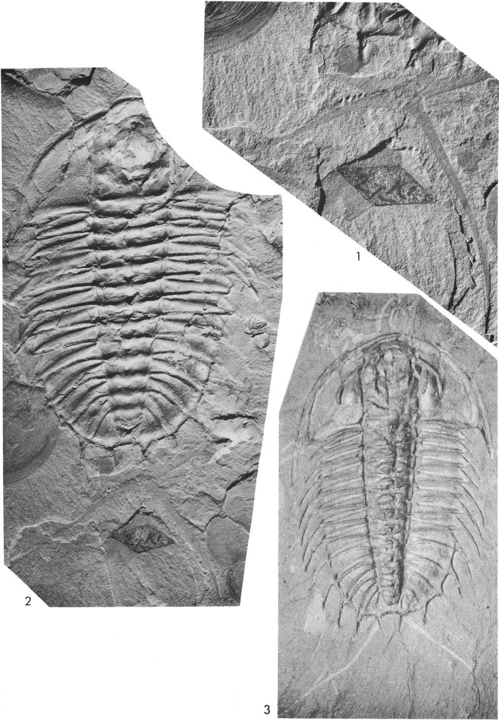
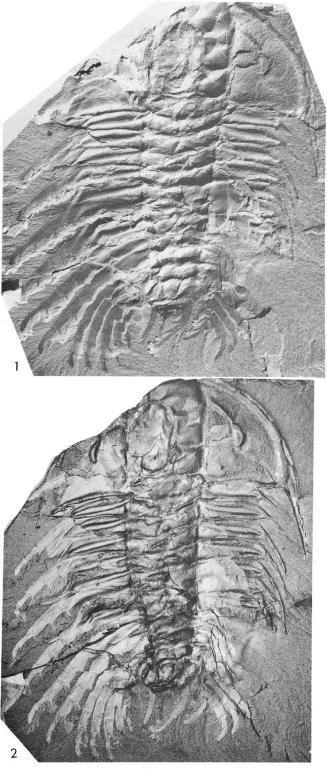
20 lomm Fig. 5. Plate 1. Olenoides serratus (Rominger, 1887), Phyllopod bed, Walcott quarry. l, USNM 57656, west, x2.5, detail of posterior appendages, see Fig. 5. 2, USNM 57656, north, xl. 7. 3, USNM 57657, west, xl.3.
21
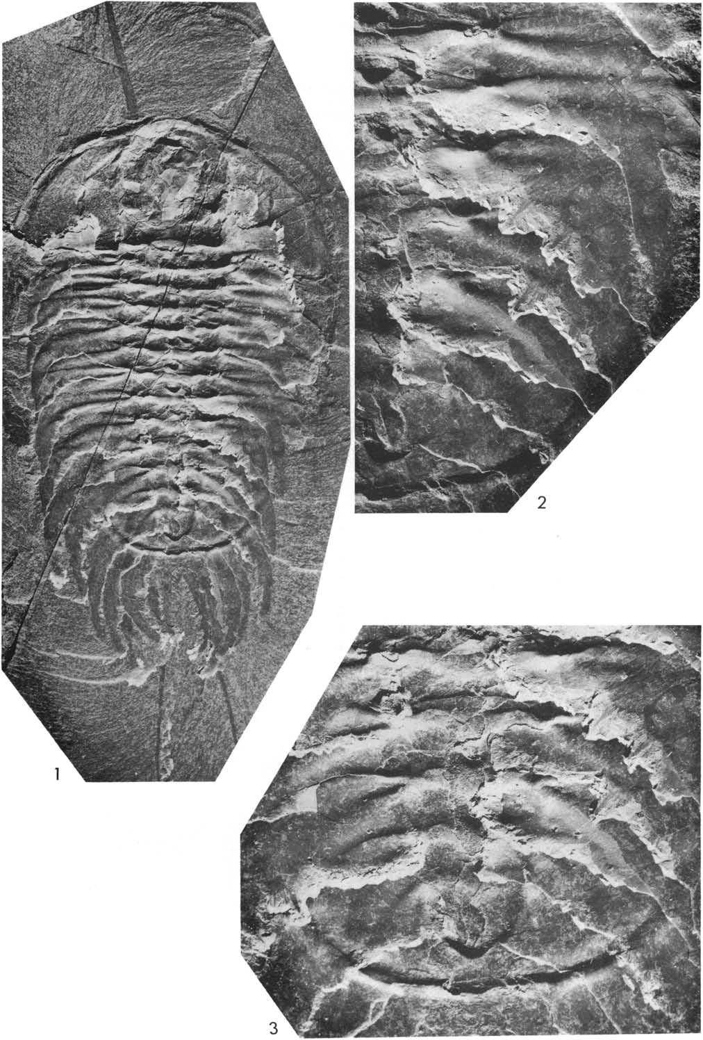
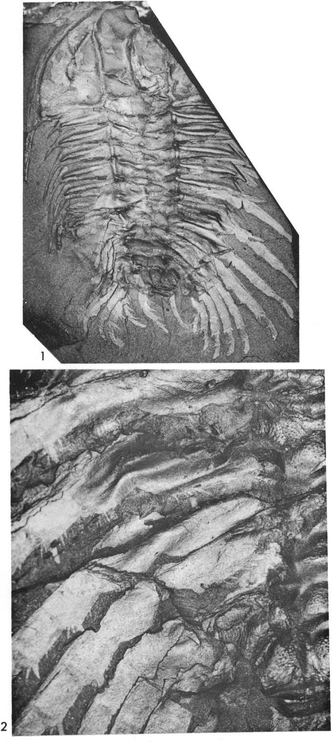
22 Fig. 6. Plate 2. Olenoides serratus (Rominger, 1887). US NM 58588A, Phyllopod bed, Walcott quarry. 1, north, x1.25, see Fig. 6. 2, northeast, x3.3, detaiis of coxae and proximal parts of leg branches R VII to XI. 3, north, x3.3, details of coxae and proximal parts of leg branches R VIn to XI, L IX to XI, see Fig. 6.
23
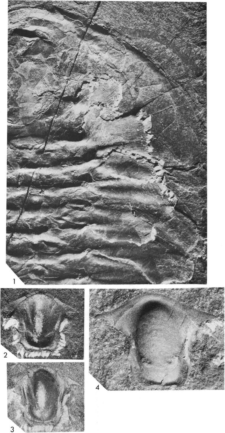
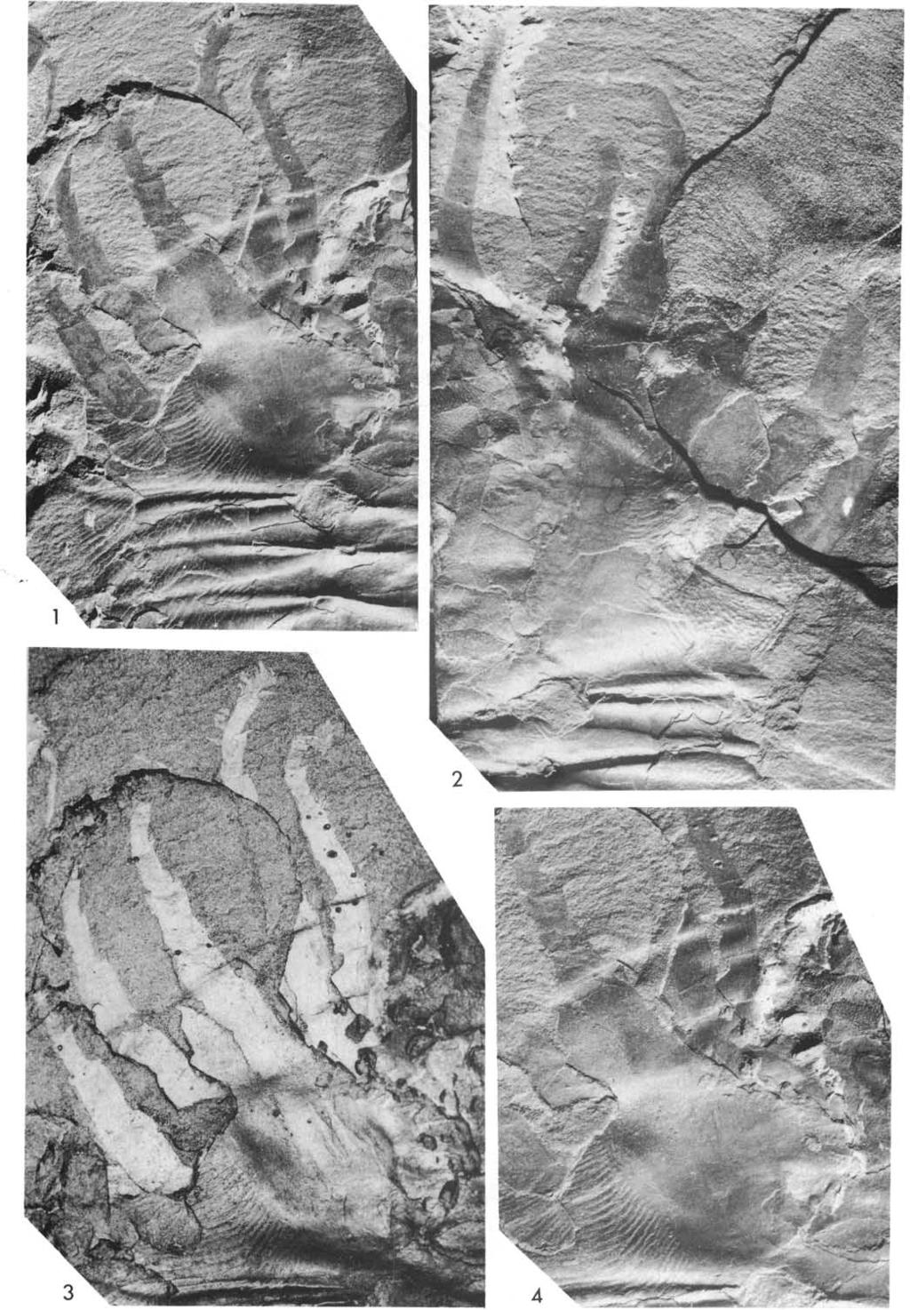
24 rs rp wp -:rl-l-+--- m Fig. 7. Plate 3. 1, Olenoides serratus (Rominger, 1887), US NM 58588A, Phyllopod bed, Walcott quarry, north, x3.3, details of appendages RI to VI, see Fig , Koo tenia burgessensis Resser, 1942, Phyllopod bed, Walcott quarry. 2, 3, USNM 65533, north, reflected, x5, see Fig. 7. 4, USNM , northwest, x5.
25
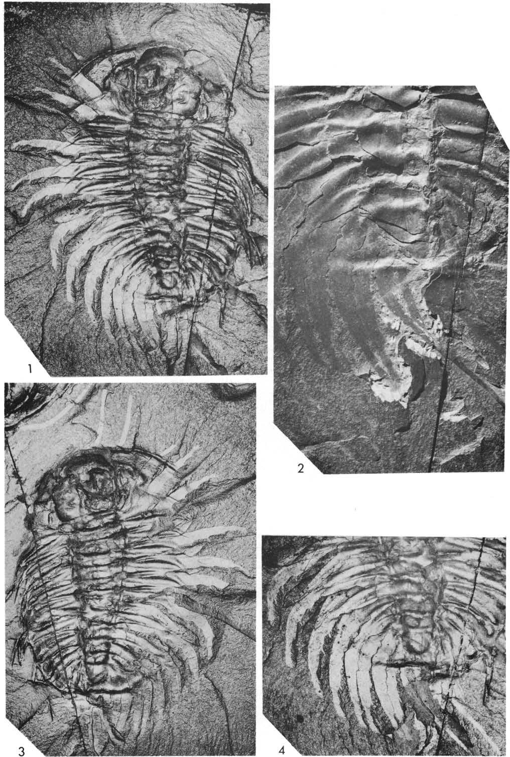
26 Ran ill lill 1- Fig. 8. Plate 4. Olenoides serratus (Rominger, 1887), USNM 58588B, Phyllopod bed, Walcott quarry. See Fig. 8. 1, 2, 4, counterpart, reflected, xl.25, north, x2.5, reflected, xl.7, showing leg branch RXV lying ventrai to right cercus. 3, reflected, ' xl.25.
27
28 Fig. 9. Plate 5. Olenol des serratus flec ted, xl 7. (Rommg. er, 1887), USNM 58589, Ph y llopo d bed, Walcott quarry. See Flg , 2, north, re
29
30 Plate 6. Olenoides serratus (Rominger, 1887), USNM 58589, Phyllopod bed, Walcott quarry. See Fig. 9. 1, counterpart, reflected, x1.7. 2, reflected, x5, showing detaiis of coxae L 5 to 8.
31
32 lomm ill oc -- It Fig Plate 7. Olenoides serratus (Rominger, 1887), Phyllopod bed, Walcott quarry. See Fig , 3, 4, USNM 65513, northwest, x2.5, reflected, x3.3, northwest, x3.3. 2, counterpart, USNM 58590, north, x3.3.
33
, USNM 58591, Phyllopod bed, Wa1cott quarry.")
34 IO mm 2p lp Fig. 11. Plate 8. Olenoides serratus (Rominger, 1887), USNM 58591, Phyllopod bed, Wa1cott quarry. See Fig , reflected, x2.5. 2, southwest, x2.5.
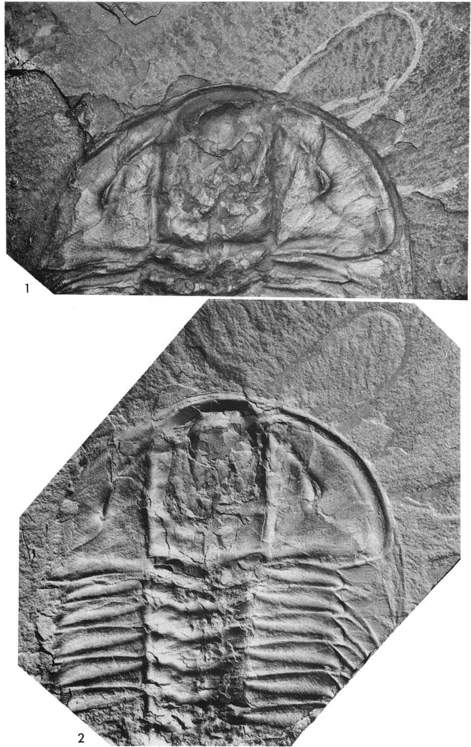
35
36 oc ',/ C /" Fig Plate 9. Olenoides serratus (Rominger, 1887), Phyllopod bed, Walcott quarry. l, USNM 65510, north, xl.7, see Figs. 12A, B, C. 2, 4, USNM 65514, counterpart, north, reflected, xl.7, see Fig , USNM , north, x3.3, incomplete isolated appendage.
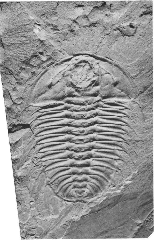
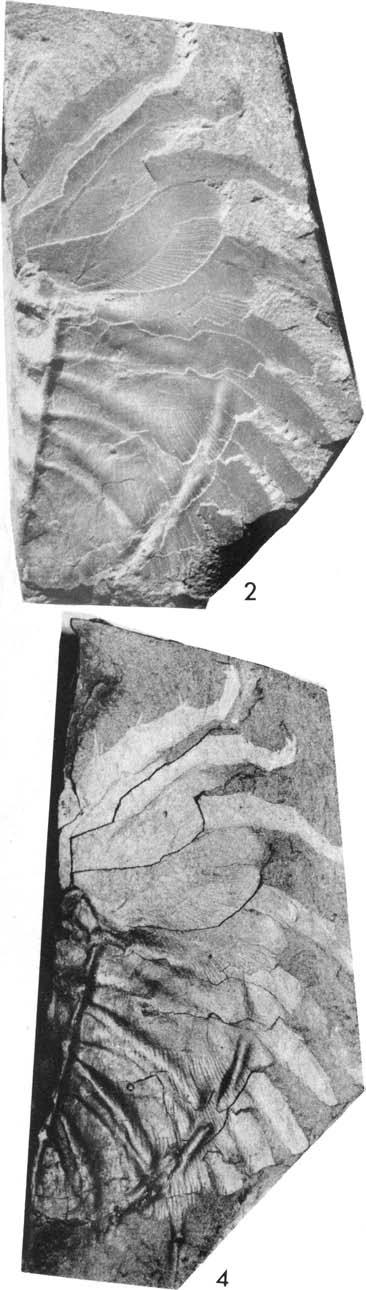

37
38 RS Fig. 13. Pla te 10. Olenoides serratus (Rominger 1887) US NM P lon o e t c ereus. x2.5. 2, northeast, x2.5. Phot graph; made be f ore e ' xc h avya ll t Po d f b l edf' Walcott quarry. See Fig , reflected '

39
40 R3cox lomm A.. Fig. 14. Plate 11. Olenoides serratus (Rominger, 1887), US NM 65515, Phyllopod bed, Walcott quarry. l, west, x2.5, see Fig. 14A. 2, reflected, x5, see Fig. 14B. 3, counterpart, reflected, x2.5, showing appendage LI, see Fig. 14A.
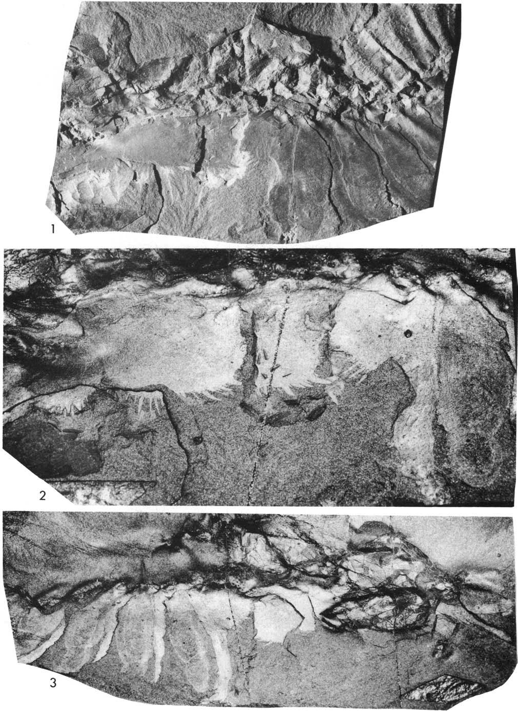
41
42 - 4t S t lomm 6t Fig. 15. Plate 12. Olenoides serratus (Rominger, 1887), USNM 65519, Phyllopod bed, Walcott quarry. See Fig , 3, north, reflected, xl. 7. 2, counterpart, northwest, x2.5. 4, northwest, x2.5, details of right appendages 5 and 6.
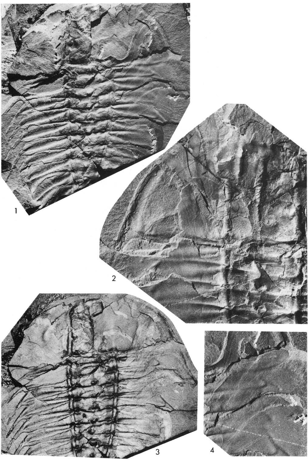
43
44 Fig. 16. Plate 13. Olenoides serratus (Rominger, 1887), USNM 65520, Phyllopod bed, Walcott quarry. See Fig , counter part, north, xl. 7. 2, 3, 4, north, xl. 7, west, x2.5, reflected, x2.5.
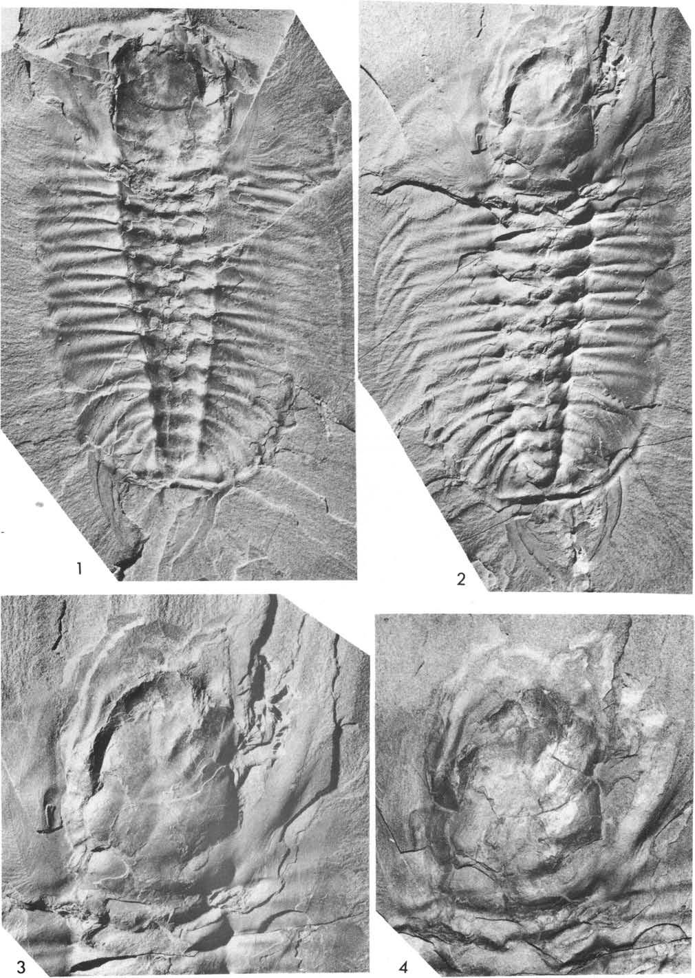
45
46 .. Fig. 17. Plate 14. Olenoides serratus (Rominger, 1887), US NM 65521, Phyllopod bed, Walcott quarry. See Figs. 4, 17. 1, north, x5, showing appendages L l to 3, see Fig. 4A. 2, north west, xl. 7, see Fig. 17, in which coxa and leg branch 8 are restored in outline. 3, west, x5, showing appendages L 8 to 14 and right appendages, see Figs. 17, 4B, 4C.
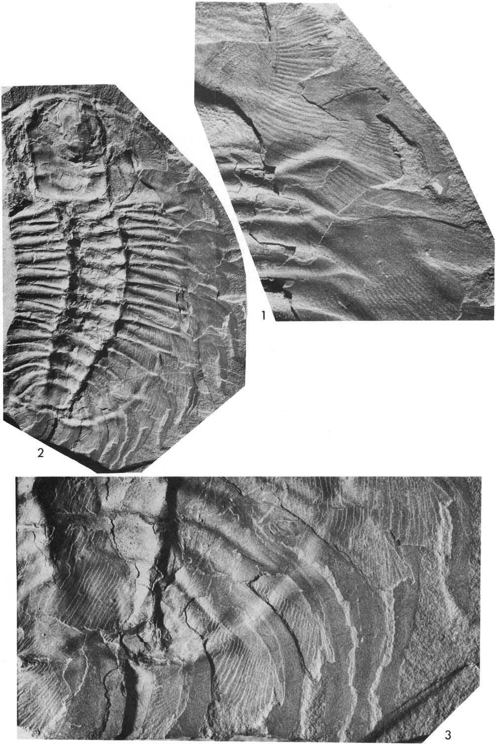
47
48 Lan lomm Fig. 18. Pla te 15. Olenoides serratus (Rominger, 1887), USNM , Phyllopod bed, Walcott quarry. See Fig. 18, in which highly reflective areas are shown by vertical lines, and granulation indicated on fragments of extemal surface. l, 2, northwest, reflected, xl. 7. 3, northwest, x3. 7.
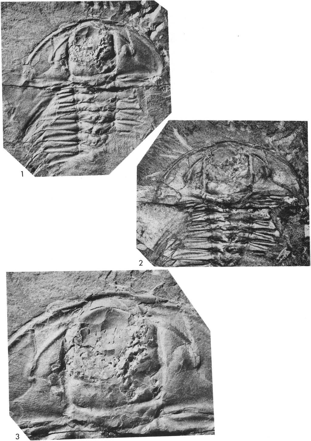
49
50 Fig. 19. Plate 16. Olenoides serratus (Rominger, 1887), US NM , Phyllopod bed, Walcott quarry. See Fig , east, xl.7. 2, counterpart, west, xl. 7. 3, northwest, x5, showing coxae and leg branches 6 to 12 and base right cercus. 4, counterpart, northwest, x5, showing leg branches 8 to 12, base cerci, and left appendage 12.
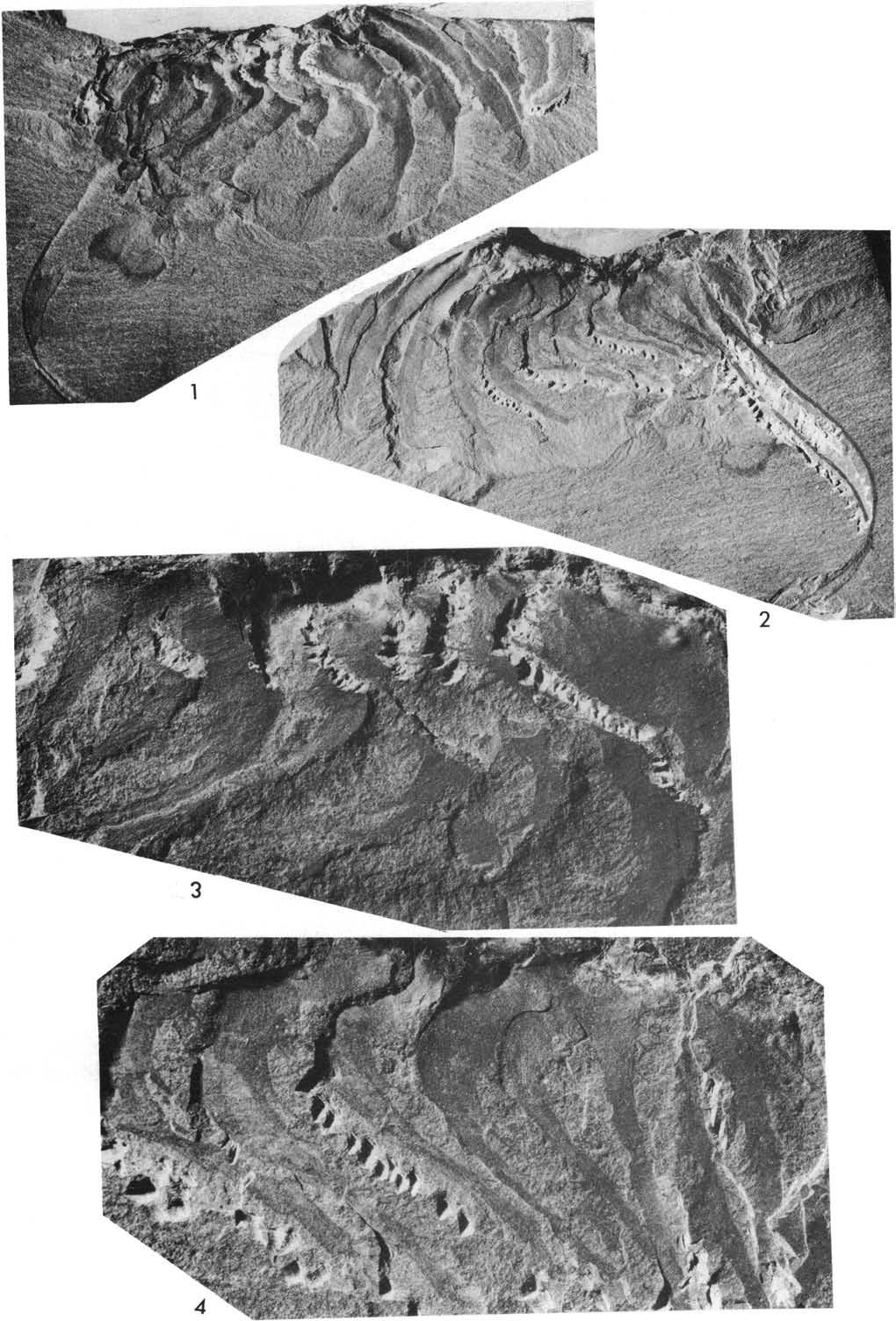
51
52 Fig. 20. Plate 17. Olenoides serratus (Rominger, 1887), GSC 34692, Walcott quarry, leve! 6ft. 10ins. to 7ft. 2ins. See Fig. 20. l, counterpart, northeast, xl. 7. 2, north, xl. 7. 3, counterpart, southwest, x3.3.
53
54 Plate 18. Olenoides serratus (Rominger, 1887), GSC 34692, Walcott quarry, leve! 6ft. 10ins. to 7ft. 2ins. See Fig. 20. l, counterpart, reflected, x3.3, compare Pl. 17:3. 2, reflected, x3.3, showing left leg branch x?, right leg branches v to y and c6xa Rx (inverted relative to Fig. 20).
55
56 Fig. 21. Plate 19. Olenoides serratus ( Rominger, 1887), esc 34693, Walcott quarry, leve! 10ft. 4 ins. to 10 ft. 8 ins. See Fig. 21. l, northeast, x3.3, details of anterior portion. 2, northwest, x3.3, detail of posterior portion. 3, reflected, xl. 7.
57
58 p m -c -- -::---- /'---- l _ 2t t \ _ l ṯ _l--- 6t 'J 5t L 7t am p y o l py \ 6 Fig. 22. Plate 20. Olenoides serratus (Rominger, 1887). 1, 2, 4, esc 34694, Walcott quarry, leve1 6ft. 7.5ins. to 7 ft. 3.5ins., see Fig. 22, 1, northeast, x3.3, 2, reflected, x3.3, 4, northwest, x1.7. 3, esc 34695, Walcott quarry, lev ei 6ft. llins. to 7ft. Dins., reflected, x3.3, see Fig. 23 and Plate 23:1.
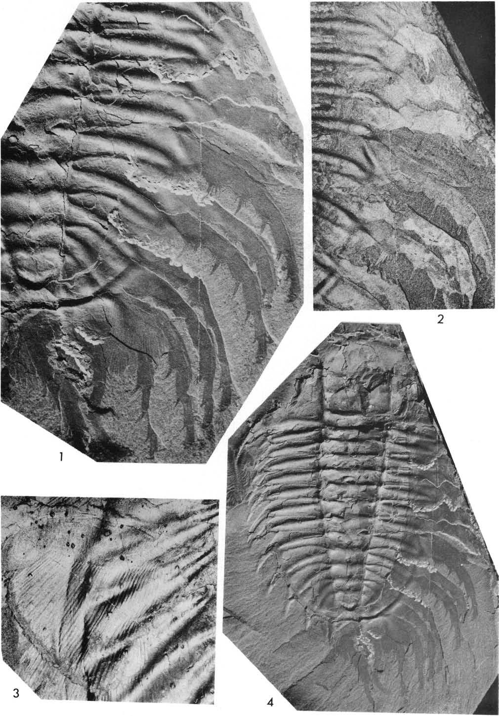
59
60 Plate 21. Olenoides serratus (Rominger, 1887), GSC 34694, Walcott quarry, leve! 6ft. 7.Sins. to 7 ft. 3.Sins. See Fig. 22. l, 2, counterpart, northwest, reflected, x3.3.
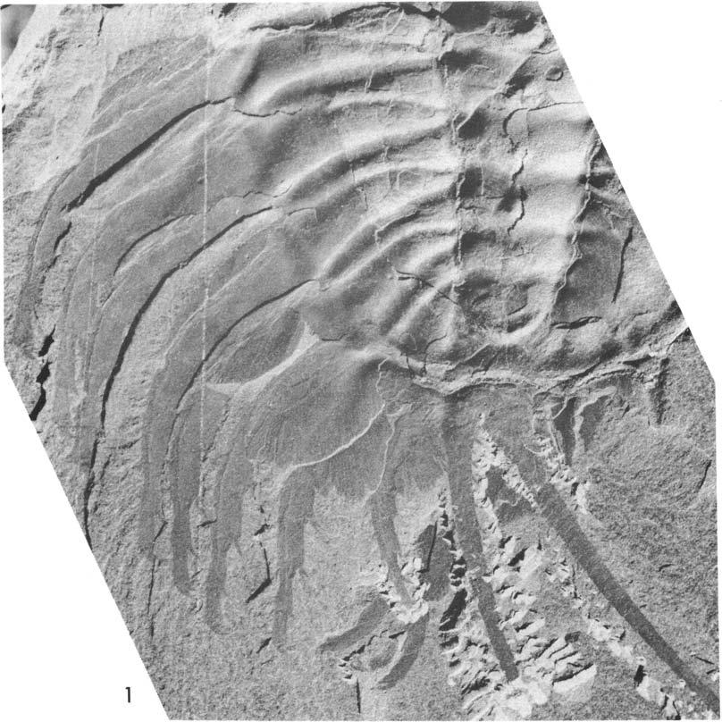
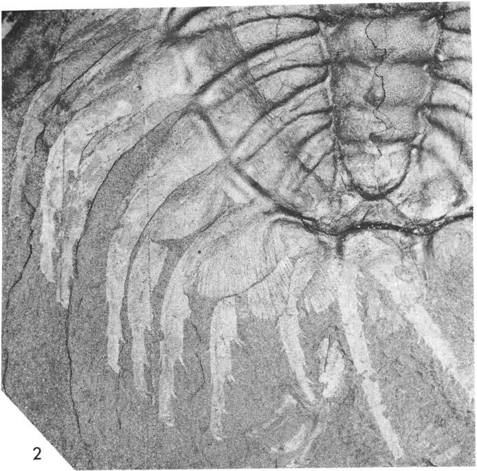
61
62 Fig. 23. Plate 22. Olenozdes. se rra tus l 4 w=""'. (;ee Pl. 23:1).. See Fig. 23. (Rominger, 1887 ), GSC 34695, Wa kott quarry, le vel 6ft. llins. to 7 t reflected, x 5. 2, 3,co unterpart, r eflecte d, northeast, x 1. 7, before exca t r;: of l amellae of 19.
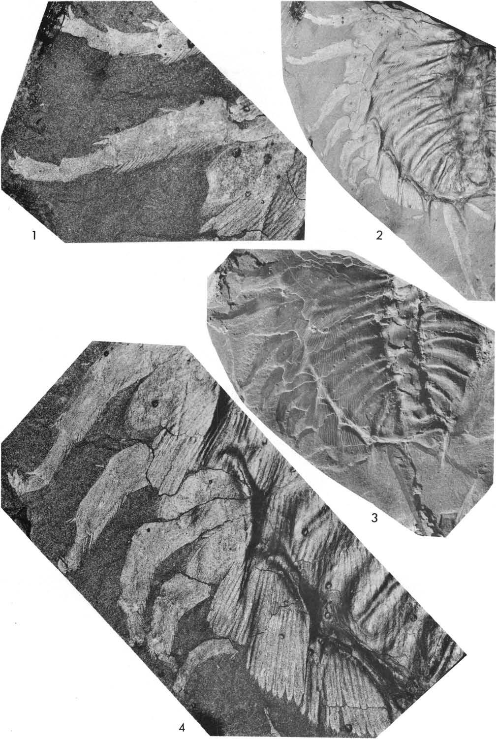
63
64 rp Fig. 24. Plate 23. Olenoides serratus (Rominger, 1887). 1, 3, GSC 34695, Walcott quarry, leve! 6ft. llins. to 7ft. Oins, l, counterpart, west, x5, showing lamellae of 19, see Fig. 23, 3, reflected, xl. 7, showing leg branches R 7, R8, and L8, see Fig , 4, SM A 89326, Phyllopod bed, Walcott quarry, 2, west, xl.7, 4, reflected, x2.5, see Fig. 24.
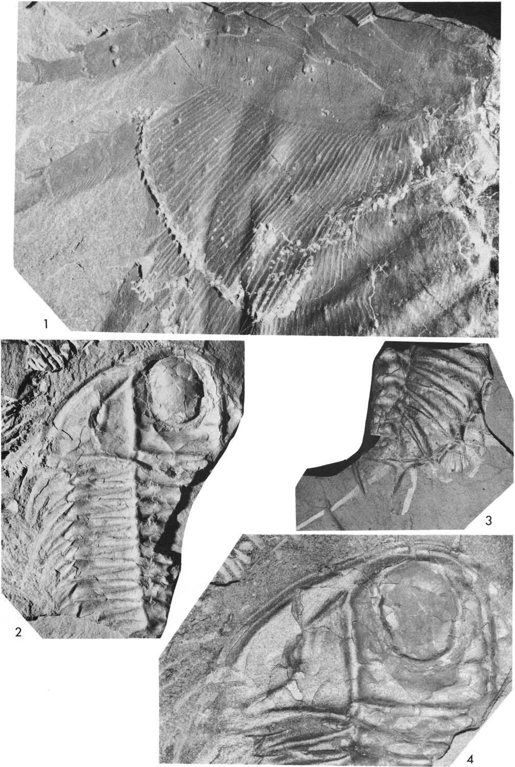
65
66 Plate 24. Olenoides serratus (Rominger, 1887), GSC 34697, Walcott quarry, leve! 6ft. llins. to 7ft. 2ins.. 1, 2, north, x3.3, xl. 7, showing details of appendages and entire speeimen. 3, 4, reflected, x5.0, x3.3.
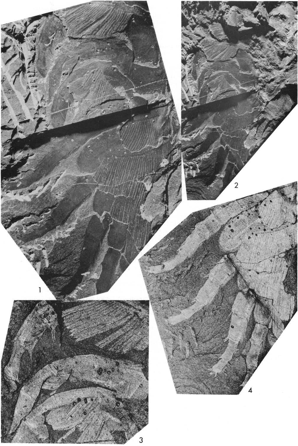
67
68 This arrangement is that the leg branch is flexed to curve backward, and lies ventrai to (i.e. nearer the observer) the anterior part of the inner and outer lobes of the gill branch (Pl. 14: 1,3). The preciseness of this arrangement, well displayed in appendages 2,3,5-9,11-13, reflects the attachment of the two branches. This attachment must be proximal, presurnably of the gill branch to the coxa, and distally segments 4-7 of the leg branch lie ventrai to (i.e. nearer the observer) the outer part of the inner lobe and outer lobe of the gill branch. Ventrai spines and dorsal setae are not preserved on the leg branches, but the direction of flexure implies that the anterior face is toward the observer (i.e. ventrally directed). This implies in turn that the leg branches have been rotated through about 90 0 to lie close to the ventrai surface of the gill branches. Presurnably this rotation was approximately along the line of attachment between the two branches, i.e. along the upper, posterior edge of the coxa, and the coxa was also rotated. Only in appendage LI0 (Pl. 14:3) is a portion of the cox a preserved. In Fig. 17 a restoration of the cox a and leg branch L8 is outlined, based on the similar sized appendage in This restoration implies that segment 4 of the leg branch was situated in life below the outer lobe of the gill branch (as also shown by 58588B and 58589). This relationship is also suggested by appendages L5 and 6 (Pl. 14:2) of this specimen. Accepting this relationship, then appendages L5, 6 and 8 show that lamellae of the gill branch are present proximally as far as at least the outer two-thirds of the coxa. Størmer suggested that the se lamellae extended inward "practically to the axial furrows" (1939: 197) of this specimen, and concluded that the gill branch ("preepipodite") was attached not to segment 2 (the basal of the leg branch) but to the proximal portion of the coxa. This latter claim appears exaggerated, but this specimen does show the relative positions and size of the two branches, and that the gill branch lay beside at least the outer two-thirds of the coxa. The regular fan form arrangement of the appendages confirms the attachment between the two branches. Distally the difference in level and relative position between segments 3-5 of the leg and gill branch in appendages L2, 3, 5, 6, indicates that the attachment was proximal to segment 3. Evidence from specimens 65514, 65519, and 65520, combines to suggest that the attachment was between the outer two-thirds of the cox a and the basal part of the inner lobe of the gill branch. In this specimen the curvature imposed on the body has imbricated the right pleurae, and the pygidium is also slightly displaced. The restoration of coxa and leg branch L8 shows that appendages of the left side are displaced outward relative to the exoskeleton. Behind the pygidium there is a large change of level beside leg branch L14 (Fig. 17, Pl. 14:3). To the left of this step, and below and adjacent to the axis of the pyg " idium, are parts of the right appendages. There are portions of two leg branches (Ra and b) and of at least four gill branches. It will be seen that the lamellae of these right gill branches are directed approximately parallei to those of the corresponding left appendages, and not inward and backward as a symmetrical arrangement would require. I explain this as meaning that the se gill branches have been folded under through so that they lie beneath the axis, dorsal surface toward the observer, the long axis directed inward and backward, the lamellae at right angles, outward and backward. Whether or not the leg branch fragments have been similarly folded over is uncertain, but the dorsal setae of segments 4 and 5 appear to be visible on Ra, sugge sting a symmetrical arrangement of the se twa branches. A falding over of more anterior right appendages, beneath the axis and the appendages of the left side, would explain the absence of appendages preserved beneath the right pleurae of the thorax and anterior part of the pygidium. Størmer cited this specimen as showing a "frontal marginal rim" (1939: 198) of the gill branch. As explained above, I interpret appendages L3, 5 and 6, as showing the distal part of the leg branch lying beneath (i.e. ventrai to) the anterior margin of the gill branch and so concealing it. The anterior margin is thus not displayed, and whether or not a marginal rim exists on the inner lobe cannot be seen. The distal lobe of L3 and 4 is exposed and shows no sign of such a rim. The gill branches show not only the length (exs.) of the lamellae - extending back above the leg branch of the succeeding appendage - but also their arrangement (Fig. 4, Pl. 14:1,3). Each lamella is rounded at the tip, and on the left side successive lamellae are seen to be imbricated so that the outer edge is dorsal to the lamella external to it. Plate 14:3 shows the ventrai side of gill branches L 8-13, and the broken edges of the lamellae form a series of scarps facing the sagittal line. In the distal part of gill branch L l (Fig. 4A), this imbrication appears to be reversed. Further, the gill branches of the righ t side (Pl. 14: 3), which it is argued above may be folded under to show the dorsal side, have the "scarp" faces of individual lamellae facing away from the sagittal line, i.e. are imbricated so that the outer edge of each lamella is below the one next to it. One interpretation is that flat lamellae could be rotated to be imbricated either way. A second interpretation is that the lamellae were originally oval in cross section, and by compaction have been flattened and imbricated in either sense. 116
69 USNM Plate 15: 1-3, Figure 18. o 1949 Størmer, in Grass e : 176, Fig. lld. O 1951 Størmer: 128, Fig. 7. O 1959 Harrington, in Moore : 056, Fig. 41. Størmer (1951) gave a drawing of the cephalon of this specimen, claiming that it showed the postoral plate or metastome. His 1949 illustration was only of the part he considered to represent the "labium" or metastome. The specimen shows the cephalon, parts of the seven thoracic segments, and the left anterior portion of the pygidium, preserved as a mould of the internal surface of the exoskeleton. Fig. 18 indicates the features displayed by the cephalon, and that some fragments of the exoskeleton adhere to it and reveal the granulate external surface. The glabeila is crushed, parts of both branches of the dorsal suture, eye ridges, and impressions of connective sutures are preserved. The latter suggest that the hypostome was approximately in place, and portions of the middle body and posterior border are visible, including granulation and terrace lines on these regions. On the left side, outside the cephalic border, part of an antenna and distal portions of four kg branches are poorly preserved. On the left cheek, outside and behind the eye lobe, outwardly directed lamellae of a gill branch are visible. These traces of appendages are more strongly reflective than adjacent areas of the exoskeleton (Pl. 15: 1,2 ). On the glabeila are are as which reflect with different intensities, those which reflect more strongly are shown in Fig. 18 by vertical ruling. In low angle radiation (Pl. 15: 3) some of these latter areas appear darker, their boundaries vaguely defined and irregular, except for the curving strip ly ing near the mid-iength of right glabellar lobe 2p. These reflective are as are on the internal mould of the glabeila, and might, as Størmer (1951) claimed, be fragments of appendages. Their size, irregular outline and positions make any interpretation dubious. The quadrangular area which Størmer outlined (1951, Fig. 7) as the metastome is just recognisable in low angle radiation (Pl. 15: 3), and less clearly so in reflected radiation (Pl. 15: 2). The triangular central area which Størmer showed is barely discernible, and the outline of the supposed plate itself by no means clear. I conclude that this specimen, as the only evidence of the metastome in O. serratus, is not convincing. USNM Plate 16: 1-4, Figure 19. Twelve appendages of the right side, the cerci, and three appendages of the left side are incompletely preserved as counterparts approximately parallel to the bedding. On the broken edge of the block may be seen fragments of the exoskeleton, the external surface facing in a direction which is towards the top in Figure 19. These fragments lie across the bedding, in surfaces which are beyond and below the edge shown in the figure. The animal was thus buried lying obliquely on its side. During the process of burial the coxae and leg branches of the right side were imbricated so that each passes marginally below the one following, swung round to lie in exsagittal planes, the leg branches curved to face convexly forward, the posterior side facing outward. The right cercus lies over the left cercus, but passes marginally below the coxa of right appendage 1 2. Left appendages 10 to 12 are curved to face in the opposite direction, and imbricated in the same sense. Since both cerci are visible, and three left as well as twelve right biramous appendages, burial of the animal was not exactly on its side, but obliquely as described above. Coxae 6-9, Il, 12 have been partially excavated and show the relative size and spinose ventrai and inner margins (Pl. 16: 3). A large spine is present on the inner, ventrai corner of coxae 6-9, other spines varying in size and also partially overlapping proximally. This overlap indicates that they were not arranged i n a single line on a sharp edge, but over the surface of a convex margin. A prominent group of long spines was present on the mid-ventral side of segment 2 of the leg branch, and groups of spines, diminishing in size distally, at the distal ends of segments 3-6. Up to three short, broad-based spines, overlapping at their bases and evidently movable, terminate the leg branch. Fragments of gill branches lie upon leg branches 3 and 5, and beside L 12, the latter showing lamellae. The cerci are broad-based, taper rapidly, then gradually, segments are short proximally, with spines at the joints, distally apparently much longer. Running below and between the cerci proximally is a narrow strip, appearing dark in low angle illumination but highly reflective. The position of the strip suggests that it may represent contents of the gut squeezed out posteriorly. ll7
70 USNM Pl. 9 :3. The specimen shows an almost complete, isolated gill branch, the posterior margin of the inner lobe bearing about 50 lamellae. These lamellae are imbricated so that each passes beneath that next inside it, shortest distally and with tips bluntly rounded. The lamellae become progressively longer inwards, the appearance that they shorten in the innermost portion may be because they are incomplete or hidden by matrix. The maximum length of the lamellae is about twothirds the axial width of the inner lobe. A distinct line separates inner and outer lobe, the latter broken around the margins and thus not showing marginal setae. Adjacent to the anterior margins of the two lobes, crumpled and ly ing at a slightly lower level, is an irregular dark strip that shows no clear subdivisions. I regard this strip as a poorly preserved portion of the leg branch, broken at each end. Since it lies at a lower level than the lobes of the gill branch, the change in level being least proximally and at a maximum distally, one may assurne that the photograph shows the dorsal side of the gill branch, in the usual relation to the leg hranch. In form the gill branch is typical of O. serratus and is so assigned. Another fragment, the distal parts of the two branches of an appendage, typical in form though crumpled, was figured by Walcott (1931, Pl. 13:1) and assigned to o. serratus by Størmer (1939:199). A third, less complete specimen was obtained by the GSC party from level 8 ft 7 ins to 8 ft 10 ins in the Walcott quarry. Thus broken portions of appendages of O. serratus do occur rarely in the Burgess Shale. esc Plate 17: 1-3, Plate 18: 1, 2, Figure 20. This unique specimen, on a bedding plane with Marrella sp lendens, shows an isolated pygidium split so that some of the external surface is revealed (Pl. 17: 1,2). Behind it are a group of appendages, not associated with the exoskeleton, and showing the coxae exceptionally well. The specimen may be part of a disrupted animal, or part of a fresh moult in which the sclerotized cuticle of the appendages, presurnably held together by the ventrai cuticle, has separated from the mineralized exoskeleton. Numbering of these appendages presents a special problem since they are not in a single series. A unique convention of lettering has therefore been adopted in Figure 20, which is a ventrai view, i.e. as seen in the counterpart, Plate 17: 1,3. The outline, particularly of the broad base, and the setae along the margins, sugge st that it is the cerci that curve back to the right of the pygidium. Imbricated successively above the cerci are four pairs of appendages (Rv to Ry and Lx? ) showing in ventrai view the anterior face of the coxa and leg branch. The gill branches are not visible, since they were on the posterior side of the appendage and so are concealed in the rock. The cox a of appendage Rx has been excavated to reveal the spinose ventrai and inner margins (Pl. 18: 2, inverted relative to Fig. 20). Three larger spines are directed ventrally in the mid-ventral region, another large spine is directed inward and ventrall y from the inner part of the ventrai margin, and two large spines are directed inward from the proximal margin. Between these large spines are many smaller spines, which overlap at their bases, suggesting that they were originally directed slightly forward or backward as well as outward. Small spines continue to the most dorsal part of the proximal margin. A further group of imbricated appendages lies adjacent to those described. In ventrai view (Fig. 20) the leg branches are preserved with the posterior face toward the observer. That nearest on the right side is labelled Rj, a fragment of the leg, visible on the part (Pl. 17:2). Those successively below it are labelled Rk to Ro, Rra being a fragment whose position in the series is uncertain. Rm and RI both underlie Rk (Pl. 17: 1,3), and may he parts of one appendage hut I consider this unlikely. Gill hranches lahelled Rmg, Rng, and Rog, are preserved extended in the same plane as the leg branch. The spinose ventrai and inner margins of cox a Rk (Pl. 17: 3, Pl. 18:1) are well shown despite a fold runn ing parallei to, and inside, the ventrai margin. The long ventrai spines of segments 2 and 3 of leg branch Rk are also cleariy shown. On the left side four appendages are preserved and labelled Li to l on the assumption that Lk and Rk, being on the same level (Pl. 17:3), belonged to the same somite. Large and small spines on the ventrai and inner margins of coxae Lj and Lk are evident, and with those of coxa Rk give an impression of the opposition of the gnathobases in life (Pl. 18:1). Fragments of gill branches, similarly extended to those of the right side, are also preserved. This specimen thus show parts of at least eleven pairs of hiramous appendages and the cerci. Too little of Ry is preserved to indicate whether or not it is the last appendage, hut 118
71 differences in level imply that it cannot be more than l or 2 from the last. It is uncertain whether Li represents the pair that originally lay next to the Rv pair, and the coxae and leg branches of the two groups of appendages have not only been rotated but their serial alignment modified. If Ry was the penultimate biramous appendage, and none is missing between Rv and Li, then appendages j and k are 7th and 8th in front of the cerci, i.e. they belonged to the middle part of the thorax. The large size and spinosity of the coxae in the posterior half of the animal's appendages is notable, and accords with the evidence from other specimens. esc Plate 19: 1-3, Figure 21. The entire exoskeleton is extended in the bedding plane, and shows the antennae, fourteen biramous appendages on the left side, the cerci, the presumed first, and the last two, leg branches of the right side. A fragment of the lamellae of a gill branch is revealed beneath the right fixed cheek, and the right margin of the block cuts off the tips of some of the pleurai spines and the first pygidial border spine. It is presumed, from the attitude of the labelled right branches, and the fragments revealed where axial rings are broken away, that the biramous appendages of the right side were folded beneath the axial and inner right pleurai region. No attempt has been made to excavate them. On the left side the antenna is preserved (Pl. 19:1) beneath the cephalic exoskeleton to a point almost in line (tr.) with the anterior margin of the eye lobe. The antenna broadens proximally, where it lies at a level above that of the proximal part of the succeeding appendage. The biramous appendages are disposed in a fan-wise arrangement, each gill branch lying dorsal to the leg branch and overlapping its successor. The posterior side of the leg branch faces dorsally and each branch passes below its successor, XIV going beneath the left cercus (Pl. 19 :2), though gill branch XIV overlaps the left cercus. The cercus in thus preserved in an attitude corresponding to that of the leg branches. The latter are flexed distally, most strongly anteriorly, where in I and Il segments 4 and 5, and 5 and 6, are flexed almost at right angles to each other, without disruption being apparent (Pl. 19: l). In III the flexure is so sharp that the distal two or three segments are apparently hidden beneath the proximal segments and gill branch. In IV to VIn the flexure is less than 90 0 between each of segments 2-7, so that the branch curves through between approximately Excavation has shown (Pl. 19:2) how leg branches IX to XI curve beneath each other below the gill branches. Curvature is less in XII to XIV. The preservation is such that proximally gill and leg branch were pressed closely one above the other, and it has not proved possible to rem ove the gill branch to reveal the coxa completely. The portion of the coxa visible in VI indicates displacement of the appendages behind the cephalon to the left of the sagittal line, as does the position of the cerci. The antenna, and possibly left appendages I to Ill, appear not far removed from their original positions. If one assurnes they originally belonged to the cephalon (Ill being the occipital appendage), then presurnably IV to X each belonged to a thoracic segment. Backward displacement during burial is evident, since coxal IV is opposite the second axial ring of the thorax, and coxa X opposite pygidial axial rings 2 and 3. The above assumptions mean that XI to XIV belonged to pygidial segments l to 4, and the cerci perhaps to pygidial segment 5. These assumptions are based on the earlier assumption that I to XIV is a complete series of biramous appendages, and the present preparation suggests that this is reasonable. esc Plate 20: 1,2,4, Plate 21: 1,2, Figure 22. The dorsal exoskeleton (Pl. 20: 4) is extended in the bedding plane, and appendages project from beneath it on the right side and posteriorly. The hypostome is displaced back and to the right, the posterior border lying beneath the right half of the occipital furrow. The rock slab terminates in ajoint which runs diagonally across the cephalon on the right side just outside the eye lobe, so that anterolaterally appendages and exoskeleton are not seen. No appendages project from beneath the left side, apparently because they have been folded under the axis and right side. This is deduced from the position of the five posterior walking legs on the left side, which are imbricated and project posteriorly below the cerci and appendage R 13. The left cercus is not so folded, and extends backward and outward on the left side. On the right side parts of 13 biramous appendages are preserved, principally the leg 119
72 branches, in dorsal view posterior face towards observer, imbricated so that each goes below the one following. Coxae 1,3,6,7, have been excavated (Pl. 20: 1,2) to show the spinose ventrai and inner margins, large spines being present in the mid-ventral and inner ventrai regions. Coxae 12 and 13 have also been excavated, and while smailer show the spinose ventrai margin. The leg branches, and ventrai spines of segments 2-6, are revealed in 6 and 8, and appear to be similarly developed on the other leg branches, induding 13 (Pl. 21: 1,2). GiB branches are imbricated dorsal to the leg branches, each going above the one fobowing, and 12 g shows the outer lobe. Gill branches 11 to 13 extend inward dose the margin of the pygidium (Pl. 21: 1,2) and thus must extend inward for much of the length of the coxa. GiIl branch 6 g is also visible (Pl. 20:1), crushed in the exsagittal direction and lying in front of the leg branch, and the counterpart shows parts of gib branches 5 and 8, lying dorsal to the leg branches. The cerci are broad proximally and taper rapidly initiaily, more gradually distaily. Stout spines project from the joints on one side (Pl. 21 :2), which appears to be the ventrai side, distally the spines are slimmer and seemingly less frequently developed. esc Plate 20:3, Plate 22: 1-4, Plate 23: 1,3, Figure 23. Eight biramous appendages of the right side, the cerci, and left leg branch 8 are preserved beneath and outside the last three thoracic segments and the pygidium. Fragments of three more anterior gib branches are preserved on the counterpart (Pl. 22: 2,3) but not numbered, and leg branches R 7, 8 and L8 are best seen on the part (Pl. 23: 3). Figure 23 is a ventrai view and the gill branches are immediately beneath the exoskeleton, overlapping so that each lies dorsal to the one following (Pl. 22: 3). The outer lobes of gill branches l to 5 are partly visible, and show the marginal setae (Pl. 22: 1,4). These setae overlie dorsally the adjacent lameilae of the inner lobe (Pl. 23: l). Each lamella is long and narrow (tr.), and their length is such that they extend above the second leg branch fobowing (Pl. 23: l). The slat-like lamebae are compacted upon each other, the inner edge ly ing dorsal to its neighbour so that across their width they slope downward and outward. The tip of each lameila is rounded and bears four or five fine hairs (Pl. 22:4). Leg branches R 1-8 are preserved with the posterior side facing ventraily, and are extended in an "S" curve, concave posteriorly over much of the length, becoming convex distaily. The concave curvature increases progressively backward, so that leg branch 8 is curved backward through more than 90 (Pl. 23:3). The proximal portions of appendages 7 and 8 have been excavated in the part, and the spinose margin of the coxa is visible in 7. The distal portions of the leg branches are well preserved, and show the groups of spines on the ventrai side at the joints, and the setae along the dorsal margins of segments 4 and 5 (Pl. 22: 1,4). The distal portion of a leg branch that has been excavated outside the right cercus (Pl. 23:3) is considered to have the posterior face directed dorsaily, and to belong to the left side, having been bent under the right appendages. The cerci shows marginal spines on both sides, and widen rapidly proximally but cannot be traced inside the pygidial border. This specimen is notable for the exceptional preservation of the appendages, and, judging from the positions of coxae 7 and 8, shows that the gill branches extend far in towards the inner edge of the coxa. This is also well shown by appendage R l (Pl. 23: l, Fig. 23), for the outer lobe of the gill branch is beside segment 4 of the leg branch, and the lamella extend far inward. The proximal part of the appendage is crumpled and poorly preserved, but inner lobe of gill branch is seen to merge in to the coxa. esc Plate 24: 1-4. In this specimen crumpled fragments of the exoskeleton, with the external surface facing the observer, lie partly above and partly beside biramous appendages (Pl. 24: 2). Anteriorly, portions of six appendages of the left side have the leg branches curved convexly forward, rotated so that the posterior side faces the observer, each branch imbricated to pass below that following. The gill branches lie dorsal to the leg branches, and are imbricated so that each lies above the one following (Pl. 24: l). Behind these appendages, and on a slightly lower level, are four biramous appendages of the right side, folded through 180 at the 1 20
73 axial region and extended below those of the left side. The distal parts of the leg branches are curved convexly backward, and have been rotated so that the posterior side faces the observer. The gill branches are extended and pass below the one following, i.e. are ventrai face toward the observer, an opposite arrangement to the gill branches of the left side (PI. 24: 1). The preservation of the appendages is exceptionally fine, and the marginal setae of the outer lobe of the gill branch, and hairs at the tips of the lamellae (PI. 24: 3 ) are elearly displayed. The distal four segments of the right leg branches (PI. 24: 1,4) show the dorsal setae on segments 4 and 5, the ventrai spines at the joints, and the three blunt, broad-based, triangular spines at the termination. The lamellae of the main lobe of the gill branches on the left side are imbricated so that the outei edge is over the adjacent lamella, i.e. across its width the lamella slopes inward and downward. On the appendages of the right side this slope is reversed, the outer edge lying under the adjacent lamella. In these right gib branches the outermost lamellae lie beneath (above in ventrai view, Pl. 24: 1 ) the edge of the outer lobe with the marginal setae. SM A l' late 23: 2,4, Figure 24. Isolated hypostomes from the Burgess Shale, originally referred to this species by Walcott, are now thought more likely to belong to Koo tenia burgessensis (see below). Part of the outline and in some cases the approximate position of the hypostome is shown in a num ber of specimens preserved from the dorsal side (57656, 58588A, B, 58589, 65510, 65520). Only the present specimen, discovered among a collection sent from the U.S. National Museum to University College, London, shows the hypostome from the ventrai side, incompletely preserved, crushed down approximately in position. The convex middle body, outlined by broad, shallow furrows, is widest at a point about one-quarter the length (sag.), the straight sides converging back to the rounded posterior portion. The highest point appears to be about central. Crushing has obscured any middle furrow and subdivision of the body. The anterior wing and lateral border are not preserved, but posteriorly a section of the gently convex border runs transversely behind the middle body, and turns posterolaterally to run forward and outward at 45 to the sagittal line, for a length similar to that of the transverse section. Judging by the lateral border furrow, the lateral border appears to be directed backward and very slightly inward. The anterior limit of the hypostome is difficult to see, but medially it may lie on the posterior slope of the anterior border furrow, at the line presumed to represent the hypostomal su ture (Fig. 24). The exoskeleton is preserved in this specimen, as in others, as a thin black layer, reflective on the smooth inner surface. Such a layer lines the cheek and glabella of this specimen, a sharp edge denoting where it is broken. The layer also elings to the hypostome, and anteriorly shows the raised, anastomosing ridges of the external surface. On the left side (right in Pl. 23: 2,4) the broken edge runs along the slope of the lateral border, matrix intervening between it and the smooth, inner side of the dorsal exoskeleton. Along the posterior and posterolateral borders of the hypostome two exoskeletal layers (outer and doublure) may be observed compacted together. Beneath the lateral border of the cheek part of the doublure exoskeleton appears to be present (Fig. 24), and fragments of an exoskeletal layer, partly obscured by matrix, eling to the anterior border. It is possible that portions of the rostrai plate are represented, especially anteriorly to the hypostome adjacent to the supposed hypostomal suture. A line impressed on the inner surface of the dorsal exoskeleton is interpreted (compare Figs. 12A, 18) as the connective suture, by analogy with the hypostome of Koo tenia burgessensis (Pl. 3: 2,3,4, Fig. 7 ). Thus, following Rasetti (1952: 889), the hypostomes and rostrai plates of Kootenia and Olenoides are considered to have been similar, and possibly fused, though in K. burgessensis the hypostomal suture is elearly preserved. The imperfectly preserved rostrai plate and hypostome of o. serratus differ from those of K. burgessensis in the more gently rounded anterior margin (line of the rostrai suture), the shorter, median transverse section of the posterior border and lunger, obliquely directed posterolateral portion. The external surface of the middle body in O. serratus bears fine tubereles connected by low, anastomosing ridges arranged concentrically (Figs. 18, 24), whereas that of K. burgessensis may be smooth. Dr. W.H. Fritz kindly investigated material of o. serratus in the GSC and American Museum of Naturai History collections, which suggests that there may have been a small anterior wing behind the sharp ly pointed distal part of the rostrai plate, i.e. a structure similar to that in K. burgessensis (Fig. 7 ). Only one other hypostome of a species of Olenoides has been described (Fritz, 1968, Pl. 43 : 23), and it appears to be similar to that of O. serratus, and shows a small anterior wing. The rostrai plate may be missing from Fritz's specimen. 121
74 MORPHOLOGY AND MODE OF LIFE OF OLENOIDES SERRA TUS Form and convexity of exosk eleton The Burgess Shale material is all flattened, but parts of unflattened exoske1etons of other Middle Cambrian species have been described by Rasetti (l948b: , Pl. 51, Pl. 52 : 9-15), Robison (1964, , Pl. 83 : 12, Pl. 84 : 1-9, ll, 14, 17), and Fritz (1968: , Pl. 39 : 1-8, Pl. 43 : 20-23). I have also examined material of Olenoz'des curtz'cez' Walcott, preserved uncrushed in nodules from the Conasauga Formation, Middle Cambrian, Alabama, in the U.S. National Museum. On this evidence I have assumed the form and convexity shown in the restorations (Figs. 25, 26). Particularly critical is the convexity of the cephalon (including the hypostome). The anterior and lateral views by Fritz (1968, Pl. 39 : 2,3, Pl. 43 : 21, 22) do not suggest that the anterior border is arched transversely more than gently, if at all, and the Alabama material is similar in this respect. These assumptions determine the angle at which he hypostome is p. laced (Fig. 27), and thus the way in which the anatom y of the cephalon IS restored. Convexity and form of thoracic segments and pygidium, and directions of pleural and border spines, are based on the Alabama material and Fritz, 1968, Pl. 39 : 4,5. The convexity of the thorax is considerably greater than assumed by Walcott ( 1918, Pl. 34 : 3, 1921, Fig. 13), Raymond (1920, Fig. 7) and Størmer (1939, Fig. 20), because of the greater con ve xi ty given to the axial ring, and because the inner part of the pleura must have been horizontal to act as a hinge between segments. Hypostome An isolated specimen (65533) referred to this species by Walcott (1918) seems to be10ng to Kootenia burgessensz's (Pl. 3: 2,3). The only available specimen from the Burgess Shale (Pl. 23: 2,4) showing the hypostome incomplete1y has been described above, and reasons given for assuming the presence of a rostrai plate. On this basis these parts are shown in the restorations, the angle at which they are placed being determined by assumptions on convexity of the dorsal exoskeleton. In crushed specimens (Pl. 9: l) the posterior margin of the hypostome lies approximate1y in a transverse line with the 2p glabellar furrows, and the restoration places it in an appropriate position. Metastome This supposed ventrai plate, not shown by Walcott (1918, Pl. 31, 1921, Pl. 94), was portrayed by Raymond (1920: 30, Fig. 8) "without any evidence for its former presence". Walcott (1921: 393) thought that this "may be misleading". Størmer (1951: 1 28, Fig. 7) described USNM (Pl. 15: 1-3) in which he be1ieved the metastome or postoral plate was visible. I do not find this specimen convincing as evidence of such a plate, and so omit it in my restorations. Antenna The antenna is broadest proximally (Pl. 7: 2, Pl. 19: l), distally the width only one-fifth that proximally, the maximum length preserved (Fig. 3 ) being three-quarters that of the exoskeleton (sag.). Segments are short and broad pro ximally, progressive1y lengthening so that the length is at least four times the width. Minute setae are preserved rare1y along the side of the flattened antenna. On ly in one specimen (Pl. 19:1) is the proximal part preserved inside the cephalic margin, as far back as a point in front of the line of lateral glabellar furrow 2p. There seems no reason to doubt that the antenna was inserted in the ventrai membrane approximately in the vertical plane passing through the axial furrow, at a point lying a short distance behind the anterior wing of the hypostome. As has been argued elsewhere (Whittington & Evitt, 1954: 20), muscles at the base of the antenna may have been attached to the anterior boss (ventrai surface of the anterior pit). 122
75 Biramous appendages Coxa There has been much discussion of this basal segment of the appendages, Walcott (1918: 127, 129, 1921: ) describing the spines and refuting Raymond's (1920: 24-25) contention that the spines were on the dorsal margin and that the coxa articulated distally with the invagination of the dorsal exoskeleton along the axial furrow. Størmer (1939: ) did not consider the spines to be any stronger than those of the leg branch, and suggested that a precoxal segment was possibly present. Preparation and reinterpretation of earlier specimens (Pl. 2:2,3, Pl. 6: 2, Pl. 11: 2 ), together with study of additional specimens (Pl. 16: 3, Pl. 18: 1,2, Pl. 20: 1,2), has given much additional information. When flattened the axial length of coxa of Il to XII is two or three times the depth, the length being equivalent to approximately half the width (tr.) of the axial ring with which it is associated. The length is more than twice that of any segment of the leg branch. The spines of the ventrai and inner side are relatively much larger than was thought (Pl. 11: 2, Pl. 18: 1,2 ). Three long, slim spines were directed downward from the outer, ventrai margin, two large, curved scimitar-like spines situated near the inner, ventrai angle, and a large spine midway along the proximal edge. Between these large spines are many smaller spines, their bases overlapping in the flattened specimens, indicating that they were closely spaced along the convex margin. The size and arrangement of the spines was, as to be expected, variable, but the larger ones are situated as indicated, particularly at or ne ar the inner, vehtral angle. All the coxae were spinose from the first (Pl. 7: 1,3,4) to the last (Pl. 16: 3, Pl. 20: 1). That of the first appendage is incompletely known, but may be relatively shorter than the next known, that of the third appendage (Pl. 7 ). The original of Plate 16 suggests that the coxae of the last four appendages are progressively reduced in relative size, but retain the spinosity. As the photographs show, the proximal parts of the appendages had the ridges and grooves of the dorsal exoskeleton impressed upon them during compaction. As explained above in describing (Pl. 6: 2), it is this effect that led Størmer (1939: 194, Fig. 16) to interpret a triangular, convexly upfolded part of the coxa as a precoxa. I do not consider there is any evidence from other specimens to support this view. As a result of preservation the coxae are imbricated, and while it has proved possible to excavate ventrai and inner margins, the dorsal margin is poorly preserved or concealed. Thus the shape of this margin is uncertain, and the coxa is not se en in its natural position relative to the rest of the body. Walking leg The leg branch is articulated to the outer end of the coxa and consists of six podomeres (numbered 2-7 in the Figures), the last bearing three terminal spines (Pl. 24: 1,4). Segments 2 to 5 are approximately equal in length, the distal two progressively shorter and slim mer. Preparation has shown (Pl. 2: 2,3, Pl. 6:2, Pl. 10:1, Pl. 20:1,2 ) that the major ventral spines, median on segment 2, distal on segments 3-5, are much longer and stouter than previously thought, and that on segments 2-4 they are flanked by a group of shorter, slimmer spines. Spines are absent on the dorsal side, but on segments 4 and 5 this surface bears many fine setae (Pl. 22: 1, Pl. 24: 4). In specimens in which the curvature of the leg branch is convex dorsad (Pl. 2: 1,2, Pl. 20: 1,2) the outline of podomeres 2 and 3, in relation to each other and the coxa, shows the dorsal margins aligned across the joints, and the ventrai margins meeting at approximately the same level. The nature of the joint between coxa and 2, and between 2 and 3, is uncertain. It may be a pivot joint (Manton, 1958: 496, Fig. 4a, z"n Moore, 1969, Figs. 1,2 ), but the podomeres are preserved flexed down (ventrally) relative to each other, not flexed upward. The joints between podomeres 3-4, 4-5, 5-6, and 6-7 may, on the other hand, be hinge joints (Manton, 1958: 496, Fig. 4a, in Moore, 1969, Figs. 1,2). The dorsal margins across these joints are aligned, whereas on the ventrai side there is a marked 'step', where the slimmer proximal part of the podomere fits inside the base of the ventrai spine of the preceding podomere (Pl. 5: 1,2, Pl. 21: 2, Pl. 22: 1, Pl. 24: 4). In the reconstructions the presence of these hinge and pivot joints is assumed, and their positions are indicated in Figure 29, right side. The originals of Plate 4 and Plate 7 suggest that there is a progressive increase in length of the coxa and leg branch of appendages I-Ill, and (Pl. 13) supports this view. The relative inc re ase in length may be predominantly in the coxa and segments 2 and 3 of the leg branch, 123
76 Fig. 25. Olenoides serratus, reconstruction of animal in dorsal view, right half of dorsal exoskeleton and ventrai cuticle removed to show appendages and inner view of exoskeleton of rostrai plate and hypostome. Antennae and cerd incomplete, gill branches of appendages I to VII removed and area of attachment to leg branch is diagonally shaded. Biramous appendages shown in 'still' position in the course of a particular gait (see text), solid drcles in transverse line with tips of leg branches which are stepping on sea floor, other leg branches in recovery phase. Diagonal shading shows area of insertion in ventrai cuticle of antenna, coxae and cercus. 124

77 ) -J j ( ' / " " I"' :> A I ) c ) o o Fig. 26. Oleno ides serra tus. A, B, C, D, cross sections of animal at appendage I, IV, XIII and XV respectively, viewed from the posterior, compare Fig Each section shows the exoskeleton, as a cross section in A, C and D, as a posterior view of the first thoracic segment in B, and the ventrai cuticle as a heavy dashed line. The coxa and leg branch is se en from the posterior, in the course of a step when in the transverse plane. The gill branch is shown in section ( passing through the inner and outer lobes ), and filaments of the next two anterior branches are shown in section between it and the ventrai cuticle. E, left lateral view of animal, in 'still ' position of gait show in Fig. 2 5, antenna and cercus incomplete. 1 25
78 Fig. 27. Olenoides serratus. Left lateral view of left appendages in 'still' position of gait shown in Figs. 25, 26E, combined with sagittal section show ing gut. Beneath tips of leg branches I to VIII (in same metachronal wave) are arrows showing direction and amount of swing (promotor and remotor) leg branch has passed through, dot under I indicates beginning of remotor stroke. Outline only of tips of filaments of gill branches shown, appendages III and IV partly cut away. Arrows with double shafts indicate directions of water currents caused by approach or separation of coxae and leg branches. but the evidence is inadequate. Coxa and leg branch of appendages Ill-XI of 58588B (Pl. 4) are similar in length, as are IV to XII or XIII in the largest speeimen, 58588A (Pl. 2 ). Coxa and leg branch of the last three or four appendages are progressively shorter (Pl. 5, Pl. 16, Pl. 21) and the original of Plate 16 shows that the reduction is particularly gre at in the coxa and podomeres 2 and 3 of the leg branch. 126
79 Outer (gill) branch This branch (Pl. 9: 2,4, Pl. 10, Pl. 14, Pl. 22, Pl. 24) lies on the dorsal side of the appendage and consists of a proximal lobe, of width about one-third the axial length, and a much smaller, ovate distal lobe. A faint diagonal line separates the two lobes, and the outer and posterior margins of the distal lobe bear dose-spaced, fine setae. From the posterior margin on the proximal lobe lamellae, preserved as flat, imbricated strips, extended backwards and outwards. The maximum length of the se lamellae (Pl. 23: 1 ) was slightly less than the axial length of the proximal lobe, the longest situated dose to the inner edge of the branch, the length reduced progressively outward. The distal lamellae were less than one-third the length of the longest, and extended outward and backward beneath the inner edge of the distal lobe. The lamellae may be imbricated in either sense, for example in Plate 24: 1, upper part of figure, appendages of the left side show the posterior face of the leg branches curving beneath the outer branches. The lamellae of the se latter branches are imbricated so that each goes below the next inside. In the lower part of the figure are appendages of the right side, the outer branches exposed from the under (ventrai) surface, and each lamell a overiaps the one inside it, sloping outward and downward (ventrally). Figures 4, 17 show that on 8g and 12g the scarp slope faces inwards, i.e. each lamella slopes below (ventrai to) the one inside it. Thus the arrangement, while similar over one side of a specimen, may be symmetrical or opposite on the two sides. The lamellae may have had the form of long, narrow thin slats, and during compaction be en rotated to imbricate in either sense. The tip of a lamella appears rounded, and bore four or five fine hairs (Pl. 24: 3). Some 50 lamellae are present on the proximal lobe, each of the order of 0.3 mm in width. Størmer (1939: 198) considered that there was a "frontal marginal rim" along the anterior margin of the outer branch, but his evidence was mainly drawn from the specimen of Koo tenia burgessensis (Fig. 31, Pl. 25: 1-4) described below. In such specimens of O. serratus as (Pl. 14), the anterior margin of the lobe of the outer branch is concealed by the leg branch e.g. in appendages 2, 3, 5 & 6 (the speeimen is exposed from the ventrai side). In (Pl. 9 : 2,4, Pl. 10) the imbrication of the outer branches again conceals this margin. In appendage 1 of (Pl. 23 : 1 ) this margin is exposed but hard to define - proximally there is a faint, irregular line separating inner lobe of outer branch and what must be the coxa. Traced outward this line becomes a distinct step between the outer portion of the gill branch and podomeres 2, 3 & 4 of the leg branch. This specimen shows no sign of an anterior rim on the gill branch, and suggests that the gill branch was attached only proximally and to the outer part of the coxa. Plate 14 and Figure 17 also show in appendages 5-8 how far inward the gill branch extended, at least to the outer half of the coxa. These speeimens also show that the inner lobe of the gill branch did not narrow so markedly proximally as Walcott (1921, Figs. 13, 14) thought. Thus I do not consider that the gill branch can have arisen from a narrow base at the coxapodomere 2 junction as Raymond (1920, Figs. 7,8) and Walcott (1921, Figs. 13, 14) portrayed, nor do es there appear to be a pre-coxal segment from which it arose (Størmer, 1939, Fig. 20). I suggest (Figs. 25, 26 A-D) that the gill branch was attached along the inner part of the anterior margin to the dorso-posterior side of the coxa. Such an attachment would tend to keep the longitudinal axis of coxa and gill branch aligned during preservation, and such an alignment is characteristic (e.g. PIs. 10, 14, 22, 24). Gill branches were present on all the appendages from I (Pl. 7 :2) to the most posterior (PIs. 10, 19, 21, 22), the last two or three having progressively shorter (exs.) filaments so that in life they did not project beyond the margin of the pygidium. The outer lobe is developed on the posterior gill branches (Fig. 22). The length of the filaments (Pl. 23: l) was such that they extended back over the two following appendages (and probably reached the third), and thus over parts of two succeeding gill branches. In life they lay in the narrow space between the ventral cutide of the pleural regions and the leg branches. As pointed out in discussing preservation, in most speeimens the gill branches are in this position but in some cases (Fig. 2 ) the gill branches have been rotated (with the leg branch?) and are crushed down along the exsagittal direction and lie over and in front of the anterior margin of the leg branch (e.g. 6g in Fig. 22, Pl. 20: 1, 2). Both orientations of gill branches may be present in parts of the same speeimen (e.g. Fig. 9, Pl. 5: 1, 2). The unusual orientations are further evidence for the type of junction between gill branch and coxa suggested above, and the interpretation of (Figs. 2, 14) implies that the two branches of each left appendage were held in relation to each other and bent through In his final restoration Walcott (1921 :368-9, 393, Figs. 13, 14, Pl. 94:1) retained a third branch of the appendage, the 'epipodite'. Størmer (1933: , 1939:200-1 ) gave reasons for not accepting the reality of this third branch, and, as explained above in discussing speeimen number 65515, I concur with this view. No new evidenee for an additional branch has come to light
80 Cercus Length when incomplete is some seven-eighths of the length (sag.) of the exoskeleton (Pl. 16: l, 2), so that length appears to have approximated to that of the axis. Proximally the cercus was broad, subtriangular in outline, the width at the base equal to, or greater than, the length of the adjacent coxa of the last biramous appendage (Pl. 16: 3, 4, Pl. 21: l, 2). The proximal onequarter of the length of the cercus tapered rapidly, the segments wider than long. Beyond this proximal portion the cercus tapered gradually, the segments becoming progressively longer. The margins bore thorn-like setae at the joints of the proximal portion, slimmer setae were present but less numerous for about half the length. The basal portions of the cerci are preserved between the last pair of biramous appendages, and in many examples (Pl. 2: l, Pl. 4: 2, Pl. 19: 2, Pl. 21: l, 2) are imbricated in series with the leg branches. The outer margin of the cercus lies above the adjacent leg branch, as the anterior margin of each leg branch lies over the preceding branch. Exactly how and where the cerci were attached is uncertain, but the serial arrangement of the basal portion with the coxae of the last pair of appendages is apparent in the fragmentary specimen (Pl. 1:1 ) as weu as others. In the reconstruction (Fig. 25) it is suggested that the cerci were attached to the telson, flanking the anal opening. Number and arrangement of biramous app endages Walcott (1918: , Pl. 31, 1921: 374, Pl. 94) considered that there were four pairs of biramous appendages on the cephalon, one pair on each of the seven thoracic segments, and five on the pygidium, a total of 16 pairs. Raymond (1920: 21-23, Fig. 8) showed the same numbers of pairs. Størmer (1939: 193) likewise accepted the 16 pairs of biramous appendages, in the same arrangement, but showed 17 pairs (6 on the pygidium) in his restoration (1951, Fig. 12). Only three specimens appear to me to show all the biramous appendages, and the size and numb rs of pairs are : 58588A (Fig. 6), sagittal length 81 mm, 16 pairs B (Fig. 8 ), sagittal length 68 mm, 15 pairs (Fig. 21), sagittal length 52 mm, 14 pairs. These specimens suggest that, while the num ber of pairs on cephalon and thorax presurnably remained constant, new pairs were aaded posteriorly as size increased. If, for example, there were three pairs on the cephalon and seven on the thorax, there were 4,5 and 6 pairs on the pygidium as size increased. In these three specimens, the axial length of coxa plus walking leg branch of the last pair is about half that of pair VI (in the anterior thoracic region where appendages are largest). This contrast in size is very different from that obtaining in Triarthrus (Raymond, 1920, Fig. 10) or Cryp tolithus (Raymond, 1920, Fig. 20). In these forms axial length of cox a pius leg branch of the last paired appendage is one-tenth that of the larger pairs in the anterior region of the thorax, i.e. the progressive reduction in size backward is much greater. As many specimens of o. serratus show, including (Fig. 5), the last pair of appendages was relatively large and the re was no diminutive series in front of the telson. This implies that if additional pairs of appendages were being added to the holaspid trilobite, growth of each additional pair was rapid, unlike the progressive growth apparent in the Ordovician species. Størmer (1939: 193) cited (Pl. 8) as evidence of four pairs of biramous appendages on the cephalon. This poorly preserved speeimen, showing distal parts of four leg branches outside the cephalic margin, does not give decisive evidence. Three additional specimens (Pls. 7, 12, 13) show the cephalic appendages more completely, but in these as in other specimens there has been considerable displacement of appendages during preservation. If one assurnes that the occipital somite and the two in front (lp and 2p, which are of similar sagittal length to the occipital) each bore a pair of biramous appendages, then it does not appear possible, after making assumptions as to convexity of cephalon, position of hypostome, foregut and stomach, to reconstruct the cephalon (Figs. 25, 27) with more than three pairs of biramous cephalic appendages. This conclusion is at variance with that of Walcott, Raymond and Størmer, who all assumed four cephalic pairs. The fourth pair could only be inserted (as by the se authors) by having the three anterior pairs beneath two segments indicated on the exoskeleton by the lp and 2p glabellar furrows. It will be observed (Figs. 26A, 27) that in the present reconstruction the cephalic convexity must be approximately as here assumed to allow space for the coxae of the anterior appendages between the hypostome and the rest of the cephalon. 128
81 It is reasonable to assurne also that each thoracic segment bore one pair of biramous appendages, though no specimen shows such an arrangement because of displacement. As noted above; if there are 10 pairs on cephalon and thorax, the pygidium bore apparently 4, 5 or 6 pairs as size increased. Coxa-body junction The ventral cutide is not preserved in any specimen, except perhaps as the dark area adaxial to coxae Rk, Rm and Rn in (Fig. 20, Pl. 17: 1, 2, Pl. 18: 1). That the ventrai cutide is not (or very rarely) preserved, and appendages are always displaced, suggests that the ventrai cutide was thinner (less heavily sderotised) than that covering the appendages. Further, the dorsal side of the coxa is not preserved in any specimen, so that the nature of the coxa-body junction is unknown, nor is its position on either the body or the coxa known. Presurnably there was no articulation at the junction, and whether coxal movement was restricted in any direction is uncertain. I have argued above that the gill branch was attached to the postero-dorsal wall of the outer part of the coxa. If this is correct, then it was the inner, dorsal surface of the coxa that was joined to the body wall (Figs. 25, 26A-D). Walcott (1921 : , Fig. 13) discussed this junction, believing i t to be between the ou ter, dorsal edge of the coxa and the body wall. He also suggested extrinsic musdes attached to the dorsal exoskeleton adjacent to the axial furrows. Raymond (1920: 24-25, Fig. 2) thought there was an articulation between the coxa and the inner side of the exoskeleton at the axial furrow. I agree with Walcott (1921: ) that (Pl. 6:2 ) does not afford evidence for such a structure. Størmer (1939: 218, Fig. 20) believed the junction was situated below the lateral quarter of the axial ring and in the anterior half of the segment. The junction was with a short pre-coxal segment, which was joined to the upper, inner edge of the coxa. I do not con sider that 58589, or any other specimen, affords evidence of a pre-coxal segment. Størmer's interpretation resulted in' a wide separation of the adaxial margins of the coxae, here shown to be strongly spinose and considered to be gnathobases. Walcott's solution to the problem does bring the inner ends of the coxae doser together, and I have done the same by placing the junction relatively dose to the midline (at about the midlength of the segments, Figs. 25, 26A-D) to bring the gnathobases in opposition. Enrollment The increased depth of the axial furrow at the anterior and posterior margins of each thoracic segment suggests the presence of axial articulating process and socket (cf. Bergstrom, 1973: 22). The margins of the segments between axial furrow and fulcrum are straight and transverse, and the anterior margin appears to bear a flange which fits beneath the posterior margin of the segment in front. Articulating half rings and facets were present on thoracic segments and pygidium. Thus the exoskeleton str u ctures associated with enrollment were present in O. serratus, but no enrolled specimen is known. Perhaps it enrolled partly, or even as dosely as the related Bonnia (Rasetti, 1948a, Pl. 4: 22-24), i.e. with the posterior border of the pygidium brought into contact with the anterior cephalic border. Particularly in the latter case, the reconstructions (Figs. 26, 27) suggest that the ventrai cutide must have been sufficiently flexible to allow some retraction of the coxae beneath the axis to bring the appendages up within the arch forrned by the pleural regions. Gait The mode of preservation suggests that the holaspid O. serratus was benthonic in habit. In the reconstruction (Figs. 25, 26E) I have shown the animal in one position of a possible gait, based upon studies of arthropod locomotory mechanisms by Manton ( ). Joints between the exoskeleton of cephalon, thoracic segments and pygidium were hingelines which did not perrnit lateral movement; i.e. in walking the body was kept straight, and locomotion was principally by action of legs Il to XII or XIII, all of similar size. It is suggested that each pair of legs moved in unison, in a promotor-remotor swing about an approximately horizontal, transverse axis at the coxa-body junction (Manton, 1958, Fig. 3e). The swing of the leg would have been implemented by extrinsic musdes arising from the proximal coxal margin and inserting within 1 29
82 n 7.m.8.N.9.]l.10. n.11. :ll:it.12. 1llIl IX x.xi.xn..xm.xill.n Fig. 28. Olenoides serratus. Trackway (each footprint indicated by solid circle) made by animal when walking in the manner shown in Figs. 25, 26E. Direction indicated along median line, footprints of appendages I to XV of first pace by roman numerals, of second pace by arabic numerals. Stride is distance between repetition of any two footprints, e.g. III to 3. See text for discussion of gait. the body. The probable nature of the joints of the walking leg (coxa to l, l to 2 being pivot joints, the remainder hinge joints) suggests a 'hanging stance' (Manton, 1952: 97, Fig. 2), the leg in a curve convex upward, which keeps the centre of gravity low and promotes stability. This stance may have been facilitated by the axis of swing at coxa-body being directed slightly upward and outward rather than exactly horizontal. In the cross-sections (Fig. 26A-D) each leg is portrayed in the vertical position, part-way through the span of the promotor-remotor 130
83 swing. It is suggested that the leg musculature may have been like that of a millepede as illustrated by Manton (1958, Figs. 4a, 17), antagonistic levator and depressor muscles would be present in podomeres 2 and 3, separated by pivot joints (Manton, 1958: 504) from each other and, the coxa. Flexor muscles only would be present in podomeres 4-7, separated from each other and 3 by hinge joints (Manton, 1958: 504) along the dorsal margin. During the earlier part of the propulsive phase the flexor muscles would produce some of the propulsive force, but in the latter part the leg is extending and the depressors produce the thrust (Manton, 1958:505-6, 508-9, Fig. 3a). In the forward, recovery swing the leg would have been lifted by the antagonistic levator muscles of podomeres 2 and 3, extended by hydrostatic pressure, and swung forward with the tip much farther from the b09y than in the propulsive phase (Manton, 1952). Raving made these assumptions, one may go on to suggest a particular gait. An approximately horizontal axis of swing of the coxa favours slow movements close to the ground (Manton, 1958:496), and for the slow movement portrayed here a relative duration of forward and backward strokes of 3 to 5 is selected. Each metachronal wave includes eight legs, five on the ground at one time, and three in the recovery phase. A leg is put on the ground one-eighth of a pace before the one in front, a phase difference of An angle of swing of the legs of 16 is selected, 4 in front of the transverse plane and 12 behind, a swing which means that the tips of the regs do not cross over each other at the maximum angles. Consideration of gaits of animals with a series of legs of equal length is given by Manton (1952: , Fig. 5, 1954: , Fig. 5, 1973, Figs. 7-9). A gait similar to that proposed is shown for Polydesmus (Manton, 1952, Fig. 5) and in Figure 5n (Manton, 1954), where the angle of swing is greater than proposed for O. serratus. In such agait, where the phase difference is less than 0.5, the metachronal wave moves forward (Manton, 1952: 103). As shown in lateral view (Fig. 26E) the body is well supported in such a gait, even when the three pairs of cephalic legs are momentarily all in the recovery phase and the cephalon supported by the longitudinal muscles. The track made in this gait is shown in Fig. 28, where allowance is made for the shorter anterior and posterior legs and posterior segments. This track is unlike that portrayed by Seilacher (1959, Fig. 2b) for a supposed Olenoides-like trilobite from the Lower Cambrian. The repeated series of footprints in a straight line on each side, the lines converging back, could only be made by simultaneous stepping of legs Ill-XV, i.e. a jump with all these pairs moving simultaneously in the propulsive backstroke. Such a 'jump' might be used by O. serratus to launch itself off the bottom in order to glide in a current or to begin swimming, but such a track is not that made by walking in the manner suggested here. Digging and rak ing The Cruziana type of trail (Crimes, 1970: 49-53, Birkenmajer & Bruton 1971: ) is regarded as typical of the activity of Cam brian trilobites, searching in the bottom sediments for food. Fig. 29 shows on the left the apparent maximum flexure possible of the walking leg of O. serratus. Pairs of legs, flexing inward in combination with a remotor swing of the coxae, could have produced the 'V'-shaped scratches (the V pointing backward) in the floor of the shauow, double longitudinal trough of Cruziana. Possibly the successive pairs of legs flexed and extended alternately, and the backward swing would push the animal forward. The cephalon may have been inclined slightly upward and forward, keeping the antennae free, the digging being done by leg branches III to XII or XIII. The inward movement of the tips of the appendages, and the ventral spines, would have aided in trapping food and pushing it toward the coxae in the mid-line. Bergstrom (1973: 54) has discussed the association between Cruziana trails and other burrows which end at the junction of the two trails, and interpreted these as the record of an arthropod catching its prey. Birkenmajer & Bruton (1971, Fig. 13) have portrayed a trilobite making a Cruziana type trail by ploughing or even burrowing in sediment. In sagittal section (Fig. 27) the cephalon of o. serratus is seen to be bluntly tapered forward, with a tubular edge, a form not weu suited to ploughing. Further, the apparent thinness and flexibility of the ventral cuticle means that to transfer a push from the legs to the cephalic exoskeleton puts a strain on extrinsic muscles of the coxae (cf. Raymond, 1920: 74). Rence I question that ploughing or burrowing in a resistant medium was a possible activity for O. serratus, but perhaps it could plough to a limited depth in wet sediment. Sideways raking (Seilacher, 1955: , Crimes, 1970: 57-59) is another activity deduced from tracks and ascribed to trilobites. Figure 29 shows on the right a leg in a raking attitude, on the extreme left is a leg flexed and pushed into the sediment. Further flexure of the left leg would enable the tip of the right leg to be raked over the surface. 131
84 Fig. 29. Olenoides serratus. Posterior view of first thoracic segment and cross section of body and gil! branches (compare Fig. 26B). Left coxa and leg branch in posterior view, leg branch in three positions of increasing flexure, possibly as us ed in Cruziana-type digging. Right CQxa and leg branch extended with tip on sea bottom, possibly as used in raking. Positions of pivot and hinge joints between podomeres indicated by heavy circles. The present investigation has revealed the rows of setae along tl;1e dorsal margins of segments 4 and 5 of the leg branch (Pl. 22: l, Pl. 24: 4). Presuming that these setae were sensory in function, they could have aided in indicating leg branch position in walking, digging and raking (Figs. 26A-D, 29). Swimming As noted above, a backward push by a series of legs in unison may. have served to lift O. serratus off the bottom. It may have progressed in a series of such jumps, taking advantage of suitable currents for drifting. The walking legs, though flattened in the axial plane, would hardly have aided in swimming since they could not be 'feathered' during the recovery swing. If, as suggested in Fig. 30, the axis of swing of the coxa remained approximately transverse, but directed somewhat downward, and the coxa and walking leg were swung back into a 'trailing' position, there would be a greater space between the leg branch and the ventrai cuticle. It is suggested that within this space the gib branch might have been flexed up and down, the axis of its attachment to the coxa directed transversely. If the gill branches moved in this manner in a metachronal rhythm (similar to that which may have occurred when the animal was walking, see below), water would have been squeezed out backward as successive lobes of the gib branches moved up against the ventrai cuticle of the pleural region. Water would have been drawn inwards as the branches moved down and away. Possibly the outer lobe was pressed against, and withdrawn from, the ventrai surface and aided in directing such currents. This does not appear to have been a strong swimming mechanism, but the size and consequent overlapping arrangement of the gib branches must have meant that their movements were confined within the space between leg branch and ventrai cuticle of the pleurai region. Both Størmer (1939: , Fig. 28) and Bergstrom (1969: , 410, Fig. 4) have suggested similar movements of the outer branches, mainly with reference to filter-feeding, though Bergstrom notes the possible propulsive effects of the water currents. A Fig. 30. Olenoides serratus. Possible positions of appendages when swimming. Axis of swing of coxa changed to outward and slightly downward in transverse plane, coxa swung back and leg branch in 'trailing' position. Up and down positions of gil! branches suggested by dashed and solid outlines, heavy dashed line is ventrai cuticle. A, left lateral view, showing left appendages of first two thoracic segments, exoskeleton cut through inner part of pleura. B, posterior view of first thoracic segment and appendage, lobes of gil! branch only shown. 132 B
85 Respt"ration Figure 27 suggests water currents sucked into the interlimb space as the leg branches move away from each other, or expelled as the interlimb space contracts, during walking. In addition, the axis of the coxa-gill branch junction was parallel to, and just below, the axis of swing of the coxa. Consequently, as the coxa swung forward in the recovery phase, the gill branch would be drawn away from the ventrai surface of the pleurai region, and driven towards it as the coxa swung back. Such movements of the gill and leg branches would have served to force water between the gill filaments and promoted aeration. The postulated swimming mechanism would prornote such aeration more strongly. It is assumed herein that the outer branches of the appendages, bearing many long (much longer than has previously be.en supposed), thin filaments, the branches strongly overlapping and extended back dose beneath the pleurai regions, were gill structures. Bergstr6m (1969: , ) has argued that the filaments were not suited to such a function, and that 50 far unknown gillstructures were situated on the ventrai cutide of the pleurai regions. Manner offeeding The entire series of coxae in O. serratus have been shown to be relatively large and strongly spinose on the inner and ventral margins. As preserved in most specimens this inner margin is far from the sagittal line (Fig. 2). This is not considered to be the naturai position, which may be more nearly represented in exceptional preservations (Pl. Il, Fig. 14, Pl. 17, Fig. 20). In Figs. 25, 26 I have placed the inner margins of the coxae relatively dose together because I interpret these margins as gnathobases which served to squeeze food and pass it forward to the mouth. This armoury of gnathobases, flanking a median groove, extended along the body from the midlength of the pygidium to the mid-iength of the cephalon. The coxa of O. serratus did not have the dorso-iateral extension which articulates with the doublure of the exoskeleton, as in Limulus (Manton, 1964: 29, Figs , in Moore, 1969, Fig. 7), so it cannot be assumed that the food-grinding mechanism of this trilobite was as powerful or efficient as that of Limulus. It is also dear that the method of moving food forward in the two animals was different. In the dorsal reconstruction (Fig. 25), the axis of swing of the coxae of the first three and last three appendages is not drawn trans v erse, but in a slightly radial arrangement to suggest the fanning of legs seen in living arthropods (Manton, 1952, Figs. 2, 3). In appendages IV to XII the axis of swing of the coxa is not drawn directly transverse, bu t ou tward and backward at 50 to the transverse plane. Such an indination would bring the large, spinose gnathobases of these appendages doser together as they swung back, enabling them to seize and crush food. In the forward swing the gnathobases would separate, so releasing the crushed mass to be seized by the next pair in front (compare drawing of coxae of an iuliform millepede by Manton, 1958, Figs. 7 a-c). Such a mechanism, perhaps aided by adduction of the coxae and a shift forward of the gnathobase edge at the end of the backs troke, may have operated during walking or during the Cruziana type digging to carry food forward along the midline. After release by the anterior gnathobases a bolus of food could have been drawn in to the gu t by peristaltic action of the oesophagus and driven along the gut cavity. It is not intended to suggest that food could only be squeezed and moved forward while the animal was walking or digging. While at rest adductor-abductor movements of coxae, possibly combined with a forward shift of gnathobases, may have been more readily achieved. A remarkable feature of the limb of O. serratus is the armoury of fixed spines on the distal, ventral side of the coxa and on the ventrai side of podomeres 2-5 of the leg branch (Figs. 26 A-D, 29). It recalls the similar spines on the cox a and leg branch of Phacops (Seilacher, 1962: , Fig. l). The spinose appendages were present along the length of the body, the spines longest on the proximal segments of the leg branch. Their function must have been to grip prey. In Fig. 29, left side, a possible maximum flexure of the leg branch is suggested. In this figure the axis of promotor-remotor swing of the cox a is in the transverse plane, directed outward and slightly upward. If this angle could have been changed to outward and downward, as suggested in Fig. 30B, but perhaps to a greater extent, prey could have been gripped between the leg branches for squeezing and shredding by the coxae. O. serratus is thus considered to have been a benthonic predator on small and soft-bodied animals as well as a scavenger, gathering food by its digging and raking activities, and pushing it into the midline using the leg branches and their armoury of ventrai spines. The importance of intrinsic limb musculature in all its activities is evident. 133
86 Størmer (1939: , 1951: ) did ot have lear evidence o. f gnatho ase. s, and both he and Bergstrom (1969: ) emphaslzed the Importance of filter-feedmg m trilobites, the filter being the lamellae of the outer branch. Both were aware of th problem of how food particles trapped by the filaments could have been scraped off and carned. to the mouth. I have preferred to emphasize the function of the gnathobases an v ntral spmes of the leg branches of o. serratus in food catching and transport along the mldlme, and to regard the outer branches as primarily used in respiration and swimming. DESCRIPTIONS OF SPECIMENS OF KOOTENIA BURGESSENSIS USNM Plate 25: 1-4, Figures 31A, B. O 1918 Koo tenia dawsoni Walcott (part): , Pl. 14:3. O 1920 Kootenia dawsoni, Raymond: 46. O 1933 Koo tenia dawsoni, Størmer: , Figs. la, 2b. O 1939 Kootenia dawsoni, Størmer: 192, 193, , Fig. 18. O 1942 Koo tenia burgessensis Resser: 27. O 1951 Koo tenia burgessensis, Rasetti: 189. The right half of this specimen is preserved as counterparts, the biramous appendages best preserved below and outside the outer'parts of the thoracic segments, the ridges and furrows of which have been impressed upon them. The total number of appendages cannot be determined, and neither antennae nor cerci (if present) are preserved. Parts of apparently the lamellae of three gill branches are preserved beneath the posterolateral part of the cheek and first thoracic segment. The next four appendages (4-7) are the best preserved, the leg branches curved outward and backward, only partially exposed, the position of the joints difficult to discern. The gill branch lies dorsal to the leg branch, and consisted of a larger inner portion, subrectangular in outline, and a tongue-shaped outer portion. The posterior margin of the inner portion bore imbricated lamellae, each lamella long and slim, rounded at the tip (7g, Fig. 31B). The lamellae are imbricated in this specimen so that each overlaps the lamella inside it. The margin of the outer portion bore slim setae (5g, 7g, Pl. 25: 4, Fig. 31B). The posterior margins of the inner and outer lobes of the gill branch curve and meet at an oblique angle, but there is no line between the two portions (Pl. 25: 4). Størmer (1939, Fig. 18) gave a drawing of the two gill branches here l.abelled 5g and 6g (Pl. 25: 4, Fig. 31B). He showed several lines traversing the inn er lobes and the outer lobe of 6g, interpreting them as possibly indicating segmentation (1939: 198). Further, Størmer (1933: 148, 1939: 198) considered that there was a distinct frontal rim along the anterior margin of the gill branch, divided into numerous 'segments'. The gill branches in this specimen have been compacted against the pleurae so that ridges and furrows, including the ridge at the outer edge, are impressed on them. The preserved anterior edges of gill branches 4g to 7g are at a slight change in level (Fig. 31B), and may not represent the original edge. In the photographs (Pl. 25:1, 4) some traces may be seen of the structures interpreted by Størmer as the frontal rim, and these are indicated by the letters 'fm' in Figure 31B. The lines crossing the gill branch are not as evident as Størmer's figure 18 suggests, and there are what I interpret as wrinkles resulting from compaction on gill branch 6g. I do not consider that there is sound evidence for a segmented frontal rim on the gill branch or subdivisions of the gill branch, that Størmer (1939, Fig. 20) shows in his reconstruction of o. serratus, based largely on evidence he drew from this specimen of K. burgessensis. I prefer to con sider the gill branch in both species to have consisted of a larger inn er, presurnably plate-shaped portion bearing lamellae, and a smaller, tongue-like outer portion bearing marginal setae and to show no divisions or supposed segments. Behind appendage 7 portions of appendages 8-11 can be seen (Fig. 31A), but behind here it is not possible to be sure of details. 134
87 USNM Plate 3:2, 3, Figure 7. o 1918 Neolenus serratus, Walcott, Pl. 36:3. O 1942 Koo tenz'a burgessensz's Resser: 27. O 1951 Kootenia burgessensis Rasetti: 189. US NM shows the exoskeleton in relief, the middle body smooth, but terrace lines are visible on the posterior and lateral borders. A second specimen (Pl. 3:4) is a mould of the inner surface of the exoskeleton viewed from the interiqr and consequently concave. Both specimens show the deep, narrow gro ove separating hypostome from rostral plate (Fig. 7), the line of the hypostomal suture. Rasetti concluded that hypostome and rostrai plate were fused in Koo tenia, Olenoides and other genera, and illustrated this in Olenoides curtz'cei (1952: , Pl. 1:3). The present two specimens show the course of the connective suture, and its junction at an acute angle with the anterior edge of the anterior wing of the hypostome. This is unlike Rasetti's drawing in which the triangular distal part of the rostrai plate, and the anterior wing of the hypostome, are fused in to a single triangular projection, and in which no trace of the hypostomal suture is shown. The middle body is divided by a shallow middle furrow, and on the posterior lobe, beside this furrow, is the ovate, slightly raised macula. The deep pit in the anterior wing indicates the position of the wing process on the inner surface. No reasons for as signing this isolated hypostome to O. serratus were given by Walcott, but presumably he used it for his restoration (1918, Pl. 31), as may have Raymond (1920, Fig. 8) and Størmer (1951, Fig. 12). The first two of these authors show the hypostome separated from the anterior doublure by a line, presumably a suture, but no connective sutures. Størmer shows hypostome and doublure fused and no ventral sutures. Isolated hypostomes of species of Koo tenia have been illustrated by Rasetti (1948b, Pl. 49: 6, 8) and Fritz (1968, Pl. 40: 6). In all the examples mentioned the anterior outline of fused hypostome and rostrai plate is bluntly and obliquely angulate, and the distal part of the rostrai plate and anterior wing are fused to form an outwardly-directed, triangular projection. The specimens illustrated here are similar in outline and form, but differ in showing the position of the hypostomal su ture by a narrow groove, and in that distal part of rostral plate and anterior wing are not completely fused into a single structure. These differences may be in preservation, for a trace of the hypostomal su ture may be visible on the left (right in the figure) side of Fritz's example. I conclude that these isolated hypostomes may belong to K. burgessensis rather than O. serratus, and reasons for this conclusion are given above in discussing SM A
88 -'<-- 7 B 7 g Fig. 31. Plate 25. Koo tenia burgessensis Resser, 1942, USNM 65512, Phyllopod bed, Walcott quarry. 1,3, north, reflected, x2.5, see Figure 31A. 2, counterpart, north, x2.5, see Figure 31A. 4, north, x5, see Figure 318.
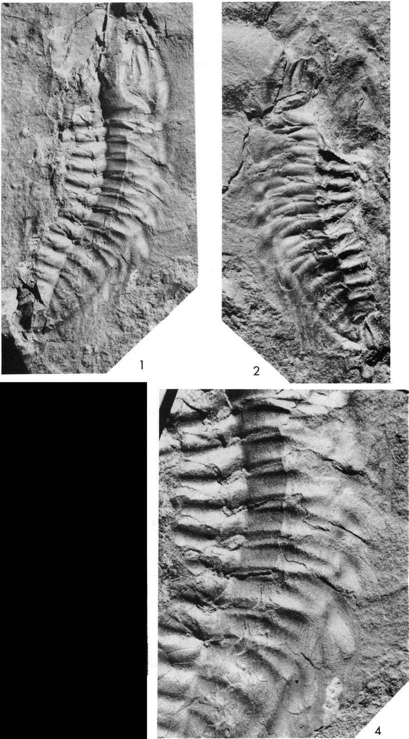
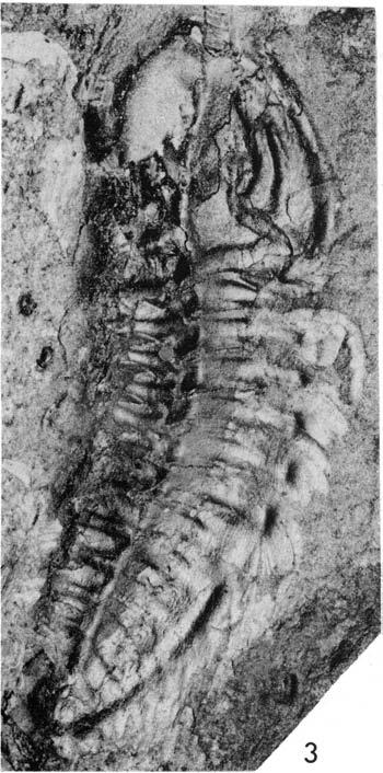
89
GSA DATA REPOSITORY Sup. Mat. 1. Figure DR1. Detailed locality information. 02
 GSA DATA REPOSITORY 008 Caron et al. Stanley Glacier - List of supplementary materials: Sup. Mat.. Figure DR. Detailed locality information. 0 Sup. Mat.. Figure DR. Claystone lithofacies. 0 Sup. Mat..
THIS IS A NEW SPECIFICATION
 THIS IS A NEW SPECIFICATION ADVANCED GCE GEOLOGY Evolution of Life, Earth and Climate F795 * OCE / 2 6692* Candidates answer on the Question Paper OCR Supplied Materials: None Other Materials Required:
SUPPLEMENTARY INFORMATION
 SUPPLEMENTARY INFORMATION doi:10.1038/nature12017 Supplementary Information 1. Spartobranchus tenuis (Walcott, 1911) from the Burgess Shale Type specimens. a-d, Lectotype, USNM 108494, specimen missing
ON THE OCCURRENCE KINNEGRAPTUS SKOGLUND IN NORWAY
 NORSK GEOLOGISK TIDSSKRIFT 42 ON THE OCCURRENCE OF KINNEGRAPTUS SKOGLUND IN NORWAY By O. M. B. BuLMAN and CYNTHIA M. Cowm (Sedgwick Museum, Cambridge) Abs t r a c t. Kinnegraptus kinnekullensis SKoGLUND,
Section 7. Reading the Geologic History of Your Community. What Do You See? Think About It. Investigate. Learning Outcomes
 Chapter 3 Minerals, Rocks, and Structures Section 7 Reading the Geologic History of Your Community What Do You See? Learning Outcomes In this section, you will Goals Text Learning Outcomes In this section,
SUPPLEMENTARY INFORMATION
 SUPPLEMENTARY INFORMATION doi:10.1038/nature11874 1. Supplementary Figures Supplementary Figure 1. Stratigraphic column of the Cambrian Stage 3 (regional Canglangpuan Stage) Xiaoshiba section in the suburb
GEOLOGY OF TODMORDEN MOOR 2 BACKGROUND
 GEOLOGY OF TODMORDEN MOOR 2 BACKGROUND 1) THE CARBONIFEROUS SERIES OF ROCKS The rocks of the Todmorden district are of the Carboniferous Series and were first laid down in an ancient sea, which covered
28. Genus Xoanon Semenov Fig. C28.1 (female dorsal habitus) Fig. C28.2 (female lateral habitus) Fig. C28.3 (male dorsal habitus)
 Fig. C28.2 (female lateral habitus) Fig. C28.3 (male dorsal habitus)") 28. Genus Xoanon Semenov Fig. C28.1 (female dorsal habitus) Fig. C28.2 (female lateral habitus) Fig. C28.3 (male dorsal habitus) Xoanon Semenov, 1921: 87. Type species: Xoanon mysta Semenov, by original
28. Genus Xoanon Semenov Fig. C28.1 (female dorsal habitus) Fig. C28.2 (female lateral habitus) Fig. C28.3 (male dorsal habitus) Xoanon Semenov, 1921: 87. Type species: Xoanon mysta Semenov, by original
MOHAVE DESERT CAMBRIAN FOSSILS FROM THE SMITHSONIAN MISCELLANEOUS COLLECTIONS VOLUME 81, NUMBER 2 CHARLES E. JULY 5, (With T?iree Plates) RESSER
 RESSER") SMITHSONIAN MISCELLANEOUS COLLECTIONS VOLUME 81, NUMBER 2 CAMBRIAN FOSSILS FROM THE MOHAVE DESERT (With T?iree Plates) BY CHARLES E. RESSER Associate Curator of Stratigraphic Paleontology: United States
SMITHSONIAN MISCELLANEOUS COLLECTIONS VOLUME 81, NUMBER 2 CAMBRIAN FOSSILS FROM THE MOHAVE DESERT (With T?iree Plates) BY CHARLES E. RESSER Associate Curator of Stratigraphic Paleontology: United States
Friday 10 June 2016 Afternoon
 Oxford Cambridge and RSA Friday 10 June 2016 Afternoon A2 GCE GEOLOGY F795/01 Evolution of Life, Earth and Climate *6011962861* Candidates answer on the Question Paper. OCR supplied materials: None Other
Shield was above sea-level during the Cambrian and provided the sediment for the basins.
 Name: Answers Geology 1023 Lab #8, Winter 2014 Platforms and Paleozoic life-forms Lab day: Tu W Th 1. Schematic geologic cross-sections A-B and C-D (shown below) cross the edge of the continental platform
Mosquito Systematics Vol. 6(Z) June 1974
 June 1974") Mosquito Systematics Vol. 6(Z) June 1974 93 Research on the Mosquitoes of Angola. VII - Redescription of the Larva of Aedes durbanensis durbanensis (Theo., 1903) and Description of Aedes durbanensis angozae
Mosquito Systematics Vol. 6(Z) June 1974 93 Research on the Mosquitoes of Angola. VII - Redescription of the Larva of Aedes durbanensis durbanensis (Theo., 1903) and Description of Aedes durbanensis angozae
OF THE LEMNA FROND MORPHOLOGY
 MORPHOLOGY OF THE LEMNA FROND FREDERICK H. BLODGETT (WITH PLATE XIV AND ONE FIGURE) In the case of structure simplified by reduction, it is sometimes necessary to trace the development of the parts through
Tuesday 10 June 2014 Afternoon
 Tuesday 10 June 2014 Afternoon A2 GCE GEOLOGY F795/01 Evolution of Life, Earth and Climate *1242977619* Candidates answer on the Question Paper. OCR supplied materials: None Other materials required: Electronic
A MEGASECOPTERON FROM UPPER CARBONIFEROUS BY F. M. CARPENTER. In I962 Professor F. Stockmans, of the Institut Royal des Sciences STRATA IN SPAIN
 A MEGASECOPTERON FROM UPPER CARBONIFEROUS STRATA IN SPAIN BY F. M. CARPENTER Harvard University In I962 Professor F. Stockmans, of the Institut Royal des Sciences Naturelles de. Belgique, kindly sent me
Bulletin Zoölogisch Museum
 Bulletin Zoölogisch Museum UNIVERSITEIT VAN AMSTERDAM Vol.11 No. 15 1988 Redescription of johanna Monod, 1926 Virgin Isls (Isopoda) from St. John, Hans Georg Müller Summary Based on the type material,
The functional morp hology of Cryptolithus
 The functional morp hology of Cryptolithus KENTON S.W. CAMPBELL Campbell, K.S.W. 1975 07 15: The functional morphology of Cryptolithus. Fossils and Strata, No. 4, pp. 65-86, Pls. 1-2. Oslo. ISSN 0300-949l.
Earth / Environmental Science. Ch. 14 THE OCEAN FLOOR
 Earth / Environmental Science Ch. 14 THE OCEAN FLOOR The Blue Planet Nearly 70% of the Earth s surface is covered by the global ocean It was not until the 1800s that the ocean became an important focus
Theory of Plate Tectonics
 Plate Tectonics Theory of Plate Tectonics Lithosphere is made of sections called plates that move around called continental drift Plates move because they float on the asthenosphere Most plates consist
Name. GEOL.5220 Structural Geology Faults, Folds, Outcrop Patterns and Geologic Maps. I. Properties of Earth Materials
 I. Properties of Earth Materials GEOL.5220 Structural Geology Faults, Folds, Outcrop Patterns and Geologic Maps Name When rocks are subjected to differential stress the resulting build-up in strain can
Lecture # 6. Geological Structures
 1 Lecture # 6 Geological Structures ( Folds, Faults and Joints) Instructor: Dr. Attaullah Shah Department of Civil Engineering Swedish College of Engineering and Technology-Wah Cantt. 2 The wavy undulations
Evolution of Life and the Atmosphere
 Evolution of Life and the Atmosphere White board used for Evolution of the Atmosphere and Early Evolution of Life The Pitfalls of Preservation It is obvious that a major change occurred in the Earth s
A FOSSIL CRAB FROM THE LAKES ENTRANCE OIL SHAFT, GIPPSLAND, VICTORIA
 gtopt JtoAtty 0f WitUxm. -..., ' -: / '.. v - - ^.. ' ' * A FOSSIL CRAB FROM THE LAKES ENTRANCE OIL SHAFT, GIPPSLAND, VICTORIA BY.'. " ; IRENE CRESPIN, R. A. Read 6** June, 1946 ' Reprinted from Proo.
Objectives: Define Relative Age, Absolute Age
 S6E5. Students will investigate the scientific view of how the earth s surface is formed. c. Classify rocks by their process of formation. g. Describe how fossils show evidence of the changing surface
1 Conducting Units: Tracheids and Vessels 1.1 Evolutionary Specialization
 1 Conducting Units: Tracheids and Vessels 1.1 Evolutionary Specialization The development of upright land plants depended on the development of a waterconducting system. Many of the earliest land plants,
F795. GEOLOGY Evolution of Life, Earth and Climate ADVANCED GCE. Wednesday 8 June 2011 Morning
 ADVANCED GCE GEOLOGY Evolution of Life, Earth and Climate F795 *F711220611* Candidates answer on the question paper. OCR supplied materials: None Other materials required: Electronic calculator Ruler (cm/mm)
LATE MIDDLE CAMBRIAN FOSSILS FROM BEACONSFIELD, TASMANIA
 Papers and Proceedings of the Royal Society of Tasmania, Volume 114, 1980 LATE MIDDLE CAMBRIAN FOSSILS FROM BEACONSFIELD, TASMANIA (ms. received 17.5.1979) by J.B. Jago School of Applied Geology, South
A) B) C) D) 4. Which diagram below best represents the pattern of magnetic orientation in the seafloor on the west (left) side of the ocean ridge?
 B) C) D) 4. Which diagram below best represents the pattern of magnetic orientation in the seafloor on the west (left) side of the ocean ridge?") 1. Crustal formation, which may cause the widening of an ocean, is most likely occurring at the boundary between the A) African Plate and the Eurasian Plate B) Pacific Plate and the Philippine Plate C)
1. Crustal formation, which may cause the widening of an ocean, is most likely occurring at the boundary between the A) African Plate and the Eurasian Plate B) Pacific Plate and the Philippine Plate C)
FoSSil Puzzler (1 Hour)
") FoSSil Puzzler (1 Hour) Addresses NGSS Level of Difficulty: 2 Grade Range: 3-5 OVERVIEW Students will examine and sketch various trace fossils from an unknown creature and then construct an image of the
FoSSil Puzzler (1 Hour) Addresses NGSS Level of Difficulty: 2 Grade Range: 3-5 OVERVIEW Students will examine and sketch various trace fossils from an unknown creature and then construct an image of the
Ch 17 Plate Tectonics Big Idea: Most geologic activity occurs at the boundaries between plates.
 Ch 17 Plate Tectonics Big Idea: Most geologic activity occurs at the boundaries between plates. 17.1 Drifting Continents 17.2 Seafloor Spreading 17.3 Plate Boundaries 17.4 Causes of Plate Motions Learning
CALLISPHENUS GRACILIS, N. GEN., N. SP. A FOSSIL ALGA FROM THE WENLOCK OF THE OSLO REGION
 CALLISPHENUS GRACILIS, N. GEN., N. SP. A FOSSIL ALGA FROM THE WENLOCK OF THE OSLO REGION BY OVE HØEG WITH 2 PLATES The specimen described below was found in August, 191 O, on the small island of Kommersøy
Stratigraphic Cross-Section
 Stratigraphic cross-sections are used to show stratigraphic relationships of strata along some path. They differ from structural cross-sections in three ways. First, their goals differ. Stratigraphic cross-sections
introduction to the Animal Kingdom (pages $55-560) Formulating a Definition: Building Vocabulary Skills
 Formulating a Definition: Building Vocabulary Skills") STUDY GUIDE CHAPTER Sponges, Cnidarians, and Unsegmented Worms Section 26-1 introduction to the Animal Kingdom (pages $55-560) SECTION REVIEW With this section you began your study of the animal kingdom.
STUDY GUIDE CHAPTER Sponges, Cnidarians, and Unsegmented Worms Section 26-1 introduction to the Animal Kingdom (pages $55-560) SECTION REVIEW With this section you began your study of the animal kingdom.
NEW AMERICAN PALEOZOIC OSTRACODA.
 New American Paleozoic Ostracoda. 179 NEW AMERICAN PALEOZOIC OSTRACODA. BY E. O. ULRICH. No. 1. CTKNOBOLBINA AND KIRKBYA. Since the publication of my paper on " New and Ivittle Known American Paleozoic
UNVERSITY OF MICHIGAN THREE NEW SPECIES OF THE CYSTID GENUS LIPSANOCYSTIS FROM THE MIDDLE DEVONIAN TRAVERSE GROUP OF MICHIGAN
 CONTRIBUTIONS FROM THE MUSEUM OF PALEONTOLOGY UNVERSITY OF MICHIGAN VOL. XII, No. 6, pp. 97-103 (2 pls.) APRIL 8, 1955 THREE NEW SPECIES OF THE CYSTID GENUS LIPSANOCYSTIS FROM THE MIDDLE DEVONIAN TRAVERSE
Geology 12 FINAL EXAM PREP. Possible Written Response Exam Questions
 Geology 12 FINAL EXAM PREP Possible Written Response Exam Questions Use this study guide to prepare for the written response portion of the final exam. Name FINAL EXAM - POSSIBLE WRITTEN RESPONSE QUESTIONS
* If occurs, fossils that formed in shallow water may be found in water. Subsidence mean the crust is.
 Name Notes: Topic 5B Topic 5B: The Dynamic Crust A. What evidence exists that proves the Earth s Crust is and has moved? Earth Science 1. Tilted, folded or faulted strata Strata is a term that refers to.
GHABBOUR, M.W. Plant Protection Research Institute, Agricultural Research Center, Ministry of Agriculture, Dokki, Egypt.
 Entomologica, Bari, 33, (1999): 73-83 GHABBOUR, M.W. Plant Protection Research Institute, Agricultural Research Center, Ministry of Agriculture, Dokki, Egypt. DESCRIPTIONS OF THE FIRST-INSTAR NYMPHS OF
Directed Reading. Section: Volcanoes and Plate Tectonics
 Skills Worksheet Directed Reading Section: Volcanoes and Plate Tectonics 1. Some volcanic eruptions can be more powerful than a(n) a. hand grenade. b. earthquake. c. geyser. d. atomic bomb. 2. The cause
Chapter Two. Figure 02_02. Geography of the Ocean Basins. The Sea Floor
 Chapter Two The Sea Floor Geography of the Ocean Basins Figure 02_02 The world ocean is the predominant feature on the Earth in total area. In the Northern Hemisphere, 61% of the total area is ocean. In
Lesson 10. References: Chapter 8: Reading for Next Lesson: Chapter 8:
 Lesson 10 Lesson Outline: General Features of the Skeletal System Axial Skeleton (Vertebral Column) Ontogenetic Origins (Morphogenesis) of the Skeletal System Functions of the Axial Skeleton Phylogenetic
A.M. TUESDAY, 12 May hours
 Candidate Name Centre Number 2 Candidate Number GCE AS/A level 1212/01 New AS GEOLOGY - GL2a Investigative Geology A.M. TUESDAY, 12 May 2009 1 1 2 hours For Examiner s Use Only ADDITIONAL MATERIALS In
EXTERNAL ANATOMY OF INSECTS
 External Anatomy of Insects 1 The insect s exoskeleton is made up of a series of plates EXTERNAL ANATOMY OF INSECTS These plates make up the insect s exoskeleton. These plates are connected by joints or
STUDY GUIDE FOR MID-TERM EXAM KEY. Color, luster, cleavage, fracture, hardness, taste, smell, fluorescence, radioactivity, magnetism
 STUDY GUIDE FOR MID-TERM EXAM KEY 1. In which type of rock are fossils most likely to be found? Sedimentary Rocks 2. Which mineral is easily identified by smell? Sulfur 3. Which natural resource makes
Plate Tectonics: A Scientific Revolution Unfolds
 Chapter 2 Lecture Earth: An Introduction to Physical Geology Eleventh Edition Plate Tectonics: A Scientific Revolution Unfolds Tarbuck and Lutgens From Continental Drift to Plate Tectonics Prior to the
Paleobiology and Geochemistry of Siliceous Concretions in the Conasauga Formation, Middle Cambrian, Northwestern Georgia
 Paleobiology and Geochemistry of Siliceous Concretions in the Conasauga Formation, Middle Cambrian, Northwestern Georgia David R. Schwimmer William J. Frazier Columbus State University William M. Montante
Streams. Water. Hydrologic Cycle. Geol 104: Streams
 Streams Why study streams? Running water is the most important geologic agent in erosion, transportation and deposition of sediments. Water The unique physical and chemical properties of water make it
5. Crater Rim Uplift and Crater Wall Collapse
 5. Crater Rim Uplift and Crater Wall Collapse The basic processes involved in crater rim uplift are understood, but there is a lot of evidence at the crater that has not been fully explored and may eventually
Theme 7. Metamorphic rocks. Distinguishing rock types
 Theme 7. Metamorphic rocks. Distinguishing rock types 7.1. Metamorphic rocks formation 7.2. Classification of metamorphic rocks 7.3. Distinguishing rock types 7.1. Metamorphic rocks formation 7.1. Metamorphic
A. Refer to Appendix F in back of lab manual for list of commonly used geologic map symbols
 Structural Geology Lab 2: Outcrop Patterns and Structure Contours I. Geologic Map Symbols A. Refer to Appendix F in back of lab manual for list of commonly used geologic map symbols 1. Emphasis: a. strike
B. Topographic maps are also called. contour maps
 Topographic Maps Introduction A. Topographic maps are essential tools in geologic and engineering studies because they show the configuration of Earth's surface in remarkable detail and permit one to measure
ANOTHER MEXICAN EARTHQUAKE! Magnitude 7.1, Tuesday Sept. 19, 2017
 ANOTHER MEXICAN EARTHQUAKE! Magnitude 7.1, Tuesday Sept. 19, 2017 Why is there no oceanic crust older than 200 million years? SUBDUCTION If new oceanic crust is being continuously created along the earth
Continental Drift and Plate Tectonics
 Continental Drift and Plate Tectonics Continental Drift Wegener s continental drift hypothesis stated that the continents had once been joined to form a single supercontinent. Wegener proposed that the
Paleo Lab #5 - Fossilization and Fossil Identification
 Paleo Lab #5 - Fossilization and Fossil Identification page - INTRODUCTION The objectives of this exercise are to understand what fossils are, to recognize the different styles of fossil preservation,
Before Plate Tectonics: Theory of Continental Drift
 Before Plate Tectonics: Theory of Continental Drift Predecessor to modern plate tectonics Shape and fit of the continents was the initial evidence Snider-Pelligrini (1858) Taylor (1908) Wegner (1915) Fig.
PLATE TECTONICS. SECTION 17.1 Drifting Continents
 Date Period Name PLATE TECTONICS SECTION.1 Drifting Continents In your textbook, read about continental drift. Circle the letter of the choice that best completes each statement. 1. Early mapmakers thought
KOCHIELLA POULSEN, 1927, AND HADROCEPHALITES NEW GENUS (TRILOBITA: PTYCHOPARIIDA) FROM THE EARLY MIDDLE CAMBRIAN OF WESTERN NORTH AMERICA
 FROM THE EARLY MIDDLE CAMBRIAN OF WESTERN NORTH AMERICA") J. Paleont., 76(1), 2002, pp. 76 94 Copyright 2002, The Paleontological Society 0022-3360/02/0076-76$03.00 KOCHIELLA POULSEN, 1927, AND HADROCEPHALITES NEW GENUS (TRILOBITA: PTYCHOPARIIDA) FROM THE EARLY
J. Paleont., 76(1), 2002, pp. 76 94 Copyright 2002, The Paleontological Society 0022-3360/02/0076-76$03.00 KOCHIELLA POULSEN, 1927, AND HADROCEPHALITES NEW GENUS (TRILOBITA: PTYCHOPARIIDA) FROM THE EARLY
Name: Mid-Year Review #2 SAR
 Name: Mid-Year Review #2 SAR Base your answers to questions 1 through 3 on on the diagram below, which shows laboratory materials used for an investigation of the effects of sediment size on permeability,
LAB 1: ORIENTATION OF LINES AND PLANES
 LAB 1: ORIENTATION OF LINES AND PLANES Read the introductory section, chapter 1, pages 1-3, of the manual by Rowland et al (2007) and make sure you understand the concepts of bearing, strike, dip, trend,
LABORATORY MEASUREMENTS OF PULLING FORCE AND GROUND MOVEMENT DURING A PIPE BURSTING TEST
 North American Society for Trenchless Technology (NASTT) NO-DIG 2004 New Orleans, Louisiana March 22-24, 2004 LABORATORY MEASUREMENTS OF PULLING FORCE AND GROUND MOVEMENT DURING A PIPE BURSTING TEST Brian
Fritzolenellus lapworthi (Peach and Horne, 1892) from the lower Cambrian (Cambrian Series 2) Bastion Formation of North-East Greenland
 from the lower Cambrian (Cambrian Series 2) Bastion Formation of North-East Greenland") Fritzolenellus lapworthi (Peach and Horne, 1892) from the lower Cambrian (Cambrian Series 2) Bastion Formation of North-East Greenland MARTIN STEIN Stein, M. 2008 01 03. Fritzolenellus lapworthi (Peach
Fritzolenellus lapworthi (Peach and Horne, 1892) from the lower Cambrian (Cambrian Series 2) Bastion Formation of North-East Greenland MARTIN STEIN Stein, M. 2008 01 03. Fritzolenellus lapworthi (Peach
Directed Reading. Section: The Theory of Plate Tectonics. to the development of plate tectonics, developed? HOW CONTINENTS MOVE
 Skills Worksheet Directed Reading Section: The Theory of Plate Tectonics 1. The theory that explains why and how continents move is called. 2. By what time period was evidence supporting continental drift,
6. Crater Rim Uplift and Crater Wall Collapse
 6. Crater Rim Uplift and Crater Wall Collapse The basic processes involved in crater rim uplift are understood, but there is a lot of evidence at the crater that has not been fully explored and may eventually
PSc 201 Chapter 3 Homework. Critical Thinking Questions
 PSc 201 Chapter 3 Homework Critical Thinking Questions 1. (adapted from text) Seawater is denser than fresh water. A ship moving from the Atlantic Ocean into the Great Lakes goes from seawater to fresh
Of the University of Colorado, Boulder.
 SOME EOCENE INSECTS OF THE FAMILY FULGORIDA~ By T. D. A. COCKERELL and GRACE SANDTIOUSE, Of the University of Colorado, Boulder. The remarkable abundance and variety of the Homopterous family Fulgoridae
PALEONTOLOGICAL CONTRIBUTIONS
 UNIVERSITY OF KANSAS PALEONTOLOGICAL CONTRIBUTIONS July 24, 1984 Paper 112 NEW OCCURRENCES OF THE UNUSUAL TRILOBITE NARAOIA FROM THE CAMBRIAN OF IDAHO AND UTAH' R. A. R.outsoN Department of Geology, University
Cambridge International Examinations Cambridge International General Certificate of Secondary Education
 Cambridge International Examinations Cambridge International General Certificate of Secondary Education *8981335670* PHYSICS 0625/31 Paper 3 Extended May/June 2015 1 hour 15 minutes Candidates answer on
GCE AS/A level 1211/01 GEOLOGY GL1 Foundation Unit
 Surname Centre Number Candidate Number Other Names 2 GCE AS/A level 1211/01 GEOLOGY GL1 Foundation Unit S15-1211-01 A.M. MONDAY, 11 May 2015 1 hour For s use Question Maximum Mark 1. 15 2. 14 Mark Awarded
Lecture 6 Folds, Faults and Deformation Dr. Shwan Omar
 Fold: A fold is a bend or wrinkle of rock layers or foliation; folds form as a sequence of ductile deformation. Folding is the processes by which crustal forces deform an area of crust so that layers of
Plate Tectonics. entirely rock both and rock
 Plate Tectonics I. Tectonics A. Tectonic Forces are forces generated from within Earth causing rock to become. B. 1. The study of the origin and arrangement of Earth surface including mountain belts, continents,
Exhibit #1: Adaptation A process that enables organisms to become better suited to their environments
 Exhibit #1: Adaptation A process that enables organisms to become better suited to their environments Better suited individuals were better able to reproduce and pass their genes on to their young (example
Name Date Class. How have geologists learned about Earth s inner structure? What are the characteristics of Earth s crust, mantle, and core?
 Chapter 4 Plate Tectonics Section 1 Summary Earth s Interior How have geologists learned about Earth s inner structure? What are the characteristics of Earth s crust, mantle, and core? Earth s surface
LINGUAU DEPOSITION IN THE WOODBINE SANDS ALONG COPPERAS BRANCH, DENTON COUNTY. TEXAS: A STUDY IN MARINE SEDIMENTATION
 LINGUAU DEPOSITION IN THE WOODBINE SANDS ALONG COPPERAS BRANCH, DENTON COUNTY. TEXAS: A STUDY IN MARINE SEDIMENTATION Ellis W. Shuler and Olin M. Millican The lack of outcrops in three dimensions often
OBSERVATIONS ON AMOEBA FEEDING ON THE CILIATE FRONTONIA.*
 OBSERVATIONS ON AMOEBA FEEDING ON THE CILIATE FRONTONIA.* BY C. DALE BEERS. (The Zoological Laboratory of the Johns Hopkins University.) THE manner of capture of rotifers and Paramecia by Amoeba has been
Chapter 15 Structures
 Chapter 15 Structures Plummer/McGeary/Carlson (c) The McGraw-Hill Companies, Inc. TECTONIC FORCES AT WORK Stress & Strain Stress Strain Compressive stress Shortening strain Tensional stress stretching
Monday 3 June 2013 Morning
 Monday 3 June 2013 Morning A2 GCE GEOLOGY F795/01 Evolution of Life, Earth and Climate *F713200613* Candidates answer on the Question Paper. OCR supplied materials: None Other materials required: Electronic
The new physiology of vision-chapter The colours of interference. SIR C V RAMAN Received October 14, 1965
 Proc. Indian Acad. Sci. A62 243-248 (1965) The new physiology of vision-chapter The colours of interference XXVII. SIR C V RAMAN Received October 14, 1965 The characteristic features and properties of
APPENDIX C GEOLOGICAL CHANCE OF SUCCESS RYDER SCOTT COMPANY PETROLEUM CONSULTANTS
 APPENDIX C GEOLOGICAL CHANCE OF SUCCESS Page 2 The Geological Chance of Success is intended to evaluate the probability that a functioning petroleum system is in place for each prospective reservoir. The
Staple this part to part one of lab 6 and turn in. Lab 6, part two: Structural geology (analysis)
") Geology 101 Staple this part to part one of lab 6 and turn in Lab 6, part two: Structural geology (analysis) Recall that the objective of this lab is to describe the geologic structures of Cougar Mountain
Geology 101 Staple this part to part one of lab 6 and turn in Lab 6, part two: Structural geology (analysis) Recall that the objective of this lab is to describe the geologic structures of Cougar Mountain
Do Now: Vocabulary: Objectives. Vocabulary: 1/5/2016. Wegener? (Can they move?) the idea that continents have moved over time?
 the idea that continents have moved over time?") Do Now: 1. Who was Alfred Wegener? 2. What was Pangaea? 3. Are continents fixed? (Can they move?) 4. What evidence supports the idea that continents have moved over time? Objectives What evidence suggests
Do Now: 1. Who was Alfred Wegener? 2. What was Pangaea? 3. Are continents fixed? (Can they move?) 4. What evidence supports the idea that continents have moved over time? Objectives What evidence suggests
With a group, get a bar magnet, some plastic wrap, iron filings and a compass.
 Name: EPS 50 Lab 8: The Earth's Magnetic Field Chapter 2, p. 39-41: The Seafloor as a Magnetic Tape Recorder Chapter 7, p. 213: Paleomagnetic Stratigraphy Chapter 14, p. 396-406: Earth s Magnetic Field
PALEONTOLOGY - the study of the history of life as reflected in the fossil record
 Paleontology PALEONTOLOGY - the study of the history of life as reflected in the fossil record FOSSIL - remains or traces of organisms that lived in the geologic past and are preserved in the crust of
Radiation and Evolution of Metazoans: The Cambrian Explosion and the Burgess Shale Fossils. Geology 331, Paleontology
 Radiation and Evolution of Metazoans: The Cambrian Explosion and the Burgess Shale Fossils Geology 331, Paleontology Marshall, 2006 Halkierids, which produced some of the small, shelly fossils of the Early
!'f \, w. Alan Stewart Colorado Exploration Company, Golden STRUCTURE OF THE FOOTHILLS AREA WEST OF DENVER, COLORADO. Introduction
 STRUCTURE OF THE FOOTHLLS AREA WEST OF DENVER, COLORADO w. Alan Stewart Colorado Exploration Company, Golden ntroduction i ; The dominant structural features west of Denver and along the mountain front
22.4 Plate Tectonics. Africa
 The Red Sea between Africa and the Arabian peninsula in Asia marks a region where two pieces of the lithosphere are slowly moving apart. Over the next 100 million years, the Red Sea could become an ocean.
Kingdom Animalia. Zoology the study of animals
 Kingdom Animalia Zoology the study of animals Summary Animals are multicellular and eukaryotic. consume and digest organic materials thereby being heterotrophs. Most are motile at some time in their lives.
Sediment and sedimentary rocks Sediment
 Sediment and sedimentary rocks Sediment From sediments to sedimentary rocks (transportation, deposition, preservation and lithification) Types of sedimentary rocks (clastic, chemical and organic) Sedimentary
Knowledge of in-slab earthquakes needed to improve seismic hazard estimates for southwestern British Columbia
 USGS OPEN FILE REPORT #: Intraslab Earthquakes 1 Knowledge of in-slab earthquakes needed to improve seismic hazard estimates for southwestern British Columbia John Adams and Stephen Halchuk Geological
Ocean Floor. Continental Margins. Divided into 3 major regions. Continental Margins. Ocean Basins. Mid-Ocean Ridges. Include:
 Ocean Floor Divided into 3 major regions Continental Margins Ocean Basins Mid-Ocean Ridges Continental Margins Include: Continental Shelves Continental Slopes Continental Rise 1 Continental Shelves Part
A L A BA M A L A W R E V IE W
 A L A BA M A L A W R E V IE W Volume 52 Fall 2000 Number 1 B E F O R E D I S A B I L I T Y C I V I L R I G HT S : C I V I L W A R P E N S I O N S A N D TH E P O L I T I C S O F D I S A B I L I T Y I N
Topic 5: The Dynamic Crust (workbook p ) Evidence that Earth s crust has shifted and changed in both the past and the present is shown by:
 Evidence that Earth s crust has shifted and changed in both the past and the present is shown by:") Topic 5: The Dynamic Crust (workbook p. 65-85) Evidence that Earth s crust has shifted and changed in both the past and the present is shown by: --sedimentary horizontal rock layers (strata) are found
Topic 5: The Dynamic Crust (workbook p. 65-85) Evidence that Earth s crust has shifted and changed in both the past and the present is shown by: --sedimentary horizontal rock layers (strata) are found
SITE SURVEY FOR SITE 410, AN EXAMPLE OF THE USE OF LONG-RANGE SIDE-SCAN SONAR (GLORIA)
") 10. SITE SURVEY FOR SITE 410, AN EXAMPLE OF THE USE OF LONG-RANGE SIDE-SCAN SONAR (GLORIA) R. C. Searle and A. S. Laughton, Institute of Oceanographic Sciences, Wormley, GU8 5UB, UK and B. D. Loncarevic,
10. SITE SURVEY FOR SITE 410, AN EXAMPLE OF THE USE OF LONG-RANGE SIDE-SCAN SONAR (GLORIA) R. C. Searle and A. S. Laughton, Institute of Oceanographic Sciences, Wormley, GU8 5UB, UK and B. D. Loncarevic,
Plate Tectonics. Essentials of Geology, 11 th edition Chapter 15
 1 Plate Tectonics Essentials of Geology, 11 th edition Chapter 15 2 3 4 5 6 7 8 9 10 11 12 13 14 15 Plate Tectonics: summary in haiku form Alfred Wegener gave us Continental Drift. Fifty years later...
Seismic measurement of the propagation speed of a fracture through a weak snowpack layer
 Seismic measurement of snow fracture Seismic measurement of the propagation speed of a fracture through a weak snowpack layer Ben Johnson 1, Bruce Jamieson 1,2, and Robert R. Stewart 2 ABSTRACT Weak layers
Marshall Shore Town Park, Liberty, Maine
 Maine Geologic Facts and Localities August, 2005 Marshall Shore Town Park, Liberty, Maine 44 22 33.04 N, 69 21 9.19 W Text by Henry N. Berry IV, Department of Agriculture, Conservation & Forestry 1 Introduction
Monday 11 May 2015 Morning
 Oxford Cambridge and RSA Monday 11 May 2015 Morning AS GCE GEOLOGY F791/01 Global Tectonics *5003095113* Candidates answer on the Question Paper. OCR supplied materials: None Other materials required:
Optical distortion in the Hubble Ultra-Deep Field
 Optical distortion in the Hubble Ultra-Deep Field COLIN ROURKE It is proposed that the strange appearance of all non-standard galaxies in the Hubble Ultra-Deep Field [1] is due to optical distortion caused
8 th Grade Science Plate Tectonics and Topography Review
 8 th Grade Science Plate Tectonics and Topography Review #1 Scientists believe that the land masses of Earth were once joined together as one supercontinent called. A: Australia B: Wegner C: Pangaea D:
Lab 7: STRUCTURAL GEOLOGY FOLDS AND FAULTS
 Lab 7: STRUCTURAL GEOLOGY FOLDS AND FAULTS This set of labs will focus on the structures that result from deformation in earth s crust, namely folds and faults. By the end of these labs you should be able
Deformation of the Crust
 Deformation of the Crust Review Choose the best response. Write the letter of that choice in the space provided. 1. The state of balance between the thickness of the crust and the depth at which it rides