ARBUSCULAR MYCORRHIZA
|
|
|
- Corey Johnson
- 5 years ago
- Views:
Transcription

1 ARBUSCULAR MYCORRHIZA Dr. Suresh. S.S. Raja Assistant Professor Department of Microbiology Bharathidasan University College Perambalur Prepared


2 Vesicles and Arbuscules Glomus Mycorrhiza is the symbiotic association between plant roots and fungal mycelia Gigaspora
3 CHARACTERISTICS OF ARBUSCULAR MYCORRHIZA Mycorrhiza increase the surface area of the root Absorb nutrients from soil especially phosphorus and micronutrients through hyphae and mobilize into the host cell. Almost 90 % of crop plants are mycorrhizal mostly of arbuscular type (AM) Mycorrhiza possess vesicles and arbuscules. Mycorrhiza species are: Glomus, Gigaspora, Scutellospora, Acaulospora, Entrophosphora and Sclerocystis
4 Gigaspora Scutellospora Glomus Gigaspora Glomus albidum Sclerocystis Gigaspora Glomus mosseae Sclerocystis
5 ROLE OF MYCORRHIZA The benefit of mycorrhizas to plants is mainly attributed to increased uptake of nutrients, especially phosphorus. This increase in uptake may be due to increase surface area of soil contact increased movement of nutrients into mycorrhizae, a modification of the root environment and increased storage. Mycorrhizas can be much more efficient than plant roots at taking up phosphorus. Phosphorus travels to the root or via diffusion and hyphae reduce the distance required for diffusion, thus increasing uptake. The rate of inflow of phosphorus into mycorrhizae can be up to six times that of the root hairs. In some cases, the role of phosphorus uptake can be completely taken over by the mycorrhizal network, and all of the plant s phosphorus may be of hyphal origin
6
7 Terminologies APPRESSORIA Hyphal swellings between two adjacent root epidermal cells. These are sites where hyphae first penetrate root cells by exerting pressure and/or enzymatic activity. ARBUSCULES These are intricately branched "haustoria" that form within root cortex cells that look like little trees. Arbuscules are formed by repeated dichotomous branching and reductions in hyphal width from an initial trunk hypha that ends in a proliferation of very fine branch hyphae. They are considered to be the major site of exchange with the host plant. Old arbuscules collapse progressively until only the trunk remains. Collapsed arbuscules are sometimes called peletons. ASEPTATE HYPHAE These are hyphae which are without cross walls (coenocytic hyphae). Cross walls may form as hyphae age.
8 AUXILIARY BODIES These structures, which are also called external vesicles /or accessory bodies, are clustered swellings on external hyphae. These are often ornamented by spines or knobs and are characteristic of Scutellospora and Gigaspora. These apparently do not function as propagules. COLONY Hyphal colonization of a root resulting from one external hypha (these may arise from several adjacent entry points). These are often called infection units. DICHOTOMOUS BRANCHING A symmetrical branching pattern which occurs when two branches arise simultaneously from the tip of a hyphae, plant or fungus organ. Divergent branches grow at the same rate.
9
10 INTERNAL HYPHAE, INTRARADICAL HYPHAE Hyphae which grow within the cortex of a root to form a colony and later develop arbuscules and vesicles. These comprise the body (thallus) of a fungus in the root. INTERCELLULAR HYPHAE Hyphae which grow between the walls of adjacent root cells. These are in the root apoplast -- the zone outside the cytoplasm of cells. INTRACELLULAR HYPHAE Hyphae which grow within root cells. These penetrate the walls of cells and grow within them, but are separated from the cytoplasm by the plasma membrane. SOIL HYPHAE These are also known as extraradical or external hyphae and are the filamentous structures which comprise the fungal thallus (body) in the soil. They acquire nutrients, propagate the association, produce spores and other structures. VAM fungi produce thick "runner" or "distributive" hyphae as well as thin, highly branched "absorptive" hyphae.
11 SPORES These are swollen structures with one or more subtending hyphae that form in the soil or in roots. Spores usually develop thick walls, which often have more than one layer. They can function as propagules. Spores of VAM fungi are sometimes called chlamydospores or azygospores. SPOROCARPS Aggregations of spores into groups, which may contain specialised hyphae and can be encased in an outer layer (peridium). Soil particles may be included in the spore mass. This term can be misleading, as the sporocarps produced by most Glomeromycotan fungi are small and relatively unorganised structures compared to those produced by larger fungi. VESICLES Intercalary (-o-) or terminal (-o) hyphal swellings formed on internal hyphae within the root cortex. These may form within or between cells. Vesicles accumulate lipids and may develop thick wall layers in older roots. The production and structure of vesicles varies between different genera of Glomeromycotan fungi. They are storage organs which may also function as propagules.
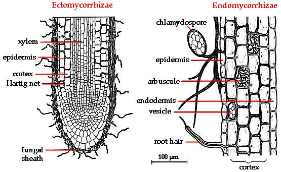
12 VAM Mycorrhizal associations produced by Glomeromycotan fungi are known as arbuscular mycorrhizas, or vesicular-arbuscular mycorrhizas (formerly also endomycorrhizas, or endotrophic mycorrhizas) and are abbreviated as VAM here. There is disagreement about whether arbuscular mycorrhizas or vesicular-arbuscular mycorrhizas is the most appropriate name to, because some fungi do not produce vesicles, but arbuscules are not consistently used to identify associations (i.e. they are absent in myco-heterotrophs and older roots).
13
14 VAM GENERAL STRUCTURE A. Structures in Soil Distributive Hyphae:A network of hyphae forms in the soil with thicker hyphae which function as conduits. Absorptive hyphae: Thin highly branched hyphae which are thought to absorb nutrients. Spores:Large (for a fungus) asexual spherical structures ( µm diameter) formed on hyphae in soil, or in roots. B. Structures in Roots Hyphae:these are non-septate when young and ramify within the cortex. Arbuscules: intricately branched haustoria in cortex cells. Vesicles:storage structures formed by many fungi.
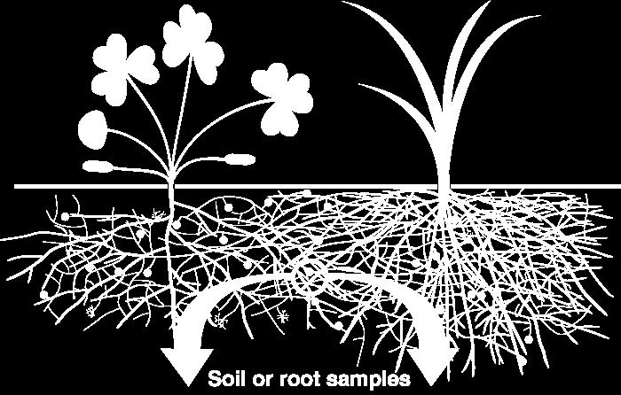
15 Looking at Associations

16 The Dissecting Microscope

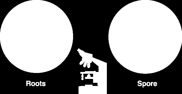
17 The Compound Microscope

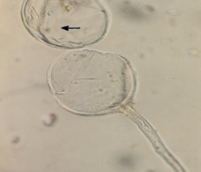
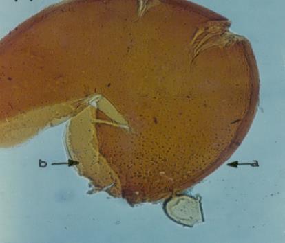
18 Structures and Developmental Stages 1. Soil Hyphae Mycorrhizal associations may be initiated by spore germination as illustrated here. Hyphae may also originate from fragments of roots. In many cases there already is a pre-existing network of hyphae resulting from previous root activity. Hyphae resulting from spore germination have a limited capacity to grow and will die if they do not encounter a susceptible root within a week or so. Hyphae emerge from a germination shield within the spore in Scutellospora and Acaulospora species. Germinating hyphae which emerged several days after spores were extracted from dry soil. These spores are of Gigaspora decipiens (left) and Scutellospora cerradensis (right). AV = accessory vesicles. GS = germination shield. Bars = 100 µm.

19 Soil Hyphae Soil hyphae, also known as extraradical or external hyphae, are filamentous fungal structures which ramify through the soil. They are responsible for nutrient acquisition, propagation of the association, spore formation, etc. VAM fungi produce different types of soil hyphae including thick "runner" or "distributive" hyphae as well as thin "absorptive" hyphae. The finer hyphae can produce "branched absorptive structures" (BAS) where fine hyphae proliferate Hyphae of Scutellospora and Gigaspora species produce clustered swellings with spines or knobs called auxiliary cells.
 with spores (S) produced by Glomus mosseae.")
20 Mycorrhizal root system washed carefully from coarse sand to reveal the intact network with external hyphae (arrow) with spores (S) produced by Glomus mosseae.

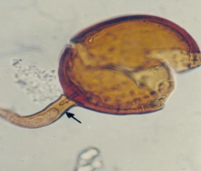
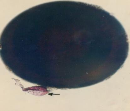
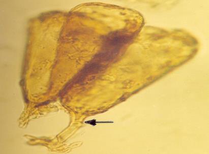
21 Soil hyphae produced by a single germinated spore of Gigaspora (arrow) used to start a mycorrhizal association. The hyphal network has produced accessory vesicles and is spreading through the root system of a clover plant.
22 2. Root Contact and Penetration Mycorrhizal associations start when soil hyphae respond to the presence of a root by growing towards it, establishing contact and growing along its surface. Next, one or more hyphae produce swellings called appressoria between epidermal cells. Root penetration occurs when hyphae from the appressoria penetrate epidermal or cortical cells to enter the root. These hyphae cross the hypodermis (through passage cells if these are present in an exodermis) and start branching in the outer cortex.
.")
23 Soil hyphae have produced 2 appressoria between epidermal cells (arrows). These are seen here in a surface view of a root with attached hyphae.
 approximately 1 day after contact with the")
24 Hyphae at an entry point (E) penetrating cortex cells (arrows) approximately 1 day after contact with the root.
.")
25 Alternating long (L) and short (S) cells in the dimorphic exodermis of a Smilacina racemosa root. Hyphae of VAM fungi have penetrated unsuberised short cells (arrows).
26 3. Hyphal Proliferation in the Cortex Aseptate hyphae spread along the cortex in both directions from the entry point to form a colony. Hyphae within root are initially without cross walls, but these may occur in older roots. two distinctive morphology types - the Arum and Paris series after host plants. These are now known as linear and coiling associations respectively. Both types of associations are important in ecosystems Linear (Arum) series associations where hyphae proliferate in the cortex by growing longitudinally between host cells. This occurs because hyphae grow through longitudinal intercellular air spaces that are present (Brundrett 2004). Coiling (Paris) series where hyphae spread by forming coils within cells because there are no continuous longitudinal air spaces.
27
 with hyphae, arbuscules (A) and vesicles (V) growing from an")
28 Part of a colony of a VAM fungus (Glomus sp.) with hyphae, arbuscules (A) and vesicles (V) growing from an entry point (arrow).
29 (i) Coiling (Paris) Arbuscular Mycorrhizas These are associations where hyphae spread primarily by intracellular growth following a convoluted path through cortex cells. The resulting colonies of VAM fungi generally have a coiled appearance, but may have more digitate branching patterns. Arbuscules may be restricted to a single layer of cells in the inner cortex.
 growing directly from coils in adjacent root cortex cells of Erythronium americanum.")
30 Colony of a VAM fungus spreading from the entry point (E) by convoluted hyphae (arrows) in the cortex of an Erythronium americanum root. Hyphal coils tend to occur in roots without prominent air channels. Higher magnification view of two arbuscules (A) growing directly from coils in adjacent root cortex cells of Erythronium americanum. Note how arbuscular branches arise from the same hyphae (arrows) which connect hyphae in adjacent cells. Arbuscules (A) and convoluted hyphae (arrow) in the inner cortex of an Asarum canadense root. Arbuscules only form in the innermost cortex cell layer next to the endodermis in this species
31 (ii) Linear (Arum) Arbuscular Mycorrhizas These are associations where hyphae grow along longitudinal intercellular air channels between the walls of root cells. A relatively rapid parallel spread of intercellular hyphae may occur along these channels. The resulting colonies of VAM fungi have a linear appearance.

 along cortex air channels in a leek root.")
32 Intercellular air channels (arrows) in a whole mount of a living leek root (Allium porrum), shown for comparison with mycorrhizal development in the same host. These channels run continuously from the apex to the base of roots. Longitudinal growth of hyphae of a VAM fungus (Glomus versiforme) along cortex air channels in a leek root. Note progressive development of arbuscules with increasing distance from the growing tips of hyphae.
33 4. Arbuscules Arbuscules are intricately branched haustoria that formed within a root cortex cell. They look like little trees. Arbuscules are formed by repeated dichotomous branching and reductions in hyphal width, starting from an initial trunk hypha (5-10 um in diameter) and ending in a proliferation of fine branch hyphae (< 1 um diameter). Arbuscules start to form approximately 2 days after root penetration. They grow inside individual cells of the root cortex, but remain outside their cytoplasm, due to invagination of the plasma membrane. Arbuscules are considered the major site of exchange between the fungus and host. This assumption is based on the large surface area of the arbuscular interface, but has not been confirmed. Arbuscule formation follows hyphal growth, progressing outwards from the entry point. Arbuscules are short-loved and begin to collapse after a few days, but hyphae and vesicles can remain in roots for months or years.
.")


")
34 Developing arbuscule of Glomus mosseae in a root cell with fine branch hyphae (arrows). The trunk (T) of this arbuscule branched from an intercellular hyphae. An arbuscule of Glomus versiforme in a root cortex cell with branch hyphae densely packed in the cortex cell of the host. Mature arbuscule of Glomus showing trunk (T) and numerous fine branch hyphae (arrows). Arbuscule of Gigaspora margarita with an elongated trunk hypha (T) and tufts of fine branch hyphae (arrows). Note how this arbuscule differs from the Glomus arbuscules above.
35 5. Vesicles Vesicles develop to accumulate storage products in many VAM associations. Vesicles are initiated soon after the first arbuscules, but continue to develop when the arbuscules senesce. Vesicles are hyphal swellings in the root cortex that contain lipids and cytoplasm. These may be interor intracellular. Vesicles can develop thick walls in older roots and may function as propagules. Some fungi produce vesicles which are similar in structure to the spores they produced in soil, but in other cases they are different.


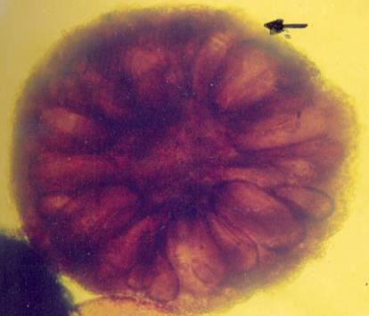
36 Vesicles (V) produced by a Glomus species in a leek root. This root also contains many intercellular hyphae Lobed vesicles of an Acaulospora species in a clover root Arrows = vesicles, A = arbuscules
37 6. Structural Diversity It is possible to identify individual Glomeromycotan fungi by recognising characteristic root morphology patterns in roots. Identification of endophytes within roots is important for culture quality control, because contaminating fungi can be identified months before they sporulate. This procedure can also be used to determine the mycorrhizal inoculum potential of different fungi by growing trap plants in a soil. It is also possible to identify Glomeromycotan fungi by colonization patterns in roots, but it is difficult to separate species. Morphological features that are important include variations in vesicles (size, shape, wall thickness, wall layers, position and abundance), hyphal branching patterns, the diameter and structure of hyphae (especially near entry points), and the staining intensity of hyphae (dark or faint). Characteristics of genera of Glomeromycotan fungi are listed and illustrated below

38 Mycorrhizas produced by Glomus species: Relatively straight hyphae ramify along the root cortex (if root anatomy permits), often producing "H" branches which result in simultaneous growth in 2 directions. Staining of these hyphae is usually relatively dark. Arbuscules can be dense and compact. Oval vesicles, which usually form between root cortex cells, are present in many cases. These vesicles persist in roots and often develop thickened and/or multilayered walls.

39 Mycorrhizas produced by Scutellospora and Gigaspora species: In Scutellospora VAM looping hyphae are often present near entry points. This genus has similar root colonisation patterns to Acaulospora, but hyphae in the cortex are generally thick-walled and stain darkly. Internal vesicles are not present. Arbuscular trunk hyphae normally are much longer and thicker than those of Glomus. Arbuscules appear wispy due to relatively long curving branches. The root colonization pattern for Gigaspora is very similar to that for Scutellospora, with wide hyphae

40 Mycorrhizas produced by Acaulospora species: Entry point hyphae have characteristic branching patterns. Hyphae in the outer cortex generally are more irregularly branched, looped or coiled than for Glomus. Colonies in roots are often relatively small. Internal hyphae are thin walled, often stain weakly and thus may be very hard to see, but may be visible due to rows of lipid droplets. External hyphae are usually also very hard to see. Intracellular oil-filled vesicles, that are initially rectangular, but often become irregularly lobed due to expansion into adjacent cells, are a characteristic feature. These have thin walls and do not persist in roots.
 and net-like growth pattern in roots.")
41 Mycorrhizas produced by fine endophytes: These unusual fungi have been called Glomus tenue, but are substantially different from other Glomus species. Fine endophytes can easily be distinguished by their very narrow hyphae (< 1 um in diameter) and net-like growth pattern in roots. Small hyphal swelling (< 5 um) can occur near entry points and may be analogous to vesicles.
42 7. Spores Spores form as swellings on one or more subtending hypha in the soil or in roots. These structures contain lipids, cytoplasm and many nuclei. Spores usually develop thick walls with more than one layer and can function as propagules. Spores may be aggregated into groups called sporocarps. Sporocarps may contain specialised hyphae and can be encased in an outer layer (peridium). Spores apparently form when nutrients are remobilised from roots where associations are senescing. They function as storage structures, resting stages and propagules. Spores may form specialised germination structures, or hyphae may emerge through the subtending hyphae or grow directly through the wall.


43 Left: Spores separated from soil and sorted into categories based on size and colour. Right: Spores (S) on a piece of filter paper used to start a "pot culture" using pasteurised soil in which a host plant was grown.

.")


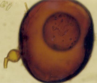
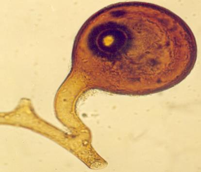
44 Spores of Glomus Relatively small white spores of a Glomus species. Spore of Glomus clarum which has a visible inner wall layer (arrow). Sporocarp of Glomus invermaium typical of the dead spores often found in field-collected soil Living spores of Glomus invermaium from a pot culture.


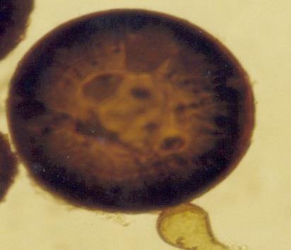
45 Spores of Acaulospora Acaulospora spore with deep pits in the outer wall and inner wall layers stained by Melzer's reagent. Acaulospora spore with several inner wall layers (arrows). One layer has stained darkly with Melzer's reagent. Spores of Scutellospora Species White Scutellospora cerradensis spores with prominent brown germination shields Large black spore with deep pits of Scutellospora reticulata.
46 Arbuscular Mycorrhizal Fungi Populations of arbuscular mycorrhizal fungi in the Glomeromycota are thought to have occupied the same soil habitats for millions of years, slowly adapting to changes in site conditions. Many of these fungi have worldwide distribution patterns, but soil factors such as ph restrict the distribution of other taxa. Consequently, habitat information is as important as knowledge of the taxonomic identity of fungi, for comparing the results of experiments, or the selection of isolates for practical use. The classification of the Glomeromycota is based on the structure of their soil-borne spores and DNA sequences. Accurate identification of these fungi often requires them to be isolated in cultures with host plants, to observe developmental stages, avoid the loss of diagnostic features and obtain healthy spores for DNA extraction. There now are many DNA sequences of Glomeromycotan fungi in databases such as Genbank
47 Arbuscular Mycorrhizal Fungi VAM fungi belong to the Glomeromycota. They are primitive fungi at the base of the tree for higher fungi (basidiomycetes). They associated with first land plants and appear to have evolved very slowly since then. They have no known sexual state. They produce microscopic structures, or relatively small sporocarps (truffle-like). Just over 200 species of these fungi are described, yet they are capable of forming mycorrhizal associations with the majority of plants.
48
49 Classification scheme for Glomeromycotan taxa
50 Classification scheme for Glomeromycotan taxa Archaeosporales Ambisporaceae Archaeosporaceae Geosiphonaceae Diversisporales Acaulosporaceae Entrophosporaceae Diversisporaceae Gigasporaceae Pacisporaceae Glomerales Glomeraceae Paraglomerales Paraglomeraceae Family Genera Acaulosporaceae Acaulospora, Kuklospora Ambisporaceae Ambispora Archaeosporaceae Archaeospora, Intraspora Diversisporaceae Diversispora Entrophosporaceae Entrophospora Geosiphonaceae Geosiphon (not a mycorrhizal fungus) Gigasporaceae Gigaspora, Scutellospora Glomeraceae Glomus (Sclerocystis) Pacisporaceae Pacispora Paraglomeraceae Paraglomus
51
52 SOURCES OF AMF INOCULUM SOIL,CRUDE,ROOT 1.Soil inoculum composed of soil, dried root fragments, and AMF spores, sporocarps, and fragments of hyphae Not a reliable inoculum - last resort Possible transfer of weed seeds and pathogens with the soil is a deterrent Figuring out how much soil to add as inoculum to a growth medium or a field is another challenge, because the abundance and viability of AMF propagules in the soil is often uncertain. Low spore viability or dead spores
53 SOURCES OF AMF INOCULUM soil or root tissue from the site can be taken to start a trap culture to boost the number of viable spore propagules for isolation and further multiplication. These roots and soil are either mixed into the growth medium or applied in a band below the soil surface. Germinated seeds of the indicator plant are then planted and grown long enough for formation of a mixed culture containing mature AMF spores, which are then Extracted, separated into morphological types, identified, and used as starter cultures.
54 SOURCES OF AMF INOCULUM 2. Crude inoculum obtained after a known isolate of AMF and a suitable host are grown together in a medium optimized for AMF development and spore formation. common type available for large-scale crop inoculation. It consists of spores, fragments of infected roots, pieces of AMF hyphae, and the medium Spores can be extracted from such an inoculum by wet-sieving and decanting, and used, alone, before or after surface disinfection spore inocula are known to initiate AMF colonization less rapidly than crude inocula, possibly because crude inocula contain a greater number of different types of infective propagules. 3. Root inoculum Infected roots of a known AMF host separated from a medium in which crude inoculum was produced can also serve as a source of inoculum.
55 MASS PRODUCTION OF VAM BY POT CULTURE TECHNIQUE STERILE POTS SOIL VAM SPORES IN WATCH GLASS SOIL:SAND VAM SPORES HOST PLANT + STERILE SOIL+VAM SPORES KEEP IN GLASS HOUSES YOUNG SEEDLINGS (remove the seedlings after few weeks) SEEDLINGS (check VAM spores microscopically. If present chop the root) CHOPPED ROOTS AS STARTER INOCULUM (Put small amount of starter inoculums one inch below soil layer in the pot) INOCULATED IN LARGER POTS (remove seedlings after 3-4 months. Inoculate seeds in soil) INOCULUM IN BULK (used in field as granular preparation THE PELLETED SEEDS ARE PACKED IN POLYTHENE BAGS
56 Producing crude/soil/root inoculums Physical Environment Media- Soil+Sand (Silica not coral) Photosynthate, light intensity, soil air temperature, soil water status Container types plastic bags and pots made of concrete, clay, and plastic. They should have holes in the bottom to ensure adequate drainage. To minimize the amount of light reaching the medium, the containers should not be translucent. If clear material must be used, it should be painted or enclosed by wrapping in an opaque material kg of medium per container Starter culture The culture must be highly infective, contain at least four infective propagules per gram, and be free of pathogenic microorganisms. The aim is to inoculate the inoculum-production medium at a rate of 500 infective AMF propagules per kilogram of medium. Nurse plant species It should grow fast, be adapted to the prevailing growing conditions, be readily colonized by AMF, and produce a large quantity of roots within a relatively short time (45 60 days). It should be resistant to any pests and diseases common in the inoculum-production environment. The best nurse plants are C. dactylon, S. grandiflora, and Z. mays
57 Producing crude/soil/root inoculums Nutrient management P and N should be optimum. Higher or lower limits growth Hogland s solution with or without P to augment growth K 250, Mg 212 (as MgSO4), Zn 10, Cu 5, B 0.1, Mo 0.5. Duration of growth Essential to grow the nurse plant in the inoculum-production medium for weeks. The medium is then allowed to dry slowly by reducing the frequency of watering over a week and then withdrawing water completely for another week. If at the end of the last week the plant is dry, it is removed from the growth medium. The roots of the plant can be chopped into fragments 1 cm long and mixed with the medium, or they can be used separately as root inoculum. The moisture content of the medium at this time should be 5% or lower. If not, the crude inoculum must be spread on a clean surface in an environment with low humidity (RH 65%) and allowed to air-dry until the desired moisture content is reached.
58
59 Extracting AMF spores from soil or crude inoculum Wet-sieving and decanting Soil samples from field sites should be taken from the rhizosphere of mycorrhizal native or crop plants at a soil depth where the most root proliferation occurs, usually 0 20 cm. The sample is then passed through a 2-mm sieve. A g soil sample (dry weight) is transferred to a beaker. If the soil is dry at sampling, make sure it is soaked for minutes before attempting to extract spores. Soil aggregates can be crushed with a spatula. Distilled or deionized water is added to obtain a 1-L suspension, and the suspension can be agitated for 1 hour in an electric stirrer. The purpose of these steps is to disperse the soil aggregates and release AMF spores. A 3.5% sodium hexametaphosphate solution can be added to increase soil dispersion.
60 Extracting AMF spores from soil or crude inoculum The soil suspension is poured through a stack of sieves (750, 250, 100, 53, and 37 μm), the finest sieve being at the bottom of the stack. A stream of tap water is added to facilitate the movement of spores. The material that remains in the 37, 100, and 250 μm aperture sieves is suspended in water and transferred to centrifuge tubes and centrifuged for 3 minutes at 2000 g. Spores are sedimented at the bottom of the tube, while organic materials remains in suspension. After removing the supernatant, the sediment is re-suspended in a 50% sucrose solution and centrifuged again for 1 2 minutes at 2000 g. After this, the spores will be in the supernatant or in the sugar-water interface. The supernatant fluid containing the spores is poured onto a 28μm aperture sieve or removed with a syringe and rinsed immediately with water to remove the sucrose. Exposure of spores to high concentration of sugar for too much time can dehydrate them, and therefore they should be transferred to tubes and stored in distilled water at least for 24 hours before mixing them with the growth medium. This will allow them to overcome osmotic shock
61 Extracting AMF spores from soil or crude inoculum The number of AMF spores in a suspension can be determined under a microscope by transferring a small volume of the suspension into a counting chamber such as the type used for counting nematodes. The standard counting chambers used in microbiological laboratories are etched with squares of known area and are constructed so that a film of the suspension of known depth can be introduced between the slide and the cover slip.
62 Extracting AMF spores from soil or crude inoculum Separation into morphotypes Spores of AMF can be transferred to a petri dish for microscopic examination and separation. Spores can be separated into distinct morphological types. Fine-tipped forceps or Pasteur pipettes can be used to transfer spores into vials or micro-dishes with water for subsequent evaluation and identification. Alternatively, spores can be collected on a filter paper and picked up from it singly with forceps or a finetipped instrument such as a dissecting needle or a paint brush. Collection of spores from water suspension is better for avoiding undesired hyphal fragments.
63 Extracting AMF spores from soil or crude inoculum Once spores are isolated and identified, they can be surface-disinfected and used as a starter inoculum for production of inoculum in one of the several ways described already. Spores of AMF are surface-sterilized by exposing them to a solution of liquid detergent (e.g., Tween 20), 0.5% sodium hypochlorite, or 2% Chloramine T, and 0.02% streptomycin sulfate in a filter unit allowing contact for 15 minutes and then rinsing with five changes of water. Alternatively, spores can be exposed to % mercuric chloride for 2 10 minutes and rinsed with three to five changes of sterile distilled or deionized water.
64
65
66
67 Extracting spores from a crude inoculum and determining their viability Sterilized soil or sand-soil mixture containing a very low concentration of available P is aseptically packed in a petri dish, leveled, and moistened with distilled water or a solution of 0.1% trypan blue to maximum available water holding capacity (Figure 17). The trypan blue solution facilitates the visibility of hyphae. On the surface of the soil, a nylon mesh (pore size 50 μm) is placed. Pieces of membrane filter 10 x 10 mm (cellulose-acetate, pore size 0.45 μm) are placed on the nylon membrane. The nylon mesh and filter squares should be sterilized by immersion for 5 minutes in 70% ethanol and rinsed with sterile deionized or distilled water prior to use. One AMF spore is placed on each filter square. The petri dish is covered and incubated in the dark at 20 C and observed regularly under a stereo microscope for 5 20 days, depending on the AMF species involved. A spore is considered to have germinated when the length of the germ tube exceeds the diameter of the spore. Except during observation for germination, the petri dish must remain closed to avoid desiccation or contamination. Alternatively, spores can be placed on a membrane filter that is folded twice and inserted into moist soil. After a 2-week incubation period, the filter is removed, unfolded, stained, and examined under a microscope
68
69 DETECTING AND QUANTIFYING AMF COLONIZATION OF ROOTS Procedure Collecting root samples After the root system is thoroughly washed free of soil, obtain a representative sample by removing four to five portions containing the entire length of the root. Chop the portions into four segments and mix them together. Transfer g (moist weight) portions of the mixture into glass or plastic vials. Rinse the roots with a couple changes of water if needed.
70 DETECTING AND QUANTIFYING AMF COLONIZATION OF ROOTS Clearing roots The aim of clearing is to get rid of nuclear and cytoplasmic materials in order to facilitate maximal penetration of the stain. Clear roots by completely covering them with 10% KOH in deionized water (w/v) for h at ambient temperature. Pour off the KOH solution and rinse the root in at least four changes of water. If roots are dark or pigmented, they can be bleached before they are acidified and stained. The most commonly used bleaching material is alkaline H2O2. It is prepared by mixing 3 ml of NH4OH with 10% H2O2 and 567 ml of tap water. NH4OH may be replaced by the same volume of household ammonia. The duration of bleaching is minutes, after which the roots are rinsed with at least three changes of tap water.
71 DETECTING AND QUANTIFYING AMF COLONIZATION OF ROOTS Acidifying roots Roots must be acidified to facilitate retention of the stain by the target specimen. Cover the roots with 10% HCl for 5 10 minutes. Remove the acid but do not rinse the root after this step. Staining roots Cover roots with an acid fuchsin-lactic acid solution and incubate them at ambient temperature for h. The staining solution is prepared by dissolving 1.5 g of acid fuchsin in a solvent consisting of 63 ml of glycerine, 63 ml of water, and 875 ml of food-grade lactic acid.
72 DETECTING AND QUANTIFYING AMF COLONIZATION OF ROOTS Destaining roots To destain roots, decant the stain from the vials containing the roots and rinse the roots with used but filtered (Whatman #1 filter paper) destaining solution to get rid of the excess stain. Cover the roots with unused destaining solution which consists of the solvent mixture used for dissolving the dye. Incubate the vials at ambient temperature for h. At the end of this period, decant the destaining solution and add unused destaining solution. The roots now should be ready for observation. In each of the above steps in which incubation is involved, the h incubation period can be replaced by heating in a water bath at 90 C for 1 h or autoclaving at 121 C for 15 min, if one has the means for doing so.
73 DETECTING AND QUANTIFYING AMF COLONIZATION OF ROOTS Observing stained roots and estimating AMF colonization level Stained root fragments can be spread in petri plates or mounted on microscope slides and examined for the occurrence of typical AMF structures. The most accurate method of determining the level of infection is the grid line intersect method. In this method, stained root preparations are spread on petri plates with grid lines on the bottom. The roots are then examined under a stereo microscope at 40x magnification. Each intersection of root and gridline is checked for the presence or absence of AMF structure(s) and scored as colonized or not colonized by AMF. Using these values the percentage of AMF colonization can be calculated. In this technique, the grid lines simply serve to systematically locate points of observation. For best accuracy, at least 200 root-gridline intersects must be tallied, although 100 root-gridline intersects are acceptable in most instances. The method can also be used to estimate the proportion of the root length that is colonized by AMF.
74 DETECTING AND QUANTIFYING AMF COLONIZATION OF ROOTS The number of root-gridline intersects to the total length of root spread is related by the formula, R = πan/2h where R = the total length of root π =.1 16 A = the area in which roots are distributed n = the number of root-gridline intersections H = the total length of straight lines.
75
 grid, and a 1 m test sample of thread cut")

76 A gridline intersection example using a 8.5 cm diameter round Petri dish with a 1/2 inch (14/11 cm) grid, and a 1 m test sample of thread cut into fragments and randomly redistributed 10 times (Figure 4.3 in Brundrett et al. 1996). Row and column totals are summarised in the table below. Redistributi on Intersects (cm) Average cm ± 2.5 (standard error)
77 DETERMINING THE ABUNDANCE OF INFECTIVE PROPAGULES IN CRUDE INOCULUM AND IN SOIL Procedures The technique is based on determining the presence or absence of microorganisms in several individual aliquots of each of several consecutive dilutions of a sample of soil or other materials containing microbial propagules. A serial dilution, usually 10-fold, of a soil or crude inoculum sample is prepared using sterile sand, soil, or sand-soil mixture as the diluent. From each dilution, a predetermined amount of material, say 20 g, is used to inoculate each of five cups containing g of sterile soil or sand-soil mixture optimized for mycorrhizal activity with a soil-solution P concentration of 0.02 mg/l. Germinated seeds or seedlings of a suitable mycorrhizal plant (onion, clover, leucaena, etc.) are sown in these cups, which are placed in a reservoir containing water or P-free nutrient solution.
78 DETERMINING THE ABUNDANCE OF INFECTIVE PROPAGULES IN CRUDE INOCULUM AND IN SOIL the indicator plant of choice for MPN determination is Leucaena leucocephala, and it is grown on a 1:1 mansand:soil mixture. The P concentration of the medium is 0.02 mg/l and its ph is 6.2. The medium is supplemented weekly with 100 ml of P-free Hoagland s solution. The plants are then allowed to grow in the greenhouse or growth chamber for four weeks. At the end of the growth period, the roots are excised, washed and cleared. The stained roots are spread in a petri dish and scored for the presence or absence of AMF colonization. Do not count detached hyphae or germinated spores.
79 DETERMINING THE ABUNDANCE OF INFECTIVE PROPAGULES IN CRUDE INOCULUM AND IN SOIL To calculate the most probable number of infective propagules in a sample, the statistical table developed by Cochran is essential. In the table, p1 stands for the number of positive replicates in the least concentrated dilution, and p2 and p3 represent the numbers of positive replicates in the next two higher dilutions. The most probable number of infective propagules in the quantity of the original sample is obtained by multiplying the reciprocal of the middle dilution by the number in the table located at the point of intersection of the experimentally observed values corresponding to p1, p2, and p3. The value represents the most probable number of infective propagules for the quantity of soil used to inoculate test plants (20 g in the current example). The number of infective propagules per gram of soil can be obtained by dividing the number of infective propagules observed by the quantity of soil.
80 DETERMINING THE ABUNDANCE OF INFECTIVE PROPAGULES IN CRUDE INOCULUM AND IN SOIL Suppose the following number of positive replicates are obtained for the following dilutions: 10 1 = = = = = 0 In this series, p1 = 5, p2 = 4, and p3 = 1 For this combination of p1, p2, and p3, Co hran s ta le gives 1.7 as the most probable number of infective propagules applied in the 10 2 dilution. Multiplying this value by the dilution factor 102 gives 107 as the number of infective propagules in the original sample. The number of infective propagules per gram of soil is calculated (107 / 20 = 5.35) to be approximately five.
81
82
Working with Mycorrhizas in Forestry and Agriculture
 Working with Mycorrhizas in Forestry and Agriculture SUB Gdttingen 206 384661 Mark Brundrett, Neale Bougher, Bernie Dell, Tim Grove and Nick Malajczuk CONTENTS Chapter I. INTRODUCTION 1.1. MYCORRHIZAL
Glomeromycota: Glomerales the arbuscular mycorrhizae
 Glomeromycota: Glomerales the arbuscular mycorrhizae Classification based on limited morphology now under revision due to molecular evidence 1 Order: Glomerales (=Glomales) About 200 species, three families
Factors Affecting the Infection of Vesicular Arbuscular Mycorrhizal Fungi in Transformed Root Culture
 Factors Affecting the Infection of Vesicular Arbuscular Mycorrhizal Fungi in Transformed Root Culture Poonpilai Suwanaritl, Savitri Ascharakul2, Omsub Nopamornbodi3 and Malee Suwana-adth4 I Department
Mycorrhiza Fungus + Plant Host (Root)
") Mycorrhiza Fungus + Plant Host (Root) Root Anatomy Mycorrhizal fungi Cryptomycota http://www.mykoweb.com/articles/index.html#apm1_4 Summary Mycorrhizal symbioses are mutualistic Fungal benefits carbohydrates
Mycorrhiza Fungus + Plant Host (Root) Root Anatomy Mycorrhizal fungi Cryptomycota http://www.mykoweb.com/articles/index.html#apm1_4 Summary Mycorrhizal symbioses are mutualistic Fungal benefits carbohydrates
The occurrence and diversity of mycorrhizal fungi found in blueberry. Susan McCallum
 The occurrence and diversity of mycorrhizal fungi found in blueberry Susan McCallum Blueberry root system Shallow rooting system mainly concentrated near the soil surface Roots that are larger than 1mm
Absorption of Mineral Salts by Higher Plant
 Article Shared by Absorption of Mineral Salts by Higher Plant Let us make an in-depth study of the Mycorrhizae. After reading this article you will learn about their role in absorption of mineral salts
COMPONENTS OF VA MYCORRHIZAL INOCULUM AND THEIR EFFECTS ON GROWTH OF ONION
 New Phytol. (1981) 87, 3 5 5.161 355 OMPONENTS OF VA MYORRHIZAL INOULUM AND THEIR EFFETS ON GROWTH OF ONION BY A. MANJUNATH AND D. J. BAGYARAJ Depart?nent of Agricultural Microbiology, University of Agricultural
Effect of host plant, cultivation media and inoculants sources on propagation of mycorrhizal fungus Glomus Mossae
 EUROPEAN ACADEMIC RESEARCH Vol. V, Issue 12/ March 2018 ISSN 2286-4822 www.euacademic.org Impact Factor: 3.4546 (UIF) DRJI Value: 5.9 (B+) Effect of host plant, cultivation and inoculants sources on propagation
Wantira Ranabuht Department of Botany, Faculty of Science Chulalongkorn University
 EFFECTS OF ARBUSCULAR MYCORRHIZAL FUNGI ON GROWTH AND PRODUCTIVITY OF LETTUCE Wantira Ranabuht Department of Botany, Faculty of Science Chulalongkorn University Lettuce Lettuce : Lactuca sativa L. Family
Comparison of two main mycorrhizal types
 Comparison of two main mycorrhizal types VAM (Endos) Ectos Plant hosts Most vascular plants, including herbs, shrubs, trees. examples of tree you know: Maples, Ash, giant Sequoia, Sequoia, Incense Cedar
ARE YOU familiar with the sayings Get to
 Root Anatomy ARE YOU familiar with the sayings Get to the root of the problem or the root of all evil? Both these sayings suggest that the root is an essential part of something. With plants, the essential
ABSTRACT I. INTRODUCTION
 2017 IJSRST Volume 3 Issue 7 Print ISSN: 2395-6011 Online ISSN: 2395-602X Themed Section: Science and Technology Effect of Arbuscular Mycorrhizal Fungi on Chemical Properties of Experimental Barren Soil
Lecture Glomeromycota. - Mycorrhizal Associations. Glomeromycota (Vesicular) Arbuscular endomycorrhizal fungi, or (V)AM fungi
 Arbuscular endomycorrhizal fungi, or (V)AM fungi") Lecture 18 - Glomeromycota - Mycorrhizal Associations Glomeromycota (Vesicular) Arbuscular endomycorrhizal fungi, or (V)AM fungi Mycorrhizal root system washed carefully from coarse sand to reveal the
Lecture 18 - Glomeromycota - Mycorrhizal Associations Glomeromycota (Vesicular) Arbuscular endomycorrhizal fungi, or (V)AM fungi Mycorrhizal root system washed carefully from coarse sand to reveal the
Vesicular-arbuscular mycorrhizal associations of sesamum
 Proc. lndian Acad. Sci. (Plant Sci.), Vol. 98, No. 1, February 1988, pp. 55-59. 9 Printed in India. Vesicular-arbuscular mycorrhizal associations of sesamum M VIJAYALAKSHMI and A S RAO Department of Botany,
Mycorrhizae in relation to crop rotation and tillage Terence McGonigle
 Mycorrhizae in relation to crop rotation and tillage Terence McGonigle, Dept. of Biology, Brandon University, Brandon, MB R7A 6A9 E- mail: mcgoniglet@brandonu.ca Abstract: Many crops form mycorrhizae,
 Question 1: What are the factors affecting the rate of diffusion? Diffusion is the passive movement of substances from a region of higher concentration to a region of lower concentration. Diffusion of
CBSE Quick Revision Notes (Class-11 Biology) CHAPTER-11 TRANSPORT IN PLANTS
 CHAPTER-11 TRANSPORT IN PLANTS") CBSE Quick Revision Notes (Class-11 Biology) CHAPTER-11 TRANSPORT IN PLANTS Plant transport various substance like gases, minerals, water, hormones, photosynthetes and organic solutes to short distance
CBSE Quick Revision Notes (Class-11 Biology) CHAPTER-11 TRANSPORT IN PLANTS Plant transport various substance like gases, minerals, water, hormones, photosynthetes and organic solutes to short distance
Appressorium formation by AM fungi on isolated cell walls of carrot roots
 New Phytol. (1997), 136, 299-304 Appressorium formation by AM fungi on isolated cell walls of carrot roots BY G. NAGAHASHI* AND D. D. DOUDS, JR USDA, Agricultural Research Service, Eastern Regional Research
Fungi are absorptive heterotrophs that secrete digestive enzymes and are major decomposers of dead organic material
 Fungi 1 2002 Prentice Hall, Inc The scarlet hood (Hygrocybe coccinea) Fungi are absorptive heterotrophs that secrete digestive enzymes and are major decomposers of dead organic material 2 Animals 3 Myxozoa
Lab 6A: Microscopic Assessment of Mycorrhiza - Part 1
 Lab 6A: Microscopic Assessment of Mycorrhiza - Part 1 What can I expect to learn in lab today? You will gain experience in assessing the degree of mycorrhizal infection of Western Wheatgrass (Agropyron
Plant and animal cells (eukaryotic cells) have a cell membrane, cytoplasm and genetic material enclosed in a nucleus.
 have a cell membrane, cytoplasm and genetic material enclosed in a nucleus.") 4.1 Cell biology Cells are the basic unit of all forms of life. In this section we explore how structural differences between types of cells enables them to perform specific functions within the organism.
4.1 Cell biology Cells are the basic unit of all forms of life. In this section we explore how structural differences between types of cells enables them to perform specific functions within the organism.
Arbuscular Mycorrhizas: Producing and Applying Arbuscular Mycorrhizal Inoculum
 Arbuscular Mycorrhizas: Producing and Applying Arbuscular Mycorrhizal Inoculum M. Habte and N. W. Osorio Department of Tropical Plant and Soil Sciences Acknowledgments We are grateful to Dr. Mark Brundrett,
Eelgrass biomass and production
 Eelgrass biomass and production Objectives To introduce methods for assessing basic parameters fundamental to many seagrass studies such as shoot size and stand structure expressed as biomass and shoot
QUANTIFYING VESICULAR-ARBUSCULAR MYCORRHIZAE: A PROPOSED METHOD TOWARDS STANDARDIZATION*
 W. (1981)87, 6-67 6 QUANTIFYING VESICULAR-ARBUSCULAR MYCORRHIZAE: A PROPOSED METHOD TOWARDS STANDARDIZATION* BY BRENDA BIERMANN Department of Botany and Plant Pathology, Oregon State University, Corvallis,
Topic 14. The Root System. II. Anatomy of an Actively Growing Root Tip
 Topic 14. The Root System Introduction. This is the first of two lab topics that focus on the three plant organs (root, stem, leaf). In these labs we want you to recognize how tissues are organized in
Fungi Coloring Worksheet
 Fungi Coloring Worksheet The basic structural features of fungi are not cells but hyphae. Hyphae are microscopic branching filaments filled with cytoplasm and nuclei. Each thread consists of a tube formed
in angiosperms 10/29/08 Roots take up water via roots Large surface area is needed Roots branch and have root hairs Cortex structure also helps uptake
 in angiosperms A. Root System Roots take up water via roots Large surface area is needed Roots branch and have root hairs Cortex structure also helps uptake 1 B. Minerals Nitrogen (NO 3-,NH 4+ ) Potassium
CHAPTER TRANSPORT
 CHAPTER 2 2.4 TRANSPORT Uptake of CO2 FOCUS: Uptake and transport of water and mineral salts Transport of organic substances Physical forces drive the transport of materials in plants over a range of distances
Absorption of Water by Plants
 Absorption of Water by Plants Absorption of water by cells and roots Availability of Water in the Soil Soil is the major source of water for plants. The plants absorb water through root hairs from the
Name: Block: FUNGI WORKSHEET
 FUNGI WORKSHEET Name: Block: The basic structural features of fungi are not cells but hyphae. Hyphae are microscopic branching filaments filled with cytoplasm and nuclei. Each thread consists of a tube
OCR (A) Biology A-level
 Biology A-level") OCR (A) Biology A-level Topic 3.3: Transport in plants Notes Plants require a transport system to ensure that all the cells of a plant receive a sufficient amount of nutrients. This is achieved through
OCR (A) Biology A-level Topic 3.3: Transport in plants Notes Plants require a transport system to ensure that all the cells of a plant receive a sufficient amount of nutrients. This is achieved through
Inoculation and Colonization of Four Saltmarsh Species with Vesicular-Arbuscular Mycorrhizal Fungi (Mississippi)
") Inoculation and Colonization of Four Saltmarsh Species with Vesicular-Arbuscular Mycorrhizal Fungi (Mississippi) Melissa Pratt-Zossoungbo (NOAA National Ocean Service, Policy, Planning and Analysis Division,
Inoculation and Colonization of Four Saltmarsh Species with Vesicular-Arbuscular Mycorrhizal Fungi (Mississippi) Melissa Pratt-Zossoungbo (NOAA National Ocean Service, Policy, Planning and Analysis Division,
3. Mycorrhiza 3.1. Introduction 3.2. Benefits of Mycorrhizal Biofertilizer
 3. Mycorrhiza 3.1. Introduction Mycorrhizal fungi are species of fungi that intimately associate with plant roots forming a symbiotic relationship, with the plant providing sugars for the fungi and the
Preservation of Spores of Vesicular-Arbuscular Endophytes by L-Drying
 APPLIED AND ENVIRONMENTAL MICROBIOLOGY, May 1979, p. 831-835 0099-2240/79/05-0831/05$02.00/0 Vol. 37, No. 5 Preservation of Spores of Vesicular-Arbuscular Endophytes by L-Drying INEZ C. TOMMERUP* AND DENIS
Contains ribosomes attached to the endoplasmic reticulum. Genetic material consists of linear chromosomes. Diameter of the cell is 1 m
 1. (a) Complete each box in the table, which compares a prokaryotic and a eukaryotic cell, with a tick if the statement is correct or a cross if it is incorrect. Prokaryotic cell Eukaryotic cell Contains
Growth and Colony Patterning of Filamentous Fungi
 Letter Forma, 14, 315 320, 1999 Growth and Colony Patterning of Filamentous Fungi Shu MATSUURA School of High-Technology for Human Welfare, Tokai University, Numazu, Shizuoka 410-0395, Japan E-mail: shum@wing.
INTRODUCTION budding, binary fission hyphae mycelium Figure 1.
 INTRODUCTION Although most of our work in this lab is done on bacteria, fungi are nonetheless an important aspect in microbiology. Besides being important food providers, fungi play central roles in recycling
Effect of Glomus sp and Gigaspora sp. on Vigna radiata (L.) Under Water Stress Condition
 Under Water Stress Condition") American-Eurasian J. Agric. & Environ. Sci., 3 (7): 935-942, 203 ISSN 88-6769 IDOSI Publications, 203 DOI: 0.5829/idosi.aejaes.203.3.07.995 Effect of Glomus sp and Gigaspora sp. on Vigna radiata (L.) Under
American-Eurasian J. Agric. & Environ. Sci., 3 (7): 935-942, 203 ISSN 88-6769 IDOSI Publications, 203 DOI: 0.5829/idosi.aejaes.203.3.07.995 Effect of Glomus sp and Gigaspora sp. on Vigna radiata (L.) Under
Microbiology. Definition of a Microorganism. Microorganisms in the Lab. The Study of Microorganisms
 Microbiology The Study of Microorganisms Definition of a Microorganism Derived from the Greek: Mikros, «small» and Organismos, organism Microscopic organism which is single celled (unicellular) or a mass
Literature. Morphology. Morphology of the mycorrhizal system. Morphology of the unramified ends
 Literature references Müller WR, Rauscher T, Agerer R, Chevalier G (1996) Tuber aestivum Vitt. + Corylus avellana L.Descr Ectomyc 1: 167-172. Rauscher T, Müller WR, Chevalier G, Agerer R (1996) Tuber aestivum.
Proc. Indian Acad. Sci. (Plaat Sci.), Vol. 95, No. 1, August 1985, pp Printed in India. K PARVATHI, K VENKATESWARLU and A S RAO
, Vol. 95, No. 1, August 1985, pp Printed in India. K PARVATHI, K VENKATESWARLU and A S RAO") Proc. Indian Acad. Sci. (Plaat Sci.), Vol. 95, No. 1, August 1985, pp. 35--40. 9 Printed in India. Response of groundnut (Arachis hypogaea L) to combined inoculation with Glomus mosseae and Rhizobium sp
Proc. Indian Acad. Sci. (Plaat Sci.), Vol. 95, No. 1, August 1985, pp. 35--40. 9 Printed in India. Response of groundnut (Arachis hypogaea L) to combined inoculation with Glomus mosseae and Rhizobium sp
Preparing the sample for determination of Viability
 Preparing the sample for determination of Viability I. For preparing one sample for analysis you will need: - 1 pcs CELLCHIP - 1 piece of colored Eppendorf with SOFIA GREEN lyophilized dye - 1 piece of
2014 Pearson Education, Inc. 1
 1 CO 2 O 2 Light Sugar O 2 and minerals CO 2 2 Buds 42 29 21 34 13 26 5 18 10 31 23 8 15 28 16 2 24 Shoot apical meristem 7 3 20 1 mm 32 11 19 12 6 4 1 25 17 14 9 40 27 22 3 Cell wall Apoplastic route
Bacterial Gram Staining
 PR021 G-Biosciences 1-800-628-7730 1-314-991-6034 technical@gbiosciences.com A Geno Technology, Inc. (USA) brand name Bacterial Gram Staining Teacher s Guidebook (Cat. # BE 202) think proteins! think G-Biosciences
EFFECTS OF NUTRIENT LEVELS ON THE COLONIZATION OF POA SECUNDA BY ARBUSCULAR MYCORRHIZAL FUNGI AND DARK SEPTATE ENDOPHYTES
 EFFECTS OF NUTRIENT LEVELS ON THE COLONIZATION OF POA SECUNDA BY ARBUSCULAR MYCORRHIZAL FUNGI AND DARK SEPTATE ENDOPHYTES Preya Sanjay Sheth Abstract Arbuscular mycorrhizal fungi (AMF) and dark septate
Movement of Molecules Biology Concepts of Biology 3.1
 Movement of Molecules Biology 100 - Concepts of Biology 3.1 Name Instructor Lab Section Objectives: To gain an understanding of: The basic principles of osmosis and diffusion Brownian motion The effects
Vesicular-arbuscular mycorrhizal fungal sporocarps associated with Pennisetum pedicillatum
 Proc. lndian Acad. Sci. (Plant Sci.), Vol. 96, No. 2, June 1986, pp. 153--158. 9 Printed in India. Vesicular-arbuscular mycorrhizal fungal sporocarps associated with Pennisetum pedicillatum K AMMANI, K
Chapter 37: Plant Nutrition - A Nutritional Network
 Chapter 37: Plant Nutrition - A Nutritional Network Every organism continually exchanges energy and materials with its environment For a typical plant, water and minerals come from the soil, while carbon
MICROBIOLOGY LAB #1 SAFETY RULES & GRAM STAIN METHOD
 MICROBIOLOGY LAB #1 SAFETY RULES & GRAM STAIN METHOD Precaution processes are extremely important when working with cultures in the lab for the safety of the microbiologist from getting diseases from bacteria
Exercise VI. Differential Staining: The Gram Stain
 Exercise VI Differential Staining: The Gram Stain The Gram stain, discovered by Dr. Hans Christian Gram in 1884, is the most useful differential stain used to aid in identifying bacteria. It divides bacterial
Mycorrhiza Fungus + Plant Host (Root)
") Mycorrhiza Fungus + Plant Host (Root) Two fungi commonly Use in ectomycorrhiza Research. Laccaria bicolor Pisolithus tinctorius Flowering Plants and mycorrhizal fungi http://mycorrhizas.info/evol.html#intro
Mycorrhiza Fungus + Plant Host (Root) Two fungi commonly Use in ectomycorrhiza Research. Laccaria bicolor Pisolithus tinctorius Flowering Plants and mycorrhizal fungi http://mycorrhizas.info/evol.html#intro
Chapter 30: Plant Nutrition & Transport
 Chapter 30: Plant Nutrition & Transport Carnivorous Plants Capture animals to supplement their nutrient intake Venus flytrap lures insects with sugary bait; closes on victim Cobra lily lures insects down
GLOMALIN EXTRACTION Introduction
 GLOMALIN EXTRACTION (Rillig, 2004; Rosier et al., 2007; and Rillig, 2003; Wright et al., 1996; Wright and Jawson, 2001; Wright, Nichols, & Schmidt, 2006; Wright & Upadhyaya, 1996; & Wright & Upadhyaya,
Amutha and Kokila, IJALS, Volume (7) Issue (2) May RESEARCH ARTICLE
 Issue (2) May RESEARCH ARTICLE") Effect of on symbiotic association of Glomus aggregatum an Arbuscular Mycorrhizal Fungus K. Amutha and V. Kokila Department of Biotechnology, Vels University, Pallavaram, Chennai, Tamilnadu, India Email
Effect of on symbiotic association of Glomus aggregatum an Arbuscular Mycorrhizal Fungus K. Amutha and V. Kokila Department of Biotechnology, Vels University, Pallavaram, Chennai, Tamilnadu, India Email
Lesson Plan: Diffusion
 Lesson Plan: Diffusion Background Particles in cells show rapid back and forth movement, or Brownian motion, which is also known as diffusion. The back and forth motion consists of random steps from a
UNIT 6 - STRUCTURES OF FLOWERING PLANTS & THEIR FUNCTIONS
 6.1 Plant Tissues A tissue is a group of cells with common function, structures or both. In plants we can find 2 types of tissues: Meristem Permanent tissues Meristem is found in regions with continuous
LOOKING AT PLANT STEMS
 Activity 4.17 Student Sheet LOOKING AT PLANT STEMS Purpose To look at the structure of xylem vessels, phloem sieve tubes and sclerenchyma fibres. To locate the position of these tissues within the stem.
Chapter 9. Fungi and Aquatic Plants. Introduction: The Big Step: DIVISION OF LABOUR
 Chapter 9. Fungi and Aquatic Plants Introduction: The Big Step: DIVISION OF LABOUR In single cell organisms (protists) all life functions are performed by specialized organelles within one cell (a.k.a.
INTRODUCTION prokaryotic eukaryotic pigments
 INTRODUCTION This exercise is intended for you to get familiar and comfortable with using a microscope as well as identifying common microbial groups. Thus, we will observe representatives of all microbes
PRODUCTION OF SPORANGIA BY PHYTOPHTHORA CINNAMOMI IN PURE CULTURE
 California Avocado Society 1969 Yearbook 53: 103-107 PRODUCTION OF SPORANGIA BY PHYTOPHTHORA CINNAMOMI IN PURE CULTURE G. A. Zentmyer and Dah-wu Chen Department of Plant Pathology, University of California,
Plant Anatomy: roots, stems and leaves
 Plant Anatomy: roots, stems and leaves The plant body has a hierarchy of organs, tissues and cells Plants, like animals, have organs composed of different tissues, which are composed of cells. Tissue is
CELLS. Structure and Function
 CELLS Structure and Function Cell Structure All plant and animal tissue consist of cells. Cells are microscopic in size. In general, each cell performs all the characteristics of life and, though in reality
Soil Microbiology. Ambarish Bhuyan Assistant Professor Botany Department MDKG College, Dibrugarh
 Soil Microbiology Ambarish Bhuyan Assistant Professor Botany Department MDKG College, Dibrugarh INTRODUCTION Nature of soils Soil arises from the weathering of rocks Soil also produced through the actions
Treat the Cause not the symptom
 Treat the Cause not the symptom A few facts about Novozymes Biologicals Bu sin ess d ivisio n o f No vo zym es w it h it s o w n R& D, Manufacturing, Sales & Marketing, Administration Headquartered in
INTRODUCTION bioactive compounds Pigmentation chromobacteria water soluble water insoluble
 INTRODUCTION So far we have witnessed several useful applications of microbes including applications in food and the bioremediation of the environment. Besides consuming the desired substrate (oil) and
General Chemistry I CHEM-1030 Laboratory Experiment No. 2 Physical Separation Techniques
 General Chemistry I CHEM-1030 Laboratory Experiment No. 2 Physical Separation Techniques Introduction When two or more substances that do not react chemically are blended together, the components of the
MYCORRHIZAE IMPACT ON BIODIVERSITY AND C-BALANCE OF GRASSLAND ECOSYSTEMS UNDER CHANGING CLIMATE MYCARBIO
 MYCORRHIZAE IMPACT ON BIODIVERSITY AND C-BALANCE OF GRASSLAND ECOSYSTEMS UNDER CHANGING CLIMATE S. DECLERCK, R. CEULEMANS, I. NIJS, L. VOETS, H. DUPRE DE BOULOIS, I. ENRIQUE DE LA PROVIDENCIA, C. ZAVALLONI,
INTRODUCTION. Gram Stain
 INTRODUCTION In microbiology, organisms are so small that additional techniques are often required for proper viewing under the microscope. Cytological stains, or dyes that stain cells or cellular features,
Useful Propagation Terms. Propagation The application of specific biological principles and concepts in the multiplication of plants.
 Useful Propagation Terms Propagation The application of specific biological principles and concepts in the multiplication of plants. Adventitious Typically describes new organs such as roots that develop
Roots and Soil Chapter 5
 Roots and Soil Chapter 5 Plant Organs Plant organs are groups of several types of tissues that together perform a particular function. Vegetative organs roots, stems, leaves make and use food, absorb water
13.4 Roots Figure 2 primary root: primary root secondary root: secondary root taproots fibrous taproots: roots. fibrous roots: adventitious roots
 10. Why is it not surprising that many hydrophytes have little or no tissue? 11. The leaves of many underwater plants are finely divided, dramatically increasing the surface area that is in contact with
Please use only the valid version of the package insert provided with the kit. This kit is intended for Research Use Only.
 Please use only the valid version of the package insert provided with the kit This kit is intended for Research Use Only. Not for use in diagnostic procedures. INTENDED USE The Vitamin Niacin test is a
Inserting grids into capsule Removing capsules from grid box Immunolabeling using template guide Preparing for TEM insertion
 Protocol Immunolabeling protocols are easily adapted to mprep/g processing. This document illustrates a typical protocol using a primary antibody and a secondary antibody conjugated to colloidal gold,
F.A. SMITH S.E. SMITH
 BIOTROPIA No. 8, 1995: 1-10 NUTRIENT TRANSFER IN VESICULAR-ARBUSCULAR MYCORRHIZAS: A NEW MODEL BASED ON THE DISTRIBUTION OF ATPases ON FUNGAL AND PLANT MEMBRANES*) F.A. SMITH Department of Botany, The
Copyright 2009 Pearson Education, Inc. FUNGI
 Copyright 2009 Pearson Education, Inc. FUNGI FUNGI Fungi are absorptive heterotrophic eukaryotes that digest their food externally and absorb the nutrients Most fungi consist of a mass of threadlike hyphae
Non Permanent Tissues - Meristematic Tissue
 PLANT TISSUES Non Permanent Tissues - Meristematic Tissue Undifferentiated plant cells that are continually dividing by mitosis Large thin walled cells No vacuole Dense cytoplasm Large nucleus Found at
Kingdom Fungi. 1. Student will be able to describe the characteristic features in the kingdom Fungi.
 Kingdom Fungi Molds, Sac Fungi, Mushrooms, and Lichens Essential Question(s): What makes fungi have their own kingdom? Objectives: 1. Student will be able to describe the characteristic features in the
Increased Sporulation of Vesicular-Arbuscular Mycorrhizal Fungi by Manipulation of Nutrient Regimenst
 APPLIED AND ENVIRONMENTAL MICROBIOLOGY, Feb. 199, p. 413-418 99-224/9/2413-6$2./ Copyright 199, American Society for Microbiology Vol. 56, No. 2 Increased Sporulation of Vesicular-Arbuscular Mycorrhizal
Recap. Waxy layer which protects the plant & conserves water. Contains chloroplasts: Specialized for light absorption.
 Recap Contains chloroplasts: Specialized for light absorption Waxy layer which protects the plant & conserves water mesophyll Layer contains air spaces: Specialized for gas exchange Vascular Tissue Exchange
PLANT STRUCTURE: PARTS (ORGANS) Roots Leaves Stems
 Roots Leaves Stems") PLANT STRUCTURE: PARTS (ORGANS) Roots Leaves Stems ROOTS El Hiquieron. Strangulating Plant Ficusjimenezii The trees you see growing on the wall are the Higueron. The Higueronsare plants that can grow in
PLANT STRUCTURE: PARTS (ORGANS) Roots Leaves Stems ROOTS El Hiquieron. Strangulating Plant Ficusjimenezii The trees you see growing on the wall are the Higueron. The Higueronsare plants that can grow in
Chapter 36~ Transport in Plants
 Chapter 36~ Transport in Plants Structural Features Used for Resource Acquistion Roots and stems to do transport of resources Diffusion, active transport, and bulk flow Work in vascular plants to transport
TESTING of AGGREGATES for CONCRETE
 TESTING of AGGREGATES for CONCRETE The properties of the aggregates affect both the fresh and hardened properties of concrete. It is crucial to know the properties of the aggregates to be used in the making
Impact of cropping system on mycorrhiza
 Impact of cropping system on mycorrhiza H. Kahiluoto 1 and M. Vestberg 2 Agricultural Research Centre of Finland 1 Ecological Production, Partala, FIN-51900 Juva, Finland 2 Laukaa Research and Elite Plant
Soil Sample Preparation Protocol (Modified from Faegri and Iversen 1 ; Moore and Webb 2 protocols)
") Soil Sample Preparation Protocol (Modified from Faegri and Iversen 1 ; Moore and Webb 2 protocols) Materials Sediment samples Volumetric sampler Metal spatula Permanent marker 15 ml Conical centrifuge
Soil Sample Preparation Protocol (Modified from Faegri and Iversen 1 ; Moore and Webb 2 protocols) Materials Sediment samples Volumetric sampler Metal spatula Permanent marker 15 ml Conical centrifuge
Plant Function. KEB no office hour on Monday 23 March. Chs 38, 39 (parts), March 2009 ECOL 182R UofA K. E. Bonine
, March 2009 ECOL 182R UofA K. E. Bonine") Plant Function Chs 38, 39 (parts), 40 KEB no office hour on Monday 23 March 10 March 2009 ECOL 182R UofA K. E. Bonine Videos: 39.3, 34.3, 39.1, 34.1 Web Browser Open 1 Video 39.3 Pollination of a night-blooming
Plant Function Chs 38, 39 (parts), 40 KEB no office hour on Monday 23 March 10 March 2009 ECOL 182R UofA K. E. Bonine Videos: 39.3, 34.3, 39.1, 34.1 Web Browser Open 1 Video 39.3 Pollination of a night-blooming
Root-Knot Nematode on Tomato Plants: Effects of Nemacur, Phosphorus and. Infection Time
 Ayman Elbuhuth Scientific Journal., Vol 5, pp. 88-107, 1996 Interaction of VA Mycorrhizal Fungi and Root-Knot Nematode on Tomato Plants: Effects of Nemacur, Phosphorus and Infection Time M. O. MIRGHANI
MYCORRHIZAL FUNGI AS BIOFERTILIZER FOR FRUIT TREE PRODUCTION IN THAILAND. Supaporn Thamsurakul 1 and Sompetch Charoensook 2
 MYCORRHIZAL FUNGI AS BIOFERTILIZER FOR FRUIT TREE PRODUCTION IN THAILAND Supaporn Thamsurakul 1 and Sompetch Charoensook 2 1 Soil Microbiology Research Group, Soil Science Division, Department of Agriculture,
Soil Biology. Chapter 10
 Soil Biology Chapter 10 The Sounds of Soil Soil as a Transition Between Aquatic and Aerial System Bacteria in a Drying Environment Wet (open structure) Dry (dense) Holden P.A., J.R. Hunt, and M. K. Firestone,
CAMBIUM, meristem, heartwood, and lenticel are
 Examining the Structures of a Tree CAMBIUM, meristem, heartwood, and lenticel are some terms that may be new to you. These terms are used to describe various tree structures. Not surprisingly, many terms
Plant Function Chs 38, 39 (parts), 40
, 40") Plant Function Chs 38, 39 (parts), 40 KEB no office hour on Monday 23 March 10 March 2009 ECOL 182R UofA K. E. Bonine Videos: 39.3, 34.3, 39.1, 34.1 Web Browser Open 1 Video 39.3 Pollination of a night-blooming
Plant Function Chs 38, 39 (parts), 40 KEB no office hour on Monday 23 March 10 March 2009 ECOL 182R UofA K. E. Bonine Videos: 39.3, 34.3, 39.1, 34.1 Web Browser Open 1 Video 39.3 Pollination of a night-blooming
BIOL 221 Concepts of Botany Spring Water Relations, Osmosis and Transpiration
 BIOL 221 Concepts of Botany Spring 2008 Topic 07: Water Relations, Osmosis and Transpiration A. Water Relations Water plays a critical role in plants. Water is the universal solvent that allows biochemical
Basic Aggregates Study Guide
 Basic Aggregates Study Guide General Conversions 1) There are pounds in one ton. 2) There are grams in one pound. 3) One kilogram consists of grams. 4) The linear distance that one station covers is feet.
Growth responses of Acacia angustissima to vesicular-arbuscular mycorrhizal. inoculation. Abstract
 Growth responses of Acacia angustissima to vesicular-arbuscular mycorrhizal inoculation ID # 04-32 N. Lucena Costa 1, V.T. Paulino 2 and T.S. Paulino 3 1 EMBRAPA - Amapá,, C.P. 10, Macapá, Amapá, 68902-208,
Zoosporic Fungi Detected and Isolated from the Environment Chytrid Lore and More. Joyce E. Longcore School of Biology & Ecology
 Zoosporic Fungi Detected and Isolated from the Environment Chytrid Lore and More Joyce E. Longcore School of Biology & Ecology Couch, J.N. (1939) Sparrow, F. K. (1943, 1960) Karling, J. S. (1945) Remy,
Mrs. Sonia Bohra* and Dr. Anil Vyas. Department of Botany, J.N.V.University, Jodhpur, Rajasthan, India.
 Received: 01 st Dec-2012 Revised: 08 th Dec-2012 Accepted: 09 th Dec -2012 Research article DISTRIBUTION OF ARBUSCULAR MYCORRHIZAL FUNGI ASSOCIATED WITH LANDSCAPE TREE GROWTH IN INDIAN THAR DESERT Mrs.
SYNTHESIS OF AN AZO DYE revisited (1 or 2 credits)
") SYNTHESIS OF AN AZO DYE revisited (1 or 2 credits) This lab you can revisit the fist experiment of this quarter and synthesize more azo dyes of your choice. The old procedure is given below followed by
SYNTHESIS OF AN AZO DYE revisited (1 or 2 credits) This lab you can revisit the fist experiment of this quarter and synthesize more azo dyes of your choice. The old procedure is given below followed by
Detection limit: grain, feed 500 ppb; milk 50 ppb; cream, cheese 5 ppb
 Product information Background Deoxynivalenol (DON) Deoxynivalenol, called vomitoxin, is a toxic metabolite mainly produced by Fusarium graminearum. It is mainly found in wheat, barley, corn and feed.
NEW CHARACTERISTICS FOR MORPHOTAXONOMY OF GIGASPORA SPECIES BELONGING TO ARBUSCULAR MYCORRHIZAL FUNGI
 SHARDA W. KHADE J. Plant Develop. 18(2011): 71-80 NEW CHARACTERISTICS FOR MORPHOTAXONOMY OF GIGASPORA SPECIES BELONGING TO ARBUSCULAR MYCORRHIZAL FUNGI SHARDA W. KHADE 1 Abstract: New characteristics for
BioWash as an Adjuvant, Translocation Promoter, and Cationic Exchange Stimulator Overview of Processes within the Plant
 BioWash as an Adjuvant, Translocation Promoter, and Cationic Exchange Stimulator Overview of Processes within the Plant Photosynthesis is the primary driver of the plant. Through a series of complex steps,
Thin Layer Chromatography
 Thin Layer Chromatography Thin-layer chromatography involves the same principles as column chromatography, it also is a form of solid-liquid adsorption chromatography. In this case, however, the solid