for Molecular Biology and Neuroscience and Institute of Medical Microbiology, Rikshospitalet-Radiumhospitalet
|
|
|
- Samuel Marsh
- 5 years ago
- Views:
Transcription
1 SUPPLEMENTARY INFORMATION TO Structural basis for enzymatic excision of N -methyladenine and N 3 -methylcytosine from DNA Ingar Leiros,5, Marivi P. Nabong 2,3,5, Kristin Grøsvik 3, Jeanette Ringvoll 2, Gyri T. Haugland 4, Lene Uldal 2, Karen Reite 2, Inger K. Olsbu 3, Ingeborg Knævelsrud 3,4, Elin Moe, Ole A. Andersen, Nils-Kåre Birkeland 4, Peter Ruoff 3, Arne Klungland 2 and Svein Bjelland 3, * The Norwegian Structural Biology Centre, University of Tromsø, N-937 Tromsø, Norway. 2 Centre for Molecular Biology and Neuroscience and Institute of Medical Microbiology, Rikshospitalet-Radiumhospitalet HF, University of Oslo, N-27 Oslo, Norway. 3 Faculty of Science and Technology, Department of Mathematics and Natural Sciences, University of Stavanger, N-436 Stavanger, Norway. 4 Department of Biology, University of Bergen, PO Box 78, N-52 Bergen, Norway. 5 These authors contributed equally to this work. *Corresponding author. Faculty of Science and Technology, Department of Mathematics and Natural Sciences, University of Stavanger, N-436 Stavanger, Norway. Tel.: ; Fax: ; svein.bjelland@uis.no
2 Derivation of kinetic equations for AfAlkA The reaction catalyzed by AfAlkA (E) can be summarized as DNA P (R) where P represents products (i.e. DNA and free base lesion). Reaction R is a first-order process with reaction rate v=d[p]/dt = k [DNA] () Equation is readily solved and the increase of product concentration [P] as a function of time t is given by the equation [P] = [DNA] ( e k t ) (2) where [DNA] denotes the al (initial) DNA substrate concentration and k is the (first-order) rate constant. The experimental results can be interpreted by considering a non-specific glycosylase binding or adsorption to DNA and looking specifically at the site where catalysis (excision) is taking place, i.e., the specific position where m A, εa or m 3 C are inserted (Figure A). If DNA i is the location i on the DNA where catalysis occurs (when E binds to this site) we can write k k E + DNA E DNA 2 i E + DNA + free base lesion (R2) k i - Considering this process, we can write the following mass balance at DNA i : [DNA] = [DNA i ] + [E DNA i ] (3) where [DNA] is the al DNA concentration, E DNA i denotes any DNA molecule where E occupies the catalytic site i, and DNA i is any DNA molecule where E has not bound to the excision site i. Calculating d[e DNA i ]/dt and assuming that E DNA i is in a steady state (leading to d[e DNA i ]/dt = ), it results in d[e DNA i ]/dt = k [E][DNA i ] (k + k 2 )[E DNA i ] = (4) By inserting [DNA i ] from Equation 3 into Equation 4, the steady state value of [E DNA i ], [E DNA i ] ss, can be expressed as 2
3 with [E][DNA] [E DNA i ] ss = K + [E] k D + K D = k k 2 (5) Because the concentration of E is in excess of the DNA template (single-turnover conditions; [E] >> [DNA] ), [E] in Equation 5 can be replaced by [E] : [E DNA i ] ss [E] = (6) K + [E] D [DNA] Since the reaction rate is expressed by ν = k 2 [E DNA i ] ss, ν can be written as [E] [DNA] ν = k2 (7) K D + [E] showing that the rate constant k of Equation is specified by the following relationship: k E] k = 2[ K + [ E] D (8) An analogous expression to Equation 7 can be derived by assuming a rapid equilibrium between E, DNA i and E DNA i. In this case K D is represented by k /k. In the case reaction R2 is irreversible (k = ), K D = k 2 /k. Due to these situations it is difficult to make a precise interpretation about the physical nature of K D based on k 2 and K D values alone. The observation that for m A, k 2 is decreased while K D is increased for most of the mutant proteins (Table I), may suggest that the binding between the DNA and the enzyme has become less effective with a somewhat slower turnover. For εa, both k 2 and K D are decreased for the mutant compared to the wild-type proteins (Table I). This may indicate the presence of a steady state with still a relatively strong binding between DNA and the enzyme, but with a less effective turnover. 3
4 Supplementary Table I Data collection, structure solution and refinement statistics Dataset Peak Native Data collection statistics Beamline ID4-4 BL4. Wavelength (Å).9.99 Resolution range (Å) 2..8(.9.8) 5.-.9(.95-.9) R sym (%) 8.4(49.9) 6.7(43.4) Multiplicity 8.(7.6) 2.9(2.9) Mean I/σI 8.(3.5).4(2.5) Completeness (%) 98.7(97.9) 99.9(.) Anom. completeness (%) 98.5(97.5) - Space group P2 P2 Unit cell parameters: Phasing statistics a(å) b(å) c(å) β( ) No. Heavy atoms/a.s.u. 2 Hg - R cullis Phasing power FOM SHARP.43 - FOM DM Refinement statistics No. Atoms 5493/49/495/4/4/4/4 5425/4826/57//4/4/ B-factors 2/9/29/33 /27 /8/33 2/9/3//34/2/ R free (%) R work (%) Geometrical deviation Bonds (Å).5.5 Angles ( ) ESU (Å).78. : Two additional Mercury atoms were identified at the refinement stage through phased anomalous fourier maps, but were not used at the preceding phasing stage. : Figure-ofmerit (FOM) from SHARP-phasing for acentric data to 2.5 Å. : FOM from DM after extending phases to.8 Å. : Total/Protein/Water/Mercury/Glycerol molecules/sodium/mes molecules. : Modeled with reduced occupancy. : Estimated overall coordinate error from REFMAC5 based on maximum likelihood. 4
5 Supplementary Table II Structural alignment of domains of AfAlkA and EcAlkA EcAlkA (#aligned/al) rmsd (Å) Domain AfAlkA (84/).55 N-term (33/48).4 Central (4/3).38 C-term (59/289).58 All residues The table illustrates structural conservation between individual domains of AfAlkA and EcAlkA, where the numbers in parenthesis are number of residues aligned out of the al number of residues in that domain and the last number lists the root mean square deviation (rmsd) between the aligned residues in AfAlkA and EcAlkA. 5

6 Supplementary Figure 6
7 Legend to Supplementary Figure Exposure of EcAlkA to m A- and m 3 C- containing DNA. Assay for cleavage of 5 [ 32 P]labeled 49-nt DNA into repair product (23 and 25 nt) is described in FigureA,B. EcAlkA was incubated with DNA ( fmol) in 5 mm Hepes [4-(2-hydroxyethyl)--piperazineethanesulfonic acid]-koh, ph 7.5,.3 mm EDTA,.5 mm 2-mercaptoethanol at 37 C for 3 min (The same result was obtained when incubation was carried out at 37 C under exactly the same conditions as employed for AfAlkA). 7


8 Supplementary Figure 2 8
9 Legend to Supplementary Figure 2 Stereo representation showing the superpositioning of the Cα-traces of AfAlkA (red) and Ec AlkA (black). Af AlkA residue numbers are labeled. 9
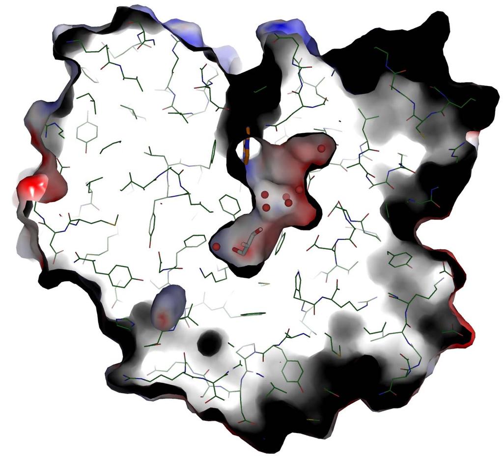
10 Supplementary Figure 3
11 Legend to Supplementary Figure 3 Sliced view of the molecular surface of AfAlkA enabling visualization of the water-accessible channel extending from the base of the substrate-binding pocket into the core of the protein. The modeled εa moiety is shown with orange carbon atoms, while protein carbon atoms are shown in dark green. Other atoms are shown in atom colors (oxygen, red; nitrogen blue; sulfur, yellow). The cavity contains a al of one glycerol and 3 water molecules. The electrostatic surface potential is calculated as for Figure 4.

12 Supplementary Figure 4 2
13 Legend to Supplementary Figure 4 SDS PAGE (2 %) of purified AfAlkA mutant and wild type proteins stained with Coomassie Blue. Samples (2 μg; μl) and protein markers ( μl; BenchMark Pre-Stained Protein Ladder, Cat. No. 748-, Invitrogen) were loaded onto the gel (Bio-Rad, Ready Gel Tris HCl Gels, 2 % Resolving Gel, Cat. # 6-56) and run for 2 h at 2 V. 3
14 Supplementary Figure 5.4 m A (Gln28Ala).6 m A (Phe33Ala) [E]= nm [E]=6 nm [E]=25 nm.4 [E]=5 nm.2 [E]= nm m A (Phe282Ala) [E]= nm [E]=6 nm [E]=25 nm [E]= nm [E]= nm [E]=6 nm [E]=25 nm [E]= nm m A (Phe33Ala/Phe282Ala).6.4 [E]= nm m A (Arg286Ala) [E]= nm [E]=6 nm [E]=25 nm [E]=5 nm [E]= nm 4
15 Legend to Supplementary Figure 5 Single-turnover kinetics for excision of m A, where nm of substrate was incubated with different concentrations of AfAlkA at 7 C for increasing time periods. Each value represents the average of three independent measurements. 5
16 Supplementary Figure 6 ε A (Gln28Ala) 8 7 [E]=25 nm [E]=5 nm 3 2 [E]= nm [E]=5 nm ε A (Phe33Ala) 6 [E]=25 nm [E]=5 nm [E]= nm [E]=5 nm 6 ε A (Phe282Ala).2 ε A (Phe33Ala/Phe282Ala) 5 [E]=25 nm 4 [E]=5 nm 3 2 [E]= nm [E]=5 nm [E]=25 nm.8.6 [E]=5 nm.4.2 [E]= nm 5 ε A (Arg286Ala) [E]=25 nm [E]= nm [E]=5 nm [E]=5 nm 6
17 Legend to Supplementary Figure 6 Single-turnover kinetics for excision of εa, where nm of substrate was incubated with different concentrations of AfAlkA at 7 C for increasing time periods. Each value represents the average of 2 3 independent measurements. 7
SUPPLEMENTARY INFORMATION
 Fig. 1 Influences of crystal lattice contacts on Pol η structures. a. The dominant lattice contact between two hpol η molecules (silver and gold) in the type 1 crystals. b. A close-up view of the hydrophobic
According to the manufacture s direction (Pierce), RNA and DNA
, RNA and DNA") Supplementary method Electrophoretic Mobility-shift assay (EMSA) According to the manufacture s direction (Pierce), RNA and DNA oligonuleotides were firstly labeled by biotin. TAVb (1pM) was incubated
Supplementary method Electrophoretic Mobility-shift assay (EMSA) According to the manufacture s direction (Pierce), RNA and DNA oligonuleotides were firstly labeled by biotin. TAVb (1pM) was incubated
SUPPLEMENTARY INFORMATION
 Table of Contents Page Supplementary Table 1. Diffraction data collection statistics 2 Supplementary Table 2. Crystallographic refinement statistics 3 Supplementary Fig. 1. casic1mfc packing in the R3
SUPPLEMENTARY INFORMATION
 Supplementary materials Figure S1 Fusion protein of Sulfolobus solfataricus SRP54 and a signal peptide. a, Expression vector for the fusion protein. The signal peptide of yeast dipeptidyl aminopeptidase
SUPPLEMENTARY INFORMATION
 Supplementary Table S1 Kinetic Analyses of the AMSH-LP mutants AMSH-LP K M (μm) k cat x 10-3 (s -1 ) WT 71.8 ± 6.3 860 ± 65.4 T353A 76.8 ± 11.7 46.3 ± 3.7 F355A 58.9 ± 10.4 5.33 ± 0.30 proximal S358A 75.1
Supplementary Materials for
 www.advances.sciencemag.org/cgi/content/full/1/7/e1500263/dc1 Supplementary Materials for Newton s cradle proton relay with amide imidic acid tautomerization in inverting cellulase visualized by neutron
SI Text S1 Solution Scattering Data Collection and Analysis. SI references
 SI Text S1 Solution Scattering Data Collection and Analysis. The X-ray photon energy was set to 8 kev. The PILATUS hybrid pixel array detector (RIGAKU) was positioned at a distance of 606 mm from the sample.
Full-length GlpG sequence was generated by PCR from E. coli genomic DNA. (with two sequence variations, D51E/L52V, from the gene bank entry aac28166),
,") Supplementary Methods Protein expression and purification Full-length GlpG sequence was generated by PCR from E. coli genomic DNA (with two sequence variations, D51E/L52V, from the gene bank entry aac28166),
Supplementary Methods Protein expression and purification Full-length GlpG sequence was generated by PCR from E. coli genomic DNA (with two sequence variations, D51E/L52V, from the gene bank entry aac28166),
SUPPLEMENTARY INFORMATION
 doi:10.1038/nature10458 Active Site Remodeling in the Bifunctional Fructose-1,6- bisphosphate aldolase/phosphatase Juan Du, Rafael F. Say, Wei Lü, Georg Fuchs & Oliver Einsle SUPPLEMENTARY FIGURES Figure
Supplementary materials. Crystal structure of the carboxyltransferase domain. of acetyl coenzyme A carboxylase. Department of Biological Sciences
 Supplementary materials Crystal structure of the carboxyltransferase domain of acetyl coenzyme A carboxylase Hailong Zhang, Zhiru Yang, 1 Yang Shen, 1 Liang Tong Department of Biological Sciences Columbia
Supplementary Information
 Supplementary Information The direct role of selenocysteine in [NiFeSe] hydrogenase maturation and catalysis Marta C. Marques a, Cristina Tapia b, Oscar Gutiérrez-Sanz b, Ana Raquel Ramos a, Kimberly L.
type GroEL-GroES complex. Crystals were grown in buffer D (100 mm HEPES, ph 7.5,
 Supplementary Material Supplementary Materials and Methods Structure Determination of SR1-GroES-ADP AlF x SR1-GroES-ADP AlF x was purified as described in Materials and Methods for the wild type GroEL-GroES
Table 1. Crystallographic data collection, phasing and refinement statistics. Native Hg soaked Mn soaked 1 Mn soaked 2
 Table 1. Crystallographic data collection, phasing and refinement statistics Native Hg soaked Mn soaked 1 Mn soaked 2 Data collection Space group P2 1 2 1 2 1 P2 1 2 1 2 1 P2 1 2 1 2 1 P2 1 2 1 2 1 Cell
SUPPLEMENTARY INFORMATION
 doi:1.138/nature1737 Supplementary Table 1 variant Description FSEC - 2B12 a FSEC - 6A1 a K d (leucine) c Leucine uptake e K (wild-type like) K (Y18F) K (TS) K (TSY) K288A mutant, lipid facing side chain
Structure and evolution of the spliceosomal peptidyl-prolyl cistrans isomerase Cwc27
 Acta Cryst. (2014). D70, doi:10.1107/s1399004714021695 Supporting information Volume 70 (2014) Supporting information for article: Structure and evolution of the spliceosomal peptidyl-prolyl cistrans isomerase
Supporting Information. UV-induced ligand exchange in MHC class I protein crystals
 Supporting Information for the article entitled UV-induced ligand exchange in MHC class I protein crystals by Patrick H.N. Celie 1, Mireille Toebes 2, Boris Rodenko 3, Huib Ovaa 3, Anastassis Perrakis
Supplementary Information. The protease GtgE from Salmonella exclusively targets. inactive Rab GTPases
 Supplementary Information The protease GtgE from Salmonella exclusively targets inactive Rab GTPases Table of Contents Supplementary Figures... 2 Supplementary Figure 1... 2 Supplementary Figure 2... 3
Supplementary Information
 Supplementary Information Structural analysis of leader peptide binding enables leaderfree cyanobactin processing Jesko Koehnke 1,2, Greg Mann 1,2, Andrew F Bent 1,2, Hannes Ludewig 1, Sally Shirran 1,
Acta Crystallographica Section D
 Supporting information Acta Crystallographica Section D Volume 70 (2014) Supporting information for article: Structural characterization of the virulence factor Nuclease A from Streptococcus agalactiae
Serine-7 but not serine-5 phosphorylation primes RNA polymerase II CTD for P-TEFb recognition
 Supplementary Information to Serine-7 but not serine-5 phosphorylation primes RNA polymerase II CTD for P-TEFb recognition Nadine Czudnochowski 1,2, *, Christian A. Bösken 1, * & Matthias Geyer 1 1 Max-Planck-Institut
Supporting Information for: Kinetic Mechanisms Governing Stable Ribonucleotide Incorporation in Individual DNA Polymerase Complexes
 Supporting Information for: Kinetic Mechanisms Governing Stable Ribonucleotide Incorporation in Individual DNA Polymerase Complexes Joseph M. Dahl, Hongyun Wang, José M. Lázaro, Margarita Salas, and Kate
The Fic protein Doc uses an inverted substrate to phosphorylate and. inactivate EF-Tu
 The Fic protein Doc uses an inverted substrate to phosphorylate and inactivate EF-Tu Daniel Castro-Roa 1, Abel Garcia-Pino 2,3 *, Steven De Gieter 2,3, Nico A.J. van Nuland 2,3, Remy Loris 2,3, Nikolay
Table S1. Overview of used PDZK1 constructs and their binding affinities to peptides. Related to figure 1.
 Table S1. Overview of used PDZK1 constructs and their binding affinities to peptides. Related to figure 1. PDZK1 constru cts Amino acids MW [kda] KD [μm] PEPT2-CT- FITC KD [μm] NHE3-CT- FITC KD [μm] PDZK1-CT-
E.Z.N.A. MicroElute Clean-up Kits Table of Contents
 E.Z.N.A. MicroElute Clean-up Kits Table of Contents Introduction... 2 Kit Contents... 3 Preparing Reagents/Storage and Stability... 4 Guideline for Vacuum Manifold... 5 MicroElute Cycle-Pure - Spin Protocol...
Supplementary figure 1 Application of tmfret in LeuT. (a) To assess the feasibility of using tmfret for distance-dependent measurements in LeuT, a
 To assess the feasibility of using tmfret for distance-dependent measurements in LeuT, a") Supplementary figure 1 Application of tmfret in LeuT. (a) To assess the feasibility of using tmfret for distance-dependent measurements in LeuT, a series of tmfret-pairs comprised of single cysteine mutants
Supplementary figure 1 Application of tmfret in LeuT. (a) To assess the feasibility of using tmfret for distance-dependent measurements in LeuT, a series of tmfret-pairs comprised of single cysteine mutants
SUPPLEMENTARY INFORMATION. Pistol Ribozyme Adopts a Pseudoknot Fold. Facilitating Site-specific In-line Cleavage
 UPPLEMENTAY INFMATIN Pistol ibozyme Adopts a Pseudoknot Fold Facilitating ite-specific In-line Cleavage Aiming en 1,2,4, Nikola Vušurović 3,4, Jennifer Gebetsberger 3, Pu Gao 2, Michael Juen 3, Christoph
Enzyme Reactions. Lecture 13: Kinetics II Michaelis-Menten Kinetics. Margaret A. Daugherty Fall v = k 1 [A] E + S ES ES* EP E + P
![Enzyme Reactions. Lecture 13: Kinetics II Michaelis-Menten Kinetics. Margaret A. Daugherty Fall v = k 1 [A] E + S ES ES* EP E + P](/thumbs/76/74341799.jpg "Enzyme Reactions. Lecture 13: Kinetics II Michaelis-Menten Kinetics. Margaret A. Daugherty Fall v = k 1 [A] E + S ES ES* EP E + P") Lecture 13: Kinetics II Michaelis-Menten Kinetics Margaret A. Daugherty Fall 2003 Enzyme Reactions E + S ES ES* EP E + P E = enzyme ES = enzyme-substrate complex ES* = enzyme/transition state complex EP
Lecture 13: Kinetics II Michaelis-Menten Kinetics Margaret A. Daugherty Fall 2003 Enzyme Reactions E + S ES ES* EP E + P E = enzyme ES = enzyme-substrate complex ES* = enzyme/transition state complex EP
Michaelis-Menten Kinetics. Lecture 13: Kinetics II. Enzyme Reactions. Margaret A. Daugherty. Fall Substrates bind to the enzyme s active site
 Lecture 13: Kinetics II Michaelis-Menten Kinetics Margaret A. Daugherty Fall 2003 Enzyme Reactions E + S ES ES* EP E + P E = enzyme ES = enzyme-substrate complex ES* = enzyme/transition state complex EP
SUPPLEMENTARY INFORMATION
 Data collection Supplementary Table 1 Statistics of data collection, phasing and refinement Native Se-MAD Space group P2 1 2 1 2 1 P2 1 2 1 2 1 Cell dimensions a, b, c (Å) 50.4, 94.2, 115.4 49.8, 94.2,
Purification, SDS-PAGE and cryo-em characterization of the MCM hexamer and Cdt1 MCM heptamer samples.
 Supplementary Figure 1 Purification, SDS-PAGE and cryo-em characterization of the MCM hexamer and Cdt1 MCM heptamer samples. (a-b) SDS-PAGE analysis of the hexamer and heptamer samples. The eluted hexamer
SUPPLEMENTARY INFORMATION
 Supplementary Results DNA binding property of the SRA domain was examined by an electrophoresis mobility shift assay (EMSA) using synthesized 12-bp oligonucleotide duplexes containing unmodified, hemi-methylated,
SUPPLEMENTARY INFORMATION
 SUPPLEMENTARY INFORMATION doi:10.1038/nature11539 Supplementary Figure 1 Schematic representation of plant (A) and mammalian (B) P 2B -ATPase domain organization. Actuator (A-), nucleotide binding (N-),
Structure and Function of Neisseria gonorrhoeae MtrF Illuminates a Class of Antimetabolite Efflux Pumps
 Cell Reports Supplemental Information Structure and Function of Neisseria gonorrhoeae MtrF Illuminates a Class of Antimetabolite Efflux Pumps Chih-Chia Su, Jani Reddy Bolla, Nitin Kumar, Abhijith Radhakrishnan,
SUPPLEMENTARY FIGURES
 SUPPLEMENTARY FIGURES Supplementary Figure 1 Protein sequence alignment of Vibrionaceae with either a 40-residue insertion or a 44-residue insertion. Identical residues are indicated by red background.
Class Business. I will have Project I graded by the end of the week. The discussion groups for Project 2 are cancelled
 Quiz 1 Class Business I will have Project I graded by the end of the week. Project 2 is due on 11/15 The discussion groups for Project 2 are cancelled There is additional reading for classes held on 10/30
Plasmid Relevant features Source. W18N_D20N and TrXE-W18N_D20N-anti
 Table S1. E. coli plasmids Plasmid Relevant features Source pdg680 T. reesei XynII AA 2-190 with C-terminal His 6 tag optimized for E. coli expression in pjexpress401 Wan et al. (in press) psbn44d psbn44h
BSc and MSc Degree Examinations
 Examination Candidate Number: Desk Number: BSc and MSc Degree Examinations 2018-9 Department : BIOLOGY Title of Exam: Molecular Biology and Biochemistry Part I Time Allowed: 1 hour and 30 minutes Marking
Acta Crystallographica Section F
 Supporting information Acta Crystallographica Section F Volume 70 (2014) Supporting information for article: Chemical conversion of cisplatin and carboplatin with histidine in a model protein crystallised
SUPPLEMENTARY INFORMATION
 www.nature.com/nature 1 Figure S1 Sequence alignment. a Structure based alignment of the plgic of E. chrysanthemi (ELIC), the acetylcholine binding protein from the snail Lymnea stagnalis (AchBP, PDB code
Supplemental Data SUPPLEMENTAL FIGURES
 Supplemental Data CRYSTAL STRUCTURE OF THE MG.ADP-INHIBITED STATE OF THE YEAST F 1 C 10 ATP SYNTHASE Alain Dautant*, Jean Velours and Marie-France Giraud* From Université Bordeaux 2, CNRS; Institut de
Supporting Information. Time-Resolved Botulinum Neurotoxin A Activity Monitored using. Peptide-Functionalized Au Nanoparticle Energy Transfer Sensors
 Electronic Supplementary Material (ESI) for Chemical Science. This journal is The Royal Society of Chemistry 2014 Supporting Information Time-Resolved Botulinum Neurotoxin A Activity Monitored using Peptide-Functionalized
Nature Structural & Molecular Biology: doi: /nsmb.3194
 Supplementary Figure 1 Mass spectrometry and solution NMR data for -syn samples used in this study. (a) Matrix-assisted laser-desorption and ionization time-of-flight (MALDI-TOF) mass spectrum of uniformly-
Supporting Information
 Supporting Information Self-Assembly of Glutathione S-transferases into Nanowires Wei Zhang, a Quan Luo,* a Lu Miao, a Yushi Bai, a Zeyuan Dong, a Jiayun Xu, a and Junqiu Liu* a a State Key Laboratory
Nature Structural & Molecular Biology: doi: /nsmb Supplementary Figure 1
 Supplementary Figure 1 Crystallization. a, Crystallization constructs of the ET B receptor are shown, with all of the modifications to the human wild-type the ET B receptor indicated. Residues interacting
Lecture 13: Data Analysis for the V versus [S] Experiment and Interpretation of the Michaelis-Menten Parameters
![Lecture 13: Data Analysis for the V versus [S] Experiment and Interpretation of the Michaelis-Menten Parameters](/thumbs/89/100978514.jpg "Lecture 13: Data Analysis for the V versus [S] Experiment and Interpretation of the Michaelis-Menten Parameters") Biological Chemistry Laboratory Biology 3515/Chemistry 3515 Spring 2018 Lecture 13: Data Analysis for the V versus [S] Experiment and Interpretation of the Michaelis-Menten Parameters 20 February 2018
Biological Chemistry Laboratory Biology 3515/Chemistry 3515 Spring 2018 Lecture 13: Data Analysis for the V versus [S] Experiment and Interpretation of the Michaelis-Menten Parameters 20 February 2018
Structural basis for catalytically restrictive dynamics of a high-energy enzyme state
 Supplementary Material Structural basis for catalytically restrictive dynamics of a high-energy enzyme state Michael Kovermann, Jörgen Ådén, Christin Grundström, A. Elisabeth Sauer-Eriksson, Uwe H. Sauer
Determining Protein Structure BIBC 100
 Determining Protein Structure BIBC 100 Determining Protein Structure X-Ray Diffraction Interactions of x-rays with electrons in molecules in a crystal NMR- Nuclear Magnetic Resonance Interactions of magnetic
Cryo-EM data collection, refinement and validation statistics
 1 Table S1 Cryo-EM data collection, refinement and validation statistics Data collection and processing CPSF-160 WDR33 (EMDB-7114) (PDB 6BM0) CPSF-160 WDR33 (EMDB-7113) (PDB 6BLY) CPSF-160 WDR33 CPSF-30
The structure of the deubiquitinase USP15 reveals a misaligned catalytic triad and an open ubiquitin-binding channel
 SUPPORTING INFORMATION The structure of the deubiquitinase USP15 reveals a misaligned catalytic triad and an open ubiquitin-binding channel Stephanie J. Ward, Hayley E. Gratton, Peni Indrayudha #, Camille
Data Sheet. Azide Cy5 RNA T7 Transcription Kit
 Cat. No. Size 1. Description PP-501-Cy5 10 reactions à 40 µl For in vitro use only Quality guaranteed for 12 months Store all components at -20 C. Avoid freeze and thaw cycles. DBCO-Sulfo-Cy5 must be stored
Overview of Kinetics
 Overview of Kinetics [P] t = ν = k[s] Velocity of reaction Conc. of reactant(s) Rate of reaction M/sec Rate constant sec -1, M -1 sec -1 1 st order reaction-rate depends on concentration of one reactant
Enzyme reaction example of Catalysis, simplest form: E + P at end of reaction No consumption of E (ES): enzyme-substrate complex Intermediate
: enzyme-substrate complex Intermediate") V 41 Enzyme Kinetics Enzyme reaction example of Catalysis, simplest form: k 1 E + S k -1 ES E at beginning and ES k 2 k -2 E + P at end of reaction No consumption of E (ES): enzyme-substrate complex Intermediate
V 41 Enzyme Kinetics Enzyme reaction example of Catalysis, simplest form: k 1 E + S k -1 ES E at beginning and ES k 2 k -2 E + P at end of reaction No consumption of E (ES): enzyme-substrate complex Intermediate
Supporting Information for. Jesinghaus, Rachael Barry, Zemer Gitai, Justin Kollman and Enoch P. Baldwin
 Supporting Information for Inhibition of E. coli CTP synthetase by NADH and other nicotinamides, and their mutual interactions with CTP and GTP Chris Habrian, Adithi Chandrasekhara, Bita Shahrvini, Brian
Supporting Information
 Supporting Information Decoding Allosteric Networks in Biocatalysts: Rational Approach to Therapies and Biotechnologies Johannes T. Cramer 1,2, Jana I. Führing 1, Petra Baruch 2, Christian Brütting 3,
Biochemistry. Lecture 8 Enzyme Kinetics
 Biochemistry Lecture 8 Enzyme Kinetics Why Enzymes? igher reaction rates Greater reaction specificity Milder reaction conditions Capacity for regulation C - - C N 2 - C N 2 - C - C Chorismate mutase -
THE CRYSTAL STRUCTURE OF THE SGT1-SKP1 COMPLEX: THE LINK BETWEEN
 THE CRYSTAL STRUCTURE OF THE SGT1-SKP1 COMPLEX: THE LINK BETWEEN HSP90 AND BOTH SCF E3 UBIQUITIN LIGASES AND KINETOCHORES Oliver Willhoft, Richard Kerr, Dipali Patel, Wenjuan Zhang, Caezar Al-Jassar, Tina
Final Chem 4511/6501 Spring 2011 May 5, 2011 b Name
 Key 1) [10 points] In RNA, G commonly forms a wobble pair with U. a) Draw a G-U wobble base pair, include riboses and 5 phosphates. b) Label the major groove and the minor groove. c) Label the atoms of
SUPPLEMENTARY INFORMATION
 Supplementary Table 1: Data collection, phasing and refinement statistics ChbC/Ta 6 Br 12 Native ChbC Data collection Space group P4 3 2 1 2 P4 3 2 1 2 Cell dimensions a, c (Å) 132.75, 453.57 132.81, 452.95
Crystal Structure of Fibroblast Growth Factor 9 (FGF9) Reveals Regions. Implicated in Dimerization and Autoinhibition
 Reveals Regions. Implicated in Dimerization and Autoinhibition") JBC Papers in Press. Published on November 1, 2000 as Manuscript M006502200 Crystal Structure of Fibroblast Growth Factor 9 (FGF9) Reveals Regions Implicated in Dimerization and Autoinhibition 1 Copyright
JBC Papers in Press. Published on November 1, 2000 as Manuscript M006502200 Crystal Structure of Fibroblast Growth Factor 9 (FGF9) Reveals Regions Implicated in Dimerization and Autoinhibition 1 Copyright
SUPPLEMENTARY INFORMATION
 Supplementary Figure 1: The HpUreI crystal used for collection of native diffraction data. The crystal belongs to spacegroup P4 2 2 1 2 and has an approximate maximal dimension of 0.25 mm. Supplementary
SUPPLEMENTARY INFORMATION
 SUPPLEMENTARY INFORMATION doi:10.1038/nature11524 Supplementary discussion Functional analysis of the sugar porter family (SP) signature motifs. As seen in Fig. 5c, single point mutation of the conserved
IgE binds asymmetrically to its B cell receptor CD23
 Supplementary Information IgE binds asymmetrically to its B cell receptor CD23 Balvinder Dhaliwal 1*, Marie O. Y. Pang 2, Anthony H. Keeble 2,3, Louisa K. James 2,4, Hannah J. Gould 2, James M. McDonnell
Supplementary Figure 1 Pairing alignments, turns and extensions within the structure of the ribozyme-product complex. (a) The alignment of the G27
 The alignment of the G27") Supplementary Figure 1 Pairing alignments, turns and extensions within the structure of the ribozyme-product complex. (a) The alignment of the G27 A40 non-canonical pair stacked over the A41 (G1-C26) three-base
Supplementary Figure 1 Pairing alignments, turns and extensions within the structure of the ribozyme-product complex. (a) The alignment of the G27 A40 non-canonical pair stacked over the A41 (G1-C26) three-base
Biological Sciences 11 Spring Experiment 4. Protein crosslinking
 Biological Sciences 11 Spring 2000 Experiment 4. Protein crosslinking = C - CH 2 - CH 2 - CH 2 - C = H H GA Cl - H 2 N N H 2 Cl - C - CH 2 - CH 2 - CH 2 - CH 2 - CH 2 - CH 2 - C DMS CH 3 CH 3 N - - C -
Nature Structural & Molecular Biology: doi: /nsmb Supplementary Figure 1
 Supplementary Figure 1 Identification of the ScDcp2 minimal region interacting with both ScDcp1 and the ScEdc3 LSm domain. Pull-down experiment of untagged ScEdc3 LSm with various ScDcp1-Dcp2-His 6 fragments.
Crystal lattice Real Space. Reflections Reciprocal Space. I. Solving Phases II. Model Building for CHEM 645. Purified Protein. Build model.
 I. Solving Phases II. Model Building for CHEM 645 Purified Protein Solve Phase Build model and refine Crystal lattice Real Space Reflections Reciprocal Space ρ (x, y, z) pronounced rho F hkl 2 I F (h,
A First Course on Kinetics and Reaction Engineering. Class 9 on Unit 9
 A First Course on Kinetics and Reaction Engineering Class 9 on Unit 9 Part I - Chemical Reactions Part II - Chemical Reaction Kinetics Where We re Going A. Rate Expressions - 4. Reaction Rates and Temperature
Supplemental Information
 Supplemental Information Combinatorial Readout of Unmodified H3R2 and Acetylated H3K14 by the Tandem PHD Finger of MOZ Reveals a Regulatory Mechanism for HOXA9 Transcription Yu Qiu 1, Lei Liu 1, Chen Zhao
SUPPLEMENTARY INFORMATION
 SUPPLEMENTARY INFORMATION doi:10.1038/nature12242 C. thermophilum 666 RPAVLDNVYIRPALE-GKRVPGKVEIHQNGIRYQSPLSTTQRVDVLFSNIRHLFFQPCQN S. pombe 659 RPAHINDVYVRPAID-GKRLPGFIEIHQNGIRYQSPLRSDSHIDLLFSNMKHLFFQPCEG
Biochemistry 3100 Sample Problems Binding proteins, Kinetics & Catalysis
 (1) Draw an approximate denaturation curve for a typical blood protein (eg myoglobin) as a function of ph. (2) Myoglobin is a simple, single subunit binding protein that has an oxygen storage function
Characterization of Reversible Kinase Inhibitors using Microfluidic Mobility-Shift Assays
 Application Note 211 Characterization of Reversible Kinase Inhibitors using Microfluidic Mobility-Shift Assays Introduction Current drug discovery efforts typically focus on developing small molecule inhibitors
Tex 25mer ssrna Binding Stoichiometry
 Figure S. Determination of Tex:2nt ssrna binding stoichiometry using fluorescence polarization. Fluorescein labeled RNA was held at a constant concentration 2-fold above the K d. Tex protein was titrated
Malachite Green Phosphate Detection Kit Catalog Number: DY996
 Malachite Green Phosphate Detection Kit Catalog Number: DY996 This Malachite Green Phosphate Detection Kit employs a simple, sensitive, reproducible, and non-radioactive method for measuring inorganic
SDS-PAGE (Sodium Dodecyl Sulfate Polyacrylamide Gel Electrophoresis):
:") SDS-PAGE (Sodium Dodecyl Sulfate Polyacrylamide Gel Electrophoresis): Aim: SDS-PAGE (Sodium Dodecyl Sulfate Polyacrylamide Gel Electrophoresis) is one of the common methods used in the molecular biology
SDS-PAGE (Sodium Dodecyl Sulfate Polyacrylamide Gel Electrophoresis): Aim: SDS-PAGE (Sodium Dodecyl Sulfate Polyacrylamide Gel Electrophoresis) is one of the common methods used in the molecular biology
SUPPLEMENTARY INFORMATION
 Supplementary Table 1: Amplitudes of three current levels. Level 0 (pa) Level 1 (pa) Level 2 (pa) TrkA- TrkH WT 200 K 0.01 ± 0.01 9.5 ± 0.01 18.7 ± 0.03 200 Na * 0.001 ± 0.01 3.9 ± 0.01 12.5 ± 0.03 200
4. What is the general expression Keq (the equilibrium constant) in terms of product and reactant concentration? tell us about the enzyme.
 in terms of product and reactant concentration? tell us about the enzyme.") Section 8 Enzyme Kinetics Pre-Activity Assignment 1. Produce a reading log for the sections in your text that discuss the Michaelis-Menten equation and including kcat. 2. Focus on the derivation of the
Section 8 Enzyme Kinetics Pre-Activity Assignment 1. Produce a reading log for the sections in your text that discuss the Michaelis-Menten equation and including kcat. 2. Focus on the derivation of the
Supplementary figure 1. Comparison of unbound ogm-csf and ogm-csf as captured in the GIF:GM-CSF complex. Alignment of two copies of unbound ovine
 Supplementary figure 1. Comparison of unbound and as captured in the GIF:GM-CSF complex. Alignment of two copies of unbound ovine GM-CSF (slate) with bound GM-CSF in the GIF:GM-CSF complex (GIF: green,
Exquisite Sequence Selectivity with Small Conditional RNAs
 Supplementary Information Exquisite Sequence Selectivity with Small Conditional RNAs Jonathan B. Sternberg and Niles A. Pierce,, Division of Biology & Biological Engineering, Division of Engineering &
Lecture 15 (10/20/17) Lecture 15 (10/20/17)
 Lecture 15 (10/20/17)") Reading: Ch6; 98-203 Ch6; Box 6- Lecture 5 (0/20/7) Problems: Ch6 (text); 8, 9, 0,, 2, 3, 4, 5, 6 Ch6 (study guide-facts); 6, 7, 8, 9, 20, 2 8, 0, 2 Ch6 (study guide-applying); NEXT Reading: Ch6; 207-20
Reading: Ch6; 98-203 Ch6; Box 6- Lecture 5 (0/20/7) Problems: Ch6 (text); 8, 9, 0,, 2, 3, 4, 5, 6 Ch6 (study guide-facts); 6, 7, 8, 9, 20, 2 8, 0, 2 Ch6 (study guide-applying); NEXT Reading: Ch6; 207-20
S-SAD and Fe-SAD Phasing using X8 PROTEUM
 S-SAD and Fe-SAD Phasing using X8 PROTEUM Kristina Djinovic Carugo Dept. for Structural and Computational Biology Max F. Perutz Labs Univ. Vienna, Austria Outline Fe-SAD on chlorite dismutase from Candidatus
SUPPLEMENTARY INFORMATION. Structural basis of laminin binding to the LARGE glycans on dystroglycan
 SUPPLEMENTARY INFORMATION Structural asis of laminin inding to the LARGE glycans on dystroglycan David C. Briggs 1, Takako Yoshida-Moriguchi 2, Tianqing Zheng 2, David Venzke 2, Mary Anderson 2, Andrea
Supporting Information. Synthesis of Aspartame by Thermolysin : An X-ray Structural Study
 Supporting Information Synthesis of Aspartame by Thermolysin : An X-ray Structural Study Gabriel Birrane, Balaji Bhyravbhatla, and Manuel A. Navia METHODS Crystallization. Thermolysin (TLN) from Calbiochem
Supporting information for: Mechanism of lignin inhibition of enzymatic. biomass deconstruction
 Supporting information for: Mechanism of lignin inhibition of enzymatic biomass deconstruction Josh V. Vermaas,, Loukas Petridis, Xianghong Qi,, Roland Schulz,, Benjamin Lindner, and Jeremy C. Smith,,
Formation and Determination of the Oxidation Products of 5- Methylcytosine in RNA
 Electronic Supplementary Material (ESI) for Chemical Science. This journal is The Royal Society of Chemistry 2016 Supporting Information For Formation and Determination of the Oxidation Products of 5-
Supplementary Information. Structural basis for precursor protein-directed ribosomal peptide macrocyclization
 Supplementary Information Structural basis for precursor protein-directed ribosomal peptide macrocyclization Kunhua Li 1,3, Heather L. Condurso 1,3, Gengnan Li 1, Yousong Ding 2 and Steven D. Bruner 1*
Supporting Information
 Supporting Information Ottmann et al. 10.1073/pnas.0907587106 Fig. S1. Primary structure alignment of SBT3 with C5 peptidase from Streptococcus pyogenes. The Matchmaker tool in UCSF Chimera (http:// www.cgl.ucsf.edu/chimera)
SUPPLEMENTARY INFORMATION
 doi:10.108/nature11899 Supplementar Table 1. Data collection and refinement statistics (+TPMP, native) (-TPMP, native) (+TPMP, recombinant) (MgCl ) (MgSO ) Data collection Space group C P 1 C P 1 1 P 1
Instantaneous and Quantitative Functionalization of Gold Nanoparticles with Thiolated DNA Using a ph-assisted and Surfactant-Free Route
 Supporting Information Instantaneous and Quantitative Functionalization of Gold Nanoparticles with Thiolated DNA Using a ph-assisted and Surfactant-Free Route Xu Zhang,, Mark R. Servos and Juewen Liu *
4 Examples of enzymes
 Catalysis 1 4 Examples of enzymes Adding water to a substrate: Serine proteases. Carbonic anhydrase. Restrictions Endonuclease. Transfer of a Phosphoryl group from ATP to a nucleotide. Nucleoside monophosphate
[Urea] (M) k (s -1 )
![[Urea] (M) k (s -1 )](/thumbs/89/98097251.jpg "[Urea] (M) k (s -1 )") BMB178 Fall 2018 Problem Set 1 Due: 10/26/2018, noon Office hour: 10/25/2018, SFL GSR218 7 9 pm Problem 1. Transition state theory (20 points): Consider a unimolecular reaction where a substrate S is converted
BMB178 Fall 2018 Problem Set 1 Due: 10/26/2018, noon Office hour: 10/25/2018, SFL GSR218 7 9 pm Problem 1. Transition state theory (20 points): Consider a unimolecular reaction where a substrate S is converted
Reconfigurable DNA Origami Nanocapsule for ph- Controlled Encapsulation and Display of Cargo
 Supporting Information Reconfigurable DNA Origami Nanocapsule for ph- Controlled Encapsulation and Display of Cargo Heini Ijäs, Iiris Hakaste, Boxuan Shen, Mauri A. Kostiainen, and Veikko Linko* Biohybrid
Supporting Protocol This protocol describes the construction and the force-field parameters of the non-standard residue for the Ag + -site using CNS
 Supporting Protocol This protocol describes the construction and the force-field parameters of the non-standard residue for the Ag + -site using CNS CNS input file generatemetal.inp: remarks file generate/generatemetal.inp
13 Determining the Efficiency of the Enzyme Acetylcholine Esterase Using Steady-State Kinetic Experiment
 13 Determining the Efficiency of the Enzyme Acetylcholine Esterase Using Steady-State Kinetic Experiment 131 Learning Objective This laboratory introduces you to steady-state kinetic analysis, a fundamental
On the status of the Michaelis-Menten equation and its implications for enzymology
 1 On the status of the Michaelis-Menten equation and its implications for enzymology Sosale Chandrasekhar 1 Department of Organic Chemistry, Indian Institute of Science, Bangalore 560 012, India 1 E-mail:
SUPPLEMENTARY INFORMATION. doi: /nature07461
 Figure S1 Electrophysiology. a ph-activation of. Two-electrode voltage clamp recordings of Xenopus oocytes expressing in comparison to waterinjected oocytes. Currents were recorded at 40 mv. The ph of
BA, BSc, and MSc Degree Examinations
 Examination Candidate Number: Desk Number: BA, BSc, and MSc Degree Examinations 2017-8 Department : BIOLOGY Title of Exam: Molecular Biology and Biochemistry Part I Time Allowed: 1 hour and 30 minutes
Nature Structural and Molecular Biology: doi: /nsmb Supplementary Figure 1. Definition and assessment of ciap1 constructs.
 Supplementary Figure 1 Definition and assessment of ciap1 constructs. (a) ciap1 constructs used in this study are shown as primary structure schematics with domains colored as in the main text. Mutations
Structure, mechanism and ensemble formation of the Alkylhydroperoxide Reductase subunits. AhpC and AhpF from Escherichia coli
 Structure, mechanism and ensemble formation of the Alkylhydroperoxide Reductase subunits AhpC and AhpF from Escherichia coli Phat Vinh Dip 1,#, Neelagandan Kamariah 2,#, Malathy Sony Subramanian Manimekalai
Biology Slide 1 of 34
 Biology 1 of 34 2 4 Chemical Reactions and Enzymes 2 of 34 2 4 Chemical Reactions and Enzymes Chemical Reactions Chemical Reactions A chemical reaction is a process that changes one set of chemicals into
Hydrophobicity-Induced Prestaining for Protein Detection in Polyacrylamide
 Electronic Supplementary Material (ESI) for ChemComm. This journal is The Royal Society of Chemistry 2016 Hydrophobicity-Induced Prestaining for Protein Detection in Polyacrylamide Gel Electrophoresis