Summary of Experimental Protein Structure Determination. Key Elements
|
|
|
- Timothy Dennis
- 5 years ago
- Views:
Transcription
1 Programme Summary of last week s lecture and quiz Structure validation Break Exercise: Structure validation tutorial Break Summary & discussion Quiz

2 Feedback Persons 2
3 Summary of Experimental Protein Structure Determination Key Elements
4 Learning Objectives After last week you should be able to: Give an outline of the most important steps (and obstacles) in protein structure determination by X-ray crystallography and NMR spectroscopy. Identify relevant parameters for evaluating the quality of protein structures determined by X-ray crystallography and NMR spectroscopy.
5 X-Ray Crystallography in Brief General method for structure determination Very accurate/well-defined structures Only little information on dynamics (Bfactors) For quality check: Remember the three Rs Resolution R/R free Ramachandran plot


6 The Experiment(s)


7 X-Ray Diffraction and Resolution Bragg s equation Diffraction image nλ = 2d sinθ " θ = sin 1 nλ % $ ' # 2d & 7
8 Other Parameters Non-crystallographic symmetry (NCS) Subunit symmetry not coinciding with crystal symmetry. Assume (=force) subunits to have identical conformation (strict/nonstrict). Reduces number of free parameters in refinement. Used when amount of data is insufficient (low resolution). B-factors Not refined Overall (from data) Grouped (aa. or bb/sc) Individual (atomic) Anisotropic (Res. < 1.5Å) Geometry Side chain rotamers Bond lengths Angles G-factor (overall geometrical quality)
9 Bottlenecks Getting the protein in sufficient quantity and purity Crystallisation (trial and error) Diffraction/resolution limit (both high and low)
10 NMR Basics NMR is nuclear magnetic resonance NMR spectroscopy is done on proteins IN SOLUTION Only atoms 1 H, 13 C, 15 N (and 31 P) can be detected in NMR experiments Proteins up to 30 kda Proteins stable at high concentration (0.5-1mM), preferably at room temperature

11 Distance Restraints Distance restraints are derived from spectra
 The ensemble shows possible structures within the space defined by the constraints The ensemble of 20 NMR structures of the U-box domain from Atpub14 deposited in PDB (1T1H)")
12 Deposited NMR structure Finally, an ensemble of the structures with lowest total energy and lowest number of violations are deposited in the Protein Data Bank The restraints are deposited in the BioMagResBank (BMRB) The ensemble shows possible structures within the space defined by the constraints The ensemble of 20 NMR structures of the U-box domain from Atpub14 deposited in PDB (1T1H) Andersen et al., JBC, 2004
13 Evaluation of NMR Structures Homo-/hetero- nuclear NMR? RMSD (be careful)? Types of constraints? Numbers of constraints? Violations? Specific regions?
14 Related DTU Protein Crystallography Spring course, 13 weeks Contact: Pernille Harris, build. 206, room 212, (+45) , ph@kemi.dtu.dk

15 Structure Validation Originally by Anne Mølgaard, University of Copenhagen
16 Structure Validation A homology model will never be better than the template it is based on So make sure the template is good!


17 Learning objective After today you will be able to tell a good crystal structure from a bad one! 1hgf 1hgf Jmol PyMOL


18 The Protein Data Bank February, 2012
19 X-ray Crystallography vs. NMR X-ray crystallography Proteins of any size Proteins in crystal Complete data/total map of structure Many details one model Resolution, R-values, Ramachandran plot NMR spectroscopy Proteins below 50 kda Proteins in solution Incomplete data Fewer details many models Restraint violations, RMSD, Ramachandran plot

20 Ramachandran plot

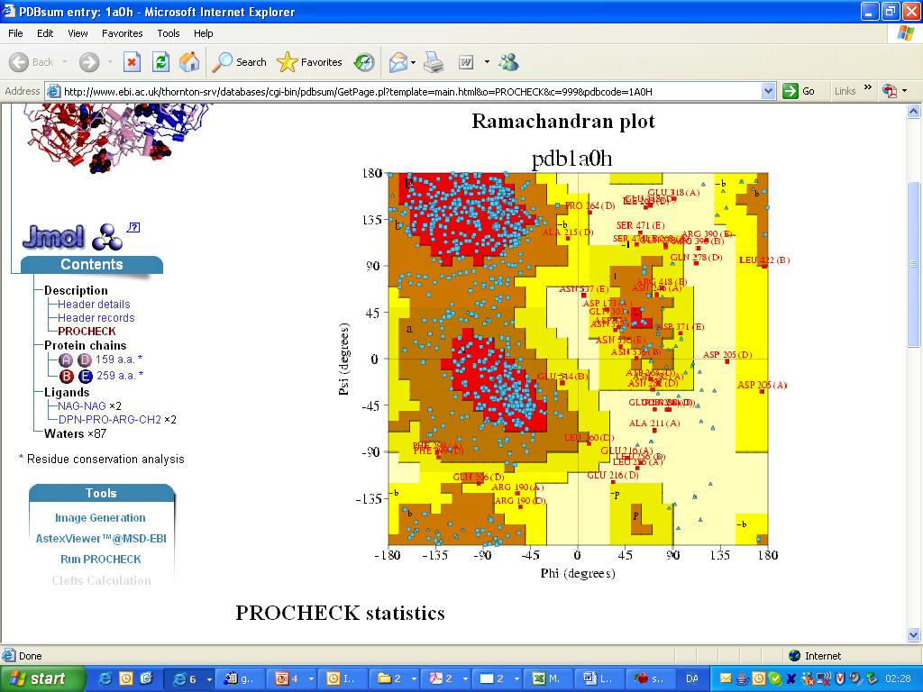
21 Ramachandran Plot I Procheck (Laskowski) 4 categories: Most favored Additionally allowed Generously allowed disallowed More than 90% structures in most favored regions and none or only a few in disallowed regions PDBSum: thornton-srv/databases/ pdbsum/
22 Ramachandran Plot II Uppsala Ramachandran server (Kleywegt & Jones): Newer analysis of PDB Two categories: Allowed Disallowed More than 95% residues in allowed regions ramachan.html
23 Ramachandran Plot III MolProbity Current PDB standard. Three categories Favoured (>98%) Allowed (>99.8%) Disallowed Also outputs plots for Glycine Proline Pre-proline molprobity.biochem.duke. edu/index.php
24 Ramachandran plot Good structures should have the majority of the residues in the most favored regions: Procheck: >90% Uppsala: >95% MolProbity: >98% and none or only a few in the disallowed regions. Residues in disallowed regions are either wrong or potentially very interesting.


25 Strange But Correct Mølgaard & Larsen (2002) Acta Cryst. D58,
![R-factor, R free Structure factor ( ) = f j exp 2πi hx j + ky j + lz j F hkl N j=1 [ ( )]](/docs-images/92/109305752/images/26-1.jpg "Individual reflections I hkl F obs (hkl) 2 Take out 5-10% of the data and use to calculate R")
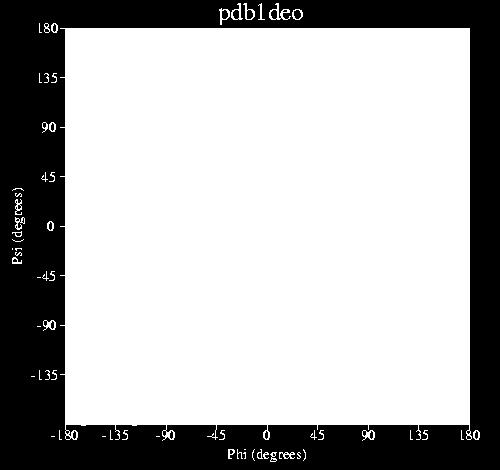
26 R-factor, R free Structure factor ( ) = f j exp 2πi hx j + ky j + lz j F hkl N j=1 [ ( )] Individual reflections I hkl F obs (hkl) 2 Take out 5-10% of the data and use to calculate R free
27 R-factor, R free R-factors vary from < 10% (high resolution structures) to 59% (theoretical random structure). Rule of thumb for good quality structures: R Resolution/10 R free should be < 30% and not much more than 5% (points) higher than R: R > R free > R


28 Key Parameters Resolution R values Agreement between data and model. Usually between 0.15 and 0.25, should not exceed R ~ Resolution / 10 R > R free > R. Ramachandran plot The majority of residues in most favoured regions. B factors Contributions from static and dynamic disorder Well determined ~10-20 Å 2, intermediate ~20-30 Å 2, flexible Å 2, invisible >60 Å 2.


29 1p7q R, Rfree: 21.8%, 30.9% Resolution: 3.4 Å Ramachandran plot B.E. Willcox et al. Nat Immunol Sep;4(9):913-9.
30 Other parameters B-factors Contributions from static and dynamic disorder Well determined ~10-20 Å 2 Intermediate ~20-30 Å 2 Flexible Å 2 Invisible >60 Å 2. Should be (mostly) > 5 Å 2 and < 50 Å 2 Occupancies Between 0 and 1 (0-100%) Rarely below 0.4 (40%)
31 B-factors 1xr9, 1.79Å atom # chain ID resi # x y z occ B-factor

32 B-factors 1p7q, 3.4Å

33 1hhh, 3.0Å Madden et al. Cell Nov 19;75(4):

34 1au4, 2.3Å, R=26.6, R free =37.0 Yamashita et al. J.Am.Chem.Soc. v119 pp.11351, 1997

35 1a0h, 3.2Å Martin et al. Structure Dec 15;5(12):

36

37 Occupancies, 1ea3, 2.3Å

38 The Electron Density Server

39
40 Something s Fishy in 2hr0 No correlation between B-factors and solvent accessibility. Large layers of solvent in the c-direction (30-40Å thick) that spans entire unit cell (i.e. nothing to hold the crystal together). Absence of bulk solvent in diffraction data. 80% solvent resolution 2.3Å. Perfect electron density around impossible geometries. Impossible merging statistics for data from four crystals (Rmerge = 0.11 in the last resolution shell, with I/sig(I) = 1.32).
41 Diffraction Precision Index DPI (with Rfree): DPI (no Rfree): Scaled by B-factor Cruickshank 1999, Acta Cryst D
42 DPI vs. Resolution 2BSU

43 2bsu, 1.60Å, R=31.9, R free =33.6 Has now been replaced by 2v2w
44 Take Home Message It is easy to find bad structures in the PDB so validate!! Resolution Ramachandran R free RMSD (NMR) Restraint violations (NMR) High impact journal high quality structure!
45 Programme Summary of last week s lecture and quiz Structure validation Break Exercise: Structure validation tutorial Break Summary & discussion Quiz
46 Break!
47 Programme Summary of last week s lecture and quiz Structure validation Break Exercise: Structure validation tutorial Break Summary & discussion Quiz
X-ray Crystallography
 2009/11/25 [ 1 ] X-ray Crystallography Andrew Torda, wintersemester 2009 / 2010 X-ray numerically most important more than 4/5 structures Goal a set of x, y, z coordinates different properties to NMR History
Introduction to" Protein Structure
 Introduction to" Protein Structure Function, evolution & experimental methods Thomas Blicher, Center for Biological Sequence Analysis Learning Objectives Outline the basic levels of protein structure.
X-ray Crystallography I. James Fraser Macromolecluar Interactions BP204
 X-ray Crystallography I James Fraser Macromolecluar Interactions BP204 Key take-aways 1. X-ray crystallography results from an ensemble of Billions and Billions of molecules in the crystal 2. Models in
7.91 Amy Keating. Solving structures using X-ray crystallography & NMR spectroscopy
 7.91 Amy Keating Solving structures using X-ray crystallography & NMR spectroscopy How are X-ray crystal structures determined? 1. Grow crystals - structure determination by X-ray crystallography relies
Ultra-high resolution structures in validation
 Ultra-high resolution structures in validation (and not only...) Mariusz Jaskolski Department of Crystallography,, A. Mickiewicz University Center for Biocrystallographic Research, Polish Academy of Sciences,
Details of Protein Structure
 Details of Protein Structure Function, evolution & experimental methods Thomas Blicher, Center for Biological Sequence Analysis Anne Mølgaard, Kemisk Institut, Københavns Universitet Learning Objectives
Direct Method. Very few protein diffraction data meet the 2nd condition
 Direct Method Two conditions: -atoms in the structure are equal-weighted -resolution of data are higher than the distance between the atoms in the structure Very few protein diffraction data meet the 2nd
Programme Last week s quiz results + Summary Fold recognition Break Exercise: Modelling remote homologues
 Programme 8.00-8.20 Last week s quiz results + Summary 8.20-9.00 Fold recognition 9.00-9.15 Break 9.15-11.20 Exercise: Modelling remote homologues 11.20-11.40 Summary & discussion 11.40-12.00 Quiz 1 Feedback
Ensemble refinement of protein crystal structures in PHENIX. Tom Burnley Piet Gros
 Ensemble refinement of protein crystal structures in PHENIX Tom Burnley Piet Gros Incomplete modelling of disorder contributes to R factor gap Only ~5% of residues in the PDB are modelled with more than
Macromolecular X-ray Crystallography
 Protein Structural Models for CHEM 641 Fall 07 Brian Bahnson Department of Chemistry & Biochemistry University of Delaware Macromolecular X-ray Crystallography Purified Protein X-ray Diffraction Data collection
Molecular Biology Course 2006 Protein Crystallography Part II
 Molecular Biology Course 2006 Protein Crystallography Part II Tim Grüne University of Göttingen Dept. of Structural Chemistry December 2006 http://shelx.uni-ac.gwdg.de tg@shelx.uni-ac.gwdg.de Overview
SOLID STATE 9. Determination of Crystal Structures
 SOLID STATE 9 Determination of Crystal Structures In the diffraction experiment, we measure intensities as a function of d hkl. Intensities are the sum of the x-rays scattered by all the atoms in a crystal.
Can protein model accuracy be. identified? NO! CBS, BioCentrum, Morten Nielsen, DTU
 Can protein model accuracy be identified? Morten Nielsen, CBS, BioCentrum, DTU NO! Identification of Protein-model accuracy Why is it important? What is accuracy RMSD, fraction correct, Protein model correctness/quality
Macromolecular Crystallography Part II
 Molecular Biology Course 2009 Macromolecular Crystallography Part II Tim Grüne University of Göttingen Dept. of Structural Chemistry November 2009 http://shelx.uni-ac.gwdg.de tg@shelx.uni-ac.gwdg.de From
Molecular Modeling lecture 2
 Molecular Modeling 2018 -- lecture 2 Topics 1. Secondary structure 3. Sequence similarity and homology 2. Secondary structure prediction 4. Where do protein structures come from? X-ray crystallography
Protein Crystallography Part II
 Molecular Biology Course 2007 Protein Crystallography Part II Tim Grüne University of Göttingen Dept. of Structural Chemistry November 2007 http://shelx.uni-ac.gwdg.de tg@shelx.uni-ac.gwdg.de Overview
SUPPLEMENTARY INFORMATION
 Table of Contents Page Supplementary Table 1. Diffraction data collection statistics 2 Supplementary Table 2. Crystallographic refinement statistics 3 Supplementary Fig. 1. casic1mfc packing in the R3
Electronic Supplementary Information (ESI) for Chem. Commun. Unveiling the three- dimensional structure of the green pigment of nitrite- cured meat
 for Chem. Commun. Unveiling the three- dimensional structure of the green pigment of nitrite- cured meat") Electronic Supplementary Information (ESI) for Chem. Commun. Unveiling the three- dimensional structure of the green pigment of nitrite- cured meat Jun Yi* and George B. Richter- Addo* Department of Chemistry
Electronic Supplementary Information (ESI) for Chem. Commun. Unveiling the three- dimensional structure of the green pigment of nitrite- cured meat Jun Yi* and George B. Richter- Addo* Department of Chemistry
HOMOLOGY MODELING. The sequence alignment and template structure are then used to produce a structural model of the target.
 HOMOLOGY MODELING Homology modeling, also known as comparative modeling of protein refers to constructing an atomic-resolution model of the "target" protein from its amino acid sequence and an experimental
Structure factors again
 Structure factors again Remember 1D, structure factor for order h F h = F h exp[iα h ] = I 01 ρ(x)exp[2πihx]dx Where x is fractional position along unit cell distance (repeating distance, origin arbitrary)
Acta Crystallographica Section F
 Supporting information Acta Crystallographica Section F Volume 70 (2014) Supporting information for article: Chemical conversion of cisplatin and carboplatin with histidine in a model protein crystallised
X-ray Crystallography. Kalyan Das
 X-ray Crystallography Kalyan Das Electromagnetic Spectrum NMR 10 um - 10 mm 700 to 10 4 nm 400 to 700 nm 10 to 400 nm 10-1 to 10 nm 10-4 to 10-1 nm X-ray radiation was discovered by Roentgen in 1895. X-rays
Scattering by two Electrons
 Scattering by two Electrons p = -r k in k in p r e 2 q k in /λ θ θ k out /λ S q = r k out p + q = r (k out - k in ) e 1 Phase difference of wave 2 with respect to wave 1: 2π λ (k out - k in ) r= 2π S r
Homology Modeling (Comparative Structure Modeling) GBCB 5874: Problem Solving in GBCB
 GBCB 5874: Problem Solving in GBCB") Homology Modeling (Comparative Structure Modeling) Aims of Structural Genomics High-throughput 3D structure determination and analysis To determine or predict the 3D structures of all the proteins encoded
Homology Modeling (Comparative Structure Modeling) Aims of Structural Genomics High-throughput 3D structure determination and analysis To determine or predict the 3D structures of all the proteins encoded
Protein Structure Determination. Part 1 -- X-ray Crystallography
 Protein Structure Determination Part 1 -- X-ray Crystallography Topics covering in this 1/2 course Crystal growth Diffraction theory Symmetry Solving phases using heavy atoms Solving phases using a model
NMR in Structural Biology
 NMR in Structural Biology Exercise session 2 1. a. List 3 NMR observables that report on structure. b. Also indicate whether the information they give is short/medium or long-range, or perhaps all three?
TLS and all that. Ethan A Merritt. CCP4 Summer School 2011 (Argonne, IL) Abstract
 Abstract") TLS and all that Ethan A Merritt CCP4 Summer School 2011 (Argonne, IL) Abstract We can never know the position of every atom in a crystal structure perfectly. Each atom has an associated positional uncertainty.
TLS and all that Ethan A Merritt CCP4 Summer School 2011 (Argonne, IL) Abstract We can never know the position of every atom in a crystal structure perfectly. Each atom has an associated positional uncertainty.
The structure of Aquifex aeolicus FtsH in the ADP-bound state reveals a C2-symmetric hexamer
 Volume 71 (2015) Supporting information for article: The structure of Aquifex aeolicus FtsH in the ADP-bound state reveals a C2-symmetric hexamer Marina Vostrukhina, Alexander Popov, Elena Brunstein, Martin
RNA protects a nucleoprotein complex against radiation damage
 Supporting information Volume 72 (2016) Supporting information for article: RNA protects a nucleoprotein complex against radiation damage Charles S. Bury, John E. McGeehan, Alfred A. Antson, Ian Carmichael,
Macromolecular Crystallography Part II
 Molecular Biology Course 2010 Macromolecular Crystallography Part II University of Göttingen Dept. of Structural Chemistry November 2010 http://shelx.uni-ac.gwdg.de tg@shelx.uni-ac.gwdg.de Crystallography
X-ray analysis. 1. Basic crystallography 2. Basic diffraction physics 3. Experimental methods
 X-ray analysis 1. Basic crystallography 2. Basic diffraction physics 3. Experimental methods Introduction Noble prizes associated with X-ray diffraction 1901 W. C. Roentgen (Physics) for the discovery
Basics of protein structure
 Today: 1. Projects a. Requirements: i. Critical review of one paper ii. At least one computational result b. Noon, Dec. 3 rd written report and oral presentation are due; submit via email to bphys101@fas.harvard.edu
NMR, X-ray Diffraction, Protein Structure, and RasMol
 NMR, X-ray Diffraction, Protein Structure, and RasMol Introduction So far we have been mostly concerned with the proteins themselves. The techniques (NMR or X-ray diffraction) used to determine a structure
Rietveld Structure Refinement of Protein Powder Diffraction Data using GSAS
 Rietveld Structure Refinement of Protein Powder Diffraction Data using GSAS Jon Wright ESRF, Grenoble, France Plan This is a users perspective Cover the protein specific aspects (assuming knowledge of
11/6/2013. Refinement. Fourier Methods. Fourier Methods. Difference Map. Difference Map Find H s. Difference Map No C 1
 Refinement Fourier Methods find heavy atom or some F s phases using direct methods locate new atoms, improve phases continue until all atoms found in more or less correct position starting point of refinement
NMR in Medicine and Biology
 NMR in Medicine and Biology http://en.wikipedia.org/wiki/nmr_spectroscopy MRI- Magnetic Resonance Imaging (water) In-vivo spectroscopy (metabolites) Solid-state t NMR (large structures) t Solution NMR
Protein Structure Determination Using NMR Restraints BCMB/CHEM 8190
 Protein Structure Determination Using NMR Restraints BCMB/CHEM 8190 Programs for NMR Based Structure Determination CNS - Brünger, A. T.; Adams, P. D.; Clore, G. M.; DeLano, W. L.; Gros, P.; Grosse-Kunstleve,
Protein crystallography. Garry Taylor
 Protein crystallography Garry Taylor X-ray Crystallography - the Basics Grow crystals Collect X-ray data Determine phases Calculate ρ-map Interpret map Refine coordinates Do the biology. Nitrogen at -180
Introduction to Comparative Protein Modeling. Chapter 4 Part I
 Introduction to Comparative Protein Modeling Chapter 4 Part I 1 Information on Proteins Each modeling study depends on the quality of the known experimental data. Basis of the model Search in the literature
Full wwpdb X-ray Structure Validation Report i
 Full wwpdb X-ray Structure Validation Report i Jan 28, 2019 11:10 AM EST PDB ID : 6A5H Title : The structure of [4+2] and [6+4] cyclase in the biosynthetic pathway of unidentified natural product Authors
HTCondor and macromolecular structure validation
 HTCondor and macromolecular structure validation Vincent Chen John Markley/Eldon Ulrich, NMRFAM/BMRB, UW@Madison David & Jane Richardson, Duke University Macromolecules David S. Goodsell 1999 Two questions
Drug targets, Protein Structures and Crystallography
 Drug targets, Protein Structures and Crystallography NS5B viral RNA polymerase (RNA dep) Hepa88s C drug Sofosbuvir (Sovaldi) FDA 2013 Epclusa - combina8on with Velpatasvir approved in in 2016) Prodrug
Protein structures and comparisons ndrew Torda Bioinformatik, Mai 2008
 Protein structures and comparisons ndrew Torda 67.937 Bioinformatik, Mai 2008 Ultimate aim how to find out the most about a protein what you can get from sequence and structure information On the way..
wwpdb X-ray Structure Validation Summary Report
 wwpdb X-ray Structure Validation Summary Report io Jan 31, 2016 06:45 PM GMT PDB ID : 1CBS Title : CRYSTAL STRUCTURE OF CELLULAR RETINOIC-ACID-BINDING PROTEINS I AND II IN COMPLEX WITH ALL-TRANS-RETINOIC
Why do We Trust X-ray Crystallography?
 Why do We Trust X-ray Crystallography? Andrew D Bond All chemists know that X-ray crystallography is the gold standard characterisation technique: an X-ray crystal structure provides definitive proof of
PROTEIN'STRUCTURE'DETERMINATION'
 PROTEIN'STRUCTURE'DETERMINATION' USING'NMR'RESTRAINTS' BCMB/CHEM'8190' Programs for NMR Based Structure Determination CNS - Brünger, A. T.; Adams, P. D.; Clore, G. M.; DeLano, W. L.; Gros, P.; Grosse-Kunstleve,
IgE binds asymmetrically to its B cell receptor CD23
 Supplementary Information IgE binds asymmetrically to its B cell receptor CD23 Balvinder Dhaliwal 1*, Marie O. Y. Pang 2, Anthony H. Keeble 2,3, Louisa K. James 2,4, Hannah J. Gould 2, James M. McDonnell
Full wwpdb X-ray Structure Validation Report i
 Full wwpdb X-ray Structure Validation Report i Mar 13, 2018 04:03 pm GMT PDB ID : 5NMJ Title : Chicken GRIFIN (crystallisation ph: 6.5) Authors : Ruiz, F.M.; Romero, A. Deposited on : 2017-04-06 Resolution
Crystal lattice Real Space. Reflections Reciprocal Space. I. Solving Phases II. Model Building for CHEM 645. Purified Protein. Build model.
 I. Solving Phases II. Model Building for CHEM 645 Purified Protein Solve Phase Build model and refine Crystal lattice Real Space Reflections Reciprocal Space ρ (x, y, z) pronounced rho F hkl 2 I F (h,
Full wwpdb X-ray Structure Validation Report i
 Full wwpdb X-ray Structure Validation Report i Mar 10, 2018 01:44 am GMT PDB ID : 1MWP Title : N-TERMINAL DOMAIN OF THE AMYLOID PRECURSOR PROTEIN Authors : Rossjohn, J.; Cappai, R.; Feil, S.C.; Henry,
Bioinformatics. Macromolecular structure
 Bioinformatics Macromolecular structure Contents Determination of protein structure Structure databases Secondary structure elements (SSE) Tertiary structure Structure analysis Structure alignment Domain
Plasmid Relevant features Source. W18N_D20N and TrXE-W18N_D20N-anti
 Table S1. E. coli plasmids Plasmid Relevant features Source pdg680 T. reesei XynII AA 2-190 with C-terminal His 6 tag optimized for E. coli expression in pjexpress401 Wan et al. (in press) psbn44d psbn44h
Linking data and model quality in macromolecular crystallography. Kay Diederichs
 Linking data and model quality in macromolecular crystallography Kay Diederichs Crystallography is highly successful Can we do better? Error in experimental data Error = random + systematic Multiplicity
Anisotropy in macromolecular crystal structures. Andrea Thorn July 19 th, 2012
 Anisotropy in macromolecular crystal structures Andrea Thorn July 19 th, 2012 Motivation Courtesy of M. Sawaya Motivation Crystal structures are inherently anisotropic. X-ray diffraction reflects this
Protein Structure Determination
 Protein Structure Determination Given a protein sequence, determine its 3D structure 1 MIKLGIVMDP IANINIKKDS SFAMLLEAQR RGYELHYMEM GDLYLINGEA 51 RAHTRTLNVK QNYEEWFSFV GEQDLPLADL DVILMRKDPP FDTEFIYATY 101
Protein NMR Structures Refined with Rosetta Have Higher Accuracy Relative to Corresponding X ray Crystal Structures
 pubs.acs.org/jacs Protein NMR Structures Refined with Rosetta Have Higher Accuracy Relative to Corresponding X ray Crystal Structures Binchen Mao, Roberto Tejero,, David Baker, and Gaetano T. Montelione*,
Full wwpdb X-ray Structure Validation Report i
 Full wwpdb X-ray Structure Validation Report i Mar 8, 2018 10:24 pm GMT PDB ID : 1A30 Title : HIV-1 PROTEASE COMPLEXED WITH A TRIPEPTIDE INHIBITOR Authors : Louis, J.M.; Dyda, F.; Nashed, N.T.; Kimmel,
Protein Structures. 11/19/2002 Lecture 24 1
 Protein Structures 11/19/2002 Lecture 24 1 All 3 figures are cartoons of an amino acid residue. 11/19/2002 Lecture 24 2 Peptide bonds in chains of residues 11/19/2002 Lecture 24 3 Angles φ and ψ in the
Full wwpdb X-ray Structure Validation Report i
 Full wwpdb X-ray Structure Validation Report i Jan 14, 2019 11:10 AM EST PDB ID : 6GYW Title : Crystal structure of DacA from Staphylococcus aureus Authors : Tosi, T.; Freemont, P.S.; Grundling, A. Deposited
Electron Density at various resolutions, and fitting a model as accurately as possible.
 Section 9, Electron Density Maps 900 Electron Density at various resolutions, and fitting a model as accurately as possible. ρ xyz = (Vol) -1 h k l m hkl F hkl e iφ hkl e-i2π( hx + ky + lz ) Amplitude
Determining Protein Structure BIBC 100
 Determining Protein Structure BIBC 100 Determining Protein Structure X-Ray Diffraction Interactions of x-rays with electrons in molecules in a crystal NMR- Nuclear Magnetic Resonance Interactions of magnetic
Resolution and data formats. Andrea Thorn
 Resolution and data formats Andrea Thorn RESOLUTION Motivation Courtesy of M. Sawaya Map resolution http://www.bmsc.washington.edu/people/verlinde/experiment.html Data quality indicators Resolution accounts
Full wwpdb X-ray Structure Validation Report i
 Full wwpdb X-ray Structure Validation Report i Mar 14, 2018 02:00 pm GMT PDB ID : 3RRQ Title : Crystal structure of the extracellular domain of human PD-1 Authors : Lazar-Molnar, E.; Ramagopal, U.A.; Nathenson,
Homology modeling of Ferredoxin-nitrite reductase from Arabidopsis thaliana
 www.bioinformation.net Hypothesis Volume 6(3) Homology modeling of Ferredoxin-nitrite reductase from Arabidopsis thaliana Karim Kherraz*, Khaled Kherraz, Abdelkrim Kameli Biology department, Ecole Normale
Modeling for 3D structure prediction
 Modeling for 3D structure prediction What is a predicted structure? A structure that is constructed using as the sole source of information data obtained from computer based data-mining. However, mixing
Proteins. Central Dogma : DNA RNA protein Amino acid polymers - defined composition & order. Perform nearly all cellular functions Drug Targets
 Proteins Central Dogma : DNA RNA protein Amino acid polymers - defined composition & order Perform nearly all cellular functions Drug Targets Fold into discrete shapes. Proteins - cont. Specific shapes
1) NMR is a method of chemical analysis. (Who uses NMR in this way?) 2) NMR is used as a method for medical imaging. (called MRI )
 NMR is a method of chemical analysis. (Who uses NMR in this way?) 2) NMR is used as a method for medical imaging. (called MRI )") Uses of NMR: 1) NMR is a method of chemical analysis. (Who uses NMR in this way?) 2) NMR is used as a method for medical imaging. (called MRI ) 3) NMR is used as a method for determining of protein, DNA,
Uses of NMR: 1) NMR is a method of chemical analysis. (Who uses NMR in this way?) 2) NMR is used as a method for medical imaging. (called MRI ) 3) NMR is used as a method for determining of protein, DNA,
Phase problem: Determining an initial phase angle α hkl for each recorded reflection. 1 ρ(x,y,z) = F hkl cos 2π (hx+ky+ lz - α hkl ) V h k l
 = F hkl cos 2π (hx+ky+ lz - α hkl ) V h k l") Phase problem: Determining an initial phase angle α hkl for each recorded reflection 1 ρ(x,y,z) = F hkl cos 2π (hx+ky+ lz - α hkl ) V h k l Methods: Heavy atom methods (isomorphous replacement Hg, Pt)
Phase problem: Determining an initial phase angle α hkl for each recorded reflection 1 ρ(x,y,z) = F hkl cos 2π (hx+ky+ lz - α hkl ) V h k l Methods: Heavy atom methods (isomorphous replacement Hg, Pt)
Full wwpdb X-ray Structure Validation Report i
 Full wwpdb X-ray Structure Validation Report i Jan 17, 2019 09:42 AM EST PDB ID : 6D3Z Title : Protease SFTI complex Authors : Law, R.H.P.; Wu, G. Deposited on : 2018-04-17 Resolution : 2.00 Å(reported)
Full wwpdb X-ray Structure Validation Report i
 Full wwpdb X-ray Structure Validation Report i Mar 8, 2018 06:13 pm GMT PDB ID : 5G5C Title : Structure of the Pyrococcus furiosus Esterase Pf2001 with space group C2221 Authors : Varejao, N.; Reverter,
Principles of NMR Protein Spectroscopy. 2) Assignment of chemical shifts in a protein ( 1 H, 13 C, 15 N) 3) Three dimensional structure determination
 Assignment of chemical shifts in a protein ( 1 H, 13 C, 15 N) 3) Three dimensional structure determination") 1) Protein preparation (>50 aa) 2) Assignment of chemical shifts in a protein ( 1 H, 13 C, 15 N) 3) Three dimensional structure determination Protein Expression overexpression in E. coli - BL21(DE3) 1
1) Protein preparation (>50 aa) 2) Assignment of chemical shifts in a protein ( 1 H, 13 C, 15 N) 3) Three dimensional structure determination Protein Expression overexpression in E. coli - BL21(DE3) 1
Protein Dynamics. The space-filling structures of myoglobin and hemoglobin show that there are no pathways for O 2 to reach the heme iron.
 Protein Dynamics The space-filling structures of myoglobin and hemoglobin show that there are no pathways for O 2 to reach the heme iron. Below is myoglobin hydrated with 350 water molecules. Only a small
Solid-state NMR and proteins : basic concepts (a pictorial introduction) Barth van Rossum,
 Barth van Rossum,") Solid-state NMR and proteins : basic concepts (a pictorial introduction) Barth van Rossum, 16.02.2009 Solid-state and solution NMR spectroscopy have many things in common Several concepts have been/will
Solid-state NMR and proteins : basic concepts (a pictorial introduction) Barth van Rossum, 16.02.2009 Solid-state and solution NMR spectroscopy have many things in common Several concepts have been/will
Roger Johnson Structure and Dynamics: X-ray Diffraction Lecture 6
 6.1. Summary In this Lecture we cover the theory of x-ray diffraction, which gives direct information about the atomic structure of crystals. In these experiments, the wavelength of the incident beam must
Supporting Information
 Electronic Supplementary Material (ESI) for Dalton Transactions. This journal is The Royal Society of Chemistry 2016 Supporting Information Over or under: Hydride attack at the metal versus the coordinated
Handout 12 Structure refinement. Completing the structure and evaluating how good your data and model agree
 Handout 1 Structure refinement Completing the structure and evaluating how good your data and model agree Why you should refine a structure We have considered how atoms are located by Patterson, direct
SHELXC/D/E. Andrea Thorn
 SHELXC/D/E Andrea Thorn What is experimental phasing? Experimental phasing is what you do if MR doesn t work. What is experimental phasing? Experimental phasing methods depend on intensity differences.
Design of a Novel Globular Protein Fold with Atomic-Level Accuracy
 Design of a Novel Globular Protein Fold with Atomic-Level Accuracy Brian Kuhlman, Gautam Dantas, Gregory C. Ireton, Gabriele Varani, Barry L. Stoddard, David Baker Presented by Kate Stafford 4 May 05 Protein
A Primer in X-ray Crystallography for Redox Biologists. Mark Wilson Karolinska Institute June 3 rd, 2014
 A Primer in X-ray Crystallography for Redox Biologists Mark Wilson Karolinska Institute June 3 rd, 2014 X-ray Crystallography Basics Optimistic workflow for crystallography Experiment Schematic Fourier
Crystals, X-rays and Proteins
 Crystals, X-rays and Proteins Comprehensive Protein Crystallography Dennis Sherwood MA (Hons), MPhil, PhD Jon Cooper BA (Hons), PhD OXFORD UNIVERSITY PRESS Contents List of symbols xiv PART I FUNDAMENTALS
Protein Structures: Experiments and Modeling. Patrice Koehl
 Protein Structures: Experiments and Modeling Patrice Koehl Structural Bioinformatics: Proteins Proteins: Sources of Structure Information Proteins: Homology Modeling Proteins: Ab initio prediction Proteins:
Protein Structure Determination from Pseudocontact Shifts Using ROSETTA
 Supporting Information Protein Structure Determination from Pseudocontact Shifts Using ROSETTA Christophe Schmitz, Robert Vernon, Gottfried Otting, David Baker and Thomas Huber Table S0. Biological Magnetic
Nature Structural & Molecular Biology: doi: /nsmb.3194
 Supplementary Figure 1 Mass spectrometry and solution NMR data for -syn samples used in this study. (a) Matrix-assisted laser-desorption and ionization time-of-flight (MALDI-TOF) mass spectrum of uniformly-
5.067 Crystal Structure Refinement Fall 2007
 MIT OpenCourseWare http://ocw.mit.edu 5.067 Crystal Structure Refinement Fall 2007 For information about citing these materials or our Terms of Use, visit: http://ocw.mit.edu/terms. Artefacts An artefact
Full wwpdb X-ray Structure Validation Report i
 Full wwpdb X-ray Structure Validation Report i Mar 8, 2018 08:34 pm GMT PDB ID : 1RUT Title : Complex of LMO4 LIM domains 1 and 2 with the ldb1 LID domain Authors : Deane, J.E.; Ryan, D.P.; Maher, M.J.;
Protein Structure Determination 9/25/2007
 One-dimensional NMR spectra Ethanol Cellulase (36 a.a.) Branden & Tooze, Fig. 18.16 1D and 2D NMR spectra of inhibitor K (57 a.a.) K. Wuthrich, NMR of Proteins and Nucleic Acids. (Wiley, 1986.) p. 54-55.
Molecular Modeling. Prediction of Protein 3D Structure from Sequence. Vimalkumar Velayudhan. May 21, 2007
 Molecular Modeling Prediction of Protein 3D Structure from Sequence Vimalkumar Velayudhan Jain Institute of Vocational and Advanced Studies May 21, 2007 Vimalkumar Velayudhan Molecular Modeling 1/23 Outline
Fourier Syntheses, Analyses, and Transforms
 Fourier Syntheses, Analyses, and Transforms http://homepages.utoledo.edu/clind/ The electron density The electron density in a crystal can be described as a periodic function - same contents in each unit
Table S1. Primers used for the constructions of recombinant GAL1 and λ5 mutants. GAL1-E74A ccgagcagcgggcggctgtctttcc ggaaagacagccgcccgctgctcgg
 SUPPLEMENTAL DATA Table S1. Primers used for the constructions of recombinant GAL1 and λ5 mutants Sense primer (5 to 3 ) Anti-sense primer (5 to 3 ) GAL1 mutants GAL1-E74A ccgagcagcgggcggctgtctttcc ggaaagacagccgcccgctgctcgg
Protein Structure Prediction II Lecturer: Serafim Batzoglou Scribe: Samy Hamdouche
 Protein Structure Prediction II Lecturer: Serafim Batzoglou Scribe: Samy Hamdouche The molecular structure of a protein can be broken down hierarchically. The primary structure of a protein is simply its
Protein Structure Analysis and Verification. Course S Basics for Biosystems of the Cell exercise work. Maija Nevala, BIO, 67485U 16.1.
 Protein Structure Analysis and Verification Course S-114.2500 Basics for Biosystems of the Cell exercise work Maija Nevala, BIO, 67485U 16.1.2008 1. Preface When faced with an unknown protein, scientists
CCP4 Diamond 2014 SHELXC/D/E. Andrea Thorn
 CCP4 Diamond 2014 SHELXC/D/E Andrea Thorn SHELXC/D/E workflow SHELXC: α calculation, file preparation SHELXD: Marker atom search = substructure search SHELXE: density modification Maps and coordinate files
Visualization of Macromolecular Structures
 Visualization of Macromolecular Structures Present by: Qihang Li orig. author: O Donoghue, et al. Structural biology is rapidly accumulating a wealth of detailed information. Over 60,000 high-resolution
Experimental Techniques in Protein Structure Determination
 Experimental Techniques in Protein Structure Determination Homayoun Valafar Department of Computer Science and Engineering, USC Two Main Experimental Methods X-Ray crystallography Nuclear Magnetic Resonance
Basic Crystallography Part 1. Theory and Practice of X-ray Crystal Structure Determination
 Basic Crystallography Part 1 Theory and Practice of X-ray Crystal Structure Determination We have a crystal How do we get there? we want a structure! The Unit Cell Concept Ralph Krätzner Unit Cell Description
Automated identification of functional dynamic contact networks from X-ray crystallography
 1 Automated identification of functional dynamic contact networks from X-ray crystallography Henry van den Bedem, Gira Bhabha, Kun Yang, Peter E. Wright and James S. Fraser Supplementary Figure 1 Supplementary
Solid State Spectroscopy Problem Set 7
 Solid State Spectroscopy Problem Set 7 Due date: June 29th, 2015 Problem 5.1 EXAFS Study of Mn/Fe substitution in Y(Mn 1-x Fe x ) 2 O 5 From article «EXAFS, XANES, and DFT study of the mixed-valence compound
Acta Cryst. (2017). D73, doi: /s
. D73, doi: /s") Acta Cryst. (2017). D73, doi:10.1107/s2059798317010932 Supporting information Volume 73 (2017) Supporting information for article: Designing better diffracting crystals of biotin carboxyl carrier protein
Acta Cryst. (2017). D73, doi:10.1107/s2059798317010932 Supporting information Volume 73 (2017) Supporting information for article: Designing better diffracting crystals of biotin carboxyl carrier protein
HADDOCK: High Ambiguity
 Determination of Protein-Protein complexes HADDOCK: High Ambiguity Driven DOCKing A protein-protein docking approach based on biochemical and/or biophysical data In PDB: >15000 protein structures but
Supporting Information
 Supporting Information Micelle-Triggered b-hairpin to a-helix Transition in a 14-Residue Peptide from a Choline-Binding Repeat of the Pneumococcal Autolysin LytA HØctor Zamora-Carreras, [a] Beatriz Maestro,