Protein Structure and Visualisation. Introduction to PDB and PyMOL
|
|
|
- Phillip Banks
- 5 years ago
- Views:
Transcription
1 Protein Structure and Visualisation Introduction to PDB and PyMOL 1

2 Feedback Persons 2
3 Program Quiz results Introduction to PDB & PyMOL Break PyMOL tutorial Break Summary Quiz 3
4 Program Quiz results Introduction to PDB & PyMOL Break PyMOL tutorial Break Summary Quiz 4


5 PDB Growth February

6 Protein Data Bank Contents File structure Types of structures Structure reports & summaries Quality check Searching Molecule of the Month 6
7 PDB File Header HEADER IMMUNE SYSTEM 16-AUG-04 1X7Q TITLE CRYSTAL STRUCTURE OF HLA-A*1101 WITH SARS NUCLEOCAPSID TITLE 2 PEPTIDE COMPND MOL_ID: 1; COMPND 2 MOLECULE: HLA CLASS I HISTOCOMPATIBILITY ANTIGEN, A-11 REMARK 3 DATA USED IN REFINEMENT. REMARK 3 RESOLUTION RANGE HIGH (ANGSTROMS) : 1.45 REMARK 3 RESOLUTION RANGE LOW (ANGSTROMS) : SEQRES 1 C 9 LYS THR PHE PRO PRO THR GLU PRO LYS HET SO4 B HETNAM SO4 SULFATE ION HELIX 1 1 ALA A 49 GLU A CRYST P
8 PDB File Atom Coordinates ATOM 1 N THR A N ATOM 2 CA THR A C ATOM 3 C THR A C ATOM 4 O THR A O ATOM 5 CB THR A C ATOM 6 OG1 THR A O ATOM 7 CG2 THR A C HETATM 3284 O3 GOL B O HETATM 3285 O HOH A O ATOM 1 N GLY A N ANISOU 1 N GLY A N ATOM 2 CA GLY A C ANISOU 2 CA GLY A C ATOM 3 C GLY A C ANISOU 3 C GLY A C
9 PDB File Fields COLUMNS DATA TYPE FIELD DEFINITION Record name "ATOM" 7-11 Integer serial Atom serial number Atom name Atom name. 17 Character altloc Alternate location indicator Residue name resname Residue name. 22 Character chainid Chain identifier Integer resseq Residue sequence number. 27 AChar icode Code for insertion of residues Real(8.3) x Orthogonal coordinates for X in Angstroms Real(8.3) y Orthogonal coordinates for Y in Angstroms Real(8.3) z Orthogonal coordinates for Z in Angstroms Real(6.2) occupancy Occupancy Real(6.2) tempfactor Temperature factor LString(2) element Element symbol, right-justified LString(2) charge Charge on the atom.

10 Worldwide Structural Genomics Fold space coverage " Complete genomes Disease-causing organisms Model organisms" Membrane proteins" Protein-ligand interactions Hou et al., PNAS 2003, 100: "

11 Protein Folds in PDB New folds Total folds
12 Protein Structure Visualisation PyMOL 12

13 What is PyMOL? Open-source molecular viewing program
14 Benefits It s free! For academia not for industry. Version 0.99rc6 Users can always compile the latest version. But you should contribute! Pay to get support, manual, latest version etc.
15 Potential Weaknesses Few! Not a fully integrated modelling environment. Not fully developed for experimental structure determination/fitting. Mostly for qualitative analyses. No undo function
16 PyMOL Representations Lines, sticks, ribbon, spheres, cartoon(s) Surfaces Transparency, quality Ray-tracing (rendering) Modes 16

17 Selections & Objects Every molecule (pdb file) is an object. Selections refer to objects Make smaller or composite objects Changes in representation can affect objects or selections.
18 Links PDB (protein structure database) PyMOL home: PyMOL manual: PyMOL Wiki: PyMOL settings (documented): modules/pymol/setting.py
19 Installation Windows Mac OS X Special behaviour Rename to get plugin menu! MacPyMOL MacPyMOLX11Hybrid Linux
20 PyMOL scripting
21 Scripts PyMOL is based on the programming language Python. PyMOL will read python commands. PyMOL also has a set of native commands. See PyMOLWiki and elsewhere for examples. pymol/
22 Automating Tasks Scripts are useful for automating tasks Repeated analyses Reading in large numbers of structures Making illustrations! get_view set_view viewport Saves space! Relative to PyMOL session files (.pse)
23 A Useful Example Using PyMOL commands: list=[]! iterate (name ca),list.append((resi,resn))! print list!! [('ASP', '1'), ('CYS', '2'), ('ALA', '3'), ('TRP', '4'), ('HIS', '5'), ('LEU',! '6'), ('GLY', '7'), ('GLU', '8'), ('LEU', '9'), ('VAL', '10'), ('TRP', '11'),! ('CYS', '12'), ('THR', '13')]!! or using a Python script (in PyMOL):!! from pymol import cmd,stored! stored.list=[]! cmd.iterate("(name ca)","stored.list.append((resi,resn))")! print stored.list!! [('1', 'ASP'), ('2', 'CYS'), ('3', 'ALA'), ('4', 'TRP'), ('5', 'HIS'), ('6', '! LEU'), ('7', 'GLY'), ('8', 'GLU'), ('9', 'LEU'), ('10', 'VAL'), ('11', 'TRP'),! ('12', 'CYS'), ('13', 'THR')]
24 color_b.py To colour proteins according to the values of the B-factor column (can be replaced with any desired value). Syntax: (see script header) color_b (c;a or c;b), mode=ramp, gradient=bwr, nbins=30, sat=0.5, value=1 to color chains A and B with the Blue-White-Red gradient in 30 colors of equal numbers of atoms in each color.
25 PyMOL Log Function Records all actions as commands in a PyMOL session GUI clicks Written commands Needs to be opened at start of session Is only written when closed (File Close log)


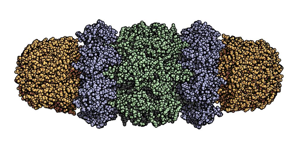
26 Example 1 rrna Large subunit: Catalytic mechanism Protein mrna Small subunit: Specificity 26

27 Example 2 27 René Magritte
28 Aesthetics of Molecular Images Personal taste General guidelines: Focus on relevant parts Strip away unnecessary information Find good viewing angle! Choice of representation No need for excessive graphics Good figures are (almost) self-explanatory. 28

29 Molecular Images Molecular graphics is an example of selective reduction of complexity. 29

30 Molecular Images Molecular graphics is an example of selective reduction of complexity. 30
31 Molecular Images Molecular graphics is an example of selective reduction of complexity. 31

32 Molecular Images Molecular graphics is an example of selective reduction of complexity. 32
33 Molecular Images Molecular graphics is an example of selective reduction of complexity. 33

34 Molecular Images Molecular graphics is an example of selective reduction of complexity. 34
35 Molecular Images Molecular graphics is an example of selective reduction of complexity. 35
36 Molecular Images Molecular graphics is an example of selective reduction of complexity. 36

37 Molecular Images Molecular graphics is an example of selective reduction of complexity. 37

38 Figures in PyMOL 38

39 Representations ray_trace_mode 0 39
40 Representations ray_trace_mode 1 40
41 Representations ray_trace_mode 2 41
42 Representations ray_trace_mode 3 42

43 Representations 43

44 Representations 44

45 Representations 45
46 Representations Standard stickrepresentation Sphere_scale = 0.3 With hydrogen atoms With shadows 46

47 Representations Standard stickrepresentation Sphere_scale = 0.3 With hydrogen atoms No shadows 47
48 Representations Standard stickrepresentation Sphere_scale = 0.3 With hydrogen atoms No shadows With depth cue 48

49 Representations Standard stickrepresentation Sphere_scale = 0.3 With hydrogen atoms No shadows With depth cue Larger image 49

50 Representations Standard stickrepresentation Sphere_scale = 0.3 With hydrogen atoms No shadows With depth cue Even larger image 50


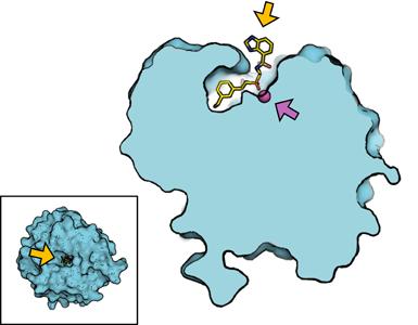
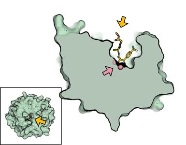
51 Focus on the Relevant Parts Homologous peptides in different proteins Intuitive colouring 51
52 Residue Conservation 52

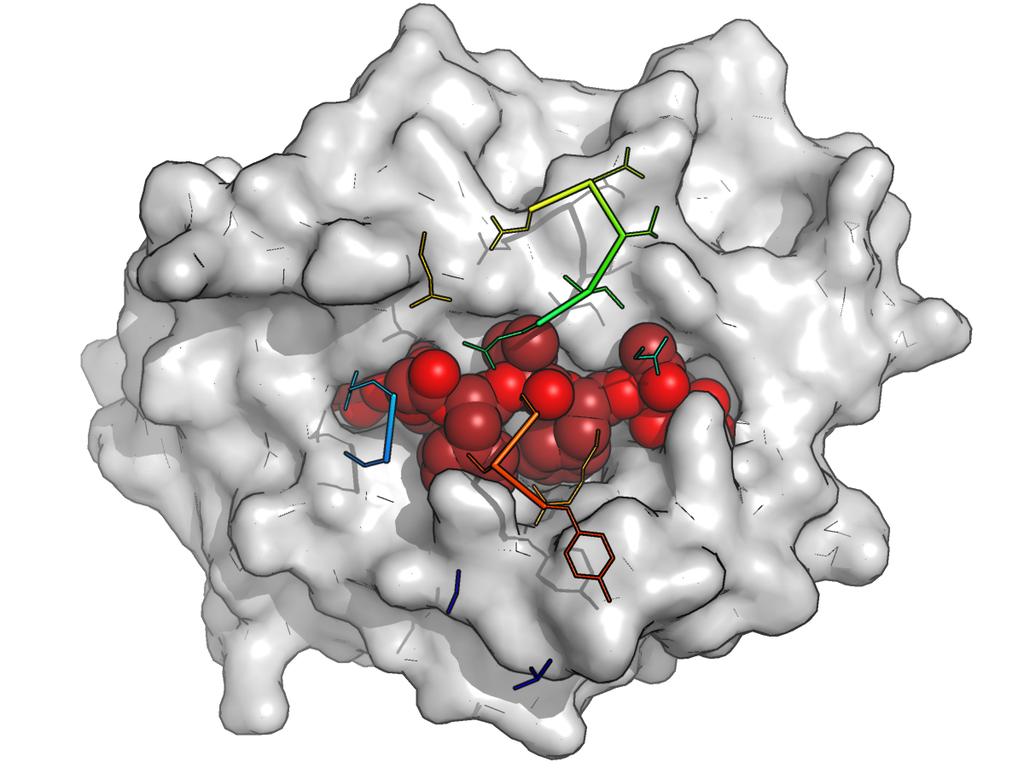
53 T-Cell Receptor Interactions I II III 53

54 Cross-Sections Proteasome 54

55 Electrostatic Surface Potentials 55


56 Composite Images MHC-I binding groove Images combined in Adobe Illustrator. C F B A E D 56
57 Simplified Composite Images C N P 2 57
58 Cross-Sections 58


59 Comparing Active Sites 59


60 Compiled Graphics Objects 60
61 PyMOL Links PyMOL home: PyMOL manual: toc.html PyMOL Wiki: PyMOL settings (documented): 61
62 Program Quiz results Introduction to PDB & PyMOL Break PyMOL tutorial Break Summary Quiz 62
63 Program Quiz results Introduction to PDB & PyMOL Break PyMOL tutorial Break Summary Quiz 63
64 Program Quiz results Introduction to PDB & PyMOL Break PyMOL tutorial Break Summary Quiz 64
Protein Data Bank Contents Guide: Atomic Coordinate Entry Format Description. Version Document Published by the wwpdb
 Protein Data Bank Contents Guide: Atomic Coordinate Entry Format Description Version 3.30 Document Published by the wwpdb This format complies with the PDB Exchange Dictionary (PDBx) http://mmcif.pdb.org/dictionaries/mmcif_pdbx.dic/index/index.html.
X-ray crystallography NMR Cryoelectron microscopy
 Molecular Graphics with PyMOL Overview of: Protein Data Bank Coordinates Jean-Yves Sgro PyMOL interface Hands-on! Experimental Methods 3 Main: X-ray crystallography NMR Cryoelectron microscopy X-ray source
Appendices. Appendix I: PDB file format
 Appendices This section contains the details of some file formats that have been used in examples in these notes. They are included for the student who wants to work on projects that use such file formats.
Working with protein structures. Benjamin Jack
 Working with protein structures Benjamin Jack Structure of Triosephosphate Isomerase PDB ID: 1HTI loop beta sheet alpha helix Different perspectives of the same structure Structure of Truncated Hemoglobin
COMP 598 Advanced Computational Biology Methods & Research. Introduction. Jérôme Waldispühl School of Computer Science McGill University
 COMP 598 Advanced Computational Biology Methods & Research Introduction Jérôme Waldispühl School of Computer Science McGill University General informations (1) Office hours: by appointment Office: TR3018
Viewing and Analyzing Proteins, Ligands and their Complexes 2
 2 Viewing and Analyzing Proteins, Ligands and their Complexes 2 Overview Viewing the accessible surface Analyzing the properties of proteins containing thousands of atoms is best accomplished by representing
Molecular Graphics with PyMOL
 Molecular Graphics with PyMOL Jean)YvesSgro Instructors Molecular Graphics & Scientific Communication Ann Palmenberg Jean-Yves Sgro Marchel Hill Holly Basta H. Adam Steinberg 1 Lab Book : Section 1 Computer
Pymol Practial Guide
 Pymol Practial Guide Pymol is a powerful visualizor very convenient to work with protein molecules. Its interface may seem complex at first, but you will see that with a little practice is simple and powerful
April, The energy functions include:
 REDUX A collection of Python scripts for torsion angle Monte Carlo protein molecular simulations and analysis The program is based on unified residue peptide model and is designed for more efficient exploration
QuartPAC: Identifying mutational clusters while utilizing protein quaternary structural data
 QuartPAC: Identifying mutational clusters while utilizing protein quaternary structural data Gregory Ryslik Genentech gregory.ryslik@yale.edu Hongyu Zhao Yale University hongyu.zhao@yale.edu August 21,
Data File Formats. There are dozens of file formats for chemical data.
 1 Introduction There are dozens of file formats for chemical data. We will do an overview of a few that are often used in structural bioinformatics. 2 1 PDB File Format (1) The PDB file format specification
ATP GTP Problem 2 mm.py
 Problem 1 This problem will give you some experience with the Protein Data Bank (PDB), structure analysis, viewing and assessment and will bring up such issues as evolutionary conservation of function,
Nitrogenase MoFe protein from Clostridium pasteurianum at 1.08 Å resolution: comparison with the Azotobacter vinelandii MoFe protein
 Acta Cryst. (2015). D71, 274-282, doi:10.1107/s1399004714025243 Supporting information Volume 71 (2015) Supporting information for article: Nitrogenase MoFe protein from Clostridium pasteurianum at 1.08
Chapter 2 Structures. 2.1 Introduction Storing Protein Structures The PDB File Format
 Chapter 2 Structures 2.1 Introduction The three-dimensional (3D) structure of a protein contains a lot of information on its function, and can be used for devising ways of modifying it (propose mutants,
Programme Last week s quiz results + Summary Fold recognition Break Exercise: Modelling remote homologues
 Programme 8.00-8.20 Last week s quiz results + Summary 8.20-9.00 Fold recognition 9.00-9.15 Break 9.15-11.20 Exercise: Modelling remote homologues 11.20-11.40 Summary & discussion 11.40-12.00 Quiz 1 Feedback
Drug targets, Protein Structures and Crystallography
 Drug targets, Protein Structures and Crystallography NS5B viral RNA polymerase (RNA dep) Hepa88s C drug Sofosbuvir (Sovaldi) FDA 2013 Epclusa - combina8on with Velpatasvir approved in in 2016) Prodrug
Computational Structural Biology and Molecular Simulation. Introduction to VMD Molecular Visualization and Analysis
 Computational Structural Biology and Molecular Simulation Introduction to VMD Molecular Visualization and Analysis Emad Tajkhorshid Department of Biochemistry, Beckman Institute, Center for Computational
Introduction to Structure Preparation and Visualization
 Introduction to Structure Preparation and Visualization Created with: Release 2018-4 Prerequisites: Release 2018-2 or higher Access to the internet Categories: Molecular Visualization, Structure-Based
VMD Tutorial Ho Chi Minh City, 12/01/2012 Emiliano Ippoliti:
 VMD Tutorial Ho Chi Minh City, 12/01/2012 Emiliano Ippoliti: e.ippoliti@grs-sim.de A Unix-like operating system is assumed to be used in this tutorial. Each file mentioned below can also be found on the
Molecular Visualization. Introduction
 Molecular Visualization Jeffry D. Madura Department of Chemistry & Biochemistry Center for Computational Sciences Duquesne University Introduction Assessments of change, dynamics, and cause and effect
3D Visualization of Drugs-Protein Complexes
 3D Visualization of Drugs-Protein Complexes Goal: Develop better understanding of Protein Database and its entries Plan Introductory information about protein structure database Learn Molsoft-browser for
Details of Protein Structure
 Details of Protein Structure Function, evolution & experimental methods Thomas Blicher, Center for Biological Sequence Analysis Anne Mølgaard, Kemisk Institut, Københavns Universitet Learning Objectives
Supplementary Figure 3 a. Structural comparison between the two determined structures for the IL 23:MA12 complex. The overall RMSD between the two
 Supplementary Figure 1. Biopanningg and clone enrichment of Alphabody binders against human IL 23. Positive clones in i phage ELISA with optical density (OD) 3 times higher than background are shown for
Introduction to Comparative Protein Modeling. Chapter 4 Part I
 Introduction to Comparative Protein Modeling Chapter 4 Part I 1 Information on Proteins Each modeling study depends on the quality of the known experimental data. Basis of the model Search in the literature
Structure and evolution of the spliceosomal peptidyl-prolyl cistrans isomerase Cwc27
 Acta Cryst. (2014). D70, doi:10.1107/s1399004714021695 Supporting information Volume 70 (2014) Supporting information for article: Structure and evolution of the spliceosomal peptidyl-prolyl cistrans isomerase
Secondary Structure. Bioch/BIMS 503 Lecture 2. Structure and Function of Proteins. Further Reading. Φ, Ψ angles alone determine protein structure
 Bioch/BIMS 503 Lecture 2 Structure and Function of Proteins August 28, 2008 Robert Nakamoto rkn3c@virginia.edu 2-0279 Secondary Structure Φ Ψ angles determine protein structure Φ Ψ angles are restricted
Nature Structural & Molecular Biology: doi: /nsmb Supplementary Figure 1
 Supplementary Figure 1 Crystallization. a, Crystallization constructs of the ET B receptor are shown, with all of the modifications to the human wild-type the ET B receptor indicated. Residues interacting
Let s continue our discussion on the interaction between Fe(III) and 6,7-dihydroxynaphthalene-2- sulfonate.
 and 6,7-dihydroxynaphthalene-2- sulfonate.") Chemistry 5995(133)-8990(013) Bioinorganic Chemistry: The Good, the Bad, and the Potential of Metals Assignment 2- Aqueous Speciation, Magnetism, Redox, UV-Vis Spectroscopy, and Pymol Let s continue our
Chemistry 5995(133)-8990(013) Bioinorganic Chemistry: The Good, the Bad, and the Potential of Metals Assignment 2- Aqueous Speciation, Magnetism, Redox, UV-Vis Spectroscopy, and Pymol Let s continue our
NMR Assignments using NMRView II: Sequential Assignments
 NMR Assignments using NMRView II: Sequential Assignments DO THE FOLLOWING, IF YOU HAVE NOT ALREADY DONE SO: For Mac OS X, you should have a subdirectory nmrview. At UGA this is /Users/bcmb8190/nmrview.
MULTIDOCK V1.0. Richard M. Jackson Biomolecular Modelling Laboratory Imperial Cancer Research Fund 44 Lincoln s Inn Fields London WC2A 3PX
 MULTIDOCK V1.0 Richard M. Jackson Biomolecular Modelling Laboratory Imperial Cancer Research Fund 44 Lincoln s Inn Fields London WC2A 3PX 1.0 Description The program MULTIDOCK was developed to provide
Proteins. Central Dogma : DNA RNA protein Amino acid polymers - defined composition & order. Perform nearly all cellular functions Drug Targets
 Proteins Central Dogma : DNA RNA protein Amino acid polymers - defined composition & order Perform nearly all cellular functions Drug Targets Fold into discrete shapes. Proteins - cont. Specific shapes
Model Mélange. Physical Models of Peptides and Proteins
 Model Mélange Physical Models of Peptides and Proteins In the Model Mélange activity, you will visit four different stations each featuring a variety of different physical models of peptides or proteins.
Preparing a PDB File
 Figure 1: Schematic view of the ligand-binding domain from the vitamin D receptor (PDB file 1IE9). The crystallographic waters are shown as small spheres and the bound ligand is shown as a CPK model. HO
Supplementary Information Intrinsic Localized Modes in Proteins
 Supplementary Information Intrinsic Localized Modes in Proteins Adrien Nicolaï 1,, Patrice Delarue and Patrick Senet, 1 Department of Physics, Applied Physics and Astronomy, Rensselaer Polytechnic Institute,
Full wwpdb X-ray Structure Validation Report i
 Full wwpdb X-ray Structure Validation Report i Mar 14, 2018 02:00 pm GMT PDB ID : 3RRQ Title : Crystal structure of the extracellular domain of human PD-1 Authors : Lazar-Molnar, E.; Ramagopal, U.A.; Nathenson,
----- Ver October 24, 2014 Bug about reading MOPAC2012 Ver.14 calculations of 1 atom and 2 atoms molecule was fixed.
 ***** Facio's Release History ***** ----- Ver.18.8.2 ----- October 24, 2014 Bug about reading MOPAC2012 Ver.14 calculations of 1 atom and 2 atoms molecule was fixed. ----- Ver.18.8.1 ----- August 14, 2014
Section III - Designing Models for 3D Printing
 Section III - Designing Models for 3D Printing In this section of the Jmol Training Guide, you will become familiar with the commands needed to design a model that will be built on a 3D Printer. As you
Build_model v User Guide
 Build_model v.2.0.1 User Guide MolTech Build_model User Guide 2008-2011 Molecular Technologies Ltd. www.moltech.ru Please send your comments and suggestions to contact@moltech.ru. Table of Contents Input
Protein Structure Prediction and Display
 Protein Structure Prediction and Display Goal Take primary structure (sequence) and, using rules derived from known structures, predict the secondary structure that is most likely to be adopted by each
Properties of amino acids in proteins
 Properties of amino acids in proteins one of the primary roles of DNA (but not the only one!) is to code for proteins A typical bacterium builds thousands types of proteins, all from ~20 amino acids repeated
Packing of Secondary Structures
 7.88 Lecture Notes - 4 7.24/7.88J/5.48J The Protein Folding and Human Disease Professor Gossard Retrieving, Viewing Protein Structures from the Protein Data Base Helix helix packing Packing of Secondary
Central Dogma. modifications genome transcriptome proteome
 entral Dogma DA ma protein post-translational modifications genome transcriptome proteome 83 ierarchy of Protein Structure 20 Amino Acids There are 20 n possible sequences for a protein of n residues!
Advanced Certificate in Principles in Protein Structure. You will be given a start time with your exam instructions
 BIRKBECK COLLEGE (University of London) Advanced Certificate in Principles in Protein Structure MSc Structural Molecular Biology Date: Thursday, 1st September 2011 Time: 3 hours You will be given a start
Protein Fragment Search Program ver Overview: Contents:
 Protein Fragment Search Program ver 1.1.1 Developed by: BioPhysics Laboratory, Faculty of Life and Environmental Science, Shimane University 1060 Nishikawatsu-cho, Matsue-shi, Shimane, 690-8504, Japan
PROTEIN ORIGAMI - A PROGRAM FOR THE CREATION OF 3D PAPER MODELS OF FOLDED PEPTIDES
 PROTEIN ORIGAMI - A PROGRAM FOR THE CREATION OF 3D PAPER MODELS OF FOLDED PEPTIDES MANUAL Protein ORIGAMI is a browser-based web application that allows the user to create straightforward 3D paper models
Molecular modeling with InsightII
 Molecular modeling with InsightII Yuk Sham Computational Biology/Biochemistry Consultant Phone: (612) 624 7427 (Walter Library) Phone: (612) 624 0783 (VWL) Email: shamy@msi.umn.edu How to run InsightII
Protein Structure Determination. Why Bother With Structure? Protein Sequences Far Outnumber Structures. Growth of Structural Data
 Protein Structure Determination Why Bother With Structure? The amino acid sequence of a protein contains interesting information. A protein sequence can be compared to other protein sequences to establish
Tutorial on molecular visualisation
 Tutorial on molecular visualisation Version: 24th January 2018 Contents 1 Introduction 1 1.1 Purpose of this tutorial............................. 1 1.2 Software..................................... 2
Biological Macromolecules
 Introduction for Chem 493 Chemistry of Biological Macromolecules Dr. L. Luyt January 2008 Dr. L. Luyt Chem 493-2008 1 Biological macromolecules are the molecules of life allow for organization serve a
Physiochemical Properties of Residues
 Physiochemical Properties of Residues Various Sources C N Cα R Slide 1 Conformational Propensities Conformational Propensity is the frequency in which a residue adopts a given conformation (in a polypeptide)
Any protein that can be labelled by both procedures must be a transmembrane protein.
 1. What kind of experimental evidence would indicate that a protein crosses from one side of the membrane to the other? Regions of polypeptide part exposed on the outside of the membrane can be probed
BIOINF527: STRUCTURAL BIOINFORMATICS LAB SESSION
 BIOINF527: STRUCTURAL BIOINFORMATICS LAB SESSION Introduction to Protein Structure Visualization and Small Molecule Docking Drs. Barry Grant & Guido Scarabelli Nov 2013 Exercise 1: Introduction to the
Major Types of Association of Proteins with Cell Membranes. From Alberts et al
 Major Types of Association of Proteins with Cell Membranes From Alberts et al Proteins Are Polymers of Amino Acids Peptide Bond Formation Amino Acid central carbon atom to which are attached amino group
Other Methods for Generating Ions 1. MALDI matrix assisted laser desorption ionization MS 2. Spray ionization techniques 3. Fast atom bombardment 4.
 Other Methods for Generating Ions 1. MALDI matrix assisted laser desorption ionization MS 2. Spray ionization techniques 3. Fast atom bombardment 4. Field Desorption 5. MS MS techniques Matrix assisted
X-Ray structure analysis
 X-Ray structure analysis Kay Diederichs kay.diederichs@uni-konstanz.de Analysis of what? Proteins ( /ˈproʊˌtiːnz/ or /ˈproʊti.ɨnz/) are biochemical compounds consisting of one or more polypeptides typically
Hands-on pdb4dna.
 Hands-on pdb4dna http://pdb4dna.in2p3.fr Emmanuel Delage Yann Perrot delage@clermont.in2p3.fr perrot@clermont.in2p3.fr Laboratoire de Physique Corpusculaire de Clermont Ferrand Pôle Physique pour la Santé
Proteins: Characteristics and Properties of Amino Acids
 SBI4U:Biochemistry Macromolecules Eachaminoacidhasatleastoneamineandoneacidfunctionalgroupasthe nameimplies.thedifferentpropertiesresultfromvariationsinthestructuresof differentrgroups.thergroupisoftenreferredtoastheaminoacidsidechain.
Full wwpdb X-ray Structure Validation Report i
 Full wwpdb X-ray Structure Validation Report i Mar 8, 2018 06:13 pm GMT PDB ID : 5G5C Title : Structure of the Pyrococcus furiosus Esterase Pf2001 with space group C2221 Authors : Varejao, N.; Reverter,
Supplementary figure 1. Comparison of unbound ogm-csf and ogm-csf as captured in the GIF:GM-CSF complex. Alignment of two copies of unbound ovine
 Supplementary figure 1. Comparison of unbound and as captured in the GIF:GM-CSF complex. Alignment of two copies of unbound ovine GM-CSF (slate) with bound GM-CSF in the GIF:GM-CSF complex (GIF: green,
Flexibility and Constraints in GOLD
 Flexibility and Constraints in GOLD Version 2.1 August 2018 GOLD v5.6.3 Table of Contents Purpose of Docking... 3 GOLD s Evolutionary Algorithm... 4 GOLD and Hermes... 4 Handling Flexibility and Constraints
Ramachandran Plot. 4ysz Phi (degrees) Plot statistics
 Plot statistics") B Ramachandran Plot ~b b 135 b ~b ~l l Psi (degrees) 5-5 a A ~a L - -135 SER HIS (F) 59 (G) SER (B) ~b b LYS ASP ASP 315 13 13 (A) (F) (B) LYS ALA ALA 315 173 (E) 173 (E)(A) ~p p ~b - -135 - -5 5 135 (degrees)
B Ramachandran Plot ~b b 135 b ~b ~l l Psi (degrees) 5-5 a A ~a L - -135 SER HIS (F) 59 (G) SER (B) ~b b LYS ASP ASP 315 13 13 (A) (F) (B) LYS ALA ALA 315 173 (E) 173 (E)(A) ~p p ~b - -135 - -5 5 135 (degrees)
Protein Structure Determination. Why Bother With Structure? Protein Sequences Far Outnumber Structures
 Protein Structure Determination How are these structures determined? Why Bother With Structure? The amino acid sequence of a protein contains interesting information. A protein sequence can be compared
BIRKBECK COLLEGE (University of London)
") BIRKBECK COLLEGE (University of London) SCHOOL OF BIOLOGICAL SCIENCES M.Sc. EXAMINATION FOR INTERNAL STUDENTS ON: Postgraduate Certificate in Principles of Protein Structure MSc Structural Molecular Biology
BIRKBECK COLLEGE (University of London) SCHOOL OF BIOLOGICAL SCIENCES M.Sc. EXAMINATION FOR INTERNAL STUDENTS ON: Postgraduate Certificate in Principles of Protein Structure MSc Structural Molecular Biology
Bacterial protease uses distinct thermodynamic signatures for substrate recognition
 Bacterial protease uses distinct thermodynamic signatures for substrate recognition Gustavo Arruda Bezerra, Yuko Ohara-Nemoto, Irina Cornaciu, Sofiya Fedosyuk, Guillaume Hoffmann, Adam Round, José A. Márquez,
Protein Structure Determination. How are these structures determined?
 Protein Structure Determination How are these structures determined? Why Bother With Structure? The amino acid sequence of a protein contains interesting information. A protein sequence can be compared
Identifying Interaction Hot Spots with SuperStar
 Identifying Interaction Hot Spots with SuperStar Version 1.0 November 2017 Table of Contents Identifying Interaction Hot Spots with SuperStar... 2 Case Study... 3 Introduction... 3 Generate SuperStar Maps
Exam I Answer Key: Summer 2006, Semester C
 1. Which of the following tripeptides would migrate most rapidly towards the negative electrode if electrophoresis is carried out at ph 3.0? a. gly-gly-gly b. glu-glu-asp c. lys-glu-lys d. val-asn-lys
SUPPLEMENTARY INFORMATION
 Supplementary Results DNA binding property of the SRA domain was examined by an electrophoresis mobility shift assay (EMSA) using synthesized 12-bp oligonucleotide duplexes containing unmodified, hemi-methylated,
Tutorial. Getting started. Sample to Insight. March 31, 2016
 Getting started March 31, 2016 Sample to Insight CLC bio, a QIAGEN Company Silkeborgvej 2 Prismet 8000 Aarhus C Denmark Telephone: +45 70 22 32 44 www.clcbio.com support-clcbio@qiagen.com Getting started
Protein Bioinformatics Computer lab #1 Friday, April 11, 2008 Sean Prigge and Ingo Ruczinski
 Protein Bioinformatics 260.655 Computer lab #1 Friday, April 11, 2008 Sean Prigge and Ingo Ruczinski Goals: Approx. Time [1] Use the Protein Data Bank PDB website. 10 minutes [2] Use the WebMol Viewer.
Hands-on Course in Computational Structural Biology and Molecular Simulation BIOP590C/MCB590C. Course Details
 Hands-on Course in Computational Structural Biology and Molecular Simulation BIOP590C/MCB590C Emad Tajkhorshid Center for Computational Biology and Biophysics Email: emad@life.uiuc.edu or tajkhors@uiuc.edu
Translation. A ribosome, mrna, and trna.
 Translation The basic processes of translation are conserved among prokaryotes and eukaryotes. Prokaryotic Translation A ribosome, mrna, and trna. In the initiation of translation in prokaryotes, the Shine-Dalgarno
Full wwpdb X-ray Structure Validation Report i
 Full wwpdb X-ray Structure Validation Report i Mar 8, 2018 08:34 pm GMT PDB ID : 1RUT Title : Complex of LMO4 LIM domains 1 and 2 with the ldb1 LID domain Authors : Deane, J.E.; Ryan, D.P.; Maher, M.J.;
Supplementary Information. Broad Spectrum Anti-Influenza Agents by Inhibiting Self- Association of Matrix Protein 1
 Supplementary Information Broad Spectrum Anti-Influenza Agents by Inhibiting Self- Association of Matrix Protein 1 Philip D. Mosier 1, Meng-Jung Chiang 2, Zhengshi Lin 2, Yamei Gao 2, Bashayer Althufairi
APBS electrostatics in VMD - Software. APBS! >!Examples! >!Visualization! >! Contents
 Software Search this site Home Announcements An update on mailing lists APBS 1.2.0 released APBS 1.2.1 released APBS 1.3 released New APBS 1.3 Windows Installer PDB2PQR 1.7.1 released PDB2PQR 1.8 released
Molecular Modeling Lecture 7. Homology modeling insertions/deletions manual realignment
 Molecular Modeling 2018-- Lecture 7 Homology modeling insertions/deletions manual realignment Homology modeling also called comparative modeling Sequences that have similar sequence have similar structure.
Examples of Protein Modeling. Protein Modeling. Primary Structure. Protein Structure Description. Protein Sequence Sources. Importing Sequences to MOE
 Examples of Protein Modeling Protein Modeling Visualization Examination of an experimental structure to gain insight about a research question Dynamics To examine the dynamics of protein structures To
Full wwpdb X-ray Structure Validation Report i
 Full wwpdb X-ray Structure Validation Report i Feb 17, 2018 01:16 am GMT PDB ID : 1IFT Title : RICIN A-CHAIN (RECOMBINANT) Authors : Weston, S.A.; Tucker, A.D.; Thatcher, D.R.; Derbyshire, D.J.; Pauptit,
7.012 Problem Set 1. i) What are two main differences between prokaryotic cells and eukaryotic cells?
 What are two main differences between prokaryotic cells and eukaryotic cells?") ame 7.01 Problem Set 1 Section Question 1 a) What are the four major types of biological molecules discussed in lecture? Give one important function of each type of biological molecule in the cell? b)
ame 7.01 Problem Set 1 Section Question 1 a) What are the four major types of biological molecules discussed in lecture? Give one important function of each type of biological molecule in the cell? b)
Ranjit P. Bahadur Assistant Professor Department of Biotechnology Indian Institute of Technology Kharagpur, India. 1 st November, 2013
 Hydration of protein-rna recognition sites Ranjit P. Bahadur Assistant Professor Department of Biotechnology Indian Institute of Technology Kharagpur, India 1 st November, 2013 Central Dogma of life DNA
Protein Structure: Data Bases and Classification Ingo Ruczinski
 Protein Structure: Data Bases and Classification Ingo Ruczinski Department of Biostatistics, Johns Hopkins University Reference Bourne and Weissig Structural Bioinformatics Wiley, 2003 More References
Right click on the link and save the file on the disk (Save link target as...). Then execute this in the command window:
. Then execute this in the command window:") Läkemedelsutveckling ht 2005 Copyright 2005 Lars Brive Excercise Analysis of structures of protein ligand complexes In this excercise you will examine the geometrical features of six x ray structures in
Läkemedelsutveckling ht 2005 Copyright 2005 Lars Brive Excercise Analysis of structures of protein ligand complexes In this excercise you will examine the geometrical features of six x ray structures in
Tutorial: Structural Analysis of a Protein-Protein Complex
 Molecular Modeling Section (MMS) Department of Pharmaceutical and Pharmacological Sciences University of Padova Via Marzolo 5-35131 Padova (IT) @contact: stefano.moro@unipd.it Tutorial: Structural Analysis
Full wwpdb X-ray Structure Validation Report i
 Full wwpdb X-ray Structure Validation Report i Jan 14, 2019 11:10 AM EST PDB ID : 6GYW Title : Crystal structure of DacA from Staphylococcus aureus Authors : Tosi, T.; Freemont, P.S.; Grundling, A. Deposited
Biochemistry Quiz Review 1I. 1. Of the 20 standard amino acids, only is not optically active. The reason is that its side chain.
 Biochemistry Quiz Review 1I A general note: Short answer questions are just that, short. Writing a paragraph filled with every term you can remember from class won t improve your answer just answer clearly,
Computational Molecular Modeling
 Computational Molecular Modeling Lecture 1: Structure Models, Properties Chandrajit Bajaj Today s Outline Intro to atoms, bonds, structure, biomolecules, Geometry of Proteins, Nucleic Acids, Ribosomes,
Assignment A02: Geometry Definition: File Formats, Redundant Coordinates, PES Scans
 Assignment A02: Geometry Definition: File Formats, Redundant Coordinates, PES Scans In Assignments A00 and A01, you familiarized yourself with GaussView and G09W, you learned the basics about input (GJF)
Report of protein analysis
 Report of protein analysis By the WHAT IF program 2010-09-19 1 Introduction what check is the name of the validation option in what if. It doesn t matter whether you use the what check program or the what
Esser et al. Crystal Structures of R. sphaeroides bc 1
 Esser et al. Crystal Structures of R. sphaeroides bc Supplementary Information Trivariate Gaussian Probability Analysis The superposition of six structures results in sextets of 3D coordinates for every
Supplementary Figure 1. Aligned sequences of yeast IDH1 (top) and IDH2 (bottom) with isocitrate
 and IDH2 (bottom) with isocitrate") SUPPLEMENTARY FIGURE LEGENDS Supplementary Figure 1. Aligned sequences of yeast IDH1 (top) and IDH2 (bottom) with isocitrate dehydrogenase from Escherichia coli [ICD, pdb 1PB1, Mesecar, A. D., and Koshland,
SUPPLEMENTARY FIGURE LEGENDS Supplementary Figure 1. Aligned sequences of yeast IDH1 (top) and IDH2 (bottom) with isocitrate dehydrogenase from Escherichia coli [ICD, pdb 1PB1, Mesecar, A. D., and Koshland,
Project Manual Bio3055. Apoptosis: Caspase-1
 Project Manual Bio3055 Apoptosis: Caspase-1 Bednarski 2003 Funded by HHMI Apoptosis: Caspase-1 Introduction: Apoptosis is another name for programmed cell death. It is a series of events in a cell that
Structural Computational Biology: Introduction and Background *
 OpenStax-CNX module: m11614 1 Structural Computational Biology: Introduction and Background * Lydia E. Kavraki This work is produced by OpenStax-CNX and licensed under the Creative Commons Attribution
HIV protease inhibitor. Certain level of function can be found without structure. But a structure is a key to understand the detailed mechanism.
 Proteins are linear polypeptide chains (one or more) Building blocks: 20 types of amino acids. Range from a few 10s-1000s They fold into varying three-dimensional shapes structure medicine Certain level
Section II Understanding the Protein Data Bank
 Section II Understanding the Protein Data Bank The focus of Section II of the MSOE Center for BioMolecular Modeling Jmol Training Guide is to learn about the Protein Data Bank, the worldwide repository
Silica surface - Materials Studio tutorial. CREATING SiO 2 SURFACE
 Silica surface - Materials Studio tutorial CREATING SiO 2 SURFACE Our goal surface of SiO2 6.948 Ǻ Import structure The XRD experiment gives us such parameters as: lattice parameters, symmetry group and
Protein Data Bank Contents Guide: Atomic Coordinate Entry Format Description. Version 3.0, December 1, 2006 Updated to Version 3.
 Protein Data Bank Contents Guide: Atomic Coordinate Entry Format Description Version 3.0, December 1, 2006 Updated to Version 3.01 March 30, 2007 1. Introduction The Protein Data Bank (PDB) is an archive
SUPPLEMENTARY INFORMATION
 Table of Contents Page Supplementary Table 1. Diffraction data collection statistics 2 Supplementary Table 2. Crystallographic refinement statistics 3 Supplementary Fig. 1. casic1mfc packing in the R3
1. What is an ångstrom unit, and why is it used to describe molecular structures?
 1. What is an ångstrom unit, and why is it used to describe molecular structures? The ångstrom unit is a unit of distance suitable for measuring atomic scale objects. 1 ångstrom (Å) = 1 10-10 m. The diameter
What makes a good graphene-binding peptide? Adsorption of amino acids and peptides at aqueous graphene interfaces: Electronic Supplementary
 Electronic Supplementary Material (ESI) for Journal of Materials Chemistry B. This journal is The Royal Society of Chemistry 21 What makes a good graphene-binding peptide? Adsorption of amino acids and
Introduction to the Ribosome Overview of protein synthesis on the ribosome Prof. Anders Liljas
 Introduction to the Ribosome Molecular Biophysics Lund University 1 A B C D E F G H I J Genome Protein aa1 aa2 aa3 aa4 aa5 aa6 aa7 aa10 aa9 aa8 aa11 aa12 aa13 a a 14 How is a polypeptide synthesized? 2