IUPAC Terms and Definitions in Mass Spectrometry
|
|
|
- Alexandrina Lawson
- 5 years ago
- Views:
Transcription
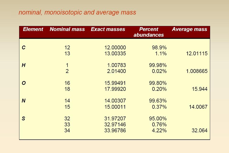
1 IUPAC Terms and Definitions in Mass Spectrometry The third Draft Document released in August 2006 by the IUPAC task group on MS Terms has fixed the basic definitions to be adopted and those to be abandoned in the MS field: Unified atomic mass unit, u - A non-si unit of mass defined as one twelfth of the mass of one atom of 12 C in its ground state and equal to (10) kg. The term atomic mass unit (amu) is deprecated. The Dalton (Da) is usually accepted. Nominal mass - Mass of an ion or molecule calculated using the mass of the most abundant isotope of each element rounded to the nearest integer value and equivalent to the sum of the mass numbers of all constituent atoms (mass number: sum of the number of protons and neutrons in an atom, molecule or ion)
2 Exact mass - Calculated mass of an ion or molecule containing a single isotope of each atom, most frequently the lightest isotope of each element, calculated from the masses of these isotopes using an appropriate degree of accuracy. Monoisotopic mass - Exact mass of an ion or molecule calculated using the mass of the most abundant isotope of each element. Average mass - Mass of an ion or molecule calculated using the average mass of each element weighed for its natural isotopic abundance.
3
4 mass formula nominal exact average C 3 H 8 O C 2 H 4 O Accurate mass - experimentally determined mass of an ion that is used to determine an elemental formula. Note: accurate mass and exact mass are NOT synonymous. The former refers to a measured mass, the latter to a calculated mass.

5
6 Isotopic distribution: carbon Natural abundance (%): 12 C: C: C: C C, no 13 C C C, 1 13 C ,200 1,202 C 100 1,204 1, ,000 12,010 C C, 2 13 C 12,020 12,030 Adapted from
7 Isotopic distribution: effects on large molecules When molecules with high molecular weight are considered, the difference between exact mass and average mass becomes progressively higher: Alanine oligo/poly-peptides

8 The available mass resolution can influence significantly the appearance of the isotopic distribution, as shown in the following example for glucagon, a 29-amino acid polypeptide hormone: High resolution Low resolution
9 Ionization Techniques Several techniques are available for the generation of charged molecules either in the gas phase, from volatile and non-volatile analytes, or from solid/liquid samples, under vacuum or in atmospheric conditions: Electron ionization (EI) Chemical Ionization (CI) Field ionization (FI) Nebulization Ionization Thermospray Ionization (TSI) Electro-Spray Ionization (ESI) Field Desorption (FD) Plasma Desorption (PD) Fast Atom Bombardment (FAB) Matrix Assisted Laser Desorption (MALDI) Desorption/Ionization on Silicon (DIOS) Desorption Electro-Spray Ionization (DESI)

10 The ionization source is one of the key components of a mass spectrometer:
 Gas phase: the sample is first vaporized and then ionized, so that the method is applicable only to volatile compounds, typically with")
11 Gas phase vs. Desorption ionization (for molecular mass spectrometry) Gas phase: the sample is first vaporized and then ionized, so that the method is applicable only to volatile compounds, typically with molecular meight lower then 1 KDa. Desorption: applicable to nonvolatile and thermally unstable samples (although volatiles can also be used). Hard vs. soft ionization methods Hard methods: energy transferred to analyte molecules is very high and produces ions in highly excited energy states. Relaxation produce estensive fragmention. Soft methods: low energy transferred; little (if any) fragmentation observed.
12 Soft vs hard ionization: effect on mass spectra The appearance of mass spectra obtained for a given compound depends strongly on the method adopted for ion formation:

13 Effect of ionization technique When possible, different ionization techniques should be used for the same analyte, since the information provided by each one is often complementary to that obtained from the others.

14 Pressure requirements for ionization sources Most ionization methods in mass spectrometry require that a relatively low pressure is achieved inside the ionization chamber. cm/s 1 s 2 volume (covered in 1 s) = 2 Actually, the relative motion between colliding molecules must be considered


15 Magnitude of relative velocity: Average velocities must be considered, since molecules do not travel at the same speed:

16
17
18

19 Vacuum Technology When electrons, atoms/molecules or ions are involved in the ionization of analyte molecules (like in EI, CI and FAB), the estimated low mean free paths imply that collisions occur between ionizing particles and neutral species, thus lowering the energy of the formers and, consequently, ionization efficiency. Rotary vane pumps usually provide vacuum conditions suitable for low pressure ionization (10-3 /10-4 torr)


20 Lower pressures (down to 10-7 torr), usually required for mass analysers and ion detectors, can be achieved using different devices, like turbomolecular or oil diffusion pumps. Turbomolecular pump Oil diffusion pump
21 Desirable properties for ion sources/ion beams generation of ion beams compatible with analyzer geometry compliance with detector sensitivity ( A) generation of ion beam with small energy spread high ionization efficiency selectivity against unwanted ions bipolarity (ability to generate either positive or negative ions) ability to ionize also high molecular weight compounds control of fragmentation absence of memory effects stability of ion emission with time minimization of chemical procedures required before MS analysis (sample pretreatment)
22 Electron (impact) ionization (EI) IUPAC definition of Electron Ionization:
23 Scheme and representation of a EI source
24 Exploded view of VG Trio 2000 electron (chemical) ionization source 7 - ion exit plate Focus plate Source block 4 ion extraction plate

25 Quadrupole entrance Vacuum lock GC transfer line LC interface Solid probe Diffusion pump

26 Focus plate Ion beam exit
 repeller filament (cathode) GC column")
27 Solid probe; LC interface (Particle Beam) trap (anode) repeller filament (cathode) GC column inlet
28 In a EI source electrons are emitted by a hot W or Re filament, due to thermoionic effect, and accelerated towards a anode target by a potential difference. Along their path electron interact with gaseous molecules coming from the sample introduction system (including the outlet of a GC column). Actually, due to the dimension difference, their interaction with a molecule is not an impact in the common meaning of this word. Ionization efficiency (IE) is the ratio between the number of ions formed and that of neutral molecules entering the source. Electron current and energy have a remarkable influence on the ionization efficiency.
29 Filament emission characteristics A roughly ohmic behaviour is observed until the filament blows, due to excessive heating
30 Aspects related to electron energy Electron energies typically adopted in EI range between 20 and 70 ev. At least 10 to 20 ev are usually transferred to a neutral molecule during interaction with these electrons. Since the ionization potential of an organic molecule is typically lower than 10 ev, the interaction leads to the generation of a molecular ion, M. +, having excess energy on it, responsible for its subsequent fragmentation. It is worth noting that the wavelengths related to ionizing electrons ( = h/mv) are in the range Å (20-70 ev). At very high energies the wavelength becomes very small and the molecule becomes transparent to the electron. In other words, there is not enough time for the electron-neutral interaction and energy transfer to occur.
31 A key parameter in determining the ionization efficiency is the ionization cross-section, that is increased with electron energy: The number of positive ions generated by EI is given by: x + where + = cross section for positive ionization, N 0 = gas density, N e = number of electrons, x = electron path.
32 Actually ionization is just one of several phenomena occurring during electron-neutral interactions. Their cross-sections for CF 4 are shown in the following graph: Note that ionization is relevant only between 20 and ev electron energies.


33 A detailed graph of CH 4 ionization cross-sections as a function of electron energy, calculated by different authors, is shown in the following:
34 The plot of Ion current vs. electron energy clearly resembles the trend observed in the rising part of the cross-section vs energy plot. Three regions can be distinguished: A) threshold region, principally molecular ions produced B) production of fragment ion becomes important C) routine operation for EI: mostly fragment ions observed
35 Ionization efficiencies for different compounds have qualitatively similar trends with respect to electron energy, yet the absolute values can be orders of magnitude different. The number of ions generated by EI (mostly fragments) is generally maximum for electron energies comprised between 50 and 100 ev.
36 Time scales of electron ionization The time available for the electron-neutral interaction is strongly dependent on the electron energy. 70 ev electrons have velocities higher that m/s; if a 10 Å (10-9 m) diameter is assumed for a molecule, a transit time of s can be easily estimated. This time scale is consistent with that pertaining to electronic transitions, whereas vibrational transitions require at least s. The Franck-Condon principle can be applied: the molecule nuclei remain frozen in their positions during the interaction. Only later internal energy excess will lead to fragmentation, eventually occurring outside the source (Post Source Decay, PSD):
37 Among activation methods used in mass spectrometry, EI is the one involving the shortest times at all: In the scheme green and blue (dotted) boxes represent timescales for vibrational and electronic excitation, respectively:
38 Energy transfer and distribution accompanying ionization Ionization of a molecule occurs very rapidly, thus the bond lengths in the molecule are assumed not to change during the ionization process. The ionization process obeys the Franck-Condon principle, which states that electronic transitions occur with no change in nuclear configuration. This corresponds to vertical transitions in the following diagram, relevant to a diatomic molecule: Transition a shows ionization from the ground state of the molecule to a higher vibrational state of the corresponding ion.
39 If the transition occurs to a state whose energy is higher than the dissociation energy (D) then the ion will be fragmented. In the case of transition b the ground state of the ion is dissociative, thus no intact molecular ion will be formed. When polyatomic molecules are considered, several different fragmentation pathways can occur. ABCD.+ A + + BCD. A. + BCD + fragm. BC + + D. rearrang. CD. + AB + A. + B + fragm. ADBC.+ A + + DBC. A + + B.
40 Unimolecular reactions Reactions in an EI source are assumed to be unimolecular, since the pressures are too low to enable significant interactions between molecules and ions. Types of ions detected 1) Molecular ions (precursor/parent) reach the detector without fragmentation 2) Product ions (fragment) - fragment by decomposition reactions in the source 3) Metastable ions ions that fragment by decomposition reactions after leaving the source
41 Factors dictating the fragmentation extent (and hence the appearance of the spectrum): - the internal energy available in the molecular ion; - the time taken for the ion to be transmitted from the source to the detector. Time scales The time factor needs to be considered since different processes occur with different time scales or rate constants; the rate constants for various types of reaction have to be considered in relation to the time spent by ions in the source and analyzer regions of the spectrometer. Residence time of ions in the source: 10-6 s s, depending on source parameters and accelerating voltage. Time taken for an ion to traverse the mass analyzer region: ca s. Decomposition times are expressed in terms of a rate constant, which can be expressed as the reciprocal of the ion lifetime, e.g. the rate constant for a decomposition of an ion with a lifetime of 10 µs is 10 5 s -1.
42 1) Fragment ions will only be detected if the parent ions possess sufficient energy for decomposition; the rate constant for decomposition is > ~10 4 s -1. 2) Parent ions will be detected only if the rate constant for decomposition is < 10 4 s -1. 3) For rates close to 10 5 s -1, metastable decompositions occur between the source and detector. 4) For rate constants of 10 6 s -1 or greater, decompositions occur in the source, giving rise to fragment ions.
43 Ionization events can be classified as: soft, if little excess energy is transferred to the ionised molecule: a molecular ion is formed; hard, if a significant fragmentation occurs.
44 The distinction can be made also by looking at potential energy curves:
45 Fragmentation is less pronounced when lower electron energies are adopted, thus favouring the molecular ion presence. On the other hand, the absolute intensity is somewhat reduced at lower electron energies, suggesting a lower ionization efficiency. 70 ev 15 ev
46 Electron energy affects the internal energy distribution of the molecule interacting with an electron. The model internal energy distribution P( ) resulting from interaction of an organic molecule with 70 ev electrons can be depicted as follows: The maximum internal energy that an ion can acquire in this case is 70 ev minus the ionization energy (IE). Actually most ions acquire small internal energies.
47 Lower electron energies imply a modification of the internal energy distribution for the molecule involved in ionization: As a consequence, the fragmentation routes available to the parent ion are different according to the electron energy. Such information can be easily described by two complementary approaches: the so-called breakdown curves, i.e. plots of fragment ion abundances as a function of the internal energy of the originally generated ion; the Wahrhaftig diagrams.
48 Breakdown curves Breakdown curves can be determined by effecting interactions of the compound of interest with electrons having a single energy, scanned over an interval, and recording the relative intensity of generated fragments. 70 ev The correlation between breakdown curves and EI spectrum is shown for n-propanol.
49 Wahrhaftig diagram for a generic ABCD. + ion In the upper side of a Wahrhaftig diagram (developed by the American chemist Austin L. Wahrhaftig) the distribution of internal energies for the parent ion is reported: In the lower side the probability of a specific fragmentation pathway is shown in the form of a rate constant: IE(M) = ionization energy
50 A rate constant of ~10 6 s -1 or greater is necessary for ion-source decomposition (lifetimes ~ 10-6 s or less): log K = 6 on the curve for the.. reaction M + AD + defines the. minimum M + internal energy. required for AD + formation,. indicated as E s (AD + ). E s is usually somewhat higher than the reaction critical energy, E 0 ; the latter is defined as the difference between the zero-point energy of M +. and that of the activated complex for the AD +. generation. AD +. M +. In the diagram m* represents a metastable ion, the corresponding required miminum internal energy is indicated as E m.

51 Vertical lines traced for each E s value divide the total area underlining the P(E) curve into different regions: each region area is related to the abundance of different generated ions Note that the area relevant to ions. like AD + and AB + refers to their initial amount, as they could eventually generate further fragments. A similar scenario is observed for metastable ions, like F*: If the ionizing electron energy is lowered, the relative proportion of M +. ions that do not dissociate will increase, although the total signal in the MS spectrum will be lower.
52 Thermodynamic vs kinetic effects The observed Wahrhaftig diagram shows a peculiar effect: the reaction of lowest critical energy does not necessarily produce the mostabundant ion. In particular, [AB + ] > [AD +. ] although the M +. AD +. process has a lower critical energy than M +. AB+. This finding can be explained if the transition states for the two processes are compared: In the rearrangement forming. AD + two new bonds are formed, offsetting the energy required to cleave A B and C D. The critical energy for AB + formation is higher, since this requires B C cleavage:
53 On the other hand, the steric requirements for AD +. formation are much more stringent than those for AB + formation. It can be said that the first process has the more favorable enthalpy, whereas the second has the more favorable entropy. For higher-energy ABCD +. ions, the B C bond dissociation can take place whenever sufficient energy accumulates in this bond. For AD +. formation, the energy requirements must be met at the same time that A and D are within bonding distance, which is true only for a small proportion of all possible conformations of ABCD +.. These offsetting enthalpy and entropy effects generally lead to a substantial number of competing primary reactions as well as consecutive secondary and further reactions, thus the mass spectrum of a large molecule can shown even hundreds of peaks. Small changes in molecular structure result in large differences in peak abundances. For this reason, when interpreting an unknown spectrum it is helpful to study the spectra of closely related compounds.
54 Effect of molecular structure When the same electron energy is adopted, the fragmentation pattern is significantly affected by the molecular structure, even when isobaric species (i.e. having the same molecular weight) are considered: As a general rule, molecules characterized by the presence of aromatic rings, especially condensed ones, do not undergo a significant fragmentation.
55 Due to: the purely physical nature of the phenomenon the involvement of gas-phase unimolecular reactions electron ionization is characterized by high reproducibility, as shown in the following comparison between n-propanol EI spectra (70 ev):
56 Quasi-equilibrium theory (QET) The quasi-equilibrium theory (QET) provides a generally accepted physical description of mass-spectral behavior, usually able to explain breakdown curves or Wahrhaftig diagrams. According to this theory, the basic steps of the ionization-fragmentation process are: 1) ionization of the molecule, taking place in approximately s, yielding the excited molecular ion without change in bond length (a Franck-Condon process); 2) except for the smallest molecules, rapid transitions between all the possible energy states of the molecular ion, leading to a "quasi-equilibrium" before ion decomposition; 3) decomposition of the molecular ion, depending only on its structure and internal energy, and not on the method used for the initial ionization.
57 Exceptions to QET are usually found for: 1) dissociations involving small molecules, having fewer, more widely separated states 2) excited electronic states that are similarly "isolated" 3) very fast decompositions (<10-11 s). The basic parameters involved in QET are: the thermochemical appearance energy, AE: the minimum energy necessary to produce a fragment from the ground-state neutral molecule.; the critical energy, E 0 : the minimum internal energy of M +. required for the decomposition to yield a specific fragment. Note that AE = IE(M) + E 0 where IE = ionization energy; the rate constant k for molecular ion decomposition, involved in the kinetic relation: ln[m +. ]0 /[M +. ] = kt.
58 Within QET a E S value reported into Wahrhaftig diagrams corresponds to the internal energy of precursor ions which lead to an equal probability of leaving the ion source (i.e. equal rate constants) for adjacent ions along the P(E) curves. The E s -E 0 difference is defined as: kinetic shift when referred to the fragment having the. lowest E 0 value (AD + in this case) competitive shift, when referred to higher E 0 fragments (like AB + ) Kinetic shift Competitive shift The kinetic shift varies between 0.01 and 2 ev; the competitive shift can be higher.
59 Metastable ions The decompositions of metastable ions (MI) in a field-free drift region have rate constant values just below the minimum required for ion-source decomposition, i.e. in the s -1 range. In the Wahrhaftig diagram shown before metastable ions arise from molecular ones having internal energies comprised between E m (AD +. ) and E s (AD +. ):
60 In a mass spectrum metastable ions appear as weaker, broader peaks occurring at non-integer m/z values (m*): The following equation can be written for an ion M 1+ that decomposes to M 2 + in the field free region of the mass spectrometer: M 1+ = M 2+ + (M 1 - M 2 ) The M 1 -M 2 difference corresponds to the mass of the neutral fragment released during decomposition.
61 If v 1 and v 2 are the velocities for M 1+ and M + 2, respectively, and the assumption is made that also the neutral species has a v 2 velocity, conservation of momentum leads to the following equation: m 1 v 1 = m 2 v 2 + (m 1 - m 2 ) v 2 which implies that: m 1 v 1 = m 1 v 2 v 1 can be calculated if the M 1 ion charge, z 1, and the accelerating potential, V, are known: z 1 V = ½ m 1 v 1 2 On the other hand, the ion actually entering the mass analyser is M 2. If a magnetic sector is used, the following equation applies: m 2 v 2 2 /r = B z 2 v 2
62 v 1 and v 2 can be calculated from the last two equations : v 12 = (2z 1 V)/m 1 v 22 = B 2 z 22 r 2 /m 2 2 Since v 1 = v 2 and z 1 = z 2 = z, we can write: (2zV)/m 1 = (B 2 z 2 r 2 )/ m 2 2 m 2 2 /(m 1 z) = B 2 r 2 /2V The last equation shows that the M 2+ ion arising from the decomposition of M 1+ behaves like an ion with mass m 22 /m 1, that is lower than m 2. For example, for m 1 = 60 and m 2 = 59 (loss of hydrogen), m 22 /m 1 = Due to the large energy dispersion characterising the generated ion, a peculiar large peak is observed in the mass spectrum: Metastable M 2 + M 2 + M 1 + m/z
63 In several cases metastable ions can provide useful information about fragmentation patterns. The electron ionization spectrum of benzene provides an interesting example: 76 In this case the m/z = 78 is related to the molecular ion (M 1 ), whereas m/z 77 is referred to the phenyl carbocation (C 6 H 5+ ), M 2, arising from H loss. If such loss occurred also in the field-free region of the mass spectrometer a metastable ion with m/z = 77 2 /78 = 76.0 would be observed. Such a peak is actually found in the spectrum. In many cases both M 2+ ions and the corresponding metastable ions are observed in EI spectra.
64 The procedure can be reversed: if the m/z ratios of metastable ions are known, masses for M 2+ ions can be retrieved starting from those of M 1 + ions: m* = m 22 /m 1 m 2 = (m* m 1 ) 1/2 Further metastable ions, observed in the benzene spectrum at m/z 74.1 and 34.7, lead to m 2 values and 52.02, respectively, that are also found directly in the EI spectrum. Such values suggest further fragmentations for the molecular ion: C 6 H 4 C 6 H H 2 C 4 H C 2 H 2 52
65 The importance of heteroatoms in electron ionization Heteroatoms may have non bonding electrons, which are more easily removed to form ions, as shown for formaldehyde: The probability of removal of non bonding electrons is related to the heteroatom: S > N > O
66 Example of heteroatom influence: electron ionization fragmentation pathways for 2-hexanone:
67 A summary of Pros and Cons of Electron Ionization
Chemistry Instrumental Analysis Lecture 34. Chem 4631
 Chemistry 4631 Instrumental Analysis Lecture 34 From molecular to elemental analysis there are three major techniques used for elemental analysis: Optical spectrometry Mass spectrometry X-ray spectrometry
Instrumental Analysis. Mass Spectrometry. Lecturer:! Somsak Sirichai
 303351 Instrumental Analysis Mass Spectrometry Lecturer:! Somsak Sirichai Mass Spectrometry What is Mass spectrometry (MS)? An analytic method that employs ionization and mass analysis of compounds in
Harris: Quantitative Chemical Analysis, Eight Edition
 Harris: Quantitative Chemical Analysis, Eight Edition CHAPTER 21: MASS SPECTROMETRY CHAPTER 21: Opener 21.0 Mass Spectrometry Mass Spectrometry provides information about 1) The elemental composition of
Lecture 8: Mass Spectrometry
 intensity Lecture 8: Mass Spectrometry Relative abundance m/z 1 Ethylbenzene CH 2 CH 3 + m/z = 106 CH 2 + m/z = 91 C 8 H 10 MW = 106 CH + m/z = 77 + 2 2 What information can be obtained from a MS spectrum?
Mass Spectrometry. Electron Ionization and Chemical Ionization
 Mass Spectrometry Electron Ionization and Chemical Ionization Mass Spectrometer All Instruments Have: 1. Sample Inlet 2. Ion Source 3. Mass Analyzer 4. Detector 5. Data System http://www.asms.org Ionization
Chemistry 311: Topic 3 - Mass Spectrometry
 Mass Spectroscopy: A technique used to measure the mass-to-charge ratio of molecules and atoms. Often characteristic ions produced by an induced unimolecular dissociation of a molecule are measured. These
Lecture 8: Mass Spectrometry
 intensity Lecture 8: Mass Spectrometry Relative abundance m/z 1 Ethylbenzene experiment CH 2 CH 3 + m/z = 106 CH 2 + m/z = 91 C 8 H 10 MW = 106 CH + m/z = 77 + 2 2 What information can we get from MS spectrum?
Fundamentals of Mass Spectrometry. Fundamentals of Mass Spectrometry. Learning Objective. Proteomics
 Mass spectrometry (MS) is the technique for protein identification and analysis by production of charged molecular species in vacuum, and their separation by magnetic and electric fields based on mass
TANDEM MASS SPECTROSCOPY
 TANDEM MASS SPECTROSCOPY 1 MASS SPECTROMETER TYPES OF MASS SPECTROMETER PRINCIPLE TANDEM MASS SPECTROMETER INSTRUMENTATION QUADRAPOLE MASS ANALYZER TRIPLE QUADRAPOLE MASS ANALYZER TIME OF FLIGHT MASS ANALYSER
Chemistry Instrumental Analysis Lecture 37. Chem 4631
 Chemistry 4631 Instrumental Analysis Lecture 37 Most analytes separated by HPLC are thermally stable and non-volatile (liquids) (unlike in GC) so not ionized easily by EI or CI techniques. MS must be at
Mass Spectrometry (MS)
") Mass Spectrometry (MS) Alternative names: Mass spectrometric (selective) detector (MSD) Spectrometry - methods based on interaction of matter and radiation Mass spectrometry - method based on formation
Mass Spectrometry (MS) Alternative names: Mass spectrometric (selective) detector (MSD) Spectrometry - methods based on interaction of matter and radiation Mass spectrometry - method based on formation
LC-MS Based Metabolomics
 LC-MS Based Metabolomics Analysing the METABOLOME 1. Metabolite Extraction 2. Metabolite detection (with or without separation) 3. Data analysis Metabolite Detection GC-MS: Naturally volatile or made volatile
Mass Spectrometry. General Principles
 General Principles Mass Spectrometer: Converts molecules to ions Separates ions (usually positively charged) on the basis of their mass/charge (m/z) ratio Quantifies how many units of each ion are formed
MS Goals and Applications. MS Goals and Applications
 MS Goals and Applications 3 Several variations on a theme, three common steps Form gas-phase ions choice of ionization method depends on sample identity and information required Separate ions on basis
Mass Spectrometry. A truly interdisciplinary and versatile analytical method
 Mass Spectrometry A truly interdisciplinary and versatile analytical method MS is used for the characterization of molecules ranging from small inorganic and organic molecules to polymers and proteins.
2. Separate the ions based on their mass to charge (m/e) ratio. 3. Measure the relative abundance of the ions that are produced
 ratio. 3. Measure the relative abundance of the ions that are produced") I. Mass spectrometry: capable of providing both quantitative and qualitative information about samples as small as 100 pg (!) and with molar masses in the 10 4-10 5 kdalton range A. The mass spectrometer
I. Mass spectrometry: capable of providing both quantitative and qualitative information about samples as small as 100 pg (!) and with molar masses in the 10 4-10 5 kdalton range A. The mass spectrometer
Mass Spectrometry. Introduction EI-MS and CI-MS Molecular mass & formulas Principles of fragmentation Fragmentation patterns Isotopic effects
 Mass Spectrometry Introduction EI-MS and CI-MS Molecular mass & formulas Principles of fragmentation Fragmentation patterns Isotopic effects 1 Introduction to MS Mass spectrometry is the method of analysis
Lecture 15: Introduction to mass spectrometry-i
 Lecture 15: Introduction to mass spectrometry-i Mass spectrometry (MS) is an analytical technique that measures the mass/charge ratio of charged particles in vacuum. Mass spectrometry can determine masse/charge
Mass Spectrometry in MCAL
 Mass Spectrometry in MCAL Two systems: GC-MS, LC-MS GC seperates small, volatile, non-polar material MS is detection devise (Agilent 320-MS TQ Mass Spectrometer) Full scan monitoring SIM single ion monitoring
Mass Spectroscopy. Base peak. Molecular Ion peak. The positively charged fragments produced are separated, based on their mass/charge (m/z) ratio. M+.
 ratio. M+.") Mass spectrometry is the study of systems causing the formation of gaseous ions, with or without fragmentation, which are then characteried by their mass to charge ratios (m/) and relative abundances.
Mass spectrometry is the study of systems causing the formation of gaseous ions, with or without fragmentation, which are then characteried by their mass to charge ratios (m/) and relative abundances.
Welcome to Organic Chemistry II
 Welcome to Organic Chemistry II Erika Bryant, Ph.D. erika.bryant@hccs.edu Class Syllabus 3 CHAPTER 12: STRUCTURE DETERMINATION 4 What is this solution Soda Tea Coffee??? 5 What is this solution Soda Tea
MS Goals and Applications. MS Goals and Applications
 MS Goals and Applications 1 Several variations on a theme, three common steps Form gas-phase ions choice of ionization method depends on sample identity and information required Separate ions on basis
MS/MS .LQGVRI0606([SHULPHQWV
 0DVV6SHFWURPHWHUV Tandem Mass Spectrometry (MS/MS) :KDWLV0606" Mass spectrometers are commonly combined with separation devices such as gas chromatographs (GC) and liquid chromatographs (LC). The GC or
Propose a structure for an alcohol, C4H10O, that has the following
 Propose a structure for an alcohol, C4H10O, that has the following 13CNMR spectral data: Broadband _ decoupled 13CNMR: 19.0, 31.7, 69.5 б DEPT _90: 31.7 б DEPT _ 135: positive peak at 19.0 & 31.7 б, negative
Mass spectrometry and elemental analysis
 Mass spectrometry and elemental analysis A schematic representation of a single-focusing mass spectrometer with an electron-impact (EI) ionization source. M: + e _ M +. + 2e _ Ionization and fragmentation
Ionization Techniques Part IV
 Ionization Techniques Part IV CU- Boulder CHEM 5181 Mass Spectrometry & Chromatography Presented by Prof. Jose L. Jimenez High Vacuum MS Interpretation Lectures Sample Inlet Ion Source Mass Analyzer Detector
Laser Dissociation of Protonated PAHs
 100 Chapter 5 Laser Dissociation of Protonated PAHs 5.1 Experiments The photodissociation experiments were performed with protonated PAHs using different laser sources. The calculations from Chapter 3
Mass Spectrometry (MS)
") Mass Spectrometry (MS) MW Molecular formula Structural information GC-MS LC-MS To Do s Read Chapter 7, and complete the endof-chapter problem 7-4. Answer Keys are available in CHB204H MS Principles Molecule
Mass Spectrometry (MS) MW Molecular formula Structural information GC-MS LC-MS To Do s Read Chapter 7, and complete the endof-chapter problem 7-4. Answer Keys are available in CHB204H MS Principles Molecule
Introduction to GC/MS
 Why Mass Spectrometry? Introduction to GC/MS A powerful analytical technique used to: 1.Identify unknown compounds 2. Quantify known materials down to trace levels 3. Elucidate the structure of molecules
Molecular Mass Spectrometry
 Molecular Mass Spectrometry Mass Spectrometry: capable of providing information about (1) Elemental composition of samples of matter: atomic mass (2) Structures of inorganic, organic, and biological molecules
RECOMMENDATIONS FOR NOMENCLATURE OF MASS SPECTROMETRY
 international UNION OF PURE AND APPLIED CHEMISTRY ANALYTICAL CHEMISTRY DIVISION COMMISSION ON ANALYTICAL NOMENCLATURE RECOMMENDATIONS FOR NOMENCLATURE OF MASS SPECTROMETRY RULES APPROVED 1973 LONDON BUTTER
The Franck-Hertz Experiment Physics 2150 Experiment No. 9 University of Colorado
 Experiment 9 1 Introduction The Franck-Hertz Experiment Physics 2150 Experiment No. 9 University of Colorado During the late nineteenth century, a great deal of evidence accumulated indicating that radiation
20.2 Ion Sources. ions electrospray uses evaporation of a charged liquid stream to transfer high molecular mass compounds into the gas phase as MH n
 20.2 Ion Sources electron ionization produces an M + ion and extensive fragmentation chemical ionization produces an M +, MH +, M +, or M - ion with minimal fragmentation MALDI uses laser ablation to transfer
Other Methods for Generating Ions 1. MALDI matrix assisted laser desorption ionization MS 2. Spray ionization techniques 3. Fast atom bombardment 4.
 Other Methods for Generating Ions 1. MALDI matrix assisted laser desorption ionization MS 2. Spray ionization techniques 3. Fast atom bombardment 4. Field Desorption 5. MS MS techniques Matrix assisted
1) In what pressure range are mass spectrometers normally operated?
 In what pressure range are mass spectrometers normally operated?") Exercises Ionization 1) In what pressure range are mass spectrometers normally operated? Mass spectrometers are usually operated in the high vacuum regime to ensure mean free paths significantly longer
Exercises Ionization 1) In what pressure range are mass spectrometers normally operated? Mass spectrometers are usually operated in the high vacuum regime to ensure mean free paths significantly longer
ICPMS Doherty Lecture 1
 ICPMS Doherty Lecture 1 Mass Spectrometry This material provides some background on how to measure isotope abundances by means of mass spectrometry. Mass spectrometers create and separate ionized atoms
MASS SPECTROMETRY. Topics
 MASS SPECTROMETRY MALDI-TOF AND ESI-MS Topics Principle of Mass Spectrometry MALDI-TOF Determination of Mw of Proteins Structural Information by MS: Primary Sequence of a Protein 1 A. Principles Ionization:
Introduction to the Q Trap LC/MS/MS System
 www.ietltd.com Proudly serving laboratories worldwide since 1979 CALL +1.847.913.0777 for Refurbished & Certified Lab Equipment ABI Q Trap LC/MS/MS Introduction to the Q Trap LC/MS/MS System The Q Trap
Mass Analyzers. Principles of the three most common types magnetic sector, quadrupole and time of flight - will be discussed herein.
 Mass Analyzers After the production of ions in ion sources, the next critical step in mass spectrometry is to separate these gas phase ions according to their mass-to-charge ratio (m/z). Ions are extracted
Mass Spectrometry: Introduction
 Mass Spectrometry: Introduction Chem 8361/4361: Interpretation of Organic Spectra 2009 Andrew Harned & Regents of the University of Minnesota Varying More Mass Spectrometry NOT part of electromagnetic
MASS SPECTRA measure a compound s Mol. Wt. This ionization type is called: electron impact MS
 MASS SPECTRA measure a compound s Mol. Wt. p. 213 M + Molecule e - Molecule + 2 e - + + Mole cule + + Mol ecule IONIZATION CHAMBER repellor plate accelerating plates variable field magnet + Mo + lecule
Auxiliary Techniques Soft ionization methods
 Auxiliary Techniques The limitations of the structural information in the normal mass spectrum can be partly offset by special mass-spectral techniques. Although a complete description of these is beyond
Structural Determination Of Compounds
 EXPERIMENT 10 Mass Spectroscopy Structural Determination Of Compounds. Introduction - In mass spectrometry, a substance is bombarded with an electron beam having sufficient energy to fragment the molecule.
Secondary Ion Mass Spectroscopy (SIMS)
") Secondary Ion Mass Spectroscopy (SIMS) Analyzing Inorganic Solids * = under special conditions ** = semiconductors only + = limited number of elements or groups Analyzing Organic Solids * = under special
Secondary Ion Mass Spectroscopy (SIMS) Analyzing Inorganic Solids * = under special conditions ** = semiconductors only + = limited number of elements or groups Analyzing Organic Solids * = under special
An ion source performs the following two functions:
 Ionization The Ion Source An ion source performs the following two functions: 1) converts sample atoms or molecules to ionized particles (ions) in the gas phase (sometimes the task of introducing the atoms
M M e M M H M M H. Ion Sources
 Ion Sources Overview of Various Ion Sources After introducing samples into a mass spectrometer, the next important step is the conversion of neutral molecules or compounds to gas phase ions. The ions could
Mass Spectrometry - Background
 Mass Spectrometry - Background In mass spectrometry, a substance is bombarded with an electron beam having sufficient energy to fragment the molecule. The positive fragments which are produced (cations
Chapter 20. Mass Spectroscopy
 Chapter 20 Mass Spectroscopy Mass Spectrometry (MS) Mass spectrometry is a technique used for measuring the molecular weight and determining the molecular formula of an organic compound. Mass Spectrometry
Skoog/Holler/Crouch Chapter 19 Principles of Instrumental Analysis, 6th ed. CHAPTER 19
 Skoog/Holler/Crouch Chapter 19 Principles of Instrumental Analysis, 6th ed. Instructor s Manual CHAPTER 19 19-1. In a continuous wave NMR experiment, the intensity of the absorption signal is monitored
Qualitative Organic Analysis CH 351 Mass Spectrometry
 Qualitative Organic Analysis CH 351 Mass Spectrometry Bela Torok Department of Chemistry University of Massachusetts Boston Boston, MA General Aspects Theoretical basis of mass spectrometry Basic Instrumentation
Chapter 5. Mass spectrometry
 ionization and fragmentation Chapter 5. Mass spectrometry which fragmentations? mass and frequency, m/z and count rate Reading: Pavia Chapters 3 and 4 Don t need 3.3 B-D, 3.4 B-D Use the text to clarify
Molecular Mass Spectrometry
 Molecular Mass Spectrometry Mass Spectrometry: capable of providing information about (1) Elemental composition of samples of matter: atomic mass (2) Structures of inorganic, organic, and biological molecules
CEE 772: Instrumental Methods in Environmental Analysis
 Updated: 10 December 2014 Print version CEE 772: Instrumental Methods in Environmental Analysis Lecture #21 Mass Spectrometry: Mass Filters & Spectrometers (Skoog, Chapt. 20, pp.511-524) (Harris, Chapt.
Mass spectrometry.
 Mass spectrometry Mass spectrometry provides qualitative and quantitative information about the atomic and molecular composition of inorganic and organic materials. The mass spectrometer produces charged
high temp ( K) Chapter 20: Atomic Spectroscopy
 Chapter 20: Atomic Spectroscopy") high temp (2000-6000K) Chapter 20: Atomic Spectroscopy 20-1. An Overview Most compounds Atoms in gas phase high temp (2000-6000K) (AES) (AAS) (AFS) sample Mass-to-charge (ICP-MS) Atomic Absorption experiment
high temp (2000-6000K) Chapter 20: Atomic Spectroscopy 20-1. An Overview Most compounds Atoms in gas phase high temp (2000-6000K) (AES) (AAS) (AFS) sample Mass-to-charge (ICP-MS) Atomic Absorption experiment
Quadrupole Mass Spectrometry Concepts. Mass spectrometers for residual gas analysis: Intermediate Level Users Guide
 Quadrupole Mass Spectrometry Concepts Mass spectrometers for residual gas analysis: Intermediate Level Users Guide What does Residual Gas Analysis allow us to do? RGA is the examination of the molecular
CHAPTER A2 LASER DESORPTION IONIZATION AND MALDI
 Back to Basics Section A: Ionization Processes CHAPTER A2 LASER DESORPTION IONIZATION AND MALDI TABLE OF CONTENTS Quick Guide...27 Summary...29 The Ionization Process...31 Other Considerations on Laser
Mass Spectrometry. Hyphenated Techniques GC-MS LC-MS and MS-MS
 Mass Spectrometry Hyphenated Techniques GC-MS LC-MS and MS-MS Reasons for Using Chromatography with MS Mixture analysis by MS alone is difficult Fragmentation from ionization (EI or CI) Fragments from
12. Structure Determination: Mass Spectrometry and Infrared Spectroscopy
 12. Structure Determination: Mass Spectrometry and Infrared Spectroscopy Determining the Structure of an Organic Compound The analysis of the outcome of a reaction requires that we know the full structure
Ion sources. Ionization and desorption methods
 Ion sources Ionization and desorption methods 1 2 Processes in ion sources 3 Ionization/ desorption Ionization Desorption methods Electron impact ionization Chemical ionization Electro-spray ionisation
MS Interpretation II. Fragmentation
 MS Interpretation II Fragmentation Ionization E Electron Ionization (EI): Even-electron neutrals yield odd-electron radical cations. M(EE) EI - 1e - M (E) Electron can come from anywhere. EI EI even electron
CEE 772 Lecture #27 12/10/2014. CEE 772: Instrumental Methods in Environmental Analysis
 Updated: 10 December 2014 Print version CEE 772: Instrumental Methods in Environmental Analysis Lecture #21 Mass Spectrometry: Mass Filters & Spectrometers (Skoog, Chapt. 20, pp.511 524) (Harris, Chapt.
Particle Position Relative Mass Relative Charge Proton Nucleus 1 +1 Neutron Nucleus 1 0 Electron Orbitals 1/ Atomic Symbol
 Atomic Structure Details of the three Sub-atomic (fundamental) Particles Particle Position Relative Mass Relative Charge Proton Nucleus 1 +1 Neutron Nucleus 1 0 Electron Orbitals 1/1840-1 Behaviour of
3 Use of Mass Spectra to Obtain Structural Information
 3 Use of Mass Spectra to Obtain Structural Information 1 Mass Spectrometry One of the most sensitive and versatile analytical tools More sensitive than other spectroscopic methods (e.g. IR spectroscopy)
Suzanne Bell Second Edition
 Forensic hemistry Suzanne Bell Second Edition Pearson Education Limited Edinburgh Gate Harlow Essex M20 2JE England and Associated ompanies throughout the world Visit us on the World Wide Web at: www.pearsoned.co.uk
MASS SPECTROSCOPY (MS)
") MASS SPECTOSCOPY (MS) Castor seeds icin (toxic protein) INTODUCTION Does not involve absorption of electromagnetic radiation. It is a spectroscopic technique, by virtue of its use in structure elucidation.
MASS SPECTOSCOPY (MS) Castor seeds icin (toxic protein) INTODUCTION Does not involve absorption of electromagnetic radiation. It is a spectroscopic technique, by virtue of its use in structure elucidation.
CHM 424 EXAM 4 CRIB - COVER PAGE FALL
 CHM 44 EXAM 4 CRIB - COVER PAGE FALL 007 There are six numbered pages with five questions. Answer the questions on the exam. Exams done in ink are eligible for regrade, those done in pencil will not be
Atomic structure Calculate the number of protons, electrons and neutrons in Important terms: quantum shells, principle quantum number, energy levels,
 Atomic structure Important terms: quantum shells, principle quantum number, energy levels, Calculate the number of protons, electrons and neutrons in Isotopes Definitions? Remember - isotopes have exactly
4. How can fragmentation be useful in identifying compounds? Permits identification of branching not observed in soft ionization.
 Homework 9: Chapters 20-21 Assigned 12 April; Due 17 April 2006; Quiz on 19 April 2006 Chap. 20 (Molecular Mass Spectroscopy) Chap. 21 (Surface Analysis) 1. What are the types of ion sources in molecular
Mass spectrometry has been used a lot in biology since the late 1950 s. However it really came into play in the late 1980 s once methods were
 Mass spectrometry has been used a lot in biology since the late 1950 s. However it really came into play in the late 1980 s once methods were developed to allow the analysis of large intact (bigger than
Because light behaves like a wave, we can describe it in one of two ways by its wavelength or by its frequency.
 Light We can use different terms to describe light: Color Wavelength Frequency Light is composed of electromagnetic waves that travel through some medium. The properties of the medium determine how light
IDENTIFICATION OF ORGANOMETALLIC COMPOUNDS USING FIELD DESORPTION IONIZATION ON THE GCT
 IDETIFICATIO OF ORGAOMETALLIC COMPOUDS USIG FIELD DESORPTIO IOIZATIO O THE GCT David Douce 1, Michael Jackson 1, Robert Lewis 1, Peter Hancock 1, Martin Green 1 and Stuart Warriner 2 1 Waters Corporation,
Welcome!! Chemistry 328N Organic Chemistry for Chemical Engineers. Professor: Grant Willson
 Welcome!! - 50750 Organic Chemistry for Chemical Engineers Professor: Grant Willson Teaching Assistants: Ji yeon Kim, Jai Hyun Koh, Paul Meyer, Qingjun Zhu http://willson.cm.utexas.edu January 19,2016
L.7. Mass Spectrum Interpretation
 L.7. Mass Spectrum Interpretation Fragmentation reactions Spectrum interpretation Confirmation of ion structural assignment Biomolecule dissociation Fragmentation reactions 1. Fragmentation reactions of
Welcome!! Chemistry 328N Organic Chemistry for Chemical Engineers. Professor: Grant Willson
 Welcome!! - 50120 Organic Chemistry for Chemical Engineers Professor: Grant Willson Teaching Assistants: Paul Meyer, Qingjun Zhu, Josh Saunders http://willson.cm.utexas.edu January 22,2019 Bureaucracy:
(Refer Slide Time 00:09) (Refer Slide Time 00:13)
 (Refer Slide Time 00:13)") (Refer Slide Time 00:09) Mass Spectrometry Based Proteomics Professor Sanjeeva Srivastava Department of Biosciences and Bioengineering Indian Institute of Technology, Bombay Mod 02 Lecture Number 09 (Refer
(Refer Slide Time 00:09) Mass Spectrometry Based Proteomics Professor Sanjeeva Srivastava Department of Biosciences and Bioengineering Indian Institute of Technology, Bombay Mod 02 Lecture Number 09 (Refer
Secondary Ion Mass Spectrometry (SIMS)
") CHEM53200: Lecture 10 Secondary Ion Mass Spectrometry (SIMS) Major reference: Surface Analysis Edited by J. C. Vickerman (1997). 1 Primary particles may be: Secondary particles can be e s, neutral species
CHEM53200: Lecture 10 Secondary Ion Mass Spectrometry (SIMS) Major reference: Surface Analysis Edited by J. C. Vickerman (1997). 1 Primary particles may be: Secondary particles can be e s, neutral species
Mass Spectrometry Course
 Mass Spectrometry Course Árpád Somogyi Mass Spectrometry Laboratory, Department of Chemistry and Biochemistry University of Arizona, Tucson, AZ Eötvös University, Budapest April 11-20, 2012 1 2 UA Chemistry
1. Cyclic voltammetry involves the measurement of a diffusion controlled at an electrode in which the is controlled. (4 points)
") Chem 454 First Exam Feb. 20, 2002 1. Cyclic voltammetry involves the measurement of a diffusion controlled at an electrode in which the is controlled. (4 points) 2. (5 points) A. Sketch a cyclic voltammogram
Chem 454 First Exam Feb. 20, 2002 1. Cyclic voltammetry involves the measurement of a diffusion controlled at an electrode in which the is controlled. (4 points) 2. (5 points) A. Sketch a cyclic voltammogram
Mass Spectrometry. What is Mass Spectrometry?
 Mass Spectrometry What is Mass Spectrometry? Mass Spectrometry (MS): The generation of gaseous ions from a sample, separation of these ions by mass-to-charge ratio, and measurement of relative abundance
for the Novice Mass Spectrometry (^>, John Greaves and John Roboz yc**' CRC Press J Taylor & Francis Group Boca Raton London New York
 Mass Spectrometry for the Novice John Greaves and John Roboz (^>, yc**' CRC Press J Taylor & Francis Group Boca Raton London New York CRC Press is an imprint of the Taylor & Francis Croup, an informa business
Discovered by German scientist Johann Hittorf in 1869 and in 1876 named by Eugen Goldstein.
 DO PHYSICS ONLINE CATHODE RAYS CATHODE RAYS (electron beams) Streams of electrons (negatively charged particles) observed in vacuum tubes - evacuated glass tubes that are equipped with at least two metal
Matti Laan Gas Discharge Laboratory University of Tartu ESTONIA
 Matti Laan Gas Discharge Laboratory University of Tartu ESTONIA Outline 1. Ionisation 2. Plasma definition 3. Plasma properties 4. Plasma classification 5. Energy transfer in non-equilibrium plasma 6.
ELECTROMAGNETIC WAVES
 VISUAL PHYSICS ONLINE MODULE 7 NATURE OF LIGHT ELECTROMAGNETIC WAVES SPECTRA PRODUCED BY DISCHARGE TUBES CATHODE RAYS (electron beams) Streams of electrons (negatively charged particles) observed in vacuum
Analytical Technologies in Biotechnology Prof. Dr. Ashwani K. Sharma Department of Biotechnology Indian Institute of Technology, Roorkee
 Analytical Technologies in Biotechnology Prof. Dr. Ashwani K. Sharma Department of Biotechnology Indian Institute of Technology, Roorkee Module - 6 Spectroscopic Techniques Lecture - 6 Atomic Spectroscopy
Complete the following. Clearly mark your answers. YOU MUST SHOW YOUR WORK TO RECEIVE CREDIT.
 CHEM 322 Name Exam 3 Spring 2013 Complete the following. Clearly mark your answers. YOU MUST SHOW YOUR WORK TO RECEIVE CREDIT. Warm-up (3 points each). 1. In Raman Spectroscopy, molecules are promoted
HONOUR SCHOOL OF NATURAL SCIENCE. Final Examination GENERAL PHYSICAL CHEMISTRY I. Answer FIVE out of nine questions
 HONOUR SCHOOL OF NATURAL SCIENCE Final Examination GENERAL PHYSICAL CHEMISTRY I Monday, 12 th June 2000, 9.30 a.m. - 12.30 p.m. Answer FIVE out of nine questions The numbers in square brackets indicate
Rb, which had been compressed to a density of 1013
 Modern Physics Study Questions for the Spring 2018 Departmental Exam December 3, 2017 1. An electron is initially at rest in a uniform electric field E in the negative y direction and a uniform magnetic
THE MODERN VIEW OF ATOMIC STRUCTURE
 44 CHAPTER 2 Atoms, Molecules, and Ions GO FIGURE What is the charge on the particles that form the beam? Experiment Interpretation Incoming a particles Beam of a particles Source of a particles Nucleus
Ionization Methods in Mass Spectrometry at the SCS Mass Spectrometry Laboratory
 Ionization Methods in Mass Spectrometry at the SCS Mass Spectrometry Laboratory Steven L. Mullen, Ph.D. Associate Director SCS Mass Spectrometry Laboratory Contact Information 31 oyes Laboratory (8:00-5:00
Choosing the metabolomics platform
 GBS 748 Choosing the metabolomics platform Stephen Barnes, PhD 4 7117; sbarnes@uab.edu So, I have my samples what s next? You ve collected your samples and you may have extracted them Protein precipitation
Mass Spectrometry and Proteomics - Lecture 2 - Matthias Trost Newcastle University
 Mass Spectrometry and Proteomics - Lecture 2 - Matthias Trost Newcastle University matthias.trost@ncl.ac.uk Previously: Resolution and other basics MALDI Electrospray 40 Lecture 2 Mass analysers Detectors
Atomic masses. Atomic masses of elements. Atomic masses of isotopes. Nominal and exact atomic masses. Example: CO, N 2 ja C 2 H 4
 High-Resolution Mass spectrometry (HR-MS, HRAM-MS) (FT mass spectrometry) MS that enables identifying elemental compositions (empirical formulas) from accurate m/z data 9.05.2017 1 Atomic masses (atomic
Proudly serving laboratories worldwide since 1979 CALL for Refurbished & Certified Lab Equipment
 www.ietltd.com Proudly serving laboratories worldwide since 1979 CALL +1.847.913.0777 for Refurbished & Certified Lab Equipment Applied Biosystems QStar Pulsar i Features of the API QSTAR Pulsar i The
Chapter 2 Fundamentals of Ion Chemistry
 Chapter 2 Fundamentals of Ion Chemistry Toshihiro Fujii 2.1 Termolecular Association Reactions 2.1.1 Association Reaction Mechanism It was experimentally found that the overall process of ion molecule
Lecture 3 Vacuum Science and Technology
 Lecture 3 Vacuum Science and Technology Chapter 3 - Wolf and Tauber 1/56 Announcements Homework will be online from noon today. This is homework 1 of 4. 25 available marks (distributed as shown). This
Analysis of Polar Metabolites using Mass Spectrometry
 Analysis of Polar Metabolites using Mass Spectrometry TransMed Course: Basics in Clinical Proteomics and Metabolomics. Oct 10-19, 2012 dd.mm.yyyy Vidya Velagapudi, Ph.D, Adjunct Professor Head of the Metabolomics
Mass Spectrometer A Comparison of Positive and Negative Ion RGA Methods
 Gas Analysis Application Note 253 Mass Spectrometer A Comparison of Positive and Negative Ion RGA Methods Summary Hiden quadrupole massspectrometers provide the user with unique data through their high
Extrel Application Note
 Extrel Application Note Real-Time Plasma Monitoring and Detection of Trace H 2 O and HF Species in an Argon Based Plasma Jian Wei, 575 Epsilon Drive, Pittsburgh, PA 15238. (Presented at the 191st Electrochemical
EQUIPMENT Beta spectrometer, vacuum pump, Cs-137 source, Geiger-Muller (G-M) tube, scalar
 tube, scalar") Modern Physics Laboratory Beta Spectroscopy Experiment In this experiment, electrons emitted as a result of the radioactive beta decay of Cs-137 are measured as a function of their momentum by deflecting
Modern Physics Laboratory Beta Spectroscopy Experiment In this experiment, electrons emitted as a result of the radioactive beta decay of Cs-137 are measured as a function of their momentum by deflecting