Michael J. Richardson
|
|
|
- Marion McDaniel
- 5 years ago
- Views:
Transcription
1 ACTA BOT. ISL. 15: 23-49, 2011 Additions to the Coprophilous Mycota of Iceland Michael J. Richardson 165 Braid Road, Edinburgh EH10 6JE U.K., ABSTRACT: Seventy-five species mostly of coprophilous fungi were recorded from 44 herbivore dung samples collected from Iceland in 2006 and 2008 and incubated in moist chambers. Nineteen species are newly recorded for Iceland: - Pilaira moreaui, Ascobolus crenulatus, Saccobolus citrinus, Pseudombrophila cervaria, Thecotheus crustaceus, Camptosphaeria citrinella, Podospora intestinacea, P. pilosa, Schizothecium tetrasporum, Sordaria alcina, Hypocopra merdaria, Syspastospora parasitica, Viennotidia fimicola, Unguiculella tityrii, Sporormiella heptamera, Sp. longispora, Sp. octonalis, Laetisaria fuciformis, and Coprinus radiatus. Collections are described and the occurrence and distribution of species is discussed. The species richness and community structure of the Icelandic coprophilous mycota from this and an earlier collection are compared, and the value of such studies for assessing diversity is discussed and validated. KEY WORDS: ascomycetes, basidiomycetes, biogeography, diversity, ecology, fimicoles, Surtsey. INTRODUCTION The coprophilous mycota of Iceland is documented in the checklists of HALLGRÍMSSON & EYJÓLFSDÓTTIR (2004, and in preparation), which list just over 100 microfungi and 26 basidiomycetes. RICHARDSON (2004) recorded 78 species from a collection of 32 herbivore dung samples made from central and northeastern areas of Iceland in These included 33 species in addition to those in the microfungus checklist, and 12 agarics that were included in the basidiomycete list above. The microfungus checklist includes some fungi that were recorded on dung, but are not part of the suite of taxa traditionally considered to be specialist coprophils. If they are excluded, then the current recorded coprophilous mycota consists of about 110 species. A further 38 samples were collected in 2006, mainly from southwestern Iceland and the Snæfellsnes peninsula. Additionally, in 2008 a sample of rabbit pellets was collected in Reykjavík and five samples of goose dung were obtained from the volcanic island of Surtsey, which was formed in The samples were subsequently incubated in damp chambers and the coprophilous zygomycetes, ascomycetes and basidiomycetes that developed were recorded. Details of the fungi recorded from the 2006 and 2008 samples are provided, and their distribution
2 24 ACTA BOTANICA ISLANDICA NO. 15 and occurrence are discussed in the context of records from the earlier Icelandic collection and from over 1000 samples providing over records from samples collected worldwide in recent years by the author, and in relation to the observations of others, especially BELL (1983), VAN BRUMMELEN (1967), and LUNDQVIST (1972). In earlier studies, the author has suggested that fungal diversity can be monitored by recording the coprophils that develop on a collection of dung samples from a particular area or habitat (RICHARDSON 2001). Construction of species richness curves allow an estimate to be made of the number of species to be expected from a standard collection of for example 50 samples. This second collection from Iceland allows comparison with the results obtained from an earlier collection (RICHARDSON 2004), to validate that suggestion, and to compare the composition of the mycota of a region as assessed on two separate occasions. MATERIAL AND METHODS Mainland samples were collected between 29 July and 7 August 2006 (Table 1, Fig. 1), and one on 11 July Samples from Surtsey were collected on 8 July and August 2008 (see EYJÓLFSDÓTTIR 2009 for the Surtsey 100 x 100 m grid system). They were collected in paper envelopes and gently air-dried, if not already dry when collected. Localities (latitude and longitude, WGS84 datum) were determined with a Magellan explorist 100 satellite navigator, and place names are given according to the 1: Kortabók (2001, Mál og menning, Reykjavík). Samples were rehydrated and incubated soon after collection on moist paper towelling in plastic boxes with lightly fitting transparent lids, under ambient light and at room temperature (ca C). Care was taken to ensure that cultures were not too wet. Samples were generally of similar size, with incubation chambers 10 x 7 cm, which would accommodate approx. 2-4 g dry wt (= 15 sheep/ptarmigan pellets), or 13 x 8 cm for horse (approx g dry wt). Samples were examined frequently at intervals of a few days, with a x7-45 magnification stereomicroscope. Fruiting bodies were removed and mounted in water for examination and identification at higher magnification. Samples were incubated for 6 to 16 wk, with observations continuing whilst new fungi were being observed (one sample was discarded after 3 wk because of damage from insect larvae). Selected material has been placed in the Herbarium of the Icelandic Institute of Natural History, Akureyri Division (AMNH). In considering diversity, an estimate of species richness was made by constructing a cumulative species curve and deriving the equation for that curve (y = ax b, where y = cumulative no. of species observed in x samples) and solving for x = 50 samples (RICHARDSON 2001). This was done for the collection of 38 samples made in 2006 and compared with the estimate from 32 samples collected in 2002, and from a combined curve for all 70 samples. The similarity of the mycotas of the two sets

3 2011 RICHARDSON: COPROPHILOUS MYCOTA OF ICELAND 25 of samples was compared using the Sørensen, Bray & Curtis and Morisita- Horn indices (MAGURRAN 2004). FIGURE 1. Map of western Iceland showing collection localities. Numbers are the MJR sample numbers of 2006, as in Table 1, with the exception of 21 and samples from Surtsey, which were collected in RESULTS AND DISCUSSION Information on the dung samples and their origin are given in Table 1. Three hundred and sixty-six records of 75 species were obtained during the period of incubation. Nineteen of the species recorded were new for Iceland. Records from the six samples from 2008 have not been included in the analyses of species richness that follow.
4 26 ACTA BOTANICA ISLANDICA NO. 15 TABLE 1. Details of Icelandic dung samples and collection localities. Sample Locality Altitute Lat. Long. Habitat Date Subno.* (m a.s.l.) ( N) ( W) strate 27/06 nr Hotel Garður, Reykjavík (Hljómskálagarður) urban grass goose 28/06 Svínárnes Hrunamannaafréttur highland grassland sheep 29/06 Svínárnes Hrunamannaafréttur highland grassland goose 30/06 Svínárnes Hrunamannaafréttur paddock horse 31/06 Deild, north of Tungufellsdalur gravelly, sparsely sheep Hrunamannahreppur vegetated, with Racomitrium/Salix/Ericaceae 32/06 Tungufellsdalur Hrunamannahreppur riverside grass horse and Betula 33/06 Tungufellsdalur Hrunamannahreppur riverside grass sheep and Betula 34/06 Laugarvatnshellar Grímsnesafréttur paddock horse 35/06 Laugarvatnshellar Grímsnesafréttur rocky grassland sheep 36/06 Laugarvatnshellar Grímsnesafréttur Racomitrium/lichen rocks ptarmigan 37/06 Þingvellir Racomitrium/lichen rocks ptarmigan 38/06 Þingvellir Racomitrium/lichen rocks ptarmigan 39/06 route 36 Þingvallahraun, Þingvellir Racomitrium/lichen ptarmigan rocks, amongst birch 40/06 Haukadalur Biskupstungur Racomitrium/Erica heath ptarmigan 41/06 path to hot pool, Hrunalaug Hrunamannhreppur rocky grassland with sheep Racomitrium 42/06 Skollagróf, Hrunamannahreppur heathy ridgetop sheep grassland 43/06 Skollagróf, Hrunamannahreppur heathy ridgetop cattle grassland 44/06 Skollagróf, Hrunamannahreppur heathy ridgetop horse grassland 45/06 by river Hvítá, Skollagróf, Hrunamannahreppur riverside vegetation goose and farm track 46/06 Skollagróf, Hrunamannahreppur paddock horse 47/06 by river Hvítá, Bryðjuholt, Hrunamannahreppur riverside vegetation cattle 48/06 by river Hvítá, Bryðjuholt, Hrunamannahreppur riverside gravel and goose vegetation 49/06 route 34 between river Ölfusá and Eyrarbakki roadside by dunes goose 50/06 route 33 lighthouse Knarrarósviti east of Stokkseyri horse track horse 51/06 Gunnarsholt Rangárvellir pasture horse 52/06 Syðra-Langholt, Hrunamannahreppur paddock horse 53/06 route 54, Stórahraun, Snæfellsnes track in lava field sheep 54/06 Barðastaðir, Snæfellsnes grass at edge of shore horse 55/06 Öndverðarnes, Snæfellsnes rough grassland sheep 56/06 by Snæfellsjökull NP Visitor Centre, Hellnar, rough grassland horse Snæfellsnes 57/06 route 54, Hamrar, Grundarfjörður, Snæfellsnes rough grassland sheep 58/06 route 54, Rjúpnaborgir, Snæfellsnes rocky moorland, sheep Racomitrium/lichen 59/06 route 54, Úlfarsfell, Álftafjördur, Snæfellsnes rocky bank, goose Salix/ Ericaceae 60/06 route 54, Narfeyri, Álftafjörður, Snæfellsnes roadside moorland sheep 61/06 route 711, nr lighthouse, Þorvaldsfjall, Vatnsnes roadside moorland sheep 62/06 route 711, nr lighthouse, Þorvaldsfjall, Vatnsnes roadside moorland horse 63/06 Hindisvik, Vatnsnes rough grassland horse 64/06 Hindisvik, Vatnsnes rough grassland sheep 21/08 Öskjuhlíð, Reykjavík suburban scrubland rabbit 26/08 Surtsey, spit (grid D16)** sand goose 27/08 Surtsey, spit (grid D16)** sand, near Honckenya goose 28/08 Surtsey, spit (grid B16)** sand goose 29/08 Surtsey, south (grid P11)** gull colony grassland goose 30/08 Surtsey, south (grid O11)** gull colony grassland goose * MJR sample no. and year identifier **Surtsey 100 x100 m grid (see EYJÓLFSDÓTTIR 2009: 107)
5 2011 RICHARDSON: COPROPHILOUS MYCOTA OF ICELAND 27 In considering diversity, the species richness of individual samples is much as expected from earlier studies (RICHARDSON 2001), with a mean of 11 per sample (range 4-20) being recorded from the 13 sheep and 12 horse and 2 cattle samples, and fewer (mean 4, range 1-8) from the avian substrates. The cumulative species curve (total no. of species recorded with successive samples, Fig. 2), plotted in the sequence of sample collection, gives an estimate of 86 species from 50 samples, which compares with a revised estimate of 95 obtained from the 2002 collection of 32 samples. The estimate from the cumulative species curve of both sets of samples (n = 70, plotted in random order, Fig. 2) is 81 species. These values of species per 50 samples for separate and combined samples from the two collections are comparable with the predicted value of 89 species per 50 samples for latitude N, obtained by solving the equation for the relationship between latitude and species richness from the data of RICHARDSON (2001), who demonstrated that there is a latitudinal gradient of species richness in coprophilous fungi, with species expected from 50 samples from latitudes N, compared to 123 from N/S and from 40-0 N/S. Earlier data sets only included Coprinus species from the basidiomycetes, so non-coprinus basidiomycetes recorded from the Icelandic samples were excluded from the calculations to ensure comparability : y = 12.33x R 2 = 0.98 Cumulative sp. total : y = 9.66x R 2 = : y = 21.06x R 2 = Sample no. FIGURE 2. Cumulative totals of taxa observed on 32 and 38 samples of dung collected from Iceland in 2002 and 2006, respectively, after incubation in moist chambers. Curves are plotted in order of collection. A curve is also given for the pooled data of the two collections, plotted in a random order. Equations and R 2 values are given adjacent to the respective curves.
6 28 ACTA BOTANICA ISLANDICA NO. 15 In addition to the second collection from Iceland allowing a comparison to be made with an earlier estimate of species richness from the same area, it has also allowed a comparison to be made of the similarity of the coprophilous mycotas observed from the two collections, to understand how well a modest collection of samples of various dungs can be used to identify the composition of the mycota with a reasonable level of confidence. The Sørensen index measures the similarity of two communities, based on the number of species common to both communities, and is considered to be one of the most effective qualitative similarity measures (SOUTHWOOD & HENDERSON 2000). The Bray & Curtis and Morisita-Horn indices are similar, but take into account, in addition to presence/absence, the abundance of the different taxa. MAGURRAN (2004) notes that the Bray & Curtis index is a particularly satisfactory measure of quantitative similarity, and that the Morisita-Horn index is highly sensitive to the abundance of the most abundant species. The values for these indices (Table 2) are quite high (1 = complete similarity, 0 = no similarity), suggesting that individual collections of samples are sufficient to provide a representative qualitative list of the mycota. The particularly high value of 0.89 for the Morisita-Horn index suggests that the sampling was adequate to provide an indication of the most prevalent species of the Icelandic coprophilous mycota. Of the total of 102 species recorded from both collections 45 (44%) were common to both, and 48% were rare, occurring only once or twice. Fourteen species comprised 50% of all records (Fig. 3). TABLE 2. Similarity of the Icelandic coprophilous mycota, as assessed from collections of 32 and 38 samples collected in 2002 and 2006, respectively collection (n=32) 2006 collection (n=38) No. of species No. of species common to both collections No. of species in only one collection Coefficients of similarity: Sørensen (presence/absence) 0.62 Bray & Curtis (quantitative) 0.59 Morisita-Horn (quantitative) 0.89 See SOUTHWOOD & HENDERSON (2000) for equations for these indices. 45
7 2011 RICHARDSON: COPROPHILOUS MYCOTA OF ICELAND 29 Schizothecium vesticola Podospora decipiens Schizothecium conicum Coprinus miser Ascobolus albidus Thelebolus microsporus Coprinus heptemerus Coniochaeta ligniaria Ascobolus stictoideus Sporormiella intermedia Coniochaeta scatigena Thelebolus stercoreus Sporormiella lageniformis Pilobolus crystallinus Trichodelitschia minuta Coprinus cordisporus Coprinus pellucidus Lasiobolus cuniculi Sporormiella grandispora Coniochaeta leucoplaca Ascobolus immersus Arnium caballinum Sordaria fimicola Lasiobolus ciliatus Delitschia perpusilla FIGURE 3. Frequency of occurrence of the 25 most abundant species of coprophilous fungi in 70 collections of herbivore dung from Iceland. BELL (2005), DOVERI (2004), and RICHARDSON (2004), have all commented on the difficulty in determining Sporormiella species. Many species have been distinguished on the basis of ascus and ascospore morphology: whether or not the asci have a long or short tapering stalk or are abruptly contracted at the base (stipitate or button-like, respectively, sensu BELL, 2005); spore size; transverse or oblique septa; alignment of germ slits and whether or not they have a kink; and the relative size and shape of the component cells. The ascus shape (tapering base or not) and spore shape (fusoid, with tapering end cells or more or less cylindrical) seem to go together and be consistent, but the other spore characters are very variable, even within the contents of a single pseudothecium, and with much overlapping from collection to collection and species to species. BELL (2005) discussed the difficulties in detail, and concluded that evidence from sources other than anatomy and morphology may elucidate the situation. As further evidence of the difficulty, Fig. 4 shows the extent of variation in spore length and width recorded from members of the stipitate-ascus complex of species studied from the 2002 and 2006 collections from Iceland. The determination of these collections have been reexamined and carefully considered. As a result, the 2002 determinations of this group have been revised, and some amended identifications of material from the 2002 col-
8 30 ACTA BOTANICA ISLANDICA NO. 15 lections are given under the individual species entries that follow. AHMED & CAIN s (1972) treatment is the most recent comprehensive treatment of the genus, but since then many new species have been described and new species are still being described, often on the basis of single collections and molecular differences (e.g. ARENAL et al. 2005). It is not clear, without cultural and mating studies, how distinct these new, or some of the older, species are. FIGURE 4. Plot of the range of spore size recorded from various collections of Icelandic Sporormiella spp. with tapered spores and ascal foot. Each box represents the minimum and maximum spore length and width observed. Different species groups are distinguished by different line styles. Spore size of the various species, as in the original description, are identified by the thickest lines as follows: 1, Sp. subtilis; 2, Sp. leporina; 3, Sp. isomera; 4, Sp. lageniformis; 5, Sp. dubia; 6, Sp. grandispora; and 7, Sp. megalospora. RECORDS Notes on species, and the sample numbers on which they were recorded, are given below. Dried material and/or slides (M) has been deposited in the herbaria of the Icelandic Institute of Natural History, Akureyri Division (AMNH) or, for one collection, the University of Gothenburg (GB). Species that have not previously been recorded for Iceland are marked with an asterisk (*). Abbreviations: NM 1 or NM 2: recorded for Iceland in Nordic Macromycetes, vol. 1 & vol. 2, respectively (HANSEN & KNUDSEN 2000, 1992). H & E: Checklist of Icelandic Fungi, HALLGRÍMSSON & EYJÓLFSDÓTTIR (2004, and in preparation). CFFI: Coprophilous fungi from Iceland (RICHARDSON, 2004).
9 2011 RICHARDSON: COPROPHILOUS MYCOTA OF ICELAND 31 Zygomycota Pilaira anomala (Ces.) J. Schröt. MJR 46, 58, 59/06. H & E, CFFI. *Pilaira moreaui Y. Ling MJR 27, 29, 45, 48/06. Pilobolus crystallinus (F.H. Wigg.) Tode MJR 35, 41, 55, 57-58, 60-61, 64/06. H & E, CFFI. Pilobolus kleinii Tiegh. MJR 43, 45, 47, 57, 60, 64/06. H & E, CFFI. Ascomycota Pezizales Ascobolus albidus P. Crouan & H. Crouan MJR 28, 30(M)-32, 41-43, 51-54(M), 58, 60-61, 63-64/06. H & E, NM 1, CFFI. Ascobolus brantophilus Dissing MJR 45(M)/06. CFFI. *Ascobolus crenulatus P. Karst. Apothecia yellowish-green, furfuraceous, <1.8 mm diam., flat at maturity, with a central point of attachment, and a crenulate margin. They appeared after 6 weeks incubation, initially on the pellet and then on the paper. Asci cylindrical, x µm, spores relatively small, x 8-9 µm, paraphyses cylindrical, yellowish-green. Although not uncommon worldwide, and originally described by Karsten from Scandinavia, it has apparently not been recorded from Iceland. MJR 26(M)/08. Ascobolus furfuraceus Pers. MJR 35, 41(M), 43, 56/06. H & E, NM 1. Ascobolus immersus Pers. MJR 35, 43(M), 47, 59, 60(M)/06. H & E, NM 1, CFFI. Ascobolus stictoideus Speg. MJR 27-29, 31, 35, 43, 45, 48, 54(M), 55, 59-61, 64/06, 26-30/08. CFFI. Iodophanus carneus (Pers.) Korf MJR 35, 47, 53, 64(M)/06. H & E, NM 1, CFFI. Lasiobolus ciliatus (J.C. Schmidt: Fr.) Boud. MJR 31, 35, 50-51, 54-55, 63/06. H & E, NM 1, CFFI.
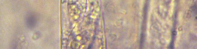
10 32 ACTA BOTANICA ISLANDICA NO. 15 Lasiobolus cuniculi Velen. MJR 28, 31(M), 35, 55, 60/06. H & E, NM 1, CFFI. Lasiobolus lasioboloides Marchal MJR 64/06. CFFI. *Pseudombrophila cervaria (W. Phillips) Brumm. (Fig. 5). Apothecia are relatively large, up to 2 mm diam., lilaceous at first becoming purplish brown with age, with a raised margin, which later becomes reflexed. Asci cylindrical x µm and KI. Spores smooth, ellipsoid, x 8-10 µm, obliquely uniseriate, with surrounding gel swelling to 3 µm wide in water. Paraphyses crowded, hyaline, cylindrical, with a septum µm below the tip, 2-3 µm diam., with little interparaphysal pigment in young material, but purplish brown in older material. The first record of Pseudombrophila species for Iceland was that of P. theioleuca Rolland (PFISTER & EYJÓLFS- DÓTTIR 2008), but no records are in NM 1 for the subarctic/subalpine or arctic/ alpine zones of Norway, Sweden and Finland, so it is interesting that P. cervaria occurred on 4 out of 11 collections of horse dung, and it was present in the field on MJR 46/06. MJR 30, 46(M), 54, 56(M)/06. *Saccobolus citrinus Boud. & Torrend (Fig. 6). Apothecia amber yellow, up to 700 µm diam., with a waxy appearance. Asci x µm. Spores clusters x 15 µm, with spores in a 4 x 2 arrangement (pattern I, BRUMMELEN 1967), asymmetrically fusoid, slightly truncate, smooth, x µm. Paraphyses with yellow green contents, slightly widened at the tip. This material was identified as S. citrinus, although it did not agree completely with BRUMMELEN S (1967) description, and the specimen had some characters nearer to those of S. truncatus, S. glaber and S. minimus as described by BRUMMELEN (1967). On balance, the current material fits S. citrinus better than the other possibilities. MJR 54(M)/06. *Thecotheus crustaceus (Starbäck) Aas & N. Lundq. (Fig. 7). Apothecia small, pale, hyaline, shortly pedicellate, x 550 µm high. Excipulum of hyaline globose-polygonal cells up to 15 µm diam. Asci cylindrical, projecting above hymenial surface when ripe, x 16 µm, KI + (blue). Spores obliquely uniseriate, smooth, ellipsoid, x µm, with an irregular, all-round gel swelling in water to 3 µm. AAS (1992) notes that T. crustaceus is mainly found on horse dung, and has been recorded from the four mainland Nordic countries. It appears to be widespread but not common, and AAS (1992) studied material from Europe, Azerbaijan, Canada, USA and Argentina, and noted records from New Zealand and Japan. I have a record from rabbit dung from Tenerife in the Canary Islands (MJR 62/02). These four Icelandic records are from a similar latitude north to those of the northernmost of the earlier records, that from the Yukon Territory at about 63.6 N (AAS 1992). MJR 30(M), 32, 51(M)-52(M)/06.


11 2011 RICHARDSON: COPROPHILOUS MYCOTA OF ICELAND 33 FIGURE 5. Pseudombrophila cervaria. Apothecia and asci. Scale bars: 2 mm (left); 25 µm (right). FIGURE 6. Saccobolus citrinus. Asci and ascospores. Scale bars: 50 µm. Thelebolales Coprotus sexdecimsporus (P. Crouan & H. Crouan) Kimbr. & Korf MJR 60/06. CFFI. Coprotus disculus Kimbr., Luck-Allen & Cain This is the same fungus as found in two 2002 collections (MJR 27, 46/02 RICHARDSON 2004), recorded as Coprotus sp. It has white discoid apothecia, up to 275 µm diam., slightly conical with a short stalk, with irregularly polygonal excipular cells µm diam. Asci ± cylindrical, x µm. Spores uni- to biseriate, ellipsoid, hyaline, x 6.5 µm. Paraphyses 1.5 µm wide, not enlarged at the apex but distinctly uncinate. Although the apothecia are
12 34 ACTA BOTANICA ISLANDICA NO. 15 consistently much smaller than described for C. disculus ( µm in KIMBROUGH et al. 1972; <700 µm in AAS 1983), all other features agree well with the original description. MJR 64/06. CFFI. Thelebolus microsporus (Berk. & Broome) Kimbr. Following DE HOOG et al. (2005), the records here include those of smaller apothecia with brownish excipular cells, 8-spored asci, but no capitate paraphyses with yellowish-green contents, as well as those of more typical apothecia, which are yellowish from the contents of the capitate paraphyses. MJR 31(M), 35, 41(M)-43, 47, 53, 55(M)-57, 59-61, 63-64/06. H & E, NM 1, CFFI. Thelebolus stercoreus Tode: Fr. Again, following DE HOOG et al. (2005), the records under this entry include those of all Thelebolus with asci with more than 8-spores. They include a range of morphological and ecological types that would previously been determined as T. nanus, T. polysporus, T. dubius, T. crustaceus etc., with from spores in single or multiple ascal apothecia. MJR 28, 31-32, 35-36, 53, 55-56, 60-61, 64/06, 26-29/08. CFFI. Helotiales *Unguiculella tityrii (Velen.) Huhtinen & Spooner (Fig. 8). Apothecia are white, in the Icelandic material µm diam. x µm high, with short erect and appressed hairs <20 x 2 µm on the upper part of the excipulum (not forming a fringe around the apothecial margin as in other material seen). Asci cylindrical, x 7 µm, with no operculum or apical structure. Spores biseriate, ellipsoid, hyaline 6.5 x 3.2 µm. Paraphyses not seen. This interesting fungus grows on the perithecia of coprophilous ascomycetes, especially Schizothecium species (HUHTINEN & SPOONER 2003). It is widely distributed in central and western Europe, Greenland and the USA and, as noted by HUHTINEN & SPOONER (2003), very variable in relation to hymenial and hair morphology. They observed that material from Greenland showed additional variability, beyond that seen in lowland populations, especially in respect of the hairs which, as in the Icelandic material, were scant or lacking, especially at the apothecial margin. MJR 33/06. Coniochaetales Coniochaeta leucoplaca (Berk. & Ravenel) Cain MJR 36(M), 38(M)-39(M), 40(M)-42, 63/06. H & E, CFFI. Coniochaeta ligniaria (Grev.) Massee MJR 29, 30, 32, 34, 39(M), 42-43, 44(M), 49(M)/06, 21/08. H & E, CFFI. Coniochaeta saccardoi (Marchal) Cain MJR 50(M)/06. H & E, CFFI.

 Cain MJR 28(M), 33(M)-34, 41-43, 49(M)-50, 53, 58(M),")

13 2011 RICHARDSON: COPROPHILOUS MYCOTA OF ICELAND 35 FIGURE 7. Thecotheus crustaceus. Apothecia. Scale bar: 500 µm. Coniochaeta scatigena (Berk. & Broome) Cain MJR 28(M), 33(M)-34, 41-43, 49(M)-50, 53, 58(M), 62-64/06. H & E, CFFI. Sordariales Arnium caballinum N. Lundq. MJR 31/06. H & E. CFFI. FIGURE 8. Unguiculella tityrii. Apothecia and asci. Scale bars: 500 µm (left); 50 µm (right).
14 36 ACTA BOTANICA ISLANDICA NO. 15 *Camptosphaeria citrinella (N. Lundq.) J.C. Krug & Jeng (Fig. 9) Perithecia semi-immersed, x µm, perithecial wall dull yellow, semi-translucent, neck and ostiole dark, opaque, surrounded by tapered tufts of yellow hyphae, up to 250 µm long x 80 µm wide at the base. Asci narrowly clavate, 230 x 20 µm, tapered below, and with a narrower cylindrical apical region ca. 16 µm wide and a small but distinct apical ring visible in most. Asci frequently with an irregularly globose, and sometimes indistinct, apical globule 6-8 µm diam. Paraphyses yellow, septate, 3 µm wide. Spores obliquely uniseriate, hyaline, smooth, clavate, broader towards the ascus tip, with globular contents, x 9-11 µm, and with a gelatinous appendage at each end ca. 16 µm long. No pigmented spores seen. MJR 21(M)/08. This would appear to be a rare fungus, with no records since its description from Sweden by LUNDQVIST (1972) and it is unlikely to be overlooked, as it is very recognisable with a crown of lemon yellow hyphae around the dark neck and ostiole, and a yellow, semitranslucent, perithecial wall, although this latter feature was not so obvious in situ as the perithecia were semi-immersed. LUNDQVIST (1972) considered Camptosphaeria as a subgenus of Cercophora, and described Cercophora citrinella as a new species. KRUG & JENG (1977) treated Camptosphaeria as distinct from Cercophora at the generic level, and provided a key to the four species they accepted. Apart from a collection they described as a new species (C. venezuelensis J.C. Krug & Jeng), however, they presented no additional records. Both C. clavispora (S.I. Ahmed & J.H. Mirza) J.C. Krug & Jeng (originally described as Podospora clavispora) have much larger spores (65-90 x µm) than those of C. citrinella and the type species C. sulphurea Fuckel, which are x 6-12 µm. All are coprophilous except C. sulphurea, which was described from stems of Peucedanum officinale. There appear to be no records of any of the four species subsequent to their original publication, although LUNDQVIST (1972) described C. citrinella on the basis of three collections from Sweden, one each from horse, hare and rabbit dung. The collection from Iceland agrees in most respects with that of LUNDQVIST (1972) for C. citrinella, apart from the extensive development of the yellow crown of hyphae around the neck, which was more developed than implied by the original description, and the apparent lack of any spore ornamentation. According to LUNDQVIST (1972) both C. sulphurea and C. citrinella have verruculose spores, but the ornamentation is illustrated as being very fine. This may be a variable feature the spores of C. venezuelensis are described as being smooth, and the generic description is that they are smooth or minutely granulate to verruculose. Chaetomium elatum Kunze: Fr. MJR 45(M)/06. H & E, but not on dung.


15 2011 RICHARDSON: COPROPHILOUS MYCOTA OF ICELAND 37 A B C D F E FIGURE 9. Camptosphaeria citrinella. A. Perithecia with crown of yellow hyphae; B. Perithecium; C. Peridium and base of neck; D. Ascus tips, with apical globule: E, F. Ascospores. Scale bars: 500µm (A, B), 25 µm (C-F).
16 38 ACTA BOTANICA ISLANDICA NO. 15 Podospora appendiculata (Auersw. ex Niessl) Niessl Particularly abundant on horse dung in the field. MJR 32-34, 40(M), 44(M), 62 (M)/06. H & E. Podospora decipiens (G. Winter ex Fuckel) Niessl MJR 28, 30-31, 34-35, 41-43, 47, 50, 51, 53, 55, 57, 60-61, 63-64/06. H & E, CFFI. *Podospora intestinacea N. Lundq. Described by LUNDQVIST (1972) with 10 finds on dung of horse (5), cattle (4) and sheep, these three records from horse and one from sheep dung are the first for Iceland, but it has also been found on sheep dung in the Faroe Islands (RICHARDSON 2005), and Doveri (2004) reported collections on pig, horse and cattle dung from Italy, and cited collections from Europe, South America and New Zealand. MJR 30, 34(M), 41, 50(M), 64(M) /06. *Podospora pilosa (Mouton) Cain Perithecia superficial, pyriform, with sparse setae at the neck up to 95 µm long. Asci round-headed (cf. Arnium) and tapered below with a long foot, x µm, with no obvious apical structure. Young spores distinctly clavate, 1-2 seriate, some almost limoniform, x µm with a pedicel 6 x 3 µm, not readily visible in mature spores, an apical germ pore and short apical appendages. MJR 30(M)/06. Schizothecium conicum (Fuckel) N. Lundq. MJR 30-31, 35(M), 41, 43, 46-47, 50-51, 53, 55(M)-58, 60-61, 64/06. H & E, CFFI. *Schizothecium tetrasporum (G. Winter) N. Lundq. There appears to be no record of S. tetrasporum from Iceland (HALLGRÍMSSON & EYJÓLFSDÓTTIR 2004; RICHARDSON 2004). It is highly probable that the finding of S. tetrasporum is associated with the presence of rabbits because from 1000 samples of the most frequently collected dung types I have obtained worldwide, S. tetrasporum occurs much more frequently on rabbit pellets than on any other dung type (Table 3). Rabbits (Oryctolagus cuniculi) are not native to Iceland, but occur in a few places as a result of naturalization from the release of unwanted domestic pets. MJR 21/08. Schizothecium vesticola (Berk. & Broome) N. Lundq. MJR 27-34, 41-42, 45, 51-53, 56, 58, 62-64/06, 26, 29/08. H & E, CFFI. *Sordaria alcina N. Lundq. MJR 50(M)/06. Sordaria baltica N. Lundq. MJR 30/06. H & E, CFFI Sordaria fimicola (Roberge ex Desm.) Ces. & De Not. MJR 30, 32, 46, 50-52, 54, 56(M)/06, 26/08. H & E, CFFI.
17 2011 RICHARDSON: COPROPHILOUS MYCOTA OF ICELAND 39 TABLE 3. Relative frequency of occurrence of Schizothecium tetrasporum on dung of different animals. Substrate (no. of samples) Sordaria minima Sacc. & Speg. See RICHARDSON (2004) for comment and observation on this fungus in Iceland. It was present on one sample of goose dung collected on Surtsey. MJR 27/08. H & E, CFFI. Xylariales *Hypocopra merdaria (Fr.: Fr.) J. Kickx f. Perithecia single in limited stromata, with the ostiole surrounded by a white mycelial weft of hyphae. Asci 350 x 30 µm. Spores uniseriate, with the pigmented cell x µm, germ slits µm long, and a distinct, small hyaline basal cell. HANSEN & KNUDSEN (2000) include H. merdaria ss. auct. as a synonym of H. lojkaeana (Rehm) J.C. Krug & N. Lundq., but this Icelandic material is not H. lojkaeana as described by them, i.e. with 1-celled spores x µm, with germ slits extending the full length. According to the treatment of KRUG & CAIN (1974) it is either H. festucacea, which is described and illustrated, or H. merdaria, which is not. DOVERI (2004) refers to the confusion between H. lojkaeana and H. merdaria, and describes both H. merdaria and H. festucacea, and it is clear from his descriptions that this material is H. merdaria, which is somewhat larger than H. festucacea in many respects. MJR 28 (M), 60(M)/06. Hypocopra parvula Griff. MJR 28(M), 63/06. CFFI. Hypocreales Selinia pulchra (G. Winter) Sacc. MJR 31(M)/06. CFFI. Occurrence of S. tetrasporum (%) Rabbit (n = 303) 74 Hare (n = 155) 25 Deer (n = 133) 20 Sheep/goat (n = 253) 10 Horse (n = 57) 4 Tetraonidae (n = 52) 2 Cattle (n = 47) 0
18 40 ACTA BOTANICA ISLANDICA NO. 15 Melanosporales Melanospora brevirostris (Fuckel) Höhn. Perithecia golden-brown, superficial, µm diam., with short beak, <65 µm, and ostiolar setae sparse, 65 x 5 µm, or absent. Asci clavate, x 22-29, 4-spored, spores ellipsoid-citriform, often slightly asymmetrical, x µm. This material agrees with the description of M. brevirostris, apart from 4- spored asci. The only consistently 4-spored Melanospora in CANNON & HAWKSWORTH (1982) is M. longisetosa which, as well as having long ostiolar setae, has discoid spores. M. zamiae is reported as having 4-8 spored asci, but is distinguished from M. brevirostris by its longer neck and smaller spores, although CANNON & HAWKSWORTH (1982) note that intermediates do occur. MJR 45(M)/06. CFFI. *Syspastospora parasitica (Tul.) P.F. Cannon & D. Hawksw. Perithecia were semi-immersed, pale golden-brown, µm diam., with a neck µm long x 65 µm wide at the base tapering to 40 µm diam. at the tip, brownish below, clearer above, composed of long, interlocking wavy cells ca. 20 µm long x 2-3 µm wide. From observation, although the neck is relatively broad, the inner diameter appears only wide enough to allow a single file of spores to be released. Asci are ephemeral, and none were seen. Spores dark grey, short barrel shaped, 4.5 x 4 µm, with a clear germ pore at each end. According to CANNON & HAWKSWORTH (1982) this is parasitic on moniliaceous hyphomycetes, so it is not strictly a coprophilous fungus. MJR 36(M)/06. Microascales *Viennotidia fimicola (Marchal) P.F. Cannon & D. Hawksw. An unmistakable fungus with long-necked, reddish brown, globose perithecia, with the immersed thecial body <250 µm diam., and a neck 1-3 mm long, terminating with cilia at the tip holding a droplet of liquid, with hyaline elliptical-allantoid spores x µm. It is a mycoparasite (WEBER & WEBSTER 1997), and is most frequently found on dung from rabbit and deer, being recorded from 21-22% of samples of those substrates, so its occurrence on sheep dung, where the incidence is about 10%, is relatively unusual. MJR 57 (M)/06. Pleosporales Delitschia perpusilla Speg. MJR 53(M)/06. CFFI. Pleospora herbarum (Pers.: Fr.) Rabenh. MJR 29/08. H & E. Sporormiella australis (Speg.) S.I. Ahmed & Cain MJR 29, 39(M)/06, 21/08. H & E. CFFI.
19 2011 RICHARDSON: COPROPHILOUS MYCOTA OF ICELAND 41 Sporormiella grandispora (Speg.) S.I. Ahmed & Cain This species is part of a morphological series that has asci with tapering stalks, and spores with end cells that are tapered to some extent. Individually they exhibit much variation in spore size, and in septa and germ slit orientation. BELL (2005) in particular has discussed the difficulties of drawing boundaries between the various taxa that have been described. SPEGAZZINI (1878) gives the spore size for Sp. grandispora as x µm, while AHMED & CAIN (1972), DOVERI (2004) and BELL (2005) all accept spores shorter than described by SPEGAZZINI (45-60 µm, µm and µm, respectively) in their understanding of Sp. grandispora. Three collections from 2006 with spores x µm (MJR 28/06), x (MJR 33/06) and x (MJR 42/06), and spore fragments from a fourth suggesting a size of 60 x , are considered to fit the description of Sp. grandispora as accepted by the above authors. Icelandic material from 2002 with a spore length ranging from (38.5-)45-67(-71) x (8-) µm over all collections was recorded as Sp. grandispora. The lower end of this range encompasses material that would be excluded from Sp. grandispora as understood above (see Fig. 4). In a re-examination of records from 2002, individual specimens had spore lengths varying, in an individual collection, over a range of <20 µm, e.g µm (MJR 47/06), supporting the acceptance by AHMED & CAIN (1972), DOVERI (2004) and BELL (2005) of spores shorter than described by SPEGAZZINI (1878) as being within the circumscription of Sp. grandispora, so eight collections from 2002 recorded as Sp. grandispora remain as initially reported (RICHARDSON 2004). Two others, however, after review, are now considered to be within the range of S. lageniformis. MJR 28(M), 33, 42, 44/06. CFFI. Amendment to records for 2002: delete records of Sp. grandispora on 42, 46/02; the material has been re-identified as Sp. lageniformis. *Sporormiella heptamera (Auersw.) S.I. Ahmed & Cain (Fig. 10). A distinctive but apparently uncommon species, with relatively large ±immersed pseudothecia <700 µm diam. Asci 320 x 35 µm, tapering to the base; ascospores 7-celled, x µm, with the third cell from the apex the widest; end cells hemi-ellipsoidal, as long as wide, the intercalary cells wider than long, deeply constricted at the septa; germ slits oblique to almost transverse. Newly recorded for Iceland, but known from North America (AHMED & CAIN 1972), France (RICHARDSON, unpublished), Italy (DOVERI 2004) and Spain (BARASSAS 1985), Chile (MUROI & UDAGAWA 1984) and Japan (FUROYA & UDAGAWA 1972). MJR 64(M)/06. Sporormiella intermedia (Auersw.) S.I. Ahmed & Cain MJR 28, 31, 33, 42, 45, 53, 58, 60/06, 21/08. H & E, CFFI.
 S.I. Ahmed & Cain The most frequent member of the stipitate ascus, tapered-spore complex in the 2002 and 2006 collections comprised 17 records of species with spores in an overlapping range from 27.")


20 42 ACTA BOTANICA ISLANDICA NO. 15 FIGURE 10. Sporormiella heptamera. Ascospores. Scale bars: 50 µm. Sporormiella lageniformis (Fuckel) S.I. Ahmed & Cain The most frequent member of the stipitate ascus, tapered-spore complex in the 2002 and 2006 collections comprised 17 records of species with spores in an overlapping range from µm x µm. Given the amount of variation observed within even a single pseudothecium, it is considered that all these collections probably belong to a single species which, depending on the particular parameter selected in the various treatments of the genus spore size, relative size of cells, germ slit orientation and septa orientation can be identified as Sp. leporina, Sp. lageniformis, Sp. isomera or Sp. dubia. In any one collection germs slits might be parallel or oblique to the long axis, and septa transverse or oblique. The original descriptions of the spore size of these species were: - Sp. leporina x 4-5 µm (NIESSL 1878) Sp. lageniformis 40 x 8 µm (FUCKEL 1869), Sp. isomera x µm (AHMED & CAIN 1972), and Sp. dubia x 8-9 µm (AHMED & CAIN 1972). The 2002 collections, recorded as Sp. leporina, all with spores in the range x µm, were slightly longer and wider than reported by AHMED & CAIN (30-37 X µm, 1972), and DOVERI ( x µm, 2004), while in BELL (2005) they key to Sp. tetramera, another AHMED & CAIN (1972) species, with spores x 6-8 µm. The 2006 collections cover a wider range of spore size, x 7-10 µm, but the narrowness of the spores, all <10 µm, preclude the largest of these collections being determined as Sp. grandispora. I believe these 17 records are all one species that, for the time being, I identify as Sp. lageniformis. One of the main distinctions between Sp. leporina and Sp. lageniformis is that the spores of the latter are described and illustrated by both AHMED & CAIN (1972) and DOVERI (2004) as having distinctly oblique septa, while Sp. leporina has is described as having spores with transverse septa; spores with both oblique and transverse septa have been

-64/06, 21/08.")
 S.I. Ahmed & Cain (Fig. 11).")
.")
 distinguish between Sp.")
21 2011 RICHARDSON: COPROPHILOUS MYCOTA OF ICELAND 43 observed in much of the material examined, but not consistently, and the majority of material has spores which are notably longer than described for the type of Sp. leporina, which I have not, however, examined. It is also worth noting here that DOVERI (2004) considers Sp. isomera to be possibly synonymous with Sp. leporina. MJR 28, 30, 33, 35, 41-42, 49, 51, 53, 62(M)-64/06, 21/08. CFFI. Additional records for 2002: 42, 46/02, to replace the records of Sp. grandispora on those samples. *Sporormiella longispora (Cain) S.I. Ahmed & Cain (Fig. 11). A striking and recognisable species by virtue of its distinctive, relatively elongated, deeply constricted at the septa, 4-celled spores, in this material x µm (Q = 6.5-7). The terminal cells are gently tapered to a rounded end, while the intercalary cells are straight-sided, almost rectangular. AHMED & CAIN (1972) distinguish between Sp. longispora, with spores (80-)90-100(-108) x µm, and Sp. longisporopsis with relatively broader spores (75-)80-100(-104) FIGURE 11. Sporormiella longispora. Ascospores. Scale bars: 50 µm.
22 44 ACTA BOTANICA ISLANDICA NO. 15 x µm. They illustrate Sp. longispora spores with the almost rectangular intercalary cells described above, while Sp. longisporopsis spores are illustrated with more rounded intercalary cells and less tapered terminal cells. BELL (2005) illustrates straight-sided spores, at the lower end of the size range x µm as Sp. longisporopsis, and DOVERI (2004) illustrates spores with rounded cells as Sp. longisporopsis, x µm. Although wider than described for Sp. longispora, the spore shape of the Icelandic material is otherwise much more in agreement with the description of Sp. longispora, as originally illustrated by CAIN (1934) than of Sp. longisporopsis, and the two species may well be synonymous. I have seen four other collections, three with broader spores, originally determined, therefore, as Sp. longisporopsis and one as Sp. longispora. Doveri notes that Sp. longisporopsis is rare, with three records from Italy and one from Kenya (KHAN & CAIN 1979). The type of Sp. longisporopsis is from Canada, with additional material from Canada, USA, Mexico and Argentina (AHMED & CAIN 1972), while the type of Sp. longispora is also from Canada, with additional material from Canada and USA (AHMED & CAIN 1972), and Kenya (KHAN & CAIN 1979). MJR 31(M)/06. Other material examined: FRANCE. Brown hare (Lepus capensis) dung: Domaine de Garenaud, Montazels, Aude (42.95 N; 2.23 E), (MJR 1/05 (E)); SPAIN (CANARY ISLANDS). Rabbit (Oryctolagus cuniculus) dung: Tafada, Anaga, Tenerife (28.58 N; W), (MJR 62/02 (E)); USA. Mule deer (Odocoileus hemionus) dung: Bryce Canyon NP, Utah (37.60 N; W), (MJR 65/01). Jack rabbit (black-tailed, Lepus californicus?) dung: Bryce Canyon NP, Utah (37.60 N; W), (MJR 66/01). Sporormiella megalospora (Auersw.) S.I. Ahmed & Cain (Fig. 12). AUERSWALD (1868) gives the spore size for Sp. megalospora as 80 x µm, while AHMED & CAIN (1972), BELL (2005) and DOVERI (2004) are in agreement, describing spores x µm, x µm and x µm, respectively. The spores of the material from Tungufellsdalur (31/06) were deeply constricted at the septa, x µm, and agree with the description of Sp. megalospora as understood by the above authors. The five Icelandic records of Sp. megalospora from 2002 have been re-examined. Samples from MJR 24, 25, 32 and 35/02, all with similar-sized spores ranging from x µm form a clear cluster (Fig. 4), and are considered to be correctly identified as Sp. megalospora. Collection MJR 33/02, with smaller, and narrower, spores, x 14.5 µm, is much closer to the range accepted for Sp. grandispora, which was also recorded from that sample, and the record of Sp. megalospora for that sample should be deleted. MJR 31(M)/06. CFFI. Sporormiella minima (Auersw.) S.I. Ahmed & Cain MJR 36, 39/06. H & E.
 S.I. Ahmed & Cain (Fig. 13).")


 original description (40 x 5-6 µm), and AHMED & CAIN s (1972) values ((37-)40-48(-50) x 7-8 µm) for Sp.")
, 50 µm (right). *Sporormiella octonalis S.I. Ahmed & Cain (Fig. 14).")
23 2011 RICHARDSON: COPROPHILOUS MYCOTA OF ICELAND 45 FIGURE 12. Sporormiella megalospora. Ascospores. Scale bar: 50 µm. Sporormiella octomera (Auersw.) S.I. Ahmed & Cain (Fig. 13). This species is particularly prevalent on ptarmigan droppings in Iceland, occurring on 3 of 5 of the 2002 samples and 4 of the 2006 collections, and not on any of the other dung types. RICHARDSON (2004) commented on the range of spore size observed in the three 2002 Icelandic collections, and the overlap with Sp. schadospora. They had spores x 6-9 µm. The spore size of the four more recent collections was less variable, all within the range (40)41-48(50) x 5-7 µm, which agrees well with AUERSWALD s (1868) original description (40 x 5-6 µm), and AHMED & CAIN s (1972) values ((37-)40-48(-50) x 7-8 µm) for Sp. octomera, although slightly narrower. MJR 37(M), 38(M), 39(M), 40(M)/06. CFFI. FIGURE 13. Sporormiella octomera. Asci and ascospores. Scale bars: 25 µm (left), 50 µm (right). *Sporormiella octonalis S.I. Ahmed & Cain (Fig. 14). Pseudothecia with dark neck and paler, more translucent body. Spores 8- celled, not deeply constricted at the septa, long ellipsoid, x µm, with intercalary cells wider than long, and the third cell from apex the widest, but not markedly so, end cells hemispherical. This is an uncommon species, reported from Canada and USA by AHMED & CAIN (1972), from Italy by FIGURE 14. Sporormiella octonalis. Ascospores. Scale bars: 50 µm.
24 46 ACTA BOTANICA ISLANDICA NO. 15 DOVERI (2004), and I have records from Scotland and France (Corsica). MJR 53 (M)/06. Sporormiella subtilis S.I. Ahmed & Cain This is one of the smaller spored species with tapered spores and tapering asci and, as with the other members of the group, it is difficult to define its boundaries. Specimens with a mean range of spore length <30 µm have been determined as Sp. subtilis, and the range x µm encompasses AHMED & CAIN s (1972) type description, BELL s (2005) understanding of the species, the spore sizes observed in the three samples from 2002 recorded as Sp. subtilis, and the three additional records from the current collection. It also includes the spore size given by NIESSL (1878) for Sp. leporina, but AHMED & CAIN (1972) and DOVERI (2004) both consider that species to have larger spores (30-37 x µm). MJR 31, 35, 41/06. CFFI. Additional record for 2002: 34/02, to replace the record of Sp. leporina. Trichodelitschia minuta (Fuckel) N. Lundq. MJR 31, 33, 58(M)/06. H & E, CFFI (as T. bisporula). Trichodelitschia munkii N. Lundq. MJR 33/06. H & E, CFFI. Basidiomycota Coprinellus heptemerus (M. Lange & A.H. Sm.) Vilgalys, Hopple & Jacq. Johnson (Coprinus heptemerus M. Lange & A.H. Sm.) MJR 28, 31, 35(M), 41, 43, 47, 53, 55(M), 57-58(M), 60-61/06. CFFI. Coprinellus pellucidus (P. Karst.) Redhead, Vilgalys & Moncalvo (Coprinus pellucidus P. Karst.) MJR 30, 34-35, 43(M), 46, 47, 50(M)-52, 57/06. CFFI. Coprinopsis cordisporus (T. Gibbs) Watling & M.J. Richardson (Coprinus cordisporus T. Gibbs) MJR 28, 30(M), 34, 46, 51-52(M), 56, 60, 62-63(M), 64/06. H & E, NM 2, CFFI. Coprinopsis ephemeroides (DC.: Fr.) Watling & M.J. Richardson (Coprinus ephemeroides (DC.: Fr.) Fr.) MJR 63(M)/06. H & E, NM 2. Also present in the field on horse dung at Hamrar, Grundarfjörður, Snæfellsnes, where sample 57/06 was collected. Coprinopsis nivea (Pers.: Fr.) Redhead, Vilgalys & Moncalvo (Coprinus niveus (Pers.: Fr.) Fr.) MJR 43(M), 47(M), 64(M)/06. H & E, NM 2. CFFI.
25 2011 RICHARDSON: COPROPHILOUS MYCOTA OF ICELAND 47 *Coprinopsis radiata (Bolton: Fr.) Redhead, Vilgalys & Moncalvo (Coprinus radiatus (Bolton: Fr.) Gray ) MJR 35(M), 47(M), 52, 56(M)/06. Coprinopsis stercorea (Fr.) Redhead, Vilgalys & Moncalvo (Coprinus stercoreus Fr.) MJR 43, 51, 57, 63-64/06. NM 2, CFFI. *Laetisaria fuciformis (McAlpine) Burds. Fruiting indeterminate over the surface of the dung, hymenium pale, whitebuff. Clamp connections not seen. Basidia 4-spored, cylindrical, x 6-7 µm. Spores hyaline, ellipsoid amygdaliform, non-amyloid, 9.5 x µm. There are no records of L. fuciformis from Iceland, and it is not known to be coprophilous, but its occurrence on goose dung is, perhaps, not suprising, since it is well known as a grass pathogen, the cause of red thread disease. MJR 30(M)/08. Det. K.-H. Larsson, material in GB. Panaeolus papilionaceus (Bull.: Fr.) Quél. MJR 63(M)/06. H & E, NM 2. Panaeolus semiovatus (Sowerby: Fr.) var. phalaenarum (Fr.) Ew. Gerhardt (P. antillarum (Fr.) Dennis) MJR 56(M)/06. H & E, NM 2, CFFI. Parasola misera (P. Karst.) Redhead, Vilgalys & Hopple (Coprinus miser P. Karst.) MJR 28, 30(M), 34-35, 43(M), 46-47, 51-53, 63(M)/06. H & E, NM 2, CFFI. Psilocybe coprophila (Bull.: Fr.) P. Kumm. MJR 34(M), 41, 44, 49(M), 62(M), 63(M), 64(M)/06. NM 2. Anamorphic Fungi Truncatella angustata (Pers.: Link) S. Hughes Described as a caulicolous, corticolous, foliicolous and fructicolous species (NAG RAJ 1993), but not known to be coprophilous, so its occurrence on a ptarmigan pellet is of interest, although perhaps not surprising, since they are composed of partially digested pieces of vegetation. MJR 36(M)/06. H & E. Mycetozoa, Myxogastria, Trichiales Arcyria cinerea (Bull.) Pers. MJR 62(M)/06. H & E.
Coprophilous fungi from Iceland
 ACTA BOT. ISL. 14: 77-102, 2004 Coprophilous fungi from Iceland Michael J. Richardson 165 Braid Road, Edinburgh EH10 6JE U.K. ABSTRACT: Eighty-one species of coprophilous fungi were recorded from 32 herbivore
Coprophilous Fungi from Brazil
 283 Vol. 44, N. 3 : pp. 283 289, September, 2001 ISSN 1516-8913 Printed in Brazil BRAZILIAN ARCHIVES OF BIOLOGY AND TECHNOLOGY AN INTERNATIONAL JOURNAL Coprophilous Fungi from Brazil Michael J. Richardson
Coprophilous fungi from the Faroe Islands
 67 Coprophilous fungi from the Faroe Islands Michael J. Richardson 165 Braid Road, Edinburgh EH10 6JE U.K. Telephone 0131 447 8165, Email: mjrichardson@clara.net Úrtak 59 sløg av taðfrekum soppum eru skrásett
Two remarkable xylariaceous ascomycetes associated with elephant dung
 Two remarkable xylariaceous ascomycetes associated with elephant dung Deepna Latha KP and Manimohan P* Department of Botany, University of Calicut, Kerala, 673 635, India Deepna Latha KP, Manimohan P 2012
Phaeocalicium populneum
 Phaeocalicium populneum markpowell222@btinternet.com After conducting a survey of the RHS garden at Wisley on 18 th August 2018, Fay Newbery kindly showed me the colony of P. populneum at Esher Common.
Mycosphere. Doveri F*
 Additions to Fungi Fimicoli Italici : An update on the occurrence of coprophilous Basidiomycetes and Ascomycetes in Italy with new records and descriptions. Doveri F* Via Baciocchi 9, I 576 Livorno. Doveri
Coprophilous Mycoflora on Different Dung Types in Southern Desert of Iraq
 Coprophilous Mycoflora on Different Dung Types in Southern Desert of Iraq S. K. ABDULLAH Department of Biology College of Science University of Basrah, Basrah, Iraq Abstract. Forty species of coprophilous
Overview. Revised through 30 June Initial Groups ("naked-eye" characters)
") Overview Revised through 30 June 2010 Initial Groups ("naked-eye" characters) Plants essentially leafless, consisting of strongly inclined, highly asymmetric capsules on a stout papillose seta; the "bug-on-a-stick"
Overview Revised through 30 June 2010 Initial Groups ("naked-eye" characters) Plants essentially leafless, consisting of strongly inclined, highly asymmetric capsules on a stout papillose seta; the "bug-on-a-stick"
PERSOONIA. Noteson cup-fungi 2. J. van Brummelen. Rijksherbarium, Leiden. groups, superficial, sessile on a narrow base,
 PERSOONIA Published by the Rijksherbarium, Leiden Volume 12, Part 3, pp. 327-334 (1984) Noteson cup-fungi2 J. van Brummelen Rijksherbarium, Leiden The coprophilous Lasiobolus monascus Kimbr. is described
Coprophily (from the Greek kópros = dung and philía = love, love for, consequently
 ISSN 1450-7153 UDK 582.282.16(450) AN UPDATED KEY TO COPROPHILOUS PEZIZALES AND THELEBOLALES IN ITALY Francesco Doveri via Baciocchi 9, 57126 Livorno, Italy f.doveri@sysnet.it Abstract The author updates
Microthyriales of Tierra del Fuego I: The Genus Parasterinella SPEGAZZINI
 Verlag Ferdinand Berger & Söhne Ges.m.b.H., Horn, Austria, download unter www.biologiezentrum. Sydowia, Annales Mycologici Ser. II. Vol. 38: 1-5 (1985) Verlag Ferdinand Berger & Söhne Gesellschaft m.b.h.,
Short guide to some common mycological terms
 Short guide to some common mycological terms Thomas Læssøe & Jens H. Petersen Macro-morphology English (latinised English) Bulb-like (= bulbous) used for swollen stem bases, can be rimmed (= marginate).
A handful of primary features are useful for distinguishing water primrose (Ludwigia) from other plants. Understand what to look for, such as leaf
 from other plants. Understand what to look for, such as leaf") A handful of primary features are useful for distinguishing water primrose (Ludwigia) from other plants. Understand what to look for, such as leaf arrangement and number of petals. Pairing morphological
A handful of primary features are useful for distinguishing water primrose (Ludwigia) from other plants. Understand what to look for, such as leaf arrangement and number of petals. Pairing morphological
Overview of Ascomycota
 Overview of Ascomycota Ascomycota ~ 6,350 Genera ~ 64,200 Species compared to Basidiomycota ~ 1,350 Genera ~31,500 Species Between 17,000-20,000 species (~ 30-40%) of Ascomycota are lichenized Many species
Kingdom Fungi. Learning Objectives. Introduction. Activity1: Zygomycota. Revised Fall 2017
 Kingdom Fungi Revised Fall 2017 ** You will require your text book Biological Science during this lab ** Learning Objectives Building on the learning objectives from your lab syllabus, you will be expected
A new variety of coprophilous Schizothecium from France
 A new variety of coprophilous Schizothecium from France DOVERI F.* & COUÉ B.** * via Baciocchi 9, 57126 Livorno-Italy. E.mail: f.doveri@sysnet.it ** 24 rue des Fours, Coudré, F-79190 Clussais la Pommeraie,
Fungi from palms. XXXIX. Asymmetricospora sp. nov. (Melanommataceae)
") Fungi from palms. XXXIX. Asymmetricospora sp. nov. (Melanommataceae) gen. et Jane Fröhlich 1 & Kevin D. Hyde 2 1 Manaaki Whenua, Landcare Research New Zealand Ltd, Private Bag 92170, Auckland, New Zealand
Fungi from palms. XXXIX. Asymmetricospora sp. nov. (Melanommataceae) gen. et Jane Fröhlich 1 & Kevin D. Hyde 2 1 Manaaki Whenua, Landcare Research New Zealand Ltd, Private Bag 92170, Auckland, New Zealand
MARINE FUNGI IN giscayne BAY, FLORIDA 1
 MARINE FUNGI IN giscayne BAY, FLORIDA 1 SAMUEL P. MEYERS The Marine Laboratory, University of Miami ABSTRACT A recent collection in Biscayne Bay, Florida, of various marine fungal forms, several previously
STUDIES OF THE BALLISTICS OF ASCOSPORES
 STUDIES OF THE BALLISTICS OF ASCOSPORES BY D. G. A. WALKEY* AND R. HARVEY Department of Botany, University College, Cardiff {Received 2^ June i()6^) SUMMARY The ballistics of horizontal spore discharge
SPORE-FORMS IN SPOROPHORES OF GANODERMA LUCIDUM (LEYSS.) KARST.
 KARST.") SPORE-FORMS IN SPOROPHORES OF GANODERMA LUCIDUM (LEYSS.) KARST. BY SACHINDRANATH BANERJEE AND ANJALI SARKAR (Department of Botany, University o[ Calcutta) Received September 12, 1958 (Communicated by Dr.
SPORE-FORMS IN SPOROPHORES OF GANODERMA LUCIDUM (LEYSS.) KARST. BY SACHINDRANATH BANERJEE AND ANJALI SARKAR (Department of Botany, University o[ Calcutta) Received September 12, 1958 (Communicated by Dr.
Pyrrolizidine Alkaloids
 Pyrrolizidine Alkaloids Pyrrolizidine alkaloids are the most common cause of liver damage. Found in numerous plant species, pyrrolizidine alkaloids are most toxic for pigs, then poultry, cattle, horses,
A SYNOPTIC GALERINA KEY
 Galerina. This synoptic key by David Savage, 2008, is intended as an alternative approach to fitting Galerina specimens to the descriptions in British Fungus Flora Vol. 7 (Watling & Gregory). Most of the
Plant Crib VERONICA. 1. Veronica serpyllifolia
 VERONICA 1. Veronica serpyllifolia Illustrations reproduced, with permission, from M. McC. Webster (1978). Flora of Moray, Nairn & East Inverness. Aberdeen. Subsp. humifusa (Dicks.) Syme Subsp. serpyllifolia
Dung-inhabiting ascomycetes from the Ukrainian Carpathians
 CZECH MYCOLOGY 70(2): 145 167, NOVEMBER 20, 2018 (ONLINE VERSION, ISSN 1805-1421) Dung-inhabiting ascomycetes from the Ukrainian Carpathians YULIA I. LYTVYNENKO 1,VERONIKA V. DZHAGAN 2,IRYNA V. TOPCHII
Simplified ascus types
 Simplified ascus types unitunicate bitunicate operculum But numerous variants are recognized pore Ascospores +/- pigmentation (fungal melanin) aseptate, uniseptate or multiseptate +/- appendages +/- sheaths
Investigation 7: Cell Division Part B: Meiosis and Crossing Over
 Background Investigation 7: Cell Division Part B: Meiosis and Crossing Over Ascomycota are a diverse group of fungi including the familiar single-celled baker s yeast, the complex morel mushroom, and the
Hyphae excluding ascogenous hyphae/ascus
 SEPTUM / PORE CAP Characters and Character States Hyphae excluding ascogenous hyphae/ascus Character location: 1N, haploid mycelium; NN, dikaryotic mycelium; 2N, diploid mycelium; XN, mycelium of unknown
Kingdom Fungi. Announcements
 Kingdom Fungi Announcements Friday lab: Fungi & Lichen Bring a Lichen to ID! Do prelab Quiz #4 Friday Study Prokaryotes & Protists Mushroom Fest extra credit due Fri Email me or bring to lab Endosymbiosis
Peziza melaleucoides Seaver. I noted that the species could not be placed easily in existing genera of the Pezizales known to me.
 614 MYCOLOGIA, VOL. 72, 1980 2. Berry, S. S. Z., and G. G. N. N. Davis. 1957. 1957. Formation of oospores of oospores by by Peronospora destructor and their possible relation to epiphytology. PI. Dis.
Literature. Morphology. Morphology of the mycorrhizal system. Morphology of the unramified ends
 Literature references Müller WR, Rauscher T, Agerer R, Chevalier G (1996) Tuber aestivum Vitt. + Corylus avellana L.Descr Ectomyc 1: 167-172. Rauscher T, Müller WR, Chevalier G, Agerer R (1996) Tuber aestivum.
Basidiomycota. Botany 201 Laboratory Spring 2007
 Botany 201 Laboratory Spring 2007 Basidiomycota As was the case of the Ascomycota, this phylum represents a very variable group of fungi. This only characteristic that is common to all species in this
A New Locality of Fossombronia mylioides (Fossombroniaceae, Marchantiophyta)
") Bull. Natl. Mus. Nat. Sci., Ser. B, 42(1), pp. 19 23, February 22, 2016 A New Locality of Fossombronia mylioides (Fossombroniaceae, Marchantiophyta) Masanobu Higuchi Department of Botany, National Museum
Bull. Natl. Mus. Nat. Sci., Ser. B, 42(1), pp. 19 23, February 22, 2016 A New Locality of Fossombronia mylioides (Fossombroniaceae, Marchantiophyta) Masanobu Higuchi Department of Botany, National Museum
21-2 Classification of Fungi Slide 2 of 44
 2 of 44 Fungi are classified according to their structure and method of reproduction. The four main groups of fungi are: Common molds (Zygomycota) Sac fungi (Ascomycota) Club fungi (Basidiomycota) Imperfect
[ 449 ] Printed in Great Britain AUSTRALIAN DISCOMYCETES ON DEAD LOGS AND BRANCHES. By G. BEATON. Hildon, Victoria
![[ 449 ] Printed in Great Britain AUSTRALIAN DISCOMYCETES ON DEAD LOGS AND BRANCHES. By G. BEATON. Hildon, Victoria](/thumbs/83/88580267.jpg "[ 449 ] Printed in Great Britain AUSTRALIAN DISCOMYCETES ON DEAD LOGS AND BRANCHES. By G. BEATON. Hildon, Victoria") [ 449 ] Trans. Br. mycol. Soc. 67 (3) 449-454 (1976) Printed in Great Britain AUSTRALIAN DISCOMYCETES ON DEAD LOGS AND BRANCHES By G. BEATON Hildon, Victoria AND GRETNA WESTE School of Botany, University
[ 449 ] Trans. Br. mycol. Soc. 67 (3) 449-454 (1976) Printed in Great Britain AUSTRALIAN DISCOMYCETES ON DEAD LOGS AND BRANCHES By G. BEATON Hildon, Victoria AND GRETNA WESTE School of Botany, University
Fungi Coloring Worksheet
 Fungi Coloring Worksheet The basic structural features of fungi are not cells but hyphae. Hyphae are microscopic branching filaments filled with cytoplasm and nuclei. Each thread consists of a tube formed
SPECIES FACT SHEET. Taxonomic Note: None.
 SPECIES FACT SHEET Common Name: Granite moss, Lantern moss Scientific Name: Andreaea nivalis Hook. Recent synonyms: Andreaea baileyi Holz. A. macounii Kindb. in Mac. Division: Bryophyta Class: Bryopsida
DONALD H. PFISTER. Farlow Reference Library and Herbarium of Cryptogamic Botany, Harvard University, Cambridge, Massachusetts 02138
 1253 Collins, O. R. 1976. Heterothallism and homothallism: a study of 27 isolates of Didymium iridis, a true slime mold. Amer. J. Bot. 63: 138-143. --, and J. Clark. 1968. Genetics of plasmodial compatibility
Working Group on Medicinal and Aromatic Plants November 2011
 Working Group on Medicinal and Aromatic Plants November 2011 Highly discriminating descriptors in this descriptor list are marked with an asterisk [ ]. Characterization should preferably be done during
PERSOONIA. Part 3, pp (1962) of Fimaria. J. van Brummelen. and F. theioleuca (Roll.) Brumm. Ascobolus. FIMARIA Vel.
 of Fimaria. J. van Brummelen. and F. theioleuca (Roll.) Brumm. Ascobolus. FIMARIA Vel.") In Lectotype: PERSOONIA Published by the Rijksherbarium, Leiden Volume 2, Part 3, pp. 321-330 (1962) Studies on DiscomycetesII. On four species of Fimaria J. van Brummelen Rijks her barium, Leiden (With
In Lectotype: PERSOONIA Published by the Rijksherbarium, Leiden Volume 2, Part 3, pp. 321-330 (1962) Studies on DiscomycetesII. On four species of Fimaria J. van Brummelen Rijks her barium, Leiden (With
Groups of Fungi. Section 2
 Groups of Fungi Section 2 Chytrid Fungi Key Idea: The chytrids are a group of aquatic fungi that provide clues about the evolution of fungi. Chytrid Fungi Chytrids were once classified with protists because
Tissue specificity by fungi endophytic in Ulex europaeus
 Tissue specificity by fungi endophytic in Ulex europaeus P. J. FISHER Department of Biological Sciences, Hatherly Laboratories, University of Exeter, Exeter, UK O. PETRINI Mikrobiologisches Institut, ETHZentrum,
(Pl. VI Fig. 36) Ramaria maculatipes sp. nov.
 Ramaria maculatipes sp. nov.") 103 Ramaria maculatipes sp. nov. (Pl. VI Fig. 36) Basidiocarpia terrestria 10 cm alta 6 cm crassa, stipite simplici 2.0-4.0 x 1.5-2.0 cm, e basi usque ad septuplo sursum ramificantia, apicibus polynodulosis,
103 Ramaria maculatipes sp. nov. (Pl. VI Fig. 36) Basidiocarpia terrestria 10 cm alta 6 cm crassa, stipite simplici 2.0-4.0 x 1.5-2.0 cm, e basi usque ad septuplo sursum ramificantia, apicibus polynodulosis,
FIMARIA HISPANICA (ASCOMYCETES) SP. NOV.
 SP. NOV.") Anal. Inst Bot. Cavanilles 34 (2): 387-392 (1978) FIMARIA HISPANICA (ASCOMYCETES) SP. NOV. by M. DE LA TORRE AND F. D. CALONGE Abstract. Fimaria hispanica Torre & Calonge is described as new taxon, being
Anal. Inst Bot. Cavanilles 34 (2): 387-392 (1978) FIMARIA HISPANICA (ASCOMYCETES) SP. NOV. by M. DE LA TORRE AND F. D. CALONGE Abstract. Fimaria hispanica Torre & Calonge is described as new taxon, being
Kingdom Fungi. 1. Student will be able to describe the characteristic features in the kingdom Fungi.
 Kingdom Fungi Molds, Sac Fungi, Mushrooms, and Lichens Essential Question(s): What makes fungi have their own kingdom? Objectives: 1. Student will be able to describe the characteristic features in the
A new species of Bertiella (Melanommataceae) from Brazil and a key to accepted species
 from Brazil and a key to accepted species") Mycosphere 8 (4): 392 396 (2017) www.mycosphere.org ISSN 2077 7019 Article Doi 10.5943/mycosphere/8/4/1 Copyright Guizhou Academy of Agricultural Sciences A new species of Bertiella (Melanommataceae) from
Mycosphere 8 (4): 392 396 (2017) www.mycosphere.org ISSN 2077 7019 Article Doi 10.5943/mycosphere/8/4/1 Copyright Guizhou Academy of Agricultural Sciences A new species of Bertiella (Melanommataceae) from
Domain: Eukarya Kingdom: FUNGI
 Domain: Eukarya Kingdom: FUNGI Fungi are eukaryotic heterotrophs that have cell walls. They are part of the nature s recycling system. They break down organic compounds. Fungi are used in wine, beer, cheese,
Fungi are absorptive heterotrophs that secrete digestive enzymes and are major decomposers of dead organic material
 Fungi 1 2002 Prentice Hall, Inc The scarlet hood (Hygrocybe coccinea) Fungi are absorptive heterotrophs that secrete digestive enzymes and are major decomposers of dead organic material 2 Animals 3 Myxozoa
Scanning ultrastructural ontogeny of eugymnohymenial apothecia in the operculate Discomycetes Ascodesrnis nigricans and A.
 Scanning ultrastructural ontogeny of eugymnohymenial apothecia in the operculate Discomycetes Ascodesrnis nigricans and A. sphaerospora K. L. O'DONNELL, G. R. HOOPER, AND W. G. FIELDS^ Dep(~rtmc>nt of
Understanding Projections
 GEOGRAPHY SKILLS 1 Understanding Projections The earth is a sphere and is best shown as a globe. For books and posters, though, the earth has to be represented as a flat object. To do this, mapmakers create
Workshop on Kingdom Fungi
 Workshop on Kingdom Fungi by Dana Krempels Introduction Kingdom Fungi is an ostensibly monophyletic assemblage of ecologically important organisms that not only perform the vital function of decomposition,
Mycorrhiza Fungus + Plant Host (Root)
") Mycorrhiza Fungus + Plant Host (Root) Two fungi commonly Use in ectomycorrhiza Research. Laccaria bicolor Pisolithus tinctorius Flowering Plants and mycorrhizal fungi http://mycorrhizas.info/evol.html#intro
Mycorrhiza Fungus + Plant Host (Root) Two fungi commonly Use in ectomycorrhiza Research. Laccaria bicolor Pisolithus tinctorius Flowering Plants and mycorrhizal fungi http://mycorrhizas.info/evol.html#intro
STRUCTURE, CHARACTERISTICS AND REPRODUCTION OF FUNGI I
 STRUCTURE, CHARACTERISTICS AND REPRODUCTION OF FUNGI I Charles Okolie, PhD Room 311 (on level 4), First College Building, Landmark University, Omu-Aran, Kwara State, Nigeria. Internal Tel. extension: 4048
Sordaria fimicola (Ascomycota, Sordariales) on Acer palmatum
 on Acer palmatum") FOLIA OECOLOGICA vol. 42, no. 1 (2015). ISSN 1336-5266 Short communication Sordaria fimicola (Ascomycota, Sordariales) on Acer palmatum Helena Ivanová Institute of Forest Ecology of the Slovak Academy
FOLIA OECOLOGICA vol. 42, no. 1 (2015). ISSN 1336-5266 Short communication Sordaria fimicola (Ascomycota, Sordariales) on Acer palmatum Helena Ivanová Institute of Forest Ecology of the Slovak Academy
Evolution Canyons occur naturally as a valley between two mountains where one slope, the
 Meiosis and Genetic Diversity in Sordaria fimicola Haley DeMartin Lab Section 001 Due October 27, 2014 Introduction Several Evolution Canyons exist in Lower Nahal Oren, Mount Carmel, Israel. These Evolution
Phylogenetic study of Diploschistes (lichen-forming Ascomycota: Ostropales: Graphidaceae), based on morphological, chemical, and molecular data
, based on morphological, chemical, and molecular data") Vol. 62 (2) April 2013 International Journal of Taxonomy, Phylogeny and Evolution Electronic Supplement to Phylogenetic study of Diploschistes (lichen-forming Ascomycota: Ostropales: Graphidaceae), based
Vol. 62 (2) April 2013 International Journal of Taxonomy, Phylogeny and Evolution Electronic Supplement to Phylogenetic study of Diploschistes (lichen-forming Ascomycota: Ostropales: Graphidaceae), based
A new species of Psathyrella (Psathyrellaceae, Agaricales) collected on dung from Punjab, India
 collected on dung from Punjab, India") Journal on New Biological Reports 2(3): 275-280 (2013) ISSN 2319 1104 (Online) A new species of Psathyrella (Psathyrellaceae, Agaricales) collected on dung from Punjab, India Amandeep Kaur 1*, NS Atri
Journal on New Biological Reports 2(3): 275-280 (2013) ISSN 2319 1104 (Online) A new species of Psathyrella (Psathyrellaceae, Agaricales) collected on dung from Punjab, India Amandeep Kaur 1*, NS Atri
Vesicular-arbuscular mycorrhizal fungal sporocarps associated with Pennisetum pedicillatum
 Proc. lndian Acad. Sci. (Plant Sci.), Vol. 96, No. 2, June 1986, pp. 153--158. 9 Printed in India. Vesicular-arbuscular mycorrhizal fungal sporocarps associated with Pennisetum pedicillatum K AMMANI, K
Mycological Notes 35. New Zealand Marasmiaceae. Jerry Cooper, 14 th Dec. 2016
 Mycological Notes 35 New Zealand Marasmiaceae Jerry Cooper, 14 th Dec. 2016 The following genera are placed within the family Marasmiaceae: Campanella, Cellypha, Crinipellis, Chaetocalathus, Lactocollybia,
The Raman Spectroscopy of Graphene and the Determination of Layer Thickness
 Application Note: 52252 The Raman Spectroscopy of Graphene and the Determination of Layer Thickness Mark Wall, Ph.D., Thermo Fisher Scientific, Madison, WI, USA Key Words DXR Raman Microscope 2D Band D
Geographic coordinate systems
 1 Geographic coordinate systems In this chapter you ll learn about longitude and latitude. You ll also learn about the parts that comprise a geographic coordinate system including Spheres and spheroids
DETAILED DESCRIPTION OF STREAM CONDITIONS AND HABITAT TYPES IN REACH 4, REACH 5 AND REACH 6.
 DETAILED DESCRIPTION OF STREAM CONDITIONS AND HABITAT TYPES IN REACH 4, REACH 5 AND REACH 6. The Eklutna River was divided into study reaches (figure 1) prior to this site visit. Prominent geologic or
INTRODUCTION budding, binary fission hyphae mycelium Figure 1.
 INTRODUCTION Although most of our work in this lab is done on bacteria, fungi are nonetheless an important aspect in microbiology. Besides being important food providers, fungi play central roles in recycling
Fungi What are they? Diverse group of eukaryotic organisms 100,000 to 1,000,000 species
 Kingdom Fungi Fungi What are they? Diverse group of eukaryotic organisms 100,000 to 1,000,000 species Fungi Characteristics Kingdom includes Molds, mushrooms & yeasts Characteristically: Most are multicellular
Banana t hri p s i n t he We s t I ndi e s
 Thrips are tiny (1-2 mm), slender insects with typical long fringes on the margins of their narrow wings. Adults may cause major damage by puncturing the fruit epidermis to suck out the contents. The fruit
Topic 18. Fungi. Web
 Topic 18. Fungi Historically fungi were considered to be plants. Molecular evidence, however, indicates that they are actually more closely allied with the animals. Fungi are all heterotrophic, and live
Key to Dermatocarpon of the Pacific Northwest
 Key to Dermatocarpon of the Pacific Northwest Doug A. Glavich, email: dglavich@yahoo.com Draft 1: September 2006 The objective of this key is to incorporate D. meiophyllizum, which has been overlooked
Mosquito Systematics Vol. 6(Z) June 1974
 June 1974") Mosquito Systematics Vol. 6(Z) June 1974 93 Research on the Mosquitoes of Angola. VII - Redescription of the Larva of Aedes durbanensis durbanensis (Theo., 1903) and Description of Aedes durbanensis angozae
Mosquito Systematics Vol. 6(Z) June 1974 93 Research on the Mosquitoes of Angola. VII - Redescription of the Larva of Aedes durbanensis durbanensis (Theo., 1903) and Description of Aedes durbanensis angozae
Name: Block: FUNGI WORKSHEET
 FUNGI WORKSHEET Name: Block: The basic structural features of fungi are not cells but hyphae. Hyphae are microscopic branching filaments filled with cytoplasm and nuclei. Each thread consists of a tube
This thesis is an outcome of efforts made on studies on the diversity, ecology and
 Chapter V SUMMARY This thesis is an outcome of efforts made on studies on the diversity, ecology and activities of microfungi associated with some medicinal plants of the forests of Western Ghats in Goa,
Chapter 12. Eukaryotes. Characterizing and Classifying. 8/3/2014 MDufilho 1
 Chapter 12 Characterizing and Classifying Eukaryotes 8/3/2014 MDufilho 1 General Characteristics of Eukaryotic Organisms Five major groups Protozoa Fungi Algae Water molds Slime molds Include both human
Tiree s great yellow bumblebee project
 Tiree s great yellow bumblebee project * * * * * Facts about the great yellow bumblebee LATIN NAME Bombus distinguendus GAELIC NAME Seillean mòr buidhe HOW RARE ARE THEY? It s one of the UK s rarest bumblebees
2 Mapping Earth Reading Essentials. 2. Contrast What is the difference between a map view and a profile view?
 Mapping Earth Maps What do you think? Read the three statements below and decide whether you agree or disagree with them. Place an A in the Before column if you agree with the statement or a D if you disagree.
Introduction to Crucifers (Brassicaceae)
") Oilseed rape (Brassica napus) Introduction to Crucifers (Brassicaceae) Tim Rich Brassicaceae (Cruciferae) - Crucifers c. 3500 species in 350 genera Lots of recent taxonomic changes in genera from DNA Found
Oilseed rape (Brassica napus) Introduction to Crucifers (Brassicaceae) Tim Rich Brassicaceae (Cruciferae) - Crucifers c. 3500 species in 350 genera Lots of recent taxonomic changes in genera from DNA Found
S. SANDHYA RANI, M. SOWGHANDIKA,
 S. SANDHYA RANI, M. SOWGHANDIKA, 76 Habitat: Rare, grows on red rocks over soil. Specimen examined: Coffee plantations in Galikonda (VSKP), MS 33959. India : Darjeeling, Sikkim, Khasi hills. World : East
infections beer Ascomycota wine Yeasts bread Dry yeast Yeast colonies
 infections beer Ascomycota wine Yeasts bread Dry yeast Yeast colonies 1. Fungi form a monophyletic group. Fungi -2011 Ascomycota No motile cells * Ascus = meiosporangium G. Wong Ascomycota Ascomycetous
Following on from the two previous
 FM 9(3) :Field mycology 16/4/09 21:02 Page 97 HYPOXYLON in Britain and Ireland 3. Hypoxylon other than the H. rubiginosum group Roy Anderson* Following on from the two previous issues of Field Mycology
THREE MITOSIS AND MEIOSIS OVERVIEW OBJECTIVES INTRODUCTION
 THREE MITOSIS AND MEIOSIS OVERVIEW In this lab you will investigate the processes of mitosis and rneiosis: 1. You will use prepared slides of onion root tips to study plant mitosis and to calculate the
COMMON CONIFERS OF THE PNW
 COMMON CONIFERS OF THE PNW The common conifers in the Pacific Northwest belong to the following genera: Abies, Calocedrus, Callitropsis, Juniperus, Larix, Picea, Pinus, Pseudotsuga, Taxus, Thuja, and Tsuga.
Identification of Balsam poplars - 1
 Identification of Balsam poplars - 1 Poplars (Populus spp.) can be split into four groups: 1. Populus tremula (Aspen) 2. Populus alba (White Poplar) and its hybrid with P. tremula, P. x canescens (Grey
ARE PLAGIOTHECIUM CAVIFOLIUM, P. NEMORALE AND P. SUCCULENTUM INDEED VARIABLE SPECIES?
 Pak. J. Bot., 50(4): 1579-1589, 2018. ARE PLAGIOTHECIUM CAVIFOLIUM, P. NEMORALE AND P. SUCCULENTUM INDEED VARIABLE SPECIES? Department of Geobotany and Plant Ecology, Faculty of Biology and Environmental
Mycological Notes 7: Resupinatus species in New Zealand
 Mycological Notes 7: Resupinatus species in New Zealand Jerry Cooper, June 2012 Resupinatus is a genus of small pleurotoid or cupulate dark coloured fungi on wood. Stevenson had a fairly broad concept
HISTOCHEMICAL STUDIES ON THE DEVELOPMENT OF CARPOPHORE OF POLYPORELLUS BRUMALIS (PERS. EX FR.) KARST.
 KARST.") J. Gen. Appl. Microbiol., 21, 211--216 (1975) HISTOCHEMICAL STUDIES ON THE DEVELOPMENT OF CARPOPHORE OF POLYPORELLUS BRUMALIS (PERS. EX FR.) KARST. MASAHIKO OKUNISHI AND KAZUO KOMAGATAI Central Research
J. Gen. Appl. Microbiol., 21, 211--216 (1975) HISTOCHEMICAL STUDIES ON THE DEVELOPMENT OF CARPOPHORE OF POLYPORELLUS BRUMALIS (PERS. EX FR.) KARST. MASAHIKO OKUNISHI AND KAZUO KOMAGATAI Central Research
Fungi. Kingdoms Fungi & Plantae. Fungi and Plants. Fungi and Plants. Phytophthora, Plasmopara. Rusts. Lecture 16
 Lecture 16 Kingdoms Fungi & Plantae Plantae Fungi Animalia Fungi and Plants Three lines of evolution from Protista based on mode of nutrition: Animals: Ingestion Fungi: Absorption Plants: Photosynthesis
First record of Hyphobasidiofera malaysiana (Basidiomycota) from Vietnam
 from Vietnam") MYCOBIOTA 5: 1 5 (2015) RESEARCH ARTICLE ISSN 1314-7129 (print) http://dx.doi.org/10.12664/mycobiota.2015.05.01 doi: ISSN 1314-7781 (online) www.mycobiota.com First record of Hyphobasidiofera malaysiana
MYCOBIOTA 5: 1 5 (2015) RESEARCH ARTICLE ISSN 1314-7129 (print) http://dx.doi.org/10.12664/mycobiota.2015.05.01 doi: ISSN 1314-7781 (online) www.mycobiota.com First record of Hyphobasidiofera malaysiana
Family placement of Ascotaiwania and Ascolacicola based on DNA sequences from the large subunit rrna gene
 Fungal Diversity 2 (March 1999) Family placement of Ascotaiwania and Ascolacicola based on DNA sequences from the large subunit rrna gene v. Mala RanghooP, Kevin D. Hydel, E.C.Y. Liewl and J.W. Spatafora2
Caloplaca citrina and C. lactea are incorrectly understood in the British Isles
 Caloplaca citrina and C. lactea are incorrectly understood in the British Isles Introduction In the British Isles the genus Caloplaca is rich in species and new taxa are still being described from there
Unit D: Controlling Pests and Diseases in the Orchard. Lesson 5: Identify and Control Diseases in the Orchard
 Unit D: Controlling Pests and Diseases in the Orchard Lesson 5: Identify and Control Diseases in the Orchard 1 Terms Abiotic disease Bacteria Biotic diseases Cultural disease control Disease avoidance
General Fungus Anatomy: Yeast: single cell fungi that reproduces by fission or budding
 Make-Up Assignment: Using the notes below as a guide, look up the organisms you are required to draw on the internet or in a book. Draw the organism in the circles provided and write a description of the
Landform Regions of Canada
 Landform Regions of Canada The movement of the earth s plates, and the resulting folding, faulting, and volcanic activity, have combined with the forces of erosion and weathering to create a variety of
Eukaryotes Most are saprobes (live on dead organisms) Grow best in warm, moist environments Mycology is the study of fungi
 Grow best in warm, moist environments Mycology is the study of fungi") KINGDOM FUNGI 1 Characteristics 2 THE CHARACTERISTICS OF FUNGI Eukaryotes Most are saprobes (live on dead organisms) Grow best in warm, moist environments Mycology is the study of fungi 3 THE CHARACTERISTICS
KINGDOM FUNGI 1 Characteristics 2 THE CHARACTERISTICS OF FUNGI Eukaryotes Most are saprobes (live on dead organisms) Grow best in warm, moist environments Mycology is the study of fungi 3 THE CHARACTERISTICS
Lecture 12. Ascomycota II
 Lecture 12 Ascomycota II - Saccharomycotina - Pezizomycotina 1 Lutzoni et al., 2004, American Journal of Botany Saccharomycotina - the true budding yeasts (--> Saccharomycetes, or the Saccharomycetales
INTRODUCTION prokaryotic eukaryotic pigments
 INTRODUCTION This exercise is intended for you to get familiar and comfortable with using a microscope as well as identifying common microbial groups. Thus, we will observe representatives of all microbes
Ecology 312 SI STEVEN F. Last Session: Aquatic Biomes, Review This Session: Plate Tectonics, Lecture Quiz 2
 Ecology 312 SI STEVEN F. Last Session: Aquatic Biomes, Review This Session: Plate Tectonics, Lecture Quiz 2 Questions? Warm up: KWL KNOW: On a piece of paper, write down things that you know well enough
NR402 GIS Applications in Natural Resources Lesson 4 Map Projections
 NR402 GIS Applications in Natural Resources Lesson 4 Map Projections From http://www.or.blm.gov/gis/ 1 Geographic coordinates Coordinates are expressed as Latitude and Longitude in Degrees, Minutes, Seconds
1967) have thrown enough light on this group to try to produce an
 have thrown enough light on this group to try to produce an") PERSOONIA Published by the Rijksherbarium, Leiden Volume 6, Part 4, pp. 415-423 (1972) Statistical methods and species delimitationin the genus Otidea A. Raitviir Institute Zoology and Botany, Tartu (With
PERSOONIA Published by the Rijksherbarium, Leiden Volume 6, Part 4, pp. 415-423 (1972) Statistical methods and species delimitationin the genus Otidea A. Raitviir Institute Zoology and Botany, Tartu (With
Monitoring and modelling hydrological fluxes in support of nutrient cycling studies in Amazonian rain forest ecosystems Tobon-Marin, C.
 UvA-DARE (Digital Academic Repository) Monitoring and modelling hydrological fluxes in support of nutrient cycling studies in Amazonian rain forest ecosystems Tobon-Marin, C. Link to publication Citation
Thanatephorus ochraceus: a saprotrophic and orchid endomycorrhizal species
 Thanatephorus ochraceus: a saprotrophic and orchid endomycorrhizal species Peter Roberts The Herbarium, Royal Botanic Gardens, Kew, Surrey TW9 3AE, England* Roberts, P. (1998). Thanatephorus ochraceus:
CALLISPHENUS GRACILIS, N. GEN., N. SP. A FOSSIL ALGA FROM THE WENLOCK OF THE OSLO REGION
 CALLISPHENUS GRACILIS, N. GEN., N. SP. A FOSSIL ALGA FROM THE WENLOCK OF THE OSLO REGION BY OVE HØEG WITH 2 PLATES The specimen described below was found in August, 191 O, on the small island of Kommersøy
Class XI Chapter 6 Anatomy of Flowering Plants Biology
 Class XI Chapter 6 Anatomy of Flowering Plants Biology Question 1: State the location and function of different types of meristem. Meristems are specialised regions of plant growth. The meristems mark
Dune habitat conservation status assessment review
 Annotated document key - Item Summary Emphasis Dune habitat conservation status assessment review CCW carried out an assessment of the condition of the dune habitats of the SAC in the summer of 2005 and