Volume dedicated to the anniversary of 200 years from the birth of Professor Anastasie Fătu and celebration of 160 years since the foundation of the
|
|
|
- Ronald Day
- 6 years ago
- Views:
Transcription
1
2

3 Volume dedicated to the anniversary of 200 years from the birth of Professor Anastasie Fătu and celebration of 160 years since the foundation of the first botanical garden from Romanian Principalities.
4
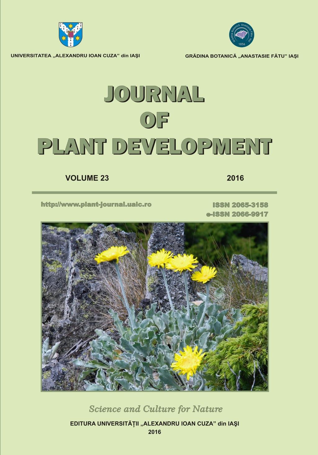
5 CONTENTS CĂTĂLIN TĂNASE, CONSTANTIN TOMA, TIBERIUS BALAEȘ Anastasie Fătu - Founder of the Botanical Garden of Iasi... 3 JAIME A. TEIXEIRA DA SILVA, YASER HASSAN DEWIR, ADHITYO WICAKSONO, MAFATLAL M. KHER, HAENGHOON KIM, MUNETAKA HOSOKAWA, SONGJUN ZENG Morphogenesis and developmental biology of african violet (Saintpaulia ionantha H. Wendl.) ALEXANDRU TELEUŢĂ, VICTOR ŢÎŢEI Economic value of some leguminous plant species of the collections from the Botanical Garden (Institute) of the Academy of Sciences of Moldova MARIUS NICUȘOR GRIGORE, CONSTANTIN TOMA Structure of salt glands of Plumbaginaceae. Rediscovering old findings of the 19 th Century: Mettenius or Licopoli organs? VINEET SONI, ANJU SHARMA, PYARE LAL SWARNKAR Clonal propagation and evaluation of peroxidase activity during in vitro rhizogenesis in Mentha arvensis L CAMELIA PAULA ŞTEFANACHE, OANA-CRINA BUJOR, RADU NECULA, DOINA DĂNILĂ, NINA CIOCÂRLAN, VEACESLAV GHENDOV, CHRISTOPH CARLEN, XAVIER SIMONNET Phenolic content of Artemisia annua L. from natural habitats in Republic of Moldova LĂCRĂMIOARA OPRICĂ, OSCAR VICENTE, MONICA BOȘCAIU, MARIUS NICUȘOR GRIGORE Enzymatic activity and soluble protein content in seedlings of Calendula officinalis L. under salt stress CORNELIA MIRCEA, OANA CIOANCĂ, CRISTINA IANCU, URSULA STĂNESCU, MONICA HĂNCIANU Microbiological and chemical evaluation of several commercial samples of Tiliae flos DANIELA ELENA ZAVASTIN, ALEXANDRA BUJOR, CRISTINA TUCHILUŞ, CORNELIA GEANINA MIRCEA, SIMONA PETRONELA GHERMAN, ANA CLARA APROTOSOAIE, ANCA MIRON Studies on antioxidant, antihyperglycemic and antimicrobial effects of edible mushrooms Boletus edulis and Cantharellus cibarius TATIANA EUGENIA ŞESAN, ANCA SÂRBU, DANIELA SMARANDACHE, FLORIN OANCEA, ANCA OANCEA, SIMONA SAVIN, AGNES TOMA, LAURA ŞTEFAN, GEORGETA NEGRU, ADRIANA FLORENTINA BIRA, GABRIELA VLĂSCEANU, MARIUS GHIUREA, LUIZA JECU, GELU VASILESCU, CRISTIAN MIHAI POMOHACI Botanical and phytochemical approach on Passiflora spp. new nutraceutical crop in Romania LIDIA ADUMITRESEI, IRINA GOSTIN Morphological and micromorphological investigations regarding the leaves of several Rosa L. species ŞTEFAN MANIC The macromycetes of the downy oak forests from Moldova ADRIAN OPREA, ION SÂRBU, NICOLAE ŞTEFAN Astragalus pseudopurpureus Guşul. in Romania's vegetation PAVEL PÎNZARU, VALENTINA CANTEMIR, ŞTEFAN MANIC Phytosociological study of the population of Paeonia peregrina Mill. (Paeoniaceae) in the Republic of Moldova
6 SILVIA OROIAN, MIHAELA SĂMĂRGHIŢAN, MARIANA HIRIŢIU, SANDA COŞARCĂ, CORNELIU TĂNASE The diversity of medicinal and aromatic plants encountered in Natura habitat from Gurghiu Mountains ECATERINA FODOR, OVIDIU HÂRUȚA Quercus robur, Q. cerris and Q. petraea as hot spots of biodiversity OVIDIU COPOȚ, CONSTANTIN MARDARI, TIBERIUS BALAEȘ, CRISTIANA PETRE, CIPRIAN BÎRSAN, CĂTĂLIN TĂNASE Spatial distribution of some ectomycorrhizal fungi (Russulaceae, Fungi, Basidiomycota) in forest habitats from the North-East Region (Romania) Aniversalia Guide to authors
7 CĂTĂLIN TĂNASE, CONSTANTIN TOMA, TIBERIUS BALAEȘ J. Plant Develop. 23(2016): 3-14 ANASTASIE FĂTU FOUNDER OF THE BOTANICAL GARDEN OF IASI Cătălin TĂNASE 1*, Constantin TOMA 2, Tiberius BALAEȘ 3 Remarkable personality, Anastasie Fătu, has organized the medical and social assistance in Moldova. The development of science in the first modern university of Romania and the establishment of the first botanical garden in Iasi (Fig. 1), recommend him as the founder of the school of medicine and natural sciences in the United Principalities. Academician Emil Pop (1967) considers that Anastasie Fătu...he is in the history of Romanian medicine as a famous physician of the poor and the main organizer of hospitals in Moldova, tasks that have taken more time and energy, probably most of his professional activity. And yet, we the botanists, consider Fătu as one of us, as an evolved botanist and extremely progressive for its time, which created solid foundations for botanic education at university and which sacrificed on the altar of science with a rare generosity and a strong love for national culture not only his entire working power, but the savings acquired with sorrow. Fătu is, during the cultural renaissance of the young Romania, an extraordinary example of the great private initiatives, on the basis of which he could call the intellectualists of his time for big collective actions for the progress of the delayed scientific research Its scientific work recommend him as a brilliantly representative of advanced social aspirations of his time, being one of the most remarkable figures in the history of medicine, science and culture of Moldova. This spiritual legacy is embodied in approximately 3000 pages, published in more than 40 years of activity [MAFTEI, 1972]. Life and professional development. Anastasie Fătu was born on 2 nd of January, 1816 in the Muşata village, the former county Fălciu (now Vaslui). His father Vasile, a priest, and his mother Ana, always wished that their son, who proved outstanding qualities since childhood, to be admitted as a Fellow of the state public schools. He attended primary school in Episcopal School of Huşi, and then Vasilian Gymnasium in Iasi. In 1834, as a distinguished prized of the Vasilian Gymnasium receives a scholarship to study in Vienna, where in 1841 he defended his doctorate in philosophy and law. During his studies understands (as will be noted later by V. A. Urechia in response to reception speech of doctor Anastasie Fătu at the Romanian Academy) that in a country widow of freedom, in the land of the whip and unjust privileges could not assert his conception of law and freedom, conception learned from books and from the courses followed at the Faculty of Law, University of Vienna. 1 Department of Biology, Alexandru Ioan Cuza University of Iaşi, Bd. Carol I, No. 20 A, Iaşi Romania 2 Romanian Academy, Prof. univ. dr. at Faculty of Biology, Alexandru Ioan Cuza University of Iaşi, Bd. Carol I, No. 20 A, Iaşi Romania 3 Anastasie Fătu Botanical Garden, Alexandru Ioan Cuza University of Iaşi, Dumbrava Rosie Street, No. 7-9, Iaşi Romania * Corresponding author. tanase@uaic.ro 3
8 ANASTASIE FĂTU - FOUNDER OF THE BOTANICAL GARDEN OF IASI Gives up a profitable career, but without clean soul enjoyments, and since 1839, enrolls also in Vienna at the Faculty of Medicine. Between 1841 and 1846 he continued his medical studies at the Sorbonne University in Paris, where he received his title of doctor in medicine, with the thesis: Des signes des maladies du cœur en général fournis par l'auscultation, la percusion, l'inspection et la mensuration. The activity of the physicians, but especially professor and academician Fătu, shows that at the two outstanding universities, was not satisfied with just the acquisition of knowledge necessary for professional career. In the two institutions Fătu closely surveyed cultural phenomena and scientific research results, comparing them to what might represent for his country. During his studies, has acquired principles that could help at organizing public health and promote natural sciences. Returned home with the firm determination to devote his entire life to initiatives for the establishment of a humanitarian and civilized ordinances in the organization of public health, on 1 st of December 1847, requests to Moldovan Health Committee to approve the free practice right to perform as a physician. After a written and oral exam, the Committee concluded that Anastasie Fătu:...is endowed with science worthy of appreciation and may be consented for free practice as doctor in medicine As a result, from 1848 to 1852 is appointed district physician and later physician of the 2 nd Regiment of Militia (1849), becoming as the historian V. A. Urechia will appreciate...doctor of the poor and consoler of the sufferer... As a doctor of the 2 nd Regiment of Police, publishes in 1853 a Project for police organization in Romania. In 1852 doctor Fătu is appointed professor and administrator of the Gregorian Institute by the prince Grigore Al. Ghica, where he will found a midwife school. Along with doctor Gheorghe Cuciureanu will offer pediatric assistance and will found the first nursery for abandoned children. As a professor he initiate the first official midwifes in Moldova, and for preparing them, wrote in 1852 The manual for midwifes (in Romanian: Manualul pentru învăţătura moaşelor). Since 1873, after professor Dimitrie Brândză went to the University of Bucharest, he occupied the department of Botany and Zoology from the Faculty of Science at the University of Iasi. In 1879, along with doctors Gheorghe Cuciureanu and Nicolae Negură, he contributed to the foundation of the Faculty of Medicine of the University of Iasi. Anastasie Fătu died in Iasi, 130 year ago, on 3 rd of March 1886, resting at the Eternitatea Cemetery [MITITIUC & TONIUC, 2006]. The personality of the great erudite was evoked in volumes [AIFTINCĂ, 2014; BOTNARIUC, 1961; BUDA, 2013; MITITIUC & TONIUC, 2006; POP, 1967; MAFTEI, 1972; POP & CODREANU, 1975; TOMA, 1986; TOMA, 2015], dictionaries [anonymous, 1982; NECULA, 2001], specialized journals [ANGELESCU & DIMA, 2006; BURDUJA & al., 1960; BURDUJA & TOMA, 1979; CONSTANTINESCU, 2009; LEOCOV, 1979, 1982; PAPP, BURDUJA & DOBRESCU, 1955; RESMERIŢĂ, 1982; TOMA, 1974, 1975, 1987, 1996], or within scientific meetings (1981, to commemorate 125 years since the establishment in Iasi of the first botanical garden, was unveiled, in the roundabout in front of the administrative building, the bust of doctor Anastasie Fătu, accomplished and donated by the sculptor Eftimie Bârleanu; in 1986, at the third Symposium of the Botanic Garden, 4
9 CĂTĂLIN TĂNASE, CONSTANTIN TOMA, TIBERIUS BALAEȘ Mandache Leocov, Gheorghe Zamfir, Petru Jitaru and Constantin Toma evoked the personality of Anastasie Fătu. The name of the great erudite is kept with gratitude at the Secondary School in commune Berezeni (Vaslui County) and in the dendrological park realized in mixed style. Founder of the botanical garden. 160 years ago, in 1856, Doctor Anastasie Fătu became renown by founding oneself the first botanical garden from Romania in Iasi, with his own financial resources, this action being considered in society as a remarkable cultural event. Like the Natural History Museum, established in Iasi in 1834, the Botanical Garden of Anastasie Fătu will contribute significantly to the natural sciences development [MITITIUC & TONIUC, 2006], the cultivated plants being used also to illustrate the botanic lessons taught in different schools and at the Academia Mihaileană, founded in This achievements constitutes favorable premises for the establishment in Iasi in 1860, of the first modern University in Romania. Science domain was based from the beginning on the two previously established prestigious institutions: Natural History Museum and the Botanical Garden, whose scientific and educational activity in training youth was coordinated by Professor Anastasie Fătu. In the period when Anastasie Fătu was attended the botanical garden development, he was collaborating with prestigious botanists known at that period: Dimitrie Brândză, Dimitrie Grecescu and Florian Porcius. Moreover, Dimitrie Brândză presented to the students of the university, at the courses of botany, the plants from Fătu s garden. This garden organized on his property, was situated on a hillside, located near the Criminalu Palace, and near the historical monument Râpa Galbenă, delimitated by Butucului street (nowadays Anastasie Fătu street), Begiului Street (nowadays Florilor Street) and the road that connected with Bohotineului Street (nowadays Arcu Street). The mission of the botanical garden was remarked by some of his contemporaries and some institutions. Thereby, Neculai Istrati, director of the Ministry of Public Instructions (Education) donated his salary for three months, and then proposed to depose an annual subvention from the public fund (exchequer). Afterward, the Town Hall of Iasi offered a subvention to the botanical garden, but those initiatives stopped in 1872, and the garden was close down under the pretext that it would block the urban development of the area [PAPP & al. 1955]. Like professor Fătu was mentioning, the purpose of this enterprise was to improve sanitation in the city of Iasi and to induce young people studying plant biology and to give natural sciences lovers, the occasion to contemplate, in their resting moments, at the natural beauties [POP, 1967]. In the press of the time, Gheorghe Asachi the organizer of national schools in Moldova- acclaims the initiative and the activity of Anastasie Fătu to support instruction and education of youth, but also the Romanian patriotism of those who sustained by various means the maintenance and development of different institutions established in Iasi, after the model of most renown education and scientific institutions of Europe. In the catalogue published, who contains more than 2500 of plant species cultivated in his garden, one may find that apart from the native species, which are growing spontaneous, the presence of some exotic gymnosperms, but also some species belonging to Ficus, Acacia and Mesembryanthemum genera. 5
10 ANASTASIE FĂTU - FOUNDER OF THE BOTANICAL GARDEN OF IASI Anastasie Fătu states about this garden that the distribution of the plants was made after the natural method the nomenclature is the Latin, adopted by the masters of Science, which enrolled people known Romanian words... [POP, 1967]. In his manuscript entitled Catalogus herbarii vivi et seminum ex horto 1870, plants cultivated in the garden are listed as follows: in the first part the species are listed in alphabetic order, on two columns on each page, one with acclimatized native plants and one with exotic species. In the second part of the catalogue there are registered some groups of ornamental plants, vegetables, herbs and fruit trees. Thereby, Professor Anastasie Fătu gives the first contribution to the knowledge of flora in the Romanian, especially Moldovan territory. The endemic plant species were obtained with the help of the botanist Iosif Szabó, and the exotic species came from Vienna or by exchanges with botanical gardens from Germany and from Chișinău. He built in his garden two greenhouses, one for the tropical species and one for the temperate ones. At that time, with his garden, Fătu achieves a true experimental field for exotic species acclimatization. The university rector, professor Nicolae Leon stated: the garden had a nice look, the ground was injured; we were descending into on a scale from Florilor Street, we were passing under the Fătu s window, from where we were arriving immediately in front of the greenhouses. Indescribable pleasure for us was to meet the pleasant botanic professor in the garden and we found him very often among the tablets that indicates genera and species names. With a great pleasure he was putting himself at our disposal, he was carrying out a clasp knife and a pocket lens and started, I can hear him: this plant, gentleman, takes part of the family, then showing us how the carpels meet each other, how ovules are inserted, different types of placentation etc. Since October 1996, the Botanical Garden of Iasi, in carrying the name of the founder Anastasie Fătu, which is mentioned on the commemorative plaque on the façade of the administrative building. Medical activity. Between , Anastasie Fătu will became physician at Sfântu Spiridon Hospital, trustee at Sfântu Spiridon House (1877 and 1880) and professor of popular medicine at the Theological Seminary Veniamin Costachi from Socola (1872), where he elaborated the Popular medicine manual (in Romanian: Manual de medicină populară), and on the inside cover is inserted immediately beneath his name the following mention: Dr. in medicine and laws, substitute teacher at the Seminary of Socola, actual member of the Romanian Academy and several local or foreign scientific societies He mentioned in the preface of this work, that when he wrote it he was thinking: in the absence of doctors in villages, priests to advice people. Priests must know the diseases causes and symptoms, to beware of them but also to teach the people [LEOCOV, 1979]. Because he was convinced of the necessity to establish hospitals for children in Moldova, Anastasie Fătu was considered to be the first pediatric physician in Moldova, and with this attribute he found the first pediatric section and assesses some scientific criteria in forensic medicine in Romania. On 1 st of November 1876, Anastasie Fătu gives Sfântu Spiridon House, franci, necessary for the maintenance of five beds for sick children under the age of 10 years. Doctor Fătu elaborated also rules (which unfortunately have not been preserved) for receiving children in this hospital, who worked in the building on the Pașcanu Street. 6
11 CĂTĂLIN TĂNASE, CONSTANTIN TOMA, TIBERIUS BALAEȘ In this perspective, he printed at Iasi, in 1863, a Project for organization of health and public hygiene police in Romania, that could be considered a truly sanitary code, in which were mentioned all of the rules and medical regulations in that period. In collaboration with Iacob Felix, coordinated the medical commission that elaborated the law for organization of health service, which was approved by the Senate in March 1872 and then, in May 1872 by the Chamber of Deputies. This first sanitary law of Romania highlighted the dependency of the administration of public health on the interior ministry [BUDA, 2013]. Academic and social activity. He was an excellent professor and patriot devoted to national progress of culture. Professor Fătu and the agronomist Ion Ionescu de la Brad had the initiative of founding in 1855, the Society for encouraging Romanian youth to study abroad, having the obligation to specialize in applied sciences, in domains such as chemistry, physics, agronomy or engineering. As a result of financial support given by Anastasie Fătu, some young students, among them the future philosopher Vasile Conta, had the opportunity of studying at renowned universities from Europe [AIFTINCA, 2014]. In the work The list of plant species cultivated in the Botanical Garden up to the year 1870 (Enumerațiunea specieloru de plante cultivate în Grădina Botanică din Iassy până în anul 1870, Fig. 2), Anastasie Fătu implement the use of a proper nomenclature, consisting of neologisms borrowed from Latin language and various words used in popular language. In this sense, he includes a many of well-defined terms which are kept in the scientific literature, thereby contributing to the creation of botanical Romanian language. After four years of didactic activity, Fătu completed in 1877 the manuscript for the first Romanian manual of botany for universities, entitled Botanic notions (in Romanian: Elemente de botanică, Fig. 3). The first part of the manual, which is 482 pages large, will be published at Iasi in 1880, containing the cytology, histology, organography and plant physiology. The second part which contains aspects referring to taxonomy, phytography and plant geography will remain as manuscript since 1920, at the Romanian Academy Bibliotheca. In addition with the original publication in medical domain, this manual recommends Anastasie Fătu as an erudite knower of the plant biology. This manual will open new perspectives and research directions less known in plant biology domain and for the last two decades of 19 th century will represent the principal source for training students from the natural sciences faculties, medicine and agriculture. Professor Fătu appreciated in the manual preface that the main wish I had in the publication of this work was to spare, as possible, the precious time of students that is lost by copying the manuscripts [POP, 1967]. Concurrently is the first work in which are popularized aspects regarding the Romanian nomenclature used in plant biology. In this manual, Fătu is giving a great attention on Plant Physiology, subject which does not had previous tackling [POP, 1967]. In carrying out this work he used the most famous branch treatises of the time (Jean Baptiste Payer, Achille Richard, Pierre Étienne Simon Duchartre, Julius von Sachs) but also the work of Dimitrie Brândză Elementary course of natural history, in three parts, Iasi, 1873 and the figures were reproduced from plates purchased from the universal exhibition in Vienna, organized in Academician Emil Pop appreciate Fătu s work as epochal, as it opens new perspectives of the Romanian academic literature, in a specialization of biological sciences which will contribute especially in forming of entire generations of students from faculties 7
12 ANASTASIE FĂTU - FOUNDER OF THE BOTANICAL GARDEN OF IASI of sciences, medicine, agronomy, from last decades of 19 th century. In this work Anastasie Fătu popularized the Romanian nomenclature in the botanical domain, some of the terms being still valid. Manual content include the following divisions: Part I General Anatomy and Plant histology: cytology, fiber tissues, vascular tissues, about epidermis, about chemical composition of plants; Part II Descriptive anatomy or Plant organography: Section I A Cotyledon plants (seeding plants); Reproduction organs (flower, inflorescence and their parts, fecundation, fruit and seed, fruit classification, seeds germination); Section II B Sporophytes and Cryptogams; Part III Plant physiology (absorption, sap circulation, transpiration, respiration, assimilation, growth, germination). This manual highlights an issue considered topical in the botanical domain, an excellent documentation and especially an evolved scientific conception of Professor Fătu, in a period in which vitalism was getting biological shades, and its philosophy was contrary with teaching about the vital force, seen supernatural in organisms. Fătu considered the plant as the unit, and the functions independent. Some of the terms are present in the specialty literature today: referring on roots: vivacious; for strain: articulated, cladodes, tight, axillar tendrils, fastigiated, geniculate, pricks, hispid, axillary, floral, mixed, terminal buds, soiled, clocks, patents, proliferating, nauseating; for leafs: alternate, acuminate, cuspidate, caduceus, crenate, cordiform, emersed, ensiform, fidate, filodii, hastate, devolve, ligules, mucrone, mesophilic, obcordate, obovate, ochree, orbiculare, petioles, peltate, pinnate, palmate, partite, reniform recurved, runcinate, sagittal, sessile, spatulate, stipele, subulate, truncate, vaginule; the flowers - epigine, hypogyne, inflorescence, involucre, corymb, perigyne, raceme; on fruit: hesperides, sorozat, sicon. Permanently preoccupied of social aspects, he studied also the curative role of mineral waters, publishing in 1851 the work Description and use of simple water and mineral waters in Moldova (Fig. 4), work translated in Russian in 1854 of Doctor Isidor Copeniki (Fig. 5) [MITITIUC & TONIUC, 2006]. Also he collaborated by publishing some articles with medical interest in Gazeta de Moldavia and other periodicals. In the year 1857, Anastasie Fătu published in the Journal of Agriculture, edited by Ion Ionescu de la Brad, a series of articles that highlighted his concern regarding the good organization of the Greogorian Institute and the public hygiene. Doctor Anastasie Fătu started his political activity since the unionist period, being elected deputy of Iasi in the ad-hoc Divan of Moldavia (Fig. 6), which proposed the unification of Romanian Principalities. He was part of the Elective Assembly of Moldavia, which chooses Alexandru Ioan Cuza as Domnitor (prince) on 5 th of January In 1868 was elected president of the Assembly of Deputies and senator in Recognition of scientific activity. On 11 th of September 1871, Doctor Anastasie Fătu was elected for his rich activity as actual member (titular) of the Romanian Academic Society (originally Romanian Literary Society on 1 st of April 1866, became the Romanian Academy in 1879), at Natural Sciences section, recent constituted by choosing two days earlier along with Petrache Poenaru, mathematician (inventor of the frame tank) of the physician Nicolae Kretzukescu and the economist Petre S. Aurelian. We have noticed the assessments of the economist Petre S. Aurelian, member of Romanian Academy, in the answer to the request of the erudite of Iasi, to publish in 1871 the first Catalogue of the Botanical Garden: how fast would go the development of science 8
13 CĂTĂLIN TĂNASE, CONSTANTIN TOMA, TIBERIUS BALAEȘ in Romania, how quickly would be studied the natural history of our country when all of those who have had interests of science, have occupied important positions at a university department, would imitate the tireless Anastasie Fătu, but, what can we say! Many are called, few are chosen Petre S. Aurelian acclaimed [TOMA, 1987]. In The speech of reception sustained in the front of the members of the Society Attempts to developments of natural sciences in Romania (in Romanian: Încercările pentru desvoltarea scientieloru naturali în România, Fig. 7) [FĂTU, 1873]. he presented the situation of sciences in Romania and the contributions on fields (geology, mineral waters, flora and fauna of Romania), of the illustrious men who take active part to the propagation of natural sciences, treatises and periodic publications elaborated by them, the public and private means which contributes at the evolution of Romanian sciences, and proposes the realization of a scientific map of the country [CONSTANTINESCU, 2009]. This speech presented in the meeting on 27 th of August 1872 dignify the cultural dimension of the personality of the great erudite and can be considered a document with exceptional history and scientific values [CONSTANTINESCU, 2009]. In speech text, written on 22 pages, Fătu realizes an argued diagnoses regarding the scientific research at the time. The document is accompanied by documented appendices elaborated on 150 pages, where are mentioned historical and statistic details related to all of the scientific societies, periodicals and the bibliography in natural sciences, the situation of museums and educational institution of all levels. The speech highlighted the remarkable education of the grand erudite, but also the special interest and preoccupation of the patriot regarding the Romanian research perspectives. Noticing the relevant analysis of the dissemination of scientific results, he insists on the directions for a fast development of them. The great culture man appreciated that the prosperity of people and the progress of national civilization are not possible without the substantial input of science and culture [AIFTINCĂ, 2014]. Professor Fătu eulogize the activity and the important role of some personalities and private associations in the development of natural sciences, but considers that this initiative must be sustained by the state. This idea is emphasized by the fact that if the private initiative is a commendable thing for those who undertake scientific investigations, it does not have sufficient financial resources to make them give all the fruits that enlightened people are expecting from the science prosperity. The role of the state has a great significance in this situation. This role and the responsibility are even greater as it dispose of the society collective means and as it can use powerful resources that neither the individual and even voluntary associations can ever have. The vision of great erudite on the present and future of Romanian society are remarkable through next directions: scientific activity of the researchers; private associations; public instruction for forming young specialists in areas that are considered to be lean. Also he considers that the scientific research must be national, pretending that the results obtained may be applied for a good evolution and economic development of Romania. In this regard, he appreciated the importance of practical application of the researches in natural sciences domain and the realization of...a complete scientific map of the country which comprehends all the braches of our geological, zoological, botanical and agricultural wealth; in it to indicate the different climatic zones with the characteristic plants and meteorological observations for each county. It must also contain the statistical notices on the population and the agriculture product of each region. From this study we gain an accurate knowledge of our 9
14 ANASTASIE FĂTU - FOUNDER OF THE BOTANICAL GARDEN OF IASI ore, of all species of plants, of animals and our agricultural development by regions and the influence of climate in plant s and animal life [POP, 1967]. Because all of his life denoted, beside a great character, that is an action man, in the final of his speech mentioned...to assert how deep I am convinced of the necessity and the usefulness of the scientific map of our country, from the modest resources that I have accumulated working for 25 years, I will deposit as fund the amount of ten thousand «franci» [POP, 1967]. In that period the amount was equivalent with the state support for one year, the money will be used by the Natural Science Section for the subvention of the project [AIFTINCĂ, 2014]. Regarding the Academy, this institution had the mission of regulator and propeller for the scientific movement. Also he considers that shall be the headquarters of all the procedures for the scientific development [AIFTINCĂ, 2014]. In his speech in the chapter dedicated Private means for science popularization exhibited alongside with the Society of Physicians and Naturalists of Iasi, Society of Natural Sciences of Bucharest, the Romanian Academic Society, the Society for encouraging Romanian youth to study abroad and Junimea Society. He considers that The objectives of the Junimea Society are to knowing the truth trough open discussions that mutually enlightens the members and illustrates with some of the themes that were touched within the meetings regarding issues with national character. Their reach is to find the truth, and for these all of the opinions must be respected, for from their meeting and counteracting one to be able to separate... The historian V.A. Urechia stated in the response, according to the academic usance, that here is a fountain filled with precious notes regarding the development between the Romanians of natural sciences, notably in the current century. There is nothing left to say for a specialists, for me not even as much, to this speech... [POP, 1967] Further, the founder of the Romanian Academy made the following greeting to the great erudite and patriot Anastasie Fătu: live long Mr. Doctor, because you have proved that your years are the country and Romanian science s years!... [POP, 1967]. From 1872 till 1886, Anastasie Fătu was the president of the Society of Physicians and Naturalists of Iasi, organizing it in sections: medicine, natural sciences and agronomy. In the year 1872 he contributed to the organization of a small botanic garden in the yard of the Society of Physicians and Naturalists of Iasi. For this purpose, some species from the old garden were planted and with the help of the botanist Iosif Szabó brings in seeds from different parts of the country. Furthermore he was corresponding member of the Medico-Surgical Society of Bucharest, president of the department from Iasi of the Society for encouraging Romanian youth to study abroad. Because he was known abroad, he was named member of the Society of Natural Sciences of Frankfurt and Silesian Society for science development. Contemporaneous personalities surrounded with unanimous respect and with high appreciation for the physician, doctor and erudite professor, who devoted his life for the common weal. 10
15 CĂTĂLIN TĂNASE, CONSTANTIN TOMA, TIBERIUS BALAEȘ References AIFTINCĂ M Timp şi valoare. Studii de istorie a culturii şi filosofiei româneşti. Colecţia Opera Omnia, Cartea de Filozofie. Edit. Tipo Moldova, Iaşi: ANGELESCU N. & DIMA A Personalităţi chirurgicale ieşene. Jurnalul de Chirurgie, Iaşi. II(4): 424. BOTNARIUC N Din istoria biologiei generale. Edit. Şti. Bucureşti: BUDA O Şcoala de medicină legală la Iaşi, Anastasie Fătu şi George Bogdan. In: Identitate naţională şi medicină socială. Antropologie culturală, psihiatrie şi eugenism în România, Edit. Muzeului Naţional al Literaturii Române, Colecţia Aula Magna, Bucureşti: BURDUJA C., FILIPESCU G. & LAZĂR M Contribuţii la cunoaşterea evoluţiei terminologiei botanice româneşti. Analele Ştiinţifice ale Universităţii Alexandru Ioan Cuza din Iaşi (serie nouă), Secţiunea II. (Ştiinţe Naturale). VI(2): BURDUJA C. & TOMA C Opera botanică a doctorului Anastasie Fătu ( ). Culegere de Studii şi Articole de Biologie, Grădina Botanică Iaşi, 1: CONSTANTINESCU R Doctorul Anastasie Fătu despre Societatea Junimea. Revista Română. Iaşi. 1(55): FĂTU A Încercările pentru desvoltarea scientieloru naturali în România. Discurs de recepţiune ( ). Soc. Acad. Rom., Imprimeria Statului, Bucureşti. LEOCOV M de ani de la înfiinţarea la Iaşi a primei Grădini Botanice din ţară. Culegere de Studii şi Articole de Biologie, Grădina Botanică Iaşi. 1: LEOCOV M Dr. Anastasie Fătu ctitorul primei grădini botanice din România. Culegere de Studii şi Articole de Biologie, Grădina Botanică Iaşi. 2: LEON N Istoria naturală medicală a poporului român. Analele Academiei Române, seria II, XXV: Bucureşti. LEON N. 1922, Amintiri. Edit. Viaţa Românească. 1, 3, Iaşi. MAFTEI I Fătu, Anastasie ( ). Medic şi naturalist. In: Personalităţi ieşene. vol. I, Omagiu, Comitetul de Cultură şi Educaţie Socialistă a Judeţului Iaşi: MITITIUC M. & TONIUC A Grădina Botanică Anastasie Fătu Iaşi. File de istorie. Edit. Univ. Alexandru Ioan Cuza din Iaşi: 8-21; NECULA P Dicţionarul personalităţilor vasluiene. Edit. Cutia Pandorei. PAPP C., BURDUJA C. & DOBRESCU C Din istoricul cercetărilor de botanică în cadrul Societăţii de Medici şi Naturalişti din Iaşi. Analele Ştiinţifice ale Universităţii Alexandru Ioan Cuza din Iaşi (serie nouă), Secţiunea II. (Ştiinţe Naturale). IV(1-2): POP E Anastasie Fătu ( ). In: Figuri de botanişti români. Edit. Şti. Bucureşti: POP E. & CODREANU R Istoria ştiinţelor în România. Edit. Acad. Rom., Bucureşti. RESMERIŢĂ I Retrospectivă din viaţa botaniştilor care au condus destinele Grădinii Botanice din Iaşi. Culegere de Studii şi Articole de Biologie, Grădina Botanică Iaşi. 2: TOMA C Din activitatea Grădinii Botanice a Universităţii ieşene. Cercetări Agronomice în Moldova, Iaşi. 3: TOMA C Der Botanische Garden der Universität Iaşi. In: Zu Problemen Botanischer Garden. Wissenschaftliche Beiträge, Halle. 6: TOMA C Anastasie Fătu ( ). In: Universitatea din Iaşi ( ), Dezvoltarea ştiinţei. Iaşi: TOMA C Profesor dr. Antasie Fătu activitatea sa didactică. Culegere de Studii şi Articole de Biologie, Grădina Botanică Iaşi. 2: TOMA C Anastasie Fătu ( ). Revista Academica, Bucureşti. 6-8: 44. TOMA C Biologi de altă dată şi de azi. Edit. Univ. Alexandru Ioan Cuza din Iaşi: * * * Fătu, Anastasie. In: Personalităţi româneşti ale ştiinţelor naturii şi tehnicii (dicţionar). Edit. Şt. Encicl., Bucureşti:

16 ANASTASIE FĂTU - FOUNDER OF THE BOTANICAL GARDEN OF IASI Fig. 1. Place of the Fătu s Botanical Garden in city plan made by engineer Fred Peytavin in 1857 (National Archives of Iasi, section Plans and Maps, no. 522) 12

17 CĂTĂLIN TĂNASE, CONSTANTIN TOMA, TIBERIUS BALAEȘ Fig. 2 Fig. 3 Fig. 4 Fig. 5 13
")
18 ANASTASIE FĂTU - FOUNDER OF THE BOTANICAL GARDEN OF IASI Fig. 6. Anastasie Fătu, deputy in Ad-hoc Divan of Moldova (1858) 14
19 JAIME A. TEIXEIRA DA SILVA & al. J. Plant Develop. 23(2016): MORPHOGENESIS AND DEVELOPMENTAL BIOLOGY OF AFRICAN VIOLET (SAINTPAULIA IONANTHA H. WENDL.) Jaime A. TEIXEIRA DA SILVA 1*, Yaser Hassan DEWIR 2, 3, Adhityo WICAKSONO 4, Mafatlal M. KHER 5, Haenghoon KIM 6, Munetaka HOSOKAWA 7, Songjun ZENG 8 Abstract: African violet (Saintpaulia ionantha H. Wendl.) has been domesticated, bred and commercialized. It is the most famous and popular of the Saintpaulia species, its ornamental value arising from its attractive leaves and flowers. African violet plants are easy to propagate by adventitious organ regeneration and are very sensitive to environmental factors including light, temperature, humidity, CO 2 concentration and photoperiod. This review offers a short synthesis on advances made in conventional vegetative propagation by adventitious organ regeneration, select early historical in vitro developmental perspectives, and vegetative and reproductive development of African violet. Keywords: development; Gesneriaceae; thin cell layers; vegetative propagation. Introduction African violet (Saintpaulia ionantha H. Wendl.; Gesneriaceae) has attractive leaves and flowers (Fig. 1) that are typical of many members of the Gesneriaceae. African violet is mainly used for ornamental purposes, thus aspects related to flower color, leaf patterning, or yield are of interest to horticulturalists, plant breeders, molecular biologists, physiologists, biotechnologists, and for the floriculture industry. Thus, the improvement of flower- and leafrelated traits via vegetative propagation, the creation of somaclonal variation, and mutation breeding are fundamental aspects of African violet research. Plant morphogenesis is regulated by complex genetic networks in a synchronized manner, and these serve as determining factors for numerous crop traits. Many constituents of these networks have been extensively studied in the model plant, Arabidopsis thaliana L. [VANHAEREN & al. 2016]. Multicellular plants have a unique group of cells that form new organs and replenish the daily loss of cells, or regenerate organs after injury, the pluripotent stem cells [AICHINGER & al. 2012]. Stem cells are located in stem cell niches that provide 1 P. O. Box 7, Miki-cho post office, Ikenobe , Kagawa-ken, Japan 2 Plant Production Department, P.O. Box 2460, College of Food & Agriculture Sciences, King Saud University, Riyadh Saudi Arabia 3 Department of Horticulture, Faculty of Agriculture, Kafrelsheikh University, Kafr El-Sheikh Egypt 4 Laboratory of Paper Coating and Converting, Centre for Functional Material, Åbo Akademi University, Porthaninkatu 3, Turku Finland 5 B. R. Doshi School of Biosciences, Sardar Patel University, Sardar Patel Maidan, Vadtal Rd., P.O. Box 39, Vallabh Vidyanagar, Gujarat, India 6 Department of Well-being Resources, Sunchon National University, Suncheon, Korea 7 Graduate School of Agriculture, Kyoto University, Sakyo-ku, Kyoto Japan 8 Key Laboratory of South China Agricultural Plant Molecular Analysis and Gene Improvement, South China Botanical Garden, the Chinese Academy of Sciences, Guangzhou, China * Corresponding author. jaimetex@yahoo.com 15

20 MORPHOGENESIS AND DEVELOPMENTAL BIOLOGY OF AFRICAN VIOLET intercellular signals for differentiation such as hormonal control [ZHAO & al. 2010], or transcription factors [STAHL & SIMON, 2010; SABLOWSKI, 2011]. This review provides several details about traits related to development that could make this ornamental plant a possible model plant for the study of morphogenesis, differentiation and organ formation. It also addresses the environmental factors regulating vegetative growth and flowering, inflorescence development, specific conditions for propagation of African violet and biotechnological methods to improve flowering. Fig. 1. A flowering African violet (Saintpaulia ionantha H. Wendl) (Gesneriaceae) plant. Initially redrawn from EASTWOOD & al (p. 50), then modified. Conventional vegetative propagation: possible model plant for adventitious organ regeneration (historical perspective) FIGDOR (1907) may have been the first official report on adventitious regeneration in the Gesneriaceae, including African violet adventitious regeneration. FAIRBURN (1936) studied the propagation of African violet and other vegetatively propagated plants using leaf cuttings, noting how the inclusion of petioles was essential to induce roots. NAYLOR & JOHNSON (1937) described how portions of the leaf blade or petiole of African violet could regenerate one or more plants within 6-15 weeks if placed on wet sand or in water in Petri dishes when placed in a moist chamber. Even ex vitro, NAYLOR & JOHNSON (1937) noted the development of callus from the cut surface, with root initials forming within as little as 10 days, emerging from, but not derived from, the callus, i.e., originating from cells within the orginal explant. Their histological evidence showed that roots formed from thin-walled cells found underneath the leaf epidermis, while shoots formed from exogenous cells of the epidermal layer, i.e., shoots originated from epidermal cells. Concept of thin cell layer and morphogenesis Basic findings within earlier attempts made by FIGDOR (1907), FAIRBURN (1936) and NAYLOR & JOHNSON (1937) would later turn out to be an important reason for the success of thin cell layers (TCLs) [TEIXEIRA DA SILVA & al. 2007; TEIXEIRA DA SILVA & DOBRÁNSZKI, 2013, 2014; TEIXEIRA DA SILVA & al. 2015], especially longitudinal TCLs, in African violet tissue culture, making African violet an important ornamental plant model for developmental studies, like tobacco, and making it a possible viable form of in vitro regeneration for other Saintpaulia species [TEIXEIRA DA SILVA & al. 2015]. To date, only 16
21 JAIME A. TEIXEIRA DA SILVA & al. two studies employed TCLs, but even so, the term TCL was not used for the earlier study. MURCH & al. (2003) used 0.25 mm thick petiole sections (i.e., transverse TCLs or ttcls) to regenerate somatic embryos while KHOSARI-NASAB & al. (2014) induced callus and shoots from petiole and pedicel ttcls. The ability to regenerate a large number of plantlets from a limited amount of tissue (in terms of surface area and volume) underlies the basic success of the TCL [TEIXEIRA DA SILVA & DOBRÁNSZKI, 2014], and thus the success of the tissue culture of African violet. The origin of knowledge about this development came from early studies on vegetative propagation in the 1930 s. Later, BROERTJES (1968), ENGELS & al. (1980), GEIER (1983), BROERTJES & VAN HARTEN (1985), Ohki (1994) and HOSOKAWA & al. (1998) confirmed similar histological evidence showing the involvement of epidermal cells (Fig. 2) in the formation of adventitious shoots from the petioles of African violet, noting, using chimeric tissue to base their assumption, the single cell origin of shoots, and thus annulling the claim by NORRIS & al. (1983) that shoots are derived from multiple cells. FINER & SMITH (1983) noticed that plastids that were in an arrested state of development in the epidermis underwent changes after placing leaf cuttings on kinetinsupplemented medium. REDWAY (1991) showed the epidermal origin of shoot primordia, after callus formed, but also showed the formation of shoot primordia from palisade tissue, which lies about 10 cell layers below the epidermal surface. This single cell origin of shoots would allow solid (i.e., non-chimeric) mutants to be produced, forming an important basis for mutation breeding studies. In Saintpaulia, some pinweel flower color cultivars are considered to be periclinal chimeras, with the petal margin arising from the L1 layer but the center of the petal from the L1+L2 layers. If shoot regeneration occurs from epidermal layers, then the flower color phenotype is identical to that of the flower margin of the mother plant. LINEBERGER & DRUCKENBROD (1985), ANDO & al. (1986) and PEARY & al. (1988) discuss shoot regeneration from the epidermal layer using pinweel-flowered periclinal cultivars in more detail. LINEBERGER & DRUCKENBROD (1985) found that whereas leaves, petioles, peduncles, sepals and inflorescences formed plantlets within 3-5 months, it took 8-10 months from subepidermal tissues, suggesting that the organogenic outcome reported in that study, and possibly in all other African violet studies, depends on the timing of sampling, which is also a function of the type of explant used [TEIXEIRA DA SILVA & DOBRÁNSZKI, 2013]. In essence, regeneration from TCLs may be faster than from regular explants since a greater area and volume of cells is exposed to the exogenously applied plant growth regulators in the in vitro medium. Fig. 2. Shoot development from epidermal tissue, as suggested by histological analyses by GEIER (1983), LO & al. (1997), and NAYLOR & JOHNSON (1937). Arrow indicates a meristematic center. 17
22 MORPHOGENESIS AND DEVELOPMENTAL BIOLOGY OF AFRICAN VIOLET In vitro morphogenesis and variability, sports and variegation ANDO & al. (1986) cultured 19 edged-type cultivars with a white corolla and a pigmented outer edge, and 9 Geneva edged-type cultivars with a pigmented corolla and a white outer edge, either as leaf cuttings, or in tissue culture. They found that single-colour cultivars did not form sports whereas single-colour sports, except for two cultivars, were produced in the remaing (edged-type and Geneva edged-type) cultivars at an average of 22%, but with a wide range of frequencies (0.9% to 68.8%), as a result of mutations. In contrast, the sports from edged-type cultivars had single-coloured corollas, and those from Geneva edged-type cultivars had white or extremely pale corollas, except for a single cultivar. Whereas the edged form ( Ms. Pretty ) could produce single colour sports through tissue culture (i.e., reversion was possible), single colour sports of the same cultivar could never produce an edged form. PEARY & al. (1988) found that the variegated pattern of a foliar variegated cultivar ( Tommie Lou ) was not caused by periclinal chimeras because the pattern of leaf variegation did not change in potted plants derived from the tissue culture of leaf and petal sections, or from subepidermal tissue. In contrast, tissue cultured plants of a variegated flower cultivar ( Candy Lou ) segregated into the chimeral components, i.e., the chimera could not be stably propagated by tissue culture. Curiously, DÜMMER (1912) observed the phenomenon of peloria (i.e., reversion of asymmetric flowers into symmetric flowers) in S. ionantha. Morphogenetic efficiency of explants: effects of age, season, and position SCOTT & MARSTON (1967) noted that 24 C and misting were suitable for the development of S. ionantha plantlets from leaf cuttings. According to HENTIG (1976), reflecting studies conducted in the early 1970 s by this author, and using Rhapsodie in Blau Typ 32 as the experimental material, there is a relationship between the length of the petiole of leaf cuttings and adventitious shoot formation. HENTIG (1976) formulated three important claims: 1) tissue from the fifth to tenth month of growth are most receptive; 2) leaves of the middle leaf zone and youngest leaves from the upper zone regenerate earlier (by 4-5 days) than older leaves (i.e., in the lower zone), forming within 56 days; 3) shorter petioles (1-3 cm) formed more adventitious shoots (and faster) than longer petioles (4-6 cm). In HENTIG s study, there were significant differences in the regeneration potential of young, middle-aged and old leaves before 56 days, but after 56 days, leaves from all three age categories were able to form adventitious shoots in 100% of explants. Even when leaves of all three age categories with petioles of different lengths were used, no less than 60% of all leaves were responsive, fortifying the notion that this is an ornamental plant that can be easily propagated vegetatively. Depending on the age of the leaf and on the length of the petiole, anything from 1 to 4 shoots could be produced per leaf. Vegetative and reproductive development: role of environmental factors JOHANSSON (1978) described members of the Saintpaulia genus as typically being shade plants while POST (1942) and STROMME (1985) described African violet as being day-neutral with respect to flower initiation and development. Inflorescences arise from leaf axils. During plug production, daily integrated photosynthetic photon flux (PPF DI) reaching African violet plants in summer range from mol/m 2 /day but are as low as < 2 mol/m 2 /day in winter. However, temperature and PPF DI can influence the vegetative growth of African violet. For example, HANCHEY (1955) noted that by increasing PPF DI from 0.31 to 1.9 mol/m 2 /day, the number of leaves doubled from 22 to 44. HILDRUM & 18
23 19 JAIME A. TEIXEIRA DA SILVA & al. KRISTOFFERSEN (1969) noted that the number of flowers, buds, and inflorescences per plant and flowers and buds per inflorescence of Biedermeier Rhapsodie and Biedermeier Rosa increased when PPF DI was augmented from 3.1 to 9.3 mol/m 2 /day. This treatment induced a maximum of 6.8 flower stalks/plant at 18 C, but a maximum number of flowers and buds/flower stalk (9.1) at 24 C. A maximum of 42 flowers/plant could be produced when daylight (i.e., most likely fluence rate) was cut by 75%, and when plants were exposed to 27 C. In contrast, flower initiation and development was inhibited when plants grown in a greenhouse were exposed to < 2 mol/m 2 /day [STINSON & LAURIE, 1954]. HILDRUM & KRISTOFFERSEN (1969) showed that both day and night temperature must be high to stimulate a greater number of flower stalks, and that flower production is linked to leaf production, given the source of inflorescences from leaf axils. This indicates that African violet is not only highly sensitive to environmental variables such as tempearture and PPF DI, but that these variables can be used to manipulate plant growth and development, both of leaves and floral organs. KWACK & KIM (1969) noted that sunlight reduced by half in the greenhouse was the best condition in terms of the growth and ornamental quality of African violet plants. As light intensity was decreased from full sunlight ( foot-candles (f.c.)) to half sunlight ( f.c.) and shade (500 f.c.), leaf area increased, i.e. 9.26, and cm 2, respectively. Full sunlight induced solarization, chlorosis in leaves or backward leaf curling, even though it produced higher dry weight (1.29, 1.21 and 0.90 g for full sunlight, half sunlight and shade, respectively). KIM & SANG (1982) argued that light intensity was critical for four varieties ( Monique, Robert O, Julianne and an undefined local variety) of S. ionantha, since it is a semi-shaded plant. They confirmed that a light intensity of 5,000-10,000 lux ( % of natural sunlight in a plastic house) was the best condition for growth and ornamental value, i.e., photosynthesis, flowering percentage, number of peduncles, number of florets/peduncle, petiole length and leaf area. In an experiment assessing the effect of illumination and culture medium on S. ionantha Oriental Red, LEE (1986) noted that lux combined with a substrate composed of 40% peatmoss, 25% sand, 20% perlite, 10% vermiculite, and 5% compost was the best for producing wider leaves with greater leaf area (34-36 cm 2 ), best petiole length ( cm), number of lateral shoots/explant (3.2), flowering rate (100%) and earliest flowering (number of days to flowering = 70 days), and number of peduncles ( ). PARK (2008) examined the effect of nitrogen fertilizer and light intensity on three varieties of S. ionantha and found that half-sunlight (4000 lux) and 200 mg/l NaNO 3 was the most effective combination for obtaining maximum fresh weight (FW) (e.g., for var. Narita 1.9 g/cutting vs 0.9 g/cutting in full-sunlight and 0 mg/l NaNO 3). The combination of 4000 lux and 500 mg/l NaNO 3 increased the number of roots, the number of shoots (4.7/cutting vs 0/cutting), and chlorophyll content (52.4 mg/g FW vs 28.2 mg/g FW). Using Utah as the model cultivar combined with linear modeling to base their assumptions, FAUST & HEINS (1993) determined maximum leaf unfolding rate (LUR) to be 0.27 leaves/day when temperature was 25 C while PPF DI was 10 mol/m 2 /day by using shadecloth to cut natural PPF DI from a maximum of around 300 mol/m 2 /day. When they decreased PPF DI from 10 to 1 mol/m 2 /day, optimal temperature was 23 C, but this resulted in a lower LUR (0.18 leaves/day). Only when the leaf blade extended from mm could an inflorescence begin to develop in the leaf axis [FAUST & HEINS, 1994]. Being able to predict and balance temperature, PPF DI and thus LUR, would allow growers to plan their greenhouse cultures for the market. For example, FAUST & HEINS (1993), by applying a
24 MORPHOGENESIS AND DEVELOPMENTAL BIOLOGY OF AFRICAN VIOLET PPF DI of 7 mol/m 2 /day at 22 C, with a LUR of leaves/day, formed 10.5 unfolded leaves/plant within 45 days (R 2 =0.99). Also using non-linear models, FAUST & HEINS (1994) showed that as daily temperature was increased from 18 to 26 C, days from leaf emergence to first open flower on the inflorescence of Utah decreased from 86 to 55. They concluded that PPF is the primary factor influencing flower initiation while average daily temperature is the primary factor influencing the rate of inflorescence development. The three cardinal temperatures (T min, T opt, and T max) for leaf appearance rate [FAUST & HEINS, 1993] are 8 C, variable (as this depends on PPFD) and 30.8 C, respectively and for leaf elongation rate [FAUST & HEINS, 1994], the values are 13.8 C, 24 C and 29 C, respectively. In contrast, STRECK (2004) found that T min was 10 C and T max was 33 C for both leaf appearance rate and leaf elongation rate, and T opt was 24 C, as also modeled by FAUST & HEINS. CONOVER & POOLE (1981) were able to increase the percentage of inflorescences that formed in African violet Inge within 9 months from 6% to 62%, and then to 100%, by exposing plants to 0.8, 1.6, and 3.2 mol/m 2 /day, respectively. However, as FAUST & HEINS (1994) cautioned, even if LUR increases, a PPF of < 4 mol/m 2 /day will negatively affect the number of developing inflorescences. Inflorescence development is acropetal [HASTON & DE CRAENE, 2007] (Fig. 3). Consequently, for African violet, the production of potted plants for season-dependent events such as Valentine s Day, Mother s Day, or Christmas, can be perfectly timed with the desired number of leaves and flowers. The leaves of African violet are very sensitive to light, temperature, humidity and photoperiod [CHEN & HENNY, 2009], and minor changes in any of these factors can cause yellow or brown leaf spots [ELLIOT, 1946; YUN & al. 1997a; YANG & al. 2001]. Leaf spot is a cellular response confined to palisade cells in leaves, whose ultrastructure is destroyed [YUN & al. 1996a, 1996b], caused by a rapid drop in temperature, as may occur in overhead irrigation, especially at night, or by transferring plants from indoor to outdoor conditions, as was shown for Ritali plants [YUN & al. 1997a]. Unlike chilling injury, leaf spot is irreversible. Outer leaves, which are more exposed to the surrounding environment, are more susceptible to leaf spot, and even exposure to 20 C water can induce this disorder [MAEKAWA & al. 1987], which results from electrolyte leakage [MAEKAWA & al. 1990]. Leaf spot is a stress response caused by the rapid production of reactive oxygen species (ROS) following sudden temperature shifts [YASUDA & al. 1997], and an increase in the activity of antioxidant enzymes (superoxide dismutase and catalase). However, this response may be cultivar-dependent since Ritali and Tamiko (with about 75% and 95% incidence of leaf spot, respectively) were more susceptible to a drop from 30 C to 15 C than Maui and New Jersey (about <5% and 22% incidence, respectively) [YANG & al. 2001]. Sudden drops in temperature causing leaf spot are also characteristic of Ritali leaves that display additional physiological disorders, namely a decrease in photosynthetic activity (especially a negative effect on PSI and PSII activity and a decrease in chlorophyll fluorescence) [YUN & al. 1997b, 1998], and plasmolysis [YUN & al. 1996a]. Recently, OHNISHI & al. (2015) revealed evidence of the involvement of calcium ions (specifically Ca 2+ channels) after the degradation of the vacuolar membrane of palisade cells. In a practical greenhouse trial to examine the response of stock plants and leaf cuttings (with 15 mm long petioles, according to HENTIG, 1976) to different commercial light sources (Grolux-low, Grolux-high, Verilux-low, Verilux-high, Fluora 77, Cool White 20, Cool White 30, Warm-White 30, Warm-White de Luxe 32, Interna 39 and Natura 36), SCHNEIDER-MOLDRICKX & AMBERGER (1982) discovered significant differences in the number of shoots that could be regenerated and in shoot FW. In their experiment, bulbs emitted µmol/m 2 /s, all test material was grown under a 16-h photoperiod, and data was pooled for three cultivars ( RH-26/74, Meta, and OP-50/75 ). Cuttings cultivated under 20
25 21 JAIME A. TEIXEIRA DA SILVA & al. light bulbs emitting higher radiant energy formed more and heavier shoots than those exposed to light bulbs emitting low levels of light energy. Grolux (emitting 63 µmol/m 2 /s) was the most effective light source, forming 81 and 89 shoots/plot from stock plants and cuttings, respectively, and 598 mg/shoot. The response was strongly cultivar-dependent, with RH- 26/74, Meta, and OP-50/75 forming 56, 94 and 80 shoots/plot and 589, 235 and 598 mg/shoot, respectively. A separate trial by the same authors showed that Warm-White 30 bulbs could induce the formation of as many as shoots/plot in a cultivar-independent manner. The use by BOSCHI & al. (2000) of red and blue spectral filters, with different red, blue and far red ratios, on greenhouse African violet (cultivar unspecified) plants reduced most growth parameters (shoot and root dry weight, chlorophyll content, chlorophyll/protein ratio, net carbon dioxide (CO 2) exchange), in some cases significantly, compared to control polyethylene film, but did not alter the transpiration rate. African violet plants ( Rosa Roccoco and Big Star ) exposed to continuous CO 2 enrichment, in which CO 2 concentration was increased from 330 to ,000 ppm, showed % higher net photosynthetic rate and 23-30% higher relative growth rate, most likely caused by the 39.6% increase in the number of formed leaves [MORTENSEN, 1984]. More refined experiments by MORTENSEN (1986) on Nicole, Lena and Rosa Roccoco showed that CO 2 enrichment (900 µl/l) had a more profound and/or significant effect in all three cultivars and in all parameters measured (greater dry weight, relative growth rate, number of leaves, leaf diameter, and number of flowers and flower buds, but reduced number of days to flowering) than ambient CO 2 (335 µl/l), a 1-h daily pulse at 900 µl/l, or a morning plus an evening pulse, each at 900 µl/l. For example, in Nicole, CO 2 enrichment resulted in 7.95 g/plant, a relative growth rate of 37.1 mg/g/day, 51.1 leaves/plant, 19.3 cm wide leaves, 56.1 days to flowering, and flowers and flower buds/plant. The equivalent values for ambient (control) CO 2 levels were 3.56, 28.4, 35.3, 17.6, 66.4 and 66.8, respectively. Vegetative growth can also be stimulated by the application of maleic hydrazide (MH). LEMATTRE (1977) found that spraying three African violet cultivars ( Rhapsodie, 2000, and 2738 ) with 0.1 to 0.2% MH resulted in a 47- to 176-fold increase in the number of vegetative shoots that formed, depending on the concentration and the cultivar. For example, in 2000, while control plants formed only an average of 0.1 vegetaive buds per plant, the application of 0.2% MH resulted in the formation of 17.6 vegetative buds, which could in essence be used as clones, and to inhibit early flowering, although the genetic stability of such clones was not tested. By applying 10 mg/l gibberellic acid (GA 3) at two-week intervals to four-month-old Rhapsody blue plants, DVORSKÁ (1979) was able to increase the number of flower stalks by about 25% (10-12 vs 6-9 in controls). Furthermore, in the same study, a constant temperature of 22 C resulted in the formation of 4-6 flower stalks in control plants, whereas a 14 C/22 C (day/night) temperature gradient resulted in the formation of 7-11 flower stalks. HERKLOTZ (1964) found that a day and night temperature of 25 C favored regeneration from leaf cuttings than all other day/night temperature combinations ranging from 15 C to 30 C. MARTÍN-MEX & al. (2005) found that the application of µm salicylic acid significantly increased the number of leaves (19 vs 16 in the control) and floral buds (14 vs 8 in the control), as well as rosette diameter (177 mm vs 139 mm in the control), and shortened the days to flowering (74 vs 89 in the control). A similar finding by JABBARZADEH & al. (2009) also confirmed the positive effects of a foliar application of M salicylic acid on vegetative growth and flowering of African violet in terms of the number of leaves, rosette diameter, the number of flower buds and the number of days from planting to anthesis. These chemical methods provide means of delaying and/or enhancing vegetative growth and/or flowering in African violet to meet market needs.
26 MORPHOGENESIS AND DEVELOPMENTAL BIOLOGY OF AFRICAN VIOLET Fig. 3. Inflorescence development typical for the Gesneriaceae. (A) A pair-flowered inflorescence displays dichasial branching. (B) A plan view of the same inflorescence represented in (A). In A and B, T = terminal flower, F = front flower, parentheses indicate directions of growth / flower expansion, and numbers indicate the level of branching on the inflorescence axis. In B, CU represents a single cyme unit with a hypopodium (peduncle supporting the cyme unit) with a terminal pair of lateral bracteoles, subtending a terminal and an associated front flower. (C) The development of a Saintpaulia ionantha inflorescence, divided into 8 stages (according to FAUST & HEINS 1994): 1) visible reproductive bud (2 mm long) in the leaf axil; 2) visible peduncle subtending the primary bud; 3) peduncle starts to curve; 4) pedicel curves 90 relative to the peduncle; 5) pedicel curvature relative to the peduncle <90 and secondary buds are at the top of the inflorescence; 6) angle between peduncle and primary bud and pedicel increases, the pedicel is at the top of the inflorescence, and the primary bud emerges from the leaf canopy; 7) upper half of pedicel and primary bud are perpendicular to lower half of pedicel (i.e., at 90 angle); 8) the petals are perpendicular to the pedicel. (A and B) retraced, redrawn and modified from HASTON & DE CRAENE (2007) Fig. 1A and 1B (p. 15); (C) redrawn and modified from FAUST & HEINS (1994) Fig. 1 (p. 728). Conclusions and future perspectives The easy regeneration potential of African violet, as evident from as far back as the NAYLOR & JOHNSON (1937) study, alongside the debate about the single or multiple cell orgin of shoot buds in earlier work, are some interesting aspects that require additional attention from molecular cell biologists to address the factors that regulate morphogenesis in African violet. African violet can be easily and rapidly cloned. This, together with its ability to regenerate organs from different tissues, has facilitated several studies with the aim of understanding organogenesis, organ development and its related in vitro physiology. Hence, 22
27 JAIME A. TEIXEIRA DA SILVA & al. African violet is valued as a model plant at the research/laboratory level and this potential can and should be further explored. Vegetative growth, flowering and floral organ development of African violet can be manipulated in a greenhouse or growth chamber by adjusting environmental variables including light source, intensity and quality, temperature, CO 2 enrichment, as well as chemical applications. Therefore, commercially, the production of potted African violet plants can be planned for the market to meet consumer demands. Acknowledgements We are thankful to Ravi R. Sonani, Sardar Patel University and Sylvia DeMar, Managing Editor, American Society for Horticultural Science for providing difficult-toaccess literature. Author contribution statement and conflicts of interest statement The authors contributed equally to all aspects of review development and writing. The authors have no conflicts of interest to declare. References AICHINGER E., KORNET N., FRIEDRICH T. & LAUX T Plant stem cell niches. Annu Rev. Plant Biol. 63: ANDO T., AKIYAMA Y. & YOKOI M Flower colour sports in Saintpaulia cultivars. Sci. Hortic. 29: BOSCHI C., DI BENEDETTO A., PAPAYANI P., CREMONA C. & BENEDICTO D Coverfilms and light quality responses in Saintpaulia ionantha. Acta Hortic. 515: BROERTJES C Mutation breeding of vegetatively propagated crops. In: Proceedings of 5 th Eucarpia Congress. Milan: BROERTJES C. & VAN HARTEN A. M Single cell origin of adventitious buds. Euphytica. 34: CHEN J. & HENNY R. J Cultural guidelines for commercial production of African violets (Saintpaulia ionantha) ENH Environmental Horticulture Department, Florida Cooperative Extension Service, Institute of Food and Agricultural Sciences, University of Florida, USA, pp CONOVER C. A. & POOLE R. T Light acclimatization of African violet. HortScience. 16: DÜMMER R. A Peloria in Saintpaulia ionantha, Wendland. Ann Bot. 26: DVORSKÁ L The effect of growth regulators and temperature gradient on flowering of Saintpaulia ionantha Wendl. Acta Hortic. 91: EASTWOOD A., BYTEBIER B., TYE H., TYE A., ROBERTSON A. & MAUNDER M The conservation status of Saintpaulia. Curtis Bot. Mag. 21: ELLIOT F.H Saintpaulia leaf spot and temperature differential. Proc. Amer. Soc. Hortic. Sci. 47: ENGELS F. M., VAN DER LAAN F. M., LEENHOUTS H. P. & CHADWICK K. H The regeneration of epidermal cells of Saintpaulia leaves as a new plant-tissue system for cellular radiation biology. Int. J. Rad. Biol. 38: FAIRBURN D. C Plant propagation. Missouri Bot Garden Bull. 24: FAUST J. E. & HEINS R. D Modeling leaf development of the African violet (Saintpaulia ionantha Wendl.). J. Amer. Soc. Hortic. Sci. 118: FAUST J. E. & HEINS R. D Modeling inflorescence development of the African violet (Saintpaulia ionantha Wendl.). J. Amer. Soc. Hortic. Sci. 119: FIGDOR W Ueber Resttutionserscheinugen an Blattern von Gesneriaceen. Jahrb. Wiss. Bot. 44: FINER J. J. & SMITH R. H Structure and development of plastids in epidermal cells of African violet (Saintpaulia ionantha Wendl.) in culture. Ann Bot. 51: GEIER T Induction and selection of mutants in tissue cultures of Gesneriaceae. Acta Hortic. 131: HANCHEY R. H Effects of fluorescent and natural light on vegetative and reproductive growth in Saintpaulia. Proc. Amer. Soc. Hort. Sci. 66: HASTON E. & DE CRAENE L. P. R Inflorescence and floral development in Streptocarpus and Saintpaulia (Gesneriaceae) with particular reference to the impact of bracteole suppression. Plant Syst Evol. 265:
28 MORPHOGENESIS AND DEVELOPMENTAL BIOLOGY OF AFRICAN VIOLET HENTIG W. U. V Results of propagation with leaf cuttings of Saintpaulia ionantha. Acta Hortic. 64: HERKLOTZ A Influence of constant and daily alternating temperatures on growth and development of Saintpaulia ionantha Wendl. Gartenbauwiss. 29: (in German with English abstract). HILDRUM H. & KRISTOFFERSEN T The effect of temperature and light intensity on flowering in Saintpaulia ionantha Wendl. Acta Hortic. 14: HOSOKAWA M., HOSSAIN M., TAKEMOTO T. & YAZAWA S Particle-gun wounding of explants with and without plant growth regulators effectively induces shoot formation in African violet. Plant Tissue Cult. Biotechnol. 4: JABBARZADEH Z., KHOSH-KHUI M. & SALEHI H The effect of foliar-applied salicylic acid on flowering of African violet. Aust. J. Basic Appl. Sci. 3(4): JOHANSSON D. R Saintpaulias in their natural environment with notes on their present status in Tanzania and Kenya. Biol. Conserv. 14: KIM J. K. & SANG C. G A study on the growth and flowering of Saintpaulia ionantha under controlled light intensities. J. Kor. Soc. Hortic. Sci. 23: (in Korean with English Abstract). KHOSARI-NASAB M., ASGHARY E., MOVAFEGHI A. & POUREBAD N Investigation of ttcls technique in regeneration of African violet (Saintpaulia ionantha). Adv. Env. Biol. 8(13): KWACK B. & KIM I Growth and ornamental value of Saintpaulia and Hypoestes under the different light conditions. J. Kor. Soc. Hortic. Sci. 6: (in Korean with English abstract). LEE Y. H Effects of BAP pretreatments and media constituents on organogenesis in vitro from leaf segments of Saintpaulia ionantha Wendl. MS thesis. Kyungsan National University, Jinju, Korea, pp LEMATTRE P Effect of maleic hydrazide on floral and vegetative development of Saintpaulia ionantha. Acta Hortic. 68: LINEBERGER R. D. & DRUCKENBROD M Chimeral nature of the pinwheel flowering African violets (Saintpaulia, Gesneriaceae). Amer. J. Bot. 72: LO K. H., GILES K. L. & SAWHNEY V. K Histological changes associated with acquisition of competence for shoot regeneration in leaf discs of Saintpaufla ionantha x confusa hybrid (African violet) cultured in vitro. Plant Cell Rep. 16: MAEKAWA S., TORISU Y., INAGAKI N. & TERABUN M Leaf injury caused by drop in leaf temperature of Saintpaulia ionantha. J. Jpn. Soc. Hortic. Sci. 55: (in Japanese with English summary). MAEKAWA S., TORISU Y., INAGAKI N. & TERABUN M Relation between development of leaf spot and electrolyte leakage. J. Jpn. Soc. Hortic. Sci. 58: (in Japanese with English summary). MARTÍN-MEX R., VILLANUEVA-COUOH E., HERRERA-CAMPOS T. & LARQUÉ-SAAVEDRA A Positive effect of salicylates on the flowering of African violet. Sci. Hortic. 103: MORTENSEN L. M Photosynthetic adaptation in CO 2 enriched air and the effect of intermittent CO 2 application on greenhouse plants. Acta Hortic. 162: MORTENSEN L. M Effect of intermittent as compared to continuous CO 2 enrichment on growth and flowering of Chrysanthemum morifolium Ramat. and Saintpaulia ionantha H. Wendl. Sci. Hortic. 29: MURCH S. J., VICTOR J. M. R. & SAXENA P. K Auxin, calcium and sodium in somatic embryogenesis of African violet (Saintpaulia ionantha Wendl. cv. Benjamin). Acta Hortic. 625: NAYLOR E. E. & JOHNSON B A histological study of vegetative reproduction in Saintpaulia ionantha. Amer. J. Bot. 24: NORRIS R. E., SMITH R. H. & VAUGHN K. C Plant chimeras used to establish de novo origin of shoots. Science. 220: OHKI S Scanning electron microscopy of shoot differentiation in vitro from leaf explants of the African violet. Plant Cell, Tiss. Org. Cult. 36: OHNISHI M., KADOHAMA N., SUZUKI Y., KAJIYAMA T., SHICHIJO C., ISHIZAKI K., FUKAKI H., IIDA H., KAMBARA H. & MIMURA T Involvement of Ca 2+ in vacuole degradation caused by a rapid temperature decrease in Saintpaulia palisade cells: a case of gene expression analysis in a specialized small tissue. Plant Cell Physiol. 56: PARK S. Y Effect of nitrogen and light treatment on the rooting and growth of African violet (Saintpaulia ionantha Wendl.) cutting. MSc thesis, Sangmyung University, Chunahn, Korea, pp PEARY J. S., LINEBERGER R. D., MALINICH T. J. & WERTZ M. K Stability of leaf variegation in Saintpaulia ionantha during in vitro propagation and during chimeral separation of a pinwheel flowering form. Amer. J. Bot. 75: POST K Effect of daylength and temperature on growth and flowering of some florist crops. Cornell Univ. Agric. Exp. Sta. Bull. 787: REDWAY F. A Histology and stereological analysis of shoot formation in leaf callus of Saintpaulia ionantha Wendl. (African violet). Plant Sci. 73:
29 JAIME A. TEIXEIRA DA SILVA & al. SABLOWSKI R Plant stem cell niches: from signalling to execution. Curr. Opin. Plant Biol. 14: 4-9. SCHNEIDER-MOLDRICKX R. & AMBERGER S Propagation of Saintpaulia ionantha under artificial lighting. Acta Hortic. 128: SCOTT M. & MARSTON M. E Effects of mist and basal temperature on the regeneration of Saintpaulia ionantha Wendl. from leaf cuttings. Hortic Res. 7: STAHL Y. & SIMON R Plant primary meristems: shared functions and regulatory mechanisms. Curr. Opin. Plant Biol. 13: STINSON R. F. & LAURIE A The effect of light intensity on the initiation and development of flower buds in Saintpaulia ionantha. Proc. Amer. Soc. Hortic. Sci. 64: STRECK N. A A temperature response function for modeling leaf growth and development of the African violet (Saintpaulia ionantha Wendl.). Ciência Rural 34: STROMME E Saintpaulia ionantha. In: A. Halevy (ed.). CRC Handbook of Flowering Vol. 3. CRC Press, Boca Raton, FL, p TEIXEIRA DA SILVA J. A., ALTAMURA M. M. & DOBRÁNSZKI J The untapped potential of plant thin cell layers. J. Hortic. Res. 23: TEIXEIRA DA SILVA J. A. & DOBRÁNSZKI J How timing of sampling can affect the outcome of the quantitative assessment of plant organogenesis. Sci. Hortic. 159: TEIXEIRA DA SILVA J. A. & DOBRÁNSZKI J Dissecting the concept of the thin cell layer: theoretical basis and practical application of the plant growth correction factor to apple, Cymbidium and chrysanthemum. J. Plant Growth Reg. 33: TEIXEIRA DA SILVA J. A., TRAN THANH VAN K., BIONDI S., NHUT D. T. & ALTAMURA M. M Thin cell layers: developmental building blocks in ornamental biotechnology. Floriculture Ornamental Biotechnol. 1: VANHAEREN H., INZÉ D. & GONZALEZ N Plant growth beyond limits. Trends Plant Sci. 21: YANG S. J., HOSOKAWA M., MIZUTA Y., YUN J. G., MANO J. & YAZAWA S Antioxidant capacity is correlated with susceptibility to leaf spot caused by a rapid temperature drop in Saintpaulia (African violet). Sci. Hortic. 88: YASUDA Y., YUN J. G., MIYOSHI N., HAYASHI T., YAZAWA S. & FUJITA S Production of radicals and acute apoptotic changes of palisade cells in early steps of Saintpaulia leaf spot appearance. Acta Histochem. Cytochem. 30: YUN J. G., HAYASHI T. & YAZAWA S. 1997a. Diurnal changes in leaf spot sensitivity of Saintpaulia (African violet). Sci. Hortic. 70: YUN J. G., HAYASHI T., YAZAWA S., KATOH T. & YASUDA Y. 1996a. Acute morphological changes of palisade cells of Saintpaulia leaves induced by a rapid temperature drop. J. Plant Res. 109: YUN J. G., HAYASHI T. & YAZAWA S. 1996b. Precise quantification of leaf spot in Saintpaulia by image analysis with transmitted light. Env. Control Biol. 34: YUN J. G., HAYASHI, T., YAZAWA S., YASUDA Y. & KATOH T. 1997b. Degradation of photosynthetic activity of Saintpaulia leaf by sudden temperature drop. Plant Sci. 127: YUN J. G., HAYASHI T., YAZAWA S., KATOH T. & YASUDA Y Abrupt and irreversible reduction of chlorophyll fluorescence associated with leaf spot in Saintpaulia (African violet). Sci. Hortic. 72: ZHAO Z., ANDERSEN S. U., LJUNG K., DOLEZAL K., MIOTK A., SCHULTHEISS S. J. & LOHMANN J. U Hormonal control of the shoot stem-cell niche. Nature. 465: How to cite this article: TEIXEIRA DA SILVA J. A., DEWIR Y. H., WICAKSONO A., KHER M. M., KIM H., HOSOKAWA M. & ZENG S Morphogenesis and developmental biology of African violet (Saintpaulia ionantha H. Wendl.). J. Plant Develop. 23: Received: 13 October 2016 / Revised: 24 November 2016 / Accepted: 5 December
30
31 ALEXANDRU TELEUŢĂ, VICTOR ŢÎŢEI J. Plant Develop. 23(2016): ECONOMIC VALUE OF SOME LEGUMINOUS PLANT SPECIES OF THE COLLECTIONS FROM THE BOTANICAL GARDEN (INSTITUTE) OF THE ACADEMY OF SCIENCES OF MOLDOVA Alexandru TELEUŢĂ 1*, Victor ŢÎŢEI 1 Abstract: The results of the evaluation of the growth and development rates, the seed productivity, the green mass yield, the biochemical composition and the content of amino acids, phosphorous and calcium, the nutritive and energy value of the forage, as well as the biomethane productivity of local ecotypes of the leguminous species maintained in monoculture, in the collection of the Botanical Garden (Institute) of the Academy of Sciences of Moldova (BG ASM): Astragalus ponticus, Coronilla varia, Lotus corniculatus, Medicago falcata, Onobrychis arenaria and Trifolium repens are presented in this article. Control variants the traditional forage crops: Medicago sativa and Onobrychis viciifolia. The local ecotypes of the studied leguminous species were characterized by different growth and development rates. Coronilla varia and Lotus corniculatus, in the 2 nd -3 rd years, could be harvested, for the first time, 5 days earlier than Medicago sativa, but Medicago falcata and Onobrychis viciifolia 18 days later. The green mass yield varied from 0.83 kg/m 2 to 4.08 kg/m 2. The studied ecotypes reached amounts of nutritive units/kg and metabolizable energy MJ/kg of dry matter, the content of digestible protein, of g/nutritive unit, met the zootechnical standards; seed production: g/m 2 ; the biomethane yield ranged from 692 to 3197 m 3 /ha. Higher yield of natural forage, dry matter and biomethane was produced by Onobrychis arenaria and Coronilla varia. Keywords: biochemical composition, biological peculiarities, economic value, perennial leguminous species. Introduction Legumes from pastures and meadows contribute nitrogen to a complex and dynamic recycling system, organic matter containing legume proteins may be mineralized in soil, liberating N as nitrates (NO 3) and NH 4 that may be used by grasses and other species of plants. They are important in livestock feeding systems because they have the potential to extend the grazing season, increase the quantity of grazed forage and hay, and reduce the amount of N fertilizer needed. Legume feed not only improves forage quality but also increases the intake of the ration, hence, gives better performance in terms of livestock production. Many legume crops are also excellent honey plants, other plants can be used as raw material in various branches of the national economy, as they are for cosmetology, pharmaceutics and bioenergetics industry [DUKE, 1981; STODDARD, 2013]. The spontaneous flora of the Republic of Moldova (RM) is relatively rich and includes 5568 species of plants (superior plants 2044 species, inferior plants 3524 species), family Fabaceae Lindl. 25 genera and 120 species [NEGRU, 2007]. The spontaneous flora of our country includes over 700 species of fodder plants and 71 species of them are leguminous plants [TELEUȚĂ, 2010]. The grasslands from the Republic of Moldova cover about 14% of the 1 Botanical Garden (Institute) of the Academy of Sciences of Moldova, 18 Pădurii str., 2002, Chişinău Republic of Moldova. * Corresponding author. ateleuta@gmail.com 27
32 ECONOMIC VALUE OF SOME LEGUMINOUS PLANT SPECIES OF THE COLLECTIONS FROM territory, they are in a deplorable condition and have very low productivity, with a share of leguminous plants decreasing from year to year [BANCIVANJI & al. 2012]. The collection of non-traditional forage plants of the Botanical Garden (Institute) of the Academy of Sciences of Moldova have 320 botanical taxa (species, varieties), including 73 leguminous plants [TELEUȚĂ, 2010; TELEUȚĂ & ȚÎȚEI, 2012; 2016]. This research was aimed at evaluating the biological peculiarities, the biochemical composition of the local ecotype of leguminous plant species and the possibility to use them as forage for ruminant animals or as biogas substrate. Materials and methods The seeds of the native leguminous species (Astragalus ponticus Pall., Coronilla varia L., Lotus corniculatus L., Medicago falcata L., Onobrychis arenaria (Kit.) DC., Trifolium repens L.) served as subjects of study and were collected from the spontaneous flora of the main botanical district from RM (region of plains of Balti and Bugeac steppe), maintained in monoculture in the collection of new and non-traditional forage plants of the BG ASM. The traditional forage crops (Medicago sativa L. cv. Ilan and Onobrychis viciifolia Scop. cv. Adam) served as control variants. The green mass and the samples for biochemical analysis were gathered after the first mowing, in the budding-flowering period. The scientific research on growth and development, yield and biochemical composition of the plants, was carried out according to the methodical indications [NOVOSIOLOV & al. 1983; PETUKHOV & al. 1989]. The carbon content of the substrates was determined by the data on volatile solids (organic dry matter), using an empirical equation [BADGER & al. 1979]. The biogas production potential and the specific methane yields were evaluated by the parameter content of fermentable organic matter [WEISSBACH, 2008]. Results and discussion As a result of the phenological observations, it has been found that, in the first growing season, the studied perennial leguminous species are characterised by different growth and development rates. Thus, it has been determined that the plantlets of Trifolium repens and Medicago falcata emerge uniformly at the soil surface, 14 days after sowing, 3 days earlier as compared with the control, Medicago sativa, and 9 days earlier as compared with Onobrychis viciifolia. The plantlets of the species: Astragalus ponticus, Lotus corniculatus and Onobrychis arenaria emerge after 20 days, so, they need the same period of time as Onobrychis viciifolia, but the plantlets of Coronilla varia emerge the latest, that is, about 26 days after sowing or 12 days later as compared with Medicago sativa. The plants of Astragalus ponticus and Coronilla varia are distinguished by a very slow growth and development of the aerial part. By the end of the growing season, Astragalus ponticus develops the rosette with leaves, and Coronilla varia reaches the budding stage and the beginning of the flowering stage. The species Lotus corniculatus, Medicago falcata, Onobrychis arenaria and Trifolium repens pass through all the ontogenetic stages. The species Lotus corniculatus and Onobrychis arenaria are characterised by faster development rates, the development and the ripening of seeds start 7 days earlier, in comparison with Onobrychis viciifolia and Trifolium repens, 32 days earlier, in comparison with Medicago sativa and 39 days earlier than Medicago falcata. 28
33 ALEXANDRU TELEUŢĂ, VICTOR ŢÎŢEI In the following years, the leguminous species, studied by us, resume their growth and development in spring, when temperatures above 3-5 C are established. The species Trifolium repens and Medicago falcata resume growth 7 days earlier than Astragalus ponticus and 3 days earlier than other species. The species: Lotus corniculatus, Astragalus ponticus and Trifolium repens are characterised by a faster development during the whole growing season, but they don t grow as tall as the traditional leguminous crops. So, by the end of April (Tab. 1), the plants reach cm high, while the control species about cm. The shoots of Coronilla varia, in this period, reach a height of cm. Tab. 1. Duration of periods of growth and plant height of the studied species Indicators Astragalus ponticus Coronilla varia Lotus corniculatus Medicago falcata Medicago sativa Onobrychis arenaria Onobrychis viciifolia Trifolium repens Resumed vegetation up to: - budding - flowering - seed ripening Plant height, cm - at the end of April - at flowering It has been determined that the period of time from the restart of active growth until the flower bud formation, in Lotus corniculatus, constitutes 63 days, in Medicago falcata 78 days, but in the control species 75 days. The flowering stage of Astragalus ponticus, Coronilla varia, Lotus corniculatus and Trifolium repens starts 22 days earlier in comparison with Onobrychis viciifolia and 5 days in comparison with Medicago sativa. The plants of Medicago falcata begin to bloom the last. During the flowering stage, the shoots of Coronilla varia and Onobrychis arenaria reach cm high, Trifolium repens and Lotus corniculatus cm, but the control cm. The studied species need a different period of time from the beginning of flowering until the full ripening of seeds. So, Trifolium repens, Astragalus ponticus and Lotus corniculatus need days, Medicago falcata and Coronilla varia days, while Onobrychis viciifolia needs 34 days and Medicago sativa 61 days. Seed production is a key pillar in the capacity of maintenance and expansion of the species. Analysing the seed productivity (Tab. 2), we conclude that the studied leguminous forage species differ from Medicago sativa and Onobrychis viciifolia in the quantity of produced seeds and in the weight of 1000 seeds. The plants of the genus Onobrychis ( g/m 2 ) and Astragalus ponticus (40.90 g/m 2 ) are characterised by high seed productivity, the mass of 1000 seeds of these species is also quite high ( g). The plants of Lotus corniculatus and Trifolium repens, even if they have lower seed productivity in comparison with the control species, produce the highest number of seeds about seeds/m 2. We have also found that Medicago falcata produces more seeds than Medicago sativa, and Onobrychis arenaria fewer than Onobrychis viciifolia.
34 ECONOMIC VALUE OF SOME LEGUMINOUS PLANT SPECIES OF THE COLLECTIONS FROM The total yield, the quality and the seasonal distribution of forage may be of greater importance to the livestock producer. As mentioned above, the studied leguminous forage species have different growth and development rates that influence the productivity of natural forage and the dry matter content. Thus, a higher yield of natural forage and dry matter, in comparison with the controls, was produced by Onobrychis arenaria (4.08 kg/m 2 or 1.11 kg/m 2 ) and Coronilla varia (3.92 kg/m 2 or 0.87 kg/m 2 ), a lower one by Trifolium repens, Astragalus ponticus and Lotus corniculatus ( kg/m 2 or kg/m 2 ). Onobrychis arenaria is distinguished by a higher productivity of natural forage and dry matter as compared with Onobrychis viciifolia, and Medicago falcata by a lower one as compared with Medicago sativa. In some papers, it was mentioned that the productivity of Coronilla varia, under the climatic conditions of Russia, reached 65 t/ha green mass [DRONOVA & al. 2009]. The ecotypes of Onobrychis arenaria from Russia are characterized by longer lifespan (5-7 years), in comparison with Onobrychis viciifolia, and by higher productivity of forage with 20-70% [MEDVEDEV & SMETANNIKOVA, 1981]. The low productivity of local ecotypes of Trifolium repens, Astragalus ponticus and Lotus corniculatus may be explained by the arid environmental conditions (low humidity, rainfall under 400 mm and high temperatures in spring) in recent years. The varieties of Trifolium repens and Lotus corniculatus, created in Romania and cultivated in areas with mm of precipitation per year, have a production potential of t/ha of green mass or 8-12 t/ha of hay [MARUȘCA & al. 2011]. It is well known that cattle eat mainly leaves, due to their high content of nutrients, and the ratio leaves/stems influences the forage value. The forage of Coronilla varia and Astragalus ponticus is characterized by a high content of leaves (56-63%), but Medicago falcata by a lower one (37%). Tab. 2. The productivity of the studied species of the family Fabaceae Species 1. Astragalus ponticus 2. Coronilla varia 3. Lotus corniculatus 4. Medicago falcata 5. Medicago sativa 6. Onobrychis arenaria 7. Onobrychis viciifolia 8. Trifolium repens fresh mass, kg/m 2 30 dry matter, kg/m 2 leaf content, % seed production, g/m 2 weight of 1000 seeds, g For growth, development, reproduction, as well as for the production of high quality milk or meat, cattle need many nutrients they receive from feed. Proteins are the most important and the largest group of natural macromolecular compounds, essential for life, are a source of nitrogen for the body and play a crucial role in the use of the genetic productive potential. The forage of Astragalus ponticus (Tab. 3), is characterised by a high content of raw protein, 23.40% of dry matter, in comparison with Medicago sativa and Onobrychis viciifolia, while Trifolium repens and Lotus corniculatus have low content of raw protein. Astragalus ponticus is distinguished by a very high content of raw protein (63.84 g/kg), which
35 ALEXANDRU TELEUŢĂ, VICTOR ŢÎŢEI is 67-75% higher compared to the species Trifolium repens and Lotus corniculatus. A high capacity of nitrogen accumulation by Astragalus ponticus is also recorded in other papers [DAVIS, 1982]. The species Coronilla varia and Medicago falcata are at the same level, regarding the content of raw protein. Other authors [ACAR & al. 2001], remarked that Medicago falcata and Coronilla varia contain % raw protein, but Lotus corniculatus and Trifolium repens % raw protein. Tab. 3. Biochemical composition and nutritional value of the dry matter of the studied species of the family Fabaceae Indicators Astragalus ponticus Coronilla varia Lotus corniculatus Medicago falcata Medicago sativa Onobrychis arenaria Onobrychis viciifolia Trifolium repens Raw protein, % 2. Raw fats, % 3. Raw cellulose, % 4. Nitrogen free extract, % 5. Minerals,% 6. Nutritive units, NU/kg 7. Metabolizable energy, MJ/kg 8. Calcium, g/kg s.a.u. 9. Phosphorus, g/ kg s.a.u. 10. Digestible protein, g/nutritive unit The vegetable fats in forage are the main source of energy for the animals because they are necessary for the organism in order to ensure the normal development of vital processes and transportation of soluble vitamins in fatty acids and it also contributes to the accumulation of fat in milk [McDONALD & al. 2011]. The forage of Lotus corniculatus and Onobrychis arenaria contains a high amount of fats ( %), at the same level as Onobrychis viciifolia, greatly exceeding Medicago sativa. The Medicago falcata has a low content of fats -1.78% (Tab. 3, line 2, column 4, 5, 6, 7, 8). The content of cellulose is quite low in the species Lotus corniculatus, Onobrychis arenaria, Medicago falcata and very high in Trifolium repens (Tab. 3, line 3, column 4, 5, 7, 9). We also mention that the optimal cellulose content has a beneficial effect on the synthesis of proteic substances in the rumen of animals and on the reduction of the nitrate content [BAHCIVANJI & al. 2012]. The nitrogen-free extract (NFE), along with fats, provides the necessary energetic material for vital processes, contributing to the formation and storage of fats [PÂRVU 1992; BAHCIVANJI & al. 2012]. The content of nitrogen-free extract varies from 32.35% to 48.60%, it is high in the species Lotus corniculatus, Onobrychis arenaria, Medicago falcata and very low in Astragalus ponticus, this fact influences the possibility of the forage to provide energy (Tab. 3, line 4, column 2, 4, 5, 6). The vegetal forage contains minerals in variable quantities, regarding the type of the elements and the proportion between them and other chemical compounds. Minerals are essential components of all tissues and organs that maintain a constant osmotic pressure, participate in the regulation of acid-base balance, activate a number of enzymes, moderate neuromuscular activities and prevent the emergence and development of diseases in animals. The presence of minerals in animal feed is indispensable for their growth and health
36 ECONOMIC VALUE OF SOME LEGUMINOUS PLANT SPECIES OF THE COLLECTIONS FROM [SUTTLE, 1982]. The forage of Lotus corniculatus and Astragalus ponticus is characterized by a high content of minerals ( %), but the forage of Trifolium repens by a lower one (6.08%), in comparison with the control species (Tab. 3, line 5, column 2, 4, 9). Calcium is the mineral element that is found in the highest quantity in the animal body, it is a structural component of bones and teeth; it plays an essential role in blood coagulation, tensing of muscles, activity of enzyme systems, at cell level. The normal assimilation of calcium by organism depends also on the quantity of phosphorus in the feed. Phosphorus plays a multiple role in the vital processes in body, and, being a constituent of nucleic acids, it participates in protein synthesis, cell multiplication and transmission of hereditary characters; it is a component of ATP (adenosine triphosphate) the main deposit of energy in tensing of muscles, nerve activity, vital processes of body; as phospholipids, it enters the structure of cells and cell membranes regulating the transportation of solutions through them; participates in the metabolization of carbohydrates, fatty acids and amino acids etc. [SUTTLE, 1982; McDONALD & al. 2011]. The content of calcium in the studied species varies from 6.37 g/kg (Astragalus ponticus) to g/kg (Onobrychis arenaria), being lower than in Medicago sativa (16.83 g/kg). Very low content of calcium has been found in the forage of Astragalus ponticus (6.37 g/kg), the other species (Coronila varia, Lotus corniculatus, Medicago falcata, Medicago sativa, Onobrichys arenaria, Trifolium repens) have higher levels of calcium ( g/kg) than Onobrychis viciifolia g/kg (Tab. 3, line 8, column 1, 6, 8). The studied species have lower content of phosphorous ( g/kg) than Onobrychis viciifolia (7.52 g/kg). The species Medicago falcata, Lotus corniculatus and Trifolium repens have very low content of phosphorous (corresponding 1.0, 2.19 and 2.51 g/kg). The species Coronilla varia and Onobrychis arenaria are distinguished by high content of phosphorous (corresponding 5.66 and 7.52 g/kg) as compared with Medicago sativa 4.39 g/kg (Tab. 3, line 9, column 1, 6, 7). The main indicators of the nutritional value of the natural forage are the content of nutritive units and metabolizable energy (MJ). The nutritional and energy value is determined by the biochemical composition and the digestibility of the organic substances from the forage that influence the health and the productivity of animals [MEDVEDEV & SMETANNIKOVA, 1981; McDONALD & al. 2011]. The nutritive units (NU) of the natural forage of the studied species reaches amounts of 0.60 (Trifolium repens) up to 0.89 NU/kg (Coronilla varia). Trifolium repens and Lotus corniculatus species containing 0.20 and 0.08 NU/kg correspondingly less than the control Medicago sativa. At the level control (0.80 NU/kg) are species Astragalus ponticus (0.77), Medicago falcata (0.83), Onobrychis arenaria (0.85) and Onobrychis viciifolia (0.84 NU/ kg). According to this index, species Coronilla varia is the most valuable forage crop (0.89 NU/kg). Metabolizable energy is an index assessing the capacity of the feed to support of the processes of the milk production. This indicator of the studied species ranges from 7.89 up to MJ/kg dry matter. The energy value of the forage of Coronilla varia and Onobrychis arenaria is at the same level as Onobrychis viciifolia (corresponding 9.90, 9.44 and MJ/kg). The value of the metabolize energy of Trifolium repens (8.05), Lotus corniculatus (8.85) and Astragalus ponticus (7.89) is same in comparison with Medicago sativa (8.65 NJ/kg) - Tab. 3, row 7, columns 2, 4, 6, 9. An important indicator of forage value is the content of digestible protein per nutritive unit. The content of digestible protein (DP) per nutritive unit (NU) ranges from g DP/NU (Lotus corniculatus) up to g DP/NU (Astragalus ponticus) and meets the zootechnical standards. Also green fodder of Onobrychis viciifolia, Onobrychis arenaria and Trifolium repens have high level of digestible protein (Tab. 3). 32
37 33 ALEXANDRU TELEUŢĂ, VICTOR ŢÎŢEI The amino acid composition is the most important factor in defining the food protein quality, followed by the digestibility of the protein and the bioavailability of its amino acids. The protein quality is determined by the ratio of certain amino acids, which provide the biological value of the feed. The efficiency of using protein crops in animal feed production strongly depends on the content of essential amino acids (EAA) in the various crops and the composition of compound feedstuffs [PÂRVU, 1992; McDONALD & al. 2011]. An essential amino acid, or indispensable amino acid, is an amino acid that cannot be synthesized de novo (from scratch) by the organism, and thus must be supplied in its diet. The nine amino acids humans cannot synthesize are phenylalanine, valine, threonine, tryptophan, methionine, leucine, isoleucine, lysine, and histidine [McDOUGALL, 2002]. By analysing the amino acid content, we have found that the forage obtained from the studied species contains different amounts of amino acids, including those essential. The total amount of amino acids ranging from mg/100 mg (Lotus corniculatus) to mg/100 mg (Astragalus ponticus). The species Onobrychis arenaria and Astragalus ponticus contain the highest amount of essential amino acids, but Lotus corniculatus, Medicago falcata and Coronilla varia contain a very low amount. It has been found that all the studied species have a lower content of methionine than Medicago sativa, but the species Onobrychis arenaria, Coronilla varia and Lotus corniculatus are distinguished by a higher content of methionine in comparison with Onobrychis viciifolia (Tab. 4). Tab. 4. The content of amino acids in forage (mg/100 mg dry matter) of the studied species of the family Fabaceae Amino acids 1. asparagine 2. threonine * 3. serine 4. glutamine 5. proline 6. glycine 7. alanine 8. valine 9. methionine 10. isoleucine 11. leucine 12. tyrosine 13. phenylalanine 14. histidine 15. lysine 16. arginine 17. Total sum of AA 18. Total sum of EAA Astragalus ponticus Coronilla varia Lotus corniculatus Medicago falcata * - essential amino acids are in bold Medicago sativa Onobrychis arenaria Onobrychis viciifolia Trifolium repens The species Astragalus ponticus and Onobrychis arenaria contain a higher amount of lysine ( mg/100 mg) than the traditional crops, but the species Coronilla varia and Lotus corniculatus a lower one ( mg/100 mg). In comparison with traditional forage crops, Astragalus ponticus is characterized by a higher content of threonine, glutamine, valine, isoleucine and histidine; Coronilla varia is very rich in proline and glycine, rich in asparagine, glutamine, glycine and alanine; Lotus corniculatus rich in threonine and serine,
38 ECONOMIC VALUE OF SOME LEGUMINOUS PLANT SPECIES OF THE COLLECTIONS FROM Medicago falcata rich in asparagine, threonine, serine, glutamine and alanine. Trifolium repens, as compared with Medicago sativa, is richer in proline, glycine, valine, leucine, tyrosine, histidine and lysine. The content of essential amino acids varies from mg/100 mg (Lotus corniculatus) up to mg/kg (Onobrychis arenaria); Medicago sativa mg/100 mg (Tab. 4). The use of forage legumes as biogas substrate contributes to an increase in the potential of bioenergy and can help reduce the greenhouse gas emissions. Through symbiotic nitrogen fixation, they compensate inorganic N fertilizer in conventional farms, if the digestate is applied as a fertilizer to the non-legume crops [STODDARD, 2013]. The content of organic matter, biochemical composition, biodegradability and ratio of carbon and nitrogen (C/N) of the raw material are essential in the production of biogas. The C/N ratio of the studied species varied from 13, in the biomass of Astragalus ponticus, to 29-30, in the biomass of Trifolium repens and Lotus corniculatus (Tab. 5). The optimal C/N ratio is expected to be in the range 15-25, when the anaerobic digestion process is carried out in a single stage, and for the situation when the process develops in two steps, the optimal C/N ratio will range: for step I: 10-45; for step II: [DOBRE & al. 2014]. Fermentable organic matter represents the proportion of organic matter which can be biologically degraded under anaerobic conditions and, thus, can be potentially utilized in biogas facilities [WEISSBACH, 2008]. The calculated gas forming potential of the fermentable organic matter of the studied Fabaceae species varied from 470 to 544 litre/kg volatile solid matter (VS), Trifolium repens, Coronilla varia lower than Medicago sativa, but Onobrychis arenaria, Medicago falcata and Lotus corniculatus exceeding Onobrychis viciifolia, they have similar content of methane. The methane yield per ha of studied species of the family Fabaceae (first mowing) ranged from 692 to 3197 m 3 /ha, Onobrychis arenaria exceeding Onobrychis viciifolia, but Coronilla varia exceeding Medicago sativa. Tab. 5. Gas forming potential of the fermentable organic matter (FOM) from the studied Fabaceae species Indicators Astragalus ponticus Coronilla varia Lotus corniculatus Medicago falcata Medicago sativa Onobrychis arenaria Onobrychis viciifolia Trifolium repens Ratio carbon/nitrogen 2. FOM, g/kg VS 3. Biogas, litre/kg VS 4. Methane, litre/kg VS 5. Methane yield, m 3 /ha Conclusions The species Coronilla varia and Lotus corniculatus can be harvested, for the first time, 5 days earlier, but Medicago falcata and Onobrychis viciifolia 18 days later than Medicago sativa. Due to this fact, fresh natural forage can be provided for animals for a longer period of time. The green mass yield varied from 0.83 kg/m 2 to 4.08 kg/m 2. The species Onobrychis arenaria and Coronilla varia have a productivity of kg/m 2 green mass, at the same level as Onobrychis viciifolia, but by 26-31% higher than Medicago sativa. The natural forage of the studied species reaches amounts of nutritive units and MJ/kg dry matter. The methionine content in the species Onobrychis arenaria, 34
39 ALEXANDRU TELEUŢĂ, VICTOR ŢÎŢEI Coronilla varia and Lotus corniculatus reaches mg/100 mg of dry matter, thus, it is higher than in Onobrychis viciifolia, but lower as compared with Medicago sativa. The calculated methane yield from the green mass of Onobrychis arenaria and Coronilla varia at the first harvest may reach m 3 /ha, exceeding Medicago sativa. The studied taxa can serve as starting material in improving and implementing new varieties leguminous species in the production of protein rich forage, as well feedstock for biogas production. References ACAR Z., AYAN I. & GULSER C Some morphological and nutritional properties of legumes under natural conditions. Pakistan Journal of Biological Sciences. 4(11): BADGER C. M., BOGUE M. J. & STEWART D. J Biogas production from crops and organic wastes. New Zeland Journal of Science. 22: BAHCIVANJI M., COŞMAN S., CARAUŞ S. & COŞMAN V Caracteristica şi valorificarea raţională a plantelor furajere naturale şi cultivate. Chişinău: Edit. Ştiinţa, 378 pp. DAVIS A. M Nitrogen production by selected Astragalus species. Agronomy Journal. 74(3): DOBRE P., FARCAŞ N. & MATEI F Main factors affecting biogas production an overview. Romanian Biotechnological Letters. 19(3): DRONOVA T. N., BURTSEVA N. I., NEVEZHIN S. Y., BOLDYREV V. V.& MOLOKANTSEVA E. I Nontraditional perennial legumes herbs under irrigation. Proceedings of Nizhnevolzhskiy agrouniversity complex. 1(13): [in Russian]. DUKE J. A Handbook of legumes of world economic importance. New York and London: Edit. Plenum Press, 345 pp. MARUŞCA M., TOD M., SILISTRU D., DRAGOMIR N. & SCHITEA M Principalele soiuri de graminee şi leguminoase perene de pajişti. Braşov. McDONALD P., EDWARDS R. A., GREENHALGH J. F. D., MORGAN C. A., SINCLAIR L. A. & WILKINSON R. G Animal nutrition. 7 th ed. Harlow: Edit. Pearson Education, 694 pp. McDOUGALL J Plant foods have a complete amino acid composition. Circulation. 105(25): e197. MEDVEDEV P. F. & SMETANNIKOVA A. I The forage crops of European part of the USSR. Leningrad: Edit. Kolos, 336 pp. [in Russian]. NEGRU A Determinator de plante din flora Republicii Moldova. Chişinău: Edit. Universul, 391 pp. NOVOSELOV Y. K., KHARKOV G. D. & SHEKHOVTSOVA N. S Methodical instructions for conducting field experiments with forage crops. Moscow: Edit.VNNIK 197 pp. [in Russian]. PÂRVU G Supravegherea nutriţional metabolică a animalelor. Bucureşti: Edit. Ceres. 392 pp. PETUKHOV E. A., BESSARABOVA R. F., HOLENEVA L. D. & ANTONOVA O. A Zoo technical analysis of the feed. Moscow: Edit. Agropromizdat, 239 pp. [in Russian]. STODDARD F. L Novel feed and non-food uses of legumes. Legume Futures Report SUTTLE N. F Mineral nutrition of livestock. 4 th Revised edition. Cambridge: CABI Publishing, 587 pp. TELEUTĂ A Introducerea și studierea plantelor furajere netradiționale: realizări și perspective. In. Conservarea diversităţii plantelor. Chişinău: Edit. Știința: TELEUŢĂ A. & ȚÎȚEI V Non-traditional plants of the legume family: their feeding value and productivity under the conditions of the Republic of Moldova. Tavricheskii naukovii visnic. 80(2): [in Russian]. TELEUŢĂ A. & ȚÎȚEI V Mobilization, acclimatization and use of fodder and energy crops. Journal of Botany. 1(12): WEISSBACH F On assessing the gas production potential of renewable primary products. Landtechnik. 6: How to cite this article: TELEUŢĂ A. & ŢÎŢEI V Economic value of some leguminous plant species of the collections from the Botanical Garden (Institute) of the Academy of Sciences of Moldova. J. Plant Develop. 23: Received: 31 October 2016 / Revised: 3 December 2016 / Accepted: 6 December
40
41 MARIUS NICUȘOR GRIGORE & CONSTANTIN TOMA J. Plant Develop. 23(2016): STRUCTURE OF SALT GLANDS OF PLUMBAGINACEAE. REDISCOVERING OLD FINDINGS OF THE 19 th CENTURY: METTENIUS OR LICOPOLI ORGANS? Marius Nicușor GRIGORE 1*, Constantin TOMA 1 Abstract: Salt (chalk) glands of Plumbaginaceae represent interesting structures involved in the excretion of calcium carbonate outside plants organs, especially on leaves surfaces. These chalk-glands, nominated by some authors as Licopoli or Mettenius organs are also very important from taxonomical point of view. Their structure has been a matter of debate for decades and a historical analysis reveals that there are still some inconsistencies regarding the contributions of earlier botanists in discovering and describing chalk-glands. The present work tries to provide a picture of historical progress recorded in the 19 th century related to investigation of these structures, focusing especially on the two important names usually mentioned in relation to them: Mettenius and Licopoli. In this respect, several useful clarifications are made, with emphasis on the role played by the two botanists in the stimulation of research interest for these glands among the generations of botanists to come. Keywords: chalk-glands, Licopoli, Mettenius, Plumbaginaceae, secretion. Introduction Plumbaginaceae constitute a well-represented cosmopolitan family in the temperate zones of the Northern Hemisphere and showing preferences for arid or saline, often coastal, environments [KUBITZKI, 1993]. The Angiosperm Phylogeny Group classification of flowering plants [APG, 2003] included this family in the Caryophyllales order, together with other families adapted to extreme environments including oligotrophic soils, arid zones, and soils with high salt content. The taxonomy and taxonomical affinities of this striking family are still very problematic and controversial [CRONQUIST, 1981; LLEDO & al. 1998, 2001, 2005; REYES, 1997; SHORT & WIGHTMAN, 2011; TAKHTAJAN, 2009]. For this reason, the number of genera and species included in the Plumbaginaceae differ greatly from one author to another: from about 12 genera and species [REYES, 1997] to genera and about 1,000 species [SHORT & WIGHTMAN, 2011], Plumbaginaceae is a well-known halophytic family [GRIGORE, 2008, 2012; GRIGORE & TOMA, 2010; GRIGORE & al. 2014] a reality since long recognized in botanical research [ENDLICHER, ; LINCEVSKII & CERNIAKOVSKOI, 1952; BENTHAM & HOOKER, 1876; VOLKENS, 1884; PAX, 1897; STRASBURGER & al. 1894; LINDLEY, 1846; RĂVĂRUȚ, 1960; MOORE, 1972; TAKHTAJAN, 2009]. When referring to the Plumbaginaceae family, one should emphasize that one of the most obvious anatomical traits of its representatives is the presence of epidermal glands (chalkglands and mucilage glands) located on leaves and stems. Actually, these glands were closely integrated in the taxonomical characteristics of Plumbaginaceae, as a significant anatomical 1 Alexandru Ioan Cuza University, Faculty of Biology, Bd. Carol I, 20 A, , Iaşi Romania. * Corresponding author. mariusgrigorepsyche@yahoo.com 37
42 STRUCTURE OF SALT GLANDS OF PLUMBAGINACEAE... feature, a tendency adopted by both older [PAX, 1897; STRASBURGER & al. 1894; VOLKENS, 1884] and recent authors [KUBITZKI, 1993; TAKHTAJAN, 2009]. When describing the secreting glands of the Plumbaginaceae species, METCALFE & CHALK (1972) and, earlier, SOLEREDER (1908) classify them into two categories: 1. Chalk (chalk-secreting) glands, also known as Mettenius glands or Licopoli glands, which generally occur on or inside the cavities on the inner side of the leaves and stem, sometimes surrounded by groups of elongated epidermal cells or by simple hairs. Individual glands of this sort are made up of 4 or 8 epidermal cells arranged in palisade surrounded by 1 or 2 layers, each one made up of 4 accessory cells. The walls between the secreting cells of the gland and the surrounding ( accessory ) cells are cutinized. The secreting organs of this sort have been generally described as chalk glands, because they exude calcium salt and water; calcium salts are sometimes scattered on the leaf or stem surface by rain drops. The amount of secreted calcium salts depends on the type of soil, although, for instance, the British Limonium species analyzed by de FRAINE (1916) do not secrete limestone-containing substances. 2. Mucilage glands occur in some representatives of the Plumbaginaceae family; those occurring in the axils of the upper side of the Limonium bellidifolium and L. binervosum basal leaf, described by de FRAINE (1916), evidence a head resting on a head borne on a base consisting of few cells with very thick cuticle-lined walls. The present contribution will actually deal only with chalk-glands ( Mettenius or Licopoli organs), and not with mucilage glands of Plumbaginaceae [GRIGORE & TOMA, 2010], which are the other type of epidermal glands found in the species of this botanical family. It is worth mentioning that an interesting phenomenon occurs regarding the semantics of these glands. Sometimes, in older botanical papers, `Mettenian gland(s)` expression is being used [de FRAINE, 1916; JACKSON, 1928]. This word derivation may suggest that authors have attributed to Mettenius the discovering and description of these intriguing structures. Throughout the present work, the authors maintained the nomenclature used in the papers consulted, without any intention to find and use instead updated synonyms. Historical approach Frequently, the structure of salt glands from Plumbaginaceae has been differently interpreted by some authors, although these controversies are related rather to details than to their basic structure. These formations drew botanists attention as early as the second half of the 19 th century, as we will describe herein. The chalk secretion and deposit on the surface of these organs have been noted long time before the detection and description of these glands. Thus, the French chemist BRACONNOT (1836, consulted paper) (and not from 1830, as he is erroneously quoted by MAURY, 1886) was the first who tried to analyze this mineral substance secreted by glands of different species of Statice: S. monopetala, S. pruinosa, S. aphylla and others and of Plumbago: P. zeylanica, P. auriculata, P. scandens and P. rosea. He investigated the inorganic scales (écailles de nature inorganique) produced by species of Plumbaginaceae family ; when examined with a magnifier glass, these white deposits on the surface of leaves appeared to Braconnot as a small parasitic fungus embedded in the tissues of host plant. He has also anticipated the existence of special secreting formations involved in the occurrence of these deposits, but he did not use a specific term to nominate them. However, he made an interesting anatomical-like observation: after washing the leaves of Statice species with acids, he observed on their surface visible cavities indicating the places where the stalks of these small scales were embedded. After having treated the leaves of 38
43 MARIUS NICUȘOR GRIGORE & CONSTANTIN TOMA several Statice species with hydrochloric acid, he performed the dissolution of the secreted substance, which he identified as calcium carbonate and which contained suspended transparent formations, which he assumed to be the organs considered to have secreted this carboncontaining substance. However, this finding remained unknown to many future botanists for a long time. MAURY (1886) and VUILLEMIN (1887) believed that the Italian botanist LICOPOLI (1866) was the first researcher who made a histological description of these calcium carbonatesecreting organs, ignoring the fact that METTENIUS had mentioned them since For this reason, even nowadays, the terms Licopoli and Mettenius organs are being used in parallel in botanical literature. The reason for this perception is perhaps explained by the fact that some authors knew only Mettenius s or only Licopoli s paper and not both of them, so that they could not have an accurate historical picture. For instance, neither MAURY nor VUILLEMIN do mention Mettenius s work, whereas, out of the two French botanists, only MAURY (1886) mentions Braconnot s earliest paper. One may assume that Mettenius s paper, published in German, was inaccessible to French botanists and thus it has not been consulted; however, METTENIUS (1856) does mention Braconnot s findings. METTENIUS (1856) described chalk-glands in a very succinct, but quite precise manner, in the way that he did not hesitate at all in using correct terms related to the chalk secreting function of these glands: Kalksecretion (chalk secretion) and Kalkschüppchen (chalk scales). He described the chalk glands of Goniolimon tataricum (Fig. 1), Limoniastrum monopetalum (Fig. 2), Plumbago europaea (Fig. 3) and P. zeylanica (Fig. 4). Nevertheless, Mettenius s work (1856) represents a significant progress in the research of chalk-glands as compared to earlier Braconnot s (1836) paper, assumed to be the first in signaling chalk-secreting process. In his brief considerations, Mettenius underlined several considerable aspects. For instance, he has correctly shown that chalkglands belong to the epidermal complex, and that they are derived from epidermal cells divisions, and most important that they are not connected with stomata or the vascular system. However, the structure of gland was incorrectly described by Mettenius as consisting of a group of four cells; his mistake was maintained subsequently by LICOPOLI (1866) and MAURY (1886). g A B Fig. 1. Chalk glands (g) in the leaf of Goniolimon tataricum (A cross section; B surface view) [METTENIUS, 1856] 39
44 STRUCTURE OF SALT GLANDS OF PLUMBAGINACEAE... Fig. 2. Chalk glands (g) in the leaf of Limoniastrum monopetalum [METTENIUS, 1856] Fig. 3. Chalk glands (g) in the leaf of Plumbago europaea, surface view [METTENIUS, 1856] Fig. 4. Chalk glands (g) in the leaf of Plumbago zeylanica, cross section [METTENIUS, 1856] The Italian botanist LICOPOLI, still considered the first who made a description of chalk glands of Plumbaginaceae species (Statice monopetala, 1866) provided a detailed analysis of these glands and depicted them in several drawings (Figs. 5-8). Indeed, his contribution is very extended and detailed; unfortunately, it has no references included, so that it is almost impossible to assert whether he knew Mettenius s paper or had other data in hand. Except for the fact that he did not nominate the exact types of gland-consisting cells, he was able however to distinguish them from an anatomical point of view and finally to deliver an accurate description of glands (known, as shown, as Licopoli organs ). In addition, he pointed out several important details with respect to these organs ; he correctly concluded that these glands are connected neither with the vascular system, nor with the stomata of plant leaf. Another important observation was that the excreted material of these glands is calcium carbonate; on his microscopical observations, he identified a chalk deposit at the top of the glands clearly nominated as glandole. LICOPOLI resumed his observations in a paper from 1879, where he used the term glandole calcifere. He states that: these glands have an organization (structure) based on the type discovered and described in Statice monopetala in my previous work that from He added in the new paper several additional data and drawings; despite very detailed descriptions of these glands, he was not able to explicitly specify the eightcell structure of these glands (1866; 1879). However, on a deeper text analysis, it could be foreseen that Licopoli may refer in 1879 on an eight-cell structure of these glands; for instance, when describing glands from Statice splendens, he referred to two distinct groups of four cells and even clearly depicted them in a surface view drawing (thus, eight cells). 40
")
![[LICOPOLI, 1866] Fig. 6.](/docs-images/71/66004488/images/45-1.jpg "Licopoli organ in the lamina of Statice monopetala, front view (ap g - aperture of the gland; st stomata) [LICOPOLI, 1866] After Licopoli s findings Fig. 7.")

, in his extensive study on the structural organization of Plumbaginaceae species, evidenced and described Licopoli organs in: Plumbago europaea (Figs. 9-10), P.")
45 MARIUS NICUȘOR GRIGORE & CONSTANTIN TOMA ch Fig. 5. Licopoli organs in the lamina of Statice monopetala (C1, C2 different types of cells; C3 a complex of cells borsetta, forming the bottom of the gland, ch chalk deposit) [LICOPOLI, 1866] Fig. 6. Licopoli organ in the lamina of Statice monopetala, front view (ap g - aperture of the gland; st stomata) [LICOPOLI, 1866] After Licopoli s findings Fig. 7. Licopoli organ in the lamina of Statice monopetala, cross section (C1, C2 different types of cells; b borsetta, forming the bottom of the gland; ch chalk deposit) [LICOPOLI, 1866] After Licopoli s findings already known and commented by the botanists to come - the interest for the study of chalk-glands was intensified towards the end of the 19 th century; the great majority of botanists recognize these glands as Licopoli rather as Mettenius glands. MAURY (1886), in his extensive study on the structural organization of Plumbaginaceae species, evidenced and described Licopoli organs in: Plumbago europaea (Figs. 9-10), P. larpentae (Fig. 11), Statice limonium (Figs ), S. elata (Fig. 14), and S. lychnidifolia (Fig. 15). 41 Fig. 8. Licopoli organ in the lamina of Statice monopetala, detail in front view (C1, C2 different types of cells; an angles formed on the intersection of different types of cells; ap g - aperture of the gland; b borsetta, forming the bottom of the gland) [LICOPOLI, 1866]
![STRUCTURE OF SALT GLANDS OF PLUMBAGINACEAE... Lc o ep Lc o stg Fig. 9. Cross section through the lamina of Plumbago europaea (ep epidermis; Lc o Licopoli organ ) [MAURY, 1886] Fig. 10.](/docs-images/71/66004488/images/46-0.jpg "Licopoli organs (Lc o) in epidermis of Plumbago europaea (st stomata) [MAURY, 1886] Fig. 11.")
46 STRUCTURE OF SALT GLANDS OF PLUMBAGINACEAE... Lc o ep Lc o stg Fig. 9. Cross section through the lamina of Plumbago europaea (ep epidermis; Lc o Licopoli organ ) [MAURY, 1886] Fig. 10. Licopoli organs (Lc o) in epidermis of Plumbago europaea (st stomata) [MAURY, 1886] Fig. 11. Cross section through the lamina of Plumbago larpentae (s c secretory cells of Licopoli organs ) [MAURY, 1886] Fig. 12. Cross section through the lamina of Statice limonium (ep epidermis; ct cuticle; p pore; s c secretory cells of Licopoli organs ) [MAURY, 1886] Fig. 13. Licopoli organs (Lc o) at epidermis level of the lamina of Statice limonium (st stomata) [MAURY, 1886] 42
47 MARIUS NICUȘOR GRIGORE & CONSTANTIN TOMA Fig. 14. Licopoli organs (Lc o) at epidermis level of the lamina of Statice elata (st stomata; t trichome) [MAURY, 1886] Fig. 15. Licopoli organs (Lc o) at epidermis level of the lamina of Statice lychnidifolia (st stomata) [MAURY, 1886] De BARY (1877) described this secreting organ in a different manner; he stated that it included 8 cells originating in the divisions of a single primary mother cell, which is round or square in surface section. This cell is divided in four by two cell wall divisions, perpendicularly on the surface and on each other. In its turn, each of them is divided again, so that one of the new cells is triangular and internal, and the other is rectangular and peripheral. VOLKENS (1884) and WORONIN (1885) adopted de BARY (1877) descriptions and interpretations. It seems that they were not aware of Licopoli s findings, since no mention is made of his interpretations. This is quite unexpectedly even for the papers of the 19 th century, which are usually well documented and supported by literature, in the manner we know nowadays. Neither Volkens nor Woronin s papers written in German have mentioned Licopoli s findings, while the Italian paper has no references, as already shown. VOLKENS (1884) maintained the basic 8-cell structure of these glands, and pointed out their irregular layout and their role in water elimination, seeing them as safety valves that start working when the absorption/transpiration ratio is altered. In his opinion, any excessive calcium salt is eliminated as carbonic acid. In Statice limonium, the cells adjacent to the gland become prominent and turn into conical protrusions. Figures show drawings of these glands in different Plumbaginaceae species, as VOLKENS (1884) depicted them. However, Volkens uses the terms: Sekretionsapparat, Kalkschuppe, and drüse corresponding to secretory structures. 43
![STRUCTURE OF SALT GLANDS OF PLUMBAGINACEAE... Fig. 16. Salt-secreting apparatus of Statice limonium [VOLKENS, 1884] Fig. 17.](/docs-images/71/66004488/images/48-0.jpg "Chalk gland (gl) at epidermis level of the lamina of Statice latifolia [VOLKENS, 188")
48 STRUCTURE OF SALT GLANDS OF PLUMBAGINACEAE... Fig. 16. Salt-secreting apparatus of Statice limonium [VOLKENS, 1884] Fig. 17. Chalk gland (gl) at epidermis level of the lamina of Statice latifolia [VOLKENS, 1884] Fig. 18. Chalk glands (gl) of Limoniastrum monopetalum (a surface view; b cross section) [VOLKENS, 1884] 44
![MARIUS NICUȘOR GRIGORE & CONSTANTIN TOMA gl Fig. 19. Salt-secreting apparatus (gl) of Statice pruinosa [VOLKENS, 1884] Fig. 20. Chalk gland (gl) of Statice rhodia [VOLKENS, 1884] Fig. 21.](/docs-images/71/66004488/images/49-1.jpg "Chalk gland of Statice occidentalis [VOLKENS, 1884] WORONIN (1885) investigated the leaf structure of Statice monopetala and evidenced the chalk glands ( Kalkdrüse ) (Figs.")

49 MARIUS NICUȘOR GRIGORE & CONSTANTIN TOMA gl Fig. 19. Salt-secreting apparatus (gl) of Statice pruinosa [VOLKENS, 1884] Fig. 20. Chalk gland (gl) of Statice rhodia [VOLKENS, 1884] Fig. 21. Chalk gland of Statice occidentalis [VOLKENS, 1884] WORONIN (1885) investigated the leaf structure of Statice monopetala and evidenced the chalk glands ( Kalkdrüse ) (Figs ); he also made a drawing of these glands in S. sareptana (Fig. 25). In addition to the anatomical description of these glands, he made an interesting ecological observation: the secretion of calcium carbonate by species of Plumbaginaceae is conditioned by soil composition, precisely by its content in calcium carbonate. Woronin correctly stated that many species of this botanical family do not show an excretory process. ch gl gl Fig. 22. Chalk glands (gl) in the lamina of Statice monopetala (cross section; ch chalk deposit) [WORONIN, 1885] 45
![STRUCTURE OF SALT GLANDS OF PLUMBAGINACEAE... Fig. 23. Chalk-glands (gl) in the lamina of Statice monopetala (surface view) [WORONIN, 1885] Fig. 24.](/docs-images/71/66004488/images/50-0.jpg "Chalk-glands (gl) in the lamina of Statice monopetala (cross section, magnified image) [WORONIN, 1885] gl Fig. 25.")
![Chalk-gland (gl) in the lamina of Statice sareptana (cross section) [WORONIN, 1885] MAURY (1886) tried to elucidate the structure of the Plumbaginaceae glands, by pointing out the possible reasons](/docs-images/71/66004488/images/50-1.jpg "for which other authors considered that these structures rely on 8 and not on 4 cells.")
50 STRUCTURE OF SALT GLANDS OF PLUMBAGINACEAE... Fig. 23. Chalk-glands (gl) in the lamina of Statice monopetala (surface view) [WORONIN, 1885] Fig. 24. Chalk-glands (gl) in the lamina of Statice monopetala (cross section, magnified image) [WORONIN, 1885] gl Fig. 25. Chalk-gland (gl) in the lamina of Statice sareptana (cross section) [WORONIN, 1885] MAURY (1886) tried to elucidate the structure of the Plumbaginaceae glands, by pointing out the possible reasons for which other authors considered that these structures rely on 8 and not on 4 cells. When viewed from the top, on a small area of the epidermis, the organ looks like a circle divided into four sectors by two diameters perpendicular on each other. Each of these sectors seems (Maury s emphasis in the text) divided itself in two by a tangential line, which is more blurred than those of the other sectors. This is actually the inner wall of each secreting cell, which borders the central intercellular space; thus, it is this wall that corresponds to this line (which may be best seen on a longitudinal section of the organ ). The secreting cells are curved, joined together at the bottom and then loosened along their whole length. Although the substance produced is mixed in this intercellular space, it expands at mid-cell height, the upper ends of which remain close to one another, so that the amount of secreted substance is not very large. The internal 46
51 MARIUS NICUȘOR GRIGORE & CONSTANTIN TOMA pressure of these 4 cells made the product exit, due to the pressure put by the inner space walls on the fluid. This fluid removal mechanism is correlated by Maury exclusively with a structure built on 4 cells. In his opinion, if there were 8 cells, the substance would be simply exuded by the outer side of the organ. In other words, de BARY (1877), VOLKENS (1884) and WORONIN (1885) argued that the calcium-containing fluid was eliminated by a mere osmotic phenomenon. MAURY (1886) also conducted experiments on some Plumbaginaceae species, designed especially to analyze the formation and nature of efflorescences, made up of very fine salt filaments, occurring on the surface of Plumbago capensis and P. zeylanica organs. These experiments also permitted several conclusions: 1. The mineral substance secreted by the Licopoli organs are shaped like filaments, due to the pressure put on the central cavity of the organ by the 4 secreting cells; 2. Under humid conditions or in the presence of water (rain water, irrigation), the mineral substance becomes hydrated and the filaments turn into small discs on the epidermis; 3. The role of this mineral substance is similar to that played by hairs in other plants; the author argues that it regulates transpiration. MAURY (1886) substantiates this last aspect in the following manner: the Plumbaginaceae living in arid or maritime environments should cope with the absence of hairs by accumulating a mineral substance on their surface. Species living in arid environments, Limoniastrum species and a specific number of Statice species are covered by a calcareous coating, which protects them against a too abundant transpiration. The data supporting his assumptions would be that the Armeria, Acantholimon species living in the uplands are less affected by these influences. The Plumbago species vegetate mostly in shady areas and, therefore, have a reduced number of Licopoli organs. Whereas MAURY (1886) was positively supporting the 4-cell structure of these Licopoli organs, VUILLEMIN (1887) claimed that the 8-secretting-cell structure was very easy to prove. Although thin, the walls of these cells are easily dissolved in reagents; the accessory cells are persistent and their boundaries are hard and cutinized, and they are joined together at the bottom of the gland. These edges are carinated and followed by two side expansions applied directly on the connection line separating the accessory cells. The latter thus form a continuous barrier between the glandular cells, on one hand, and the parenchyma and epidermis, on the other; all substances that shift from one to the other have to pass through the accessory cells. The cutinized ridges have a rather constant layout in the various genera of the Plumbaginaceae family; each of them is made up of a lateral and a deep side. The lateral side makes up a triangle pointing towards the inside of the gland; the 4 deep sections, which form a cross, are almost parallel to the surface of the epidermis. Unlike MAURY (1886), who claimed that the Limoniastrum monopetalum organs are full of limestone-containing substances, the analyses made by VUILLEMIN (1887) on the same species, did not reach a similar conclusion. Instead, he used another research method: he burned a piece of leaf in potassium; this action, even when lasting for a long time, does not modify the limestone-containing product. The epidermis is easily dissociated and each isolated gland remains stuck to the excreted mass. The dissolution process led to the disappearance of the thin walls separating the glandular cells; the accessory cells often persist with the cutinized ridges, which support and separate them. When one examines this type of skeleton (in Limoniastrum monopetalum Fig. 26 and 47
 diverticula, which precedes the gland, is made up of two parts joined together by a constriction: the outer part, found on the surface of the")


52 STRUCTURE OF SALT GLANDS OF PLUMBAGINACEAE... Statice latifolia Fig. 27), one may notice the completely loose and empty gland, despite the limestone covering the external side. The concretion stuck to the inner chamber (inner space) diverticula, which precedes the gland, is made up of two parts joined together by a constriction: the outer part, found on the surface of the epidermis, and the inner four-lobed part, which resembles the shape of the actual gland. In Statice imbricata (Fig. 28), 6 cells, separated by very thin angled walls, can be noticed. There are actually 4 glandular cells flanked by two accessory cells. The thin cellulosic walls stretching between the accessory cells and the secreting components are almost always partially masked by cutinized borders. Glandular cells usually stick out from the surface of the leaf, since the accessory cells sink between the gland and the adjacent portions of the epidermis. The parenchyma cells have an oblong shape and a palisade-like layout (with much reduced meatuses) in the gland (Fig. 28b). In the section joined to the epidermis, the accessory cells are often much thicker than in the deep section. The epidermal cells have punctuations both on their lateral sides and on their deep side. These punctuations are evenly scattered on the lateral sides and grouped on the deep one in round surfaces (corresponding to parenchyma cell insertions), whereas the opaque ones correspond to intercellular meatuses. The cuticle is interrupted in the hypostomatic chambers (Fig. 26), and fenestrated outside these chambers. Generally speaking, the basic structure of the glands detected and studied by VUILLEMIN in the Plumbaginaceae species remains constant. Only 4 of the 8 glandular cells are excretive. The two rows of cells are sometimes similar in terms of their dark and fine-grained content, which clearly differentiates them from the accessory cells and from epidermal or cortical elements. Exchanges occur easily among them, due to their thin walls. The external secreting cells communicate easily with the accessory cells through osmosis, along their walls, which are also thin, but separated from the latter by other leaf tissues. Cutinized ridges prevent any communication between the parenchyma and glandular cells in the interstice separating the accessory cells, as well as the formation of any meatus, by providing a proper sealing of the latter. e a b c A B d C Fig. 26. Structure of chalk-gland in Limoniastrum monopetalum (A gland observed in front view, without chalk mass; B skeleton of gland, without accessory cells; C a, external limit of cutinized frame that forms the edge of the internal chamber; b, orifice of the chamber in which basis gland opens; c, basis of chambers diverticula; e, extremity of free side of accessory cells) [VUILLEMIN, 1887] 48
 [VUILLEMIN, 1887] In the species whose accessory cells are very well developed and partly sealed on their sides, like Limoniastrum guyonianum (Fig.")


53 MARIUS NICUȘOR GRIGORE & CONSTANTIN TOMA gl gl h Fig. 27. Statice latifolia. Epidermis, surface view (gl chalk glands; h hairs) [VUILLEMIN, 1887] In the species whose accessory cells are very well developed and partly sealed on their sides, like Limoniastrum guyonianum (Fig. 29), a cuticle sheet grows between them and bifurcates on their outer side, so that to prevent wall detachment. The accessory cells are connected with the epidermis and parenchyma cells, appearing as bridges connecting the leaf tissues with the gland; from this point of view, they behave like the basal cells of glandular hairs. The above-cited author considers the two anatomic structures, i.e. gland and hair, as homologous. The accessory cells would correspond to the foot, whereas the secreting cells to the head of a glandular hair, yet one that underwent an extreme shortening. The surface section of glandular cells differs from that of the other walls due to its complete cutinization. The cutinized plate was best noticed on the front view of an epidermis. In Statice tatarica (Fig. 30), the depth of the chamber preceding the gland (which is almost as thick as the epidermis) and the plate are located at the level of inner side of this layer. After having treated the epidermis with a chlorine-iodine solution, the author viewed it as a violet lamella covered with yellow discs (representing glands). Each disc still leaves the impression of two dividing walls in a cross-like layout and other four walls in a rhombus-like layout. The surface is also divided into 4 triangles close to the middle and 4 neighboring trapezoids close to the borders. gl gl Fig. 28. Statice imbricata. Chalk-gland (a - cuticular network of the deep side of epidermis, continued in the proximity of a stoma; b gland, in cross section, with 4 secretory cells and 2 accessory cells) [VUILLEMIN, 1887] 49

![gland opens; e external limit of cutinized frame constituting the limit of the chamber; f basis of diverticula of the chamber; g the most external segment of the accessory cells) [VUILLEMIN, 1887]](/docs-images/71/66004488/images/54-1.jpg "Conclusions Fig. 30.")
54 STRUCTURE OF SALT GLANDS OF PLUMBAGINACEAE... Fig. 29. Limoniastrum guyonianum (a frame delimitating the free surface of secretory cells; b cutinized edges supporting the gland; c projection of edges between accessory cells; d orifice at whose basis the gland opens; e external limit of cutinized frame constituting the limit of the chamber; f basis of diverticula of the chamber; g the most external segment of the accessory cells) [VUILLEMIN, 1887] Conclusions Fig. 30. Statice tatarica (a orifice of excavation in the depth of which the gland opens; b frame delimitating the free surface of secretory cells) [VUILLEMIN, 1887] The salt glands (chalk-glands) of Plumbaginaceae represent striking structures involved in the excretion of calcium carbonate at the level of aerial organs (leaves, stems) of halophytes from arid and saline environments. According to our analysis, their secretion product has been evidenced about 20 years prior to their anatomical description. While many authors still consider that LICOPOLI (1866) was the first botanist who mentions them, it is by now obvious that, actually, METTENIUS (1856) did this prior to the Italian botanist. Indeed, Licopoli gave an extended and accurate description of them and his research could be considered as exclusively focused on the chalk-glands of Statice monopetala. As a matter of fact, all experiments developed by the plant anatomists of the 19 th century in this direction were intense attempts at clarifying the structure and functions of these chalk-glands. Acknowledgements Special thanks are extended to Roberta GASPARRI, from the Department of Agricultural, Food and Environmental Sciences, Polytechnic University of Marche, Ancona - Italy, who helped us in providing Licopoli s (1866) paper, a valuable resource for this work. 50
55 MARIUS NICUȘOR GRIGORE & CONSTANTIN TOMA References APG - Angiosperm Phylogeny Group An update of the Angiosperm Phylogeny Group Classification for the orders and families of flowering plants: APG II. Bot. J. of the Linn. Soc. 141: BARY A. de Vergleichende Anatomie der Vegetationsorgane der Phanerogamen und Farne, Leipzig: Wilhelm Engelmann, Handbuch der physiologischen Botanik. 3: 663 pp. BRACONNOT H Sur les écailles de nature inorganique produites par les plantes de la famille des Plumbaginées. Ann. de Chimie et de Physique. 73: BENTHAM G. & HOOKER J. D Genera plantarum ad exemplaria imprimis in Herbariis Kewensibus servata definita, vol. 2, p. 2, Reeve & Co., 5 Henrietta Street, Covent Garden, Williams & Norgate, 14 Henrietta Street, Covent Garden: 1279 pp. CRONQUIST A An integrated system of classification of flowering plants, second ed. New York: Columbia University Press, 1262 pp. ENDLICHER S Genera plantarum secundum ordines naturales disposita, Vindobonae apud Fr. Beck Universitatis Bibliopolam, 1483 pp. FRAINE E. de The morphology and anatomy of the genus Statice as represented at Blakeney Point. I. Statice binervosa G. E. Smith and S bellidifolia D.C. (= S reticulata). Ann. Bot. 30: GRIGORE M. N Introducere în halofitologie. Elemente de anatomie integrativă. Iaşi: Edit. Pim, 238 pp. GRIGORE M. N Romanian Salt Tolerant Plants. Taxonomy and Ecology. Iași: Edit. Tehnopress, 455 pp. GRIGORE M. N. & TOMA C Structuri secretoare de săruri la halofite. O abordare integrativă. București: Edit. Academiei Române, 289 pp. GRIGORE M. N., IVĂNESCU L. & TOMA C Halophytes. An integrative anatomical study, Springer, Cham, Heidelberg, New York, Dordrecht, London: 548 pp. JACKSON B. D A glossary of botanic terms with their derivation and accent, fourth edition, Gerald Duckworth & CO. LTD., London, Hafner Publishing CO. INC., New York: 481 pp. KUBITZKI K Plumbaginaceae, In: KUBITZKI K., ROHWER J. G. & BITTRICH V. (eds.) The families and genera of vascular plants, vol. 2, Springer, Berlin, Germany: LLEDO M. D., CRESPO M. B., CAMERON K. M., FAY M. F. & CHASE M. W Systematics of Plumbaginaceae based upon cladistic analysis of rbcl sequence data. Systematic Bot. 23: LLEDO M. D., KARIS P. O., CRESPO M. B., FAY M. F. & CHASE M. W Phylogenetic position and taxonomic status of the genus Aegialitis and subfamilies Staticoideae and Plumbaginoideae (Plumbaginaceae): evidence from plastid DNA sequences and morphology. Plant Systematics and Evol. 229: LLEDO M. D., CRESPO M. B., FAY M. F. & CHASE M. W Molecular phylogenetics of Limonium and related genera (Plumbaginaceae): biogeographical and systematic implications. Am. J. Bot. 92(7): LICOPOLI G Richerche microscopiche sopra alcuni organi particolari della Statice monopetala. Sulla formazione di alcune organi nella Statice monopetala destinati all escrezione di sostanza minerale, (Extract from) Ann. dell Acad. d. aspir. natural. di Napoli: LICOPOLI G Gli stomi e le glandole delle piante. Atti dell R. Acad. d. Sci. Fis. e Mat. 8(5): LINDLEY J The vegetable kingdom; or, the structure, classification, and class of plants, illustrated upon the natural system, London, Bradbury & Evans, Printers, Whitefriars: 908 pp. LINCEVSKII I. A. & CERNIAKOVSKOI E. G Plumbaginaceae, In: SHISHKIN B. K. & BOBROV E. G. (eds.) Flora SSSR, vol. 18, Izdatel stvo Akademii Nauk SSSR, Moskva, Leningrad: MAURY P Etude sur l organisation et distribution géographique des Plombaginacées. Ann. Sci. Nat., sér. 7, Bot. 4: METCALFE C. R. & CHALK L Anatomy of the Dicotyledons. Vol. 2, Oxford: Clarendon Press, METTENIUS G Filices Horti Botanici Lipsiensis. Die Farne Des Botanischen Gartens zu Leipzig, Leipzig: von Leopold Voss Verlag, 135 pp. MOORE D. M Plumbaginaceae, In: TUTIN T. G., HEYWOOD V. H., BURGES N. A., MOORE D. M., VALENTINE D. H., WALTERS S. M. & WEBB D. A. (eds.), Flora Europaea, vol. 3 (Cambridge University Press: PAX F Plumbaginaceae, In: ENGLER A. & PRANTL K. (eds.) Die natürlichen Pflanzenfamilien, 4(1-2), Leipzig, von Wilhelm Engelmann Verlag: RĂVĂRUȚ M Plumbaginaceae, In: SĂVULESCU T. (ed.), Flora R.P.R, vol. 7. București: Edit. Academiei R. P. R., REYES S. A Plumbaginaceae, In: SOSA V. (ed.), Flora de Veracruz Instituto de Ecologia, A. C. Xalapa, Ver., University of California, Riverside, CA. 97:
56 STRUCTURE OF SALT GLANDS OF PLUMBAGINACEAE... SHORT P. S. & WIGHTMAN G. M Plumbaginaceae, In: SHORT P. S. & COWIE I. D. (eds.), Flora of the Darwin Region, vol. 1 Northern Territory Herbarium, Palmerston, Australia: 1-4. SOLEREDER H Systematic anatomy of the Dicotyledons. A handbook for laboratories of pure and applied Botany. Vol. 1, Clarendon Press Oxford: 644 pp. STRASBURGER E., NOLL F., SCHENCK H. & SCHIMPER A. F. W Lehrbuch der Botanik für Hochschulen, Jena, von Gustav Fischer Verlag: 710 pp. TAKHTAJAN A Flowering Plants (second ed.), Springer Science + Business Media B. V.: 871 pp. VOLKENS G Die Kalkdrüsen der Plumbagineen. Ber. deutsch. bot. Ges. 2: VUILLEMIN P Recherches sur quelques glandes épidermiques. Ann. Sci. Nat., sér. 7, Bot. 5: WORONIN M Notiz über die Structur der Blätter von Statice monopetala L. Bot. Zeit. 43: How to cite this article: GRIGORE M. N. & TOMA C Structure of salt glands of Plumbaginaceae. Rediscovering old findings of the 19 th Century: Mettenius or Licopoli organs? J. Plant Develop. 23: Received: 13 July 2016 / Revised: 2 September 2016 / Accepted: 6 December
57 VINEET SONI, ANJU SHARMA & PYARE LAL SWARNKAR J. Plant Develop. 23(2016): CLONAL PROPAGATION AND EVALUATION OF PEROXIDASE ACTIVITY DURING IN VITRO RHIZOGENESIS IN MENTHA ARVENSIS L. Vineet SONI 1*, Anju SHARMA 2, Pyare Lal SWARNKAR 3 Abstract: A rapid and efficient plant propagation system through nodal explants was developed for in vitro propagation of Mentha arvensis. Nodal explants exhibited high frequency shoot proliferation on Murashige and Skoog s (MS) basal medium supplemented with 0.5 mg l -1 BAP (6- benzylaminopurine). Microshoots were best rooted on ½ strength MS medium with 0.5 mg l -1 IBA (indole-3-butyric acid) and 2% (w/v) sucrose. Plantlets with high ( 0.8) Fv/Fm (φ P0 maximum quantum yield of primary photochemistry) were successfully shifted to natural conditions. The overall survival rate from in vitro growth to field transfer was 74%. The developed micropropagation protocol can be successfully used for large-scale multiplication and genetic modification of this high value medicinal plant species. In addition, the results of this study also indicate the key role of Gpx (guaiacolperoxidase) as a marker of in vitro rhizogenesis in M. arvensis. Keywords: Fv/Fm, guaiacol-peroxidase, in vitro rhizogenesis, Mentha arvensis, Micropropagation. Introduction Mentha L. is a genus of aromatic perennial herbs belonging to the family Lamiaceae, distributed widely in temperate and sub-temperate areas of the world. Several Mentha species are considered industrial herbs as they are a source of essential oils enriched in certain monoterpenes, commonly used in food, flavor, cosmetic and pharmaceutical industries. Mentha arvensis L., known as common mint, is often used as a folk medicine to treat indigestion, nausea, sore throat, and toothache since prehistoric time. The fresh leaves contain essential oils consisting of monoterpenes like menthol, menthone, carvone and pulegone as major constituents, which are widely used by industries in food, pharmaceutical, flavor and/or fragrance formulations. The plant also contains anti-viral [ALI & al. 1996], antiinflammatory [VERMA & al. 2003], anti-mycotic [PANDEY & al. 2003], anti-fungal [DUARTE & al. 2005], anti-oxidative [ZAKARIA & al. 2008] and anti-bacterial [WANNISSORN & al. 2008; COUTINHO & al. 2008; NASCIMENTO & al. 2010; JOHNSON & al. 2011] activities. Owing to the diverse biological activities and increasing consumer interest, few efforts were carried out to propagate this valuable plant species through tissue culture techniques [CHISHTI & al. 2006; ISLAM & BARI, 2012]. In spite of earlier studies, more efficient and rapid in vitro propagation techniques are required for the genetic transformation and conservation of this medicinal plant. In the present investigation, we report a new plant 1 Mohanlal Sukhadia University, Department of Botany,Udaipur , Rajasthan India 2 Jaipur National University, School of Life Science, Jaipur , Rajasthan India 3 University of Rajasthan, Department of Botany, Jaipur , Rajasthan India * Corresponding author. vineetsonijnu@gmail.com 53
58 CLONAL PROPAGATION AND EVALUATION OF PEROXIDASE ACTIVITY regeneration method for M. arvensis through nodal explants. This efficient, rapid and simple regeneration system facilitates the application of plant tissue culture and genetic engineering approach in M. arvensis. In addition, efforts were also carried out to understand the involvement of guaiacol-peroxidase (Gpx) in rhizogenesis in vitro in this plant species. Materials and methods Culture media and growth conditions: The young shoots of M. arvensis were collected from plants grown at the botanical garden of Jaipur National University, Jaipur, India. The stem segments dissected and washed thoroughly under running tap water for 30 min to eliminate dust particles and then with 5% teepol for 8-10 min and rinsed several times in sterile distilled water. Thereafter, the explants were surface sterilized under a laminar flow chamber with aqueous solution of 0.1% (w/v) solution of HgCl 2 for 3 min and finally washed with sterile distilled water for 2 3 times. After trimming both the cut ends, the sterilized stem explants were inoculated vertically on MS Medium [MURASHIGE & SKOOG, 1962] supplemented with 3% (w/v) sucrose and various concentrations ( mg l -1 ) of BAP and KIN (kinetin), singly or in combinations for multiple shoot induction. MS medium lacking plant growth regulators served as control. These cultures were transferred to a culture room and maintained at 25 ± 2 o C and 65 70% relative humidity with photoperiod of 16 h using a photosynthetic photon flux density (PPFD) of 40 µmol/m 2 s 1 provided by cool white fluorescent tubes (Philips, India). To induce rhizogenesis, elongated shoots with 2 3 pairs of healthy leaves were cultured on MS supplemented with various auxins. Hardening and acclimatization: Well rooted plantlets were gently washed in sterile water and transferred to plastic cups (10 cm 8 cm) containing sterilized mixture of sterile soil, sand and coco peat (1:2:1). The plantlets covered in transparent polyethylene bags were kept for 3 weeks in growth chamber at 25 ± 2 o C with 16 h photoperiod and 40 µmol/m 2 s 1 of irradiation. The plantlets were irrigated with tap water. The hardened plantlets were subsequently transferred to earthen pots containing normal garden soil and were maintained in greenhouse conditions. Measurement of Fv/Fm (φp0): Photosynthetic screening during hardening and acclimatization process can help to improve the performance and survival of micropropagated plants. Thus, the maximum quantum yield of primary photochemistry (φ P0= Fv/Fm) of plantlets growing under growth chamber and green house conditions was regularly measured using a Plant Efficiency Analyser, PEA (Hansatech Instruments, Kings Lynn, Norfolk, U.K.) according to HEBER & al. (2011). The leaf samples were dark adapted for 2 h before the fluorescence measurements. The plants with high φ P0 ( 0.8) were subsequently transferred in natural conditions. The survival rate of plantlets was recorded after 1 month of transfer to natural conditions. Guaiacol peroxidase (GPx) activity ( ): To determine the Gpx activity during the rhizogenesis in vitro, the nodal explants were cultured on root-induction medium [½ strength MS medium + 2% (w/v) sucrose mg l -1 IBA] and withdrawn daily, until the emergence of root primordia, to homogenize in pre-chilled mortar and pestle using phosphate buffer (ph 7.0). The homogenate was centrifuged at 10,000 rpm for 20 min and used to essay GPx activity. GPx activity was measured spectrophotometrically at 25 o C by following the method of RACUSEN & FOOTE (1965). The reaction mixture consisted of 1.5 ml of 50 mmol l -1 potassium phosphate buffer (ph 7.0), 0.2 ml of enzyme extract, and 1 54
59 VINEET SONI, ANJU SHARMA & PYARE LAL SWARNKAR ml of 1% guaiacol, and the absorbance was set zero at 470 nm. The reaction was started by the addition of 0.3% hydrogen peroxide (0.2 ml) and changes in absorbance were recorded for every 15 s up to 5 min. Enzyme activity was calculated in terms of units g -1 fresh wt. One unit of enzyme was defined as the amount of enzyme necessary to decompose 1 mmol of substrate per minute at 25 o C. Experimental design and statistical analysis: MS medium without hormone was treated as control in all experiments. All experiments were conducted with a minimum of 30 replicates per treatment and each experiment was repeated thrice. The data were analyzed statistically by one-way analysis of variance (ANOVA) followed by Tukey s test at P = 5 % using SPSS software version 17.0 (SPSS Inc., Chicago, IL, USA) and data represented as mean ± standard error (SE). Results and discussion In vitro establishment and multiplication: Among the two different cytokinins tested, BAP proved better than KIN for accelerating the multiple shoot induction in nodal explants of M. arvensis. KIN at low concentration (0.5 mg l -1 ) could provoke the development of single shoot per node after 12 days of culture (Fig. 1 A). Regenerated shoots elongated further on same medium (Fig. 1 B). Higher concentrations of KIN could not alter the frequency of shoot bud proliferation in vitro. All concentrations of BAP ( mg l -1 ) induced multiple shoot induction in nodal explants of M. arvensis (Tab. 1). The best response in terms of explants response (%), mean number of shoots and mean shoot length was achieved on MS medium supplemented with 0.5 mg l -1 BAP (Fig. 1 C). Frequency of shoot induction was drastically decreased with increasing concentration of BAP. Low frequency of shoot bud induction and callus formation at basal part of nodal explants was noted on medium enriched with high concentration of BAP (Fig. 1 D). Similar, inhibitory effect of higher concentrations of BAP on in vitro shoot proliferation has also been reported earlier in Pterocarpus marsupium [ANIS & al. 2005], Arachis hypogaea [BANERJEE & al. 2007], Doritis pulcherrima [MONDAL & al. 2013] and Salvia splendens [SHARMA & al. 2014]. Combinations of BAP and KIN could not initiate any significant morphogenetic responses in nodal explants of M. arvensis. Tab. 1. Influence of BAP on direct shoot organogenesis in M. arvensis L. Values represent the mean of 30 replicates ± SE, n= 3. BAP concentration (mg l -1 ) Explant response (%) 55 Mean number of shoots per node Mean shoot length (cm) ± ± ± ± ± ± ± ± ± ± ± ± ± ± ± ± ± ± 0.76 In vitro rhizogenesis: The rooting characters like percentage of cultured shoots producing roots, mean number of root/shoot and mean root length (cm) have been significantly affected by the MS medium strength and IBA concentration. Overall, MS
60 CLONAL PROPAGATION AND EVALUATION OF PEROXIDASE ACTIVITY medium of ½ strength proved better for induction of in vitro rhizogenesis as compared to full strength MS medium whether with or without IBA of any concentration (Tab. 2). ½ strength MS medium supplemented high concentrations of IBA ( mg l -1 ) could lead the development of aerial roots (Fig. 1 E). High frequency (96 ± 6.2) induction of rooting was obtained on ½ strength MS medium with 0.5 mg l -1 IBA and 2% (w/v) sucrose. Similarly, IBA-induced rhizogenesis in vitro has been reported in many plant species, i.e. Tectona grandis [MENDOZA DE GYVES & al. 2007], Phaseolus vulgaris [KWAPATA & al. 2010], Gentiana kurroo [SHARMA & al. 2014], Cattleya [DEWIR & al. 2015]; Hemidesmus indicus [SHEKHAWAT & MANOKARI, 2016]. Tab. 2. Influence of IBA and MS strength on in vitro rhizogenesis in M. arvensis L. after 4 weeks of culture. Values represent the mean of 30 replicates ± standard error (SE). Media Combinations [(MS strength + IBA (mg1-1 )] % Rooting 56 Mean no. of root/shoot Mean root length (cm) Full ± ± ± 0.2 Full ± ± ± 0.2 Full ± ± ± 0.7 Full ± ± ± 0.4 Half ± ± ± 0.8 Half ± ± ± 0.3 e Half ± ± ± 0.5 Half ± ± ± 0.4 Hardening and acclimatization: The increased use and efficiency of plant tissue culture techniques is still limited by the high percentage of plants which are lost or damaged when transferred from in vitro to in vivo conditions [POSPÍŠILOVÁ & al. 1999]. It is due to a poor photosynthetic capacity of in vitro-cultured plantlets, caused by insufficient inflow of carbon dioxide and the sucrose added to growth medium causing negative feedback for photosynthesis. Therefore, screening of photosynthetic potential of in vitro raised plants is essential to optimize hardening and acclimatization duration to ensure their high survival rate in field conditions. Plantlets regenerated in vitro were successfully acclimatized in the growth chamber (84% survival) and then in the greenhouse (88% survival). The φ P0 value of in vitro developed plantlets was found 0.42, which was slightly increased during hardening process in culture chamber (Fig. 2 A). Micropropagated plants achieved φ P0 value 0.73 in first week of their transfer at green house conditions. All the plants gained φ P0 value 0.8 within three weeks of acclimatization process. Fully hardened plants having high PSII photochemical efficiency (φ P0 0.8) were shifted from green house to natural conditions with 100% survival rate (Fig. 2 B). The regeneration method standardized in the present investigation relies on the fact that it is efficient, quick and highly reproducible method for micropropagation and genetic transformation studies of M. arvensis. GPx activity during in vitro rhizogenesis: The biochemical changes drive cellular differentiation and organization as tissues and finally growth as organs. As per GASPAR & al. (1992) rhizogenesis includes three biochemical phases: 1) an induction phase, characterized by a sharp reduction in peroxidase activity, 2) a phase of root initiation with corresponding increase in peroxidase activity, and 3) an expression phase, characterized by a gradual decline in peroxidase activity, which is followed by the emergence of root

61 VINEET SONI, ANJU SHARMA & PYARE LAL SWARNKAR primordia. In this study, we determined the possible involvement of Gpx in rhizogenesis in vitro in M. arvensis. In the present study, nodal explants of M. arvensis showed a slight decline in the Gpx activity on the 2 th day, a gradual increase till the emergence of root primordia (8 th day) and a drastic reduction on the 10 th day on the root-induction medium (½ strength MS medium with 0.5 mg l -1 IBA and 2% (w/v) sucrose). An increase followed by a decrease in peroxidase activity preceding root appearance has also been observed in Psoralea corylifolia [ROAT & al. 2000], Plumbago zeylanica [SAXENA & al. 2000] and Petunia hybrida microshoots [KOTIS & al. 2009]. According to the GASPAR & HOFINGER (1988), the increase in Gpx activity during the induction phase is related with a reduction in the content of endogenous auxin level; the reduction of peroxidase activity during the initiation phase is associated to an increase in the endogenous auxin level, leading to the differentiation of root primordia. Increase in peroxidase activity could also be related to the lignification of root primordia cells [MARJAMAA & al. 2009; NOVO-UZAL & al. 2013]. The results of this study suggest a key role of Gpx in in vitro rooting of microshoots of M. arvensis. Fig. 1. In vitro propagation of M. arvensis. A- B. Shoot induction from nodal explant on MS medium fortified with 0.5 mg l -1 KIN. C. Multiple shoot proliferation on MS medium containing 0.5 mg l -1 BAP, 4-week-old culture. D. Shoot bud induction and callus formation at basal part of nodal explants on MS medium augmented with 5.0 mg l -1 BAP. E. In vitro development of aerial roots on MS medium supplemented with IBA (5.0 mg l -1 ). F.-H. In vitro rhizogenesis on ½ strength MS medium with 0.5 mg l -1 IBA and 2% (w/v) sucrose. 57
 n=30]. B. Acclimatized plantlet. Fig. 3. Gpx activity during in vitro rhizogenesis in nodal explants of M. arvensis.")
62 CLONAL PROPAGATION AND EVALUATION OF PEROXIDASE ACTIVITY Fig. 2. A. Fv/Fm (φp0) values of in vitro raised plantlets during hardening and acclimatization [Different letters mean that there are significant differences among treatments. Tukey Test HSD (p > 0.05) n=30]. B. Acclimatized plantlet. Fig. 3. Gpx activity during in vitro rhizogenesis in nodal explants of M. arvensis. [Different letters mean that there are significant differences among treatments. Tukey Test HSD (p > 0.05) n=30]. Conclusions The present study describes an efficient and reproducible protocol for in vitro propagation of M. arvensis using nodal explants. The protocol can be used for clonal propagation and genetic modification of this high value medicinal plant species. In addition, biochemical study also indicates the key role of Gpx as a marker of rhizogenesis in M. arvensis. References ALI A. M., MACKEEN M. M., EL-SHARKAWY S. H., ABDUL HAMID J., ISMAIL N. H., AHMAD F. B. H. & LAJIS N. H Antiviral and cytotoxic activities of some plants used in Malaysian indigenous medicine. Pertanika J. Trop. Agric. Sci. 19(2/3): ANIS M., HUSAIN M. K. & SHAHZAD A In vitro plantlet regeneration of Pterocarpus marsupium Roxb., an endangered leguminous tree. Curr. Sci. 88(6): BANERJEE P., MAITY S., MAITY S. S. & BANERJEE N Influence of genotype on in vitro multiplication potential of Arachis hypogaea L. Acta Bot. Croat. 66(1):
63 VINEET SONI, ANJU SHARMA & PYARE LAL SWARNKAR CHISHTI N., SHAWL A. S., KALOO Z. A., BHAT M. A. & SULTAN P Clonal propagation of Mentha arvensis L. through nodal explants. Pak J Biol Sci. 9(8): COUTINHO H. D. M., COSTA J. G. M., FALCÃO-SILVA V. S., LIMA E. O. & SIQUEIRA-JÚNIOR J. P Enhancement of the antibiotic activity against a multiresistant Escherichia coli by Mentha arvensis L. and chlorpromazine. Chemotherapy. 54(4): DEWIR Y. H., EL-MAHROUK M. E., MURTHY H. N. & PAEK. K. Y Micropropagation of Cattleya: Improved in vitro rooting and acclimatization. Horticulture, Environment, and Biotechnology. 56(1): DUARTE M. C., FIGUEIRA G. M., SARTORATTO A., REHDER V. L. & DELARMELINA C Anticandida activity of Brazilian medicinal plants. J. Ethnopharmacology. 97(2): GASPAR T. & HOFINGER M Auxin metabolism during adventitious rooting. Pp In T. D. DAVIS & al. (eds). Adventitious root formation in cuttings. Portland: Dioscorides Press. ISBN: (HB). GASPAR T., KEVERS C., HAUSMAN J. F., BERTHON J. Y. & RIPETTI V Practical uses of peroxidase activity as a predictive marker of rooting performance of micropropagated shoots. Agronomie. 12: HEBER U., SONI V. & STRASSER R. J Photoprotection of reaction centers: thermal dissipation of absorbed light energy vs charge separation in lichens. Physiologia Plantarum. 142(1): ISLAM M. S. & BARI M. A In vitro regeneration protocol for artificial seed production in an important medicinal plant Mentha arvensis L. J. Bio-Sci. 20: JOHNSON M., WESELY E. G., KAVITHA M. S. & UMA V Antibacterial activity of leaves and inter-nodal callus extracts of Mentha arvensis L. Asian Pacific J. Tropical Medicine. 4(3): KOTIS M., YUPSANIS T. A., SYROS T. D. & ECONOMOU A. S Peroxidase, acid phosphatase, RNase and DNase activity and isoform patterns during in vitro rooting of Petunia hybrida microshoots. Biologia Plantarum. 53: KWAPATA K., SABZIKAR R., STICKLEN M. B. & KELLY J. D In vitro regeneration and morphogenesis studies in common bean. Plant Cell, Tissue and Organ Culture. 100(1): MARJAMAA K., KUKKOLA E. M. & FAGERSTEDT K. V The role of xylem class III peroxidases in lignifications. J. Exp. Bot. 60(2): MENDOZA DE GYVES E., ROYANI J. I. & RUGINI E Efficient method of micropropagation and in vitro rooting of teak (Tectona grandis L.) focusing on large-scale industrial plantations. Ann. For. Sci. 64(1): MONDAL T., ADITYA S. & BANERJEE N In vitro axillary shoot regeneration and direct protocorm like body induction from axenic shoot tips of Doritis pulcherrima Lindl. Plant Tissue Culture and Biotechnology. 23(2): MURASHIGE T. & SKOOG. F A revised medium for rapid growth and bio assays with tobacco tissue cultures. Physiologia Plantarum. 15: NASCIMENTO M. M., RODRIGUES F. G., CAMPOS A. R. & COSTA G. M Phytochemical prospection, toxicity and antimicrobial activity of Mentha arvensis (Labiatae) from northeast of Brazil. J. Young Pharm. 1(3): NOVO-UZAL E., FERNÁNDEZ-PÉREZ F., HERRERO J., GUTIÉRREZ J., GÓMEZ-ROS L. V., BERNAL M. A., DÍAZ J., CUELLO J., POMAR F. & PEDREÑO M. A From Zinnia to Arabidopsis: Approaching the involvement of peroxidases in lignification. J. Exp. Bot. 64(12): PANDEY A. K., RAI M. K. & ACHARYA D Chemical composition and antimycotic activity of the essential oils of corn mint (Mentha arvensis) and lemon grass (Cymbopogon flexuosus) against human pathogenic fungi. Pharmaceutical Biology.41(6): POSPÍŠILOVÁ J., TICHÁ I., KADLEČEK P., HAISEL D. & PLZÁKOVÁ Š Acclimatization of micropropagated plants to ex vitro conditions. Biologia Plantarum. 42(4): RACUSEN D. & FOOTE M Protein synthesis in dark grown bean leaves. Can. J. Bot. 43(7): ROUT G. R., SAMANTARAY S. & DAS P In vitro rooting of Psoralea corylifolia Linn: Peroxidase activity as a marker. Plant Growth Regulation. 30(3): SAXENA C., SAMANTARAY S., ROUT G. R. & DAS P Effect of auxins on in vitro rooting of Plumbago zeylanica: Peroxidase activity as a marker for root induction. Biologia Plantarum. 43(1): SHARMA A., KAUR R. & SHARMA N In vitro morphogenic response of different explants of Gentiana kurroo Royle from Western Himalayas an endangered medicinal plant. Physiol. Mol. Biol. Plants. 20(2): SHARMA S., SHAHZAD A., KUMAR J. & ANIS M In vitro propagation and synseed production of scarlet salvia (Salvia splendens). Rend. Fis. Acc. Lincei. 25(3):
64 CLONAL PROPAGATION AND EVALUATION OF PEROXIDASE ACTIVITY SHEKHAWAT M. S. & MANOKARI M In vitro regeneration frequency, micro-morphological studies and ex vitro rooting of Hemidesmus indicus (L.) R. Br.: a multi-potent endangered climber. Ind. J. Plant Physiol. 21(2): VERMA S. M., ARORA H. & DUBEY R Anti inflammatory and sedative-hypnotic activity of the methanolic extract of the leaves of Mentha arvensis. Anc. Sci Life. 23(2): WANNISSORN B., JARIKASEM S., SIRIWANGCHAI T. & THUBTHIMTHED S Antibacterial properties of essential oils from Thai medicinal plants. Fitoterapia. 76(2): ZAKARIA Z., AZIZ R., LACHIMANAN Y. L., SREENIVASAN S. & RATHINAM X Antioxidant activity of Coleus blumei, Orthosiphon stamineus, Ocimum basilicum and Mentha arvensis from Lamiaceae family. Int. J. Nat. Eng. Sci. 2(1): How to cite this article: SONI V., SHARMA A. & SWARNKAR P. L Clonal propagation and evaluation of peroxidase activity during in vitro rhizogenesis in Mentha arvensis L. J. Plant Develop. 23: Received: 1 November 2016 / Revised: 24 November 2016 / Accepted: 30 November
65 CAMELIA PAULA ŞTEFANACHE & al. J. Plant Develop. 23(2016): PHENOLIC CONTENT OF ARTEMISIA ANNUA L. FROM NATURAL HABITATS IN REPUBLIC OF MOLDOVA Camelia Paula ŞTEFANACHE 1*, Oana-Crina BUJOR 1, Radu NECULA 1,2, Doina DĂNILĂ 1, Nina CIOCÂRLAN 3, Veaceslav GHENDOV 3, Christoph CARLEN 4, Xavier SIMONNET 5 Abstract: The aim of our study was to assess the phenolic compounds content and profile of A. annua samples harvested from natural habitats in R. Moldova. The samples, consisting in leaves, were harvested in August 2014 (before flowering) from different growing sites in north, centre and south regions. Phenolic extracts were obtained by methanol extraction of the residual plant material resulted from artemisinin separation. The phenolic compounds were identified and quantified by TLC and HPLC- DAD analyses, respectively. In all samples, four phenolic acids (caffeic, p-coumaric and chlorogenic acids, cynarin) and two flavonoids (isoquercitrin and luteolin-7-glucoside) were identified and quantified, cynarin being the major compound. The variations in phenolic composition between samples harvested from the same growing site and also for the samples from different growing areas (south, centre, north), were mostly quantitative. Similar phenolic profiles were obtained for all samples, regardless of the growing site. Phenolic acids were the dominant components in the phenolic extracts. Keywords: A. annua leaves, flavonoids, phenolic acids, HPLC, TLC. Introduction Artemisia annua L. is an annual species ( 250) cm in height. It has a pioneer strategy characterized by a high degree of morphological and reproductive plasticity and massive seed production. This species is native in East Asia, most probably Inner Mongolia in China, where it is part of the grassland and steppe vegetation. A. annua has become widespread in temperate regions worldwide [TUTIN & al. 1976; VALLES & al. 2001]. In the flora of the Republic of Moldova A. annua is present in all regions of the country, but the distribution is very uneven. It regularly occurs in association with human settlements, ruderal habitats associated with transport infrastructure like roads and railways, rarely occurs in fields, as well as in semi-natural habitats [NEGRU, 2007; TZVELEV, 1994; VISJULINA, 1962]. A. annua was widely studied due to the biological effects of its extracts. The chemical composition of A. annua consists of volatile and non-volatile constituents. The volatile components are mainly represented by essential oils ( %) [MOHAMMADREZA, 2008]. The main non-volatile compounds include sesquiterpenoids, phenolic acids, flavonoids and coumarins, steroids [WHO Monograph on GACP for 1 National Institute of Research and Development for Biological Sciences Bucharest / Stejarul Biological Research Centre, Alexandru cel Bun, 6, , Piatra Neamt Romania 2 Alexandru Ioan Cuza University of Iasi, Faculty of Chemistry, Bd. Carol I 20A, , Iasi Romania 3 Botanical Garden (Institute) of ASM, Padurii no. 18, MD-2002, Chisinau Republic of Moldova 4 Agroscope, Institute for Plant Production Sciences, 1964, Conthey Switzerland 5 Mediplant, Swiss Research Centre in Medicinal and Aromatic Plants, 1964, Conthey Switzerland * Corresponding author. camelia.stefanache@yahoo.com 61
66 PHENOLIC CONTENT OF ARTEMISIA ANNUA L. FROM NATURAL HABITATS Artemisia annua, 2006]. A. annua is the main source of artemisinin, a sesquiterpene lactone used for the treatment of falciparum type malaria in many countries [SUBERU & al. 2013]. In addition, phenolic compounds are an important group of bioactive molecules found in high amounts in A. annua plants. According to the literature, the most representative phenolic compounds in A. annua are flavones and their glycosides (luteolin, luteolin-7-glucoside, apigenin), flavonols and their glycosides (kaempferol, quercetin, isoquercitrin, rutin, patuletin), coumarins (coumarin, 6,7-dimethoxy-coumarin) and phenolic acids (ferulic acid) [CAI & AL. 2004, IVANESCU & al. 2010]. The phenolic extracts of A. annua showed antioxidant and antitumor activity [IQBAL & al. 2012; ZHU & al. 2013]. It is well known that the radical scavenging capacities of plant extracts is correlated with the phenolic content [FERREIRA & al. 2010; SYTAR & al. 2016]. The antioxidant properties of A. annua phenolic extracts were reported in several in vitro tests such as ABTS, ORAC, ferric reducing antioxidant power and lipid peroxidation in emulsion model and also in vivo mouse models [SKOWYRA & al. 2014; KIM & al. 2014]. Regarding the role of phenolic compounds in the plant, it was demonstrated that they are important molecules in plant stress responses, thus having an adaptive role for environmental parameters such as altitude, temperature, evapotranspiration [BAUTISTA & al. 2016]. Flavonoids are involved in plants interactions with other organisms and their response to the environmental stress, mainly due to their strong antioxidant properties [MIERZIAK & al. 2014]. Thus, the synthesis and accumulation of phenolic compounds is influenced by the environmental factors (biotic and abiotic), which are characteristic to each growing site. The study aimed at assessing the phenolic compounds content and composition for A. annua samples harvested from natural habitats in R. Moldova, in order to identify high yielding plants to be used in breeding programs. Material and method Plant material The plant samples consisted in A. annua leaves harvested before flowering stage from several habitats in the southern, central and northern regions of the Republic of Moldova in August We harvested the samples at this plant development stage, when artemisinin content is higher, since our main goal was to isolate the phenolic compounds from the residual plant material resulted from artemisinin extraction. For each growing site, a habitat assessment was performed, and the plant associations were described (Tab. 1). Description of the associations was done according to the phytosociological research method of the central European school, based on the traditional ecological-floristic systems developed by TÜXEN (1955) and J. BRAUN- BLANQUET (1964). Voucher specimens of identified species are deposited in the herbarium of the Botanical Garden (Institute) of ASM. R. Moldova has a temperate-continental climate. The average yearly air temperature is 8-10 C. The average annual amount of precipitation goes down from 620 mm at the northwest to 490 mm at the south-east (Ministry of Ecology, Constructions and Territorial Development of the Republic of Moldova, National Institute of Ecology. Republic of Moldova State of the Environment Report 2002). 62
67 CAMELIA PAULA ŞTEFANACHE & al. Chemicals and reagents Methanol (for analysis and HPLC grade), acetonitril, dichloromethane, formic acid and 2-aminoethyldiphenyl borate (NP reagent) were from Merck (Darmstadt, Germany), ethyl acetate was from SC Chimreactiv SRL (Romania), Kollisolv PEG E 400 (Macrogol 400), quercetin, rutin and cynarin were from Sigma Aldrich (Steinheim, Germany), caffeic acid, p-coumaric acid, isoqercitrin and luteolin-7-glucoside were from Roth (Karlsruhe, Germany), hyperoside and chlorogenic acid were from Hwi Analytik GmbH (Ruelzheim, Germany). Phytochemical analysis Extract preparation The dried and milled plant material was extracted with chloroform in order to isolate the sesquiterpene lactone fraction (especially artemisinin). Afterwards, the residual plant material was extracted 3 times at 40 C in the ultrasonic bath (40 KHz) with methanol 100% for the isolation of phenolic compounds (phenolic acids and flavonoids). The extracts were vacuum dried using a rotary evaporator, and stored at -20 C until analyzed. The extraction yields for phenolic compounds were calculated for each sample and the data is presented in Fig. 1. Thin Layer Chromatography (TLC) analysis For the TLC analysis the dried extract was re-dissolved in methanol, at a concentration of 35 mg dry extract/ ml. Stationary phase: HPTLC 20x10cm, silica gel 60 F 254, plates (Merck); mobile phase: ethyl acetate/formic acid/water (80/10/10, v/v/v); development distance: 8 cm; derivatization: NP solution (10 g/l, in ethylacetate) and PEG solution (Macrogol 400, 50 g/l, in dichloromethane); visualization: 366 nm. High Performance Liquid Chromatography (HPLC) analysis For the HPLC analysis, the dried extract was re-dissolved in methanol, at a concentration of 3.5 mg dry extract/ml. The phytochemical analysis was performed using an Agilent 1200 HPLC system coupled with a DAD G1315D detector, G1311A quaternary Pump, G1329A autosampler and G1322A degasser. The chromatographic conditions where: Nucleodur C18 Isis (250 x 4.6 mm, 5 µm) column; mobile phase water adjusted to ph 2.5 with phosphoric acid (A) and acetonitrile (B); elution gradient % B for min. after which we switched back to the initial conditions for 10 min; flow 1 ml/min. Detection was performed at 320 nm for phenolic acids and 350 nm for flavonoids. The phenolic compounds were identified and quantified according to their UV-VIS spectra and available standards. The quantitative results were expressed as mg/100 g dry plant material (d.w.). Results and discussion Habitat assessment In R. Moldova Artemisia annua is tending to populate some anthropogenic habitats with a high number of segetal and ruderalised vascular plants, such as: Amaranthus deflexus L., Anagallis arvensis L., Anagallis foemina Mill., Anchusa pseudoochroleuca Shost., Atriplex tatarica L., Atriplex oblongifolia Waldst. et Kit., Ballota nigra L., Berteroa incana (L.) DC., Bidens tripartita L., Brachyactis ciliata (Ledeb.) Ledeb., Capsella bursa-pastoris (L.) Medik., Cuscuta campestris Yunck, Cyclachaena xanthiifolia (Nutt.) Fresen., Daucus carota L., Descurainia sophia (L.) Webb ex Prantl, Diplotaxis muralis (L.) DC. etc., forming 63
 (Photo 1) where species becomes mono-dominant in some ruderal")
 Sanda et al.")

68 PHENOLIC CONTENT OF ARTEMISIA ANNUA L. FROM NATURAL HABITATS sometimes pure vegetal associations (Artemisietum annuae Fijalkowski 1967) (Photo 1) where species becomes mono-dominant in some ruderal phytocoenoses or being a part of floristic component of other phytocoenoses: Galinsogo-Euphorbietum pepli Mititelu 1972, Portulacetum oleracei Felföldy 1942, Portulacetum oleracei-amaranthetosum deflexi (Grigore 1968) Sanda et al. 2001, Capsello-Descurainietum sophiae Mucina 1993 (Photo 2), Hordeetum murini Libbert 1939, Chenopodio vulvariae-urticetum urens (Slavnić 1951) Soó (Photo 3), etc. The description of the samples and their harvest site is presented in Tab. 1. Photo 1. Artemisietum annuae Fijalkowski 1967 pure vegetal association, Rascaieti, Stefan Voda district Photo 2. Capsello-Descurainietum sophiae Mucina 1993 vegetal association (antropogenic habitat), Naslavcea, Ocnita district Photo 3. Chenopodio vulvariae-urticetum urens (Slavnić 1951) Soó vegetal association, Cosauti, Soroca district 64
69 65 CAMELIA PAULA ŞTEFANACHE & al. Tab. 1. Description of A. annua samples Samples Growing site/district Plant association Portulacetum oleracei-amaranthetosum deflexi (Grigore 1-3 Ciumai, Taraclia 1968) Sanda et al South 4-6 Rascaieti, Stefan Voda Artemisietum annuae Fijalkowski Colibasi, Cahul Galinsogo-Euphorbietum pepli Mititelu Bacioi, Chisinau Portulacetum oleracei Felföldy 1942, Centre Trebujeni, Orhei Artemisietum annuae Fijalkowski Naslavcea, Ocnita Capsello-Descurainietum sophiae Mucina North Cosauti, Soroca Chenopodio vulvariae-urticetum urens (Slavnić 1951) Soó Branzeni, Edinet Hordeetum murini Libbert 1939 Phenolic compounds assessment Extraction of phenolic compounds was made using 100% methanol under ultrasound assisted extraction. The extraction yield for was calculated for each sample and the data is presented in Fig. 1. It varied from 10.22% (sample 6) to 13.57% (sample 10), with lowest average values for the samples harvested from Rascaieti (Stefan Voda district) and Trebujeni (Orhei district) and the highest average values for the samples harvested from Bacioi (Chisinau). Identification of phenolic compounds was performed by two chromatographic methods, namely TLC and HPLC. The TLC fingerprint (Fig. 2) showed the presence of the following phenolic compounds: rutin (Rf=0.26), chlorogenic acid (Rf=0.46), hyperoside (Rf=0.50), luteolin-7- glucoside (Rf=0.54) and cynarin (Rf=0.86). In addition, two other flavonoids (green spot with Rf=0.12 and orange spot with Rf=0.18) and three phenolic acids were separated (blue spots with Rf=0.30; 0.58; 0.68). By HPLC analysis, in all samples 25 phenolic acids and derivatives were separated (Fig. 3a), four of which being identified and quantified: caffeic acid in amounts of mg/100 g d.w., p-coumaric acid mg/100 g d.w., chlorogenic acid mg/100 g d.w. and cynarin to mg/100 g d.w. (Fig. 4). Furthermore, five flavonoids were separated, among which isoquercitrin and luteolin-7-o-glucoside (Fig. 3b). Isoquercitrin was found in amounts of mg/100 g d.w., while luteolin-7-glucoside content was mg/100 g d.w. (Fig. 5). Cynarin was the major phenolic compound in all A. annua samples, but in literature there are few reports on it regarding only its presence in A. annua species, with no quantitative data [ZAO & al. 2014; ZAO & al. 2015]. Phenolic compounds such as chlorogenic acid, p-coumaric acid, cynarin, caffeic acid, hyperoside, isoquercitrin, rutin and luteolin-7-glucoside were identified in previous studies on A. annua aerial parts and leaves [CAI & al. 2004; IVANESCU & al. 2010; ZHAO & al. 2015]. In contrast with our study, showing phenolic acids as dominant constituents, in the study of SONG & al. (2016) on fresh leaves harvested at flowering stage, the flavonoid composition was more diverse and the flavonoid content was higher. This difference in phenolic profile can be attributed to the plant development stage of A. annua, but further studies are needed to confirm this hypothesis. The values of the phenolic compounds content for the samples harvested from the north region had a narrower variability range, compared with the samples from the centre
.")
 Fig. 2.")
70 PHENOLIC CONTENT OF ARTEMISIA ANNUA L. FROM NATURAL HABITATS and south. The samples from north were characterized by a higher content of flavonoids. Interestingly, high variations of phenolic compounds content were obtained for the samples harvested from the south and centre regions (Fig. 4 and Fig. 5). The lowest phenolic compounds contents were determined for the samples harvested from Rascaieti (Stefan Voda district) and Trebujeni (Orhei district) growing sites which are characterised by Artemisietum annuae Fijalkowski 1967 plant association. Fig. 1. Extraction yields for methanolic extracts of A. annua samples (% dry weight) Fig. 2. TLC chromatogram for phenolic acids and flavonoids of A. annua samples. a. A. annua samples 1 to 8; b. Standards for phenolic acids: 1) caffeic acid, 2) chlorogenic acid, 3) cynarin; c: Standards for flavonoids: 1) rutin; 2) isoquercitrin; 3) hyperoside; 4) quercetin; 5) luteolin-7-glucoside 66
. a. phenolic acids at 320 nm.")

71 CAMELIA PAULA ŞTEFANACHE & al. Fig. 3. Chromatographic profile of A. annua (sample 1). a. phenolic acids at 320 nm. b. flavonoids at 350 nm Fig. 4. Phenolic acids conent in A. annua samples Fig. 5. Flavonoids content in A. annua samples 67
72 PHENOLIC CONTENT OF ARTEMISIA ANNUA L. FROM NATURAL HABITATS Conclusions To our knowledge, this study presents the first data on phenolic profile of A. annua leaves harvested before flowering. Furthermore, it is the first time when quantitative data on cynarin in A. annua were obtained. The variation on phenolic compound composition between samples, for the samples harvested from the same growing site and also for the samples from different growing areas (south, centre, north), were mostly quantitative. Similar phenolic profile was obtained for all samples, regardless of the growing site. Phenolic acids were dominant components in the phenolic extracts, both qualitatively and quantitatively. Several accessions had high amount of phenolic compounds, being promising candidates for breeding programs. Considering the high amounts of phenolic compounds in our samples, it is feasible to use the residual plant material resulted from artemisinin extraction as a source of phenolic compounds, and thus achieving also the sustainable exploration of raw materials. Acknowledgements The work is financed through SCOPES program of SNF (Switzerland), Project no. IZ73Z0_ References BAUTISTA I., BOSCAIU M., LIDON A., LLINARES J.V., LULL C., DONAT M. P., MAYORAL O. & VICENTE O Environmentally induced changes in antioxidant phenolic compounds levels in wild plants. Acta Physiol Plant. 38: 9. BRAUN-BLANQUET J Pflanzensoziologie, Spinger Verlag, Wien-New-York, Aufl. 3: CAI Y., LUO Q., SUN M. & CORKE H Antioxidant activity and phenolic compounds of 112 traditional chinese medicinal plants associated with anticancer. Life Sci. 74: FERREIRA J. F. S., DEVANAND L. D. L., SASAKI T. & HEYERICK A Flavonoids from Artemisia annua L. as antioxidants and their potential synergism with artemisinin againstmmalaria and cancer. Molecules. 15: IQBAL S., YOUNAS U., CHAN K. W., ZIA-UL-HAQ M. & ISMAIL M Chemical composition of Artemisia annua L. leaves and antioxidant potential of extracts as a function of extraction solvents. Molecules. 17: IVANESCU B., VLASE L., CORCIOVA A. & LAZAR M. I HPLC/DAD/MS study of polyphenols from Artemisia absinthium, A. annua, and A. vulgaris. Chem Nat Compd. 46(3): KIM M. H., SEO J. Y., LIU K. H. & KIM J. S Protective effect of Artemisia annua L. extract against galactose-induced oxidative stress in mice. PLoS ONE. 9(7): e MIERZIAK J., KOSTYN K. & KULMA A Flavonoids as important molecules of plant interactions with the environment. Molecules. 19: MOHAMMADREZA V Variation in the essential oil composition of Artemisia annua L. of different growth stage cultivate in Iran. Afr J Plant Sci. 2(2): NEGRU A Determinator de plante din flora Republicii Moldova. Chișinău: Edit. Universul, pp. SKOWYRA M., GALLEGO M. G., SEGOVIA F. & ALMAJANO M. P Antioxidant properties of Artemisia annua extracts in model food emulsions. Antioxidants. 3(1): SONG Y., DESTA K. T., KIM G-S., LEE S. J., LEE W. S., KIM Y. H., JIN J. S., EL-ATY A. M. ABD., SHIN H. C., SHIM J. H. & SHIN S. C Polyphenolic profile and antioxidant effects of various parts of Artemisia annua L. Biomed Chrom. 30(4):
73 CAMELIA PAULA ŞTEFANACHE & al. SUBERU J., SONG L., SLADE S., SULLIVAN N., BARKER G. & LAPKIN A. A Rapid method for the determination of artemisinin and its biosynthetic precursors in Artemisia annua L. crude extracts. J Pharm Biomed Anal. 84: SYTAR O., HEMMERICH I., ZIVCAK M., RAUH C. & BRESTIC M Comparative analysis of bioactive phenolic compounds composition from 26 medicinal plants. Saudi J Biol Sci. TUTIN T. G., HEYWOOD V. H., BURGES N. A. & VALENTINE D. H Flora Europaea. Volume 4 Plantaginaceae to Compositae (and Rubiaceae). Cambridge, Cambridge University Press, pp. TÜXEN R Das System der nordwestdeutschen Pflanzengesellschaften, Mitt Floristic-Sociologie Arbeitsgen, n. Folge. 5: TZVELEV N. N Flora partis europaea URSS. Volume 7: VALLÈS J. & MCARTHUR E. D Artemisia systematics and phylogeny: cytogenetic and molecular insights. In: McArthur E. D. & Fairbanks D. J. (Eds.), Shrubland Ecosystem Genetics and Biodiversity, Utah: Department of Agriculture Forest Service, Rocky Mountain Research Station: 67 pp. VISJULINA O. D Flora Ucrainae. Volume 11. Kyiv: ZHAO W., ZHANG W., CHEN Y., YANG F., CAO Q., CHEN, LIU J. & DAI K Identification and purification of novel chlorogenic acids in Artemisia annua L. JEBAS. 3(5): ZHAO Y. W., NI F. Y., SONG Y. L., WANG S. Y., HUANG W. Z., WANG Z. Z. & XIAO W Chemical constituents from Artemisia annua. Zhongguo Zhong Yao Za Zhi. 39(24): ZHU X. X., YANG L., LI Y. L., ZHANG D., CHEN Y., KOSTECKA P., KMONICKOVA E. & ZIDEK Z Effects of sesquiterpene, flavonoid and coumarin types of compounds from Artemisia annua L. on production of mediators of angiogenesis. Pharmacol Rep. 65: *** WHO monograph on good agricultural and collection practices (GACP) for Artemisia annua L., 2006, ISBN How to cite this article: ŞTEFANCHE C. P., BUJOR O. C., NECULA R, DĂNILĂ D., CIOCÂRLAN N., GHENDOV V., CARLEN C. & SIMONNET X Phenolic content of Artemisia annua L. from natural habitats in Republic of Moldova. J. Plant Develop. 23: Received: 11 November 2016 / Revised: 25 November 2016 / Accepted: 30 November
74
75 LĂCRĂMIOARA OPRICĂ & al. J. Plant Develop. 23(2016): ENZYMATIC ACTIVITY AND SOLUBLE PROTEIN CONTENT IN SEEDLINGS OF CALENDULA OFFICINALIS L. UNDER SALT STRESS Lăcrămioara OPRICĂ 1*, Oscar VICENTE 2, Monica BOȘCAIU 3, Marius Nicușor GRIGORE 1 Abstract: Enzymatic activity and soluble protein content in relation to salt stress tolerance were investigated in Calendula officinalis seedlings after 24 days of treatment with different salt treatments, including NaCl, CaCl 2, MgCl 2 and mixtures of them. The marigold seedlings were used in order to investigate the possible salt-inducible responses and the possible alleviative role of calcium and magnesium salts in respect with adverse salinity conditions. Activity of superoxide dismutase (SOD; EC ) and peroxidase (POD; EC ) generally slightly decreased under salt treatments, with minor variations from the value recorded for control series and within applied treatments. Catalase (CAT; EC ) activity was stimulated by NaCl salinity and MgCl 2 addition; its activity was found to be lowest under calcium and magnesium chloride treatments solely. Keywords: antioxidant system, calcium, flavonoids, magnesium, salinity, seedlings. Introduction Marigold (Calendula officinalis L.) is the common name for Calendula genus of Asteraceae family. Calendula is stated to possess antispasmodic, mild diaphoretic, antiinflammatory styptic, antihemorrhagic, emmenagogue, vulnerary, and antiseptic properties [DUKE & al. 2002; BARNES & al. 2007; EBADI, 2007; YBERT & DE LAAGE, 2007]. Traditionally, it has been used to treat gastric and duodenal ulcers, amenorrhea, dysmenorrhea and epistaxis; crural ulcers, varicose veins, hemorrhoids, anal eczema, proctitis, lymphadenoma, inflamed cutaneous lesions (topically) and conjunctivitis (as an eye lotion) [EBADI, 2007; KHALID & al. 2012]. Phytochemical studies have reported four main groups of constituents, for Calendula, namely flavonoids, polysaccharides, volatile oil and triterpenes [AZZAZ & al. 2007; BARNES & al. 2007]. Animal studies have reported wound-healing and anti-inflammatory effects, supporting the traditional uses of calendula in various dermatological conditions. The antiinflammatory effect is due to the triterpenoid constituents, although flavonoids may contribute to the activity. The reputed antispasmodic effect may be attributable to the volatile oil fraction. In addition, immunostimulant activity has been reported for high molecular weight polysaccharide components. Clinical research assessing the effects of calendula 1 Alexandru Ioan Cuza University, Faculty of Biology, Bd. Carol I, 20A, , Iasi Romania. 2 Institute of Plant Molecular and Cellular Biology (IBMCP, UPV-CSIC), Universitat Politècnica de València, Camino de Vera s/n, 46022, València Spain. 3 Mediterranean Agroforestal Institute (IAM, UPV), Universitat Politècnica de València, Spain, Camino de Vera s/n, 46022, València Spain. * Corresponding author. iasilacra@yahoo.com 71
76 ENZYMATIC ACTIVITY AND SOLUBLE PROTEIN CONTENT IN SEEDLINGS OF CALENDULA preparations is limited, and rigorous randomized controlled clinical trials are required [BARNES & al. 2007]. Salinity is one of the most severe environmental factors limiting the productivity of agricultural crops. Most crops are sensitive to salinity caused by high concentrations of salts in the soil [PITMAN & LAUCHLI, 2004]. The cost of salinity to agriculture is estimated conservatively to be about $US 12 billion a year, and is expected to increase as soils are further affected [GHASSEMI & al. 1995]. An alarming scenario has been described in this context, including a future dominated by salinity and aridity in agriculture, fresh water scarce and consequently food crisis [GRIGORE & al. 2014]. Hence, the necessity to elucidate the salt-mechanism tolerance in plants with medicinal properties is a condition of great interest. There are several studies focused to salt stress in C. officinalis; they refer to influence on germination [GHARINEH & al. 2013; SEDGHI & al. 2010; TORBAGHAN, 2012], or impact of exogenous salicylic acid on growth parameters in marigold under salt stress [BAYAT & al. 2012]. Few studies refer on antioxidant activity under salt stress conditions [CHAPARZADEZ & al. 2004]. Therefore, the aim of this study is to investigate several biochemical responses of the medicinal plant C. officinalis subjected to different salt treatments; in addition, calcium and magnesium salts are discussed in relation to the possible role in alleviating NaCl effects. Materials and methods Plant material, treatment and growth conditions In order to investigate the effect of salt solutions on several biochemical parameters in C. officinalis L. seedlings, an experiment was conducted in controlled laboratory conditions based on a completely randomized design. Marigold (C. officinalis) seeds were obtained from Agricultural Research and Development Station, Secuieni Neamt, Romania. Intact seeds, which were homogeneous and identical in size and colour, and free from wrinkles, were chosen. These seed were then sterilized with sodium hypochlorite 10% for 30 seconds and were washed with sterile distilled water. After that, 30 marigold seeds were sown and germinated in plastic pots. After 7 days (corresponding to a uniform seedlings emergence) salt treatments started and were carried out by adding 100 ml of salt solutions (or distilled water for the control treatments) to pots once per week. The follow treatments were applied: 50mM NaCl, 50mM NaCl + 10mM CaCl 2, 50mM NaCl + 20mM MgCl 2, 100mM NaCl, 100mM NaCl + 10mM CaCl 2, 100mM NaCl + 20 MgCl 2, 150mM NaCl, 150mM NaCl + 10mM CaCl 2, 150mM NaCl + 20mM MgCl 2, 10mM CaCl 2, 20mM MgCl 2. The NaCl concentrations were chosen as optimal after performing previous trial at different concentrations. Concentrations of calcium and magnesium chloride were preferred according to previous research experiments [GRIGORE & al. 2012]. The biochemical analyses were conducted at 24-days old seedlings; five different individuals from each treatment were selected to measure all the analyzed parameters. Preparation of extracts and assay enzyme Marigold seedling sample (0.3g) were homogenized with phosphate buffer (ph=7.5). After that the homogenates were centrifuged the supernatants were used for enzyme assays. 72
77 73 LĂCRĂMIOARA OPRICĂ & al. Superoxide dismutase (SOD) activity was estimated by recording the decrease in absorbance of superoxide-nitroblue tetrazolium complex by the enzyme [WINTERBOURN & al. 1975]. About 3 ml of reaction mixture, containing 0.1 ml of 1.5 mm nitroblue tetrazolium (NBT), 0.2 ml of 0.1 M EDTA, 2.55 ml of M potassium phosphate buffer, and 0.01 ml of enzyme extraction, were taken in test tubes in duplicate from each enzyme sample. One tube without enzyme extract was taken as control. The reaction was started by adding 0.05 ml of 0.12 mm riboflavin and placing the tubes below a light source of 215 W florescent lamps for 5 min. The reaction was stopped by switching off the light and covering the tubes with black cloth. Tubes without enzyme developed maximal colour. A nonirradiated complete reaction mixture, which did not develop colour, served as blank. Absorbance was recorded at 560 nm and 1 unit of enzyme activity was taken as the quantity of enzyme that reduced the absorbance reading of samples to 50% in comparison with tubes lacking enzymes. Peroxidase (POD) activity was determined spectrophotometrically by measuring the oxidation of o-dianisidine at 540 nm [MOLLER & OTTOLENGHI, 1966] with slight modification. The reaction was started by adding 0.1 H 2O % on mixture reaction containing 0.2 ml of enzyme extraction, 0.8 ml distillated water and 1.5 ml 1% o- dianisidine. After 5 min. the reaction was stopped with 2.5 ml H 2SO 4 50%. One unit of POD activity was expressed as the amount of enzyme that produced a change of 1.0 absorbance per min. Catalase (CAT) activity was measured according to the method described by Sinha, Briefly, the assay mixture consisted of 0.4 ml phosphate buffer (0.01 M, ph 7.0), 0.5 ml hydrogen peroxide (0.16 M) and 0.1 ml enzymatic extract in a final volume of 3.0 ml. About 2 ml dichromate acetic acid reagent was added in 1 ml of reaction mixture, boiled for 10 min, cooled. Changes in absorbance were records at 570 nm. CAT activity was expressed as the amount of enzyme needed to reduce 1 µmol of H 2O 2 per min. The activity of these enzymes (SOD, POD and CAT) was expressed as unit per mg proteins (U/mg protein). The determination of soluble protein content was determined according to BRADFORD method (1976) with bovine serum albumin as standard. Thus, this assay is refers to the binding of Coomassie Brilliant Blue G-250 at aromatic amino acid radicals and measuring the colour at 595nm. Statistical analysis. The statistical analysis was performed using Student t-test. Values with p<0.005 were considered as statistically significant. Results and discussion Enzymatic activity There is no uniform response of Calendula seedlings with respect to salinity stress, regarding the investigated enzymatic activity: SOD, CAT, and POD (Fig. 1, Fig. 2 and Fig. 3). Superoxide dismutase activity was registered to be visibly higher only in plants subjected to elevated concentration of 150 mm NaCl (14.27%), comparatively with unstressed plants and other salt treatments. However, the other values remain only slightly lower than in control samples. In all cases, the addition of calcium and magnesium chloride to NaCl corresponds to lower values than those recorded for NaCl treatments solely. SOD
78 ENZYMATIC ACTIVITY AND SOLUBLE PROTEIN CONTENT IN SEEDLINGS OF CALENDULA activity showed a significant decrease (p<0.05) in case of singular treatment with CaCl 2 and, respectively, MgCl 2. SOD activity (U mg-1 protein) Control 50 mm NaCl 50 mm NaCl+CaCl2 50 mm NaCl+MgCl2 100 mm NaCl 100 mm NaCl+CaCl2 100 mm NaCl+MgCl2 150 mm NaCl 150 mm NaCl+CaCl2 150 mm NaCl+MgCl2 10 mm CaCl2 * 20 mm MgCl2 * Fig. 1. Effect of NaCl, CaCl2, MgCl2 and their combination on SOD activity of C. officinalis (Values are means ± SE of 5 replicates); significant at p< 0.05 SOD is an important antioxidant enzyme and is the first line of defence against oxidative stress in plants [ALSCHER & al. 2002; JITESH & al. 2006]. The production of toxic superoxide free radicals appears to be a universal problem in aerobic cells exposed to different type of stress. The elimination of superoxide by superoxide dismutase (SOD) produces H 2O 2 which in turn is removed by various peroxidase or catalase. Usually, salt stress in cultivars differing in salt tolerance evidenced an increased SOD activity in salt-tolerant genotypes of pea, cotton, tomato and wheat; therefore, induction of SOD activity was suggested as a reason for improved tolerance to salinity in these species [HERNANDEZ & al. 1993, 1995, 1999; GOSSETT & al. 1994; MITTOVA & al. 2003; SAIRAM & al. 2005] and in general [SHARMA & al. 2011]. However, there are exceptions from this general statement; SOD activity may decrease in several species under salt conditions or it may vary in the same species in different investigated organs. For instance, in tobacco leaves, SOD activity increased, while in the roots of the same plant, SOD activity decreased [MYTINOVA & al. 2010]. In Plantago major exposed to NaCl stress, the roots were characterized, unlike leaves, by high constitutive activity of SOD [RADYUKINA & al. 2009]. In forage sorghum seedlings, salinity of 50 and 100 mm NaCl induced significant increase in SOD activity, in tolerant genotypes compared to sensitive group [HEFNY & ABDEL-KADER, 2009]. Other data report that under salinity conditions (50 and 100 mm NaCl), CAT (and POD) activity decreased in Calendula officinalis [CHAPARZADEH & al. 2004]. CAT activity has been found to be higher in NaCl stressed plants (50 and 100 mm NaCl), while under 150 mm NaCl, the activity was almost imperceptibly lower than the control (Fig. 2). Interestingly, the smallest value of CAT in plants under 150 mm NaCl salinity treatments (-1,6%) is inversely correlated to SOD activity under the same conditions (14.27%). The sodium chloride mixed to CaCl 2 and MgCl 2 induces different responses. The addition of magnesium chloride to all NaCl treatments implies higher values of CAT activity 74
79 LĂCRĂMIOARA OPRICĂ & al. than the control plants; addition of calcium chloride show lower and higher values comparatively with the control. However, only under elevated 150 mm NaCl salinity, the added calcium and magnesium chloride seem to increase the CAT activity compared to NaCl treatment alone. By application of the singular treatment with CaCl 2 the activity of CAT showed a significant decrease (p < 0.01). CAT activity (UC mg-1 protein) Control 50 mm NaCl 50 mm NaCl+CaCl2 50 mm NaCl+MgCl2 100 mm NaCl 100 mm NaCl+CaCl2 100 mm NaCl+MgCl2 150 mm NaCl 150 mm NaCl+CaCl2 150 mm NaCl+MgCl2 ** 10 mm CaCl2 20 mm MgCl2 Fig. 2. Effect of NaCl, CaCl2, MgCl2 and their combination on CAT activity of C. officinalis (Values are means ± SE of 5 replicates); significant at p < 0.01 Catalases are haem-containing tetrameric enzymes involved in the removal of H 2O 2 and during salt stress and other abiotic stress conditions [WILLEKENS & al. 1997]. Abiotic stresses cause either enhancement or depletion of CAT activity [EL-SHINTINAWY & al. 2004; SHARMA & DUBEY, 2005; NOREEN & ASHRAF, 2009]. For instance, in strawberry (Fragaria x ananassa Duch., cv. Selva) leaves under NaCl stress, there was an inhibition in CAT activity [TANOU & al. 2009]. In Plantago major roots subjected to NaCl stress a lower CAT activity has been reported [RADYUKINA & al. 2009]. In nine genetically diverse pea (Pisum sativum) cultivars exposed to salt stress, a decreased CAT activity has been found, while SOD activity was enhanced by salinity conditions [NOREEN & ASHRAF, 2009]. In tobacco leaves, CAT activity declined under salinity stress [MYTINOVA & al. 2010]. POD activity in NaCl stressed plants show fairly lower values when compared with control plants (Fig. 3). The mixed salts (NaCl with calcium and magnesium chloride) induced different values, as compared with control or series stressed with NaCl solely. For instance, MgCl 2 added to 50 and 150 mm NaCl imply slightly higher values than those recorded in plants subjected only to corresponding NaCl treatments. 75
80 ENZYMATIC ACTIVITY AND SOLUBLE PROTEIN CONTENT IN SEEDLINGS OF CALENDULA POD activity (U mg-1 protein) 3 2,5 2 1,5 1 0,5 0 Control 50 mm NaCl 50 mm NaCl+CaCl2 50 mm NaCl+MgCl2 100 mm NaCl 100 mm NaCl+CaCl2 100 mm NaCl+MgCl2 150 mm NaCl 150 mm NaCl+CaCl2 150 mm NaCl+MgCl2 10 mm CaCl2 20 mm MgCl2 Fig. 3. Effect of NaCl, CaCl2, MgCl2 and their combination on POD activity of C. officinalis (Values are means ± SE of 5 replicates) Plants contain abundant amounts of peroxidases that are involved in H 2O 2 scavenging; they are associated with the cell wall where they generate phenoxy compounds from cinnamic acids [JITESH & al. 2006]. Activity of POD has been markedly enhanced by salt stress in several genetically diverse pea cultivars [NOREEN & ASHRAF, 2009]. Soluble protein content Under salinity conditions, the registered values are generally higher than in control (Fig. 4). The NaCl treatment induces values very closed to those registered for control; however, the mixture of NaCl with calcium and magnesium chloride increased the soluble protein content as compared with control series. Plants treated only with CaCl 2 and MgCl 2 have the highest values of soluble protein content. As regards the protein amount in stress condition, the data reported from literature is abundant numerous and greatly variable. Therefore, some authors [AGASTIAN & al. 2000] published that soluble protein content increases at low salinity and decreases at high salinity in Morus sp. Depending on NaCl concentrations, in some wheat varieties (Faur, Iasi, Fundulea) the protein content was reduced in 4 days old seedlings, but it tends to increase after 168 and 240h of salt treatment, as compared to control [OPRICĂ, 2011]. On the other hand soluble protein amounts of leaves significantly decreased in response to salinity at some species like Paulownia imperialis and P. fortunei [AYALA-ASTORGA & ALCAREZ-MELENDEZ, 2010; PARVAIZ & SATYAVATI, 2008; PARIDA & DAS, 2005]. However, in other study [FAYEZ & BAZAID, 2014], the soluble protein of barley leaves was slightly changed in response to salt and water deficit stresses as well as with the combined treatments (salicylic acid and KNO 3). 76
81 LĂCRĂMIOARA OPRICĂ & al. Soluble protein content (mg g-1fw) 4,5 4 3,5 3 2,5 2 1,5 1 0,5 0 Control 50 mm NaCl 50 mm NaCl+CaCl2 50 mm NaCl+MgCl2 100 mm NaCl 100 mm NaCl+CaCl2 100 mm NaCl+MgCl2 150 mm NaCl 150 mm NaCl+CaCl2 150 mm NaCl+MgCl2 10 mm CaCl2 20 mm MgCl2 ** Fig. 4. Effect of NaCl, CaCl2, MgCl2 and their combination on soluble protein content of C. officinalis (Values are means ± SE of 5 replicates); significant at p < 0.01 Conclusions There is no uniform response of Calendula seedlings with respect to salinity stress, regarding the investigated enzymatic activity: SOD, CAT, and POD. SOD activity, usually considered as a first line of defense against oxidative stress, shows no important increase as a response of salt stress. The soluble protein content registered values generally higher than in control plants, with highest values recorded in the case of plant series subjected to calcium and magnesium chloride. References AGASTIAN P., KINGSLEY S. J. & VIVEKANANDAN M Effect of salinity on photosynthesis and biochemical characteristics in mulberry genotypes, Photosynthetica. 38: ALSCHER R. A., ERTURK N. & HEATH L. S Role of superoxide dismutases (SODs) in controlling oxidative stress in plants. J. Exp. Bot. 53: APEL K. & HIRT H Reactive oxygen species: metabolism, oxidative stress, and signal transduction. Annu. Rev. Plant Biol. 55: AYALA-ASTORGA GLORIA IRMA & ALCARAZ-MELÉNDEZ L Salinity effects on protein content, lipid peroxidation, pigments, and proline in Paulownia imperialis and Paulownia fortunei grown in vitro. Electron. J. Biotechnol. 13(5): AZZAZ N. A., HASSAN E. A. & EL-EMAREY F. A Physiological, anatomical, and biochemical studies on pot marigold (Calendula officinalis L.) plants. African Crop Science Conference Proceedings. 8: BARNES J., ANDERSON L. A. & PHILLIPSON J. D Herbal medicines (third ed.). Pharmaceutical Press, London, Chicago: 710 pp. BAYAT H., ALIREZAIE M. & NEAMATI H Impact of exogenous salicylic acid on growth and ornamental characteristics of calendula under salinity stress. J. Stress Physiol. Biochem. 8(1): BRADFORD M A rapid and sensitive method for microgram quantities of protein utilizing the principle of protein-dye binding. An. Biochem. 7(72): CHAPARZADEH N., D'AMICO M. L., KHAVARI-NEJAD R. A., IZZO R. & NAVARI-IZZO F Antioxidative responses of Calendula officinalis under salinity conditions. Plant Physiol. Biochem. 42(9):
82 ENZYMATIC ACTIVITY AND SOLUBLE PROTEIN CONTENT IN SEEDLINGS OF CALENDULA DUKE J. A., BOGENSCHUTZ-GODWIN M. J., DU CELLIER J. & DUKE P-A. K Handbook of medicinal herbs (second ed.). CRC Press, Boca Raton, London, New Tork, Washington, D.C.: 870 pp. EBADI M Pharmacodynamic basis of herbal medicine (second edition), Taylor & Francis Group Eds.: 699 pp. EL-SHINTINAWY F., EBRAHIM M. K. H., SEWELAM N. & EL-SHOURBAGY M. N Activity of photosystem 2, lipid peroxidation, and the enzymatic antioxidant protective system in heat shocked barley seedlings. Photosynthetica. 42: FAYEZ K. A.&BAZAID S. A Improving drought and salinity tolerance in barley by application of salicylic acid and potassium nitrate. J. Saudi Soc. Agri. Sci. 13(1): GHARINEH M. H., KHODDAMI H. R. & RAFIEIAN-KOPAEI M The influence of diffrent levels of salt stress on germination of marigold (Calendula officinalis L.). Int. J. Agr. Crop Sci. 5(14): GHASSEMI F., JAKEMAN A. J. & NIX H. A Salinisation of Land and Water Resources. University of New South Wales Press Ltd, Canberra, Australia: 526 pp. GOSSETT D. R., MILLHOLLON E. & LUCAS M. C Antioxidant response to NaCl stress in salt-tolerant and salt-sensitive cultivars of cotton. Crop Sci. 34: GRIGORE M. N., BOSCAIU M., LINARES J. & VICENTE O Mitigation of salt stressed-induced Inhibition of Plantago crassifolia reproductive development by supplemental calcium or magnesium. Not. Bot. Horti Agrobot. 40(2): GRIGORE M. N., IVĂNESCU L. & TOMA C Halophytes. An integrative anatomical study. Springer, Cham, Heidelberg, New York, Dordrecht, London: 548 pp. HATIER J. H. B. & GOULD K. S Foliar anthocyanins as modulators of stress signals. J. Theor. Biol. 253: HEFNY M. ABDEL-KADER D. Z Antioxidant-enzyme system as selection criteria for salt tolerance in forage sorghum genotypes (Sorghum bicolor L. Moench). In: ASHRAF M., OZTURK M., ATHAR H. R. (eds.). Salinity and Water Stress, Amsterdam, Netherlands, Springer: HERNANDEZ J. A., CORPAS F. J., GOMEZ M., DEL RIO L. A. & SEVILLA F Salt-induced oxidative stress mediated by activated oxygenspecies in pea leaf mitochondria. Physiol. Plant. 89: HERNANDEZ J. A., OLMOS E., CORPAS F. J., SEVILLA F. & DEL RIO L. A Salt-induced oxidative stress in chloroplasts of pea plants. Plant Sci. 105: JITESH M. N., PRASHANTH S. R., SIVAPRAKASH K. R. & PARIDA A. K Antioxidative response mechanisms in halophytes: their role in stress defence. J. Genet. 85(3): KHALID A. K. & TEIXEIRA DA SILVA J. A Biology of Calendula officinalis Linn: Focus on Pharmacology, Biological activities and Agronomic Practices. Medicinal Aromatic Plant Sci. Biotech. 6(1): MITTOVA V., TAL M., VOLOKITA M. & GUY M Up-regulation of the leaf mitochondrial and peroxisomal antioxidative systems in response to salt-induced oxidative stress in the wild salt-tolerant tomato species Lycopersicon pennelli. Plant Cell Environ. 26: MÖLLER K. M. & OTTOLENGHI P The oxidation of o-dianisidine by H2O2 and peroxidase at neutral ph. Compt. Rend. Trav. Lab. Carlsberg. 35: MÝTINOVÁ Z., HAISEL D., MOTYKA V., GAUDINOVÁ A. & WILHELMOVÁ N Effect of abiotic stresses on antioxidative enzymes and contents of phytohormones in wild type and AtCKX transgenic tobacco plants. Biol. Plant. 54: NOREEN Z. & ASHRAF M Assessment of variation in antioxidative defense system in salt-treated pea (Pisum sativum) cultivars and its putative use as salinity tolerance markers. J. Plant. Physiol. 166(16): OPRICA L Early effect of NaCl treatment on the protein content in the seedling of three wheat cultivars. An. St. Univ. Al. I Cuza Iasi, Sect. Genetica si Biol. Molec. 12(4): PARIDA A. K. & DAS A. B Salt tolerance and salinity effect on plants: a review. Ecotoxicol. Environ. Saf. 60: PARVAIZ A. & SATYAVATI S Salt stress and phyto-biochemical responses of plants- a review. Plant Soil Environ. 54: PITMAN M. G. & LÄUCHLI A Global impact of salinity and agricultural ecosystems. In: LAUCHLI A. & LUTTGE U. (eds.). Salinity: Environment-Plants-Molecules Kluwer Academic Publishers, New York: RADYUKINA N. L., MAPELLI S., IVANOV Y. V., KARTASHOV A. V., BRAMBILLA I. & KUZNETSOV V. V Homeostasis of polyamines and antioxidant systems in roots and leaves of Plantago major under salt stress. Russ. J. Plant. Physiol. 56:
83 LĂCRĂMIOARA OPRICĂ & al. REMORINI D., MELGAR J. C,. GUIDI L., DEGL INNOCENTI E., CASTELLI S., TRAVERSI M. L., MASSAI R. & TATTINI M Interaction effects of root-zone salinity and solar irradiance on the physiology and biochemistry of Olea europaea. Environ. Exp. Bot. 65: ROMERO ARANDA R., SORIA T. & CUARTERO J Tomato plant-water uptake and plant-water relationships under saline growth conditions. Plant Sci. 160: SAIRAM R. K., SRIVASTAVA G. C., AGARWAL S. & MEENA R. C Differences in antioxidant activity in response to salinity stress in tolerant and susceptible wheat genotypes. Biol. Plant. 49: SEDGHI M., EMATI A. & ESMAIELPOUR B Effect of seed priming on germination and seedling growth of two medicinal plants under salinity. Emir. J. Food Agric. 22(2): SINHA K. A Colorimetric assay of catalase. An. Biochem. 47: SHARMA P., JHA A.B. & DUBEY R. S Oxidative stress and antioxidative defense systems in plants growing under abiotic stresses. In: PESSARAKLI M. (ed.). Handbook of plant and crop stress, third edition, CRC Press, Taylor & Francis Group, Boca Raton, London, New York: SHARMA P. & DUBEY R. S Drought induces oxidative stress and enhances the activities of antioxidant enzymes in growing rice seedlings. Plant Growth Regul. 46: TANOU G., MOLASSIOTIS A. & DIAMANTIDIS G Induction of reactive oxygen species and necrotic deathlike destruction in strawberry leaves by salinity. Environ. Exp. Bot. 65: TORBAGHAN M. E Effect of salt stress on germination and some growth parameters of marigold (Calendula officinalis L.). Plant Sci. J. 1(1): WALIA H., WILSON C., CONDAMINE P., LIU X., ISMAIL A. M., ZENG L., WAMAKER S. I., MANDAL J., XU J., CUI X. & CLOSE T. M Comparative transcriptional profiling of two contrasting rice genotypes under salinity stress during the vegetative growth stage. Plant Physiol. 139: WILLEKENS H., CHAMNONGPOL S., DAVEY M., SCHRAUDNER M., LANGEBARTELS C. & VAN MONTAGU M Catalase is a sink for H 2O 2 and is indispensable for stress defence in C3 plants. EMBO J. 16: XU C., SULLIVAN J. H., GARRETT W. M., CAPERNA T. J. & NATARAJAN S Impact of solar Ultraviolet- B on the proteome in soybean lines differing in flavonoid contents. Phytochemistry. 69: YBERT E. & DE LAAGE DE MEUX A Larousse Encyclopédie des Plantes Médicinales. Identification, preparations, soins. Larousse, 21 Rue du Montparnasse 75283, Paris: 332 pp. WINTERBOURN C., HAWKINS R., BRIAN M. & CARRELL R The estimation of red cell superoxide dismutase activity. J. Lab. Clin. Med. 85: 337. How to cite this article: OPRICĂ L., VICENTE O., BOȘCAIU M. & GRIGORE M. N Enzymatic activity and soluble protein content in seedlings of Calendula officinalis L. under salt stress. J. Plant Develop. 23: Received: 29 October 2016 / Revised: 27 November 2016 / Accepted: 5 December
84
85 CORNELIA MIRCEA & al. J. Plant Develop. 23(2016): MICROBIOLOGICAL AND CHEMICAL EVALUATION OF SEVERAL COMMERCIAL SAMPLES OF TILIAE FLOS Cornelia MIRCEA 1, Oana CIOANCĂ 1*, Cristina IANCU 1, Ursula STĂNESCU 1, Monica HĂNCIANU 1 Abstract: The aim of our study was to evaluate the chemical and microbiological composition of seven commercial samples of Tiliae flos cum bracteis. Sample S2 presents the maximum level of microbiological contamination with 1.2x10 4 colony forming units (CFU) aerobic germs/g, 9.6x10 3 CFU fungi/g, and 360 CFU Salmonella sp./g. For all samples the number of colony forming units for each type of microorganisms was under the limits of European Pharmacopoeia, 8th edition. The amount of flavonoids ranges between 489±1.25 mg/100 g (S2) and 647±1.32 mg/100 g (S4). For polyphenols the quantities range between 663±2.12 mg/100 g (S2) and 1,169±2.76 mg/100 g (S3). The maximum levels of metals determined in mineralised extracts were: 10.35±0.22 mg Mn/100 g (S5), 22.66±0.23 mg Zn/100 g (S3), 1.36±0.12 mg Cu/100 g (S4), 9.92±0.12 mg Fe/100 g (S2), and 9.15±0.12 mg Pb/100 g (S3). Keywords: flavonoids, metals, microorganisms, polyphenols, Tiliae flos cum bracteis. Introduction Tiliae flos cum bracteis, generally named lime flowers, represent the flowers and bracteis of different species of Tilia. Lime flowers are used in traditional medicine in feverish cold, respiratory diseases and migraine, but also for their sedative, antispasmodic, antioxidant and hepatoprotective effects [KARIOTI & al. 2014; YAYALACI & al. 2014]. The dichloromethane extract from Tilia cordata showed antiproliferative activity on lymphoma cell line, and the extract from Tilia x viridis proved antiproliferative effect on tumour lymphocytes and stimulated normal lymphocytes [MANUELE & al. 2008]. In cosmetics, extracts from lime flowers are used for hydrating and astringent effects. Aqueous extracts from Tilia are used in folk medicine to trait anxiety. Ethanol and dichloromethane extracts from Tilia cordata Mill. had antiproliferative activity on murine lymphoma cell line BW 5147 [BARREIRO ARCOS & al. 2006]. Tilia tomentosa Moench bud extracts (TTBEs) are used in folk medicine for their sedative effect. Compounds from these extracts have the capacity to fix on GABA A and benzodiazepine receptors from hippocampal neurons and to mimic agonist effects of GABA and benzodiazepines [ALLIO & al. 2015]. European Pharmacopoeia, 8th edition, sets rules for microbiological quality of medicinal plants that are used to prepare aqueous extracts, so the maximum limits are 10 7 colony forming units (CFU) aerobic germs/g vegetal product, and 10 5 CFU fungi/g vegetal product [EUROPEAN PHARMACOPOEIA, 2014]. 1 Grigore T. Popa University of Medicine and Pharmacy from Iaşi, Faculty of Pharmacy, 16 Universităţii Street, , Iaşi Romania. * Corresponding author. oana.cioanca@gmail.com 81
86 MICROBIOLOGICAL AND CHEMICAL EVALUATION OF SAMPLES OF TILIAE FLOS The aim of our study was to evaluate the chemical (polyphenols, flavonoids, heavy metals) and microbiological quality of lime flowers. Material and method In our study, we evaluated seven samples of commercial sorts of Tiliae flos cum bracteis. Microbiological evaluation For each sample, extracts have been prepared according to European Pharmacopoeia - 1 g sample was mixed with 10 ml peptone-phosphate buffer (ph 7). 1 ml of each extract and diluted extracts (1:10; 1:100) was inoculated on agar broth (aerobic germs), MacConkey broth (Escherichia coli, Salmonella sp.), and Sabouraud broth (fungi). The Petri dishes were incubated for 5 days at 37 C (for bacteria) and 7 days at 25 C (for fungi). The results were expressed in number of colony forming units/g sample (CFU/g) [EUROPEAN PHARMACOPOEIA, 2014]. Chemical evaluation 5 g of sample was extracted with 100 ml methanol at 80 o C and the extract was used for quantification of polyphenols and flavonoids. Flavonoids were quantified by a spectrophotometric method based on the reaction of flavonoids with aluminium chloride and sodium nitrite resulting in a pink compound with a maximum absorbance at 510 nm. The results are expressed in mg rutoside/100 g sample [HATANO & al. 1988]. Polyphenols were determined by a spectrophotometric method based on the reaction with Folin-Ciocalteau reagent resulting in a blue compound with a maximum absorbance at 765 nm. The results are expressed in mg caffeic acid/100 g sample [SINGLETON & ROSSI, 1965]. Each determination was made in triplicate and the results represent the average ± standard deviation. Metals content 1 g of sample was mineralized with nitric acid and the metals were determined by atomic absorption spectrophotometry. For each sample, an aqueous extract was prepared according to the rules for water extracts used as pharmaceutical solutions (6 g vegetal sample extracted with 100 ml of boiling water). The results are expressed in mg metal/100 g sample for each extract Results and discussion Firstly, we analysed the macroscopic quality of the samples. Because in our samples the vegetal product is minced, it is difficult to identify the species of linden that have been harvested. According to the European Pharmacopoeia, linden flowers are represented by inflorescence of Tilia cordata Miller, Tilia platyphyllos Scop., Tilia vulgaris Heyne or a mixture of these [EUROPEAN PHARMACOPOEIA, 2014.]. Romanian producers indicate on the label of the box of medicinal product the presence of flowers from Tilia cordata and Tilia platyphyllos but it is very difficult to establish the species when samples are minced. Microbiological quality The results of the microbiological evaluation are presented in Tab
87 CORNELIA MIRCEA & al. For all samples, the level of microbiological contamination is under the limits of European Pharmacopoeia, 8th edition [EUROPEAN PHARMACOPOEIA, 2014.]. Sample 2, having the highest level of microbiological contamination, also presented a lot of brown fragments indicating sample processing deficiencies during drying of vegetable product or during storage. Tab. 1. Microbiological evaluation of Tiliae flos cum bracteis samples Sample Microorganisms (CFU/g) Aerobic germs Salmonella sp. Fungi S 1 2.4x x10 2 S 2 1.2x x x10 3 S x10 3 S 4 3.6x x10 3 S 5 4.2x x10 3 S 6 5.4x x10 3 S 7 3.8x x10 3 Microbiological contamination is an important parameter for the quality of medicinal herbs because microorganisms, by their enzymes, could modify bioactive phytochemicals. On the other hand, the microorganisms could contaminate aqueous extracts prepared from medicinal herbs, modify their composition and influence the patient s health. Fungal strains identified in tested samples are different but Rhizopus nigricans was found in all samples. Other fungal strains were identified: Mucor sp. in S 1 and S 3, Aspergillus niger in S 3 and S 5, Penicillium sp. and Aspergillus niger in S 2. Chemical composition The quantities of flavonoids and polyphenols determined in lime flower samples are presented in Fig. 1 and 2. Flavonoids and polyphenols are compounds with antioxidant properties and could pass in water extracts. By HPLC-DAD and HPLC-ESI-MS analysis, Karioti and Ieri identified in water extracts from Tilia platyphylos different quantities of epicatechin, tiliroside, quercetin and kaempferol derivatives and ferulic acid [IERI & al. 2015; KARIOTI & al. 2014] mg rutoside% S 1 S 2 S 3 S 4 S 5 S 6 S 7 sample Fig. 1. Flavonoid content in Tiliae flos cum bracteis samples (mg rutoside/100 g sample) 83
88 MICROBIOLOGICAL AND CHEMICAL EVALUATION OF SAMPLES OF TILIAE FLOS mg caffeic acid% 0 S 1 S 2 S 3 S 4 S 5 S 6 S 7 sample Fig. 2. Polyphenol content in Tiliae flos cum bracteis samples (mg caffeic acid/100 g sample) Flavonoids and polyphenols present antioxidant properties. These compounds have hydroxyl groups that neutralize reactive species of oxygen or different radicals involved in oxidative processes. Metals content The metals content in samples depends on the type of metal and its properties. The results are presented in Tab. 2. Tab. 2. The metals content of Tiliae flos cum bracteis samples Sample mg metal/100 g sample Mn Zn Cu Fe Pb S ± ± ± ± ±0.02 S ±0.18 4,41± ± ± ±0.02 S ± ± ± ± ±0.12 S ± ± ± ± ±0.11 S ± ± ± ± S ± ± ± ± ±0.04 S ± ± ± ± European Pharmacopoeia indicates the necessity to evaluate the metals content of vegetal samples used as medicinal herbs but it does not specify limits for metals content. In mineralised extracts, there have been identified some metals that could induce toxic effects on humans such as: manganese, copper, iron, zinc, and lead. In water extracts, a small quantity of metals passes because these metals have low water solubility and the method used for extraction does not destroy the complexes between metals and other cell constituents. The ratio of water extraction depends on metals: manganese - between 7.35% (S 2) and 25.92% (S 3); zinc between 4.53% (S 2) and 20.42% (S 5); copper between 9.33% (S 1) and 14.11% (S 5); iron between 0% (S 1, S 2) and 4.95% (S 4). Water extracts didn t contain lead. In small quantities, these metals, excepting lead, are necessary for the body because they are involved in many biochemical processes. Romanian laws do not mention limits for the content of metals in medicinal plants, but limit the metals content in foods. Thus, the 84
89 85 CORNELIA MIRCEA & al. limits are 0.3 mg% for lead, 5 mg% for zinc and 5 mg% for copper [REGULATION OF HEALTH MINISTRY, 1998]. Cells of the body could use metals present in aqueous extract in different biochemical processes. So, zinc is used for immunity, enzymes structure, hormones synthesis and insulin structure. Normal daily intake of zinc for adults is 7-10 mg. The excess of zinc (over 15 mg/day) might affect the nervous system and heart [DART, 2004]. CELECHOVSKA analysed samples of Tiliae flos cum bracteis and found zinc contents between 13.8 and 32.5 μg/g. Similar to our evaluation, the yield of water extraction for metals from vegetal samples was under 25% [CELECHOVSKA & al. 2004]. Copper is present in the structure of enzymes that catalyze dopamine and epinephrine synthesis, melanin synthesis and in enzymes of respiratory chain. Normal daily intake for adults is mg and an intake over 4 mg/day for a long time could induce Wilson s disease. Manganese is a metal used for enzymes structure, collagen synthesis and glucose synthesis. In excess, this metal will affect neurons that produce dopamine and, by this mechanism, could induce symptoms similar to Parkinson s disease [DART, 2004]. Most of countries have different limits for manganese in medicinal herbs with maximum value of 1 mg%. Iron is used for haemoglobin synthesis and also for ATP synthesis. The normal daily intake is 10 mg for men and 15 mg for women. Iron excess induces oxidative processes and affects the structure of DNA and lipids. When iron is present in water extracts, it could modify the colour of these extracts or could precipitate active compounds in extracts. The yield of water extraction for iron is low because it reacts with polyphenols from vegetal samples or is present on vegetal samples as powder with low water solubility. Lead is a toxic metal that blocks haemoglobin synthesis and affects neurons [DART, 2004]. Lead is present in samples but boiling water extraction is not enough to separate it. The WHO standard is 1 mg% for lead in raw materials for herbal medicines [WHO, 1998]. Samples S 3 and S 4 contain lead over this limit. Heavy metals are present in plants that have been harvested from polluted area and sometimes are used like biomonitors to assess the level of atmospheric pollution [ANNAN & al. 2013; HÜLSMANN & al. 2005]. Tomašević and Marković analysed linden leaves and bark from different areas and they found a direct correlation between the level of heavy metals and air, respectively soil pollution of the area where these trees are growing [MARKOVIĆ & al. 2013; TOMAŠEVIĆ & al. 2004]. Comparing the results for metals, we observe differences between values depending on metal, so this could indicate the type of pollution of area from where flowers have been harvested. Conclusions The chemical composition will influence the quality of plant extracts obtained from them and ultimately, the therapeutic effects. Microorganisms and metals present in plant samples can alter the biological properties of plant extracts or can cause toxicities in some patients. By increasing the time and temperature for water extraction, more important quantities of metals could pass in herbal extracts.
90 MICROBIOLOGICAL AND CHEMICAL EVALUATION OF SAMPLES OF TILIAE FLOS References ALLIO A., CALORIO C., FRANCHINO C., GAVELLO D., CARBONE E. & MARCANTONI A Bud extracts from Tilia tomentosa Moench inhibit hippocampal neuronal firing through GABAA and benzodiazepine receptors activation. J Ethnopharmacol. 172: ANNAN K., DICKSON R. A., AMPONSAH I. K. & NOONI I. K The heavy metal contents of some selected medicinal plants sampled from different geographical locations. Pharmacognosy Res. 5(2): BARREIRO ARCOS M. L., CREMASCHI G., WERNER S., COUSSIO J., FERRARO G. & ANESINI C Tilia cordata Mill. extracts and scopoletin (isolated compound): differential cell growth effects on lymphocytes. Phytother Res. 20(1): CELECHOVSKA O., PIZOVA M. & KONICKOVA J The content of zinc and cadmium in medicinal plants and their infusions. Ceska Slov Farm. 53(6): DART C. R Medical Toxicology. Third Edition, Philadelphia: Lippincott Williams & Wilkins, 1914 pp. HATANO T., KAGAWA H., YASUHARA T. & OKUDA T Two new flavonoids and other constituents in licorice root: their relative astringency and radical scavenging effects. Chemical and Pharmaceutical Bulletin. 36: HÜLSMANN O., STRÖHLE A., WOLTERS M. & HANN A Ist eine Mineralstoff Suplementierung Immer Sinnvoll?, Deutsche Apoteker Zeitung. 145(8): IERI F., INNOCENTI M., POSSIERI L., GALLORI S. & MULINACCI N Phenolic composition of bud extracts of Ribes nigrum L., Rosa canina L. and Tilia tomentosa M. J Pharm Biomed Anal. 115: 1-9. KARIOTI A., CHIARABINI L., ALACHKAR A., FAWAZ CHEHN M., VINCIERI F. F. & BILIA A. R HPLC-DAD and HPLC-ESI-MS analyses of Tiliae flos and itspreparations. Journal of Pharmaceutical and Biomedical Analysis. 100: MANUELE M. G., FERRARO G. & ANESINI C Effect of Tilia x viridis flower extract on the proliferation of a lymphoma cell line and on normal murine lymphocytes: contribution of monoterpenes, especially limonene. Phytother Res. 22(11): MARKOVIĆ D. M., MILOŠEVIĆ I. R. & VILOTIĆ D Accumulation of Mn and Pb in linden (Tilia platyphyllos Scop.) bark and wood. Environ Sci Pollut Res Int. 20(1): SINGLETON V. L., ROSSI J. A. Jr Colorimetric of total phenolics with phosphomolybdic-phosphotungstic acid reagents. American Journal of Enology and Viticulture. 16: TOMAŠEVIĆ M., RAJŠIĆ S., ĐORDEVIĆ D., TASIĆ M., KRSTIĆ J. & NOVAKOVIĆ V Heavy metals accumulation in tree leaves from urban areas. Environ Chem Lett. 2: YAYALACI Y., CELIK I. & BATI B Hepatoprotective and antioxidant activity of linden (Tilia platyphyllos L.) infusion against ethanol-induced oxidative stress in rats. J Membr Biol. 247(2): European Pharmacopoeia th Edition, France, EDQM, Strasbourg: 6514 pp. WHO Quality control methods for medicinal plant materials. Geneva, Switzerland. Regulation of Health Ministry no. 975/1998 approving Sanitary Standards for Food. The Official Journal of Romania no. 268/ How to cite this article: MIRCEA C., CIOANCĂ O., IANCU C., STĂNESCU U. & HĂNCIANU M Microbiological and chemical evaluation of several commercial samples of Tiliae flos. J. Plant Develop. 23: Received: 20 October 2016 / Revised: 1 December 2016 / Accepted: 8 December
91 DANIELA ELENA ZAVASTIN & al. J. Plant Develop. 23(2016): STUDIES ON ANTIOXIDANT, ANTIHYPERGLYCEMIC AND ANTIMICROBIAL EFFECTS OF EDIBLE MUSHROOMS BOLETUS EDULIS AND CANTHARELLUS CIBARIUS Daniela Elena ZAVASTIN 1, Alexandra BUJOR 1, Cristina TUCHILUŞ 2, Cornelia Geanina MIRCEA 1, Simona Petronela GHERMAN 1, Ana Clara APROTOSOAIE 1, Anca MIRON 1* Abstract: The study evaluated the antioxidant, antihyperglycemic and antimicrobial effects of both ethanolic and hydromethanolic extracts of the fruiting bodies of wild edible mushrooms Boletus edulis (penny bun) and Cantharellus cibarius (golden chanterelle) sampled in Poiana Stampei (Suceava county, Romania). The total phenolic contents of extracts were also determined. Boletus edulis hydromethanolic extract showed the highest total phenolic content (72.78±0.29 mg/g). This extract was also the most active as scavenger of DPPH and ABTS radicals (EC 50=151.44±0.85 and 65.4±0.4 µg/ml, respectively) and reducing agent (EC 50=46.77±0.34 µg/ml). Cantharellus cibarius ethanolic extract showed high ferrous ion chelating (EC 50=82.9±0.6 µg/ml), 15-lipoxygenase (EC 50=236.7±1.5 µg/ml) and α- glucosidase (EC 50=9.77±0.06 μg/ml) inhibitory activities. For both mushrooms, the ethanolic extracts were more active against Staphylococcus aureus ATCC than the hydromethanolic ones. The antioxidant and antihyperglycemic effects revealed in this study support further investigations for a possible valorization of both mushrooms in the dietary supplement and pharmaceutical industries. Keywords: Boletus edulis, Cantharellus cibarius, ferrous ion chelation, free radical scavenging, α-glucosidase, 15- lipoxygenase, reducing power. Introduction Edible mushrooms are consumed for their nutritional and functional properties in fresh or dried form [CHEUNG 2013; VALVERDE & al. 2015]. Bioactive compounds such as polysaccharides, proteins, triterpenoids, phenols and flavonoids have been isolated from edible mushroom species [LIU & al. 2016]. Moreover, numerous studies have reported that some edible mushrooms have antioxidant, antitumor, antiallergic, anti-inflammatory, anticholesterolemic, antiviral, antibacterial and immunomodulatory effects [CHANG & WASSER, 2012]. In oriental medicine, many edible mushrooms are widely used to prevent chronic diseases [SARIKURKCU & al. 2008]. Edible mushrooms are also known for their low glycemic index and high mannitol and dietary fibers content that recommend them for diabetic patients diet [CHANG & WASSER, 2012]. Evaluation of chemical composition and pharmacological activities of edible mushrooms is still an active research area. Boletus edulis Bull. (Boletaceae, penny bun) is a widespread mushroom that grows in deciduous and coniferous forests in Europe, North America and Asia. Due to its nutritional value and unique taste, it is considered a culinary delicacy and a functional food [TSAI & al. 1 Grigore T. Popa University of Medicine and Pharmacy Iaşi, Faculty of Pharmacy, 16 Universitatii, , Iaşi Romania 2 Grigore T. Popa University of Medicine and Pharmacy Iaşi, Faculty of Medicine, 16 Universitatii, , Iaşi Romania * Corresponding author. anca.miron@umfiasi.ro 87
92 STUDIES ON ANTIOXIDANT, ANTIHYPERGLYCEMIC AND ANTIMICROBIAL EFFECTS 2007; WANG & al. 2014]. Fruiting bodies of Boletus edulis are an important source of carbohydrates (mannose, rhamnose, glycans), lectins (boledulin A, B, C), organic acids (malic, oxalic, quinic, ketoglutaric acids), aminoacids (glutamine, alanine, serine, proline) and microelements (Co, Cu, Fe, Ni) [FAURE & al. 2014]. Polysaccharides isolated from Boletus edulis are responsible for many biological activities such as antitumor, antiinflammatory and antioxidant effects. Oral administration of a water-soluble polysaccharide purified from Boletus edulis proved to have antitumor effect on renal cell carcinoma in mice [WANG & al. 2014]. Ethyl acetate fractions rich in boledulin A, B and C showed moderate cytotoxic activity against human myeloid leukemia HL-60, breast cancer MCF-7, hepatocellular carcinoma SMMC-7721, colon cancer SW480 and lung cancer A-549 cells [FENG & al. 2011]. Hot water extract of Boletus edulis (5-320 μg/ml) showed immunomodulatory activity due to a stimulatory effect on splenic lymphocytes proliferation [WANG & al. 2013]. Boletus edulis extracts exhibited antioxidant and antiviral effects. Strong free radical scavenging activity was reported for methanolic and hot-water extracts of Boletus edulis fruiting bodies [SARIKURKCU & al. 2008; TSAI & al. 2007]. Methanolic and water extracts rich in polyphenols and α,β-glycans exhibited antiviral activity on type-1 Herpes simplex virus (HSV-1), while hot-water extract rich in lectins showed antiviral properties against type-1 human immunodeficiency virus (HIV-1) [SANTOYO & al. 2012; ZHENG & al. 2007]. Cantharellus cibarius Fr. (Cantharellaceae, golden cantherelle) is the most common wild edible mushroom in European coniferous forests and hardwood forests that can be harvested from early spring to fall [DREWNOWSKA & FALANDYSZ, 2015; HONG & al. 2012]. Cantharellus cibarius mushrooms are rich in ergocalciferol but also carotenoids; the latter are responsible for the yellow-to gold pigmentation of the fruiting bodies [FALANDYSZ & al. 2012; DREWNOWSKA & FALANDYSZ, 2015]. Other constituents such as polysaccharides, lectins, phenolic acids, lipids, sterols and indolic compounds have been recently isolated from Cantharellus cibarius extracts. Similar to Boletus edulis, Cantharellus cibarius is of great interest due to its antitumor, anti-inflammatory and immunomodulatory effects but also for its antimicrobial and antigenotoxic potential [VALENTAO & al. 2005; DREWNOWSKA & FALANDYSZ, 2015]. The immunomodulatory effect is apparently related to acetylenic acid derivatives as these compounds, isolated from the methanolic extract of Cantharellus cibarius, were able to enhance gene expression of peroxisome proliferator-activated receptor gamma (PPAR-γ) [HONG & al. 2012]. At the same time, a polysaccharide-rich fraction of Cantharellus cibarius stimulated the proliferation of mouse splenocytes [HAN & al. 2013]. Regarding possible benefits of Cantharellus cibarius in Alzheimer s disease, a slight inhibition of acetylcholinesterase was reported for the methanolic extracts rich in polyphenols [ORHAN & USTUN, 2011]. This mushroom could also have a beneficial role in other chronic diseases as it contains phytochemicals with anti-inflammatory properties. MORO & al. (2012) investigated the anti-inflammatory mechanism of Cantharellus cibarius methanolic extracts and concluded that these extracts could reduce the expression of inducible nitric oxide synthase (inos), interleukins IL-1β and IL-6 in lipopolysaccharide-stimulated macrophages. Further investigation is necessary to broaden the therapeutic applications of Boletus edulis and Cantharellus cibarius in pharmaceutical and functional food industries. The purpose of our study was to evaluate the polyphenolic content of edible mushrooms Boletus edulis and Cantharellus cibarius sampled in Suceava county, Romania. Our further objective 88
93 89 DANIELA ELENA ZAVASTIN & al. was to assess their antioxidant, antihyperglycemic and antimicrobial effects. In this respect, ethanolic and hydromethanolic extracts were prepared and investigated. Materials and methods Mushroom material Fruiting bodies of Boletus edulis (Bull.) and Cantharellus cibarius Fr. were collected in September 2011 in Poiana Stampei, Suceava county, Romania. The mushroom material was cleaned and stored at -18 C. For further analysis, the samples were defrosted and air dried in shade. Voucher specimens are deposited in the Laboratory of Pharmacognosy, Faculty of Pharmacy, Grigore T. Popa University of Medicine and Pharmacy, Iasi, Romania. Ethanolic extracts preparation Dried and powdered mushroom samples (50 g) were extracted twice with 500 ml of 96% ethanol at room temperature for 3 h under continuous stirring. The combined ethanolic extracts were evaporated at 40 C under reduced pressure resulting in the final ethanolic extracts. Hydromethanolic extracts preparation After ethanolic extraction, the mushroom residue was further extracted with methanol:water mixture (1:1, v/v) using the same procedure. The extracts were evaporated at 40 C under reduced pressure resulting in the final hydromethanolic extracts. Total phenolic content The total phenolic contents of both extracts were evaluated by Folin-Ciocalteu assay [WANGENSTEEN & al. 2004]. DPPH radical scavenging effect DPPH (1,1-diphenyl-2-picrylhydrazyl) radical scavenging activity was assessed according to a previously described method [LIU & al. 2012; WANGENSTEEN & al. 2004] with slight modifications. Briefly, DPPH radical scavenging activity was determined after 60 min reaction time in darkness at room temperature. ABTS radical cation scavenging effect ABTS [2,2'-azino-bis(3-ethylbenzothiazoline-6-sulfonic acid] radical cation scavenging activity was determined using the method of RE & al. (1999). Reducing power Reducing power was evaluated according to the method described by ZHANG & al. (2011). Ferrous ion chelating effect Ferrous ion chelation assay was performed according to VENDITTI & al. (2010). 15-Lipoxygenase inhibition The ability to inhibit the peroxidation of polyunsaturated fatty acids was investigated using 15-lipoxygenase inhibition assay as previously described [BITO & al. 2014; WANGENSTEEN & al. 2004]. α-glucosidase inhibition The capacity to inhibit α-glucosidase was performed as described by LIU & al. (2012) with slight modifications. α-glucosidase from Saccharomyces cerevisiae was dissolved in phosphate buffer (67 mm, ph 6.8 at 37 C) to a concentration of 0.86 IU/mL. An aliquot of 0.05 ml of each extract was mixed with 0.5 ml phosphate buffer, 0.02 ml glutathione (3 mm) and 0.02 ml α-glucosidase. After 5 min incubation at 37 C, a volume
94 STUDIES ON ANTIOXIDANT, ANTIHYPERGLYCEMIC AND ANTIMICROBIAL EFFECTS of 0.05 ml p-nitrophenyl α-d-glucopyranoside (10 mm) was added followed by 15 min incubation at 37 C. The reaction was stopped with 2.36 ml sodium carbonate (0.1 M). The absorbance was determined at 400 nm. Antibacterial and antifungal effects Antibacterial activity was evaluated against Gram-positive bacteria (Staphylococcus aureus ATCC 25923, Sarcina lutea ATCC 9341), Gram-negative bacteria (Escherichia coli ATCC 25922, Pseudomonas aeruginosa ATCC ), while the antifungal activity was tested against Candida albicans ATCC 10231, Candida glabrata ATCC MYA 2950 and Candida parapsilosis ATCC The strains belonged to the Culture Collection of the Microbiology Department, Grigore T. Popa University of Medicine and Pharmacy, Iasi, Romania. Antibacterial and antifungal effects were evaluated by agar diffusion assay [WAYNE, 2015]. Results and discussion Extraction The solvent used for extraction has a great influence on the phenolic content and consequently on the biological effects of vegetal extracts. In our study, the hydromethanolic extracts showed higher yields than the ethanolic ones (Tab. 1). Total phenolic content The highest phenolic contents were detected in the hydromethanolic extracts (Tab. 1). Regarding Boletus edulis extracts, the values found in our study were higher than those reported by TSAI & al. (2007) for Boletus edulis samples collected in Taiwan (5.73±0.05 and 5.81±0.10 mg/g for the ethanolic and hot water extracts, respectively). Lower phenolic contents were also reported for the methanolic extract of Boletus edulis from Portugal (5.03±0.11 mg/g) [BARROS & al. 2008]. Cantharellus cibarius extracts showed higher phenolic contents than those reported for the methanolic extract of Cantharellus cibarius from India (3.20±0.05 mg/g) [RAMESH & PATTAR, 2010] and Portugal (1.75±0.5 mg/g) [BARROS & al. 2009]. These different values reported in literature might be due to the harvest moment, substrate on which mushrooms grew, storing conditions and duration [HELENO & al. 2010]. Our results indicate that the total phenolic content depends on the mushroom species and solvent used for extraction. Tab. 1. Extraction yields and total phenolic contents of Boletus edulis and Cantharellus cibarius extracts Mushroom Extract Abbreviations Yields (%) Total phenolic content (mg/g) Boletus edulis ethanolic Be-E ±0.92 hydromethanolic Be-HM ±0.29 Cantharellus cibarius ethanolic Cc-E ±0.32 hydromethanolic Cc-HM ±0.03 DPPH radical scavenging effect For all tested concentrations, the hydromethanolic extracts showed higher scavenging activity against DPPH radical than the ethanolic extracts. As it can be concluded from the low effective concentrations 50% (EC 50) (μg/ml) (Tab. 2), Boletus edulis extracts exhibited stronger DPPH radical scavenging effects than Cantharellus cibarius extracts. In the same assay, an EC 50 of 2.33±0.06 μg/ml was found for quercetin [ZAVASTIN & al. 90
95 DANIELA ELENA ZAVASTIN & al. 2015]. In contrast to our study, TSAI & al. (2007) reported lower scavenging activity for the ethanolic and hot water extracts from commercial samples of Boletus edulis from Taiwan (EC 50=1.75±0.02 and 15.78±0.10 mg/ml, respectively). Moreover, other researchers reported higher EC 50 values for the methanolic extracts of Boletus edulis from Portugal (EC 50=1.54±0.03 mg/ml) [FERNANDES & al. 2013] and Poland (EC 50=1.80±0.01 mg/ml) [HELENO & al. 2015]. With respect to Cantharellus cibarius, KOSANIC & al. (2013) found lower EC 50 values for the methanolic and acetonic extracts of Cantharellus cibarius from Serbia (EC 50= and μg/ml, respectively). ABTS radical cation scavenging effect In this assay, hydromethanolic extracts displayed higher ABTS radical cation scavenging effects than the ethanolic ones (Tab. 2). Boletus edulis hydromethanolic extract was the most active; at 250 μg/ml, it almost completely scavenged ABTS radical cation (90.62±0.15% scavenging activity). In the same assay, quercetin showed an EC 50 value of 1±0 μg/ml [ZAVASTIN & al. 2015]. Reducing power The highest reducing power was determined for Boletus edulis hydromethanolic extract. However, both Boletus edulis extracts were more active than the extracts of Cantharellus cibarius (Tab. 2). The reducing effects of the tested extracts were lower than that found for quercetin in our previous studies (EC 50 =2.98±0.12 μg/ml) [ZAVASTIN & al. 2015]. In contrast to our study, other researchers reported a lower reducing power for the methanolic extracts of Boletus edulis from Portugal (EC 50=0.71±0.01 mg/ml) [FERNANDES & al. 2013] and Poland (EC 50=0.63±0.02 mg/ml) [HELENO & al. 2015]. However, the ethanolic extract of Cantharellus cibarius from Turkey had a similar reducing capacity (0.315±0.10 at 500 µg/ml) as the one found in our study (0.34±0.00 at µg/ml) [ORHAN & USTUN, 2011]. Ferrous ion chelating effect In this assay, the ethanolic extracts were more active than the hydromethanolic ones. The strongest ferrous ion chelating effect was exerted by Cantharellus cibarius ethanolic extract (Tab. 2). At 576 µg/ml, this extract chelated ferrous ions by 91.47±0.44%. At the same concentration, Boletus edulis ethanolic extract showed 55.32±0.26% chelating activity. In the same assay, an EC 50 value of 6.34±0.06 μg/ml was determined for ethylenediaminetetraacetic acid (EDTA), a very potent metal chelator [ZAVASTIN & al. 2015]. KHALILI & al. (2015) also reported the capacity of the methanolic and ethyl acetate extracts of Cantharellus cibarius to chelate plasmatic ferrous ions in iron overloaded mice. In our study, Boletus edulis ethanolic extract showed a weaker capacity of chelating ferrous ions than the methanolic extract of Boletus edulis from Turkey (90.2±0.85% chelating activity at 500 μg/ml). Extracts with strong chelating capacity might be able to chelate the excess of pro-oxidant ferrous ions in the human body [SARIKURKCU & al. 2008]. 15-Lipoxygenase inhibition In contrast to the hydromethanolic extracts, the ethanolic ones exhibited a stronger inhibition of 15-lipoxygenase (Tab. 2). It is worth noting that at µg/ml, both Boletus edulis and Cantharellus cibarius ethanolic extracts almost completely inhibited 15- lipoxygenase (100% and 97.53% inhibition, respectively). At the same concentration, the percentages of 15-lipoxygenase inhibition showed by the hydromethanolic extracts were very low (20.14±0.23% for Boletus edulis extract and 1.02±0.18% for Cantharellus cibarius extract). With respect to the EC 50 values, all extracts were less effective than quercetin; the 91
96 STUDIES ON ANTIOXIDANT, ANTIHYPERGLYCEMIC AND ANTIMICROBIAL EFFECTS latter was found to inhibit 15-lipoxygenase with an EC 50 value of 19.5±0.7 µg/ml [ZAVASTIN & al. 2015]. The inhibition of 15-lipoxygenase could not be associated with the phenolic content; other compounds seem to be responsible for this effect. 15-Lipoxygenase inhibitors are able to restrain lipid peroxidation in the human body [BITO & al. 2014]. Antihyperglycemic activity α-glucosidase is an important enzyme involved in the hydrolysis of starch and disaccharides to glucose units. α-glucosidase inhibitors slow the absorption of carbohydrates thus being able to control postprandial hyperglycemia [KUMAR & al. 2013]. The ethanolic extracts proved to be strong α-glucosidase inhibitors (Tab. 2). Their activity was significantly higher than that of acarbose (anti-diabetic drug) that inhibits α-glucosidase. In the same assay, acarbose inhibited the enzyme with an EC 50 of 70.7±0.3 µg/ml, as determined previously [ZAVASTIN & al. 2015]. Boletus edulis ethanolic extract had a remarkable α-glucosidase inhibitory activity (84.27±0.19% at µg/ml). Further studies should be done to identify the constituents responsible for this activity. Other researchers stated that polysaccharides from mushrooms are involved in the antihyperglicemic effect as they elevate insulin level in plasma, increase hepatic glycogen and reduce carbohydrates decomposition by restraining α- glucosidase [WANG & al. 2016]. Tab. 2. Antioxidant and antihyperglycemic effects of Boletus edulis and Cantharellus cibarius extracts Type of activity EC 50 values (µg/ml) Be-E Be-HM Cc-E Cc-HM DPPH radical scavenging ± ±0.85 > ±3.05 effect ABTS radical cation ± ± ± ±1.65 scavenging effect Reducing power 98.54± ± ± ±1.20 Ferrous ion chelating effect ± ± ± ± Lipoxygenase inhibition ± ±1.5 - Antihyperglycemic activity 13.2± ± ±2.2 Antibacterial and antifungal effects In the last decades, antibiotic resistance among microbial strains has dramatically increased. Since the current treatment often failed to overcome multidrug-resistance, researchers have investigated the antimicrobial activity of natural products [NOWACKA & al. 2014]. In our study, all extracts acted selectively against Gram-positive bacteria (Sarcina lutea ATCC 9341 and Staphylococcus aureus ATCC 25923). According to the inhibition zone diameter (IZD), the most sensitive bacteria was Sarcina lutea ATCC 9341 (Tab. 3). The ethanolic extracts showed the highest antimicrobial activity. According to IZD, Boletus edulis ethanolic extract had a slightly lower antibacterial activity against Sarcina lutea ATCC than chloramphenicol (20 vs. 25 mm). These results are in agreement with other data reporting antibacterial activity against Staphylococcus aureus ESA 7 (strain isolated from pus) for the methanolic extracts of Boletus edulis and Cantharellus cibarius showing minimum inhibitory concentrations (MIC) of 5 and 50 µg/ml, respectively [BARROS & al. 2008]. NOWACKA & al. (2015) found that the extracts of wild growing mushrooms from Poland were more active against Gram-positive bacteria than Gram-negative bacteria. Not only the activities of individual compounds in mushroom extracts, but also the interactions between them might be responsible for these differences in the antibacterial potencies [KOSANIC & al. 2016]. 92
97 93 DANIELA ELENA ZAVASTIN & al. Our study also revealed that all extracts were inactive against Gram-negative bacteria although other studies found that Cantharellus cibarius ethanolic extract was active against Escherichia coli ATCC (MIC=15 µg/ml) and Pseudomonas aeruginosa ATCC (MIC=13 µg/ml) [RAMESH & PATTAR, 2010]. Boletus edulis ethanolic extract was the only extract with antifungal activity against Candida parapsilosis ATCC but its efficacy was lower than that of nystatin. In contrast to our results, KOSANIC & al. (2013) reported that the methanolic and acetonic extracts of Cantharellus cibarius were active against Candida albicans IPH 1316 (MIC=10 and 5 mg/ml, respectively). Differences in the microbial cell wall structure might also explain, in part, the different antimicrobial effects of investigated mushroom extracts. Gram-positive bacteria cell wall is composed of several layers of peptidoglycans, Gram-negative bacteria cell wall consists of one peptidoglycan layer and an outer membrane containing phospholipids and lipopolysaccharides, whereas the fungal cell wall contains chitin and other polysaccharides [KOSANIC & al. 2016; SILHAVY & al. 2010]. Tab. 3. Antibacterial and antifungal activity of Boletus edulis and Cantharellus cibarius extracts Diameter of inhibition zone (mm) Extract/ S. P. C. S. lutea E. coli C. glabrata C. Positive aureus aeruginosa albicans ATCC ATCC ATCC parapsilosis control ATCC ATCC ATCC MYA 2950 ATCC Be-E Cc-E Be-HM Cc-HM Ampicillin (25 μg/disc) n.d. n.d. n.d. Chloram phenicol n.d. n.d. n.d. (30 μg/disc) Nystatin (100 μg/disc) n.d. n.d. n.d n.d. not determined Conclusions As far as we know, this is the first report that underlines the antioxidant, antihyperglycemic and antimicrobial effects of Boletus edulis and Cantharellus cibarius mushrooms from Suceava county, Romania. The present study demonstrates that these mushrooms are valuable sources for the development of antioxidant and antihyperglycemic dietary supplements. Boletus edulis hydromethanolic extract showed a remarkable free radical scavenging activity which is related to its high content in phenolic compounds, while Cantharellus cibarius ethanolic extract proved to be an important 15-lipoxygenase inhibitor and ferrous ion chelator. Furthermore, Boletus edulis and Cantharellus cibarius ethanolic extracts were effective α-glucosidase inhibitors. The components of both ethanolic extracts should be further investigated for antidiabetic activity. The ethanolic extracts showed a moderate antimicrobial activity against Gram-positive bacteria and therefore a possible synergism of these extracts with conventional antibiotics should be further evaluated. Taking into consideration the results of the present study, we can conclude that Boletus edulis and Cantharellus cibarius can bring important positive effects on the human health as functional foods.
98 STUDIES ON ANTIOXIDANT, ANTIHYPERGLYCEMIC AND ANTIMICROBIAL EFFECTS References BARROS L., CRUZ T., BAPTISTA P., ESTEVINHO L. M. & FERREIRA I. C Wild and commercial mushrooms as source of nutrients and nutraceuticals. Food Chem. Toxicol. 46: BARROS L., DUEÑAS M., FERREIRA I. C., BAPTISTA P. & SANTOS-BUELGA C Phenolic acids determination by HPLC-DAD-ESI/MS in sixteen different Portuguese wild mushrooms species. Food Chem. Toxicol. 47(6): BITO T., TENG F. & OHISHI N Characterization of vitamin B12 compounds in the fruiting bodies of shiitake mushroom (Lentinula edodes) and bed logs after fruiting of the mushrooms. Mycoscience. 55: CHANG S.T. & WASSER S. P The role of culinary-medicinal mushrooms on human welfare with a pyramid model for human health. Int. J. Med. Mushrooms. 14(2): CHEUNG C. K. P Mini-review on edible mushrooms as source of dietary fiber: Preparation and health benefits. Food Sci. Hum. Wellness. 2: DREWNOWSKA M. & FALANDYSZ J Investigation on mineral composition and accumulation by popular edible mushroom common chanterelle (Cantharellus cibarius). Ecotox. Envir. Safe. 113: FALANDYSZ J., WIDZICKA E., KOJTA A. K., JARZYŃSKA G., DREWNOWSKA M., DANISIEWICZ- CZUPRYŃSKA D., DRYŻAŁOWSKA A., LENZ E. & NNOROM I. C Mercury in Common Chanterelles mushrooms: Cantharellus spp. update. Food Chem. 133: FAURE N., JESU J. & GARNIER S Connaissances médicales utiles autour de la consommation du champignon Boletus edulis en 2014: une revue de la littérature. Cah. Nutr. Diét. 49(5): FENG T., LI Z. H., DONG Z. J., SU J., LI Y. & LIU J. K Non-isoprenoid botryane sesquiterpenoides from basidiomycete Boletus edulis and their cytotoxic activity. Nat. Prod. Bioprospect. 1: FERNANDES A., BARREIRA J. C. M., ANTONIO A. L., SANTOS P. M. P., MARTINS A., OLIVEIRA M.B. P. P. & FERREIRA I. C. F. R Study of chemical changes and antioxidant activity variation induced by gamma-irradiation on wild mushrooms: comparative study through principal component analysis. Food Res. Int. 54: HAN X. Q., LI W. J., KO C. H., GAO X. M., HAN C. X. & TU P. F Structure characterization and immunocompetence of a glucan from the fruiting bodies of Cantharellus cibarius. J. Asian Nat. Prod. Res. 15(11): HELENO S. A., BARROS L., SOUSA M. J., MARTINS A. & FERREIRA I. C Tocopherols composition of Portuguese wild mushrooms with antioxidant capacity. Food Chem. 119: HELENO S. A., FERREIRA R. C.,. ANTONIO A. L, QUEIROZ M. J. R. P., BARROS L. & FERREIRA I. C. R. F Nutritional value, bioactive compounds and antioxidant properties of three edible mushrooms from Poland. Food Biosci. 2: HONG S. S., LEE J. H., JEONG W., KIM N., JIN H. Z., HWANG B. Y., LEE H. J., LEE S. J., JANG D. S. & LEE D Acetylenic acid analogues from the edible mushroom Chanterelle (Cantharellus cibarius) and their effects on the gene expression of peroxisome proliferator-activated receptor-gamma target genes. Bioorg. Med. Chem. Lett. 22(6): KHALILI M., ALI E. M., MEHRNOUSH K., ALI A. & MOHAMMAD A Iron chelation and liver disease healing activity of edible mushroom (Cantharellus cibarius), in vitro and in vivo assays. RSC Advances. 5(7): KOSANIC M., RANKOVIC B. & DASIC M Antioxidant and antimicrobial properties of mushrooms. Bulg. J. Agric. Sci. 19(5): KOSANIC M., RANKOVIC B., RANCIC A. & STANOJKOVIC T Evaluation of metal concentration and antioxidant, antimicrobial, and anticancer potentials of two edible mushrooms Lactarius deliciosus and Macrolepiota procera. J Food Drug Anal. 24( 3): KUMAR D., GHOSH R. & PAL B. C α-glucosidae inhibitory terpenoids from Potentilla fulgens and their quantitative estimation by validated HPLC method. J. Funct. Foods. 5(3): LIU Y., CHEN D., YOU Y., ZENG S., LI Y., TANG Q., HAN G., LIU A., FENG C., LI C., SU Y., SU Z. & CHEN D Nutritional composition of boletus mushrooms from Southwest China and their antihyperglycemic and antioxidant activities. Food Chem. 211: LIU Y. T., SUN Y. & LUO Z. Y Chemical composition of five wild edible mushrooms collected from Southwest China and their antihyperglycemic and antioxidant activity. Food Chem. Toxicol. 50: MORO C., PALACIOS I., LOZANO M., DÁRRIGO M., GUILLAMÓN E., VILLARES A., MARTÍNEZ J. A. & GARCÍA-LAFUENTE A Anti-inflammatory activity of methanolic extracts from edible mushrooms in LPS activated RAW macrophages. Food Chem. 130:
99 DANIELA ELENA ZAVASTIN & al. NOWACKA N., NOWAK R., DROZD M., OLECH M., LOS R. & MALM A Analysis of phenolic constituents, antiradical and antimicrobial activity of edible mushrooms growing wild in Poland. LWT- Food Sci. Technol. 59: NOWACKA N., NOWAK R., DROZD M., OLECH M., LOS R. & MALM A Antibacterial, antiradical potential and phenolic compounds of thirty-one polish mushrooms. PLoS One. 10(10): e ORHAN I. & USTUN O Determination of total phenol content, antioxidant activity and acetylcholinesterase inhibition in selected mushrooms from Turkey. J. Food Comp. Anal. 24(3): RAMESH C. & PATTAR M. G Antimicrobial properties, antioxidant activity and bioactive compounds from six wild edible mushrooms of western ghats of Karnataka, India. Pharmacogn. Res. 2(2): RE R., PELLEGRINI N., PROTEGGENTE A., PANNALA A., YANG M. & RICE-EVANS C Antioxidant activity applying an improved ABTS radical cation decolorization assay. Free Radic. Biol. Med. 26(9-10): SANTOYO S., RAMIEZ-ANGUIANO A. C., ALDARS-GARCIA L., REGLERO G. & SOLER-RIVAS C Antiviral activities of Boletus edulis, Pleurotus ostreatus and Lentinus edodes extracts and polysaccharide fractions against Herpes simplex virus type 1. J. Food. Nutr. Res. 51(4): SARIKURKCU C., TEPE B. & YAMAC M Evaluation of the antioxidant activity of four edible mushrooms from the Central Anatolia, Eskisehir-Turkey: Lactarius deterrimus, Suillus collitinus, Boletus edulis, Xerocomus chrysenteron. Bioresource Technol. 99(14): SILHAVY T. J., KAHNE D. & WALKER S The bacterial cell envelope. Cold Spring Harb. Perspect. Biol. 2(5): a TSAI S. Y., TSAI H. L. & MAU J. L Antioxidant properties of Agaricus blazei, Agrocybe cylindracea, and Boletus edulis. LWT - Food Sci. Technol. 40(8): VALENTAO P., ANDRADE P. B., RANGEL J., RIBEIRO B., SILVA B. M., BAPTISTA P. & SEABRA R. M Effect of the conservation procedure on the contents of phenolic compounds and organic acids in chanterelle (Cantharellus cibarius) mushroom. J. Agric. Food Chem. 53(12): VALVERDE M. E., HERNÁNDEZ-PÉREZ T. & PAREDES-LÓPEZ O Edible Mushrooms: Improving Human Health and Promoting Quality Life. Int. J. Microbiol. 2015: VENDITTI E., BACCHETTI T., TIANO L., CARLONI P., GRECI L. & DAMIANI E Hot vs. cold water steeping of different teas: do they affect antioxidant activity? Food Chem. 119: WANG G., WU S. & WU Q Separation, purification and identification of acidic polysaccharide fraction extracted from Boletus edulis and its influence on mouse lymphocyte proliferation in vitro. J. Chem. Pharm. Res. 5(12): WANG C. P., ZHAO S., YANG B. Y., WANG Q. H. & KUANG H. X Anti-diabetic polysaccharides from natural sources: A review. Carbohydr. Polym. 148: WANG D., SUN S. Q., WU W. Z., YANG S. L. & TAN J. M Characterization of a water-soluble polysaccharide from Boletus edulis and its antitumor and immunomodulatory activities on renal cancer in mice. Carbohyd. Polym. 105: WANGENSTEEN H., SAMUELSEN A. B. & MALTERUD K. E Antioxidant actitivity in extracts from coriander. Food Chem. 88(2): WAYNE P. A Performance standards for antimicrobial susceptibility testing; Twenty-Fifth Informational Supplement. Clinical and Laboratory Standards Instituite. CSLI document M100-S25. ZAVASTIN D. E., MIRCEA C., APROTOSOAIE A. C., GHERMAN S., HANCIANU M. & MIRON A Armillaria mellea: phenolic content, in vitro antioxidant and antihyperglycemic effects. Rev. Med. Chir. Soc. Med. Nat., Iasi. 119(1): ZHANG A., XIAO N., HE P. & SUN P Chemical analysis and antioxidant activity in vitro of polysaccharides extracted from Boletus edulis. Int. J. Biol. Macromol. 49(5): ZHENG S., LI C., NG T. B. & WANG H. X A lectin with mitogenic activity from the edible wild mushroom Boletus edulis. Process Biochem. 42: How to cite this article: ZAVASTIN D. E., BUJOR A., TUCHILUŞ C., MIRCEA C. G., GHERMAN S. P., APROTOSOAIE A. C. & MIRON A Studies on antioxidant, antihyperglycemic and antimicrobial effects of edible mushrooms Boletus edulis and Cantharellus cibarius. J. Plant Develop. 23: Received: 9 November 2016 / Revised: 26 November 2016 / Accepted: 30 November
100
101 TATIANA EUGENIA ŞESAN & al. J. Plant Develop. 23(2016): BOTANICAL AND PHYTOCHEMICAL APPROACH ON PASSIFLORA SPP. NEW NUTRACEUTICAL CROP IN ROMANIA Tatiana Eugenia ŞESAN 1,2*, Anca SÂRBU 1, Daniela SMARANDACHE 1, Florin OANCEA 2, Anca OANCEA 3, Simona SAVIN 3, Agnes TOMA 3, Laura ŞTEFAN 3, Georgeta NEGRU 4, Adriana Florentina BIRA 4, Gabriela VLĂSCEANU 4, Marius GHIUREA 2, Luiza JECU 2, Gelu VASILESCU 2, Cristian Mihai POMOHACI 5 Abstract: It has been performed a complex investigation morpho-anatomical, physiological, taxonomical and phytochemical one of Passiflora nutraceutical plants from Hofigal S.A., in the frame of project PN- II-PCCA , contract 160 (MAIA)/2014. Anatomic analysis of leaf lamina, petiole and stem, provided data with taxonomical importance, leading to the conclusion that plant material belongs to Passiflora caerulea L., in concordance with world monographers of Passiflora genus: VANDERPLANK (2000) and ULMAN & MacDOUGAL (2004). Physiological investigation referred to the following parameters: coefficient k, leaf area index (LAI), chlorophyll fluorescence, stomatal conductance and yield of green plant biomass. Phytochemical investigation consisted in analyzing active principles (polyphenols, flavonoids) content, in correlation with their antioxidant activity and determination of cytotoxicity of Passiflora extracts in NCTC cell line. At µg/ml concentrations, it was recorded a normal cell morphology. At concentrations over 250 µg/ml, the plant extract become cytotoxic, altering the cell membrane structure, cells viability and proliferation. Key words: Passiflora plants, leaf (lamina, petiole) and stem structure, taxonomical considerations, physiological parameters, phytochemical aspects, cell viability, Romania. Introduction Known and cultivated in Romania at the beginning as an ornamental plant [GRINŢESCU, 1955], Passiflora L. (Fam. Passifloraceae) became a promising nutraceutical crop, relatively newly acclimatized for its benefic proprieties. There are mentioned in the literature a large biodiversity of Passiflora spp. in the world, more than 95% originary from South America, and 5% from Asia, Australia, North America. Passiflora spp. are wild in North and South America, the West Indies, the Galapagos Islands, Africa, Australia, the Philippines, Asia and many Islands in the Pacific Ocean [VANDERPLANK, 2000; ULMAN & MacDOUGAL, 2004]. According to VANDERPLANK (2000), Passiflora comprises 18 genera and approximately 630 species, distributed in the tropical regions of America, Asia and Africa. The genus Passiflora, whose 1 University of Bucharest, Biology Faculty, 1-3 Portocalelor Alley, sector 6, Bucharest 35 Romania 2 National Research & Development Institute for Chemistry and Biochemistry (ICECHIM) Bucharest, 202 Splaiul Independenţei, sector 6, P.O. Box 174, Bucharest 35 Romania 3 National Research & Development Institute for Biological Sciences (INCDSB) Bucharest, 296 Splaiul Indenpendenţei, sector 6, P. O. Box 17-16, Bucharest 35 Romania 4 S.C. HOFIGAL Export-Import S.A., 2 Intrarea Serelor, sector 4, P.O. Box 8, Bucharest 8 Romania 5 University of Agronomical Sciences and Veterinary Medicine Bucharest, 59 Mărăşti Bd., sector 1, Bucharest 32 Romania * Corresponding author. tatianasesan@yahoo.com 97
102 BOTANICAL AND PHYTOCHEMICAL APPROACH ON PASSIFLORA SPP. NEW center of origin is South America and center of geographic distribution is the northern and central regions of Brazil, has the largest number of species in the family Passifloraceae, 400 [VITTA & BERNACCI, 2004; OLIVEIRA & al. 2013]. However, only approximately 60 species produce fruits with commercial values (SCHULTZ, 1968; SCHULTZ & HUBER, 1968; MANICA, 1997; OLIVEIRA & al. 2013], either for consumption or medicinal use [OLIVEIRA & al. 2013]. Approximately 90% of the species originated in the Americas [LOPES, 1991; OLIVEIRA & al. 2013] and more than 120 of them are native from Brazil [CERVI, 1997; SOUZA & LORENZI, 2005; OLIVEIRA & al. 2013]. Brazil is the largest world producer with approximately 650,000 tons/year, 83% of world production [GAMA & al. 2013]. Also, the literature refers to an extensive variety of Passiflora pharmacological proprieties [DHAWAN & al. 2004; PATEL & al. 2011; SINGH & al. 2012; MIRRODI & al. 2013; SAHELIAN, 2014; SHI & al. 2014; DEVESA & al a.o.] (Fig. 2). Passiflora accumulation of biological active compounds depends significantly to the environmental conditions [CARNEVALLI DIAS & al. 2010; INGALE & HIVRALE, 2010; IZAGUIRRE & al. 2013; CHAGUT & al. 2014], that is why we had the following objectives to investigate the influence of Passiflora cultivation in Romania on: (1) the morpho-anatomical aspects of leaves and stems, with importance in the taxonomy of Passiflora; (2) physiological aspects mainly leaves and stems and on (3) the production and accumulation of biological active compounds in plants. Till now there were not performed histo-anatomical studies with taxonomic, ecological and technological importance on Passiflora spp. cultivated in Romania [TOMA & RUGINĂ, 1998; TOMA & TONIUC, 2008; TOMA, personal communication]. Also, the phytochemical investigations of Passiflora plants cultivated into Romania conditions represents a novelty. Our aim was to characterize botanically and phytochemically the local Passiflora population cultivated in the open-greenhouse on Hofigal experimental fields. Material and methods Biological material for morpho-anatomical evaluation consists in Passiflora L. leaves, stems and shoots, collected on 23 June 2015 from the Hofigal experimental field. These were preserved in 70% ethylic alcohol. For histological analysis, the usual methods used in plant anatomy [ŞERBĂNESCU-JITARIU & al. 1983] have been followed. Passiflora leaves, stems and shoots have been manually cross cut in the median zone of lamina lobs, petiole, stem and shoots. Paradermal sections were prepared for analyzing the characteristics of the epidermis in apical view. Differential and successive colorations of crossed material with Iodine green and Carmine Alum have been applied [ŞERBĂNESCU-JITARIU & al. 1983; ŞESAN & al. 2015]. All microscopic slides have been analyzed with a DOCUVAL optical microscope in normal and polarized lights (crystal study). Photomicrographs have obtained with a microscope incorporated Nikon D90 digital camera. For physiological evaluation, Passiflora plants have been monitorized during the whole experiment and measured their morpho-physiological parameters, at the 7 th day ( ), at the beginning of experiment and after 20 days ( ), at the end of our trials. Passiflora samples were prelevated for biochemical analysis of bioactive compounds and for alternative tests on cell cultures at the same dates. It has been evaluated finally the yield level (green biomass) for each plot/replicate (15 plant samples/replicate). Results have been evaluated statistically by the variance analysis. There were used the following methods: (a) classic and computational methods for evaluation of leaf area (cm 2 ), (b) fluorometry method for determining chlorophyll fluorescence [Y(II)] in arbitrary units 98
 Method for leaf area determining (LA Leaf Area; LAI Leaf Area Index).")
![This method have been suggested by MONTGOMERY in 1911 [CHANDA & SINGH, 2002], using formula: LA (cm 2 ) = length (L cm) x width (l cm) x k (coefficient), Coefficient k for Passiflora leaves is known](/docs-images/71/66004488/images/103-1.jpg "from the literature [REIMBERG & al. 2009; MORGADO & al. 2013].")
103 TATIANA EUGENIA ŞESAN & al. and (c) porometry method for the stomatal resistance (s/cm) and stomatal conductance (nmol/ m -2 s -1 ). (a) Method for leaf area determining (LA Leaf Area; LAI Leaf Area Index). This method have been suggested by MONTGOMERY in 1911 [CHANDA & SINGH, 2002], using formula: LA (cm 2 ) = length (L cm) x width (l cm) x k (coefficient), Coefficient k for Passiflora leaves is known from the literature [REIMBERG & al. 2009; MORGADO & al. 2013]. We have been calculated it using the classic method of drawing of scanned fresh leaves on the A4 sheet of paper divided in square of 5 mm. For our experiment we have randomized collected 10 leaves/replicate, in total 30 leaves/plot/variant (Fig. 1). In order to calculate coefficient k, it has been used the formula K = Leaf area calculated by another method / L x l. Data of length, width and leaf area have been statistically discussed by test t. The computational method is based on different softwares [DOBRE & LAZĂR, 2014 a.o.], used in our researches, too [RĂUT & al. 2015; GHIUREA & al. 2015] for the scanned sampled leaves / plots. By this method the advantage is obtaining rapidly and precisely the value of leaf area (cm 2 ), after a proper calibration of equipment. A B Fig. 1. A. Working A4 squared sheet of paper used for classical method of leaf area estimation; B. Passiflora simple, palmate-partite leaves (herborized and scanned) [ŞESAN, 2015] (b) Fluorometry. The light energy absorbed by chlorophyll molecules on leaves follows 3 pathways: (i) it is used to produce reduced equivalents (NADPH+H + ) and metabolic energy (ATP), (ii) it is dissipated as heat or (iii) could be re-emitted as light - chlorophyll fluorescence, respectively. All three processes are in competition, thus increasing of efficiency of a certain pathway, will determine a reducing of output of the other two ways. Through evaluation of chlorophyll fluorescence output it is possible to obtain information on the changes in photochemical efficiency and in the dissipation of heat. The measurements of chlorophyll fluorescence of Passiflora plants have been performed with the fluorometer Walz Pam-2500 on different representative, healthy leaves of a plant (3 leaves/variant). It has been calculated the average value Y(II) of each plant, as well as the average value of replicates. (c) Porometry. This method is based on the study of the gas diffusion through pores, especially through leaf stomata. Plant transpiration being adjusted by the opening and closing of stomata, these parameters are essential in many plant research domains. This is a measure of plant resistance to the loss of vapors through stomata and it is an indicator of plant physiological status. The used equipment is working through the measurement of the 99
104 BOTANICAL AND PHYTOCHEMICAL APPROACH ON PASSIFLORA SPP. NEW necessary time for a leaf to release sufficient water vapors for changing the relative humidity in a certain chamber with a standard quantity. It has been used a porometer Delta-T Devices AP4 for the measurement of stomatal resistance (s/cm) and stomatal conductance (nmol/m - 2 s -1 ) of Passiflora leaves (Tab. 4). Phytochemical investigations. Processing plant material: Fresh plants (leaves, sprouts with leaves and flowers) were dried at 50 C for 24 hrs. and grinded. Each part was extracted in ethanol and petroleum ether 70 % (v/v) (Et-OH), in a ratio of 1.5:10 (v/v), for 10 days, filtered through vacuum filter and the filtrate was stored at 4 C in dark until use. The dry weight (dw) was determined on a moisture analyzer (Radwag). Qualitative analysis: The extracts were qualitatively analyzed for different phytochemicals, like tannins (FeCl 3 test), polysaccharides (Molisch test), glycosides (Borntrager test), triterpenoides (Salkowski test), saponins (foam test) and alkaloids (Hager test) [OANCEA & al. 2013; SINGH & al. 2012]. Quantitative analysis: Quantitative analysis consisted in total polyphenolic content (determined by Folin-Ciocâlteu method), flavonoids content (assessed using the aluminum chloride colorimetric method) [POURMORAD, 2006], and the antioxidant capacity using two methods. The first of them which measures the scavenging of the stable radical 2,2- diphenyl-1-picrylhydrazyl (DPPH), based on the decrease of the DPPH maximal absorbance at 516 nm in the presence of extracts [OLLANKETO, 2002; RICE-EVANS, 1996] and the second method which assesses the inhibition of 2,2'-azino-bis(3-ethylbenzothiazoline-6- sulphonic acid) (ABTS) cationic radical [KAVITHA & al. 2012]. Plant extracts preparation. To be tested in cell culture, hydro-alcoholic extracts were brought to dryness by evaporation of the solvent, and then were re-dissolved in PBS. The obtained extracts were sterilized using Millipore filters with a porosity of 0.22 mm. Dry substance for each sample was determined using a thermobalance (Moisture Analyser Balance, RADWAG Poland). In vitro assays for vegetal material cytotoxicity determination were realized using a stabilized line of mouse fibroblast L929 cells (ATCC, cell line, NCTC clone 929) provided from the European Collection of Cell Cultures (ECACC). The NCTC cells were cultured in Eagle's MEM supplemented with 10% fetal bovine serum (FBS) and antibiotics (100 mg/ml penicillin, 100 mg/l streptomycin and 500 mg/l neomycin), maintained at 37 C in a humidified incubator with 5% CO 2, for 24 hrs. They were seeded in 24- well plates at a cell density of 5x10 4 cells/ml for 24 hours to allow adherence and then were incubated in the presence of different dilutions (1, 10, 50, 100, 150, 250 and 500 µg / ml) of Passiflora extracts for 24 hrs., respectively 48 hrs. The cell viability was determined by colorimetric method with Neutral Red (NR). After removal of the plant extract from the wells, the solution of the NR (50 mg/ml) prepared in MEM medium supplemented with 10 % fetal bovine serum was added. After an incubation period of 3hrs at 37 C in 5% CO 2 atmosphere, the NR solution was removed and it was added an equal volume of fixative solution. The absorbance of the solution in the wells was measured at 540 nm, using a plate reader Mithras LB 940 (Berthold Technologies). Results were reported as percent viability depending on the control sample (cells incubated without the plant extract) considered as 100 % viability [FOTAKIS, 2006]. Cell morphology: The L-929 line cultured in the presence of various dilutions of plant extracts for 48 hrs., were fixed in methanol and stained with Giemsa solution for 20 minutes, examined under the optical microscope Zeiss Observer D1 20X objective. 100
105 TATIANA EUGENIA ŞESAN & al. Statistical analysis. All phytochemical analyses were made in triplicate and the cell culture experiments were separated performed in tree replicates. Significant statistical differences were considered at p<0.05. Results and discussion Importance of Passiflora plants as medicinal and nutraceutical ones is determined by the effects on human (patho)physiology and is illustrated in Fig. 2. ANTI-ANXIETY, ANTIDEPRESSIVE, SEDATIVE, ANTI-SLEEP-DISORDERS Anxiety: [4], [6], [7], [11], [12], [15], [16], [17], [19], [25], [26], [34], [36], [40], [41], [42], [43], [46], [49], [50], [54], [62], [63], [64], [66], [68], [73], [80], [81], [82], [86], [87], [90], [91], [97], [100], [103], [105], [110], [111], [113], [115], [116], [117], [119], [124], [143], [150]; Anxiety in dentary treatment: [53], [71], [73]; Stress, depression: [11], [60], [68], [110]; Sleep disorders: [6], [12], [36], [55], [68], [90], [110], [115], [116], [124], [140] ANALGESIC for children, adults: [11], [12], [60], [66], [103], [110] ANTICONVULSIVE, ANTISPASMODIC [5], [6], [11], [12], [26], [53], [55], [89], [98], [117], [119] ANTIOXIDANT [20], [29], [30], [102], [108], [116] INHIBITOR OF NARCOTICS (Cannabis, Nicotine, Morphine) & ALCOHOL DEPENDENCES [3], [6], [12], [39], [44], [45], [66], [89], [98], [103], [119] Passiflora actions by their bioactive compounds ANTI-ASTHMA & RESPIRATORY DISORDERS [12], [38], [48], [80], [97], [98], [110], [119] ANTI-CANCER [98], [100], [109], [119], [127] ANTIDIABETIC [8], [35], [70], [80], [88], [116], [121] ANTIINFLAMATORY, ANTIMICROBIAN (antifungic, antibacterial) [2], [12], [15], [74], [116], [117], [150] ADJUSTING OF CARDIO-VASCULAR ABNORMALITIES, HYPOTENSIVE ACTION [6], [11], [14], [60], [82], [98], [99], [110], [116], [119], [120], [122], [150] RECOVERY OF SEXUAL DISFUNCTIONS, APHRODISIAC [6], [39], [40], [47], [98], [110], [119] Fig. 2. Main medicinal and nutraceutical importance of Passiflora spp. Numbers in square brackets are the numbers from the References Section. Botanical evaluation. Morpho-anatomical investigations Leaves (PLATES I-II, Figs. 3-12, Tab. 1). Lamina is simple, palmat-partite (Fig. 1A, 1B), until 12 cm length, with 5 entire inequally lobes, ovate-lanceolate, acute apex, with serrate lamina border and penate nervation. Mediane nervure of leaf lobe (350 m width) presents the adaxiale part relatively plane or slightly convex and the abaxiale proeminent semi-circular 101
106 BOTANICAL AND PHYTOCHEMICAL APPROACH ON PASSIFLORA SPP. NEW (Fig. 3). Epidermis (adaxiale and abaxiale) presents isodiametric cells in cross section, with the extern tangential wall secondary thickened (3.5 m) and covered with a cuticula (1.20 m) with clear cuticulare ridges. Mechanical tissue is an angular collenchyma, consisting in 3-4 layers adaxially and, respectively, 1-2 layers abaxially. Conducting tissues are represented by 1-2 conducting bundles of colateral type, disposed in a compact ground parenchyma. Xylem belt is adaxially oriented, and the phloem belt is abaxially located. Lamina of the leaf lobe is approximately m width, with dorsi-ventral structure and hypostomatic. In cross sections epidermal cells appear as rectangular, tangentially elongated. Adaxiale (superior) epidermis is formatted only by proper epidermal cells (20-30 m width/60-70 m length) (Fig. 5). Abaxiale epidermis (inferior) is formatted by proper epidermal cells (20-30 m width/30-40 m length), stomatal cells (17/22 m) and stomatal annexes cells (Fig. 6). Stomata are as anomocitic and anisocitic types and they are differentiated only at the level of abaxiale epidermis (390 stomates/1 mm 2 ) (Figs. 5, 6). Tector and secretor trichoms are not present on the level of both epidermis. Mesophyll is differentiated in palisadic 1-layered tissue, with vertically elongated cells (80-90 m), abaxially localized and in lacunous parenchyma sphaerical and board cells with small abaxial lacunes (Fig. 4). Calcium oxalate druses (10-15 m diameter) are frequent in the lamina tissues. They were identified mainly along nervures, disposed in uniseriate strand (Figs. 7, 8). Petiole (1.5 mm diameter) is circular in sectional view, with adaxial shallow groove and monosimetrical structure (Fig. 9). Epidermis presents thick rectangular cells in cross section, with thick cuticle. Mechanical tissue consists on 3 or 4 subepidermal layers of angular collenchyma. The remaining ground tissue is a parenchyma with thin walled cells. Conducting tissues are organized in about 8 collateral bundles of different size. Xylem elements are in endarch position and phloem elements forms compact mass on the outer part of the xylem. Calcium oxalate druses are abundant in the phloemic and ground parenchyima (Fig. 10). Tab. 1. Analyzed Passiflora caerulea leaf parameters Leaf parameters Measurements / size Median nervure of leaf lobe 350 m thickness Epidermis - extern wall 3.5 m thickness Cuticule 1.2 m thickness Leaf lamina m thickness Cells in adaxiale (superior) epidermis m width x m length Cells in abaxiale (inferior) epidermis m width x m length Stomatal cells 10 m width x 22 m length Stomatal index 390 stomata / 1 mm 2 Mesophyll cells m length Petiole 1.5 mm diameter Druses size m diameter 102


107 TATIANA EUGENIA ŞESAN & al. Passiflora caerulea LAMINA Collenchyma Adaxial epidermis Adaxial epidermis Palisade mesophylle PLATE I Phloem Lacunous parenchyma Xylem Abaxial epidermis Ground parenchyma Collenchyma Abaxial epidermis Fig. 3 Fig. 4 Fig. 5 Fig. 6 Fig. 7 Fig


108 BOTANICAL AND PHYTOCHEMICAL APPROACH ON PASSIFLORA SPP. NEW Passiflora caerulea PETIOLE PLATE II Adaxial groove Adaxial bundle Ground parenchyma Lateral bundle Fig. 9 Epidermis Collenchyma Ground parenchyma Phloem Xylem Druse Fig
109 TATIANA EUGENIA ŞESAN & al. Stem (PLATE III, Figs. 1, 11-12, Tab. 2) has a circular outline, irregular-ribbed, approximately mm diameter and secondary structure (Fig. 11). Epidermis forms a continuous layer of cells, with slightly and uniform, regular thickened walls, covered with a thickened cuticule (6-7 m). At the level of ribs some epidermal cells are divided. Cortex contains a subepidermal angular collenchyma, more developed at the level of ribs (3-4 cellular layers) and a cortical meatic (with small empty spaces) parenchyma (4-5 cellular layers). The central cylinder is voluminous (approximately 2 mm diameter). Secondary vascular tissues form a continuous vascular ring, consisting of an outer layer of secondary phloem and an inner layer of secondary xylem (xylem vessels up to 100 m) (Fig. 12). The primary xylem is present in the inner part of the secondary xylem cylinder. The secondary phloem is accompanied by the packs of sclerenchymatic periphloemic fibres of different sizes. At the maturity of the organ, the ground parenchyma of central cylinder suffers a disorganization and forms a large pity cave. Calcium oxalate druses are abundant, being present in the epidermal cells, in the cortical cells and in the phloemic parenchyma, too (Fig. 11). Tab. 2. Analyzed Passiflora caerulea stem parameters Stem parameter Measurements / Size Stem mm diameter Central cylinder 2 mm diameter Epidermis cells cuticule 6-7 m thickness Xylem vessels m diameter The following structural aspects analyzed in our study are important in characterization of analyzed Passiflora samples: i. Leaf lamina with a dorsi-ventral structure, hipostomatal and with lack of trichoms; ii. Stomata of anomocitic and anizocitic types; iii. Leaf petiole with an adaxial large kennel and a mono-symmetrical structure with vascular distinct fascicles; iv. Irregular-ribbed stem which differentiates a secondary structures with concentric xylem and phloem rings, respectively; v. Stem presenting collenchyma and sclerenchyma tissues; vi. Leaves generating only collenchyma; vii. Calcium oxalate crystals present in different cells from the leaf and stem structure of the druse type. These aspects can be compared only partially with literature data [PÉREZ-CORTÉZ & al. 2005; GARCIA & PÉREZ, 2008; ZERPA & GOMEZ, 2014, DE FARIAS, 2014; CHINNIAH & THIAGARAJAN, 2015; WOSCH & al. 2015] because they have studied exotical species, non-cultivated in our country. In Romania there were not performed histoanatomical studies with taxonomic importance up to the present [TOMA & RUGINĂ, 1998; TOMA & TONIUC, 2008; TOMA, personal communication], our investigations being the first ones with a significant highlighting to define more accurately the taxa of Passiflora genus. 105


110 BOTANICAL AND PHYTOCHEMICAL APPROACH ON PASSIFLORA SPP. NEW Passiflora caerulea STEM PLATE III Sclerenchyma Ribs Secondary phloem Secondary xylem Pith cavity Cortex Primary xylem Epidermis Sclerenchyma Secondary phloem Cortex Fig. 11 Secondary xylem Vascular cambiu zone Primary xylem Fig
111 Taxonomical considerations 107 TATIANA EUGENIA ŞESAN & al. All morpho-anatomical investigations have been performed in order to complete the botanical identification and description of the Passiflora material from Hofigal experimental field (Vouchers are deposited in the Collection of Botanical Garden Bucharest). Data on this material is presented in the Tab. 3. Tab. 3. Botanical identification characters of Passiflora caerulea L. [GRINŢESCU, 1955; SCHULTZ, 1968; TUTIN & al. 1968; CERVI, 1997; DEGIGNANI, 2001; VANDERPLANK, 2000, 2003; BERNACCI & al. 2003, 2008;ULMAN & MacDOUGAL, 2004; VITTA & BERNACCI, 2004; SOUZA & LORENZI, 2005; REGINATTO & al. 2006; REJMÁNEK & REJMÁNEKOVÁ, 2009; BLANKESPOOR, 2012; HUTCHINSON, 2012; DE FARIAS, 2014; SAHELIAN, 2014; FRANCO, 2014; Wikipedia, accesed 2016]. Common name: Watch, Passion flower. Plant type: Woody climbing plant, with tendrils, growing to a height of 15 m height; glabrous vine. Stem: subangular, striate, grooved, stout. Leaves: Glabrous leaves, sectate-palmate, with oblong acuminate lobes; 3-, 7- or 9-lobate, generally 5-palmate-lobed, leaf 5-18 x 6-18 cm, with entire margin. Petiole: 1.5-4(5) cm long. Petiole glands: 2-4 petiole glands, occasionally 6, stipitate. Stipules: falciforme; semi-ovate to subreniform, slightly dentate to subentire, 1-2 cm x cm. Peduncle: Slender, fine, 3-7 cm long, solitary. Bracts: Broadly ovate bracts; 3 oval large bracts, green pale, x cm, pale green, entire, free. Flowers: Blue and pink, white, blue, and blackish purple, (6-)7-9(-10) cm diameter. Hypanthium (floral tube) absent or saucer-shaped and inconspicuous, corolla white. Blue and white flowers, up to 10 cm diameter. Sepals 5, petaloid, with a gland subapical green, white to pale green, oblong, x cm, petals white inside, green outside, oblong, x cm, slightly keeled outside, keel terminating in a short awn. Calyx tube cup-shaped. Petals (corolla) 5, similar as form and size with sepals. Petals white inside and outside, sometimes tinged pink, oblong, 2-4 x cm. Corolla disposed in 2 verticils, outer verticil consisting in many filiform apendices (radia), purple or blackish purple at the base, white in the middle and blue at the apex. Corona filaments in 4 series, outer 2, (0.8) cm, usually up to 2 cm long, purple at the base, white in the middle and blue towards apex; inner series cm long. Gynophore well developed, cm high. Stamens 5 green pale with yellow anthers. Unilocular green ovary, ovoid or subglobous, glabrous. Styles 3, reddish, unit at the base, with 3 reniform stigmats. Flowering time: V-X. Fruit: ovoid pulpy berry, yellow at maturity; fruiting in greenhouse; fruit brightly orange when ripe, (4-)5-7 x 3-4 cm. Green fruit contain cyanhidric acid ( % HCN. Seeds: obcordate or cuneate, x 0.4 cm, coarsely reticulate. Place of origin: Brazil, Peru; introduced and spread in Europe at the beginning of XVIII th century. Spread: South America in USA - California, New Zealand, Australia, South Africa. Varieties: Passiflora Constance Elliot (The Garden I, 1887: 595) a.o. Propagation: Best propagation by cuttings of a reliable source. Uses: Ornamental plant, covering fences, walls, kiosks, and to decorate windows. Bioactive compounds: Alkaloid passiflorine with therapeutical proprieties. Fruits contain glucose and fatty acids; a flavone chrysin, cyanogenic glucoside sulphate tetraphyllin 8-4-sulphate and epitetraphyllin B-4-sulphate. Analyzing plant materials from Hofigal experimental field, we obtained a first botanical evaluation, proving that our plants present the botanical characteristics of P. caerulea L. (PLATES IV-VI, Figs ), the oldest citation of Passiflora in the Flora
112 BOTANICAL AND PHYTOCHEMICAL APPROACH ON PASSIFLORA SPP. NEW R.P.R. [GRINŢESCU, 1955]. In Passiflora taxonomy, it was a long confusion between P. incarnata and P. edulis, the two species being estimated as synonyms by J. HOOKER & al. (1843), in Index Kewensis (1895). After long time, DHAWAN & al. (2001) have focused on the correct identification of P. incarnata L. and P. edulis Sims. Comparing the recognized taxonomical references on Passiflora [GRINŢESCU, 1955; SCHULTZ, 1968; TUTIN & al. 1968; CERVI, 1997; DEGINANI, 2001; VANDERPLANK, 2000, 2003; BERNACCI & al. 2003, 2008; ULMAN & MacDOUGAL, 2004; VITTA & BERNACCI, 2004; SOUZA & LORENZI, 2005; REGINATTO & al. 2006; REJMÁNEK & REJMÁNEKOVÁ, 2009; BLANKESPOOR, 2012; HUTCHINSON, 2012; DE FARIAS, 2014; SAHELIAN, 2014; FRANCO, 2014 etc.] We have concluded that our material belongs to P. caerulea L. This species has given origin to more hybrids than any other species, because other species from sub-genus Passiflora accept its pollen easily [FRANCO, 2014] (Tab. 3). Different authors were focused on different taxonomical characters of Passiflora spp.: GARCIA & PÉREZ (2008) analyzed the presence of trichoms as scales on adaxiale epidermis and the presence of druses in the parenchyma cells in the main vein and in the parenchyma and colenchyma of petiole for P. guazumaefolia Juss., ornamentations of adaxiale cuticule on the leaf and petiole levels for P. aff. tiliaefolia L.; REJMÁNEK & REJMÁNEKOVA (2009) have been focused on the hypanthium (floral tube) to define P. caerulea. VIANA & al. (2010) performed morphological investigations on Passiflora spp. organs as: leaves (length, width, area), flowers (number), fruit (number, mass, diameter, length), seeds (number, mass, length). Their results have showed high inter- and intra-specific morphological variation for traits of interest in Passiflora plants; RAPD analyses indicated that there is polymorphism within and among accession of the studied species. The indication of the most divergent accessions was very important for the Passiflora breeding program, since the information will be used for selecting parents for interspecific crosses to produce ornamental hybrids [VIANA & al. 2010]. Other authors insisted on leaf constants number of vein-islets, number of vein endings, number of stomata/stomatal index [LIM & al. 2012], size of stomata µm, stomatal index 38.2%, number of 22 stomata per cm 2, leaf size of 112 cm 2 [SREELAKHAMI & al. 2014] as taxonomical characters for P. edulis Sims. Recently, CHINNIAH & THIAGARAJAN (2015), WOSCH & al. (2015) have performed detailed studies on the leaf anatomy in order to characterize species of Passiflora. These data presume to continue the botanical analysis in the conditions of Romania with a higher number of samples in order to evaluate additional identification aspects as: plant phenotypic plasticity, hybridization, influence of environmental and technological factors [CARNEVALLI DIAS & al. 2010; INGALE & HIVRALE, 2010; IZAGUIRRE & al. 2013; OLIVEIRA & al. 2013; CHAGUT & al a.o.]. Macroscopic aspects on the Passiflora cultivated in experimental field of Hofigal are presented in the PLATES IV V and Figs


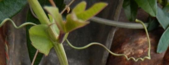
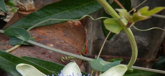
113 TATIANA EUGENIA ŞESAN & al. Passiflora caerulea L. TAXONOMICAL CHARACTERS PLATE IV Fig. 14 Fig. 15 Fig. 13 Fig. 17 Fig. 16 Fig. 18 Fig. 19 Fig


114 BOTANICAL AND PHYTOCHEMICAL APPROACH ON PASSIFLORA SPP. NEW Passiflora caerulea L. TAXONOMICAL CHARACTERS PLATE V Fig. 23 Fig. 21 Fig. 24 Fig. 22 Fig


115 TATIANA EUGENIA ŞESAN & al. Passiflora caerulea L. TAXONOMICAL CHARACTERS PLATE VI Fig. 26 Fig. 27 Fig. 28 Fig
116 BOTANICAL AND PHYTOCHEMICAL APPROACH ON PASSIFLORA SPP. NEW Physiological parameters In order to determine the morpho-anatomical (leaf length, leaf width, leaf area) and physiological (leaf growing rate, photosynthesis intensity / chlorophyll fluorescence and stomatal conductance) parameters, Passiflora caerulea plants from experimental plots have been monitorized during the whole time of this. Classic method has been used for the estimation of leaf area for many plant species by different researchers [LAZAROV, 1965; BOLDOR & al. 1983; MONTGOMERY & al. 1985; CHANDA & SINGH, 2002; CRISTOFORI & al. 2008; DEMIROY & LANG, 2010; PANDAY & SINGH, 2011; AHMED & KHAN, 2011 a.o.]. For Passiflora spp., this method for estimation leaf area has been published by REIMBERG & al. (2009), MORGADO & al. (2013) a.o. Leaf area. Using classic method of leaf area calculation in the formula LA = L x l x k (cm 2 ) appear the coefficient k. Average values calculated by us for the coefficient k have been 0.53, at and 0.47, after 20 days. Coefficient k, analyzed statistically by the test χ 2 (Hi/chi square) did not vary for the 3 factors (length, width and area), being the aspect which support and argue the specificity of this coefficient for each type of plant. These data confirm the literature data published by MOLGADO & al. (2013), which established the specificity of coefficient k for Passiflora spp., classifying the values calculated by us (k = ) as moderate and sufficient for the characterization of Passiflora plants from the point of view of the leaf morphologic parameters length, width, leaf area. Analyzing leaf growing rate during 14 days of experiment between the two determinations, at 7 and 20 days respectively (Tab. 4.) at Passiflora caerulea, the values were 4.1 cm 2 (calculated by classic method) and 6.1 cm 2 (calculated by computational method) in 14 days. Tab. 4. Morpho-physiological parameters of Passiflora caerulea leaves from the experimental field Hofigal: Leaf area (cm 2 ), Leaf growing rate (cm 2 ), Chlorophyll fluorescence [Y(II)], Stomatal conductance (nmol m -2 s -1 ), 2015 Parameters At 7 days At 20 days Difference Test t (p) Leaf length (cm) *** Leaf width (cm) * Leaf area (L x l x k) (cm 2 )* * Coefficient k Leaf area (cm 2 )** Leaf growing rate (cm 2 )* 4.1 Leaf growing rate (cm 2 )** 6.1 Chlorophylle fluorescence [Y(II)] (arbitrar unit) Stomatal conductance (nmol m -2 s -1 ) Yield green plant mass (kg/5 plants/replicate) Legend: * - classical method; ** - computational method Intensity of photosynthesis has been estimated by the parameter chlorophyll fluorescence. Through evaluation of chlorophyll fluorescence output it is possible to obtain information on the changes in photochemical efficiency and in the dissipation of heat [MAXWELL & JOHNSON, 2000; PASK & al. 2012]. Measurements on Passiflora caerulea 112
117 113 TATIANA EUGENIA ŞESAN & al. leaves have been showed the following values of chlorophyll fluorescence: 3341 arbitrary units, at and as 3046 arbitrary units, for the measurements at These values are quite similar at the both moments ( and ), they have not significantly varied. Stomatal conductance. This physiological parameter [MONTEITH & al. 1988; DEWAR 2002; PASK & al. 2012] for Passiflora was determined through porometric method, with a porometer Delta-T Devices AP4, based on gas diffusion through pores, especially through leaf stomata. Plant transpiration being adjusted by the opening and closing of stomata, using of these parameters is essential in many plant research domains. This is a measure of plant resistance to the loss of vapors through stomata and it is an indicator of plant physiological status. The equipment is working through the evaluation of the necessary time for a leaf to release sufficient water vapors for changing the relative humidity in a certain chamber with a fixed/standard quantity. Our determinations (Tab. 4.) showed values of m -2 s -1, which have been compared with PASK & al. (2012) results obtained for irrigated wheat. After these data, the normal values for this parameter in the wheat irrigated crop, in the conditions of Mexico, are nmol m -2 s -1, and values between v nmol m -2 s -1 show a reduced stress action on the plants [PASK & al. 2012]. As a conclusion, our data, the value of nmol m -2 s -1 is normal, belonging to the value interval nmol m -2 s -1. The parameter green mass production/yield (leaves, shoots, stems) determined at the end of our experiment has been estimated of kg/5plants replicate (Tab. 5). As a conclusion for this research group, our investigations have given us data for characterization of some physiological parameters (Tab. 5), as leaf area, leaf growing rate, coefficient k, chlorophyll fluorescence, stomatal conductance. Comparing with the literature [MOLGADO & al. 2013], coefficient k = can be evaluated as moderate and sufficient for Passiflora plants specific characterization. It has been estimate the chlorophyll fluorescence for Passiflora leaves over 3000 au ( au). Photosynthetic mechanisms respond very quickly to most of the stress that plants encounter [TOTH & al. 2007; GAMA & al. 2011; STRIBET & GOVINDJEE, 2011; IZAGUIRRE & al. 2013; CHAGUT & al a.o.]. Characteristics of gas exchange photosynthetic pigment determination and chlorophyll α fluorescence have been widely tested in many plant species as a parameter for the plant developments and for possible use in breeding programs to improve plant stress tolerance [GAMA & al. 2013]. Our data on stomatal conductance (554.2 nmol m -2 s -1 ), compared with the literature [PASK & al. 2012] have showed normal value, between 300 and 700 nmol m -2 s -1, for this parameter in an irrigated crop, as Passiflora is under the greenhouse conditions in Hofigal experimental field. All the physiological data are newly for the botanical and horticultural Romanian literature. Phytochemical investigations Qualitative analyses. After performing the qualitative methods we obtained the results shown in Tab. 5. Phytochemical screening showed that glycosides were detected in both studied plant extracts of Passiflora caerulea. In turn, tannins and alkaloids could not be detected. We observed that the alcoholic extracts contained more bioactive compounds than petroleum ether extracts, and saponins were detected only in ethanolic extracts. In the ethanolic extract of Passiflora caerulea leaves, saponins, glycosides and triterpenoids were detected, while the petroleum ether extract contained only glucosides and polysaccharides.
 Phytoconstituents PE Et-OH Glycosides + + Triterpenoids - +")
118 BOTANICAL AND PHYTOCHEMICAL APPROACH ON PASSIFLORA SPP. NEW Tab. 5. Qualitative bioactive compounds from the extracts of Passiflora caerulea (PE petroleum ether extract; Et-OH ethanol extract) Phytoconstituents PE Et-OH Glycosides + + Triterpenoids - + Polysaccharides + - Saponins - ++ Tannins - - Alkaloids - - Quantitative analyses. Quantitative data showed that high amount of total polyphenols (expressed in mg gallic acid/ g dw) and flavonoids (expressed as mg quercitine/ g dw) were found in ethanolic extracts of Passiflora leaves. The highest value determined in Passiflora leaves ethanolic extract was mg GA/g dw. Similar results were also obtained for flavonoids content, the maxim value being mg Q/g dw (Fig. 30). Fig. 30. The content of total phenols (determined by the Folin Ciocâlteu) and flavonoids for ethanol (Pf - EtOH) and petroleum ether extracts of Passiflora caerulea leaves (Pf - EP) Antioxidant capacity evaluation. Data obtained by the DPPH method shows that ethanol extract of Passiflora caerulea leaves had the largest content of free radical DPPH, with a concentration IC50 value of µg/ml, similar to the standard BHT (57.16 µg/ml) (Fig. 31). 114

119 TATIANA EUGENIA ŞESAN & al. Fig. 31. Antiradical activity of Passiflora caerulea extracts measured by DPPH method Similar results were obtained for the antioxidant capacity of plant extracts studied through inhibition of the radical ABTS assay. In this regard, Passiflora ethanol extract of leaves showed the highest antioxidant activity (79.77 ± 6.76 µmol TE / GMU) (Tab. 6). Tab. 6. The antioxidant activity of the ethanolic and petroleum ether extracts of Passiflora caerulea leaves, determined by ABTS and DPPH methods Trolox content DPPH test Analyzed extract (µmol/g dw) IC50 (µg/ml) Passiflora caerulea leaves ethanol 70% ± Passiflora caerulea leaves petroleum ether ± BHT Cell viability determination by Neutral Red method (NR). Test results showed a higher values of cell viability for concentrations between µg/ml Passiflora caerulea extracts, that did not inhibited cells growth at 24 hrs respectively 48 hrs, the cell viability being greater than 80% (98.3%, 97.86%, 94.26% and respectively 93.10%, compared to the control culture (considered 100%) (Fig. 32). After 48 hrs, we observed a slight decrease in the cell proliferation, but all values were maintained above 80% at the mentioned concentrations. Extract concentrations between 250 and 500 µg/ml affected the normal development of culture and had a toxic effect on individual cells. It was observed a significant inhibition on the cell viability, less than 70%, at 24 hrs and 48 hrs, respectively (Fig. 32). 115
, after 24 hrs respectively 48 hrs after cultivation, determined by Neutral Red method The results obtained by")
120 BOTANICAL AND PHYTOCHEMICAL APPROACH ON PASSIFLORA SPP. NEW Fig. 32. The effect of Passiflora caerulea extracts on the culture of fibroblasts (NCTC clone 929), after 24 hrs respectively 48 hrs after cultivation, determined by Neutral Red method The results obtained by Neutral Red method showed that Passiflora caerulea extract concentrations between 1 and 150 µg/ml did not inhibit cells growth at 24 hrs and 48 hrs, respectively, cells viability being greater than 80%. We also observed a slight decrease in cell viability at 48 hrs. At concentrations of 250 and 500 μg/ml, P. caerulea extract determined a decrease of cell viability after 24 hrs and 48 hrs, respectively. Cell morphology analysis through light microscopy. Our results indicated that the influence of different concentrations of Passiflora extracts on the NCTC cells morphology depend on the concentration. At extract concentrations between 1 and 150 µg/ml there are not an important changes in the cells morphology and density. Cells had a normal aspect, with 2-3 cytoplasmatic extensions, monochrome cytoplasm and very few rounded cells were observed (Fig. 33.B). At the concentration of 250 µg/ml it was observed an obvious decrease in cell density and the cells showed an altered morphology (Fig. 33.C). At the concentration of 500 µg/ml, P. caerulea extracts had a toxic effect, determined cells membranes integrity alteration and affecting the normal development of cell culture (Fig. 33.D). In summary, our results have proved that: (i) The highest concentration in polyphenols and flavonoids was recovered in the ethanolic extracts of P. caerulea; (ii) A correlation between the polyphenols/flavonoids content of P. caerulea extracts and their antioxidant activity was observed. The highest values of antioxidant activity were calculated for the ethanolic plant extracts; (iii) The NR method showed a good biocompatibility of P. caerulea extracts in NCTC cell line, up to µg/ml concentration, sustained by a normal cell morphology. At concentrations higher than 250 µg/ml, the plant extract become cytotoxic, altering the cell membrane structure, the cells viability and proliferation. 116
 These data allowed us to select the optimal")

 Leaf lamina with a dorsi-ventral structure, hipostomatal and with lack of trichoms; (ii) Stomates of anomocitic and anizocitic types; (iii) Leaf petiole with an adaxial large")
 Leaves generating only collenchyma; (vii) Calcium oxalate crystals present in different cells from the leaf and stem structure of the druse")
121 TATIANA EUGENIA ŞESAN & al. A. Control (NCTC) B. Extract of P. caerulea, 100 µg/ml C. Extract of P. caerulea, 250 µg/ml D. Extract of P. caerulea, 500 µg/ml Fig. 33. Effect of P. caerulea extracts on culture of fibroblasts NCTC at concentrations of 100, 250 and 500 µg/ml after 48 hrs of cultivation (Giemsa stain, 20X objective) These data allowed us to select the optimal range of non-cytotoxic concentrations of the Passiflora extracts (less than 250 µg/ml) that will be use in further experiments. Conclusions Our complex investigation, morpho-anatomical, physiological, taxonomical and phytochemical on Passiflora nutraceutical plants from Hofigal experimental field, lead us to the following conclusions: 1. Structural aspects analyzed are important in characterization of P. caerulea samples: (i) Leaf lamina with a dorsi-ventral structure, hipostomatal and with lack of trichoms; (ii) Stomates of anomocitic and anizocitic types; (iii) Leaf petiole with an adaxial large kennel and a monosimetrical structure with vascular distinct fascicles; (iv) Irregularribbed stem which differentiates a secondary structures with concentric xylem and phloem rings, respectively; (v) Stem presenting collenchyma and sclerenchyma tissues; (vi) Leaves generating only collenchyma; (vii) Calcium oxalate crystals present in different cells from the leaf and stem structure of the druse type. In Romania, present histo-anatomical studies with taxonomic importance are the first ones with a significant highlighting to define more accurately the taxa of Passiflora genus. We will continue the botanical analysis, investigating a higher number of samples in order to evaluate additional identification aspects as: plant phenotypic plasticity, hybridization, influence of environmental and technological factors. 117
122 BOTANICAL AND PHYTOCHEMICAL APPROACH ON PASSIFLORA SPP. NEW 2. From taxonomical point of view, plant materials sampled for these studies from S.C. Hofigal Export-Import S.A. have been analyzed, obtaining a botanical evaluation that these plants belong taxonomical to Passiflora caerulea L., in concordance with the data of the monographers of Passiflora genus in the world: VANDERPLANK (2000) and ULMAN and MacDOUGAL (2004). 3. Physiological aspects determined in this approach have given us data for characterization of some parameters, as: (i) Coefficient k = , evaluated as moderate and sufficient for Passiflora plants specific characterization; (ii) Leaf area varied between 30 cm 2 and 34.1 (calculated by classic method), respectively 36.1 cm 2 (computational method); (iii) Leaf growing rate varied between 4.1 cm 2, approximately 0.29 cm 2 /experimental day (classic method) and 6.1 cm 2, approximately 0.43 cm 2 /day (computational method); (iv) Chlorophyll fluorescence was evaluated over 3000 arbitrary units ( ); (v) Stomatal conductance (554.2 nmol m -2 s -1 ), with a normal value (554.2 nmol m -2 s -1 ), between 300 and 700 nmol m -2 s -1, in an irrigated crop, as Passiflora is under the greenhouse conditions in Hofigal Company; (vi) Yield green plant mass (kg/5 plants/replicate) has reached in our experiment to an average value of kg/5 plants. All these results are new for the Romanian botanical literature. 4. Referring to the phytochemical approach, the influence of the extraction medium on the content of some active principles was analyzed and (i) the highest concentration in polyphenols and flavonoids was recovered in the ethanolic extracts of Passiflora; (ii) a correlation between the polyphenols/flavonoids content of P. caerulea extracts and their antioxidant activity was observed, the highest values of antioxidant activity being calculated for the ethanolic plant extracts; (iii) The NR method showed a good biocompatibility of P. caerulea extracts in NCTC cell line, up to µg/ml concentration, sustained by a normal cell morphology. At concentrations higher than 250 µg/ml, the plant extract become cytotoxic, altering the cell membrane structure, the cells viability and proliferation. All these data are new for the Romanian phytochemical literature and allowed us to select the optimal range of non-cytotoxic concentrations of the Passiflora extracts (less than 250 µg/ml) that will be use in further experiments. Authors contributions: T. E. ŞESAN coordinated and monitorized the project 160/2014, performed designed experiments, prepared the manuscript, analyzed and gave general interpretation of data (leaf morphology, physiological data, taxonomical evaluation, phytochemical data, photos), prepared periodical project reports, techno-manufactured the manuscript. A. SÂRBU and D. SMARANDACHE performed section of vegetal material, provided anatomical data and their interpretation for taxonomical evaluation, took microscopic photos; F. OANCEA prepared and supervised finishing of manuscript. A. OANCEA, S. SAVIN, A. TOMA and L. ŞTEFAN performed phytochemical analyses and their interpretation. G. NEGRU, A. F. BIRA and G. VLĂSCEANU applied in the experimental field of Hofigal the experiment designed by the coordinator of project, managed P. caerulea crop and provided samples for laboratory analyzes. M. GHIUREA, G. VASILESCU and L. JECU obtained and interpreted physiological experimental data. C. M. POMOHACI analyzed statistically experimental data. Acknowledgements These results are based upon work supported by the Ministry of National Education Research Activity, CNDI UEFISCDI, in the frame of the project number PN-II-PT-PCCA (project 160-MAIA). 118
123 TATIANA EUGENIA ŞESAN & al. Explanation of plates and figures Fig. 1. A. Working A4 squared sheet of paper/page used for classical method of leaf area estimation; 1.B. Passiflora caerulea simple, palmat-partite leaves (herborized and scanned) (Şesan, 2015) Fig. 2. Main medicinal and nutraceutical importance of Passiflora spp. PLATE I (photo: Anca Sârbu) Passiflora caerulea LAMINA Cross section through median zone of the leaf segment, colorants Iodine Green and Carmine Alum (Fig. 3) Cross section through the median zone of leaf segment, with evidence of epidermis and mesophyll, colorants iodine green and Carmine alum (Fig. 4) Adaxiale epidermis (superior) in apical image (Fig. 5) Abaxiale epidermis (inferior) in apical image (Fig. 6) Abaxiale epidermis in apical image, with evidence of calcium oxalate crystals (polarized light) (Fig. 7) Abaxiale epidermis in apical image, with evidence of calcium oxalate crystals (polarized light) (Fig. 8) PLATE II (photo: Anca Sârbu) Passiflora caerulea PETIOLE Cross section through petiole, colorants Iodine Green and Carmine Alum (Fig. 9) Cross section through petiole, colorants Iodine Green and Carmine Alum (Fig. 10) PLATE III (photo: Anca Sârbu) Passiflora caerulea STEM Cross section through stem, colorants Iodine Green and Carmine Alum (Fig. 11) Cross section through stem, with evidence of epidermis, cortex and of central cylinder elements, colorants Iodine Green and Carmine Alum (Fig. 12) PLATE IV (photo: Tatiana Eugenia Şesan, ) Passiflora caerulea TAXONOMIC CHARACTERS General aspect of the Passiflora caerulea vine in the experimental field of Hofigal (climbing vine, flowers, floral calyx, 5-diveded lobate leaves) (Fig. 13) Passiflora stipules and tendrils (Fig ) Passiflora stipule (Fig. 17) Passiflora stipule and 2 nectary glands at the petiole base (Fig. 18) Passiflora tendrils (Fig. 16, 19, 20) PLATE V (photo: Tatiana Eugenia Şesan, ) Passiflora caerulea TAXONOMIC CHARACTERS Passiflora caerulea flowering in the greenhouse at experimental field of Hofigal (climbing vine, flowers, floral calyx, 5-diveded lobate leaves) (Fig. 21) Passiflora caerulea flower, 5-lobed leaf, tendrils (Fig. 22) Passiflora caerulea white flowers, corolla white; sepals 5, white to pale, calyx tube cup-shaped, petals 5, similar as form and size with sepals; petals white inside and outside, corona (petals) disposed in 2 verticils, extern verticil consisting in many filiform appendices (radia), purple at the base, in the middle white and blue at the apex, corona filaments in 4 series, outer 2, purple at the base, then white and blue towards apex, gynophore well developed; stamens 5, unilocular ovary, styles 3, reddish (Fig ) PLATE VI (photo: Tatiana Eugenia Şesan) Passiflora caerulea TAXONOMIC CHARACTERS Passiflora caerulea floral buds in the experimental field of Hofigal ( ) (Fig. 26) Passiflora caerulea at the phenofase of green fruits ( ) (Fig. 27) Passiflora caerulea at the phenofase of orange fruits at the ripe status: , left (Fig. 28); , right (Fig. 29) Fig. 30. The content of total phenols (determined by the Folin Ciocâlteu method) and flavonoids for ethanol (Pf - EtOH) and petroleum ether extracts of Passiflora caerulea leaves (Pf -EP) Fig. 31. Antiradical activity of extracts of Passiflora caerulea measured by DPPH method Fig. 32. The effect of Passiflora caerulea extracts on the culture of fibroblasts (NCTC clone 929), after 24 hrs. respectively 48 hrs. after cultivation, determined by neutral red method Fig. 33. Effect of Passiflora caerulea extracts on culture of fibroblasts NCTC at concentrations of 100, 250 and 500 mg/ml after 48 hrs. of cultivation (Giemsa stain, 20X objective) 119
124 BOTANICAL AND PHYTOCHEMICAL APPROACH ON PASSIFLORA SPP. NEW References 1. AHMED N. & KHAN D Leaf area estimation in Jatropha curcas L. Int. J. Biol. Biotech. 8(3): AKANBI B. O., BODUNRIN O. D. & OLAYANJU S Phytochemical screening and antibacterial activity of Passiflora edulis, Researcher. 3(5): AKHONDZADEH S., KASHANI L., MOBASERI M., HOSSEINI H., NIKZAD S. & KHANI M. 2001a. Passionflower in treatment of opiates with drawal: a double-blind randomized controlled trial. Journal of Clinical Pharmacy and Therapeutics. 26(5): AKHONDZADEH S., HAGHAVI H. R., VAZIRIAN M., SHAYEGABPOUR A., RASHIDI H. & KHANI M. 2001b. Passionflower in the treatment of generalized anxiety: a pilot double-blind randomized controlled trial with oxazepam. Journal of Clinical Pharmacy and Therapeutics. 26: AKHONDZADEH S., MOHAMMADI M. R. & MOMENI F Passiflora incarnata in the treatment of attention-deficit hyperactivity disorder in children and adolescents. Future Drugs. 2: APPEL K., ROSE T., FIEBICH B., KAMMLER T., HOFFMAN C. & WEISS G., 2010/2011. Modulation of the γ- aminobutyric acid (BABA) system by Passiflora incarnata L. Phytotherapy Research. 25(6): , published Online Library (wileyonlinelibrary.com) DOI: /ptr.3352; (2011) 25: ASLANARGUN P., CUVAS O., DIKMEN B., ASLAN E. & YUKSEL M. U Passiflora incarnata Linneus as an anxiolytic before spinal anesthesia. Journal of Anesthesia. 26(1): 39-44; doi: /s BARBALHO S. M., DE SOUZA M. D. S. S., DE PAULA E SILVA J. C., MENDES C. G., DE OLIVEIRA G. A., COSTA T. & FARINAZZI-MACHADO M. V Yellow passion fruit rind (Passiflora edulis): an industrial waste or an adjuvant in the maintenance of glycaemia and prevention of dyslipidemia? Journal of Diabetes Research & Clinical Metabolism; 9. BERNACCI L. C., VITTA F. A. & BAKKER Y. V Passifloraceae. In: WANDERLEY M. G. L., SHEPHERD G. J., GIULIETT A. M. & MELHEM T. S. (eds.). Flora Fanerogâmica de Estado de São Paulo. São Paulo, RiMa/FAPEST, v. 3: ; SciELO Brazil BERNACCI L. C., SOARES-SCOTT M. D., JUNQUEIRA N. T. V., DA SILVA PASSOS I. R. & MELETTI L. M. M Passiflora edulis Sims: the correct taxonomic way to cite the yellow passion fruit (and others colors). Rev. Bras. Frutic. 30(2): 17 pp; BLANKESPOOR J Passionflower Ecology, Cultivation, Botany, and Medicinal and Edible Uses. Blog Castanea: BOERICKE W Passiflora incarnata. Homoeopathic Materia Medica. Retrived 2015 Oct 16 from BOLDOR O., RAIANU O. & TRIFU M Fiziologia plantelor. Edit. Didactică şi Pedagogică, Bucureşti: BORAWSKA M. H., MARKIEWICZ-ŹUKOWSKA R., SAWICKA D., NALIWAJKO S. K., SOCHA K., OMELJANIUK W. & CAR H Effects of chrysin on haematological parameters in rats. Farmacia. 62(2): BORRELLI F., PINTO L., IZZO A. A., MASCOLO N., CAPASSO F., MERCATI V., TOJA E. & AUTORE G Anti-inflammatory activity of Passiflora incarnata L. in rats. Phytotherapy Research. 10: S104-S BOURIN M., BOURGEROL T., GUITTON B. & BROUTIN E. A A combination of plant extracts in the treatment of outpatients with adjustment disorder with anxious mood: controlled study versus placebo. Fundamentals of Clinical Pharmacology. 11: BROWN E., HURD N. S., MCCALL S. & CEREMUGA T. E Evaluation of the anxiolytic effects of chrysin, a Passiflora incarnata extract, in the laboratory rat. American Association of Nurse Anesthetists (AANA) Journal. 75(5): CARNEVALLI DIAS L. L., RIBEIRO D. M., CATARINA C. S., BARROS R. S., FLOH E. I. S. & OTONI W. C Ethylene and polyamine interactions in morphogenesis of Passiflora circinnata: effects of ethylene biosynthesis and action modulators, as well as ethylene scavengers. Plant Growth Regulators. 62: CARRASCO M. C., VALLEJO J. R., PARDO-DE-SANTAYANA M., PERAL D., MARTIN M. A. & ALTIMIRAS J Interactions of Valeriana officinalis L. and Passiflora incarnata L. in a patient treated with lorazepam. Phytotherapy Research. 23(12): ; doi: /ptr CAZARIN C. B. B., DA SILVA J. K., COLOMEN T. C., ZOLLUER R. D. L. & MARÓSTICA Jr. M. R [Antioxidant capacity and chemical composition of passion fruit peel (Passiflora edulis)]. Ciência Rural (Santa Maria). 44(9): [in Portuguese]. 21. CERVI A. C Passifloraceae do Brasil. Estudo do gênero Passiflora L, subgênero Passiflora. Fontqueria. XLV: CHAGUT N., SOBOL S., NAVE N. & SAMACH A Shielding flowers developing under stress: translating theory to field application. Plants. 3:
125 121 TATIANA EUGENIA ŞESAN & al. 23. CHANDA S. V. & SINGH Y. D Estimation of leaf area in wheat using linear measurements. Plant Breeding and Seed Science. 46(2): CHINNIAH V. & THIAGARAJAN V.R.K Anatomical investigation on the leaves and stem of Passiflora incarnata (Passifloraceae), International Journal Research Ayurveda Parmacognosy. 6(4), July-August: ; CHITRA V., GOWRI K., TOMILABAN T. & SONI D Neuropharmacological screening of aerial parts of Passiflora incarnata Linn (Passifloraceae). Scholars Research Library. Archives of Applied Science Research. 1(2): ; CILLA F Passiflora and its anxiolytic and anticonvulsant effects, file:///c:/documents%20and%20setting/tatiana/desktop/passiflora, accessed CRISTOFORI V., ROUPHAEL Y., MENDOZA-DE GYVES E. & BIGNAMI C A simple model for estimating leaf area of hazelnut from linear measurements. Scientia Horticulturae. 113: CRISTOFORI V., FALLOVO C., MENDOZA-DE GYVES E., BIGNAMI C. & ROUPHAEL Y Nondestructive, analogue model for leaf area estimation in persimon (Diospyros kaki L.f.) based on leaf length and width measurement. European Journal of Horticultural Sciences. 73: DA SILVA J. K., CAZARIN C. B. B., COLOMEU T. A., BATISTA A. G., MELETTI L. M. M., PASCHOAL J. A., R., BOGUSZ JR. S., FURLAN M. F., REYES F. G. R. & AUGUSTO F Antioxidant activity of aqueous extract of passion fruit (Passiflora edulis) leaves: In vitro and in vivo study, Food Research International. 53: DA SILVA J. K., CAZARIN C. B. B., BATISTA A. G. & MARÓSTICA Jr. M Effects of passion fruit (Passiflora edulis) bioproduct intake in antioxidant status of Wistar rets tissues. LWT Food Science and Technology. 59: DE FARIAS V Anatomia foliar de Passiflora L. (Passifloraceae): Aspectos taxonômicos e evolutivos. Universidade Federal Do Paraná, Curitiba: Ph Dissertation: 63 pp. 32. DEGINANI N. B The species of Passiflora (Passifloraceae) [In Portuguese]. Darwiniana. 39(1-2): DEMIRSOY H. & LANG G. A Short communication. Validation of a leaf area estimation model for sweet cherry. Spanish Journal of Agricultural Research. 8(3): ; DE-PARIS F., PETRY R. D., REGINATTO F. H., GOSMANN G., QUEVEDO J., SALGUEIRO J. B., KAPCZINSKI F., GONZÁLEZ-ORTEGA G. & SCHENKEL E. P Pharmacochemical study of aqueous extracts of Passiflora alata Dryander and Passiflora edulis Sims. Acta Farm Bonaer. 21: DE QUEIROZ M. S., JANEBRO D. I., DE CUHNA M. A. L. D., MEDEIROS J. D. S., SABAA-SHUR A. U. O., DINIZ M. D. F. F. M. & DOS SANTOS S. C Effect of the yellow passion fruit peel flour (Passiflora edulis f. flavicarpa Deg.) in insulin sensitivity in type 2 diabetes mellitus patients. Nutrition Journal. 11: 89-96; doi: / DEVESA A. J. G., MARTINEZ A. M. & ORTEGA-ALBÁS J. J Herbal drugs and sleep: what can we expect?. Journal of Sleep Disorders & Therapy. 4(3): 3 pp.; DEWAR R. C The Ball-Berry-Leuning and Tardieu-Davies models: Synthesis and extension within a spatially aggregated picture of guard cell formation. Plant, Cell and Environment. 25: DHAWAN K. & SHARMA A. 2002a. Antitussive activity of the methanol extract of Passiflora incarnata leaves. Fitoterapia. 73(5): DHAWAN K. & SHARMA A. 2002b. Prevention of chronic alcohol and nicotine-induced zospermia, sterility and decreased libido, by a novel tri-substituted benzoflavone moiety from Pasiflora incarnata Lin. in healthy male rats. Life Sciences. 71: DHAWAN K. & SHARMA A Restoration of chronic-d9-thc-induced decline in sexuality in male rats by a novel benzoflavone moiety from Passiflora incarnata Linn, British Journal of Pharmacology. 138: DHAWAN K., DHAWAN S. & CHHABRA S Attenuation of benzodiazepine dependence in mice by a trisubstituted benzoflavone moiety of Passiflora incarnata Linneus: a non-habit forming anxiolytic. Journal of Parmmacy & Pharmaceutical Sciences. 6(2): DHAWAN K., KUMAR S. & SHARMA A. 2001a. Anxiolytic activity of aerial and underground parts of Passiflora incarnata. Fitoterapia. 72: DHAWAN K., KUMAR S. & SHARMA A. 2001b. Anti-anxiety studies on extracts of Passiflora incarnata Linneus. Journal of Ethnopharmacology. 78: DHAWAN K., KUMAR S. & SHARMA A. 2002a. Reversal of cannabinoids (delta9-thc) by the benzoflavone moiety from methanol extract of Passiflora incarnata Linn in mice: a possible therapy for cannabonoid addiction. Journal of Pharmacy and Pharmacology. 54(6): DHAWAN K., KUMAR S. & SHARMA A. 2002b. Nicotine reversal effects of the benzoflavone moiety from Passiflora incarnata Linn in mice. Addiction Biology. 7(4): DHAWAN K., KUMAR S. & SHARMA A. 2002c. Suppression of alcohol-cessation-oriented hyper-anxiety by the benzoflavone moiety of Passiflora incarnata Linn. in mice. Journal of Ethnopharmacology. 81(2):
126 BOTANICAL AND PHYTOCHEMICAL APPROACH ON PASSIFLORA SPP. NEW 47. DHAWAN K., KUMAR S. & SHARMA A. 2003a. Aphrodisiac activity of methanol extract of leaves of Passiflora incarnata Linn in mice. Phytotherapy Research. 17(4): DHAWAN K., KUMAR S. & SHARMA A. 2003b. Antiasthmatic activity of the methanol extract of leaves of Passiflora incarnata. Phytotherapy Research. 17(7): DHAWAN K., KUMAR S. & SHARMA A Passiflora: a review update. Journal of Ethnopharmacology. 94: DHAWAN K., KUMAR R., KUMAR S. & SHARMA A Correct identification of Passiflora incarnata Linn., a promising herbal anxiolytic and sedative. J. Med. Food. 4(3): DOBRE S. P. & LAZĂR C Determinări de suprafaţă foliară pentru frunza steag la un set de linii DH mutante la grâu. Analele I.N.C.D.A. LXXXII: 7-16; DOS SANTOS L. F., DE OLIVEIRA E. J., DOS SANTOS SILVA A., DE CARVALHO F. M., COSTA J. L., PÁDUA J. G ISSR Markers as a tool for three assessment of genetic diversity in Passiflora. Biochemical Genetics. 48(7): ELSAS S. M., ROSSI D. J., RABER J., WHITE G., SEELEY C.A., GREGORY W. L., MOHR C., PFANKUCH T. & SOUMYANATH A Passiflora incarnata L. (Passionflower) extracts elicit GABA currents in hippocampal neurons in vitro, and shows anxiogenic and anticonvulsant effects in vivo, varying with extraction method. Phytomedicine. 17(12): ERNST E Herbal remedies for anxiety a systematic review of controlled clinical trials. Phytomedicine. 13: FOSTER S Passionflower - Passiflora incarnata; Stevenfoster.com/education/monograph/ pflower.html. 56. FOTAKIS G. & TIMBRELL J. A In vitro cytotoxicity assays: comparison of LDH, neutral red, MTT a protein assay in hepatoma cell lines following exposure to cadmium chloride. Toxicol Lett. 160: In: CIOFI N. & RAI M. (eds.) Nano-Antimicrobials Berlin Heilderberg: 555 pp. ISBN FRANCO J Passiflora. The flowers of passion. Printed in USA pp. 58. GAMA V. N., CUNHA J. T., DE MELO LIMA I., BACARIN M. A. & SILVA D. M Photosynthetic characteristics and quality of five passion fruit varieties under field conditions. Acta Physiol Plant. 35: ; DOI /s GARCIA M. & PÉREZ D. J. D Anatomia foliar de Passiflora guazumaefolia Juss. y Passiflora aff. tiliaefolia (Passifloraceae) presentes in Venezuela. Ernstia, Caracas. 18(2): 7 pp; GRINŢESCU I Passifloraceae. Passiflora. Flora Republicii Populare Romîne, Edit. Academiei Republicii Populare Romîne, Bucuresti: GHIUREA M., RĂUT I., ŞESAN T. E., OANCEA F., NEGRU G., VASILESCU G. & JECU L Physical methods applied for the investigation of the plant protection against the diseases produced by phytopathogens, Symposium PRIOCHEM 2015, Proceedings (ISSN ): GRUNDMANN O., WANG J., MCGREGOR G.P. & BUTTERWECK V Anxiolytic activity of a phytochemically characterized Passiflora incarnata extract is mediated via the GABAergic system. Planta Medica. 74(15): GRUNDMANN O., WHALING C., STAIGER C. & BUTTERWECK V Anxiolytic effects of a passion flower (Passiflora incarnata L.) extract in the elevated plus maze in mice. Pharmazie. 64(1): HOLBIK M., KRASTEVA S., MAYER N., KÄHLIG H. & KRENN L Apparently no sedative benzoflavone moiety in Passiflora. Planta Medica. 76(7): ; DOI: /s HOOKER J. D., JACKSON D. B. & DARWIN Ch Index Kewensis Plantarum Phanerogamarum. Clarendon PressVol.2: ; suppl. I, : 316; suppl. II, : 135; suppl. III, : 130; suppl. IV, : ; suppl. V, :187; suppl. VI, :146; suppl. VII, : ; suppl.viii, :173; suppl.ix, :204; suppl. X, :162; suppl. XI, :179; suppl. XII, :103; suppl. XIII, :100; suppl :100; suppl. XV, : HURCIKOVA A. 2014/2015. An herbal approach to the treatment of withdrawal from opioid and benzodiazepine dependence. In Student Herbal Thymes Winter Edition: HUTCHINSON M Passiflora incarnata Purple Passionflower. Native Plant Owners Manual. Florida Native Plant Society: INGALE A. G. & HIVRALE A. U Pharmacological studies of Passiflora sp. and their bioactive compounds. African Journal of Plant Science. 4(10): IZGUIRE M. M., MAZZA C. A., ASTIGUETA M. S., CIARLA A. M. & BALLARÉ C. L No time for candy: passionfruit (Passiflora edulis) plants down-regulate damage-induced extra floral nectar production in response to light signals of competition. Oecologia. 173: KANAKASABAPATHI D. & GOPALAKRISHNAN V. K Evaluation of antidiabetic potential of aqueous extract of Passiflora edulis Sims on alloxan induced Diabetes Mellitus in Wistar albino rats. Int. J. Pharm. Sci. Rev. Res. 34(1):
127 TATIANA EUGENIA ŞESAN & al. 71. KAVIANI N., TAVAKOLI M., TABANMEHR M. R. & HAVAEI R. A The efficacy of Passiflora incarnata Linnaeus in reducing dental anxiety in patients undergoing periodontal treatment. Journal Dentistry of Shiraz University of Medical Sciences. 14(2): KAVITHA KRISHNA R., KRISHNA KUMARI S., VINODHA M., SEBASTIN A. & GOMATHI R In vitro free radical scavenging activity of Amorphophallus commutatus an endemic aroid of Western Ghats south India. International Research Journal of Pharmacy. 3(2): LAKHAN S. E. W. & VIEIRA K. F Nutritional and herbal supplements for anxiety and anxiety-related disorders: systemic review. Nutrition Journal. 9: LAM S. K. & NG T. B Passiflin, a novel dimeric antifungal protein from seeds of the passion fruit. Phytomedicine. 16(2-3): LAZAROV R Coefiţienti za opredeliane na listnata povârhnost pri niakoi selskostopanski culturi, [Coefficients for the determination of leaf area in certain agricultural crops]. Rastenievdnii Nauki Sofia [Plant Science]. II(2): LIM T. K. (ed.) Edible medicinal and non-medicinal plants, vol. 4, Fruits, Springer Dordrecht, Heidelberg, London, New York: LOPES S. C Citogenética do Maracujá, Passiflora spp. In: SÃO JOSÉ A. R. (ed.). A culturado maracujá no Brasil Funep. Funep, Jaboticabal: MANICA I Maracujá: Taxonomia-anatomia-morfologia. In: SÃO JOSÉ A. R., BRUCKNER C. H., MANICA I., HOFMANN M. (eds.). Maracujá: Temas selecionados-melhoramento, morte prematura, polenização, taxonomia. Cinco Continentes, Porto Alegre: MAXWELL K. & JOHNSON G. N Chlorophyll fluorescence a practical guide. J. Exp. Bot. 51(345): MIRODDI M., CALAPAI G., NAVARRA M., MINCIULLO P. L. & GANGERNI S Passiflora incarnata L.: ethnopharmacology, clinical application, safety and evaluation of clinical trials. Journal of Ethnopharmacology. 150(3): ; doi: /j.jep MIYASAKA L. S., ATALLAH A. N. & SOARES B. G. O Passiflora for anxiety disorder (Review). Cochrane Database of Systematic Reviews. 24(1): CD MOHAMMED ALDUGMAN, MUBARAK AL GAHTANY & GAITHAMAN K Neuroprotective effect of methanolic extract of Passiflora nepalensis against oxidative stress in middle cerebral artery occlusion model of stroke in rats. Journal of Global Trends in Pharmaceutical Sciences (JGTPS). 6(4): MONTEITH J. L., CAMPBELL G. S. & POTTER E. A Theory and performance of a dynamic diffusion porometer. Agricultural & Forest Meteorology. 44: MONTGOMERY E. G Correlation studies in corn. Nebraska Agricultural Experimental Station, Annual Report. 24: MORGADO M. A. D O., BRUCKNER C. H., ROSADO L. D. S., ASSUNAÇÃO W. & DOS SANTOS C. E. M Estimação da áres foliar por método não destrutivo, utilizado medidas lineares das folhas de espécies de Passiflora, [Estimation of leaf area by non-destructive method using linear measurements of leaves of Passiflora species]. Revista Ceres [on line] 60(5): ; MORI A., HASEGAWA K., MURASAKI M., YAMAUCHI T., ITO S., ASAI M., KUDO Y., NAKAJIMA T., NISHIMURA T., SAITO M., HANADA M., IKAWA G., HIGASHI Y. & KAWAKITA Y Clinical evaluation of Passiflamin (Passiflora extract) on neurosis. Multicenter, double-blind study in comparison with mexazolam. Rinsho Hyoka [Clinical Evaluation for Drugs]. 21(3): MOVAFEGH A., ALIZADEH R., HAJIMOHAMADI E., ESFEHANI F. & NEJATFAR M Preoperative oral Passiflora incarnata reduces anxiety in ambulatory surgery patients: a double-blind, placebo-controlled study. Anesthesia and Analgesia. 106(6): ; doi: /ane.0b013e318172c3f NASCIMENTO C. S. K. & SABAA-SRUR U. O Influence of passion fruit (Passiflora edulis f. flavicarpa, DEG) juice sweetened with sucrose, saccharin and cyclamate on blood glucose in normal and diabetic rats (Rattus norvegicus). The Natural Products Journal. 2(2): NASSIRI-ASL M., SHARIATI-RAD F. & ZAMANSOLTANI F Anti-convulsant effects of arial parts of Passiflora incarnata extract in mice: involvement of benzodiazepine and opioid receptors. BioMed Central Complementary and Alternative Medicine. 7: 26-31; doi: / /7/ NGAN A. & CONDUIT R. A A double-blind, placebo-controlled investigation of the effects of Passiflora incarnata (Passionflower) herbal tea on subjective sleep quality. Phytotherapy Research. 25: ; doi: /ptr NORIEGA P., DE FREITAS MAFUD D., STRASSER M., KATO E. T. M. & BACCHI E. M Passiflora alata Curtis: a Brazilian medicinal plant. Boletin Latinoamericano y del Caribe de Plantas Medicinales y Aromáticas. 10(5):
128 BOTANICAL AND PHYTOCHEMICAL APPROACH ON PASSIFLORA SPP. NEW 92. OANCEA A., ROATĂ G., POPESCU S., PĂUN L., MATEESCU I., TOMA A. E., GASPAR A. & SIDOROFF M Phytochemical screening of the bioactive compounds in the most widespread medicinal plants from Cǎlǎraşi-Silistra cross-border area. Buletin of Transilvania University of Brasov. 6(55): OLIVEIRA G. A. F., PÁDUA J. G., COSTA J. L., DE JESUS O. N., DE CARVALHO F. M. & DE OLIVEIRA E. J Cross-species amplification of microsatellite loci developed for Passiflora edulis Sims. in related Passiflora species. Braz. Arch. Biol. Technol. 57(5): OLLANKETO M., PELTOKETO A., HARTONEN K., HILTUNEN R. & RIEKKOLA M.L Extraction of sage (Salvia officinalis L.) by pressurized hot watter and conventional methods: antioxidant activity of the extracts. European Food Research and Tehnology. 215(2): PANDEY S. K. & SINGH H A simple, cost-effective method for leaf estimation. Journal of Botany, Hindawi Publishing Corporation, vol. 2011, Article ID , 6 pp; doi: /2011/ PASK A. J. D., PIETRAGALLA J., MULLAN D. & REYNOLDS M. (eds.) Physiological breeding II: a field guide to wheat phenotyping. Mexico DF: CIMMYT (International Maize and Wheat Improvement Center), ISBN: L152, Institutional Multimedia Publications Repository: 133 pp; PATEL S. S Morphology and pharmacology of Passiflora edulis: a review. Journal of Herbal Medicine and Toxicology. 3(5): PATEL S. S., MOHAMED SALEEM T. S., RAVI V., SHRESTHA B., VERMA N. K. & GAUTHAMAN K Passiflora incarnata Linn: A phytopharmacological review. International Journal of Green Pharmacy. October-December: PATEL S. S., VERMA N. K., RAVI V., GAUTHAMAN K. & SONI N Antihypertensive effect of an aqueous extract of Passiflora nepalensis Wall. International Journal of Applied Research in Natural Products. 3(2): PATEL S. S., SONI H., MISHRA K. & SINGHAI A. K Recent updates on the genus Passiflora: A review. International Journal of Research in Phytochemistry & Pharmacology. 1(1): PÉREZ-CORTEZ S., ESCALA S. M. & TILLETT S Anatomia de la cubierta seminal en ocho especies de Passiflora L., subgénero Passiflora. Acta Bot. Venez. 28: POURMORAD F., HOSSEINIMEHR S. J. & SHAHABIMAJD N Antioxidant activity, phenol and flavonoid contents of some selected Iranian medicinal plants. African Journal of Biotechnology. 5(11): RASMUSSEN PH A role of phytotherapy in the treatment of benzodiazepine and opiate drug withdrawal. Modern Phytotherapist. 3(3): RĂUT I., GHIUREA M., ŞESAN T. E., NEGRU G., JECU L., CĂLIN M., VASILESCU G. & OANCEA F Influenţa tratamentelor cu elicitori ai rezistenţei sistemice asupra parametrilor morfofiziologici au unor plante nutraceutice. Sesiunea anuală de comunicări a ICDPP Bucureşti Protecţia plantelor, cercetare interdisciplinară în slujba dezvotării durabile a agriculturii şi protecţiei mediului, Rezumatele lucrărilor prezentate, ISBN , 6 noiembrie 2015: REGINATTO F. H., DE-PARIS F., PETRY R. D., QUEVEDO J., ORTEGA G. G., GOSMANN G. & SCHENKEL E. P Evaluation of anxiolytic activity of spary dried powders of two South Brazilian Passiflora species. Phytotherapy Research. 20(5): ; doi: /ptr REIMBERG M. C. H., COLOMBO R. & YARIWAKE J. H [Análise multivariada dos efeitos dos parâmetros do solo e fatores ambientais no teor de flavonoides das folhas de Passiflora incarnata L.], Multivariate analysis of the effects of soil parameters and environmental factors on the flavonoid content of leaves of Passiflora incarnata L., Passifloraceae. Revista Brasileira de Farmacognosia. 19(4) João Pessoa Oct./Dec.: 1-8; REJMÁNEK M. & REJMÁNEKOVÁ E Passiflora (Passifloraceae) in California: a key and comments on naturalized species, BEN (Botanical Electronic News), 406, March 26: 4pp.; RICE-EVANS C. A., NICHOLAS J. MILLER N.J. & PAGANGA G Structure-antioxidant activity relationships of flavonoids and phenolic acids. Free Radical Biology and Medicine. 20(7): ROWE C. A., NANTZ M. P., DENIERA C., GREEN K., TALCOTT S. T. & PERCIVAL S. S Inhibition of neoplasmic transformation of benzo [alpha] pyrene-treated BALB/c 3T3 murine cells by a phytochemical extract of passion fruit juice. Journal of Medicinal Food. 7: ; doi: /jmf.2004/7/ SAHELIAN R Passiflora incarnata herb information and use to relieve stress, anxiety and help with sleep and relaxation a review of research. Retrived 2015 Oct 16 from SAMPATH C., HOLBIK M., KRENN L. & BUTTERWECK V Anxiolitytic effects of fractions obtained from Passiflora incarnata L. in the elevated plus maze in mice. Planta Medica. 76: P337; DOI: /s SCHULTZ A Introdução ao estudo da botânica sistemática. Porto Globo:
129 125 TATIANA EUGENIA ŞESAN & al SCHULZ H. & HÜBNER J. M The quantitative EEG as a screening instrument to identify sedative effects of single doses of plant extracts in comparaison with diazepam. Phytomedicine. 5(6): ; doi: /S (98)8004-X SEIGLER D. S., SPENCER K. C., STATIER W. S., CONN E. E. & DUNN J. E Tetraphyllin B and epitetraphyllin B sulphates: novel cyanogenic glucosides from Passiflora caerulea and P. alata-caerulea. Phytochemistry. 21: SHI Y., DONG J. W., ZHAO J. H., TANG L. N. & ZHANG J. J Herbal insomnia medications that target GABAergic systems: a review of the psychopharmacological evidence. Current Neuropharmacology. 12: SHIAMALA D. R., JAPAR S. B. & MUTA H. Z Assessment of total phenolic, antioxidant, and antibacterial activity of Passiflora species. The Scientific World Journal. article ID : 10 pp.; SINGH B., SINGH D. & GOEL R. K Dual protective effect of Passiflora incarnata in epilepsy and associatedpost-istal depression. Journal of Ethnopharmacology. 139: ; doi: /jep SINGH D., SINGH P., GUPTA A., SOLANKI S., SHARMA E. & NEMA R Qualitative estimation of the presence of bioactive compound in Centella asiatica: An important medicinal plant. International Journal of Life Science and Medicinal Science. 2(1): SITA S. P., NEELESH K. V. & KURANAKARAN G Passiflora incarnata Linn: A review on morphology, phytochemistry and pharmacological aspects. Phcog. Rev. 3(5): ; SITA S. P., NEELESH K. V., BHUNPENDRA S. & KURANAKARAN G Antihypertensive effect of methanolic extract of Passiflora nepalensis. Revista Brasileira de Farmacognosa. 21(1): SMITH E. R., DA SILVA MENEZES M. E. & SABAA-SRUR U. O Potential health benefits of passion fruit peel flour. The Natural Products Journal. 2(2): SMITH G. W., CHALMERS T. M. & NUKI G Vasculitis associated with herbal preparation containing Passiflora extract. British Journal of Rheumatology. 32: SOUZA V. C. & LORENZI H Botânica sistemática: Guia ilustradopara identificaçâo das familias de fanerógamas nativas e exóticas no Brasil, baseado em APGII. Instituto Plantarum, nova ODESSA (SP), Brazil SPERONI E., BILLI R., PERELLINO N. C. & MINGHETTI A A role of Chrysin in the sedative effects of Passiflora incarnata L. Phytotherapy Research. 10: SREELAKSHMI V. V., SRUTHY E. P. M. & SHEREENA J Relationship between the leaf are and taxonomic importance of foliar stomata. International Journal of Research in Applied, Natural and Social Sciences (IMPACT: IJRANSS). 2(7): STIRBET A. & GOVINDJEE F On the relation between the Kautsky effect (chlorophyll α fluorescence induction) and Photosystem II: basics and applications of the OPJIP fluorescence transient. Journal of Photochemistry and Photobiology B: Biology. Elsevier /S; doi: /j.jphotobiol SUJANA N., RAMANATHAN S., VIMALA V., MUTHURAMAN M. S. & PEMAIAH B Antitumour potential of Passiflora incarnata L. against Ehrlich ascites carcinoma. International Journal de Pharmacy and Pharmaceutical Sciences. vol. 4 (Suppl 2): ŞERBĂNESCU-JITARIU G., ANDREI M., RĂDULESCU-MITROIU N. & PETRIA E Practicum de biologie vegetală. Edit. Ceres, București: 296 pp ŞESAN T. E., SÂRBU A., SMARANDACHE D., OANCEA F., OANCEA A., SAVIN S., TOMA A., ŞTEFAN L. & NEGRU G Cercetări morfo-anatomice şi de fitochimie la Passiflora L., plantă de cultură nutraceutică nou aclimatizată în România, Sesiunea de Comunicări Ştiinţifice a Grădinii Botanice D. Brândza Bucureşti, 7 nov. 2015, Program şi Rezumate: TOMA C Personal communication 131. TOMA C. & RUGINĂ R Anatomia plantelor medicinale. Atlas. Edit. Academiei Române, Bucuresti: 320 pp TOMA C. & TONIUC A Dezvoltarea cercetărilor de fitomorfologie în ultima jumătate de secol la Universitatea din Iaşi. Analele Ştiinţifice ale Universităţii Al. I. Cuza Iaşi, s. II a. Biologie vegetală. 54(1): TÓTH S. Z., SCHANSKER G. & ASSERR. J Photosynthetic electron transport activity in heat-treated barley leaves: the role of internal alternative electron donors to photosystem. II. Biochim Biophys Acta 1767: TUTIN T. G., HEYWOOD V. H., BURGES N. A., MOORE D. M., VALENTINE D. H., WALTERS S. M. & WEBB D. A Flora Europaea. vol. 2. Rosaceae to Umbelliferae, Cambridge at the University Press: ULMAN T. & MacDOUGAL M Passiflora: pasionflowers of the world TIMBER PRESS Portland Cambridge: 430 pp VANDERPLANK J Passion Flowers. 3 rd MIT Press Edition Cambridge, Massachusetts: 221 pp VANDERPLANK R. J. R Passiflora Society International: Passiflora hybrids and cultivars registration. In: BOENDER R., BLANCO E. G., FEUILLET C., FRANK A., KING L., KUGLER E. LAURENS C., MacDOUGAL J., SKIMINA T., VANDERPLANK R. J. R. & VECCHIA M. The International Passiflora
130 BOTANICAL AND PHYTOCHEMICAL APPROACH ON PASSIFLORA SPP. NEW register. Surrey (UK, Passiflora Online 2003: 3-36; VIANA A. J. C., SOUZA M. M., ARAÚJS., CORRÊA R. X. & AHNERT D Genetic diversity in Passiflora species determined by morphological and molecular characteristics. Biologia Plantarum. 54(3): VITTA F. A. & BERNACCI L. C A new species of Passiflora in section Tetrastylis (Passifloraceae) and two overlooked species of Passiflora from Brazil. Brittonia. 56(1): WHEATLEY D Medicinal plants for insomnia: a review of their pharmacology, efficacy and tolerability. Journal of Psychopharmacology. 19: WOSCH L., IMIG D. C., CERVI A. C., MOURA B. B., BUDEL J. M. & DE MORAES SANTOS C. A Comparative study of Passiflora taxa leaves: A morpho-anatomic profile. Revista Brasileira de Farmacognosia. 25: ZERPA M. A. Z. & GÓMEZ V. A. O Morfoanatomia del tallo de tres especies del género Passiflora L. Passifloraceae. Agrobiologia, Venezuela. 26(2): *** Metagenics. Children s stress and anxiety relief. July 2011: *** Factsheets. Passiflora edulis (Passion Fruit) 145. *** Passiflora. Wikipedia.org/wiki/Passiflora 146. *** Passiflora caerulea. Wikipedia.org/wiki/Passiflora caerulea 147. *** Passiflora edulis. Wikipedia.org/wiki/Passiflora edulis 148. *** Passiflora incarnata. Wikipedia.org/wiki/Passiflora?incarnata: *** USDA Natural Ressources Conservation Service (NRCS) *** WHO monographs on selected medicinal plants Vol. 3. Herba Passiflorae. World Health Organization Geneva: &activeingredientname=passionflo How to cite this article: ŞESAN T. E., SÂRBU A., SMARANDACHE D., OANCEA F., OANCEA A., SAVIN S., TOMA A., ŞTEFAN L., NEGRU G., BIRA A. F., VLĂSCEANU G., GHIUREA M., JECU L., VASILESCU G. & POMOHACI C. M Botanical and phytochemical approach on Passiflora spp. new nutraceutical crop in Romania. J. Plant Develop. 23: Received: 22 September 2016 / Revised: 7 October 2016 / Accepted: 7 December
131 LIDIA ADUMITRESEI & IRINA GOSTIN J. Plant Develop. 23(2016): MORPHOLOGICAL AND MICROMORPHOLOGICAL INVESTIGATIONS REGARDING THE LEAVES OF SEVERAL ROSA L. SPECIES Lidia ADUMITRESEI 1*, Irina GOSTIN 2 Abstract: Multicellular secretory glands are present on Rosa L. species, at least on the stipellae edge; some of them have a larger number and different particularities. The researches have been made on leaves of the following species: Rosa agrestis, R. x damascena, R. multibracteata, R. pimpinellifolia and R. rubiginosa. Micromorphological studies emphasized a large number of cells which forming both terminal secretory part and foot, even though their dimensions are small. These researches highlight the micromorphological aspect of these glands, tector hairs and epicuticular wax. All of them were examined through scanning electron microscopy method. Key words: epicuticular wax, micromorphology, Rosa, secretory glands, tector hairs. Introduction The classical literature concerning to morpho-anatomy of Rosa L. genus mentioned both names, either secretory gland (especially in plant morphology papers) [BELDIE & PRODAN, 1956; CIOCÂRLAN, 2009], or glandular trichomes (especially in plant anatomy papers without express reference to Rosa genus) [FAHN, 1987]. The multitude of writings about Rosa L. species places secondly after cereals [BELDER & MISSONE, 1994]. Furthermore, there still are things to clarify. The present scientific literature present many electron-microscopy researches for many genera and, some of them, are about rings of few Rosa species or varieties. Likewise, epicuticular wax has been studied at a limited number of species [WERLENMARK & al. 1999; WISSEMANN, 2000] Often, secretory emergencies from Rosa are treated as glandular trichomes [CAISSARD & al. 2006; HASHIDOKO & al. 2001; SULBORSKA & WERYSZKO- CHMIELEWSKA, 2014]. It can be observed that these emergencies are pediculous formations, which has a foot with variable length, branched or not, multiseriate (generally series of cells), multicellular (each series being composed of 8-12 cells for short pediculous glands from the teeth of leaflets and for some stipellae, cells in glands with branched foot from R. x damascena, R. multibracteata, R. pimpinellifolia). These pediculous formations shows, most often, terminal but sometimes from the side, on their pediculous ramifications, a secretory part more or less globulous or hairy-shaped; this character varies with species. The external 1 Alexandru Ioan Cuza University of Iaşi, Anastasie Fătu Botanic Garden, 7-9, Dumbrava Roşie St., Iaşi Romania 2 Alexandru Ioan Cuza University of Iaşi, Faculty of Biology, 1A, Bd. Carol I, Iaşi Romania * Corresponding author. lidia.adumitresei@yahoo.com 127
132 MORPHOLOGICAL AND MICROMORPHOLOGICAL INVESTIGATIONS REGARDING layer of secretory part consists of at least 40 cells. For these reasons we agree with the term secretory gland instead of the term glandular trichomes [GOSTIN & ADUMITRESEI, 2010]. Secretory emergences found in Rosa L are structures more massive than the ones described as glandular trochomes from Acer campestre [ASCENSAO & PAIS, 1985], Hibiscus permambucensis [ROCHA & MACHADO, 2009], Salvia officinalis [VENCLATACHALAMK & al 1984]. Material and methods The investigated material is from collection of The Botanical Garden Anastasie Fătu from Iaşi. We have studied the following species: Rosa agrestis Savi, R. x damascena Herrm., R. multibracteata Hemsl. & E. H. Wilson, R. pimpinellifolia L. (syn. R. spinosissima L.), R. rubiginosa L. Among them, R. agrestis, R. pimpinellifolia, R. rubiginosa are spontaneous species from Romanian Flora and R. x damascena and R. multibracteata are non-indigenous species. The investigated species belong to a polyploid series. Some of them are tetraploid species (for example R. x damascena, considered by many authors with hybrid origin), R. multibracteata and R. pimpinellifolia, or pentaploid species (for example R. agrestis and R. rubiginosa). Microphotographs that refer to morphologic aspects of a leaf have been made at Karl Zeiss Jena eyeglass with an Olympus SP 500UZ camera. Scanning electron microscopy (SEM) investigations fragments of leaves, rachis and stipellae were fixed in FAA in ethanol 50% for 48 hours, stored in 70% ethanol (Johansen 1940). After dehydration in a graded ethanol series, the material was dried at critical point with CO 2, sputter-coated with a thin layer of gold (30 nm) and, finally, examined in a scanning electron microscopy (Tescan Vega II SBH) at an acceleration voltage of kv. Results and discussion The leaf is odd-pinnate composed by 5-9 leaflets, several at R. multibracteata, R. pimpinellifolia, and R. rubiginosa. The leaflets shape is different from oblong-elliptic to cuneate-obovate at R. agrestis, oblong-ovate at R. x damascena, wide-ovate at R. multibracteata, elliptical at R. pimpinellifolia, and ovate at R. rubiginosa. The leaflets edge is either double-glandulous serrate (at R. agrestis, R. pimpinellifolia and R. rubiginosa) or simple serrate (at R. x damascena and R. multibracteata). Most often, on the teeth of leaflets are present glands or hydathodes. The leaflets have different size: from 1.4/0.7 cm (at R. pimpinellifolia) and 2.2/3 cm (at R. multibracteata), which are the smallest leaflets, up to 6/3 cm (at R. x damascena), which are the largest leaflets. The leaflets surface is glabrous on both sides (i.e. R. x damascena and R. multibracteata), short-hairy on the upper surface (i.e. R. agrestis, R. pimpinellifolia and R. rubiginosa), hairy and glandulous on the lower surface (at R. agrestis, R. pimpinellifolia which have spherical glands with a foot slightly longer than in other species, or R. rubiginosa which have glands located only on the nervure of all orders). On the median nervure, both hairs and glands are more numerous than in other segments of leaf, for all investigated species. 128
133 LIDIA ADUMITRESEI & IRINA GOSTIN Usually, leaf rachis presents thorns typical for species on abaxial surface, very common at R. x damascena (it has thorns with a disposition in 2-3 parallel rows and some of them ending with a gland), and at R. pimpinellifolia (with acicular thorns and some glands have either a relatively short foot or an extremely long and lignified foot, seeming to be acicular-like with a gland on extremity). Generally, hairs and glands are more abundant on rachis than on leaflets surface, except the adaxial channel, where they are missing or they are rarely and shorter. Also, hairs and glands have an increased frequency on petiolules in the case of species where they are present. The stipellae are fused with leaf petiole and they have a specific shape for each species. Thus, at R. agrestis stipellae are slightly rounded in the middle, with auricles oriented apart, with a rich-glandulous and hairy edge. The glands are situated side by side, which secretory spherical part and foot with different lengths (but not too long). The glands are reddish with a translucent secretion that smells like green apples (Pl. I, Fig. 1). In the same time, the stipellae of R. x damascena, are long and not too wide (2.1/ cm), with parallel edges and auricles with a parallel disposition to leaf rachis; edges presents glands and tector hairs. At R. multibracteata, stipellae are very short (less than 1 cm length) and relatively narrow, with parallel edges, which have relatively rare glands, but more frequent on auricles. They have short foot, are approximately spherical and red colored at maturity. During the young stage, gland secretion is translucent (Pl. I, Fig. 2) and at R. pimpinellifolia stipellae are narrow, with widened auricles and with an external orientation, with glands both on their edges and lower side. Stipellae have glands on terminal side. They are almost spherical, slightly flattened on the top, and the foot has variable length (Pl. IV, Fig. 21). At R. rubiginosa, stipellae have parallel edges and rounded auricles, with outward disposition with medium size ( cm), they have highglandulous edges with spherical gland and foot with different length (but relatively short compared to that of other species and varieties). The secretion of these glands smells like green apples and it is translucent (Pl. V, Fig. 25, 26). We have to mention that, glands begin to regress at some point, and in most cases it overlap with dropping flowers and, rarely, the glands remain active much later - R. agrestis, R. pimpinellifolia, R. rubiginosa. The regression only partially and somewhat later than flowering of glands from leaf can be explained by the fact that a significant part of these gland secretions are sesquiterpenes, which are not characteristic secretions of flower, leaf by going to its metabolism beyond the flower (flavones and volatile oils are characteristic for both leaves and petals) [ADUMITRESEI & al. 2009; CAISSARD & al. 2006; HASHIDOKO & al. 2001; ZAMFIRACHE & al. 2006]. Micromorphology of leaf appearance highlights the peculiarities of the investigated species. Thus, the species more closely related genetically are R. agrestis and R. rubiginosa and they have some similarities. R. agrestis presents clavate secretory glands (stipitate) on leaflets underside, along some different orders of nervure, leaflets teeth, stipellae edges, petiole and rachis (Pl. I, Fig. 3-6). On the lower surface of leaflets, on petiole and rachis glands alternate with tector hairs: on leaflets are more glands, while on rachis there are more hairs; only glands are present on stipellae edges. At the young leaf, glands are very close to each other; as much as leaf grows and epidermal cells between nervures are dividing, these distances became larger. Glands have pluriseriate and pluricellular feet; their terminal part are underglobulous and it has a small central concavity at the apical extremity. The glands are of two 129
134 MORPHOLOGICAL AND MICROMORPHOLOGICAL INVESTIGATIONS REGARDING types: some of them are big, with a foot length between μm, and the diameter of secretory part is between μm; the others have the diameter of secretory part between μm. Glands situated on rachis have slightly longer foot, reaching up to 65 μm. On the stipellae edges a gland with a longer foot alternating with a small one with a shorter foot. Glands situated on leaflets teeth have a very short foot (15-20 μm). The number of cells from the external layer are more than 40. The tector hairs are numerous on the upper surface of leaflets and rachis; they are missing on stipellae (Pl. I, Fig. 3-6) and they are very rare on the leaflets underside. The hairs from those two epidermis from leaflets are relatively long ( μm) and flexuous. The hairs from rachis are of two types: some are long ( μm) and rare, and the others are short (30-90 μm) and more frequently. Epidermal cells from around the base of tector hairs form a slight protuberance around them. At R. rubiginosa, clavate secretory glands are present in the same leaf segments with some peculiarities. Both on rachis and underside leaflets, and on stipellae edges, the glands alternate with tector hairs; on rachis, hairs are more frequent than glands, and on leaflets and stipellae proportions of these parts are balanced, tilting slightly for glands (Pl. V, Fig. 25, 26, 28, 30). The glands of this species are somewhat smaller compared with R. agrestis having foot length of μm, and diameter of secretory part is around 100 μm. At R. agrestis, glands from leaflets teeth have an extremely short foot (about 20 μm), and some glands from rachis have a foot much longer than the majority of them (about 180 μm), as the secretory part (170 μm). Form of the secretory part is almost globulous (in the case of glands situated on leaflets and stipellae, but even some glands situated on rachis) with a slight central protuberance on apical pole and under-globulous protuberance on glands with a long foot on the rachis. The number of cells from external layer of secretory part varies between 20 and 50, and this number is correlated with gland size. On leaflets teeth hydathodes are present (Pl. V, Fig. 29). Tector hairs are present on both sides of leaflets, on stipellae edges and rachis. Also, some of them are long (up to 200 μm) and flexuous, while others are shorter ( μm) and straight (Pl. V, Fig. 27, 30). Rosa pimpinellifolia also shows a large number of glands on all leaf segments, and it has a specific series of particular aspects. Some glands have a relatively short foot, and others had extremely long and lignified foot (seeming to be a needle-like emergence with a gland at its extremity) (Pl. IV, Fig. 19, 20, 22, 23). On the stipellae edges under-globulous glands are present, with a foot of different size, gland diameter varies between 120 and 150 μm. Also, at this species the branching of foot glands can be noticed; the ramifications ends with the same type of glands. The foot which shows lateral branches is extremely long (up to 1600 μm), and it have stretched strong base ( μm), but also its upper part is considerably widened (about 150 μm) in which the foot thickness is adjusted, this part is similar with gland foot for the most investigated species. Such foot shows 2-3 lateral branches (on both sides) with typical glands (Pl. IV, Fig. 21). 130
135 LIDIA ADUMITRESEI & IRINA GOSTIN In addition, on stipellae edges can be observed globulous glands; they have a long foot ( μm) and widened to near the top ( μm), and also common globulous glands with short foot ( μm) or average (250 μm) and with the same thickness throughout its length, gland diameter varying around μm. The upper part of the gland is slightly concave. R. x damascena and R. multibracteata species presents only few glands located on stipellae edges, and on rachis at R. x damascena species. Thus, at R. x damascena on stipellae edge, both secretory glands and tector hairs are present, these last in a smaller number. The glands are of two types: some are globulous (with short foot by 80 mm, and gland by μm) and others are piriform (with foot long 300 μm, and 150 x gland axes μm). The bottom part of the pyriform gland foot is stretched. The top of globulous gland is, in this case, as a perfect sphere (Pl. II, Fig. 9-12). Tector hairs have a medium length ( μm). At R. multibracteata, in the adaxial channel of the petiole (the area where it accrete with stipellae) are present short and thick hairs, which are rarer on rachis. The rachis presents glands (rare and only on lower surface) and tector hairs, especially on insertion points of leaflets; tector hairs are present in adaxial channel. This specie presents on the stipellae edges both pyriform and globulous glands, both categories are active in young leaf, the pyriform glands ending his their activity first (Pl. III, Fig ). The pyriform glands have a foot slightly widened at the bottom, with a medium length ( μm) and glands with axes x 100 μm. Globulous glands have a shorter foot (150 μm) and the secretory part about 100 μm being strongly curved at the top. Epicuticular wax is of granular type at R. agrestis (Pl. I, Fig. 6), R. x damascena (Pl. II, Fig. 12) and R. multibracteata (Pl. III, Fig. 18) species, and of triangular type at R. pimpinellifolia (Pl. IV, Fig. 24). Stomata length varies between 15 μm at R. agrestis (among the lowest compared to other investigated species), 20 μm at R. x damascena and R. multibracteata, 25 μm at R. pimpinellifolia; in the case of R. rubiginosa stomata have variable lengths, between 15 and 20 μm. Conclusions Secretory glands are spread on leaves in all cases, the differences are concerning to number, shape and scattered thereof (only on the stipellae edges and/or leaflets teeth, rachis, median nervure, etc.). Presence, shape, size and disposition of glands (characteristic for some other species of Rosaceae family) are characters used for diagnosis and they are very interesting by their own great variability within the Rosa genus. On the stipellae edge glands are present in all species. At some species (R. agrestis, R. pimpinellifolia and R. rubiginosa), both hairs and glands are present in all leaf segments, in different proportions. However, in most species, leaflets are hairless, tectors and secretory formations are located only on the stipellae edges, rachis, and sometimes on median nervure and leaflets teeth. 131
136 MORPHOLOGICAL AND MICROMORPHOLOGICAL INVESTIGATIONS REGARDING The shape of secretory glands is generally elliptical or under-globulous, with variations between them, sometimes encountered both forms at the same taxa (R. x damascena and R pimpinellifolia). Secretory glands are present in many cases even on rachis at R. agrestis, R. x damascena, R. pimpinellifolia and R. rubiginosa, their frequency considerably increases at their insertion points of leaflets and on their petiolules. On rachis, glands can alternate with hairs as at the following species: R. agrestis, R. x damascena, R. multibracteata (where glands are rare) and R. rubiginosa. The leaflets teeth are ending with glands, as in the following species: R. agrestis, R. pimpinellifolia, R. rubiginosa (where glands alternating with hydathodes). The presence of hydathodes in large numbers on leaflets teeth was observed at R. rubiginosa. Secretory glands at Rosa present a pluriseriate and multicellular foot, and its clavate terminal part is either globulous or under-globulous in some cases; either ellipticaloblongated or piriform in some other cases. Among the investigated taxa, some of them show only under-globulous to globulous glands (R. agrestis, R. rubiginosa and R. pimpinellifolia). R. x damascena and R. multibracteata species present both type of glands: more or less globulous and elliptical to piriform. The branching of foot in elliptical or pyriform gland is present at R. multibracteata and R. pimpinellifolia. The length of the foot glands vary within some wide limits, from 20 μm in underglobulous glands from leaflets teeth of R. agrestis, to 1600 μm from stipellae edges glands from R. pimpinellifolia. Broadly, globulous and under-globulous glands (unless the case shown above) have shorter foot which ranging from 20 to 200 μm, and elliptical glands present a foot longer ranging from 300 to 600 μm. The diameter of under-globulous to globulous secretory part varies between 80 μm (at R. agrestis) and 180 μm to those on the rachis (at R. rubiginosa). The dimensions of secretory part of elliptical or piriform glands ranges from 90 to 110 μm for minor axis and from 150 and 260 μm for major axis. In terms of number of cells which forming external layer of secretory glands part, it varies between 40 and 50, and it is correlated with glands size. The number of cells (in cross-section) that forms a foot gland is approximatively 12-13, the foot external layer which consists of some series by at least 8 to 12 cells. The tector hairs have different lengths ranging: from 30 to 90 μm for short hairs (R. agrestis and R. rubiginosa); from 250 to 270 μm for long hairs (R. x damascena); μm for hairs present on rachis (R. agrestis). At the same species was observed both short hairs and long hairs. Generally, epidermal cells forming a slight protuberance around tector hairs. The stomata are either irregular dispersed among epidermal cells (R. agrestis, R. pimpinellifolia and R. rubiginosa), or with a grouped disposition on hybrids (R. x damascena). In terms of cell stomata length, we distinguished species with smaller stomata (15-20 μm): R. agrestis (not exceeding 15 μm); R. x damascena, R. multibracteata and species with larger stomata (greater than 25 μm) as R. pimpinellifolia. The epicuticular wax could be of granular type (R. agrestis, R. x damascena, R. multibracteata and R. rubiginosa), and by triangular type (R. pimpinellifolia). 132
137 LIDIA ADUMITRESEI & IRINA GOSTIN References ADUMITRESEI L., GOSTIN I., APROTOSOAIE C., SPAC A., STĂNESCU I. & TOMA C Chemical compounds identified in the leaf glands of Rosa agrestis Savi and Rosa rubiginosa L. An. şt. Univ. Al. I. Cuza Iaşi. s. II a (Biol. Veget.). 55(1): ASCENSAO L. & PAIS M. S. S Différenciation et processus sécréteur des trichomes d Acer campestris subsp. maritima (Compositae). Annales des Sciences Naturelles, Botanique, 13e serie. 7: BARTHLOTT W., NEINHUIS C., CUTLER D., DITSCH F., MEUSEL I., THEISEN I. & WILHELMI H Classification and terminology of plant epicuticular waxes. Botanical Journal of the Linnean Society. 126: BELDER JELENA DE & MISONNE X Arbres et arbustes pour parcs et jardins. Paris: La Maison Rustique: BELIN-DEPOUX M. & CLAIR-MACZULAJTYS D Introduction à l étude des glandes foliaires de l Aleurites moluccana Willd. II Aspects histologiques de la glande petiolaire funcionnelle. Revue générale de Botanique. 82: BUIA A. & PRODAN I Rosaceae. În Flora R.P.R., IV, Bucureşti: Edit. Acad. R. P. Române: CAISSARD J. C., BERGOGANOUX V., MARTIN M. & BAUDINO S Chemical and histochemical analysis of 'Quatre Saissons Blanc Mousseux' a Moss Rose of the Rosa damascena group. Ann. Bot. 97(2): CIOCÂRLAN V Flora ilustrată a României. Bucureşti: Edit. Ceres: DENISSOVA G. A Attempt at classification of terpenoid containing conceptacles in plants. Botanicheskii Zhurnal SSSR [In Russian]. FAHN A Secretory tissues in vascular plants. New Phytol. 108: FAHN A Functions and location of secretory tissues in plant and their possible evolutionary trends. Isr. J. Plant Sci. 50 (supl. I): S59-S64. GOSTIN I. N. & ADUMITRESEI L Micromorphological aspects regarding the leaves of some roses with emphasis on secretory glands. J. Plant Develop. 17: HASHIDOKO Y., ENDOH K., KUDO T. & TAHARA S Capability of wild Rosa rugosa and its varieties and hybrids to produce sesquiterpene components in leaf glandular trichomes. Bioscience, Biotechnology and Biochemistry. 65(9): KAUSSMANN B. & SCHIEWER U Funktionelle Morphologie und Anatomie der Pflanzen. VEB Gustav Fischer Verlag, Jena: 465 pp. LÜTTGE U Structure and function of plant glands. Annual Rewew of Plant Physiol. 22: MARTINET J Organes de sécrétion des végétaux. Ann. des Sci. Nat., Bot., sér. 5, 14: METCALFE C. R. & CHALK L Anatomy of the Dicotyledons. (2 nd ed.). I. Systematic anatomy of the leaf and stem. Clarendon Press, Oxford: NAPP-ZINN K. L Anatomie des Blattes. II. Angiospermen. In: Handbuch der Pflanzenanatomie. 8, 2 A 1-2, B 1. Gebrüder Borntraeger, Berlin, Stuttgart: ROCHA J. F. & MACHADO S. R Anatomy, ultrastructure and secretion of Hibiscus pernambucensis Arruda (Malvaceae) extrafloral nectary. Rev. Bras. Bot. 32(3). http//dx.doi. org/ /s SULBORSKA A. & WERYSZKO-CHMIELEWSKA E Characteristics of the secretory structures in the flowers of Rosa rugosa Thunb.Acta Agrobotanica. 67(4): VENCLATACHALAM K. V., KJONAAS R. & CROTEAU R Development and essential oil content of secretory glands of sage (Salvia officinalis). Plant Physiol. 76: WISSEMANN V Epicuticular wax morphology and the taxonomy of Rosa (section Caninae, subsection Rubiginosae). Plant Syst. Evol. 221: ZAMFIRACHE M. M., TOMA C., BURZO I., ADUMITRESEI L., TOMA I., OLTEANU Z., MIHĂESCU D., TĂNĂSESCU V., APETREI R. I. & SURDU Ş Morphological, anatomical, biochemical and physiological researches upon taxa of Rosa genus cultivated in Iaşi Botanical Garden (note II). The 4 th Conference on Medicinal and Aromatic Plants of South-East European Countries, Iasi - Romania: How to cite this article: ADUMITRESEI L. & GOSTIN I Morphological and micromorphological investigations regarding the leaves of several Rosa L. species. J. Plant Develop. 23: Received: 2 November 2016 / Revised: 30 November 2015 / Accepted: 8 December


")
; 4) leaflet (200 μm)")
138 MORPHOLOGICAL AND MICROMORPHOLOGICAL INVESTIGATIONS REGARDING Plate I Rosa agrestis: 1) leaflet, lower epidermis (scale bar 6.3 x 2.5); 2) leaflet (6.3 x 1.6); 3) margin of leaflet (200 μm, details = 100 μm) (ADUMITRESEI & al. 2009); 4) leaflet (200 μm) (ADUMITRESEI & al. 2009); 5) rachis (500 μm); 6 rachis (100 μm). 134


; 10), 11)")
; 12)")
139 LIDIA ADUMITRESEI & IRINA GOSTIN Plate II Rosa x damascena: 7) stipellae and rachis (6.3 x 4); 8) leaflets (6.3 x 1.6); 9) stipellae (500 μm); 10), 11) stipellae glands (100 μm); 12) epicuticular wax and stomata (20 μm). 135


")
; 14) leaflet (6.")
; 15) stipellae (200")

140 MORPHOLOGICAL AND MICROMORPHOLOGICAL INVESTIGATIONS REGARDING Plate III Rosa multibracteata: 13) stipellae (6.3 x 4); 14) leaflet (6.3 x 1.6); 15) stipellae (200 μm); 16), 17) stipellae glands (100 μm); 18) epicuticular wax and stomata (20 μm) 136

")
; 20) leaflet (6.")
; 22)")
; 23) stipellae")

141 LIDIA ADUMITRESEI & IRINA GOSTIN Plate IV Rosa pimpinellifolia: 19) stipellae (6.3 x 1.6); 20) leaflet (6.3 x 4); 21) stipellae glands (50 μm); 22) stipellae glands (500 μm); 23) stipellae glands (100 μm); 24) epicuticular wax and stomata (20 μm). 137


; 26)")
 mature leaflet (1 mm")
; 28) leaflet")

142 MORPHOLOGICAL AND MICROMORPHOLOGICAL INVESTIGATIONS REGARDING Plate V Rosa rubiginosa: 25) leaflet (500 μm); 26) mature leaflet (50 μm); 27) mature leaflet (1 mm and 100 μm) (ADUMITRESEI & al. 2009); 28) leaflet gland (500 μm); 29) leaflet hydathode (100 μm and 20 μm), 30) rachis (500 μm). 138
143 ŞTEFAN MANIC J. Plant Develop. 23(2016): THE MACROMYCETES OF THE DOWNY OAK FORESTS FROM MOLDOVA Ştefan MANIC 1 Abstract: The article contains original data, accumulated over several years, about mushrooms from the forests of downy oak, inventoried during the growing seasons in The paper includes a systematic list of all the species inventoried to date, with short eco-chorological descriptions. The rare and protected species are also listed. Keywords: chorology, downy oak forests, macromycetes. Introduction The downy oak associations are spread, mainly, throughout the southern part of Moldova, where they can be found in separate small areas. Insignificant areas of downy oak forests are also found in the southern part of Codri, alongside Dniester and Prut rivers, on southern slopes. The first layer of stands consists, mainly, of downy oaks. The distribution of woody species is uneven; trees are arranged in groups, forming groves alternating with glades. Rarely, in the first layer, pedunculate oak, sessile oak, Tatarian maple, common pear and sweet cherry can be found [POSTOLACHE, 1995]. An important part in the distribution of mushrooms throughout the phytocoenoses of downy oaks is played by the placement of trees in groups. Based on that, the saprotrophic foliicolous fungi and the mycorrhizal fungi are met in groves, while in the glades, among groves, there are favourable conditions for the development of sporophores and species of the genera Hygrocybe, Lepiota and Macrolepiota, which prefer open areas [MANIC, 1980]. Materials and methods The research on macromycetes from downy oak forests was systematic, taxonomic, chorological and ecological in nature. Each of the studied issues was scientifically documented by examining a wide range of specialized literature: [EYSSARTIER & ROUX, 2011; GALLI, 1996; KIRK & al. 2008; MOSER, 1993; SĂLĂGEANU & SĂLĂGEANU, 1985; SINGER, 1986]. The field research started with the delimitation of the territory to be investigated and the establishment of the itineraries to be covered, according to methodological recommendations [CONSTANTINESCU, 1974; VASILIEVA, 1965]. The sampling of biological material was carried out during the years , according to the methodical recommendations [COURTECUISSE, 2000], in downy oak forests from southern Moldova and not too large, separate stands of downy oak which are 1 Botanical Garden (Institute) of the Academy of Sciences of Moldova, str. Pădurilor 18, Chișinău MD2008, Moldova. manic.stefan@gmail.com 139
144 THE MACROMYCETES OF THE DOWNY OAK FORESTS FROM MOLDOVA also found in the southern part of Codri and the along Dniester and Prut rivers, on southern slopes. Macromycetes were collected from the appearance of the first mushrooms until late autumn, in different biotopes from the territory of the Republic, in various stages of development and in all seasons. This was preceded by the macroscopic analysis, on the spot, of sporophores with the registration of all phenotypic characters as follows: size, shape, colour and area of the cap, type of hymenophore and spore print, type of universal and partial veil, shape and colour of the stipe (stem), presence or absence of the ring, flesh and its consistency, colour and changes caused by air, taste, smell, presence/absence of latex etc. [MOSER, 1993]. The macro-chemical reactions of various structural-functional components of sporophores, especially the cuticle and the external part of the stipe, the hymenophore, the spores and the latex were studied with particular attention. The changes that took place as a result of the action of chemicals, which are incontestable taxonomic characters, have also been studied: [COURTECUISSE, 2000; MOSER, 1993]. The macroscopic analyses were supplemented by photonic-microscopic analyses that targeted the structure of the hymenial layer, with particular emphasis on the qualities of asci and ascospores as well as basidia and basidiospores, respectively, that have been focused on colour, size, ornamentation and amyloid reaction of spores, phenotypic characteristics of great taxonomic value. Special attention was paid to biometrics, which was very useful in determining various species. The identification of taxa was accomplished by using the well-known methodology, that is, the study of specialised literature that contains keys to the determination and diagnosis of species [EYSSARTIER & ROUX, 2011; GALLI, 1996; KIRK & al. 2008; MOSER, 1993; SĂLĂGEANU & SĂLĂGEANU, 1985]. Results and discussion In the associations of downy oak, 240 species have been inventoried. The inventoried macromycobiota, in terms of systematics, belong to 95 genera, 43 families, 12 orders and 4 classes, included in 2 phyla of the kingdom Fungi (Tab. 1) The phylum Ascomycota is represented by 16 species of macromycetes, which belong to 14 genera, included in 8 families, 4 orders and 3 classes. The most prevalent order of the phylum Ascomycota is Pezizales with 11 species of 10 genera, belonging to 6 families. The phylum Basidiomycota is represented by 224 species of 78 genera, 18 families and 9 orders of the class Agaricomycetes. The most prevalent order of the phylum Basidiomycota is Agaricales with 144 species, followed by the order Russulales with 40 species and Polyporales with 16 species. The most common families are Russulaceae with 36 species, Agaricaceae with 25, followed by Tricholomataceae with 21 and Boletaceae with 12 species, respectively. A high diversity, in this phylum, is characteristic of the genera: Russula with 22 taxa, Lactarius with 14 taxa, Boletus and Mycena with 10 taxa each (Tab. 1). An annotated list of species from downy oak forests is presented below. The nomenclature and synonyms used in this list are in full compliance with the decisions taken at the 18 th International Botanical Congress (Melbourne, Australia), in July 2011, when the new International Code of Nomenclature for algae, fungi and plants was adopted. 140
145 141 ŞTEFAN MANIC The scientific name, chorological data, the phenophase when sporophores formed and some information on gastronomic importance are given for each species from the annotated list. Tab. 1. Taxonomic diversity of macromycetes from downy oak forests Family Genus Species Trophic Phenophasdance Abun- group Helotiaceae Bisporella B. citrina Lg IX-XI Rare Chlorociboria Ch. aeruginascens Lg IX-XI Rare Bulgariaceae Bulgaria B. inquinans Lg IX-XI Rare Helvellaceae Helvella H. acetabulum Hum III-V Rare Morchellaceae Morchella M. deliciosa Hum III-V Rare Verpa V. digitaliformis Hum III-V Rare P. echinospora Hum III-V Rare Peziza Pezizaceae P. vesiculosa Hum IX-XI Rare Terfezia T. arenaria M V-XI Rare Pyronemataceae Aleuria A. aurantia Hum IX-XI Rare Scutellinia S. scutellata Lg V-XI Rare Tarzetta T. catinus Hum V-XI Rare Sarcoscyphaceae Sarcoscypha S. austriaca Lg IX-III Rare Tuberaceae Tuber T. aestivum M IX-XI Rare Xylariaceae Xylaria X. digitata Lg IX-ZI Rare X. polymorpha Lg I-XII Com. A. altipes Hum IX-XI Com. A. arvensis Hum IX-XI Com. A. campestris Hum VI-XI Com. A. koelerionis Hum IX-XI Com. Agaricus A. langei Hum VI-XI Com. A. phaeolepidotus Hum VI-XI Com. A. pseudopratensis Hum VI-XI Com. A. silvicola Hum VI-XI Com. A. subperonatus Hum III-XI Com. Bovista B. tomentosa Hum IX-XI Com. Calvatia C. candida Hum IX-XI Freq. Chlorophyllum Ch. rachodes Hum VI-XI Freq. Agaricaceae Cyathus C. striatus Lg VI-XI Freq. Echinoderma E. asperum Hum IX-XI Freq. L. clypeolaria Hum IX-XI Com. L. forquignonii Hum IX-XI Com. Lepiota L. helveola Hum VI-XI Com. L. oreadiformis Hum VI-XI Com. L. severiana Hum VI-XI Rare Leucoagaricus L. meleagris Hum VI-XI Rare L. pilatianus Hum VI-XI Rare Lycoperdon L. pratense Hum VI-XI Freq. M. excoriata Hum IX-XI Freq. Macrolepiota M.konradii Hum VI-XI Freq. M. procera Hum IX-XI Freq. Amanitaceae Amanita A. battarrae M VI-XI Freq.
146 THE MACROMYCETES OF THE DOWNY OAK FORESTS FROM MOLDOVA Family Genus Species Trophic Phenophasdance Abun- group A. crocea M VI-XI Freq. A. excelsa M VI-XI Rare A. fulva M V-XI Com. A. phalloides M VI-XI Rare A. rubescens M VI-XI Com. A. solitaria M VI-XI Rare A. vaginata M VI-XI Com. Conocybe C. percincta Hum VI-XI Rare Bolbitiaceae P. papilionaceus Hum IX-XI Rare Panaeolus P. semiovatus. Hum IX-XI Com. C. boudieri M IX-XI Rare C. collinitus M IX-XI Rare C. elatior M IX-XI Rare Cortinariaceae Cortinarius C. galeobdolon M IX-XI Rare C. infractus M IX-XI Rare C. meinhardii M IX-XI Rare C. torvus M IX-XI Rare E. aprile M III-IX Com. E. farinolens Hum III-V Com. Entolomataceae Entoloma E. politum Hum IX-XI Rare E. prunuloides M III-XI Com. E. rhodopolium M IX-XI Com. Hygrocybe H. acutoconica Hum IX-XI Com. H. conica Hum VI-XI Com. Cuphophyllus C. pratensis Hum VI-XI Com. H. chrysodon M IX-XI Com. Hygrophoraceae H. cossus M IX-XI Com. Hygrophorus H. eburneus M IX-XI Com. H. penarius M VI-XI Com. Lichenomphalia L. umbellifera Fol IX-XI V. rar. Crepidotus C. mollis Lg VI-XI Com. I. asterospora Hum VI-XI Freq. I.cookei. Hum VI-XI Freq. I. geophylla Hum IX-XI Freq. Inocybe Inocybaceae I. godeyi Hum VI-XI Freq. I. margaritispora Hum VI-XI Freq. I. rimosa Hum VI-XI Freq. Phaeomarasmius Ph. rimulincola Lg VI-XI Rare Tubaria T. furfuracea Lg VI-XI Com. Lyophyllaceae Calocybe C. gambosa M III-V Com. Lyophyllum L. rhopalopodium Gs Hum IX-XI Vul. M. bulliardii Fol IX-XI Rare Marasmius M. cohaerens Fol VI-XI Rare Marasmiaceae M. epiphyllus Fol IX-XI Com. M. rotula Fol VI-XI Com. Megacollybia M. wynneae Fol IX-XI Rare M. platyphylla Lg VI-XI Com. 142
147 143 ŞTEFAN MANIC Family Genus Species Trophic Phenophasdance Abun- group M. aetites Gs III-V Rare M. corticola Lg IX-XI Rare M. filopes Lg IX-XI Rare M. galopus Fol III-V Rare Mycenaceae Mycena M. niveipes Lg VI-XI Com. M. polygramma Lg VI-XI Com. M. pura Gs IX-XI Com. M. romagnesiana Lg VI-XI Com. M. rosea Fol IX-XI Com. M. vitilis Lg VI-XI V. rar. G. brassicolens Fol IX-XI Com. G. dryophilus Fol IX-XI Com. G. erythropus Fol IX-XI Com. Omphalotaceae Gymnopus G. foetidus Fol IX-XI Com. G. fusipes Fol IX-XI Com. G. impudicus Fol IX-XI Com. G. peronatus Fol VI-XI Com. G. subpruinosus Fol IX-XI Com. A. cepistipes Lg IX-XI Com. Armillaria A. mellea Lg IX-XI Com. Physalacriaceae Hymenopellis H. radicata Hum VI-XI Com. Xerula X. pudens Hum VI-XI Com. Pleurotaceae Pleurotus P. cornucopiae Lg VI-XI Freq. P. cervinus Lg V-XI Com. Pluteus P. leoninus Lg V-XI Rare Pluteaceae P. petasatus Lg IX-XI Freq. Volvariella V. caesiotincta Lg V-XI Rare Psathyrellaceae Coprinellus V. pusilla Lg V-XI Rare C. micaceus Lg III-XI Com. C. radians Cop III-XI Com. Coprinopsis C. laanii Cop V-XI Com. Coprinus C. alopecia Cop IX-XI Rare Parasola P. plicatilis Cop IX-XI Freq. P. candolleana Lg III-V Freq. P. leucotephra Hum VI-XI Rare Psathyrella P. melanthina Hum IX-XI Rare P. piluliformis Hum VI-XI Freq. P. spintrigera Hum VI-XI Rare Schizophyllaceae Schizophyllum S. commune Lg I-XII Com. Strophariaceae Agrocybe A. dura Hum III-XI Freq. Hebeloma H. crustuliniforme M IX-XI Rare H. quercetorum M IX-XI Rare H. sinapizans M IX-XI Rare Hypholoma H. fasciculare Lg III-XI Com. H. lateritium Lg VI-XI Com. Kuehneromyces K. mutabilis Lg VI-XI Freq. Pholiota P. lenta Hum VI-XI Rare
148 THE MACROMYCETES OF THE DOWNY OAK FORESTS FROM MOLDOVA Family Genus Species Trophic Phenophasdance Abun- group P. squarrosoides Lg VI-XI Freq. Arrhenia A. rustica Fol V-XI Rare C. gibba Gs V-XI Com. C. houghtonii Fol IX-XI Com. Clitocybe C. infundibuliformis Hum VI-XI Com. C. metachroa Fol IX-XI Com. C. odora Fol IX-XI Com. C. phyllophila Fol IX-XI Com. Delicatula D. integrella Lg VI-XI Com. Haasiella H. splendidissima Lg IX-XI Com. Infundibulicybe I. geotropa Gs IX-XI Com. Tricholomataceae L. flaccida Fol VI-XI Rare Lepista L. panaeolus Fol IX-XI Rare L. sordida Hum IX-XI Rare M. arcuata Fol IX-XI Rare Melanoleuca M. brevipes Hum IX-XI Rare M. grammopodia Hum IX-XI Rare M. stridula Fol VI-XI Rare Resupinatus R. applicatus Lg VI-XI Rare T. basirubens M IX-XI Rare Tricholoma T. gausapatum M IX-XI Rare T. orirubens M IX-XI Rare T. saponaceum M IX-XI Rare Typhulaceae Macrotyphula M. fistulosa M IX-XI V. rar. B. aereus M VI-XI Freq. B. badius M VI-XI Freq. B. chrysenteron M VI-XI Freq. B. edulis M III-XI Freq. B. erythropus M V- XI Freq. Boletus B. impolitus M VI-XI Freq. Boletaceae B. luridus M III-XI Freq. B. queletii M III-XI Freq. B. reticulatus M V-XI Freq. B. subtomentosus M V-XI Freq. Xerocomellus X. armeniacus M III-XI Freq. X. pruinatus M III-XI Freq. Sclerodermataceae Scleroderma S. citrinum Hum IX-XI Freq. S. verrucosum Hum IX-XI Freq. Corticiaceae Vuilleminia V. comedens Lg I-XII Rare G. fimbriatum Hum IX-XI Freq. Geastraceae Geastrum G. melanocephalum Hum IX-XI Freq. G. pectinatum Hum IX-XI Freq. G. triplex Hum IX-XI Freq. Gomphaceae Ramaria R. stricta Lg VI-XI Rare I. cuticularis Lg IX-XI Freq. Hymenochaetaceae Inonotus I. hispidus Lg VI-XI Freq. I. obliquus Lg I-XII Freq. 144
149 145 ŞTEFAN MANIC Family Genus Species Trophic Phenophasdance Abun- group Schizoporaceae Basidioradulum B. radula Lg IX-XI Freq. Daedalea D. quercina Lg I-XII Freq. Fomitopsidaceae Phaeolus Ph. schweinitzii Lg VI-XI Freq. Postia P. stiptica Lg VI-XI Freq. Ganodermataceae Ganoderma G. applanatum Lg I-XII Freq. G. lucidum Lg V-XI Freq. Meripilaceae Junghuhnia J. nitida Lg IX-XI Freq. Meruliaceae Abortiporus A. biennis Lg IX-XI Freq. Merulius M. tremellosus Lg Ix-XI Freq. Phanerochaetaceae Byssomerulius B. corium Lg IX-XI Freq. Lenzites L. betulina Lg VI-XI Freq. P. arcularius Lg III-V Freq. Polyporus P. leptocephalus Lg VI-XI Freq. Polyporaceae P. squamosus Lg VI-XI Freq. T. gibbosa Lg VI-XI Freq. Trametes T. suaveolens Lg IX-XI Freq. T. versicolor Lg I-XII Freq. Auriscalpiaceae Artomyces A. pyxidatus Lg IX-XI Freq. L. acerrimus M IX-XI Freq. L. azonites M IX-XI Freq. L. citriolens M IX-XI Freq. L. decipiens M VI-XI Freq. L. evosmus M VI-XI Freq. L. flexuosus M IX-XI Freq. Lactarius L. illyricus M V-XI Freq. L. mairei M VI-XI Freq. L. pergamenus M VI-XI Freq. Russulaceae L. piperatus M VI-XI Freq. L. quietus M VI-XI Freq. L. vellereus M VI-XI Freq. L. volemus M IX-XI Freq. L. zonarius M VI-XI Freq. R. adusta M VI-XI V. rar. R. amarissima M V-XI Freq. Russula R. atropurpurea M VI-XI Rare R. cyanoxantha M VI-XI Com. R. decipiens M V-XI Rare R. delica M VI-XI Com. R. farinipes M VI-XI Com. R. foetens M VI-XI Com. R. furcata M VI-XI Com. R. heterophylla M V-XI Com. R. integra M V-XI Rare R. luteotacta M V- XI Rare R. minutula M V- XI Rare R. nana M V- XI Rare R. nuragica M V- XI Rare
150 THE MACROMYCETES OF THE DOWNY OAK FORESTS FROM MOLDOVA Family Genus Species Trophic Phenophasdance Abun- group R. pectinata M V- XI Rare R. risigallina M V- XI Rare R. subfoetens M VI-XI Rare R. subterfurcata M VI-XI Com. R. vinosa M VI-XI Rare R. virescens M VI-XI Com. R. zvarae M V-XI Freq. Stereaceae Stereum S. hirsutum Lg I-XII Freq. Sebacinaceae Sebacina S. incrustans Lg VI-XI Freq. Thelephoraceae Thelephora T. penicillata Hum V-XI Freq. Abbreviations: M mycorrhizal; Hum humicolous; Lg lignicolous; Fol foliicolous; Cop coprophilous; Com. common; Freq. frequent; Rare rare; V. rar. very rare; vul. vulnerable. The ecotrophic spectrum of macromycetes from associations of downy oak is dominated, as in the other associations that include plants of the genus Quercus, by mycorrhizal fungi with 79 (32.92%) species. The following categories of saprotrophic macromycetes have been identified: lignicolous 68 (28.33%); humicolous 65 (27.08%); foliicolous 24 (10%) and coprophilous 4 (1.67%) species (Fig. 1). 35,00% 30,00% 25,00% 20,00% 15,00% 10,00% 5,00% 0,00% mycorrhizal - 79 lignicolous - 68 humicolous - 65 foliicolous - 24 coprophilous - 4 Fig. 1. Ecotrophic categories of macromycetes in downy oak forests In the specific composition of macromycetes from downy oak stands, the species of the genera Macrolepiota and Lepiota play an important role. They form very often the socalled fairy rings. Among mycorrhizal fungi, Calocybe gambosa, Entoloma aprile and Entoloma prunuloides often form spore-producing structures. These species are found only during spring and summer and are usually associated with trees and shrubs of the family Rosaceae. Besides the mycorrhizal fungi associated with woody species of the family 146
151 ŞTEFAN MANIC Rosaceae, such species as Amanita solitaria, Amanita vaginata and Xerocomellus chrysenteron associated with downy oak are quite common. The most common saprotrophic foliicolous fungi, in the years with high humidity, is Clitocybe geotropa, and from lignicolous fungi the species of the genus Polyporus. Conclusions The research on macromycetes was conducted in phytocoenoses of downy oak forest, and as a result, 240 species were identified. Considering the specific habitats of phytocoenoses with downy oak, the spectrum of ecotrophic categories denotes a high diversity of mycorrhizal macromycetes. Symbiotrophic macromycetes are important elements, characteristic of forest stands, where their spatial distribution is closely related to the composition of the stand, that s why the fungus-plant relationship is largely specific. The saprotrophic species from glades and the mycorrhizal species that are associated with trees and shrubs of the family Rosaceae are characteristic of downy oak forests from the Republic of Moldova. References CONSTANTINESCU O Metode și tehnici în micologie. București: Edit. Ceres, 215 p. POSTOLACHE G Vegetația Republicii Moldova. Chişinău: Edit. Știința, 340 p. VASILIEVA L. N. Metodica izuceniea macromițetov v lesnîh fitoțenozah. V: cn.: Problemî izuceniea gribov i lișainicov. Tartu: MANIC S. I C flore agaricovîh gribov Moldavii. V: Izv. AN MSSR, Seria biol. i himic. nauc. 1: COURTECUISSE R Champignons d Europe. Paris: Delachaux et Niestlé Lausanne, 465 p. EYSSARTIER G. & ROUX P Le guide des champignons de France et d Europe. Paris: Belin, 1119 pp. GALLI R Le Russule. Milano: Edinatura, 490 pp. KIRK P. & al Dictionary of the fungi. 10 th Edition. CABI Bioscience, UK: 784 pp. MOSER M Guida alla determination dei funghi. Polyporales, Boletales, Agaricales, Russulales). 3, Trento: Saturnia: 565 pp. SĂLĂGEANU G. & SĂLĂGEANU A Determinator pentru recunoaşterea ciupercilor comestibile, necomestibile și otrăvitoare din România. București: Edit. Ceres, 330 pp. SINGER R The Agaricales in modern taxonomy. Koenigstein: Koeltz Sci. Books, 981 pp. *** /Index.htm How to cite this article: MANIC S The macromycetes of the downy oak forests from moldova. J. Plant Develop. 23: Received: 17 October 2016 / Revised: 29 November 2016 / Accepted: 5 December
152
153 ADRIAN OPREA, ION SÂRBU & NICOLAE ŞTEFAN J. Plant Develop. 23(2016): ASTRAGALUS PSEUDOPURPUREUS GUŞUL. IN ROMANIA'S VEGETATION Adrian OPREA 1*, Ion SÂRBU 1, Nicolae ŞTEFAN 2 Abstract: Astragalus pseudopurpureus was described as a new species for science by the botanist Mihail Guşuleac, from Bicaz and Şugău Gorges. It is a very rare and endangered plant species in Romania, being endemic for Eastern Carpathians (it is a Dacian floristic element). From phytosociological point of view, this species has been previously attributed, by various authors, to class Elyno-Seslerietea or to alliance Seslerion bielzii. It is made a proposal of a new syntaxon in this paper, namely: subassociation astragaletosum pseudopurpurei subass. nova, subordinate to the association Thymetum comosi Pop et Hodişan This new subassociation is described from Şugău Gorges (Neamţ county, Romania). This new syntaxon is framed, as follow: Class Thlaspietea rotundifolii Br.-Bl Order Thlaspietalia rotundifolii Br.-Bl Alliance Achnatherion calamagrostis Br.-Bl Asociation Thymetum comosi Pop et Hodişan 1963 subassociation astragaletosum pseudopurpurei subass. nova The differential species for this subassociation, Astragalus pseudopurpureus, together with all the other species, edify phytocoenoses with soil coverages between 30% and 50%, having a stabilizing role of limestone screes. Keywords: Astragalus pseudopurpureus, Eastern Carpathians, endemic, new syntaxon, Şugău Gorges, Romania. Introduction Astragalus pseudopurpureus is an endemic plant species in Romanian flora, distributed only in the Eastern Carpathians Mountains. The vernacular name is that of cosaci bicăjean. It was described by the romanian botanist Mihail Guşuleac, as a new species for science, in It was cited from Bicaz Gorges, being identified on Piatra Panţârului, at 840 m a.s.l. and Dealul Glodului, between 800 m a.s.l. and 1300 m a.s.l. [GUŞULEAC, 1933]. Materials and methods Individuals of Astragalus pseudopurpureus have been examined right on the field in Şugău Gorges, Eastern Carpathians, Romania (Fig. 1-2). It has also been looked after in Nemira Mountains, Hăşmaş Mountains, and Bicaz Gorges (Eastern Carpathians), without any trace of existence, at least, for the time being. 1 University Alexandru Ioan Cuza, Botanic Garden Anastasie Fătu, 7-9, Dumbrava Roşie St., Iaşi Romania 2 University Alexandru Ioan Cuza, Faculty of Biology Iaşi, 1A, Bd. Carol I, Iaşi Romania * Corresponding author. a_aoprea@yahoo.co.uk 149
 (Adrian Oprea) Fig. 2.")
")
![and I (abbreviations follow Thiers) [THIERS, 2009]. Fig. 3.](/docs-images/71/66004488/images/154-2.jpg "A herbarium sheet with Astragalus pseudopurpureus (in IAGB) (Adrian Oprea) The plant populations were followed")
154 ASTRAGALUS PSEUDOPURPUREUS GUŞUL. IN ROMANIA'S VEGETATION Fig. 1. Astragalus pseudopurpureus (habitus) (Adrian Oprea) Fig. 2. Astragalus pseudopurpureus (flowers) (Adrian Oprea) Herbarium sheets were also examined for distribution, in IAGB (Fig. 3) and I (abbreviations follow Thiers) [THIERS, 2009]. Fig. 3. A herbarium sheet with Astragalus pseudopurpureus (in IAGB) (Adrian Oprea) The plant populations were followed between the years of 2007 and 2016, for their dynamics. The phytosociological surveys were made on the field in Şugău Gorges, Eastern Carpathians. The methodology for phytosociological surveys follow that one of Central European School of Phytosociology [BRAUN-BLANQUET, 1964]. 150
155 ADRIAN OPREA, ION SÂRBU & NICOLAE ŞTEFAN Results and discussion Plant species nomenclature The nomenclature of Astragalus pseudopurpureus follow that existed in Flora României, vol. 5 [GUŞULEAC, 1957], Flora Europaea, vol. 2 [CHATER, 1968], Plante endemice în România [DIHORU & PÂRVU, 1987], Cartea Roşie a Plantelor Vasculare din România [DIHORU & NEGREAN, 2009], and in two monographs special dedicated to genus Astragalus [PODLECH, 1987, 2008]. The name and author(s) of all the plant species in this paper follow the most recent field identification book for plants in Romania [SÂRBU & al. 2013]. Distribution Astragalus pseudopurpureus is distributed in the middle part of Eastern Carpathian Mountains only. The next distribution points for this species, are given as they are: Şugău Gorges: Piatra Glodului, between 800 m a.s.l. and 1300 m a.s.l. (meters above sea level) [GUŞULEAC, 1932; SOÓ, 1940; ŢOPA, 1953 (in herbarium BUCA), 1960, 1968 (in herbarium IAGB), 1969; HOREANU, 1979 a, b; NEGREAN, 2009 (in herbarium BUCM]; Bicaz Gorges: Piatra Panţârului, at 840 m a.s.l. [GUŞULEAC, 1932]; Bicaz Gorges: Dealul Glodului and Dealul Panţa, in pineto, at cca 900 m a.s.l., leg. (collected, in latin language), at 28.V.1934 [BORZA, 1935]; Munticelu [SOÓ, 1940]; Surduc, at 1267 m a.s.l. [GUŞULEAC, 1932; NYÁRÁDY, 1937; LÖRINCZI & GERGELY, 1977];? Suhardul Mic, between 900 m a.s.l. and 1300 m a.s.l. [GUŞULEAC, 1932], Suhardul Mic above the chalet called Suhard [LÖRINCZI & GERGELY, 1977];? Poliţele Bardosului, at 1132 m a.s.l. [GUŞULEAC, 1932; SOÓ, 1940];? Suhardul Mare [SOÓ, 1940; BUCA];? Nemira Mountains, at Dărmăneşti [MITITELU & al. 1971, 1993; CHIFU & al. 1987];? Limestone rocks. E. Carpathians (near Târgu Mureş and Bacău) [CHATER, 1968]. As a matter of fact, the locations of Astragalus pseudopurpureus known for sure till now, are the Şugău Gorges only, on different local points, as they are Piatra Glodului /known also as Dealul Glodului / and Piatra Panţârului /known also as Dealul Panţa / [GUŞULEAC, 1932]. This species was identified on the entire length of the ridge Munticelu in the recent years, having as a maximum distribuiton area (in terms of individual numbers and populations) in Şugău Gorges (which are the southernmost part of the Nature Reserve Munticelu-Şugău Gorges, Neamţ county) (Fig. 4 a, b). 151
 Romanian Mountain Club) Plant ecology a.")
, being in blossom in May-June, fruits reaching their maturity in July (sometimes, at the end of June).")
156 ASTRAGALUS PSEUDOPURPUREUS GUŞUL. IN ROMANIA'S VEGETATION a b Fig. 4 a, b. Geographical location of the Nature Reserve Munticelu-Şugău Gorges (copyright: a) b) Romanian Mountain Club) Plant ecology a. Geology Astragalus pseudopurpureus grows on limestone screes, moderately moisted, on medium inclined slopes, with various aspects, in lower mountain belt of vegetation (beech trees belt of vegetation), being in blossom in May-June, fruits reaching their maturity in July (sometimes, at the end of June). The limestone screes are mobile or semi-mobile, with diameters of stones most often between 3 cm and cm, underlying at the base of limestone walls of the Şugău Gorges (debris coming from the degradation of rocks or boulders detached walls). The rocks are of upper age Jurassic limestones (Neocomian and Urgonian divisions), rich in fossils [CIOBANU & al. 1972; GRASU & al. 2010]. The peak called Munticelu is of 1387 m a.s.l.; the length of Şugău Gorges is about 350 m, with openings between the walls of 3-4 m only. There are also travertine rock formations situated at the northern part of the Şugău Gorges [OPREA & al. 2007]. b. Pedology Lithomorphyc soils, of proto-rendzines and rendzines types, associated with outcrops of limestones and dolomites [BARBU & al. 1977]. c. Climate Temperate-continental, with maximum absolute amplitudes, annual and diurnal quite large and winters over 4 months long. Annual average temperatures = cca 6 C, the average annual precipipitations = cca 760 mm [HOREANU, 1979a]. The synthetic ecological factors that characterize this species, are the next ones: L 9T 4U 3R 8N x, meaning that Astragalus pseudopurpureus is a plant growing in full sunlight (=heliophylous), in mountain and high mountain belts of vegetation (=mesothermal), on relatively dry soils at surfaces (=xero-mesophylous), usually indicative of limestone rocks (=neutral-basophylous) and euri-nitrophylous [SÂRBU & al. 2013]. Astragalus pseudopurpureus is accompanied by other plant species, normally growing among the limestone rock crevices, as: Campanula carpatica, Hieracium pojoritense, Helictotrichon decorum, Androsace villosa subsp. arachnoidea, Silene zawadskii and so on [LÖRINCZI & GERGELY, 1977]. 152
157 ADRIAN OPREA, ION SÂRBU & NICOLAE ŞTEFAN Plant description Astragalus pseudopurpureus is a perennial, hemicriptophyte, heliophylous, saxicolous, and calciphyluous plant species, cm tall, with procumbent-ascending stems (Obs.: in Flora României, vol. V, it is said that the stems are erected! [GUŞULEAC, 1957]); the stipules are connate around the stem at least on 1/3 of their length; the colour of corolla is bluish to violet, with standard weakly emarginate, of mm long, keel connate, with a mucron at the apex on adaxial side; the fruit (legume) is of mm long, villous, inflated at the rippen time (Fig. 1-2). A black and white ink-drawing accompanies the description of the species in Flora României, vol. V, page 283, fig. 1 a-e [GUŞULEAC, 1957]. The chromosoms number (2n) is unknown [DIHORU & NEGREAN, 2009]. Pathways of reproduction It is an amphymictic and apomictic species, being reproduced by seeds and stolons, exhibiting also the phenomenon of polycormy; it is an allogamous, anemochorous, endozoochorous or barochorous species [DIHORU & NEGREAN, 2009]. This species does not produce viable seeds each year on the field, so the vegetative reproduction by stolons is more presumable. Sozology Astragalus pseudopurpureus is one of the very rare plant species in Romanian flora, being endemic for Eastern Carpathians (it is a Dacian floristic element), distributed in the mountain inferior belt of vegetation [BELDIE, 1967; ŢOPA, 1960]. It is an endangered (EN) plant species [DIHORU & NEGREAN, 2009]. Aspects on this topics are added in other papers [MANOLIU & al. 1979; HOREANU, 1979b; DIHORU & PÂRVU, 1987; DIHORU & NEGREAN, 2009], but with no any new or valuable information. It is parasited by the fungus Ovularia tuberculiformis Höhnel 1987, matrix nova Negrean 2009 (in BUCM) [DIHORU & NEGREAN, 2009]. Specimens examined Astragalus pseudopurpureus was collected for FRE, no. 1277, by the romanian botanist Emilian Ţopa, from Bicaz Gorges, on Dealul Glodului and Dealul Panţa, in pineto, at cca 900 m a.s.l., in In herbarium IAGB (abbreviations according to Thiers [THIERS, 2009]) there is a single herbarium sheet only, under no. 4267, collected also by Emilian Ţopa, on July, the 2 nd, 1968, from Şugău Gorges, on Dealul Glodului and Dealul Panţir, at cca 900 m a.s.l. Another herbarium sheet (no ) is in herbarium IASI, a specimen collected in Bicaz Gorges, on Dealul Glodului and Dealul Panţa, on May, 28 th, 1934, by the same Emilian Ţopa, for FRE [BORZA, 1935]. Phytosociology From phytosociological point of view, Astragalus pseudopurpureus, has been previously attributed by various authors, either to Class Elyno-Seslerietea Br.-Bl [CHIFU & al. 2006; SÂRBU & al. 2013) or to the Alliance Seslerion bielzii Br.-Bl [SANDA & al. 1983], an alliance formerly named Seslerion bielzii (Pawł em. A. Nyár. 1967) calcophilum I. Pop 1968 (also from Class Elyno-Seslerietea Br.-Bl. 1949) [SANDA & al. 1980]. Due to the fact that Astragalus pseudopurpureus is a strictly, local, and endemic species, located in Eastern Carpathians only, being accompanied by other endemic or near- 153
158 ASTRAGALUS PSEUDOPURPUREUS GUŞUL. IN ROMANIA'S VEGETATION endemic plants, there is proposed a new syntaxon, namely subassociation astragaletosum pseudopurpurei subass. nova (ICPN, app. 1-11) [WEBER & al. 2000], subordinated to the association Thymetum comosi Pop et Hodişan This plant association, Thymetum comosi, was originally named as Asoc. Thymus comosus, by I. Pop and I. Hodişan [POP & HODIŞAN, 1963]. Later on, this association has received in synonymy [COLDEA, 1991, 1997], based on the Art. 25 of ICPN, the association Galietum erecti, initially described by I. Pop and collaborators, from Runc Gorges [POP & al. 1964b], and the association Teucrietum montani, described by I. Csűrös, from the Nature Reserve Scăriţa-Belioara [CSŰRÖS, 1958], under two names: Teucrietum montani in text of the paper, and As. de Teucrium montanum in table no. 5 attached to the same paper, based on the Art. 8, of ICPN [COLDEA, 1991, 1997]. The association Thymetum comosi Pop et Hodişan 1963, was described from Bulzeşti Gorges (Bihor-Vlădeasa Mountains) [POP & HODIŞAN, 1963]; it has also been identified in Runc Gorges (Gilău-Muntele Mare Mountains) [POP & al. 1964b], located on brown soils, rich in sand and gravel, of different sizes. So far, within the plant association Thymetum comosi Pop et Hodişan 1963, there were described some infrataxa, as they are: subassociation typicum Coldea 1991 [COLDEA, 1991, 1997], described from Bulzeşti Gorges [POP & HODIŞAN, 1963]; it was cited also from Grohot Massif [ŞUTEU & FAUR, 1977], Runc Gorges [POP & al. 1964], Iadu valley [RAŢIU & al. 1984], Leaota Massif [DIACONESCU, 1973], and Tălmaciu-Podu Olt [SCHNEIDER-BINDER, 1970]; subassociation galietosum albi (Pop et al., 1964b) Coldea 1991 [COLDEA, 1991, 1997], described from Runc Gorges [POP & al. 1964]; it was also identified in Vadu Crişului [BOŞCAIU & al. 1966], Râmeţ Gorges [ŞUTEU, 1968], Grohot Massif [ŞUTEU & FAUR, 1977], Iadu valley [RAŢIU & al. 1984], Sighiştel valley [POP & HODIŞAN, 1969], Ascunsa valley [POP, 1971], and Leaota Massif [DIACONESCU, 1973]; subassociation teucrietosum montani (Csűrös 1958) Coldea 1991 [COLDEA, 1991, 1997] (syntaxon synonym association Galio albi-teucrietum montani Ştefan et al [ŞTEFAN & al ]), described from the Nature Reserve Scăriţa-Belioara [CSŰRÖS, 1958]; it was also cited from Crăciuneşti valley [POP & HODIŞAN, 1964a], Leaota Massif [DIACONESCU, 1973], and Bicaz Gorges/Eastern Carpathians [ŞTEFAN & al ]. There is a relatively high similarity of phytocoenoses with Astragalus pseudopurpureus from the Nature Reserve Munticelu-Şugău Gorges (Eastern Carpathians), with the phytocoenoses of the other subassociations (i. e. typicum, galietosum albi, teucrietosum montani), assigned to ass. Thymetum comosi Pop et Hodişan 1963 [POP & HODIŞAN, 1963]. Thus, we assumed that this new subassociation, astragaletosum pseudopurpurei subass. nova, is a vicariant one of the previous three syntaxa. Coenotaxonomical framing of the newly proposed syntaxon is as follow: Class Thlaspietea rotundifolii Br.-Bl Order Thlaspietalia rotundifolii Br.-Bl Alliance Achnatherion calamagrostis Br.-Bl Association Thymetum comosi Pop et Hodişan 1963 subassociation astragaletosum pseudopurpurei subass. nova Holotype relevé: Tab. 1, relevé no. 5 (*) From phytosociologic point of view, Astragalus pseudopurpureus, together with all the other taxa belonging to association and alliance (e.g. Teucrium montanum, Galium album 154
159 ADRIAN OPREA, ION SÂRBU & NICOLAE ŞTEFAN subsp. album, Geranium macrorrhizum etc.), order (e.g. Acinos alpinus subsp. alpinus, Gymnocarpium robertianum), and class (e.g. Carduus defloratus subsp. glaucus, Sedum telephium subsp. maximum etc.), made a strong core of this new subassociation. Other taxa belong to alliance Bromo-Festucion pallentis (e.g. Alyssum saxatile subsp. saxatile, Biscutella laevigata subsp. laevigata, Seseli libanotis subsp. libanotis etc.) and to class Festuco-Brometea (e.g. Anthemis tinctoria subsp. tinctoria, Centaurea stoebe subsp. australis, Teucrium chamaedrys, Campanula sibirica subsp. sibirica etc.), indicating thus some xeric features of this habitat type. Other taxa are belonging to class Asplenietea trichomanis (e.g. Asplenium ruta-muraria, Jovibarba heuffelii, Silene nutans subsp. dubia, Gypsophila petraea etc.), thus indicating a series of species who entered here from the limestone walls surrounding the screes substratum of this new syntaxon. Other taxa are coming into the phytocoenoses of this coenotaxa from nearby, as: either from the meadows (many species belong to Arrhenatheretalia et Molinio-Arrhenatheretea: Leucanthemum vulgare, Linum catharcticum subsp. catharcticum, Anthyllis vulneraria subsp. polyphylla etc.), or forests (species belonging to Querco-Fagetea, Vaccinio-Piceetea and Franguletea: Epipactis atrorubens subsp. atrorubens, Polygonatum verticillatum, Frangula alnus juv., Melica nutans etc.), or from the weed communities (belonging to Trifolio-Geranietea, Galio- Urticetea, Epilobietea, or Dauco-Melilotion: Geranium phaeum, Pteridium aquilinum, Cirsium erisithales, Epilobium montanum, Melilotus albus etc.) (Tab. 1). Tab. 1. The phytosociological table of association Thymetum comosi Pop et Hodişan 1963 subassociation astragaletosum pseudopurpurei subass. nova * = holotype relevé Live's Geoelement Relevé area (sq. m) form Altitude (m a.s.l.) Aspect S W S S-W W Slope (degrees) Herb layer coverage (%) Relevé no * Difer. subass. H End. Astragalus pseudopurpureus Achnatherion calamagrostis H Centr. eur.- Teucrium montanum submedit. H Eur. Galium album subsp. album H Centr. eur.- Melica ciliata subsp. ciliata medit. T-Ht Cosm. Geranium robertianum H Euras. Origanum vulgare subsp vulgare H Pont.-medit. Scutellaria altissima H Euras. cont. Vincetoxicum hirundinaria subsp. hirundinaria H Alp.-carp.-balc. Geranium macrorrhizum Thlaspietalia rotundifolii H Alp.-carp. Acinos alpinus subsp alpinus G Circ. Gymnocarpium robertianum Thlaspietea rotundifolii T-H End. carp. Campanula carpatica
160 ASTRAGALUS PSEUDOPURPUREUS GUŞUL. IN ROMANIA'S VEGETATION H E alp.-carp. Carduus defloratus subsp glaucus Ch Euras. arct.- Saxifraga paniculata alp.-eur. H Eur. Sedum telephium subsp maximum Ch End. Romanian Carp. Thymus comosus Bromo-Festucion pallentis H Grecia, Gentiana phlogifolia Romania (E & S Carp.) Ch Cont. eur. Alyssum saxatile subsp saxatile T-Ht S-E Eur. Sedum hispanicum (naturalized also in N Eur.) H Balc.-pan. Silene flavescens Ht End. E Carp. Erysimum witmannii subsp witmannii H Near-end. Dianthus spiculifolius (Ch) carp.(romania, Ukraine) H Near-end. Dianthus tenuifolius (Carp.) H Centr. eur. Biscutella laevigata subsp (mont.) laevigata H End. Romanian Helictotrichon decorum Carp. H End. E Carp. Hieracium pojoritense subsp. pojoritense Ph Eur. mont. - V Juniperus sabina As. H Dacian / End. E Poa rehmannii Carp. H Medit. Primula veris subsp columnae H Carp.-balc. Scrophularia heterophylla subsp. laciniata Ht-H Euras. cont. Seseli libanotis subsp libanotis H Carp.-balc. Sesleria bielzii H Centr. - S eur. (mont.) Pedicularis comosa subsp. comosa Stipo-Festucetalia pallentis H Euras. Aconitum anthora G Centr. eur.- Allium lusitanicum submedit. (mont.) H Euras. cont. Carex humilis
161 ADRIAN OPREA, ION SÂRBU & NICOLAE ŞTEFAN H Centr. eur. Festuca pallens subsp (mont.) pallens H Alp.-eur. Kernera saxatilis subsp saxatilis Ch Centr. eur.- Minuartia setacea subsp balc.-pont setacea H Euras. cont. Thalictrum foetidum Festuco-Brometea H Euras. Agrimonia eupatoria subsp eupatoria H Euras. Ajuga genevensis H Euras. cont. Anthemis tinctoria subsp tinctoria H Centr. eur.- Asperula cynanchica medit Ht-H Pont.-pan.-balc. Centaurea stoebe subsp australis Ch Eur. Helianthemum nummularium subsp. nummularium H Pont.-pan.-balc. Inula ensifolia Ht-H Euras. cont. Isatis tinctoria subsp tinctoria H Euras. Pimpinella saxifraga subsp (submedit.) saxifraga H Eur. cont. Potentilla heptaphylla H Centr. eur.- Salvia verticillata medit. H Pont.-medit.- Stachys recta subsp. recta centr. eur. Ch Centr. eur. Teucrium chamaedrys (submedit.) H Euras. cont. Veronica teucrium subsp teucrium Ht Euras. Echium vulgare H Carp.-balc.- Phleum montanum cauc.-anat. Ht Euras. cont. Campanula sibirica subsp sibirica Ht-H Circ. Erigeron acris subsp. acris H Pont.-pan.-balc. Linum flavum Asplenietea trichomanis H Euras. Asplenium ruta-muraria H Cosm. Asplenium trichomanes subsp. trichomanes Ch Carp.-balc. Jovibarba heuffelii (Dacian) G Circ. Polypodium vulgare H End. Romanian Carp. Silene nutans subsp. dubia
162 ASTRAGALUS PSEUDOPURPUREUS GUŞUL. IN ROMANIA'S VEGETATION Ch End. E Carp. Silene zawadzkii Ch End. E & S Gypsophila petraea Carp. H Euras. Sedum acre Arrhenatheretalia et Molinio-Arrhenatheretea H Euras. Leucanthemum vulgare subsp. vulgare T-H Eur. Linum catharcticum subsp catharcticum H Euras. Lotus corniculatus H Euras. Potentilla erecta T Eur. Trifolium campestre H Euras. Valeriana officinalis (submedit.) H Carp.-balc. Ranunculus montanus subsp pseudomontanus H Eur. Scabiosa columbaria subsp columbaria G Eur. Gymnadenia conopsea subsp. conopsea H Eur. cont. Anthyllis vulneraria subsp polyphylla Vaccinio-Piceetea H Cosm. Athyrium filix-femina G Euras. Epipactis atrorubens subsp atrorubens Ph Circ. Juniperus communis subsp communis juv. Querco-Fagetea H Eur. Carex muricata H Euras Hieracium murorum s. lato H (G) Euras. Melica nutans G Euras. Platanthera bifolia G Euras. Polygonatum verticillatum Ph Eur. Ribes uva-crispa subsp grossularia G Euras. Polygonatum odoratum subsp. odoratum Franguletea Ph Euras. Frangula alnus Trifolio-Geranietea H Alp.-eur. Bupleurum falcatum subsp falcatum H Centr. eur. Geranium phaeum H Eur. (mont.) Laserpitium latifolium G Cosm. Pteridium aquilinum H Circ. Solidago virgaurea subsp virgaurea Ht Eur. Verbascum lychnitis subsp. lychnitis
163 ADRIAN OPREA, ION SÂRBU & NICOLAE ŞTEFAN H Euras Cruciata glabra subsp glabra H Centr. eur.- Coronilla varia submedit. H Alp. eur. Geum urbanum Galio-Urticetea H Centr. eur. Cirsium erisithales (mont.) H Eur. cont. Sisymbrium strictissimum Epilobietea H Euras. Epilobium montanum Rhamno-Prunetea Ph Euras. Rhamnus catharcticus juv Dauco-Melilotion Ht Euras. Melilotus albus T-H Euras. Medicago lupulina Aliae H Euras. Euphorbia cyparissias Date and place of relevés: May, 23 rd, 2015, the Nature Reserve Munticelu - Şugău Gorges (Neamţ county) i. The differential species for this newly proposed subassociation is Astragalus pseudopurpureus, which, together with the other species, edify pioneer phytocoenoses, covering the soil between 30% and 50%, sometimes even more. Previously, Astragalus pseudopurpureus has been recorded in a single phytosociological relevé only, within the association Calamintho baumgarteni-galietum anisophylli Beldie 1967, an association identified in Bicaz Gorges, as a characteristic species attributed to class Elyno-Seslerietea [ŞTEFAN & al ]. In a monography dedicated to flora and vegetation from Bicaz Gorges, Red Lake and Hăşmaş Mountains [NECHITA, 2003], Astragalus pseudopurpureus was not attributed to any coenotaxa. ii. The ecology of these phytocoenoses is: slopes of various aspects (South, West, or South-West), with different slope degrees (from 20 to 35 ), on surfaces of sq. m, achieving coverages up to 50% of soil. All the species fits into a thin soil layer, mostly of them being heliophilous or helio-sciadophilous, xero-mesophilous, and calciphilous. a) The live's forms spectrum of the new syntaxon shows the next value (both in terms of absolute figures and percentage): H=62 (60,19%); Ch=10 (9,70%); G=9 (8,73%); T-H=3 (2,91%); Ht=5 (4,85%); Ht-H=4 (3,88%); H(Ch)=1 (0,97%); T-Ht=2 (1,94%); Ph=5 (4,85%); H(G)=1 (0,97%); T=1 (0,97%) (in bold are given the prevailing categories) (Fig. 5). 159
 The floristic elements (geoelements) spectrum of the new syntaxon shows the next value: Euras.=34 (33%); Eur. (incl. eur. mont.)=25 (24.27%); Endemic=10 (9.70%); Nearendemic=3 (2.91%); Centr.")
164 ASTRAGALUS PSEUDOPURPUREUS GUŞUL. IN ROMANIA'S VEGETATION Fig. 5. The live's forms spectrum of subassociation astragaletosum pseudopurpurei subass. nova b) The floristic elements (geoelements) spectrum of the new syntaxon shows the next value: Euras.=34 (33%); Eur. (incl. eur. mont.)=25 (24.27%); Endemic=10 (9.70%); Nearendemic=3 (2.91%); Centr. eur.-medit.-submedit.=11 (10.67%); Alp.-carp.-balc.=6 (5.82%); Pont.-pan.-balc.=5 (4.85%); Circ.=5 (4.85%); Cosm.=4 (3.88%) (in bold are given the prevailing categories) (Fig. 6). Beside the Astragalus pseudopurpureus itself, there are other several endemic or near-endemic species into the Romanian flora, (e.g. Thymus comosus, Gentiana phlogifolia, Dianthus tenuifolius, Helictotrichon decorum, Poa rehmannii, Campanula carpatica etc.), which increase the value of this new proposed subassociation. Fig. 6. The floristic elements spectrum of subassociation astragaletosum pseudopurpurei subass. nova c) The soil moisture/humidity spectrum of the new syntaxon shows the next value: U 1=1 (0.97%); U 2=8 (7.76%); U3=31 (30.09%); U4=34 (33%); U 5=18 (14.47%); U 6=2 160
 The temperature spectrum of the new syntaxon shows the next value: U 1=1 (0.97%); U 2=8 (7.76%); U3=31 (30.09%); U4=34 (33%); U 5=18 (14.47%); U 6=2 (1.94%); U 7=3 (2.")
 The ph soil spectrum of the new syntaxon shows the next value: R 1=0; R 2=1 (0.97%); R 3=1 (0.97%); R 4=4 (3.88%); R 5=8 (7.76%); R 6=13 (12.62%); R7=24 (23.30%); R 8=16 (15.")
165 ADRIAN OPREA, ION SÂRBU & NICOLAE ŞTEFAN (1.94%); U 7=3 (2.91%); U 8=0; U 9=0; U x=7 (6.79%) (in bold are given the prevailing categories) (Fig. 7). Fig. 7. The soil moisture spectrum of subassociation astragaletosum pseudopurpurei subass. nova d) The temperature spectrum of the new syntaxon shows the next value: U 1=1 (0.97%); U 2=8 (7.76%); U3=31 (30.09%); U4=34 (33%); U 5=18 (14.47%); U 6=2 (1.94%); U 7=3 (2.91%); U 8=0; U 9=0; U x=7 (6.79%) (in bold are given the prevailing categories) (Fig. 8). Fig. 8. The temperature spectrum of subassociation astragaletosum pseudopurpurei subass. nova e) The ph soil spectrum of the new syntaxon shows the next value: R 1=0; R 2=1 (0.97%); R 3=1 (0.97%); R 4=4 (3.88%); R 5=8 (7.76%); R 6=13 (12.62%); R7=24 (23.30%); R 8=16 (15.53%); R 9=0; Rx=36 (34.95%) (in bold are given the prevailing categories) (Fig. 9). 161
166 ASTRAGALUS PSEUDOPURPUREUS GUŞUL. IN ROMANIA'S VEGETATION R (soil ph) R2 R3 R4 R5 R6 R7 R8 Rx Fig. 9. The soil reaction (ph) spectrum of subassociation astragaletosum pseudopurpurei subass. nova f) The light preferences spectrum of the of the plant species of the new syntaxon shows the next value: L 1=0; L 2=0; L 3=0; L 4=9 (8.73%); L 5=2 (1.94%); L 6=11 (10.67%); L7=35 (33.98%); L8=35 (33.98%); L 9=11 (10.67%) (in bold are given the prevailing categories) (Fig. 10). L (light) L4 L5 L6 L7 L8 L9 Fig. 10. The light spectrum of subassociation astragaletosum pseudopurpurei subass. nova iii. The distribution area of this newly proposed infrataxa, subass. astragaletosum pseudopurpurei, is the Nature Reseve Munticelu-Şugău Gorges (Eastern Carpathians, Romania), solely. 162
 [Habitat Directive 92/43/EEC, 1992; GAFTA & al. 2008; European Commission, 2013] (Fig. 11-")

167 ADRIAN OPREA, ION SÂRBU & NICOLAE ŞTEFAN Concerning the natural habitat of this new subassociation, it seems to be the best alloted to 8120 Calcareous and calcashist screes of the montane to alpine levels (Thlaspietea rotundifolii) [Habitat Directive 92/43/EEC, 1992; GAFTA & al. 2008; European Commission, 2013] (Fig ). Fig. 11. Calcashist stones habitat of subassociation astragaletosum pseudopurpurei subass. nova (Adrian Oprea) Fig. 12. Calcareous screes habitat of subassociation astragaletosum pseudopurpurei subass. nova (Adrian Oprea) From 2007, this nature reserve is a Natura 2000 site, namely ROSCI0033 Cheile Şugăului-Munticelu [Ord. MESD, 2008], thus being protected the natural habitats, the most precious plant and animal species, as well as the natural landscapes, inside those 318 hectaries. Conclusions Astragalus pseudopurpureus is a very rare and endangered (EN) plant species, being endemic for Eastern Carpathians (Romania). From phytosociological point of view, this species has been formerly attributed by various authors, to Class Elyno-Seslerietea, or to Alliance Seslerion bielzii. It was made a proposal for a new coenotaxa in this paper, namely subassociation astragaletosum pseudopurpurei subass. nova, subordinate to the ass. Thymetum comosi Pop 163
168 ASTRAGALUS PSEUDOPURPUREUS GUŞUL. IN ROMANIA'S VEGETATION et Hodişan 1963, alliance Achnatherion calamagrostis, order Thlaspietalia rotundifolii, and class Thlaspietea rotundifolii. The differential species for this new subassociation is Astragalus pseudopurpureus. The phytocoenoses are installed on limestone screes, on various aspects (South, West, or South-West), with different slope degrees (from 20 to 35 ). All the plant species fits into a thin soil layer, mostly of them being heliophilous or helio-sciadophilous, xero-mesophilous, and calciphilous. Phytocoenoses of this new subassociation are distributed on nature reserve Munticelu- Şugău Gorges (Neamţ county, Romania) only. The natural habitat of this subassociation is 8120 Calcareous and calcashist screes of the mountain to alpine levels (Thlaspietea rotundifolii). Abbreviations used in this paper: BUCA herbarium of the Institute of Biology Bucharest, Romanian Academy BUCM mycological herbarium of the Institute of Biology Bucharest, Romanian Academy FRE Flora Romaniae Exsiccata I herbarium of the University Alexandru Ioan Cuza, Iaşi, Romania IAGB herbarium of the Botanic Garden Anastasie Fătu - University Alexandru Ioan Cuza, Iaşi, Romania ICPN International Code of Phytosociological Nomenclature NGO Non-Guvernamental Organisation Ord. MESD Order of the Minister of Environment and Sustainable Development Acknowledgements The authors thanks to the support of the NGO Romanian Mountain Club, namely to Mr. Constantin Lăcătuşu, who put at our disposal his cottage in Şugău Gorges for this field study. References BARBU N., LUPAŞCU G., RUSU G. & BARBU A Solurile munţilor Hăşmaş. Analele Şti. Univ. Alexandru Ioan Cuza, Iaşi, ser. Geol.-Geogr., s. II. 23: BELDIE A Endemismele şi elementele dacice din flora Carpaţilor României. Comunic. Bot., S. S. B. R.: BORZA AL Schedae ad Floram Romaniae Exsiccatam a Museo Botanico Universitatis Clusiensis Editam. Centuriae XII-XIV, 138 pp. BOŞCAIU N., GERGELY I., CODOREANU V., RAŢIU O. & MICLE F Descrierea asociaţiilor. In: Flora şi vegetaţia rezervaţiei naturale Defileul Crişului Repede. Contrib. Bot. 1: BRAUN-BLANQUET J Pflanzensoziologie. Grundzüge der Vegetationskunde. Ed. 3. Vienna: Springer Verlag, 865 pp. CHATER A. O Astragalus L. In: TUTIN T. G., HEYWOOD V. H., BURGES N. A., MOORE D. M., VALENTINE D. H., WALTERS S. M. & WEBB D. A. (eds.). Flora Europaea. 2, 1 st ed., Cambridge: Cambridge University Press, p CHIFU T., MITITELU D. & DĂSCĂLESCU D Flora şi vegetaţia judeţului Neamţ. Memor. Secţ. Şti., Acad. Română, Ser. IV, X(1): CHIFU T., MÂNZU C. & ZAMFIRESCU O Flora şi vegetaţia Moldovei (România). Flora. Iaşi: Edit. Univ. Alexandru Ioan Cuza. I, p. 80. CIOBANU M., GRASU C. & IONESCU V Monumentele naturii din Judeţul Neamţ. Piatra Neamţ. COLDEA G Prodrome des associations vegetales des Carpates du sud-est (Carpates Roumaines). Camerino, Documents phytosociologiques. 13:
169 ADRIAN OPREA, ION SÂRBU & NICOLAE ŞTEFAN COLDEA G. (ed.) Les associations végétales de Roumanie. Tome 1. Les associations herbacées naturalles. Cluj-Napoca: Presses Universitaires de Cluj, 261 pp. CSŰRÖS I Cercetări de vegetaţie pe masivul Scărişoara-Belioara. Studia Univ. Babeş-Bolyai, t. III, nr. 7, Ser. II, Biol., f. 2: DIACONESCU F Aspecte din vegetaţia Masivului Leaota. I. Analele Şti. Univ. Alexandru Ioan Cuza, Iaşi, s. II, Biol. 19(2): DIHORU G. & PÂRVU C Plante endemice în flora României. Bucureşti: Edit. Ceres, p DIHORU G. & NEGREAN G Cartea roşie a plantelor vasculare din România. Bucureşti: Edit. Acad. Române, p GAFTA D., MOUNTFORD J. O. (Eds.), ALEXIU V., ANASTASIU P., BĂRBOS M., BURESCU P., COLDEA GH., DRĂGULESCU C., FĂGĂRAŞ M., GAFTA D., GOIA I., GROZA G., MICU D., MIHĂILESCU S., MOLDOVAN O., NICOLIN L. A., NICULESCU M., OPREA A., OROIAN S., PAUCĂ COMĂNESCU M., SÂRBU I. & ŞUTEU A Manual de interpretare a habitatelor Natura 2000 din România / Handbook for interpretation of Natura 2000 habitats from Romania/. Cluj-Napoca: Edit. Risoprint, 101 pp. GRASU C., MICLĂUŞ C., BRÂNZILĂ M. & BACIU D. C Munţii Hăşmaşului. Monografie geologică şi fizico-geografică. Iaşi: Edit. Univ. Alexandu Ioan Cuza, 434 pp. GUŞULEAC M Astragalus pseudopurpureus Guşul., eine neue Art aus den Ostkarpathen. Bul. Fac. Şti., Cernăuţi. 6(1-2): GUŞULEAC M Astragalus L. In: Flora Republicii Populare Române. Bucureşti: Edit. Acad. Române, red. pp. Tr. Săvulescu, V: HOREANU C. 1979a. Flora rezervaţiei naturale Munticelu-Cheile Şugăului, Judeţul Neamţ. Anuar. Muz. Şti. Nat. Piatra Neamţ, Ser. Bot.-Zool. IV: HOREANU C. 1979b. Consideraţii asupra importanţei ştiinţifice a florei şi vegetaţiei complexului carstic Cheile Bicazului-Lacul Roşu. Ocrotirea Naturii Nemţene, a VI-a sesiune de comunicări ştiinţifice: Conservarea naturii pe baze ecologice în judeţul Neamţ, Piatra Neamţ, p LÖRINCZI F. & GERGELY I Micromicetele izolate din rizosfera unor specii endemice de Astragalus. Contrib. Bot.: MANOLIU A., HOREANU C. & BORCEA M Funcţia ştiinţifică, ecologică şi socială a rezervaţiilor naturale din judeţul Neamţ. Ocrotirea Naturii Nemţene, a VI-a sesiune de comunicări ştiinţifice: Conservarea naturii pe baze ecologice în judeţul Neamţ, Piatra Neamţ, p MITITELU D., PASCAL P., BARABAŞ N. & HAJA S Completări la flora Moldovei. Stud. Com. Muz. Şti. Nat. Bacău: MITITELU D., BARABAŞ N., BÂRCĂ C. & COSTICĂ M Contribuţii noi la cunoaşterea florei şi vegetaţiei judeţului Bacău. Stud. Com. Muz. Şti. Nat. Bacău, / /: NECHITA N Flora şi vegetaţia cormofitelor din Masivul Hăşmaş, Cheile Bicazului şi Lacul Roşu. Piatra Neamţ: Edit. Constantin Matasă, 383 pp. NYÁRÁDY E. I A Gylkosto es a Bekaszoros korniekenek növezete. Sibiu, p OPREA A., ŞTEFAN N., SÂRBU I. & LĂCĂTUŞU C A natural habitat (7220*) threatened in Romania. Analele Şti. Univ. Alexandru Ioan Cuza Iaşi, s. II, a. Biol. veget. 53: PODLECH D Thesaurus astragalorum. Index of all taxa described within the genus Astragalus L. and other genera but belonging to the genus Astragalus taxa of the Old World and related taxa of the New World. PODLECH D The genus Astragalus L. (Fabaceae) in Europe with exclusion of the former Soviet Union*. Feddes Repertorium. 119(5-6): POP I Vegetaţia dealurilor de pe cuprinsul Văii Ascunse (Sălciua de Jos, jud. Alba). Stud. Univ. Babeş- Bolyai, Ser. Biol. 2: POP I. & HODIŞAN I Aspecte din flora şi vegetaţia Cheilor Bulzeşti. Stud. Univ. Babeş-Bolyai, Ser. Biol. 2: POP I. & HODIŞAN I. 1964a. Studii floristice şi de vegetaţie de la Cheile Crăciuneşti (Raion Brad). Stud. Univ. Babeş-Bolyai, Ser. Biol. 1: POP I., CSURÖS ST., KOVACS A., HODISAN I. & MOLDOVAN I. 1964b. Flora şi vegetaţia Cheilor Runc (Reg. Cluj, Raion Turda). Contrib. Bot.: POP I. & HODIŞAN I Consideraţii asupra florei şi vegetaţiei masivelor calcaroase de pe valea Sighiştel (Munţii Bihor). Stud. Univ. Babeş-Bolyai, Ser. Biol. 1: RAŢIU O., GERGELY I. & ŞUTEU Ş Flora şi unităţile fitosintaxonomice de pe valea Iadului (jud. Bihor). Importanţa economică şi ştiinţifică. Caracterizarea lor ecologică. III. Contrib. Bot.:
170 ASTRAGALUS PSEUDOPURPUREUS GUŞUL. IN ROMANIA'S VEGETATION SANDA V., POPESCU A. & DOLTU M. I Cenotaxonomia şi corologia grupărilor vegetale din România. Stud. Comunic., Muz. Brukenthal Sibiu, Şti. Nat. 24(supl.): SANDA V., POPESCU A., DOLTU M. I. & DONIŢĂ N Caracterizarea ecologică şi fitocenologică a speciilor spontane din flora României. Stud. Comunic., Muz. Brukenthal Sibiu, Şti. Nat. 25(supl.): SÂRBU I., ŞTEFAN N. & OPREA A Plante vasculare din România. Determinator ilustrat de teren. Bucureşti: Edit. "Victor B Victor", 1326 pp. SCHNEIDER-BINDER E Aspecte din flora şi vegetaţia conglomeratelor Tălmaciu-Podu Olt (jud. Sibiu). Stud. Comunic., Muz. Brukenthal Sibiu, Şti. Nat. 15: SOÓ R A Székelyföld flórájának előmunkálatai. Prodromus Florae Terrae Siculorum (Transsilvaniae orientalis). Inst. Syst.-Geob. Univ. Kolozsvar. Magy. Floraművek. 3, 146 pp. ŞTEFAN N., MÂNZU C. & MARDARI C Contribution to the study of saxicolous vegetation from Bicaz Gorges (Eastern Carpathians). Rom. J. Biol. - Plant Biol : ŞUTEU Ş Vegetaţia ierboasă de stâncărie din Cheile Rîmeţului. Contrib. Bot.: ŞUTEU Ş. & FAUR N Aspecte de vegetaţie din masivul Grohot (jud. Hunedoara). Contrib. Bot.: THIERS B [continuously updated]. Index Herbariorum: a global directory of public herbaria and associated staff. New York Botanical Garden's Virtual Herbarium. ŢOPA E Endemisme floristice în R. P. Romînă. Natura. XII(4): ŢOPA E Călăuza monumentelor naturii din Moldova. Univ. Alexandru Ioan Cuza Iaşi/Comit. Cult. Artă Jud. Iaşi, Consil. Îndrumare Ocrot. Monum. Naturii, p WEBER H. E., MORAVEC J. & THEURILLAT J. P International Code of Phytosociological Nomenclature. 3rd edition. Journal of Vegetation Science. 11: /IAVS; Opulus Press Uppsala. Printed in Sweden/ ***1992. Council Directive 92/43/EEC of 21 May, on the conservation of natural habitats and of wild fauna and flora. European Commission, DG Environment, Nature, and Biodiversity. Official Journal of the European Union L 206, 22/07/1992 P Finnish special edition: Chapter 15, Volume 11, 114 pp. ***European Commission Interpretation Manual of European Union Habitats, EUR 28. DG Environment - Nature ENV B.3, 144 pp. ***2008. Anexa nr. 6 la Ordinul ministrului mediului și dezvoltării durabile nr privind instituirea regimului de arie naturală protejată a siturilor de importanță comunitară, ca parte integrantă a rețelei ecologice europene Natura 2000 în România. Monitorul oficial al României, anul 176 (XX), nr. 98 bis: How to cite this article: OPREA A., SÂRBU I. & ŞTEFAN N Astragalus pseudopurpureus Guşul. in Romania's vegetation. J. Plant Develop. 23: Received: 31 October 2016 / Accepted: 21 November
171 PAVEL PÎNZARU, VALENTINA CANTEMIR & ŞTEFAN MANIC J. Plant Develop. 23(2016): PHYTOSOCIOLOGICAL STUDY OF THE POPULATION OF PAEONIA PEREGRINA MILL. (PAEONIACEAE) IN THE REPUBLIC OF MOLDOVA Pavel PÎNZARU 1*, Valentina CANTEMIR 1, Ştefan MANIC 1 Abstract: The article contains information on the phytosociological study of the population of Paeonia peregrina Mill. in the Central Moldavian Plateau, in the Republic of Moldova. Three releveés (plots), with a total area of 3 ha, are described. These phytocoenoses are proposed to be grouped in the association Paeonio peregrinae-quercetum pubescentis (Sârbu 1982) Popescu et Sanda Keywords: Paeonia peregrina, phytosociological study, Republic of Moldova. Introduction The species Paeonia peregrina Mill. (Paeoniaceae) is a Balkan geophyte, xeromesophile, common in the thermoxerophilous forests from Italy (Basilicata, Calabria), Albania, Greece, Macedonia, Serbia, Bulgaria, Romania (Dobrogea Plateau) and Turkey (Anatolia) [AKMAN & al. 1979; AKSOY & GEMICI, 2010; CHIFU & POPA, 2006; CONTI & al. 2005; DONIŢĂ & al. 1992, 2005; DUMAN, 1988; EKIM & AKMAN, 1991; PETRESCU, ; SANDA & al. 2008; SÂRBU, 1982; SÂRBU & al. 1995; ŢUPU & CHIFU, 2009; CHIFU (ed.), 2014]. In the Republic of Moldova, this species of peony has been found in Boltun village, Nisporeni district, it has been protected by the State since 1975 and included in the Red Book of the Republic of Moldova (1978, 2001, 2015, category: Critically Endangered [CR]). Populations of Paeonia peregrina, in Moldova, are found at the eastern part of the studied area. Uprooting of plants, grazing and mowing in glades and forest edges are limiting factors for this species [GHEJDEMAN & al. 1982; PÎNZARU & al. 2016]. No phytosociological research on the populations of this species in our country has been done so far, therefore the phytosociological study has been the main objective of this work. Materials and methods Phytosociological studies were carried out in May and August 2016, in the Central Moldavian Plateau, using the methods of the European Central school [BRAUN- BLANQUET, 1964]. Three relevés (plots), of m² each, have been described; the area of the releveés according to [IVAN & SPIRIDON, 1983]. The floristic nomenclature according to [PÎNZARU & SÎRBU, 2014]. 1 Botanical Garden (I) of the Academy of Sciences of Moldova, Chişinău Republic of Moldova * Corresponding author. p_panzaru@yahoo.it 167
172 PHYTOSOCIOLOGICAL STUDY OF THE POPULATION OF PAEONIA PEREGRINA Results and discussion Peony (Paeonia peregrina), in the Republic of Moldova, is found in thermoxerophile forests of downy oak (Quercus pubescens Willd.) with European smoketree (Cotinus coggygria Scop.), on hillsides with south-western and western aspect and an angle of slope degree of 10-25º, at the altitude of m.a.s.l., in levigated chernozems. In the vertical structure of phytocoenoses, 3 layers are observed: 1. The degree of canopy 60-75%. The height of trees about 7-8 m, the dominant species is downy oak (Q. pubescens), with the diameter at breast height cm, secondary species: Tilia tomentosa Moench, T. cordata Mill., Acer campestre L., Cerasus avium (L.) Moench, Fraxinus excelsior L., less frequently Acer platanoides L. 2. The degree of canopy 30-80%. The height of the layer is about m, it consists of the species: Acer tataricum L., Cotinus coggygria, Crataegus monogyna Jacq., Rhamnus catartica L., Cornus sanguinea L., Rosa canina L., Euonymus verrucosus Scop., E. europaeus L., Ligustrum vulgare L. Young trees of several species are also part of this layer. 3. Herbaceous cover of (5-) 50-80%, in glades up to 100%, consists, mostly, of herbaceous plants, small shrubs, branches of young shrubs and trees. The height of this layer varies between cm (if it consists of medium-sized herbaceous plants) and cm (in areas where the abundance+dominance is up to 3-5 of the species Alliaria petiolata (M.Bieb.) Cavara et Grande, in thickets of Cotinus coggygria or in glades with tall herbaceous plants). In the thickets of European smoketree (Cotinus coggygria), there are few herbaceous plants, their abundance varies between ʺrʺ and 1. In the described phytocoenoses, only the species: Convallaria majalis L., Buglossoides purpurocaerulea (L.) I. M. Johnst., Fragaria viridis Duchesne, Vinca herbacea Waldst. & Kit., Anemone sylvestris L. have an abundance+dominance of 2-3; the other herbaceous species are represented by an insignificant number of individuals. The lower layer also consists of small groups of Amygdalus nana L., Rosa pimpinellifolia L. small shoots and plantlets grown from seeds of woody species. Peony (Paeonia peregrina) is also found in this layer, in groups or sporadically. Other rare species that have been found are: Clematis integrifolia L., Pulsatilla montana (Hoppe) Rchb., Adonis vernalis L., Iris graminea L., I. variegata L. Altogether, the floristic composition of the three relevés, in summer, comprises about 140 species; this information has been completed by additional data obtained as a result of the research carried out in spring. In Romania, the phytocoenoses containing P. peregrina have been grouped in several associations: Paeonio peregrinae-quercetum pubescentis (Sârbu 1982) Popescu et Sanda 1999, Cotino-Quercetum pubescentis Soó 1932, Paeonio peregrinae-carpinetum orientalis Doniţă 1970, Galio dasypodi-quercetum pubescentis Doniţă 1970; the last two associations, are considered by some authors synonymous with Cotino-Quercetum pubescentis Soó 1932 [SANDA & al. 2008]. We consider questionable the inclusion of the associations with P. peregrina, found on Dobrogea Plateau, in different alliances: Paeonio perigrinae Quercetum pubescentis (Sârbu 1982) Popescu et Sanda 1999 in the alliance Quercion petraeae Zólyomi et Jakucs in Soó 1963 and Cotino-Quercetum pubescentis Soó 1932 in the alliance Fraxino orni-cotinion Soó 1960 [SANDA & al. 2008], because both associations are thermoxerophilous, found on levigated chernozems, at the same altitude, differing only in the absence or the presence of the species Cotinus coggygria. Analysing the list of species from the associations of Quercus pubescens with Paeonia peregrina (Tab. 1) and taking into consideration the characteristics 168
173 PAVEL PÎNZARU, VALENTINA CANTEMIR & ŞTEFAN MANIC of the sites described by the authors who determined these associations, we propose to combine all the phytocoenoses of Quercus pubescens with Paeonia peregrina into a single association: Paeonio peregrinae-quercetum pubescentis (Sârbu 1982) Popescu et Sanda Tab. 1. The associations of the alliance Quercion pubescenti-sessiliflorae Number of the column Number of the relevés Abundance (AD) or constancy (K) AD AD AD K K K K K K Characteristic species: Paeonia peregrina r + + III IV V III IV V Quercus pubescens V V IV IV V V Diff. sous-ass. Cotinus coggygria V III IV - V - Differentiated species: Carpinus orientalis IV V V - - Galium dasypodum I - - IV - Tree layer [A = 60-75%] Quercus dalechampii II - III Or. Quercus pedunculiflora I Or. Quercus virgiliana I Cl. Acer campestre I II - IV - I Acer platanoides I - - I - - Al. Carpinus orientalis IV V Cl. Cerasus avium III Fraxinus coriariifolia I Cl. Fraxinus excelsior I - V II - I Or. Fraxinus ornus I V II III - I Cl. Malus sylvestris I - I - - I Morus alba I Pyrus elaeagrifolia II II Pyrus pyraster III II I II II IV Robinia pseudacacia I Sorbus domestica I - - I - Sorbus torminalis II - II - - Tilia cordata Tilia tomentosa III - II - II Ulmus procera II - - I - II Cl. Ulmus minor I Shrub layer [B = %]: Acer tataricum III - I - - IV Berberis vulgaris I Clematis vitalba I I - - I Cornus mas III V V V III I Cornus sanguinea
174 PHYTOSOCIOLOGICAL STUDY OF THE POPULATION OF PAEONIA PEREGRINA Crataegus monogyna V III V - V V Euonymus europaeus I - I Euonymus verrucosus II II V - I III Ligustrum vulgare III II V II II V Prunus spinosa s.l V IV V Rhamnus cathartica IV III Rosa canina V - I - - V Rosa glauca I Viburnum lantana II II V - - I Brushwood: Quercus pubescens Cerasus avium Acer campestre Acer platanoides Fraxinus excelsior Tilia cordata Tilia tomentosa Herbaceous layer [C = 5-60(100)%]: Achillea pannonica I Achillea setacea I - - II - IV Adonis vernalis I I Agrimonia eupatoria III I IV Agropyron cristatum s.l I - - Ajuga genevensis Ajuga laxmannii I - - I I Ajuga reptans I Alliaria petiolata II - - I - III Allium oleraceum Allium paniculatum I - - I - Allium rotundum I Althaea cannabina I II I Althaea officinalis I - - Anemone sylvestris Anemonoides ranunculoides [=Anemone ranunculoides] I I - - Anthericum ramosum Anthriscus longirostris [= A. cerefolium ssp I - - trichosperma] Anthriscus sylvestris I Arabis turrita I Arctium lappa Arenaria serpyllifolia I Artemisia austriaca I - - I - II Arum orientale I - IV - - Asparagus officinalis r r - I III Asparagus tenuifolius r + + IV III - III IV III 170
175 PAVEL PÎNZARU, VALENTINA CANTEMIR & ŞTEFAN MANIC Asparagus verticillatus III IV II V I Asperula cynanchica I - - Asperula tenella I I Astragalus glycyphyllos II I - II II II Ballota nigra I Bothriochloa ischaemum I II Brachypodium pinnatum I II II Brachypodium sylvaticum II III III - - II Bromus benekenii Bromus inermis I - - V - Bromus sterilis I Buglossoides arvensis Buglossoides purpurocaerulea IV III IV - V V Bupleurum affine I - - I - Calamagrostis epigejos I I Campanula bononiensis Campanula glomerata var. cervicarioides - r r Campanula grossekii IV Campanula persicifolia III II Campanula rapunculoides I I Campanula rapunculus I I Capsella bursa-pastoris I - - Carduus crispus I - - Carex contigua (= Carex spicata) III II Carex divulsa III II Carex halleriana II I Carex michelii III - - IV - Carex pilosa I Carex praecox I II - II - I Carex tomentosa I - - II - Centaurea orientalis Centaurea stenolepis Chenopodium album I - - II - I Chaerophyllum bulbosum I - - Chaerophyllum nodosum [= Myrroides nodosa] I - II I - Chaerophyllum temulum I - - II - I Chamaecytisus austriacus I Chamaecytisus lindemanii II - Chondrilla juncea I - - Chrysopogon gryllus I I Cichorium intybus I - - Clematis integrifolia Clinopodium vulgare V II V Convallaria majalis Convolvulus arvensis II - - Corydalis cava II
176 PHYTOSOCIOLOGICAL STUDY OF THE POPULATION OF PAEONIA PEREGRINA Corydalis solida II - - Crepis foetida s.l I - - Cruciata glabra I - - Cruciata laevipes I - - I - Cynanchum acutum IV - - Dactylis glomerata III - - II - III Dactylis polygama III III Dianthus andrzejowskianus I Dictamnus albus V II Digitalis lanata I - - II - I Echium maculatum I Eryngium campestre I - I Elytrigia intermedia I - Elytrigia repens (= Elymus repens) I II I Epipactis helleborine [=E. latifolia] III Erysimum canescens Euphorbia agraria I - Euphorbia glareosa II II Euphorbia illyrica Euphorbia stepposa Falcaria vulgaris I Fallopia convolvulus (= Polygonum convolvulus) III III Ferulago sylvatica I - Festuca rupicola II - - I - Festuca valesiaca IV I - II I II Ficaria verna s.l.[= R. ficaria subsp. calthifolius] I - - Filipendula vulgaris V I - - III V Fragaria vesca II - - Fragaria viridis IV III I III II V Fumaria schleicheri I I Galanthus graecus V Galanthus plicatus I - - Galatella lynosiris [= Lynosiris vulgaris] I - Galium aparine III - - I - II Galium dasypodum I - - IV - Galium humifusum I I Galium mollugo I I I Galium octonarum I I Galium odoratum III Galium schultesii I - - Galium verum V - - I II IV Geranium robertianum I Geranium sanguineum II I - Geum urbanum IV III II V - V Glechoma hirsuta III - - II - III 172
177 PAVEL PÎNZARU, VALENTINA CANTEMIR & ŞTEFAN MANIC Hedera helix I I - - Helichrysum arenarium I Hesperis tristis Hieracium robustum Himantoglossum hyrcinum I Hyacinthella leucophaea I - - Hypericum hirsutum I Hypericum perforatum III I - II - I Inula germanica I I I Inula hirta IV III Inula salicina I - - II - Iris graminea Iris variegata Koeleria macrantha I I Knautia arvensis II I Lactuca muralis (= Mycelis muralis) I Lamium amplexicaule I - - Lamium maculatum I - - Lamium purpureum III - II Lapsana communis II III Lappula squarosa I I Laser trilobum I I - III - Lathyrus lacteus V III Lathyrus niger III III III Lathyrus pallescens Lathyrus pannonicus Leonurus cardiaca I - - I - I Lepidium draba [= Cardaria draba] I - - Lepidium ruderale I - - Leopoldia comosa Linaria genistifolia I I Malva thuringiaca (= Lavathera thuringiaca) I I Marrubium peregrinum III - I Medicago falcata II I Medicago lupulina I I Melampyrum arvense Melica ciliata III - - I - III Melica nutans Melica transsilvanica Melica uniflora III - V - - Mercurialis ovata III II Milium effusum I - - Muscari racemosum I - - Myosotis arvensis I - - Nepeta nuda - r r I I Orchis purpurea I I - - Origanum vulgare II I 173
178 PHYTOSOCIOLOGICAL STUDY OF THE POPULATION OF PAEONIA PEREGRINA Orlaya grandiflora I - - Ornithogalum fimbriatum I - I - - Peucedanum alsaticum - r Phleum phleoides I I Phlomis tuberosa Physospermum cornubiensis[=danaa cornubiensis] IV Pilosella bauhinii (=Hieracium bauhinii) I I Pilosella cymosa Piptantherum virescens I III - II III II Plantago media I I Plantago lanceolata I I Plantago urvillei Poa angustifolia V II I I I III Poa bulbosa I - - Poa nemoralis II II I - - II Polygonatum odoratum III II III Polygonatum latifolium II II IV - II II Potentilla alba I Potentilla argentea III III Potentilla patula Potentilla recta r + r II I Primula veris IV Pulmonaria mollis III - III Pulsatilla montana - r Ranunculus auricomus Rubus caesius I I Rubus tomentosus I - Rumex acetosa L Salvia austriaca I Salvia verticillata I I Scilla bifolia I - II - - Scutellaria altissima I II - - Securigera varia (=Coronilla varia) I II Sedum maximum II - - I - III Silene alba I Silene coronaria (= Lycnis coronaria) II I I Silene nutans Silene otites II Silene vulgaris Solanum dulcamara I I Stachys germanica I II Stachys officinalis III I Stachys recta I Stachys sylvatica I - - Stellaria graminea Stellaria media I - I 174
179 PAVEL PÎNZARU, VALENTINA CANTEMIR & ŞTEFAN MANIC Stipa capillata I Stipa pennata Tanacetum corymbosum [=Pyretrum corymbosum I III IV - V IV Taraxacum camyloides (= T. officinalis) I - - III - II Taraxacum serotinum II Teucrium chamaedrys III II I II I III Thalictrum aquilegiifolium II I Thalictrum minus III II I I III II Thlaspi perfoliatum II Thymus pannonicus s.l. [= T. pannonicus subsp. auctus] I - - Torilis arvensis I Tragopogon orientalis Trifolium alpestre III II IV Trifolium campestre I Trifolium medium III Trifolium ochroleucon I Trinia kitaibelii Turritis glabra I I Urtica dioica I - - I - I Valeriana collina Valerianella locusta I - - Veratrum nigrum II - IV - Verbascum chaixii I - - I - Verbascum nigrum I - Verbascum phlomoides II - - I - I Verbascum phoeniceum I - - II - Verbascum speciosum I Veronica arvensis Veronica austriaca II I Veronica chamaedrys III I III Veronica hederifolia I - - Veronica jacquinii Veronica orchidea I Vicia angustifolia I - Vicia cassubica II Vicia cracca II Vicia grandiflora I - Vicia hirsuta I - - Vicia pisiformis Vicia tenuifolia II - Vicia tetrasperma I Vinca herbacea III II - II II III Vincetoxicum hirundinaria III III Viola arvensis Viola hirta V - - II - V 175
180 PHYTOSOCIOLOGICAL STUDY OF THE POPULATION OF PAEONIA PEREGRINA Viola jordanii I - - II - Viola mirabilis I - I - - I Viola odorata II - I Viola reichenbachiana IV - - Viola suavis I I - III - III Amygdalus nana II R. pimpinellifolia Brushwood: Cotinus coggygria Quercus pubescens Acer campestre Cerasus avium Euonymus verrucosus Content of the columns: 1 (3 relevés) from R. Moldova; 2 -as. Cotino-Quercetum pubescentis Soó 1932: CHIFU & POPA, 2006; as. Paeonio peregrinae-carpinetum orientalis Doniţă 1970: DONIŢĂ & al. 1992, tab. 32; 5 - as. Paeonio perigrinae-carpinetum orientalis Doniţă 1970: ŢUPU & CHIFU, 2009; 6 - As. Galio dasypodi- Quercetum pubescentis Doniţă 1970: DONIŢĂ & al. 1992, tab. 32; 7 - as. Paeonio peregrinae-quercetum pubescentis (Sârbu 1982) Popescu et Sanda 1999: CHIFU & POPA, Conclusions The studied plant communities with Paeonia peregrina from the forests of Quercus pubescens and Cotinus coggygria, from the Republic of Moldova, fit in the association Paeonio peregrinae-quercetum pubescentis (Sârbu 1982) Popescu et Sanda The association Paeonio peregrinae-quercetum pubescentis (Sârbu 1982) Popescu et Sanda 1999 is proposed to be included in the alliance Quercion pubescenti-petraeae Br.- Bl. 1932, ord. Quercetalia pubescenti-petraeae Klika 1933, cl. QUERCO-FAGETEA Br.-Bl. et Vlieger in Vlieger References AKMAN Y., BARBERO M. & QUEYEL P Contribution to the study of forest vegetation on Mediterraneen Anatolia. Phytologia. 5: AKSOY A. & GEMICI Y Studies on forest vegetation of Mahmut moutain of Imir in Turkey. Journal of Environmental Biology. 31: BRAUN-BLANQUET J Pflanzensoziologie. Grundzüge der Vegetationskunde. Ed. 3. Wien: Springer Verlag, 865 pp. Cartea Roşie a Republicii Moldova = The Red Book of the Republic of Moldova, 2015, ed. 3. Col. red. Gh. Duca & al. Chişinău: Ştiinţa, Part I, pp CHIFU T. (ed.), IRIMIA I. & ZAMFIRESCU O Diversitatea fitosociologică a vegetaţieie României. Part III. Vegetaţia pădurilor şi tufărişurilor. Iaşi: Institutul European, 551 pp. CHIFU T. & POPA D Contributions a l etude des forests de Quercus pubescens du Sud de la Moldavie (Roumanie). Analele ştiinţifice ale Universităţii Al. I. Cuza, Iaşi. 52(2): CONTI F., ABBATE G., ALESSANDRINI A. & BLASI C. (eds.) An annotated checklist of the Italian vascular flora. Roma: Palombi & Partener S.R.L., 253 pp. DONIŢĂ N., IVAN D., COLDEA G., SANDA V., POPESCU A., CHIFU T., PAUCĂ-COMĂNESCU M. C., MITITELU D. & BOŞCAIU N Vegetaţia României. Bucureşti: Editura Tehn. Agric., 882 pp. DONIŢĂ N., POPESCU A., PAUCĂ-COMĂNESCU M., MIHĂLESCU S. & BIRIŞ. A Habitatele din România. Bucureşti: Edit. Tehn. Silvica, 496 pp. DUMAN H Two new plant community from Manisa Mountain National Park. IX. National Biology Congres, September Cilt. 3:
181 PAVEL PÎNZARU, VALENTINA CANTEMIR & ŞTEFAN MANIC EKIM T. & AKMAN Y An investigation on plants sociology of forest vegetation Sündiken Montain of Eskisehir. Turk. J. Bot. 15: GHEJDEMAN T. S., VITKO K. R., ISTRATI A. I., KIRTOKA V. A., NIKOLAEVA L. P. & POSTOLACHE G. G Redkie vidy flory Moldavii. Kišinev: Štiinţa, 103 pp. IVAN D. & SPIRIDON L Fitocenologie şi vegetaţia R. S. România. Manual de lucrări practice. Bucureşti: Tipografia Universităţii Bucureşti. PETRESCU M Characterization of two new habitats from the Dobrogea Platoeau- Romania. Rom. J. Biol. Plant Biol. Bucharest : PÎNZARU P. & SÎRBU T Flora vasculară din Republica Moldova (Lista speciilor şi ecologia). Chişinău: Garomont-Studio, 134 pp. PÎNZARU P., CANTEMIR V. & MANIC Ş Phytocenological study of population Paeonia peregina Mill. (Paeoniaceae). Rezumate la Simpozionul Ştiinţific Conservarea diversităţii plantelor in situ şi ex situ. Iaşi: Editura Universitatea Alexandru Ioan Cuza, sept. 2016: 31. SANDA V., ÖLLERER K. & BURESCU P Fitocenozele din România. Sintaxonomie, structura, dinamica şi evoluţia. Bucureşti. Ars Docendi, 570 pp. SÂRBU I Cercetări fitosociologice asupra pădurilor din silvostepă din bazinul Chinejii (jud. Galaţi). Culeg. Stud. Art. Biol., Iaşi. 5: SÂRBU I., OPREA A. & TĂNASE C Rezervaţia naturală pădurea Pogăneşti-Suceveni (jud. Galaţi). Bul. Grăd. Bot. Iaşi. 5: ŢUPU E. & CHIFU N Paeonio peregrinae-carpinetum orientalis Association on Tulcea hills Doniţă Rom. J-Biol.-Plant Biol. 54(1): How to cite this article: PÎNZARU P., CANTEMIR V. & MANIC Ş Phytosociologycal study of the population of Paeonia peregrina Mill. (Paeoniaceae) in the Republic of Moldova. J. Plant Develop. 23: Received: 12 October 2016 / Revised: 27 November 2016 / Accepted: 30 November
182
183 SILVIA OROIAN & al. J. Plant Develop. 23(2016): THE DIVERSITY OF MEDICINAL AND AROMATIC PLANTS ENCOUNTRED IN NATURA HABITAT FROM GURGHIU MOUNTAINS Silvia OROIAN 1, Mihaela SĂMĂRGHIŢAN 2, Mariana HIRIŢIU 1, Sanda COŞARCĂ 1, Corneliu TĂNASE 1* Abstract: Researches on medicinal plants used in various human disorders are particularly important and timely, as an alternative to medication. The studied semi-natural mountainous grasslands occur in the Gurghiu mountains. Special interest today worldwide for herbal medicine has led us to study the Gurghiu Mountains medicinal plants used in various diseases. In order to identify the taxa we used classical methods, described in the literature and statistical analyze was also carried out. In the study area 2 plant associations rich in medicinal plants were identified. They belong to 6520 Mountain hay meadows habitat. Keywords: grasslands, habitats, medicinal plants, Mureş county. Introduction The study area stretches on the part of Gurghiu Mountains that belong to Mureş County. In terms of vegetation this area is characterized by the predominance of forest ecosystems, along with semi-natural mountainous meadows with much lower share. Special interest today, worldwide, for herbal medicine has led us to study especially medicinal plants used in phytotherapy, and the risk of their irrational collection. Also, we plan an assessment of habitat conservation. Material and methods The study of medicinal plants was carried out on the basis of own research in the field and of bibliography data [OROIAN, 1998; SĂMĂRGHIȚAN & OROIAN, 1999; OROIAN & SĂMĂRGHIȚAN, 2000; SĂMĂRGHIȚAN, 2005] by using classic techniques and procedures promoted by the literature [CRISTEA & al. 2004], to which was added the statistical analysis. The name of the species was given in accordance to CIOCÂRLAN (2009) and SÂRBU & al. (2013). The type of habitat has been coded in accordance with Manual interpretation of habitats in Romania [GAFTA & MOUNTFORD, 2008] and Habitats from Romania [DONIŢĂ & al. 2005, 2006]. Habitat structure characterization was done using phytosociological surveys. The inventory of the medicinal species was based on the active principles contained therein, and data obtained from bibliographic information [RÁCZ & al. 1 University of Medicine and Pharmacy, Târgu-Mureş, Faculty of Pharmacy, Pharmaceutical Botany, 38 Gh. Marinescu Street, RO Târgu Mureş Romania 2 Mureş County Museum, Department of Natural Sciences, 24 Horea Street, RO Târgu Mureş Romania * Corresponding author. tanase.corneliu@yahoo.com 179
184 THE DIVERSITY OF MEDICINAL AND AROMATIC PLANTS ENCOUNTRED IN NATURA ; CIULEI, 1993; ISTUDOR, 1998, 2001; KATHE & al. 2003; STĂNESCU & al. 2004; OROIAN, 2011; YBERRT & al. 2013; EŞIANU & al. 2016]. Results and discussions The field studies were carried out in 2014 and data analysis was made in Two plant associations were identified: Poo Trisetetum flavescentis Knapp ex Oberdorfer 1957 and Festuco Agrostetum capillaris Horv They were classified according to COLDEA (2012) and SANDA & al. (2001, 2008) as follows: Cls. Molinio-Arrhenatheretea Ord. Arrhenatheretalia All. Arrhenatherion Koch 1926 Ass. Poo Trisetetum flavescentis Knapp 1951 em. Oberdorfer 1983 All. Cynosurion R.Tx.1947 Ass. Festuco Agrostetum capillaris Horv The phytocoenosis of these two associations belong to 6520 Mountain hay meadows habitat of community interest listed in Annex I of Habitats Directive [Council Directive 92/43/E, 1992]. These meadows have a good and very good conservation status. The field observations found that the trend of global conservation status of the habitat type 6520 is unfavourable-inadequate. The most important risk factors/threats observed in the area are: overgrazing, land abandonment and the intrusion of invasive species. A major risk is also the massive collection of medicinal plants, which results in the future severe drop in the number of medicinal species which entails changes in the structure and composition of vegetation due to the proliferation of invasive species. The flora of these associations includes many medicinal and fodder varieties, species having indicators of high natural value grasslands. This kind of meadows are considered to be among the best pastures in the country, with a very high productivity and very good nutritional value [SÂRBU & al. 2004; VEEN & al. 2009]. The diversity of medicinal and aromatics plants in the identified plant associations Ass. Poo-Trisetetum flavescentis Knapp 1951 em. Oberdorfer 1983 The coenosis edified by Trisetum flavescens (Photo 1), were described in the low montane level on altitudes between 504 and 1634 m a.s.l. They occur on less moist and acid soils, rich in nutrients. Eight phytosociological surveys were taken in following localities of Mureş County: Sălard, Neagra, Ciobotani, Răstoliţa and Androneasa. In the floristic composition of the coenosis edified by Trisetum flavescens and Poa pratensis 141 species were recorded. In mesophilous to xerophilous phytocoenosis prevail the species characteristic to Arrhenatherion alliance: Arrhenatherum elatius ssp. elatius, Campanula patula, Centaurea nigrescens, Centaurea phrygia ssp. phrygia, Crepis biennis, Geranium pratense, as well as those of Cynosurion alliance: Cynosurus cristatus, Trifolium repens ssp. repens, Leontodon autumnalis, Phleum pratense and Polygono-Trisetion alliance: Centaurea pseudophrygia, Polygonum bistorta. 180

185 SILVIA OROIAN & al. In these coenosis can be found also a significant number of species of Arrhenatheretalia order: Achillea millefolium ssp. millefolium, Briza media, Campanula glomerata ssp. glomerata, Carum carvi, Dactylis glomerata ssp. glomerata, Galium mollugo, Knautia arvensis ssp. arvensis, Leontodon hispidus ssp. hispidus, Leucanthemum vulgare ssp. vulgare, Plantago media, Rhinanthus rumelicus, Tragopogon pratensis ssp. orientalis, Veronica chamaedrys etc., and of Molinio-Arrhenatheretea class: Agrostis capillaris, Anthoxanthum odoratum, Cerastium holosteoides, Euphrasia rostkoviana, Holcus lanatus, Plantago lanceolata, Prunella vulgaris, Stellaria graminea, Trifolium pratense ssp. pratense, Vicia cracca, Viola tricolor ssp. tricolor etc. Photo habitat, mountain hay meadows with Trisetum flavescens and Poa pratensis (foto M. Sămărghiţan) Ass. Festuco-Agrostetum capillaris Horv Mesophilic phytocoenosis of the association (Photo 2) were identified in the grasslands that stretch on cleared forests, planes or on slopes with various inclinations and aspects and also on mountain plateaux. 21 phytosociologic surveys were conducted in the following locations of Mureş County: Chiheru de Sus; Câmpul Cetății Pârâul Cald; Câmpul Cetății Nirajul Mic Valley; Chiheru de Jos Nirajul Mic Valley; Chiheru de Jos Pârâul Diceau; Gurghiu Orsova; Lăpușna; Sovata Platoul Repaş; Ilieși Sig; Sălard Belciu Valley; Gurghiu Toaca, Poiana Căpităneasa; Lăpușna Poiana Borta; Lăpuşna Gitea; Răstolița Peșcoasa Mare; Răstolița Listeș; Răstolița Podirei; Răstolița Borzia; Meștera; Neagra Jinga Valley; Gudea. The surveys were taken on various elevations between 518 and 1255 m a.s.l. The coenosis occur on low acid soils, moderately moist, rich in humus, developed on crystalline schists. They are cohesive, have very good vegetation coverage (97-100%) 181

186 THE DIVERSITY OF MEDICINAL AND AROMATIC PLANTS ENCOUNTRED IN NATURA 2000 with a continuous and dense plant canopy. The grasslands edified by Festuca rubra and Agrostis capillaris have a remarkable floristic diversity proved by the 186 taxa recorded in 21 surveys. Along with the association`s diagnose species Festuca rubra and Agrostis capillaris, there are many species belonging to the superior syntaxonomic units, which the association was reported to, as well a series of transgressive species of the xeromesophilous grasslands. Thus were recorded species from Arrhenatherion alliance such as: Arrhenatherum elatius ssp. elatius, Campanula patula, Centaurea nigrescens ssp. nigrescens, Centaurea phrygia ssp. phrygia, Crepis biennis, Geranium pratense, Orchis coriophora ssp. coriophora, Trisetum flavescens and from Cynosurion alliance: Carum carvi, Centaurea pseudophrygia, Cynosurus cristatus, Leontodon autumnalis, Phleum pratense, Trifolium repens ssp. repens, Veronica serpyllifolia ssp. serpyllifolia. Also numerous species of Arrhenatheretalia order were identified: Achillea millefolium ssp. millefolium, Ajuga reptans, Briza media, Dactylis glomerata ssp. glomerata, Daucus carota, Galium mollugo, Heracleum spondylium ssp. spondylium, Knautia arvensis ssp. arvensis, Leontodon hispidus ssp. hispidus, Leucanthemum vulgare ssp. vulgare, Lotus corniculatus, Plantago media, Rhinanthus rumelicus, Senecio jacobea, Stellaria graminea, Tragopogon pratensis ssp. orientalis, Trifolium hybridum, Veronica chamaedrys etc., and species belonging to Molinio-Arrhenatheretea class such as: Alopecurus pratensis, Anthoxanthum odoratum, Cerastium holosteoides, Euphrasia rostkoviana, Festuca pratensis, Holcus lanatus, Ononis arvensis, Plantago lanceolata, Primula veris, Prunella vulgaris, Rumex acetosa, Poa pratensis, Trifolium pratense ssp. pratense, Viola tricolor ssp. tricolor, Myosotis arvensis, Medicago lupulina etc. Photo habitat, Festuco Agrostetum capillaris association (foto Mihaela Sămărghiţan) These mountain hay meadows assigned to 6520 habitat, are rich in medicinal species. As they are a vegetable treasure of inestimable value and therefore their inventory enables us their quantitative knowledge in order to rational exploitation without jeopardizing 182
187 183 SILVIA OROIAN & al. their existence. A comparative analysis of the floristic composition of the two associations included in 6520 habitat (Tab. 1), shows that of the 148 species present in the phytosociologic surveys of Festuco Agrostetum capillaris, 74 species have certain content in therapeutic chemical compounds. These species are classified as medicinal and aromatic herbs and are used in traditional medicine and phytotherapy. In the second association Poo Trisetetum flavescentis, recording 141 species, 57 species are considered medicinal and aromatic. The most common herb, whose presence in phytosociologic surveys is very high (81-100%) are: Achillea millefolium ssp. millefolium, Plantago lanceolata, Prunella vulgaris and Trifolium pratense ssp. pratense, followed by those with high frequency (61-80 %): Alchemilla xanthochlora, Carum carvi, Equisetum arvense, Euphrasia rostkoviana, Pimpinella saxifraga, Plantago media, Rumex acetosella, Thymus pulegioides, Veronica chamaedrys, Viola tricolor ssp. tricolor etc. The following species have an average frequency between 41-60%: Daucus carota, Fragaria vesca, Galium mollugo, Galium verum, Mentha longifolia, Polygala vulgaris, Potentilla erecta etc. SPECIES Tab. 1. Presence of medicinal and aromatic species in 6520 habitat Ass. Festuco Ass. Ass. Poo rubrae Festuco Trisetetum SPECIES Agrostietum Agrostetum flavescentis capillaris capillaris Ass. Poo Trisetetum flavescentis K K K K Achillea millefolium V IV Mentha longifolia III II Agrimonia eupatoria I - Ononis arvensis I - Ajuga reptans II II Origanum vulgare II - Alchemilla III IV Pastinaca sativa I - xanthochlora Anchusa officinalis I - Petasites hybridus - I Anthyllis vulneraria I II Picea abies II - Artemisia vulgaris II I Pilosella II I officinarum Bellis perennis I - Pimpinella IV IV saxifraga Carum carvi IV III Plantago lanceolata IV V Centaurium erythraea II - Plantago major II - Cichorium intybus I II Plantago media III IV Clematis vitalba I - Polygala comosa - I Colchicum autumnale I I Polygala vulgaris III III Convolvulus arvensis II II Polygonum bistorta - II Crataegus monogyna I - Populus tremula I - Cruciata glabra - II Potentilla argentea I - Cruciata laevipes II II Potentilla erecta III III Daucus carota II III Potentilla recta I - Digitalis grandiflora I - Potentilla reptans II I Echium vulgare II - Primula veris II II Equisetum arvense I IV Prunella vulgaris IV V Eupatorium I - Rosa canina I - cannabinum Euphrasia rostkoviana IV IV Rubus idaeus I - Filipendula ulmaria I I Rumex acetosa II III Fragaria vesca II III Rumex acetosella III IV Fragaria viridis I - Rumex crispus I I Galium mollugo III III Salix alba - I Galium verum III II Senecio jacobaea II II
188 THE DIVERSITY OF MEDICINAL AND AROMATIC PLANTS ENCOUNTRED IN NATURA 2000 Genista tinctoria I - Stachys germanica II - Gentiana asclepiadea I - Stachys officinalis II II Geranium I I Symphytum I I robertianum officinale Geum urbanum - I Taraxacum - II officinale Glechoma hederacea I I Thymus glabrescens II 1 Heracleum II II Thymus - I sphondylium pulcherrimus Hypericum maculatum II II Thymus pulegioides III IV Hypericum perforatum II - Trifolium pratense IV V Juniperus communis I - Tussilago farfara I - Linaria vulgaris I I Urtica dioica I I Lysimachia I II Veratrum album I - nummularia Lythrum salicaria I I Verbascum lychnitis I I Malus sylvestris I - Veronica II IV chamaedrys Medicago falcata I II Veronica officinalis II I Medicago lupulina II I Vincetoxicum I - hirundinaria Melilotus officinalis I - Viola tricolor IV III Mentha arvensis I - These species were gathered according to the dominant active principles for which they are used in traditional medicine or phytotherapy [Farmacopeea Română, 1993; European Pharmacopoeia, 2015] The most numerous species contain: tannins (16.66%), essential oils (12.22%), coumarins (11.11%), flavonoids (10%), saponins (8.88%), alkaloids and mucilage (6.66% each), iridoids, bitter compounds and organic acids, vitamins and provitamins (4.44% each) etc. (Fig. 1) Conclusions The purpose of this study was a better knowledge of taxa of medicinal plants with high content of active compounds in Gurghiu Mountains. There were identified semi-natural mountainous meadows rich in medicinal plants. The most numerous species contain: tannins (16.66%), essential oils (12.22%), coumarins (11.11%), flavonoids (10%) etc. These meadows have a good and very good conservation status. The most important risk factors/threats observed in the area are: overgrazing, land abandonment and the intrusion of invasive species. A major risk is also the massive collection of medicinal plants, which results in the future severe drop in the number of medicinal species which entails changes in the structure and composition of vegetation due to the proliferation of invasive species. 184

189 SILVIA OROIAN & al. 4,44 1,11 4,44 6,66 3,33 6,66 3,33 2,22 1,11 1,11 4,44 1,11 8,88 12, ,11 16,66 Mucilage Phenolic glycosides Anthraquinone derivatives Naphtodianthrones Cardiac glycosides Saponins Flavonoids Coumarins 11,11 Fig. 1. The spectrum of active principles contained in the identified medicinal plants References CIOCÂRLAN V Flora ilustrată a României. Pteridophyta et Cormophyta. Bucureşti: Edit. Ceres. CIULEI I., GRIGORESCU E. & STĂNESCU U Plante medicinale, fitochimie şi fitoterapie, vol. 1-2, București: Edit. Medicală, 733/741 pp. COLDEA G. (red) Les associations végétales de Roumanie, tom 2. Les associations herbacées anthropogène. Cluj-Napoca: Edit. Presses Universitaires de Cluj, 482 pp. CRISTEA V., GAFTA D. & PEDROTTI F Fitosociologie. Cluj-Napoca: Edit. Presa Universitară Clujeană, 394 pp. DONIŢĂ N., POPESCU A., PAUCĂ-COMĂNESCU M., MIHĂILESCU S. & BIRIŞ I. A Habitate din România. Bucureşti: Edit. Tehnică Silvică Bucureşti, 496 pp. DONIŢĂ N., POPESCU A., PAUCĂ-COMĂNESCU M., MIHĂILESCU S. & BIRIŞ I. A Habitatele din România, modificări conform amendamentelor propuse de România şi Bulgaria la Directiva Habitate (92/43/EEC). Bucureşti: Edit. Tehnică Silvică, 95 pp. EŞIANU S. & al Manual de farmacognozie, vol. 1, Litografia U.M.F. Tg-Mureş, 266 pp. EŞIANU S Manual de farmacognozie, vol. 2, Litografia U.M.F. Tg-Mureş, 278 pp. GAFTA D. & MOUNTFORD O. (eds) Manual de interpretare a habitatelor Natura 2000 din România. Cluj- Napoca: Edit. Risoprint, 101 pp. KATHE W., HONNEF S. & HEYM A. 2003, Medicinal and aromatic plants in Albania, Bosnia-Herzegovina, Bulgaria, Croatia and Romania (BfN-Skripten 91), Bundesamt für Naturschutz, Bonn, 200 pp. ISTUDOR V. 1998, 2001, Farmacognozie, Fitochimie, Fitoterapie, vol. 1, 2, 3, Bucureşti: Edit. Medicală. 399/385/406 pp. OPREA A Lista critică a plantelor vasculare din România. Iaşi: Edit. Univ. Al. I. Cuza Iași, 488 pp. OROIAN S Flora şi vegetaţia Defileului Mureşului între Topliţa şi Deda. Târgu-Mureş: Casa de Editură Mureş, 426 pp. 185
190 THE DIVERSITY OF MEDICINAL AND AROMATIC PLANTS ENCOUNTRED IN NATURA 2000 OROIAN S. & SĂMĂRGHIŢAN M Plantele medicinale din flora spontană a Defileul Mureşului între Topliţa şi Deda, Acta Horti Botanici Bucurestiensis, Bucureşti, 2: OROIAN S Botanică Farmaceutică. Târgu-Mureş: Edit. University Press,701 pp. RÁCZ G., LAZA A. & COICIU A Plante medicinale şi aromatice. Bucureşti: Edit. Ceres, 682 pp. SANDA V., POPESCU A. & STANCU D. I Structura cenotică şi caracterizarea ecologică a fitocenozelor din România, București: Edit. Conphis, 539 pp. SANDA V., ÖLLERER K. & BURESCU P Fitocenozele din România Sintaxonomie, structură, dinamică şi evoluţie. Bucureşti: Edit. Ars Docendi, 570 pp. SÂRBU A., COLDEA G., NEGREAN G., CRISTEA V., HANGANU J. & VEEN P Grasslands of Romania. Final report on National Grassland Inventory , University of Bucharest & Royal Dutch Society for Nature Conservation. SĂMĂRGHIŢAN M Flora şi vegetaţia văii Gurghiului. Târgu-Mureş: Edit. University Press, 510 pp. SĂMĂRGHIŢAN M. & OROIAN S Inventarierea plantelor medicinale din valea Gurghiului. Note Botanice, în Catalogul de Seminţe al U.M.F.Tg-Mureş, XXV: SÎRBU I., ŞTEFAN N. & OPREA A Plante vasculare din România, Determinator ilustrat de teren. Bucureşti: Edit. Victor B. Victor, 1320 pp. STĂNESCU U., MIRON A., HĂNCIANU M. & al Plante medicinale de la A la Z. Monografii ale produselor de interes terapeutic, I-II, Iași: Edit. Gr. T. Popa, 646 pp. VEEN P., JEFFERSON R., DE SMIDT J. & VAN DER STRAATEN J. (eds.) Grasslands in Europe of high nature value. KNNV Publishing, Zeist, Netherlands, 320 pp. YBERRT E. & DELESALLE-FEAT T. (coord.) Larousse des Plantes Medicinales-Identification, preparation, soins, Franţa: Edit. Encyclopedia of Medicinal Plans, ed. a II-a, 335 pp. *** Council Directive 92/43/EEC of 21 May 1992 on the conservation of natural habitats and of wild fauna and flora. *** Farmacopeea Română Editia a X-a, Bucureşti: Edit. Medicală. *** European Pharmacopoeia 8 th Edition EDQM, How to cite this article: OROIAN S., SĂMĂRGHIŢAN M., HIRIŢIU M., COŞARCĂ S. & TĂNASE C The diversity of medicinal and aromatic plants encountred in Natura habitat from Gurghiu Mountains. J. Plant Develop. 23: Received: 11 November 2016 / Revised: 7 December 2016 / Accepted: 9 December
191 ECATERINA FODOR, OVIDIU HÂRUȚA J. Plant Develop. 23(2016): QUERCUS ROBUR, Q. CERRIS AND Q. PETRAEA AS HOT SPOTS OF BIODIVERSITY Ecaterina FODOR 1*, Ovidiu HÂRUȚA 1 Abstract: Three different bipartite networks (pathogenic, ectomycorrhizal and galling insects) established by Quercus robur L., Q. cerris L. and Q. petraea (Matt.) Liebl. were merged in order to investigate the topological properties of the complex network, shading light on how biodiversity was organized through complex interactions. The complex network contains 290 species 137 are pathogens (parasitic interaction), 72 are mycorrhizal fungi (mutualists) and 81 species of galling insects (herbivores). Most relevant network descriptors, connectivity, nestedness and modularity were analyzed. The main network and subnetworks displayed different behaviors in terms of topological properties, three of four networks showing significant modularity (galling insects network was marginally significant in what regards modularity). High connectivity and different degrees of nestedness characterized all networks. Clustering and Non Metric Multidimensional scaling refined the information provided by network analysis showing that networks occupy distant positions in ordination space and there are differences in terms of resemblance patterns. Key words: biodiversity, community assembly, composite bipartite network, modularity, nestedness, subnetworks. Introduction One of the recurrent questions in ecology, why are there so many species, an inspiring interrogation since the seminal paper of G. E. HUTCHINSON (1959) is still in the quest of a conclusive answer. The answer is not simple and there are only partial explanations for the phenomenon of biodiversity. However, a common ground for investigation lies in the assembly rules which govern the structure of communities and ecosystems [GÖTZENBERGER & al. 2011] and in the structure itself. There is an obvious link between biodiversity as system property [HOLLAND, 1995] and the functioning of the system [LEVIN, 1998]. Network theory provides tools for the analysis of complex networks, ecological networks included [STROGATZ, 2001]. These tools permit the analysis of links simultaneously across the whole network. In a larger context, ecological networks incorporate interaction networks (established by antagonistic and mutualistic interactions, facilitation included) and spatial networks (such as habitat structure at landscape and metaecosystem levels), being complex real world networks with a specific small world and scale free topology [BARABÁSI & REKA, 1999; DUNNE & al. 2003; WATTS & STROGATZ, 1998]. Network approach facilitates the description of complex interactions summarized by large matrices, providing insight into ecological processes such as species co-existence, biogeographic frameworks, vulnerability to invasion or to extinction, population genetics and also, mechanisms of biodiversity patterns [ECONOMO & KEITT, 2008; THÉBAULT & 1 University of Oradea, Faculty of Environmental Protection, Forestry and Forest Engineering Department, 26 General Magheru Bld., Bihor County, Oradea Romania * Corresponding author. ecaterina.fodor@gmail.com 187
192 QUERCUS ROBUR, Q. CERRIS AND Q. PETRAEA AS HOT SPOTS OF BIODIVERSITY FONTAINE, 2010; SIMARD & al. 2012; BAHRAM & al. 2014]. From a different perspective, species interaction diversity is analogous to species diversity in its simplest form, as interaction richness with characteristic network properties added [TYLIANAKIS & al. 2010]. Ecological networks depicting species interactions display non-random structure that allows them to persist despite their complexity [BASCOMPTE, 2010] however, the assembly of species within webs is modeled by deterministic and stochastic events which must be considered in the assessment of community structure [GÖTZENBERGER & al. 2011]. Network architecture is described by four main properties: number of nodes, connectance which describes the relative number of interactions, nestedness which describes the level of sharing of interaction patterns among species and modularity showing the degree of compartmentalization of the network [THÉBAULT & FONTAINE, 2010]. Species interactions are generally depicted by unipartite (trophic) and bipartite graphs (depicting mutualistic or antagonistic interactions). Bipartite networks are graphs with constrained wiring, with links between two groups of species, but not within each group. The two distinct sets of species are linked through coevolution and, the bipartite graph captures the main properties of the coevolved interactions such as asymmetry and modularity [JORDANO, 2010]. Intimate interactions established by symbiotic organisms (mutualistic or antagonistic) build networks of different pattern and structure as compared to free living organisms such as consumers and their prey [THOMPSON, 2005]. Being more specialized, networks integrating symbiotic organisms and their hosts display low modularity [PIRES & GUIMARÃES, 2013] and high nestedness [BASCOMPTE & al. 2003; FORTUNA & al. 2010]. It is generally accepted that low intimacy interactions which define consumers and their prey (consumption of plant tissue included), generate networks of different architecture, with high modularity and low nestedness. Connectance, as traditional network topology measure is dependent on species richness (higher in small networks) but has particular signature in trophic and mutualistic networks, with consumers network being less connected than mutualistic network [THÉBAULT & FONTAINE, 2010]. Network analysis does not exclude multivariate analysis as traditional class of analytic tools but offers a new perspective and complements it [MELLO & al. 2011], with multivariate analysis focusing on species and network analysis on interactions. Compared to multivariate analysis, networks integrate different aspects including specificity and association breadth [CHAGNON & al. 2012]. Trees are complex organisms in terms of architecture, size, diversity of microhabitats, types of resources and longevity [LAWTON, 1983]. By consequence, biodiversity of associated species from higher trophic levels is vast [TEWS & al. 2004]. Organizing in a meaningful way this type of data becomes an important scientific goal in order to understand why indeed so many species are connected to trees and if trees are biodiversity generators in ecosystems, what consequences will have the unprecedented tree species decline that we witness [BUTCHART & al. 2010]. From higher trophic level perspective, trees are complex resources providing habitat and food for many categories of consumers and mutualists. Galling insects are typical consumers but with an elaborate strategy of reprograming the host-plant development, resulting in new structures beneficial to insects at the expense of the plant, being considered the most sophisticated herbivores [SHORTHOUSE & al. 2005]. These new structures are the galls which are composed of plant tissue generated as response to stimuli produced by gall inducers [STONE & SCHÖNROGGE, 2003]. The intimate level of interaction involving 188
193 ECATERINA FODOR, OVIDIU HÂRUȚA molecular recognition mechanisms and genetic matching makes this category of highly specialized consumers superficially similar to pathogens. Also they are ideal models for the study of ecological diversity due to their abundance, specificity and richness [PORTUGAL- SANTANA & DOS SANTOS-ISAIAS, 2014]. Trees harbor a vast community of pathogens, the interaction with this category of organisms being antagonistic and the result of the co-evolution process, also intimate, modulated at molecular and genetic level. The impact of herbivores and pathogens on tree diversity was first hypothesized by JANZEN (1970) and CONNELL (1971). This was confirmed experimentally by BAGCHI & al. (2014) who showed that specialized consumers harmed more when their host were abundant, consequently leaving way to the establishment of less competitive tree species and therefore leading to higher biodiversity, at least in tropical areas. On the other hand, these groups of co-evolving organisms include rare parasitic species, mainly wood degrading fungi which are considered reliable indicators of ecosystem s biodiversity [CHRISTENSEN & al. 2004; ADAMČÍK & al. 2007]. Ectomycorrhizal fungi (ECM) establish beneficial mutualistic relationships with trees, essentially trophic interactions established through co-evolution, with different levels of specificity. There are ECM fungi with broad host range while others colonize few host species or closely related species within one genus [MOLINA & al. 1992; VAN DEN HEIJDEN & al. 2015]. Mycorrhizal interactions only recently were approached in the frame of mutualistic network studies focused more on pollinator and seed dispersal webs [BAHRAM & al. 2014]. The present study addresses the bipartite network properties of three types of communities (pathogens, ectomycorrhizal fungi and galling insects) linked to three important late successional tree species within same genus, dominant in mixed broadleaved forests of North-Western Transylvania, Quercus robur L., Q. cerris L. and Q. petraea (Matt.) Liebl. The summary networks for the three communities were merged in one complex network in order to investigate the topology and properties of the emerged network, assessing the contribution of each subnetwork. The assembly of the three communities was devised to highlight interaction diversity on taxonomically resolved networks. Combined with the information provided by the alternate multivariate analysis, it permits the analysis of possible assembly rule governing the association of different tree dependent communities and is an indirect indication of the importance of tree species as biodiversity key species. Materials and methods Compilation of species lists We constructed complex bipartite network summarizing species of galling insects, pathogens and ectomycorrhizal fungi and three, phylogenetically close tree host species Q. robur, Q. cerris and Q. petraea. Each of the corresponding subnetworks resulted from long term field observations. The number of species is considered to be a first order property of the network [DORMANN & al. 2009] and one of the fundamental network properties [THÉBAULT & FONTAINE, 2010]. The cumulative nature of the networks excludes strict spatial and temporal synchrony but is useful in the search of non-randomness in community assemblies. The interactions are resource mediated in the sense of host providing food and shelter for the associated species. Species inventory was assembled in one complex presenceabsence matrix, used to generate the complex network and corresponding subnetworks (of galling insects, pathogens and ectomycorrhizal fungi). 189
194 QUERCUS ROBUR, Q. CERRIS AND Q. PETRAEA AS HOT SPOTS OF BIODIVERSITY The bipartite network approach in the case of galling insects which are linked to trees as consumers (traditionally included in unipartite trophic webs) was preferred due to the particular intimate nature of interaction resembling host-pathogen association. Q. cerris, Q. robur and Q. petraea co-occur in forests located in hilly regions of North Western Transylvania, but our observations are restricted to areas in the proximity of cities as Cluj-Napoca and Oradea, in pure or mixed stands. As dominant, late stage species. Q robur and Q. petraea are closely related species, hybridizing frequently [SAMUEL & al. 1995; JENSEN & al. 2009], sharing many common features and being sympatric in most of their areal [ELLENBERG, 2009]. Q. cerris is more tolerant to drought, a feature correlated with its predominantly southern distribution in Europe and Asia minor, more restricted than Q. robur and Q. petraea and expected to extend toward North due to the climate change [HLÁSNY & al. 2011]. ECM fungi inventory was based almost exclusively on aboveground observations on EC fungi fructifications over the last 20 years in mixed broadleaved forests in hilly areas of North Western Transylvania, on Q. robur, Q. cerris and Q. petraea, with the exception of Coenococcum geophilum observed on detached metabolically active roots of the investigated host species. The link to a specific host was assessed by the attachment of the sporophore to the root system. The long period of field observations suggests that at least most important and frequent active mycobionts were assessed but molecular data are needed to cover completely the diversity of ectomycorrhizal fungi [BAHRAM & al. 2014]. Observations on pathogenic, sapro-parasitic and parasitic fungi were gathered over the same extended period, within same types of ecosystems. Galling insects were inventoried beginning with 2006, within same locations. The nomenclature of ectomycorrhizal fungi and pathogens follows Index Fungorum [ Global Biodiversity Information and Mycobank [ For galling insects, nomenclature follows Fauna Europaea [ and Melika (2006). Network analysis The complex network and separately, galling insects, pathogens and ECM fungi subnetworks were generated in Pajek ver.4.09 [BATAGELJ & MRVAR, 1998] using Kamada- Kawai layout. Unipartite versions of bipartite networks were also generated in Pajek. Network metrics were calculated using several software packages for better significance testing, since different software authors provided different significance testing methods. Small networks (below 1000 nodes) are characterized by unstable metrics, a problem that arises when using iterative algorithms as in the case of the calculation of modularity and nestedness indices. In fact, many indices are affected by network size [OLESEN & al. 2007] and in the search of most stable results, using different software is helpful. Network size is the product of total number of interacting species, more precisely the product between the number of hosts and number of their interaction partners, consumers or/and mutualists. Connectance is a second order property of the network being related to specialization [DORMANN & al. 2009]. Is the proportion of realized interactions from all possible interactions in the network [MAY, 1972]. In bipartite webs it is measured as: L C I J 190
195 ECATERINA FODOR, OVIDIU HÂRUȚA Where: J stands for lower trophic level species and I for higher trophic level species. Web asymmetry is a simple measure of the balance between the lower (J) and higher trophic (I) level in bipartite networks [DORMANN & al. 2009]. J I W J I Positive numbers indicate higher trophic level species while negative numbers indicate lower trophic level species prevalence. The index scales within the interval [-1; 1]. When there are more species in the higher trophic level set, the value approaches -1. Modularity is a network property measuring the tendency of groups of nodes (species) to interact more strongly among themselves than with other nodes in the network, compartmentalization defining the real world networks as opposed to random networks lacking this property [BARABÁSI, 2016; BASCOMPTE, 2010]. Detection of communities in networks is considered to reveal links between topologies and functional traits of biological systems and is considered to be more obvious in antagonistic than in mutualistic networks [OLESEN & al. 2007]. There are many algorithms proposed to search and measure modularity. We chose Louvain algorithm [BLONDEL & al. 2008] (provided by the software Pajek) which estimated the modularity index (Q) using a greedy optimization algorithm. It is the same Newman - Girvan (2004) metric which incorporates hierarchy in successive community search iterations. The optimization is performed in two steps: first it optimizes the modularity locally by clustering the neighboring nodes and during the second phase clusters are aggregated until modularity ceases to increase. N M E kic k j R i Q 2 i 1 E E Where: k ic represents the sum of degrees of nodes within module i that belong to set C and k jr represents the sum of degrees of nodes within module i that belong to set R; E i represents the number of links in module i; E the number of links in the complete network. The partition of nodes that gives rise to the maximum Q value is considered as community structure of a graph or network. Modularity index quantifies the degree of modules clear delimitation [OLESEN & al. 2007]. The results are considered to be robust and comparable to simulated annealing results [POISOT & al. 2013]. We used the default settings of the algorithm: resolution parameter set to 1, number of random restarts was 5, maximum number levels at each iteration - 20 and the number of repetitions at each level set to 50. The algorithm was repeated 50 times until a stable number of modules and of modularity value was obtained. For comparisons, NETCARTO software [GUIMERÀ & AMARAL, 2005] was employed. The metric used for the present study was the most popular with modularity analysis, Q metric of Girvan and Newman (2002). Guimerà and Amaral (2005) developed an algorithm for modularity optimization over possible partitions by using simulated annealing. Significance testing is performed according to null model III, number of links in random models is the same with number of links in observed network reproducing in this way the real data [GOTELLI, 2000]. MODULAR software [MARQUITTI & al. 2014] was employed for comparing the possibility of significance testing using two null models: ER null model and null model II, (number of links is kept constant during randomizations but marginal matrix distributions vary). The alternative hypothesis states that observed modularity should be greater than the average modularity calculated on 100 simulated matrices. An alternative significance testing uses z 191
196 QUERCUS ROBUR, Q. CERRIS AND Q. PETRAEA AS HOT SPOTS OF BIODIVERSITY scores, which use the empirical modularity, value Q calc, mean and standard deviation of simulated matrices modularity [GUIMERÀ & al. 2004]. Significant modularity implies z 1. Z= (Q calc-μ)/σ The roles and degree of connectivity of each node can be assessed in parameter z-p space (P standing for participation coefficient which measures how well are distributed the links of node i among modules and z for within module degree, which measures how well is connected node i to other nodes in module). Nodes are classified in this space as non-hub and hub nodes while non-hub nodes can play different roles as: ultra-peripheral, peripheral, connector and kinless nodes [GUIMERÀ & AMARAL, 2005]. Nestedness is a presence-absence matrix property displayed also by the corresponding matrix quantified by an index which is the measure of how much of the matrix elements can be packed without holes [ARAUJO & al. 2010]. In nested matrices, specialist species interact with generalists hosts. The concept was first used in biogeographic context to explain the diversity pattern of species from poor sites as being subsets of species rich sites [ATMAR & PATTERSON, 1993] and became a key tool to characterize network structure [ARAUJO & al. 2010]. To calculate matrix temperature, the authors used an analogy from thermodynamics, characterizing matrix order as 0 C temperature and complete disorder as 100 C. At zero degrees temperature, a thermodynamic system presents particles in the state of minimal energy analogous to a completely nested structure while, in the state of complete disorder particles present high energy level analogous to a non-nested matrix. Nestedness is calculated as N=(100-T)/100, [BASCOMPTE & al. 2003], N being defined in the range [0,1] where 1 corresponds to a perfectly nested network and 0 corresponds to systems where interactions occur completely at random. Matrix temperature was calculated using BINMATNEST software [RODRÍGUEZ-GIRONÉS & SANTAMARÍA, 2006] using 100 randomizations for significance testing. We employed also results yielded by the package bipartite for R [DORMANN & al. 2008]. Different software use different null models to test significance of the results: the actual network against a randomly assembled network under more or less liberal restrictions. BINMATNEST provides the results of testing null models I, II and III but authors recommend to use as benchmark, null model III (FE-fixed row totals and equiprobable column totals) [ULRICH & GOTELLI, 2007]. R bipartite package employs null model r00 or null model I, the most liberal of null models assuming that column and row total vary freely. The alternative hypothesis states that observed statistic is greater or less than simulated statistic (1000 simulations). Alternative significance testing is provided by SES (Standardized Effect Size) which measures the number of standard deviations that the observed statistic is above or below the mean statistic of simulated null matrices. SES above 2 and below -2 indicates significant result at error level of 5% [ULRICH & GOTELLI, 2007]. C score, the index devised for community analysis for the search of possible assembly mechanism [STONE & ROBERTS, 1990] is also included in network analysis as characteristic metric for qualitative bipartite networks [DORMANN & al. 2009] originally being used as biogeographic index. C-score is the average number of checkerboard units that are for a pair of species [STONE & ROBERTS, 1990; GOTELLI, 2000]. In order to quantify the direction of deviation from the null model, standardized effect size (SES) is calculated. The calculations were performed in EcoSim ver. 7.0 [GOTELLI & ENTSMINGER, 2001]. 192
197 ECATERINA FODOR, OVIDIU HÂRUȚA Classical community analysis approach To test the level of pattern detection provided by network analysis we employed classical community analysis methods such as cluster analysis and Non Metric Multidimensional Scaling (NMDS). Clustering is an exploratory method of grouping entities (species) according to a resemblance measure. Clustering was performed using UPGMA (Unweighted Pair Group Method with Arithmetic Mean) algorithm and as dissimilarity measure, Euclidean distance. The graphical representation, the dendrogram records the sequences of merges of entities, for instance species, and splits which generate groups. The species are compared pairwise. Clustering depends highly on the similarity measure used: one robust measure is Euclidean distance [HAMMER & al. 2001] which we employed in our cluster analysis. We tested for correlation between similarity matrices of the three categories of organisms and also between the complex matrix and each of the categories using Mantel test [MANTEL, 1967]. Mantel test analyzes the correlation between 2 distance matrices being a non-parametric significance test. It computes the significance of correlation through permutation of rows and columns of the input matrices. The test statistic is Pearson product moment, R coefficient taking values in the [-1:1] range. Non-metric Multidimensional Scaling ranks distances among objects and uses the ranks to map the objects onto two-dimensional ordination space preserving the ranked differences [SHEPARD, 1966] and operating in most parsimonious way. The essence of the method consists in finding the configuration of points compatible with a given dissimilarity relation among them permitting the visualization of a structure hidden in the original data [KENKEL & ORLÓCI, 1986; BORG & GROENEN, 2005]. The method is iterative and several iterations are performed until the lowest stress value (the best goodness of fit) is obtained. The proximity among objects in the ordination space corresponds to their similarity. We used Euclidean distance as similarity measure. Clustering and NMDS were performed using software PAST [HAMMER & al. 2001]. Results Interaction matrix The assembled interaction matrix contains 290 species of which, higher trophic level incorporates galling insects (81 species), tree pathogens (137 species) and ECM fungi (72 species). The lower trophic level is represented by Q. robur, Q. cerris and Q. petraea (Tab. 1). The constructed matrix and networks are taxonomically resolved at species level. Closely related tree species are expected to share their higher level partners, a fact that should be reflected in the topology of the complex network incorporating three different types of interactions. The repartition on categories and hosts of the species is depicted in Fig
198 QUERCUS ROBUR, Q. CERRIS AND Q. PETRAEA AS HOT SPOTS OF BIODIVERSITY Tab 1. Matrix of pathogenic, ectomycorrhizal and galling insects species associated to host trees Q. robur Nr.1 2 Nr.2 3 Species Q. cerris Q. petraea Pathogens Abortiporus biennis (Bull.) Singer Aleurodiscus disciformis (DC.) Pat Aleurodiscus oakesii (Berk. & M. A. Curtis) Cooke Alternaria alternata (Fr.) Keissl Amphiporthe leiphaemia (Fr.) Butin Anthostoma decipiens (DC.) Nitschke Antrodia albida (Fr.) Donk Antrodiella semisupina (Berk. & M. A. Curtis) Ryvarden Apiocarpella quercicola Tak. Kobay. & K. Sasaki Apiognomonia errabunda (Roberge ex Desm.) Höhn Armillaria gallica Marxm. & Romagn Armillaria mellea (Vahl) P. Kumm Armillaria tabescens (Scop.) Emel Ascochyta quercus Sacc. & Speg., Aureobasidium apocryptum (Ellis & Everh.) Herm.-Nijh Bjerkandera adusta (Willd.) P. Karst Botryodiplodia sp Botryosphaeria stevensii Shoemaker Botryotinia fuckeliana (de Bary) Whetzel Buglossoporus pulvinus (Pers.) Donk Cerrena unicolor (Bull.: Fr.) Murrill Chondrostereum purpureum (Pers.) Pouzar Ciboria batschiana (Zopf) N. F. Buchw Colpoma quercina (Pers.) Wahll Cryphonectria parasitica (Murrill) M. E. Barr Cryptocline cinerascens (Bubák) Arx Daedalea quercina (L.) Pers Daldinia childiae J. D. Rogers & Y. M. Ju Diaporthe insularis Nitschke Diatrypella quercina (Pers.) Cooke Dicarpella dryina Belisario & M. E. Barr Diplodia corticola A. J. L. Phillips, A. Alves & J. Luque Diplogelasinospora grovesii Udagawa & Y. Horie Elsinoe quercicola Bitanc. & Jenkins Entonaema cinnabarinum (Cooke & Massee) Lloyd Erwinia herbicola (Lohnis 1911) Dye Erysiphe alphitoides (Griffon & Maubl.) U. Braun & S. Takam Erysiphe hypophylla (Nevod.) U. Braun & Cunningt Erysiphe quercicola S. Takam. & U. Braun Eutypa quercicola Berk Fistulina hepatica (Schaeff.) With Fomes fomentarius (L.) Fr Fuscoporia contigua (Pers.) G. Cunn Fuscoporia ferruginosa (Schrad.) Murrill Fuscoporia torulosa (Pers.) T. Wagner & M. Fisch Fusicoccum quercus Oudem Complex matrix 3 Separate matrices 194
199 ECATERINA FODOR, OVIDIU HÂRUȚA Nr.1 2 Nr.2 3 Species Q. Q. Q. robur cerris petraea Ganoderma applanatum (Pers.) Pat Ganoderma lucidum (Curtis) P. Karst Gibberella baccata (Wallr.) Sacc Gibberella pulicaris (Kunze) Sacc Gibberella zeae (Schwein.) Petch Globisporangium spiculum (B. Paul) Uzuhashi, Tojo & Kakish Globisporangium ultimum (Trow) Uzuhashi, Tojo & Kakish Gnomoniopsis clavulata (Ellis) Sogonov Grifola frondosa (Dicks.) Gray Gymnopus fusipes (Bull.) Gray Hapalopilus croceus (Pers.) Bondartsev et Singer Hapalopilus nidulans (Fr.) P. Karst Helicomyces roseus Link Hericium cirrhatum (Pers.) Nikol Hericium erinaceus (Bull.) Pers Hymenochaete rubiginosa (Dicks.) Lév Hypospilina pustula (Pers.) M. Monod, Inonotus andersonii (Ellis & Everh.) Nikol Inonotus hispidus (Bull.) P. Karst Inonotus nidus-pici Pilat ex Pilat Inonotus obliquus (Ach. ex Pers.) Pilát Inonotus rheades (Pers.) Fiasson & Niemelä Inonotus rickii (Pat.) Reid Irpex mollis Berk. & M.A. Curtis Laetiporus sulphureus (Bull.) Murrill Lentinus arcularius (Batsch) Zmitr Lenzites betulina (L.) Fr Loranthus europaeus Jacq Meripilus giganteus (Pers.) P. Karst Microsphaera silvatica Vlasov Microstroma album (Desm.) Sacc Monochaetia monochaeta (Desmaz.) Allesch Mycelium radicis-atrovirens Melin Nemania serpens (Pers.: Fr.) S. F. Gray Neofusicoccum parvum (Pennycook & Samuels) Crous, Slippers & A. J. L. Phillips Nodulisporium corticioides(ferraris & Sacc.) S. Hughes Peniophora quercina (Pers.) Cooke Pesotum piceae J. L. Crane & Schokn Pestalotiopsis monochaeta Maharachch. K. D. Hyde & Crous Pestalotiopsis neglecta (Thüm.) Steyaert Pezicula cinnamomea (DC.) Sacc Pezicula melanigena (T. Kowalski & Halmschl.) P. R. Johnst Phellinopsis conchata (Pers.) Y. C. Dai Phialocephala dimorphospora W. B. Kendr Phlebia radiata Fr Pholiota squarrosa (Vahl) P. Kumm Phomopsis glandicola (Lév.) Grove Phomopsis quercina (Sacc.) Höhn. ex Died Phyllactinia guttata (Wallr.) Lév
200 QUERCUS ROBUR, Q. CERRIS AND Q. PETRAEA AS HOT SPOTS OF BIODIVERSITY Nr.1 2 Nr.2 3 Species Q. Q. Q. robur cerris petraea Phytophthora plurivora T. Jung & T. I. Burgess Phytophthora cactorum (Lebert & Cohn) J. Schröt Phytophthora citricola Sawada Phytophthora cryptogea Pethybr. & Laff Piptoporus quercinus(schrad.) P. Karst Podoscypha multizonata (Berk. & Broome) Pat Polyporus squamosus (Huds.) Fr Porostereum spadiceum (Pers.) Hjortstam & Ryvarden Postia subcaesia (A. David) Jülich Pseudoinonotus dryadeus (Pers.) T. Wagner & M. Fisch Pseudomonas quercus Schern Pythium inflatum V. D. Matthews Pythium sterilum Belbahri & Lefort Ramularia endophylla Verkley & U. Braun Rigidoporus lineatus (Pers.) Ryvarden Rosellinia necatrix Berl. ex Prill Schizophyllum commune Fr Schizopora paradoxa (Schrad.) Donk Septoria quercicola Sacc Spongipellis litschaueri Lohwag Spongipellis spumeus (Sowerby) Pat Steccherinum ochraceum (Pers.) Gray Stereum gausapatum (Fr.) Fr Stereum hirsutum (Willd.) Pers Stereum ochraceoflavum (Schwein.) Sacc Stereum rameale (Schwein.) Burt Stereum rugosum Fr Stereum subtomentosum Pouzar Taphrina coerulescens (Desm. & Mont.) Tul Trametes hirsuta (Wulfen) Lloyd Trametes trogii Berk Trametes versicolor (L.) Lloyd Tyromyces fissilis (Berk. & Curt.) Donk Valsa intermedia Nitschke Verticillium dahliae Kleb Viscum album L Vuilleminia comedens (Nees.) Maire Vuilleminia cystidiata Parmasto Vuilleminia megalospora Bres Xanthomonas campestris (Pammel, 1895) Dowson, Xylebolus frustulatus (Berk & M. A. Curtis) Boidin Xylobolus subpileatus (Berk. & M. A. Curtis) Boidin Ectomycorrhizal fungi Coenococcum geophilum Fr Lactarius circellatus (Battara) Fr Amanita rubescens (Pers.:Fr.) Gray Amanita phalloides (Vaill. ex Fr.) Link Russula foetens (Pers.) Pers Russula cyanoxantha (Schaeff.) Fr Lactarius quietus (Fr.) Fr Russula fragilis sensu Cooke Lactarius vellereus (Fr.) Fr Russula delica Fr
201 ECATERINA FODOR, OVIDIU HÂRUȚA Nr.1 2 Nr.2 3 Species Q. Q. Q. robur cerris petraea Xerocomellus chrysenteron (Bull.) Šutara Butyriboletus appendiculatus (Schaeff.) O. A. Arora & J. J. Frank Xerocomellus porosporus (Imler ex. Bon) Šutara Russula nobilis Velen Russula consobrina (Fr.) Fr Boletus reticulatus Schaeff Suillellusqueletii (Schulzer) Vizzini, Simonini & Gelardi Suillellus luridus (Schaeff.) Murrill Gyroporus castaneus (Bull.) Quél Lactarius piperatus (L.) Pers Lactarius volemus (Fr.) Fr Craterellus cornucopioides (L.) Pers Inocybe rimosa (Bull.) P. Kumm Scleroderma citrinum Pers Russula cessans A. Pearson Boletus subtomentosus L Amanita vaginata (Bull.) Lam Russula virescens (Schaeff.) Fr Tricholoma saponaceum (Fr.) P. Kumm Russula claroflava Grove Russula fellea (Fr.) Fr Hygrophorus eburneus (Bull.) Fr Russula vesca Fr Laccaria laccata (Scop.) Cooke Laccaria amethystina (Huds.) Cooke Hebeloma crustuliniforme (Bull.) Quél Russula pectinatoides Peck Suillellus satanas (Lenz) Blanco-Dios Cortinarius sp Amanita lividopallescens (Secr. ex Boud.) Kühner & Romagn Russula rosea Pers Russula parazurea Jul. Schäff Paxillus involutus (Batsch) Fr Inocybe sp Tricholoma virgatum (Fr.) P. Kumm Leccinum aurantiacum (Bull.) Gray Russula nigricans Fr Lactarius chrysorrhaeus Fr Lactarius camphoratus (Bull.) Fr Amanita echinocephala (Vittad.) Quél Russula romellii Maire Russula ionochlora Romagn Russula atropurpurea (Krombh.) Britzelm Amanita virosa Bertill Amanita ceciliae (Berk. & Broome) Bas Amanita pantherina sensu Gonnermann & Rabenhorst Lactarius fulvissimus Romagn Russula brunneoviolacea Crawshay Scleroderma areolatum Pers Hemileccinum impolitum (Fr.) Šutara Russula viridicans Carteret & Reumaux Hydnum repandum L
202 QUERCUS ROBUR, Q. CERRIS AND Q. PETRAEA AS HOT SPOTS OF BIODIVERSITY Nr.1 2 Nr.2 3 Species Q. Q. Q. robur cerris petraea Ramaria stricta (Pers.) Quél Lactarius blennius (Fr.) Fr Russula aurea Pers Cyanoboletus pulverulentus (Opat.) Gelardi, Vizzini & Simonini Russula decolorans (Fr.) Fr Lactarius scrobiculatus (Scop.) Fr Suillus granulatus (L.) Roussel Russula grata Britzelm Boletus edulis Bull Entoloma sinuatum (Bull.) P. Kumm Galling insects Andricus lucidus (= A. aestivalis Giraud, 1859 ) Andricus amblycerus Giraud, Andricus aries Giraud, Andricus autumnalis Mayr, Andricus caliciformis Giraud, Andricus callidoma (= A. cirratus Adler, 1881 ) Andricus callidoma Hartig, Andricus collaris Hartig, Andricus collaris (= A. curvator Hartig, 1840 ) Andricus conglomeratus Giraud, Andricus coriarius Hartig, Andricus corruptrix Schlechtendal, Andricus crispator Tschek, Andricus cydoniae Giraud, Andricus foecundatrix Hartig, Andricus galeatus Giraud, Andricus gallaetinctoriae Olivier, Andricus gemmeus Giraud, Andricus gemmeus Giraud, Andricus glandulae Hartig, Andricus glutinosus Giraud, Andricus grossulariae (= A. mayri Mayr, 1882 ) Andricus grossulariae (= A. grossulariae Giraud, 1859 ) Andricus inflator Hartig, Andricus inflator (= Cynips globuli Hartig, 1840 ) Andricus kollari Hartig, Andricus lignicolus Hartig, Andricus lucidus Hartig, Andricus malpighii Adler, Andricus mitratus Mayr, Andricus multiplicatus Giraud, Andricus paradoxus Radoszkowski, Andricus polycerus Giraud, Andricus quadrilineatus Hartig, Andricus quercuscalicis Burgsdorff, Andricus quercuscorticis Linnaeus, Andricus quercusradicis Fabricius, Andricus quercusradicis (A. trilineatus Hartig, 1840 ) Andricus schroeckingeri Wachtl, Andricus seminationis Giraud, Andricus serotinus Giraud, Andricus solitarius Fonscolombe,
203 ECATERINA FODOR, OVIDIU HÂRUȚA Nr.1 2 Nr.2 3 Species Q. Q. Q. robur cerris petraea Andricus seckendorffi Wachtl, Andricus gallaeurnaeformis (= A. sufflator Mayr, 1882 ) Andricus superfetationis Giraud, Andricus testaceipes Hartig, Andricus testaceipes Hartig, Andricus truncicola Giraud, Andricus vindobonensis Müllner, Aphelonyx cerricola Giraud, Biorhiza pallida Olivier, Chilaspis nitida (= C. lowi Wachtl, 1882 ) Chilaspis nitida Giraud, Contarinia quercicola Rubsaamen, Cynips agama Hartig, Cynips disticha Hartig, Cynips divisa Hartig, Cynips longiventris Hartig, Cynips quercusfolii (= C. taschenbergi Schlechtendal, 1870 ) Cynips quercusfolii Linnaeus, Trigonaspis megaptera (= Cynips renum Hartig, 1840 ) Dryomyia circinans Giraud, Epinotia festivana Hübner, Heliozela sericella Haworth, Janetia nervicola Kieffer, Macrodiplosis pustularis Bremi, Macrodiplosis roboris Hardy, Neuroterus anthracinus Curtis, Neuroterus lanuginosus Giraud, Neuroterus quercusbaccarum (= Diplolepis lenticularis Olivier, 1791 ) Neuroterus minutulus Giraud, Neuroterus numismalis Fourcroy, Neuroterus numismalis (= N. vesicatrix Schlechtendal, 1870 ) Neuroterus politus Hartig, Neuroterus quercusbaccarum Linnaeus, Neuroterus saliens Kollar, Polystepha malpighi Kieffer, Pseudoneuroterus macropterus Hartig, Synophrus pilulae Houard, Synophrus politus Hartig, Trioza remota Foerster,
. The complex network (Fig.")

204 QUERCUS ROBUR, Q. CERRIS AND Q. PETRAEA AS HOT SPOTS OF BIODIVERSITY Fig. 1. The repartition of galling insects, tree pathogens and ECM fungi according to the hosts. Network analysis Networks are characterized by high asymmetry caused by high number of associated higher trophic level species (network size = 870). The complex network (Fig. 2) shows the highest level of asymmetry, followed by tree pathogens network, and galling insects together with ECM fungi with very close asymmetry values (Tab. 2). Fig. 2. Bipartite complex network and subnetworks: the radius of nodes is proportional to weighted node degree and the numbers of complex network correspond to species as they are listed in the adjacency matrix, first column. The node numbers in subnetworks correspond to second column in the adjacency matrix (A ECM network, B galling insects network, C pathogenic network, D complex network) 200
205 201 ECATERINA FODOR, OVIDIU HÂRUȚA Tab. 2. Nestedness results provided by package bipartite in R. Modularity results produced by software MODULAR with significance expressed in z score values (z scores 1, not significant result). C score estimated in EcoSim. Descriptor Composite Galling insects Tree pathogens Mycorrhizal fungi Web asymmetry C score SES= SES= SES= Connectance Modularity Z=2.15 Z=0.86 Z=2.12 Z=1.05 SES= (Q simul.=0.190±0.010) (Q simul.=0.311±0.02) (Q simul=0.190±0.07) (Q simul=0.191±0.01) Nestedness P=0.002** P=0.0009*** P=0.0009*** P=0.0009*** Number of nodes Number of links Note: *significant at p<0.05; **significant at p<0.01; ***significant at p<0.001 The complex network and subnetworks are highly connected, lowest connectance characterizing galling insects subnetwork, the most host specialized organisms included in this study and herbivorous consumers, a pattern observed also elsewhere [THÉBAULT & FONTAINE, 2010]. For each network (complex and subnetworks) we ran the modularity detecting algorithm SA (simulated annealing) in MODULAR and NETCARTO and Louvain method in Pajek. All employed software produced same modularity indices but with different significance levels. According to significance testing using z scores, all networks were significantly modular (z scores 1), except for galling insects network (Tab. 2 reports the NETCARTO results). The unipartite versions of the networks (Fig. 3) illustrate de level of species sharing among hosts, this display suggesting niche overlap [MELLO & al. 2011]. Lowest overlap is observed in galling insects and the highest in complex and pathogenic networks. Host specialization is greater in galling insects than in other associated species, a feature that explains differences in sharing pattern. A rule of a thumb indicates that modularity refers to the situation with more links within modules than outside the modules and this situation is depicted in P-z space [GUIMERÀ & AMARAL, 2005] by assigning to nodes different roles. The complex network contains 23% connector nodes, 30% peripheral nodes and 47.58% ultraperipheral nodes (linked to only one host). Pathogenic network displays a similar repartition of node roles: 33.57% are connectors, 29.19% are peripheral nodes and 40.14% are ultraperipheral nodes. Galling insects establish with their hosts a peculiar network, without connectors, 40.24% peripheral nodes and 57.75% ultraperipheral nodes. This repartition of node roles generates an apparently modular network but the lack of connector nodes is causing a defective structure which cannot be tracked as significant. ECM network displays 30.55% connector nodes, 20.83% peripheral nodes and 50% ultraperipheral. All networks contain three module hubs corresponding to tree hosts. Nestedness analysis yielded interesting results: complex network was highly nested together with ECM fungi subnetwork while galling insects showed lower but significant nestedness. Pathogens displayed marginally significant high nestedness under conservative null model III (results produced by BINMATNEST software) while all networks showed significant

206 QUERCUS ROBUR, Q. CERRIS AND Q. PETRAEA AS HOT SPOTS OF BIODIVERSITY nestedness under the liberal null model I (r00) (results provided by bipartite package of R). Nestedness results are shown in Tab. 2, as provided by R, considering null model r00. Fig. 3. Unipartite versions of the networks (A ECM network, B galling insects network, C pathogenic network, D complex network) The highest C score (Tab. 2) corresponds to galling insects community, apparently assembled by aggregation (indicated by negative SES scores). Same pattern at lower scores is encountered in complex network and the rest of subnetworks. C scores close to 0 however, indicate a pattern close to random suggesting that there is a degree of stochasticity in community assemblages. Network topologies reflect the phylogenetic closeness among hosts, the number of shared species being high and suggesting large niche overlap in associated species sharing among hosts. The topology of the complex, with mixed interactions network is characterized by high connectance, low modularity and high nestedness, being closer to symbiotic networks pattern. Subnetworks follow the already observed pattern of low modularity for symbionts, higher for consumers, and high nestedness for symbionts, lower for consumers. Community analysis Clustering: Analyzing similarities among hosts in terms of shared associated species using the complex network and matrices summarizing galling insects, ECM fungi and tree pathogens, a general pattern emerged: ECM fungi and pathogens share same clustering pattern while galling insects and the complex network display a similar species share pattern (Fig. 4). Mantel test used for the search of correlations among resemblance matrices yielded non-significant correlations, showing that there are differences in sharing pattern among hosts with respect to different categories of associated organisms. 202
207 ECATERINA FODOR, OVIDIU HÂRUȚA Fig. 4. Dendrograms of clustering pattern in ECM, galling insects, pathogenic, and complex communities (UPGMA clustering algorithm and Euclidean distance as resemblance measure) Fig 5. Mapping of ECM, galling insects, pathogenic and complex communities in NMDS ordination space (A ectomycorrhizal network, B galling insects network, C pathogenic network, D complex network) 203
208 QUERCUS ROBUR, Q. CERRIS AND Q. PETRAEA AS HOT SPOTS OF BIODIVERSITY Modularity analysis suggests that there are three significant clusters meaning that a splitting line should be traced above the branching level in dendrograms. Non Metric Multidimensional Scaling: The mapping of complex community and separate associated to tree hosts organisms communities shows consistent differences between the three selected tree hosts in terms of shared communities, in this case the positioning being different for all investigated communities (Fig. 5). For the obtained configurations, minimum stress of 0 corresponds to best agreement between dissimilarity matrices and distances in ordination space (R 2 for complex network of , for pathogens, , for mycorrhizal fungi and for galling insects, ). The analysis of the resemblance matrix using NMDS highlighted more subtle differences among analyzed communities, a feature confirmed by Mantel tests (resemblance matrices were not significantly correlated). Discussion The evolution of plants in terrestrial environments triggered the evolution of various groups of associated organisms by species interactions, trophic webs and diversification of available niches. Closely related hosts tend to share many of their interaction partners, even more if they co-exist in the same geographic area or even location. All the interactions of the three categories of organisms presented in this study merged in one complex network are intimate, resource oriented. The three functional groups of organisms affect tree hosts modifying the level of productivity and their fitness, hosts in turn present as functional trait susceptibility to herbivory and to pathogens/mutualists. The consumption/mutualistic interaction and susceptibility of hosts are community level traits affecting the ecosystem functions (productivity, nutrient cycling) and structure (habitat optimization via mutualistic interactions) [MATTHEWS & al. 2011]. The very same traits are responsible for the assemblage of associated communities. The diversity of merged summary network revealed the fact that the number of species using trees is impressive. As networks are open structures, the attachment of new nodes and links covering other types of interactions (other categories of consumers, enemies of herbivorous insects, fungal endophytes, corticolous lichens, etc.) could increase consistently the size of the network. The networks reflect the phylogenetic closeness of hosts and also the differences in their ecology. Q cerris is clearly separated from Q. petraea and Q. robur in terms of biogeographic range (confined more to Central and Southern Europe, also to Asia Minor) and fundamental niche (in terms of tolerance to drought and ability to vegetate on different types of soil, etc.) [ELLENBERG, 2009]. Dependent species assemblages (ECM, galling insects and pathogens) on Q. cerris are more dissimilar to the other two Quercus species (at greater extent observed for galling insects and the complex network). The complex network displays interesting properties: subgraphs are smaller versions of the main graph in terms connectivity and modularity. High connectance and nestedness are reported for low intimacy antagonistic networks while significant nestedness and modularity are correlated with high and intermediate intimacy antagonistic interactions (as pathogenic, for instance) [PIRES & GUIMARAES, 2013]. High connectance is generally 204
209 ECATERINA FODOR, OVIDIU HÂRUȚA linked to lower specialization [POISOT & GRAVEL, 2014], differences among hosts being small in terms of resource type use. Connectance as network first order property is the result of co-evolution [PETANIDOU & POTTS, 2006; POISOT & GRAVEL, 2014] and the lower the connectance, the higher is the specialization, a structural feature observed in parasitic networks [BELLAY & al. 2013], applied also to pathogenic network under study. Higher diversity and connectance promotes stability in mutualistic networks and destabilize trophic networks (based on free living consumers) [THÉBAULT & FONTAINE, 2010]. However, small networks are characterized by default by high connectance [THÉBAULT & FONTAINE, 2010], a rule applying to our data as well. Modularity is linked to ecosystem stability and co-evolution [OLESEN & al. 2007]. The striking result of low-modular structure of the investigated networks has an explanation found in the evolution of other biological systems such as genomes, showing that modular structures are usually less optimal than non-modular [KASHTAN & ALON, 2005]. Authors have shown that modularity evolves spontaneously in biological systems and causes emergent properties: stability, robustness under changing environment pressures because designs including modularity manifest higher survival rates. Modularity requires at least networks of 150 nodes to be significantly non-random [OLESEN & al. 2007]. Closely related hosts on the other hand share many of their associated species. As modularity evolves as a response to changes in environment, closely related hosts adapt in a correlative way. When several hosts from distantly related genera are used to construct the network (considering mycorrhizal species or pathogens), modularity is a significant structural feature [VACHER & al. 2008; CHAGNON & al. 2012; TOJU & al. 2014]. Using several software applications, same modularity index value was obtained and same number of modules, demonstrating that non-random modularity characterized three of the four analyzed networks. Our analysis show that there is a pattern in the repartition of different categories of nodes according to their roles, similar in complex, mycorrhizal and pathogenic networks and different, lacking connector nodes in galling insects network. Under null model II (more conservative), galling insects network appear as marginally modular. As consumers, galling insects are expected to be organized in networks displaying modularity [PIRES & GUIMARÃES, 2013]. Trees are interconnected through mycorrhizae [KOTTKE & al. 2010], the level of share is recognized to be high among closely related and distant hosts, therefore modularity, if is expressed, must take only modest values. Many species are connectors among modules of which many are super-generalists. They are important for network topology because they link host species even when massive extinction affects other symbionts [GUIMARÃES & al. 2006] such is the case of the mutualist Coenococcum geophilum, a generalist mycorrhizal species with large ecological range. The conservation of ECM fungi biodiversity is an important issue recently highlighted by this category of fungi decline in various parts of Europe and North America [AMARANTHUS, 1998; SENN-IRLET & al. 2007; SUZ & al. 2015]. The galling insects selected as antagonistic consumers and free living organisms only during adult stage are presumed to reflect this mixed position between free living and intimately depending antagonistic organisms in network topology. Gall inducing insects and mining insects display an intermediate level of intimacy with their hosts but like pathogens they need specific host recognition systems and mechanisms to avoid defense reactions of the host [HESPENHEIDE, 1991; HARRIS & al. 2003]. Their species-specific interaction with hosts makes them highly predictive organisms of the level of diversity in various 205
210 QUERCUS ROBUR, Q. CERRIS AND Q. PETRAEA AS HOT SPOTS OF BIODIVERSITY ecosystems [DE ARAÚJO, 2011]. The network shows high nestedness, high connectance, aggregative mechanism in community assemblage (closer to random) and defective modularity (only marginally significant), characteristics of both antagonistic symbiotic and trophic networks. Global climatic change will affect directly and through interspecific interactions the trees [HLÁSNY & al. 2011]; it is expected that emerging pathogens will put new threats to woody species, the case of Phytophthora species participation to trees decline being largely documented [HANSEN, 2008; SANTINI & al. 2012; KEČA & al. 2016]. Modularity of pathogenic network enhances the pathogenic spread to other host species as for instance Phytophthora plurivora which could jump from the position of peripheral species to connector species, affecting all three host species in the future, in Romania. Phytophthora citricola is on the other hand a typical connector species. Also, pathogens are important density dependent population control factors. It might be hypothesized that in the Q. petraea and Q. robur range, the greater documented number of pathogens exerts a more significant control whereas for more xeric Quercus species (Q. cerris), abiotic factors play a more important role in population control. Also, contiguous host ranges imply that a pathogen parasitizing one host is more likely to parasitize another host placed in a close range [WARREN & al. 2010], a component of the local ecological network. Among pathogenic and parasitic fungi, however there are species important for biodiversity conservation, protected species such as Piptoporus quercinus [CROCKATT & al. 2010]. Wood decomposers attacking live and dead trees are hallmarks for forest ecosystems and important players of biodiversity. Nestedness is ubiquitous in nature however, stochastic processes contribute greatly to the emergence of significant nestedness [HIGGINS & al. 2006] deriving from passive sampling, dynamics of extinction and colonization and use of inappropriate null models. As a consequence, caution must be taken in interpreting nestedness results; null model r00 employed by package bipartite in R more liberal than null model III provided by software BINMATNEST but significance analysis provided similar results confirming the fact that nestedness is a structural feature of the analyzed networks and not a methodological bias. For instance, it was stated that ECM networks display non-nested pattern of association which is unclear if this fact is determined by biased methodology or ecological pattern [BAHRAM & al. 2014]. Also, nestedness generating processes are determined by host abundance (sometimes equivalent to dominance in given ecosystems) and reciprocal specialization [JOPPA & al. 2010], more obvious with phylogenetically distant hosts. Our results show that ECM network is significantly nested, at least in interactions with closely related hosts. High nestedness is a common feature for all analyzed networks under the present study. However, larger networks of hosts and mycorrhizal partners analyzed elsewhere show no nestedness [TOJU & al. 2014] or low level of nestedness [BAHRAM & al. 2014]. As a proxy for niche overlap, the unipartite version of the investigated networks shows different patterns: lowest niche overlap is characterizing galling insects network, other networks showing high overlap. High overlap suggests species redundancy [BLÜTHGEN & al. 2006] which in the case of mutualists is an insurance policy in the case of a series of extinctions. Tree species were arranged in NMDS ordination space according to the variables represented by associated communities, symbiotic and herbivorous. The provided information adds dimensionality to species similarity pattern showing that hosts are 206
211 ECATERINA FODOR, OVIDIU HÂRUȚA positioned distantly in ordination space. Clustering pattern suggest which hosts are closer in terms of shared species, the pattern being different for each network. Differences in similarity pattern lead to lack of correlation among similarity matrices (Mantel test results). Modularity analysis revealed that the splitting line in dendrograms depicting the clustering pattern should separate three distinct branches corresponding to three tree hosts. As modularity splits networks in functional subsets, the separation is not always similar to classical clustering which is constructed on pairwise comparisons generating a resemblance matrix. Network analysis of complex interactions and, at the smaller scale of one type interaction highlighted the differences in topologies which reflected differences in functional roles of species assembling communities, based on trees as food resources and habitats. At regional scale, it is presumed that community assemblage is incorporating stochastic events and mostly biotic drivers such as host affinity and predisposition, ecosystem type and interspecific interactions. As a conclusive remark, diversity of interactions is important from conservation perspective as much as the conservation of species or communities and new efforts must be made in the direction of the study of complex ecological networks, their structure and functioning to improve our understanding about biodiversity mechanisms. References ADAMČÍK S., CHRISTENSEN M., HEILMANN-CLAUSEN J. & WALLEYN R Fungal diversity in the Poloniny National Park with emphasis on indicator species of conservation value of beech forests in Europe. Czech Mycol. 59(1): AMARANTHUS M. P The importance and conservation of ectomycorrhizal fungal diversity in forest ecosystems: lessons from Europe and Pacific Northwest. US Dept. of Agric. Forest Service. General Technical Report PNW-QTR-431. ARAUJO A. L. L., CORSO G., ALMEIDA A. M. & LEWINSOHN T An analytic approach to the measurement of nestedness in bipartite networks. Physica A. 389: ATMAR W. & PATTERSON B. D The measure of order and disorder in the distribution of species in fragmented habitat. Oecologia. 96: BAGCHI R., GALLERY E. R. E., GRIPENBERG S., GURR S. J., NARAYAN L., ADDIS C. E., FRECKLETON R. P. & LEMS O. A Pathogens and insect herbivores drive rainforest plant diversity and composition. Nature. 506: BAHRAM M., HAREND H. & TEDERSOO L Network perspectives of ectomycorrhizal association. Fungal Ecology. 7: BARABÁSI A. L. & RÉKA A Emergence of scaling in random networks. Science. 286(5439): BARABÁSI A. L Network Science. Cambridge University Press. BASCOMPTE J Structure and dynamics of ecological networks. Science. 329: BASCOMPTE J., JORDANO C. J., MELIAN J. M. & OLESEN J. M The nested assembly of plant animal mutualistic networks. Proc. Natl Acad. Sci. USA. 100: BATAGELJ V. & MRVAR A Pajek A program for large network analysis. Connections. 21: BELLAY S., DE OLIVEIRA E. F., ALMEIDA-NETO M., LIMA Junior D. P., TAKEMOTO R. M. & LUQUE J. L Developmental Stage of Parasites Influences the Structure of Fish-Parasite Networks. PLoS ONE. 8(10): e doi: /journal.pone BLONDEL D. V., GUILLAUME J. L., LAMBIOTTE R. & LEFEBRE E Fast unfolding of communities in large networks. Journal of Statistical Mechanics: Theory and Experiment. 10: P 10008t. BLÜTHGEN N. MENZEL F. & BLÜTHGEN N Measuring specialization in species interaction networks. BMC Ecology. 6: 9 DOI: / BORG J. & GROENEN P. J. F Modern Multidimensional Scaling; theory and applications. Springer Verlag, New York. BUTCHART S. H. M. & al Global biodiversity: indicators of recent declines. Science. 328: CHAGNON P. L., BRADLEY R. L. & KLIRONOMOS J. N Using ecological network theory to evaluate the causes and consequences of arbuscular mycorrhizal community structure. New Phytol. 194:
212 QUERCUS ROBUR, Q. CERRIS AND Q. PETRAEA AS HOT SPOTS OF BIODIVERSITY CHRISTENSEN M., HEILMANN-CLAUSSEN J., WALEYN R. & ADAMCIK S Wood-inhabiting Fungi as Indicators of Nature Value in European Beech Forests. Monitoring and Indicators of Forest Biodiversity in Europe - From Ideas to Operationality. CONNELL J. H On the role of natural enemies in preventing competitive exclusion in some marine animals and in rain forest trees. Proc. Adv. Study Inst. Dynamics Numbers Popul. (Oosterbeck, 1970): CROCKATT M. E., CAMPBELL A, ALLUM L., AINSWORTH A. M. & BODDY L The rare oak polypore Piptoporus quercinus. Population structure, spore germination and growth. Fungal Ecology. 3(2): DE ARAÚJO W. S Can host plant richness be used as a surrogate for galling insect diversity? Tropical Conservation Science. 4(4): DORMANN C. F., FRÜND J., BLÜTHGEN N. & GRUBER B Indices, graphs and null models: analyzing bipartite ecological networks. The Open Ecology Journal. 2: DUNNE J. A., WILLIAMS R. J. & MARTINEZ N. D Food-web structure and network theory: the role of connectance and size. PNAS. 99(20): ECONOMO E. P. & KEITT T. H Species diversity in neutral metacommunities: a network approach. Ecology Letters. 11, 52e62. ELLENBERG H. H Vegetation ecology of Central Europe. Cambridge University Press. FORTUNA M. A., STOUFFER D. B., OLESEN J. M., JORDANO P., MOUILLOT D., KRASNOV B. R., POULIN R. & BASCOMPTE J Nestedness versus modularity in ecological networks: two sides of the same coin? J. Anim. Ecol. 79(4): GIRVAN M. & NEWMAN M. E. J Community structure in social and biological networks. PNAS. 99(12): GOTELLI N. J. & ENTSMINGER G. L EcoSim: Null models software for ecology. Version 7.0. Acquired Intelligence Inc. & Kesey-Bear. GOTELLI N. J Null model analysis of species co-occurrence patterns. Ecology. 81: GÖTZENBERGER L, DE BELLO F., BRÅTHEN K. A., DAVISON J., DUBUIS A., GUISAN A., LEPŠ A. J., LINDBORG R., MOORA M., PÄRTEL M., PELLISSIER L., POTTIER J., VITTOZ P. & ZOBEL K Ecological assembly rules in plant communities approaches, patterns and prospects. Biol. Rev. 87: GUIMARÃES P. R., RICO-GRAY V., DOS REIS S. F. & THOMPSON J. N Asymmetries in specialization in ant-plant mutualistic networks. Proceedings of the Royal Society B 273, 2041e2047. GUIMERÀ R. & AMARAL L. A. N Functional cartography of complex metabolic networks. Nature. 433: GUIMERÀ R., SALES-PARDO M. & AMARAL L. A Modularity from fluctuations in random graphs and complex networks. Phy. Rev. E. Stat. Nonlin. Soft Matter Phys. 70(2pt2): HAMMER Ø., HARPER D. T. & RYAN P. D PAST: paleontological statistics software package for education and data analysis. Paleontologia Electronica: org. HANSEN E. M Alien forest pathogens, Phytophthora species are changing world forests. Boreal Env. Res. 13: HARRIS M. O., STUART J. J., MOHAN M., NAIR S., LAMB R. J. & ROHFRITSCH O Grasses and gall midges; plant defense and insect adaptation. Annu. Rev. Entomol. 48: HESPENHEIDE H. A Bionomics of leaf-minig insects. Annu. Rev. Entomol. 36: HIGGINS C. L., WILLIG M. R. & STRAUSS R. E The role of stochastic processes in producing nested patterns of species distributions. OIKOS. 114: HLÁSNY T., HOLUSKA J., ŠTEPÁNEK P., TURČÁNI M. & POLČCAK N Expected impacts of climate change on forests:czech Republic as a case study. Journal of Forest Science. 57(10): HOLLAND J Hidden order: how adaptation builds complexity. Reading (MA): Addison-Westley. HUTCHINSON G. E Homage to Santa Rosalia or why are there so many kinds of animals? The American Naturalist. 93(870): JANZEN D. H Herbivores and the number of tree species in tropical forests. The American Naturalist. 104: JENSEN J., LARSEN A., NIELSEN L. R. & COTRELL J Hybridization between Quercus robur and Quercus petraea in mixed oak stand in Denmark. Ann. For. Sci. 66: 706. JOPPA L. N., MONTOYA J. M., SOLÉ R., SANDERSON J. & PIMM S. L On nestedness in ecological networks. Evolutionary Ecology Research. 12: JORDANO P Coevolution in multispecific interactions among free-living species. Evo.Edu.Outreach. 3: KASHTAN N. & ALON U Spontaneous evolution of modularity and network motifs. PNAS. 102:
213 209 ECATERINA FODOR, OVIDIU HÂRUȚA KEČA N., KOUFAKIS I., DIETERSHAGEN J., NOWAKOWSKA J. A. & OSZAKO T European oak decline phenomenon in relation to climatic changes. Folia Forestalia Polonica, series B. 58(3): KENKEL N. C. & ORLÓCI I Applying matrix and non-metric multidimensional scaling to ecological studies: some new results. Ecology. 67(4): KOTTKE I., PABLO SUAREZ J., HERRERA P., CRUZ D., BAUER R., HAUG I. & GARNICA S Atractiellomycetes belonging to the rust lineage (Pucciniomycotina) form mycorrhizae with terrestrial and epiphytic neotropical orchids. Proceedings of the Royal Society B Biological Sciences. 277: LAWTON J. H Plant architecture and the diversity of phytophagous insects. Annual Review of Entomology. 28: LEVIN S. A Ecosystems and the biosphere as complex adaptative systems. Ecosystems. 1: MANTEL N The detection of disease clustering and a generalized regression approach. Cancer Research. 27: MARQUITTI F. M. D., GUIMARÃES JR P. R., PIRES M. M. & BITTENCOURT L. F MODULAR: software for the autonomous computation of modularity in large network sets. Ecography. 37: (ver. 0.1). MATTHEWS B., NARWANI A., HAUSCH S., NONAKA E., PETER H., YAMAMICHI M., SULLAM K. E., BIRD K. C., THOMAS M. K., HANLEY T. C. & TURNER C. B Toward an integration of evolutionary biology and ecosystem science. Ecology Letters. 14: MAY R. M What is the chance that a large, complex system will be stable? Nature. 237: MELIKA G Gall wasps of Ukraine.Cynipidae Volume 1. Kyiv: Vestnik zoologii. MELLO M. A. R., MARQUITTI F. M. D., GUIMARÃES P. R. JR., KALKO E. K. V., JORDANO P. & MARTINEZ DE AGUIAR M. A The Missing Part of Seed Dispersal Networks: Structure and Robustness of Bat-Fruit Interactions. PLoS ONE. 6(2): e doi: /journal.pone MOLINA R., MASSICOTTE H. & TRAPPE J. M Specificity phenomena in mycorrhizal symbioses: community-ecological consequences and practical implications. In: Allen MF, ed. Mycorrhizal functioning: an integrative plant fungal process. New York, NY, USA: Chapman & Hall, NEWMAN M. E. J. & GIRVAN M Finding and evaluating community structure in networks. Physical Review E. 69(2): OLESEN J. M., BASCOMPTE J., DUPONT Y. L. & JORDANO P The modularity of pollination networks. PNAS. 104 (50): PETANIDOU T. & POTTS S. G In: WASER N. M. & OLLERTON J. Eds. Mutual use of resources in Mediterranean plant pollinator communities: How specialized are pollination webs? Plant Pollinator Interactions: From Specialization to Generalization, University of Chicago Press, Chicago, PIRES M. M. & GUIMARÃES P. R. Jr Interaction intimacy organizes networks of antagonistic interactions in different way. J.R.Soc. Interface 10: POISOT T. & GRAVEL D When is an ecological network complex? Connectance drives degree distribution and emerging network properties. PeerJ 2: e251; DOI /peerj.251. POISOT T., STANKO M., MIKLISOVA D. & MORAND S Facultative and obligate parasite communities exhibit different network properties. Parasitology. 140(11): PORTUGAL-SANTANA A. & DOS SANTOS ISAIAS R. M Galling insects as bioindicators of environmental quality in a Conservation Unit. Acta Bot.Bras. 28(4): RODRÍGUEZ-GIRONÉS M. A. & SANTAMARÍA L A new algorithm to calculate the nestedness temperature of presence-absence matrices. Journal of Biogeography. 33: SAMUEL R., PINSKER W. & EHRENDORFER E Electrophoretic analyisi of genetic variation within and between populations of Quercus cerris, Quercus pubescens, Quercus petraea and Quercus robur from Eastern Austria. Plant Biology. 108(4): SANTINI A., GHELARDINI L., DE PACE C., DESPREZ-LOUSTAU M. L., CAPRETTI P., CHANDELIER A., CECH T., CHIRA D., DIAMANDIS S., GAITNIEKIS T., HANTULA J., HOLDENRIEDER O., JANKOVSKY L., JUNG T., JURC D., KIRISITS, T., KUNCA A., LYGIS, V., MALECKA, M., MARCAIS, B., SCHMITZ S., SCHUMACHER J., SOLHEIM H., SOLLA A., SZABÒ I., TSOPELAS P., VANNINI A., VETTRAINO A. M., WEBBER J., WOODWARD S. & STENLID J Biogeographical patterns and determinants of invasion by forest pathogens in Europe. New Phytologist. DOI: /j x. SENN-IRLET B., HEILMAN-CLAUSE J., GENNEY B. & DAHLBERG A Guidance for conservation of macrofungi in Europe. Directorate of culture and Cultural and Natural Heritage. Council of Europe, Strasbourg. SHEPARD R. N Metric structures in ordinal data. J. Math. Psychol. 3:
214 QUERCUS ROBUR, Q. CERRIS AND Q. PETRAEA AS HOT SPOTS OF BIODIVERSITY SHORTHOUSE J. D., WOOL D. & RAMAN A Gall-inducing insects Nature s most sophisticated herbivores. Basic and Applied Ecology. 6: SIMARD S. W., BEILER K. J., BINGHAM M. A., DESLIPPE J. R., PHILIP L. J. & TESTE F. P Mycorrhizal networks: mechanisms, ecology and modelling. Fungal Biology Reviews. 26, 39e60. STONE G. N. & SCHÖNROGGE K The adaptive significance of insect gall morphology. Trends Ecol. Evol. 18: STONE L. & ROBERTS A The checkerboard score and species distributions. Oecologia. 85: STROGATZ S. H Exploring complex networks. Nature. 410: SUZ L. M., BARSOUM N., BENHAM S., CHEFFINGS C., COX F., HACKETT L., JONES A. G., MUELLER G.M., ORME D., SEIDLING W., VAN DER LINDE S. & BIDARTONDO M. I Monitoring ectomycorrhizal fungi at large scales for science, forest management, fungal conservation and environmental policy. Annals of Forest Science. 72 (7): TEWS J., BROSE U., GRIMM V., TIELBORGER K., WICHMANN M. C., SCHWAGER M. & JELTSCH F Animal species diversity driven by habitat heterogeneity/diversity: the importance of keystone structures. Journal of Biogeography. 31: THÉBAULT E. & FONTAINE C Stability of ecological communities and the architecture of mutualistic and trophic networks. Science. 239: THOMPSON J. N The geographic mosaic of coevolution. Chicago, IL: University of Chicago Press. TOJU H., GUIMARÃES P. R., OLESEN J. M. & THOMPSON J. N Assembly of complex plant-fungus networks. Nature Communications. 5: 5273/DOI; /ncomms6273. TYLIANAKIS J. M., LIBERTÉ E., NIELSEN A. & BASCOMPTE J Conservation of species interaction networks. Biological Conservation. 143: ULRICH W. & GOTELLI N. J Disentangling community patterns of nestedness and species co-occurrence. OIKOS. 116: VACHER C., PIOU D. & DESPREZ-LOUSTAU M. L Architecture of an Antagonistic Tree/Fungus Network: The Asymmetric Influence of Past Evolutionary History. PLoS ONE. 3(3): e1740. doi: /journal.pone VAN DER HEIJDEN M. C. A., MARTIN F. M., SELOSSE M. A., SANDERS J. R Mycorrhizal ecology and evolution: the past, the present, and the future. New Phytologist. 205: WARREN C. P., PASCUAL M., LAFFERTY K. D. & KURIS A. M The inverse niche model for food webs with parasites Theor. Ecol. 3: WATTS D. J. & STROGATZ S. H Collective dynamics of small world networks. Nature. 393: Index Fungorum, accessed between 1-15 September, Mycobank, accessed between 1-15 September, Fauna Europaea, accessed between 1-15 September, 2016 How to cite this article: FODOR E. & HÂRUȚA O Quercus robur, Q. cerris and Q. petraea as hot spots of biodiversity. J. Plant Develop. 23: Received: 2016 / Revised: 2016 / Accepted: 5 December
215 OVIDIU COPOȚ & al. J. Plant Develop. 23(2016): SPATIAL DISTRIBUTION OF SOME ECTOMYCORRHIZAL FUNGI (RUSSULACEAE, FUNGI, BASIDIOMYCOTA) IN FOREST HABITATS FROM THE NORTH-EAST REGION (ROMANIA) Ovidiu COPOȚ 1*, Constantin MARDARI 1, Tiberius BALAEȘ 1, Cristiana PETRE 1, Ciprian BÎRSAN 1, Cătălin TĂNASE 2 Abstract: Ectomycorrhizal macromycetes are, generally, an important ecological component for forest habitats, and a valuable resource in the context of sustainable development of rural communities in the North- East Region of Romania. The woody species distribution is an extremely important factor for the ECM macromycetes presence. The purpose of this study was to elaborate maps of potential distribution for some ECM edible macromycetes from Russula and Lactarius genera, based on chorological information, ICAS Forest Types Map, vegetation tables and bibliographical sources. These information allowed the elaboration of 15 potential maps of distribution for 15 edible species of Russula and Lactarius. The study was based entirely on the plant fungal associations. The results highlighted that in the North-East Region of Romania there is a noteworthy potential for Russulaceae species. As expected, there is a large amplitude of species presence in the field depending on the fungal specificity for tree host and tree species distribution. Keywords: ECM, Lactarius, Russula, North-East Region, potential distribution map. Introduction There are approximately known species of fungi on Earth [BOA, 2004] and many have economic value [BOA, 2004]. More than 3000 species possess some degree of edibility and are used in kitchens across world [GARIBAY-ORIJEL, 2009]. In temperate zones, many edible fungi are ectomycorrhizal (ECM). ECM fungi have a special relationship with plants, being in symbiosis with them [TĂNASE & MITITIUC, 2001]. Extensions of radicular system of plants, ECM fungi absorb nutrients and water from soil and give these resources to the plant [MARTINOVÁ & al. 2015; PEŠKOVA & al. 2012; WOLFE & PRINGLE, 2012]. On the other way, the plant supplies fungi the carbohydrates and other organic compounds necessary for development [TĂNASE & MITITIUC, 2001]. Plant diversity and composition is influenced by mycorrhizae [PEAY & al. 2009] and across forest ecosystems, fungi play a vital functional role [BUÉE & al. 2011]. On the other side, ECM fungi fruit bodies apparition is influenced especially by the host phenology [BUNTGEN, 2013]. Any change in the forest ecosystem can drive important phenology shifts, especially if the disturbance is powerful, like those induced by hazards (insect attacks [ŜTURSOVA & al. 2009], fires [KUTORGA & al. 2012], droughts [PEŠKOVA & 1 Alexandru Ioan Cuza University from Iaşi, Anastasie Fătu Botanical Garden, 7-9 Dumbrava Roşie, , Iaşi Romania 2 Alexandru Ioan Cuza University from Iaşi, Faculty of Biology, Department of Biology, 20A Carol I, , Iaşi Romania * Corresponding author. copot_ovidiu2008@yahoo.com 211
216 SPATIAL DISTRIBUTION OF SOME ECTOMYCORRHIZAL FUNGI (RUSSULACEAE... al. 2012], strong storms [MILLER & LODGE, 1997]). In temperate zones, vegetation composition and structure influences fungal diversity and fungal distribution patterns [ANGELINI & al. 2014]. Important plant families with distribution in temperate areas are associated with ECM fungi: Betulaceae, Fagaceae, Pinaceae, Tiliaceae [MARTINOVÁ & al. 2015]. Numerous studies confirmed differences between different forest types, in relation to fungal diversity and composition, especially between broad-leaved and coniferous forests [ANGELINI & al. 2014; KUTSEGI & al. 2015]. Therefore, fungal geographical distribution is driven by the type of forests, as well as other related variables like soil upper layer s parameters [PEAY & al. 2009; SHI & al. 2013; MARTINOVÁ & al. 2015; DINCĂ & DINCĂ, 2015; KUTSEGI & al. 2015]. The forest type and its abiotic characteristics can be used as a powerful predictive variable for the macromycetes distribution, in our case from Russula and Lactarius genera. Lactarius and Russula genera are included in Russulaceae family, Agaricales order, Hymenomycetes class and Basidiomycotina phylum. These genera are the main members of the Russulaceae family in Europe [ADAMČÍK & al. 2006]. The total number of species is unknown, but the family s diversity is high [ADAMČÍK & al. 2006].These macromycetes can form mycorrhizae with one tree species (e.g. Lactarius deliciosus with Pinus, L. deterrimus with Picea; COURTECUISSE & DUHEM, 2013) or with many woody hosts (e.g. Russula delica with Carpinus, Fagus, Quercus, Tilia; GERHARDT, 1999). There are two different ways of using edible fungi: personal subsistence or marketing [BOA, 2004], and in North-East Region, most of forest collected fungi are used for personal culinary use or for local marketing, Russula and Lactarius fungi having their part. Numerous studies have also highlighted the effect of some Russulaceae species to the good human health [SUN & al. 2010; NANDI & al. 2012; KOSANIĆ & al. 2016; RUTHES & al. 2013; SARIKURKCU & al. 2008]. On the other hand, Russulaceae family can prove as an important group for fungal diversity in North-East Region s forests, as PAVEL (2007) found that Russulaceae family and Russula genus was the most diverse genus in Quercus sp. edified forests. The value of Russula and Lactarius is therefore multiple, many species having commercial value, being important for human health or important from ecological perspective. The importance of forest can be emphasized (among other important services and products) through their potential for wild edible fungi. The fact that a large part of these fungi are ECM [BOA, 2004] and the Russulaceae form an important group in forest s myco-diversity, made us to approach this group in the study. The aims of the study were: (i) to generate maps with common woody hosts across North-East Region; (ii) to generate maps of potential distribution for edible Russula and Lactarius species in North-East Region s forests; (iii) to prove that any literature source can be useful in primary assessment of potential distribution mapping; (iiii) to show, through mapping, that forests types have a powerful impact on fungi presence. Materials and methods In order to generate potential distribution maps, the first step was to determine the area in which those maps are set to be created. Forest ecosystems distribution across North- East Region of Romania was obtained from the ICAS forest types map (1997). After 212

217 OVIDIU COPOȚ & al. clipping the interest region, a number of 76 forest types was obtained. Next, a correspondence between forest types and plant communities was realized using CHIFU & al. (2006). Thus, a distribution map of 12 plant communities in the North-East Region resulted. The Pino Quercetum association, mapped in the south-western part of the region, was excluded because the study s purpose was to select only Quercus sp. forests, without coniferous species in their composition. Another reason for Pino Quercetum exclusion was that it coveredless than 1% of the total area (Fig. 1). Fig. 1. Distribution mapof broad-leaved forests in North-East Region of Romania (adapted after ICAS Forest Types Map, 1997) The investigated fungal species were selected as having a minimal edibility. Five Lactarius species and ten Russula species were chosen on this criterion, using literature [TĂNASE & al. 2009; GERHARDT, 1999; COURTECUISSE & DUHEM, 2013]. For these species, four tree genera were identified as hosts: Carpinus, Fagus, Quercus and Tilia. 213
218 SPATIAL DISTRIBUTION OF SOME ECTOMYCORRHIZAL FUNGI (RUSSULACEAE... Depending on the number of tree - hosts, probability numbers were assigned to fungi plant mycorrhizae: 0 for no mycorrhizae, 100 for common mycorrhizae and 25 for rarely mycorrhizae (Tab. 2). For each studied tree genus all constancy classes were extracted from CHIFU & al. (2006). The maximum percentage value was chosen as the value for the tree genera in each plant community. Tab. 1. Forest communities with Quercus sp. in the North-East Region of Romania Dominant tree genera Code Association Class (maximum constancy class >= 4) P1 Carpinus, Aro orientalis Carpinetum Querco Fagetea Quercus, *quercetosum roboris Tilia P2 P3 P4 P5 P6 P7 Aro orientalis Carpinetum *quercetosum pedunculiflorae Lathyro venetus Fagetum *fagetosum Lathyro venetus Fagetum *quercetosum dalechampii Dentario quinquefoliae Carpinetum *typicum Genisto tinctoriae Quercetum petraeae Tilio tomentosae Quercetum dalechampii *typicum Querco Fagetea Querco Fagetea Querco Fagetea Querco Fagetea Quercetea robori petraeae Quercetea pubescentis P8 Aceri tatarico Quercetum roboris Quercetea pubescentis P9 Communities with Acer campestre and Quercus robur Quercetea pubescentis P10 Quercetum robori petraeae Quercetea pubescentis Carpinus, Quercus, Tilia Fagus, Carpinus Carpinus, Fagus, Quercus Carpinus, Quercus Carpinus, Quercus Quercus, Tilia Quercus, Tilia, Carpinus Quercus Quercus, Carpinus P11 Quercetum pedunculiflorae Quercetea pubescentis Quercus P12 Cotino Quercetum pubescentis Quercetea pubescentis Quercus The next step in map processing consisted in importing the final ecosystem map in R software and transforming map s data to a more appropriate type, the raster format. For each tree genera, the area (in ha) of each forest type per each constancy value was calculated. The total number of pixels, where the rasters resolution was 100 x 100 m (Fig. 2) were considered for this purpose. After importing the potential distribution map of plant communities in R software, a data frame was generated for further analysis. There was a 30 columns with minimum and maximum probability of occurrence of fungi based on tree fungi association and tree constancy values. In total, 360 cells. For computing the probability of fungi occurrence, a modified form of the law of total probability was used. 214

219 OVIDIU COPOȚ & al. Tab. 2. Occurrence probability of selected Lactarius and Russula species and their tree genera hosts code mushroom species Carpinus Fagus Quercus Tilia m1 Lactarius pallidus m2 Lactarius piperatus m3 Lactarius quietus m4 Lactarius subdulcis m5 Lactarius vellereus m6 Russula aurea m7 Russula cyanoxantha m8 Russula delica m9 Russula graveolens m10 Russula grisea m11 Russula heterophylla m12 Russula olivacea m13 Russula vesca m14 Russula violeipes m15 Russula virescens Fig. 2. Occurrence probability and occupied area (ha) of tree-hosts genera in the North-Eastern Romania Suppose that B 1, B 2,, B n is a collection of exclusive events, each one with its specific probability space, for any event A, the probability of A s occurrence in the same total space is: P(A) = P(A B i) = P(A B i)*p(b i) [ZWILLINGER & KOKOSKA, 2000]. 215
 Because the formula states that, given the total space, only one")
220 SPATIAL DISTRIBUTION OF SOME ECTOMYCORRHIZAL FUNGI (RUSSULACEAE... Fig. 3. Quercus, Fagus, Carpinus and Tilia genera potential distribution map (the corresponding colors used are: green, blue, brown and pink) Because the formula states that, given the total space, only one event A i can happen, the formula was adapted so that each event can happen at the same time. In this way, the theorem s limitation that in a given forest type, a fungal species can occur only because of a single association, is eliminated. The fungi probability associations with tree genera were modified according to formula 1: PFm i(f kt j) = Pm i(f kt j) * 100 / Pm i(f kt j). The law of total probability was therefore generated in formula 2: 216
= Pf k(t j) * PFm i(f kt j) / 100, where Pf k(m i) it s the probability that the mushroom species m i can be found in polygon f k; Pf k(t j) is the maximum probability that the woody plant")
221 OVIDIU COPOȚ & al. Pf k(m i)= Pf k(t j) * PFm i(f kt j) / 100, where Pf k(m i) it s the probability that the mushroom species m i can be found in polygon f k; Pf k(t j) is the maximum probability that the woody plant genera t j can be found in polygon f k; PFm i(f kt j) is the maximum probability that the fungi species m i can be found in polygon f k only on available tree genera t j while PFm i(f kt j) have the same interpretation without being transformed through formula 1; where j is from 1 to 4 and the number of tree genera; where i is from 1 to 15 and the number of fungi and k is from 1 to 12 and the number of plant communities. In this formula, were used: the total number of trees genera available in the polygon forest (NTtF), the total number of tree genera, 4 (NTt4) or the total number of trees available for the mushroom (NTtM). For an in situ fungi probability of occurrence, only the tree species growing on that site can be considered. Therefore, a fourth variable was taken, the total number of trees that grows on that site and have mycorrhizae with fungi (NTtMF). After incorporation of the final matrix in to the shapefile s data, 15 maps with the maximum distribution of the majority of edible Russula and Lactarius species in North- East Region s forests with Quercus sp. (Fig. 5 and Fig. 6a, 6b) were generated. Another objective of this study was to observe if some groups of forest types could be differentiated by fungi probability occurrence. Thus, an attempt to group forest types depending on fungi probability of occurrence by K-means clustering was carried out. Silhouette index was calculated in order to identify the optimum number of clusters. This was obtained using fviz_nbclust function from factoextra package in R software (R Core Development Team, 2015). The data used was composed only from the mushrooms maximum probability occurrence values, in the 12 plant communities. In this case, the optimum number was three clusters (Fig. 4). Fig. 4. Identification of optimum number of clusters using the Silhouette index 217
222 SPATIAL DISTRIBUTION OF SOME ECTOMYCORRHIZAL FUNGI (RUSSULACEAE... Fig. 5. Lactarius sp. maximum potential distribution maps (variation in color intensity corresponds to different probabilities of occurrence of fungi species). 218
223 OVIDIU COPOȚ & al. Fig. 6a. Russula sp. maximum potential distribution maps (variation in color intensity corresponds to different probabilities of occurrence of fungi species). 219
224 SPATIAL DISTRIBUTION OF SOME ECTOMYCORRHIZAL FUNGI (RUSSULACEAE... Fig. 6b. Russula sp. maximum potential distribution maps (variation in color intensity corresponds to different probabilities of occurrence of fungi species) Results and discussion The study methodology consisted in systematically use of the probabilities for fungi and tree species occurrence. This algorithm uses at maximum the plant constancy values and cartographic representations for generation of the potential distribution maps. These generated maps are a product of mathematics and one explanatory variable: fungi preference to particular plant tree species. The data base used was stretched from fungi to woody plant relations and from woody plant presence and constancy in certain plant communities. A significant difference from a fungal species to another (in respect to probability of occurrence) was observed, due to different host preferences, different total number of hosts and different constancy values of trees in different plant communities. It was also found that there is a similarity of distribution probabilities between fungal species making mycorrhizae with identical or merely identical hosts. According to the maps of fungi 220
225 OVIDIU COPOȚ & al. maximum probability of occurrence (Fig. 5 and Fig. 6a, Fig. 6b), the majority of fungi have a large distribution area. The Russulaceae species with lower probability of occurrence in different plant communities were L. subdulcis (code m4) and R. olivacea (code m12), because of their affinity only to Fagus species. On the other side, L. quietus (code m3), which form mycorrhizae only with Quercus genus, had a larger spectrum of occurrence. Fig. 7. Map of the three groups (resulted in k-means clustering) of forest types aggregated depending on fungi probability of occurrence in North-East Region of Romania. Comparing the potential area of occurrence for each plant community and each fungal species, it was observed that, for the majority of fungi, association Lathyro venetus Fagetum *fagetosum comprised the largest part of 100% probability of occurrence, about ha. The next three plant communities were Dentario quinquefoliae Carpinetum, Aro orientalis Carpinetum *quercetosum roboris and Quercetum robori petraeae, with values between and ha. These four plant communities became the most important when considering edible Russula and Lactarius species in Quercus forests of North-East Region of Romania. Appreciated culinary species like Russula cyanoxantha, R. delica or R. virescens had a 100 % presence surface of approximately ha, mostly in the former four plant communities. The species with the largest area of 100% probability occurrence were R. vesca and R. grisea, with over ha, both having 3 genera of trees as mycorrhizae partners: Quercus, Fagus, Carpinus. 221
226 SPATIAL DISTRIBUTION OF SOME ECTOMYCORRHIZAL FUNGI (RUSSULACEAE... The first group included Tilio tomentosae Quercetum dalechampii, Quercetum pedunculiflorae, Cotino Quercetum pubescentis and Lathyro venetus Fagetum subassociation quercetosum dalechampii plant communities, all characterized by high frequency and dominance of Quercus species. Excepting the last plant community, all other were characterized by no Fagus sp. presence. Most of the studied mushrooms had a high probability of occurrence in this plant communities within this cluster. This shows that Quercus species was an important driver for the ECM fungi found in North-East Region forests. The last community was characterized by codominance of both Fagus and Quercus genera. It was a particular case in all clustered data. In the first 3 plant communities can be observed that the most frequent (and dominant) tree genera (Quercus and Tilia) were represented by xerophytic species (particularly Q. pedunculiflora, Q. pubescens and T. tomentosa) [SÂRBU & al. 2013]. Mean annual temperature was highlighted as an important driver for ECM distribution [SHI & al. 2013]. In this context, the above mentioned tree species, which are more thermophilous could explain the more restricted spatial distribution (only in the south-eastern part of the study area) of plant communities. The second group comprised Aro orientalis Carpinetum, Dentario quinquefoliae Carpinetum, Aceri tatarico Quercetum roboris, Quercetum robori petraeae, Genisto tinctoriae Quercetum petraeae and Acer campestre with Quercus robur communities. This group was characterized by increased frequencies and dominance of Quercus sp., lower values for Fagus sp., while the other two tree genera had maximum probabilities of occurrence ranging from 40% to 100%. The last cluster included Lathyro venetus Fagetum *fagetosum and was characterized (distinctively from other plant communities) by Fagus sp. dominance and Quercus sp. low occurrence probability (maximum of 20%). Also, the cluster groups the only combination of low Quercus sp. probability occurrence with high Carpinus sp. occurrence probability. Both sub-associations of Lathyro venetus Fagetum provides maximum probability of occurrence for mushrooms specialized with Fagus genera, as there were, in this study, Lactarius subdulcis and Russula olivacea. Regarding the differentiation between the second and the third clusters, the fungi that had large amplitude probability of occurrence were Lactarius quietus, L. subdulcis and Russula olivacea, all having only one tree genera host. This showed that, mushrooms affinity for particular tree genera can prove as an important driver for an entire fungal group. As others authors found, dominant tree species is one of the main drivers for shaping fungal communities in temperate forests [GOLDMANN & al. 2015; SHI & al. 2013]. Fungal species having multiple tree hosts, like Russula delica, R. graveolens, R. grisea, R. heterophylla and R. vesca, could be found in more forest types having at least one tree genera host in its composition. Because of that, at regional scales, some mushrooms can be found in many ecosystem types, from Mediterranean [ANGELINI & al. 2015; AZUL & al. 2011] to temperate forests [BUÉE & al. 2011], as shown for ECM species like Russula cyanoxantha or R. delica. Regarding the distribution of these groups (Fig. 7), the clusters 2 and 3 had a large latitudinal and longitudinal amplitude, while the first cluster was preponderantly located in the southern and south-eastern part of the region. Clusters 1 and 2 comprised 7.3%, respectively 33.7% of the total forest area, while Fagus sp. dominated clustered had 59.0% of the total forest surface. Therefore, edible Russula and Lactarius species may prove a particular regional affinity, considering the forests dominant trees. Among the main factors separating the forests was the occurrence probability of Quercus sp. (especially clusters 1 222
227 OVIDIU COPOȚ & al. and 2 from 3) and of Fagus sp. The fact that Fagus sp. is an important driver for edible Lactarius and Russula species could be explained by the fact that the number of these species associations with Fagus sp. is far larger (14 vs. 5 and 3) than other codominant species (Carpinus sp. and Tilia sp.) with Quercus sp. Dominant species in phytocenoses covering large areas stimulates diversity of other organism groups [FODOR & al. 2002]. The successful host genera Quercus (Tab. 2) in number of associative fungi - 50% from Russulaceae family was also demonstrated by RICHARD & al. (2004) who found that Russula and Cortinarius accounted for 34.4% of ECM fruitbodies and 50% of species diversity. As shown by TOJU & al. (2013), in a Quercus sp. temperate Japanese forest, roots colonized by both ECM fungi (including many species of Lactarius and Russula genera) and root endophytes may explain the complexity of fungal communities in oak dominated forests. Also, deep-rooted tree species like Quercus robur and Q. petraea [ȘOFLETEA & CURTU, 2007] might enhance survival of ECM fungi during prolonged drought, as shown with other Quercus species in California [SMITH & al. 2007]. PAVEL (2007) found more edible and non-edible Russula species in Quercus sp. edified forests than in Picea abies forests, both in Dofteana and Hemeiuș forests. O HANLON (2011) found that Irish oak forests are the home for as many ECM species as the Irish Scots pine forests are. He found that oak forests hosts with 50% more Lactarius species than Scots pine, ash or Sitka spruce forests and has similar numbers in terms of Russula species as Scots pine forests. In total, Russula and Lactarius genera occupied the fourth and fifth places in a ranking fungal genera across all four forests types. Significant variation in composition of fungal communities was found also by GOLDMANN & al. (2015), who analyzed them in a comparative study between beech and coniferous forests. The most important driver which controls the fungal distribution, especially the ECM ones is the plant host [FODOR & al. 2002]. Russula and Lactarius genera are known to have a wider distribution than other ECM species [GOLDMANN & al. 2015]. Conclusions In this study, 15 species with edibility potential included in Russula and Lactarius genera were investigated, in the forest ecosystems from the North-East Region of Romania, and maps of the potential distribution for each fungal species were generated. There are differences and similarities between distribution maps, determined by the fungal host specificity and host s probability of occurrence in the field. There are fungal species whose distribution maps are projections of the ecosystem edified by the dominant tree - in this case, the genus of tree-host. Significant differences between distribution maps of species preferring broad-leaved forests with Quercus sp. in North-East Region were observed. Acknowledgements We thank CS I dr. eng. Iovu-Adrian Biriș, Prof. univ. dr. Ecaterina Fodor, and Lecturer dr. eng. Ovidiu Hâruță for their precious help in providing us with the map of forest types for Romania. 223
228 SPATIAL DISTRIBUTION OF SOME ECTOMYCORRHIZAL FUNGI (RUSSULACEAE... References ADAMČÍK S., RIPKOVÁ S. & ZALIBEROVÁ M Diversity of Russulaceae in the Vihorlatskévrchy Mts. (Slovakia). Czech Mycol. 58(1-2): ANGELINI P., COMPAGNO R., ARCANGELI A., BISTOCCHI G., GARGANO M. L., VENANZONI R. & VENTURELLA G Macrofungal diversity and ecology in two Mediterranean forest ecosystems. Plant Biosystems. 12: AZUL A. M., MENDES S. M., SOUSA J. P. & FREITAS H Fungal fruitbodies and soil macrofauna as indicators of land use practices on soil biodiversity in Montado. Agroforestry Systems. 82(2): BOA E Wild edible fungi. A global overview of their use and importance to people, Publishing Management Service, Food And Agriculture Organization of the United Nations, Roma, 147 pp. BUÉE M., MAURICE J. P., ZELLER B., ANDRIANARISOA S., RANGER J., COURTECOUISSE R., MARҪAIS B. & LE TACON F Influence of tree species on richness and diversity of epigeous fungal communities in a French temperate forest stand. Fungal Ecology. 4(1): CHIFU T., MÂNZU C. & ZAMFIRESCU O Flora și Vegetația Moldovei. Vol II. Edit. Univ. Al. I. Cuza, Iași, 691 pp. COURTECUISSE R. & DUHEM B Champignons de France et d Europe. Edit. Delachaux et Niestlé, 542 pp. DINCA M. & DINCA L. C Truffles and soil. Research Journal of Agricultural Science. 47(3): DONIȚĂ N., BÂNDIU C., BIRIȘ I., STAN D. & ZOLOTOVICI G Harta forestieră a României pe unităţi ecosistemice, scara 1: / Forest Map of Romania based on forest ecosystem types, scale 1: Editura Silvică, RegiaNațională a Pădurilor, Institutul de Cercetări și Amenajări Silvice. FODOR E Biodiversitatea ciupercilor de micoriză la gorun în arborete afectate și neafectate de uscare din nord-vestul României. Anale ICAS, București. 45(1): GARIBAY-ORIJEL R., CORDOVA J., CIFUENTES J., VALENZUELA R., ESTRADA-TORRES A. & KONG A Integrating wild mushrooms use into a model of sustainable management for indigenous community forests. Forest Ecology and Management. 258(2): GERHARDT E Guide Vigot des Champignons. Paris: Edit. VIGOT, 714 pp. GOLDMANN K., SCHÖNING I., BUSCOT F. & WUBET T Forest management type influences diversity and community composition of soil fungi across temperate forest ecosystems. Frontiers in Microbiology.6: 1300.doi: /fmicb KOSANIĆ M., RANKOVIĆ B., RANČIĆ A. & STANOJKOVIĆ T Evaluation of metal concentration and antioxidant, antimicrobial, and anticancer potential of two edible mushrooms Lactarius deliciosus and Macrolepiota procera. Journal of Food and Drug Analysis. 24: KUTORGA E., ADAMONYTE G., IRŠĖNAITĖ R., JUZENAS S., KASPARAVIČIUS J., MARKOVSKAJA S., MOTIEJŪNAITĖ J. & TREIGIENĖ A Wildfire and post-fire management effects on early fungal succession in Pinus mugo plantations, located in Curonian Spit (Lithuania). Geoderma. 191: KUTSEGI G., SILLER I., DIMA B., TAKÁCS K., MERÉNYI Z., VARGA T., TURCSÁNYI G. & BIDLÓ A Drivers of macrofungal species composition in temperate forests, West Hungary: functional groups compared. Fungal Ecology. 17: MARTINOVÁ V., VANGEEL M., LIEVENS B. & HONNAY O Strong differences in Quercus roburassociated ectomycorrhizal fungal communities along a forest-city soil sealing gradient. Fungal Ecology. 20: MILLER R. M. & LODGE D. J fungal responses to disturbance: agriculture and forestry. The Mycota IV Environmental and Microbial Relationships, (a.2007): NANDI A. K., IPSITA K. S., SAMATA S., MAITY K., DEVI K. S. P., MUKHERJEE S., MAITI T. K., ACHARYA K. & ISLAM S. S Glucan from aqueous extract of an ectomycorrhizal edible mushroom, Russula albonigra (Krombh.) Fr.: structural characterization and study of immunoenhancing properties. Carbohydrate Research. 363: O HANLON R The diversity of fungi in four Irish forest types. PhD Thesis. University of Limerick, Ireland, 387 pp. PAVEL O. C Cercetări sistematice și ecologice asupra ciupercilor din arboretumul Hemeiuș și Dofteana, județul Bacău. PhD. Thesis. Univ. Al. I. Cuza, Iași, 233 pp. PEAY K. G., KENNEDY P. G., DAVIES S. J., TAN S., BRUNS T. D Potential link between plant and fungal distributions in a dipterocarp rainforest: community and phylogenetic structure of tropical ectomycorrhizal fungi across a plant and soil ecotone. New Phytologist. 185:
229 OVIDIU COPOȚ & al. PEŠKOVÁ V., LANDA J. & MODLINGER R Long term observation of mycorrhizal status and aboveground fungi fruiting body production in oak forest. Dendrobiology. 69: R CORE TEAM R: a language and environment for statistical computing. R Foundation for Statistical Computing, Vienna, Austria. URL: ]. RICHARD F., MOREAU P. A., SELOSSE M. A. & GARDES M Diversity and fruiting patterns of ectomycorrhizal and saprobic fungi in an old-growth Mediterranean forest dominated by Quercus ilex L. New Phytologist. 166(3): RUTHES A. C., CARBONERO E. R., CÓRDOVA M. M., BAGGIO C. H., SANTOS A. R. S., SASSAKI G. L., CIPRIANI T. R., GORIN P. A. J. & IACOMINI M Lactarius rufus (1 3), (1 6)-β-d-glucans: Structure, antinociceptive and anti-inflammatory effects. Carbohydrate Polymers. 94: SARIKURKCU C., TEPE B. & YAMAC M Evaluation of the antioxidant activity of four edible mushrooms from the Central Anatolia, Eskisehir Turkey: Lactarius deterrimus, Suillus collitinus, Boletus edulis, Xerocomus chrysenteron. Bioresource Technology. 99: SÂRBU I., ȘTEFAN N. & OPREA A Plante vasculare din România. Determinator ilustrat de teren. București: Edit. Victor B Victor, 1317 pp. SHI L. L., MORTIMER P. E., SLIK J. W. F., ZOU X. M., XU J., FENG W. T. & QIAO L Variation in forest soil fungal diversity along a latitudinal gradient. Fungal Diversity. 64(1): SMITH M. E., DOUHAN G. W. & RIZZO D. M Ectomycorrhizal community structure in a xeric Quercus woodland based on rdna sequence analysis of sporocarps and pooled roots. New Phytologist. 174(4): SUN Z. W., ZHANG L. X., ZHANG B. & NIU T. G Structural characterization and antioxidant properties of polysaccharides from the fruiting bodies of Russula virescens. Food Chemistry.118: ŜTURSOVA M., ŜNAJDR J., CAJTHAML T., BÁRTA J., ŠANTRŮČKOVÁ H. & BALDRIAN P When the forest dies: the response of forest soil fungi to a bark beetle-induced tree dieback. The ISME Journal.8: ȘOFLETEA N. & CURTU L Dendrologie. Brașov:Edit. Univ. Transilvania, 419 pp. TĂNASE C. & MITITIUC M Micologie. Iași: Edit. Univ. Alexandru Ioan Cuza, 285 pp. TĂNASE C., BÎRSAN C., CHINAN V. & COJOCARIU A Macromicete din România. Iași: Edit. Universităţii Alexandru Ioan Cuza, 537 pp. TOJU H., YAMAMOTO S., SATO H., TANABE A. S., GILBERT G. S. & KADOWAKI K Community composition of root-associated fungi in a Quercus-dominated temperate forest: codominance of mycorrhizal and root-endophytic fungi. Ecology and Evolution. 3(5): WOLFE B. E. & PRINGLE A Geographically structured host specificity is caused by the range expansions and host shifts of a symbiotic fungus. The ISME Journal. 6: ZWILLINGER D. & KOKOSKA D CRC Standard Probability and Statistics Tables and Formulae. CRC Press, 537 pp. How to cite this article: COPOȚ O., MARDARI C., BALAEȘ T., PETRE C., BÎRSAN C. & TĂNASE C Spatial distribution of some ectomycorrhizal fungi (Russulaceae, Fungi, Basidiomycota) in forest habitats from the North- Eastern Region (Romania). J. Plant Develop. 23: Received: 10 November 2016 / Revised: 30 November 2016 / Accepted: 9 December
230

231 ANIVERSALIA PROFESSOR PhD TOADER CHIFU AT THE 80 TH ANNIVERSARY On February 27, 2016, we celebrated the 80 th anniversary of Professor PhD Toader CHIFU, a jubilee moment for the entire academic community as well as for his family. We celebrated a great scientific career, a Professor with high moral and professional standards. He was born in Târnăuca, (Dorohoi County, nowadays in Ukraine Republic). In 1960 he graduated the Faculty of Natural Sciences- Geography of the Alexandru Ioan Cuza" University in Iaşi. After graduation he become assistant professor at Botany Department ( ). In next three years ( ) worked as botanist in the Botanic Garden of Iaşi, where he organized the Systematic Section. Since 1968 he was Scientific Researcher in the Geobotany Laboratory of the Romanian Academy (Iaşi Filial) and since 1976 the Chief of the Terrestrial Ecology Collective in the Biological Research Center from Iaşi. In 1971 he was awarded with the PhD diploma in Phytosociology- Mycology at Alexandru Ioan Cuza University for the thesis: Floristic, ecologiccoenologic and economic research of the macromycetes from Neamțului Depression. As a recognition of the value of his research he was invited to hold courses, talks, lectures and research stages in the National Agronomical Institute from Alger ( ). In 1981 he returns to the Faculty of Biology and held courses of Systematic Botany, Tropical and Subtropical Flora, Vegetal taxonomy, Photosynthesis and productivity of ecosystems and Environment Protection. In 1995 he become full Professor at the Faculty of Biology and, currently, he is associate Professor in the same institution. The research activity of Professor Chifu in domains as mycology, mycocoenology, botany, phytosociology, nature conservation is reflected by over than 180 articles and 10 monographies published. The most remarkable achievements are represented by over 30 coenotaxa new for science and numerous species of fungi and plants new for Romania. His newest achievement, synthesizing his enormous experience in vegetation classification is the Phytosociological diversity of Romania s vegetation, in four volumes, where there are described over 1200 coenotaxa. For this valuable work, he was distinguished with the Emanoil Teodorescu Prize of the Romanian Academy. Also, he participated at numerous scientific and didactic expeditions in Africa (Algeria, Morocco, Sahara Dessert etc.) and Europe (Switzerland, France, Andorra, Italy etc). He was responsible or coordinator of more than 50 research grants. As a recognition of his experience, he is now a distinguished member in the scientific board of numerous prestigious journals and member in many scientific societies from Romania (Romanian Phytosociological Society) and abroad (Amicale Internationale de Phytosociologie (Bailleul France) or Association pour l'étude de la végétation from Uppsala Sweden). Also, he attended prestigious international scientific meetings and 227
232 conferences, presenting important aspects of Romanian vegetation: Excursion International de Phytosociologie en Suisse; International Excursion of Phytosociology in Apuseni Mountains, Colloque Végétation et sols de montagnes, Grenoble France, II-ème Congrès de la Fédération Internationale de Phytosociologie; XXVIII-ème Colloque Phytosociologique etc. The passion for fungi, plants and plants communities, his tenacity, and perseverance made him a model and mentor for numerous PhD students, specialists, collaborators and friends. Therefore, on the behalf of all colleagues in the Anastasie Fătu Botanic Garden of Iaşi, we wish Professor Toader Chifu long years of health, energy to continue his prolific scientific career and all the best for many years to come. Happy Anniversary! Constantin MARDARI, Cătălin TĂNASE 228
233 229
234
235 ) JOURNAL OF PLANT DEVELOPMENT GUIDE TO AUTHORS Types of contributions: Original research papers, as well as short communications. Review articles will be published following invitation or by the suggestion of authors. "Journal of Plant Development" also publishes book reviews, as well as conference reports. Submission of a paper implies that it has not been published previously (except in the form of an abstract or as part of a published lecture or academic thesis), that it is not under consideration for publication elsewhere, that its publication is approved by all authors, and that, if accepted, will not be published elsewhere in the same form, in English or in any other language, without the written consent of the publisher. Authors are requested to submit their original paper and figures in digital format, to the Editor-in-Chief. The corresponding author should be indicated with an asterisk. Manuscripts must be single-spaced, with wide margins. A font as Times New Roman, normal, is required. The mirror of the page would be as follows: 13 x 20 cm (top 4.85 cm, bottom 4.85 cm, right 4 cm, left 4 cm). The papers will be published only in a foreign language, structured as follows: title (the title would be also in the romanian language, if it is possible for the authors), authors, affiliation of the authors (including s), abstract, keywords, introduction, material and method, results & discussions, conclusions, acknowledgements, references. Titles would be written with bold, capital letters, 12 points, centered. Names of the authors will be written with Times New Roman, 10 points, centered, capitals for surname (family name) and no capitals for first name (except initial letter). The names will not be abbreviated; each author name would be accompanied by a complete address, as a footnote on the first page. The affiliation should be provided in the following order: university (institution) name; faculty/department name; number and street name; city; country and address. Abstract: A concise and factual abstract is required (about words). The abstract should state briefly the purpose of the research, the principal results and major conclusions. An abstract is often presented separately from the article, so it must be able to stand alone. References should therefore be avoided, but if essential, they must be cited in full, without reference to the reference list. Non-standard or uncommon abbreviations should be avoided but, if essential, they should be defined at their first mention in the abstract itself. Key Words: five to seven words, the most important ones, in alphabetical order, after someone could discover your paper on the internet engines. Key words should not repeat the title of the manuscript. 231
236 Units: The SI system should be used for all scientific and laboratory data. In certain instances, it might be necessary to quote other units. These should be added in parentheses. Temperatures should be given in degrees Celsius. The main text would be written at a single space, on A4 format page, Times New Roman, of 10 points. The scientific names of taxa would be italicized. Tables should be numbered consecutively in accordance with their appearance in the text and given suitable captions. Be sparing in the use of tables and ensure that the data presented in tables do not duplicate results described elsewhere in the article. Illustrations: photographs, charts and diagrams are all to be referred to as Figure(s), should be numbered consecutively in accordance with their appearance in the text. The mentions at the drawings, figures, pictures and tables will be placed inside the round brackets for instance (Fig. 2); (Tab. 2); all illustrations should be clearly marked with the figure number and the author s name. Obs.: all the schemes, drawings, etc. would be accompanied by a scale; the pictures must be very clear, being accompanied by the explanations. The diagrams should be made in Excel; pictures, ink drawings must be saved in JPG, JPEG, or BMP format, having a good resolution. Other than the cover page, every page of the manuscript, including the title page, references, tables etc. should be numbered; however, no reference should be made in the text to page numbers. All publications cited in the text should be presented in a list of references following the text of the manuscript. In the text, references are made using the author (s) name of a certain paper (e.g.: other authors [GÉHU, 2006] mentioned that ). The full reference should be given in a numerical list in the end of the paper. References should be given inside the square brackets. Obs.: if there are two authors only, there must be written down both names (ex. [BOX & MANTHEY, 2006]); if there are more authors, there would be written the first author followed by & al. (ex. [AMORFINI & al. 2006]). References For scientific papers: the name of the author (s) would be given in capital letters. The Christian name (s) would be abbreviated. Before the last but one and the last author you must insert the sign &. In the reference list you must mention all the authors of a certain paper. The year of a paper publication is put after the author (s). Title: it should be fully written. The title of a book is written in italics. Between the year and the title we recommend to be inserted a dot sign. Next to it is the town and the publishing house of it (for books) or the periodical for papers. For periodicals, the abbreviations would be according to the international standards (BRIDSON & SMITH, 1991 or BROWN & STRATTON (eds), ). Each periodical name is to be written in italics. A certain 232
237 volume must be given in bolds. After it is placed the number of the issue, inserted between the round brackets; next to it would be inserted the page numbers of the paper. For books, after the title, is placed the name of the town, the publishing house and the number of pages. The chapter in books: author (s), year, title, pages, a dot sign, followed by In : author (s) of the book, city, publishing house, number of pages. Serial papers or chapters in serial papers: like the previous, but with the specification of the serial volume. Obs.: it is compulsory that all the papers from the reference list to be cited in the manuscript. The punctuation signs would not be in bold or italics. The references should be written in alphabetic order only. If a certain author has more papers with other contributors, the papers would be given also in alphabetic order (and not after the year of appearance). The number of a volume, the issue etc. would be given with Arabian numbers. Examples of papers quotation: References for papers in periodicals: CIOCÂRLAN V Lathyrus linifolius (Reichard) Bässler in the Romanian flora. J. Plant Develop. 15: 3-6. MEHREGAN I. & KADEREIT J. W Taxonomic revision of Cousinia sect. Cynaroideae (Asteraceae, Cardueae). Willdenowia. 38(2): References for books: BOŞCAIU N Flora şi Vegetaţia Munţilor Ţarcu, Godeanu şi Cernei. Bucureşti: Edit. Acad. Române, 494 pp. HILLIER J. & COOMBES A The Hillier Manual of Trees & Shrubs. Newton Abbot, Devon, England: David & Charles, 512 pp. Serials: JALAS J., SUOMINEN J., LAMPINEN R. & KURTTO A. (eds) Atlas Florae Europaeae. Distribution of vascular plants in Europe. Vol. 12. Resedaceae to Platanaceae. Helsinki: Committee for Mapping the Flora of Europe and Societas Biologica Fennica Vanamo. Maps , 250 pp., ill (maps), ISBN TUTIN T. G., BURGES N. A., CHATER A. O., EDMONDSON J. R., HEYWOOD V. H., MOORE D. M., VALENTINE D. H., WALTERS S. M. & WEBB D. A. (eds, assist. by J. R. AKEROYD & M. E. NEWTON; appendices ed. by R. R. MILL) Flora Europaea. 2nd ed., 1993, reprinted Vol. 1. Psilotaceae to Platanaceae. Cambridge: Cambridge University Press, xlvi, 581 pp., illus. ISBN X (HB). Chapters in books: TUTIN T. G Helleborus L. Pp In: T. G. TUTIN et al. (eds). Flora Europaea. 2nd ed., 1993, reprinted Vol. 1. Psilotaceae to Platanaceae. Cambridge: Cambridge University Press, xlvi, 581 pp., illus. ISBN X (HB). 233
238 Short Communications: follow the same format as for the full papers, except that the Results and Discussion section (these should be combined). Manuscripts should not exceed 2000 words. Special Issues: Proposals for Special Issues of full research papers that focus on a specific topic or theme will be considered. Proofs will be sent to the corresponding author and should be returned within 48 hours of receipt. Corrections should be restricted to typesetting errors. All queries should be answered. Manuscripts should be sent to: gbot.is@uaic.ro or ana.cojocariu@uaic.ro Botanic Garden Anastasie Fatu Iasi, 2016 All rights (including translation into foreign languages) reserved. Except for the abstracts and keywords, no part of this issue may be reproduced, without the prior written permission of the publisher. 234
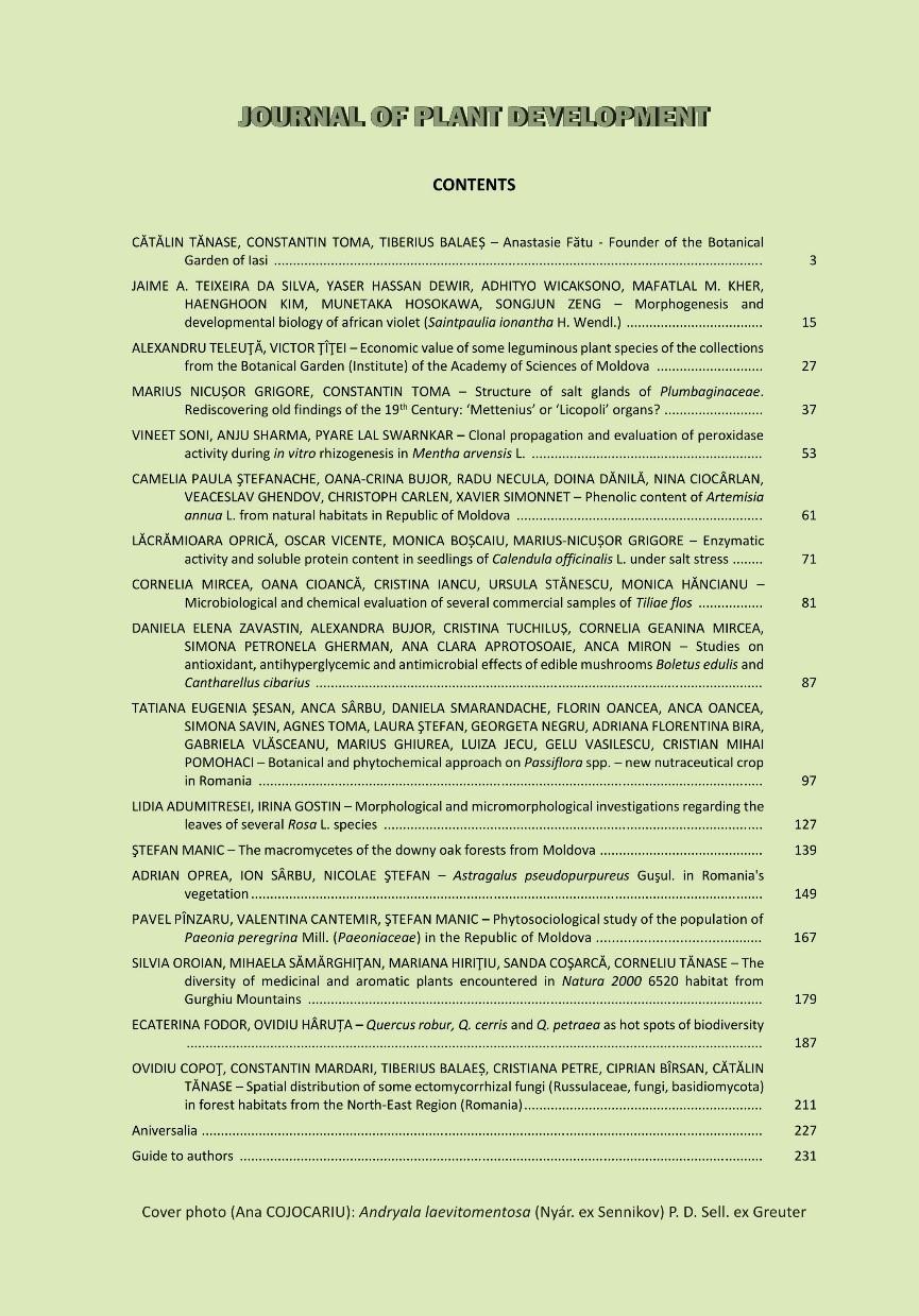
239
Useful Propagation Terms. Propagation The application of specific biological principles and concepts in the multiplication of plants.
 Useful Propagation Terms Propagation The application of specific biological principles and concepts in the multiplication of plants. Adventitious Typically describes new organs such as roots that develop
Plant Structure and Organization - 1
 Plant Structure and Organization - 1 In our first unit of Biology 203 we will focus on the structure and function of the higher plants, in particular the angiosperms, or flowering plants. We will look
PLANT BIOLOGY (PBIO) Plant Biology (PBIO) 1
 Plant Biology (PBIO) 1") Plant Biology (PBIO) 1 PLANT BIOLOGY (PBIO) PBIO 1052 How Plants Shaped Our World (LN) Description: This course is an eclectic dive into the world of plants and their influence on human society. Students
Plant Biology (PBIO) 1 PLANT BIOLOGY (PBIO) PBIO 1052 How Plants Shaped Our World (LN) Description: This course is an eclectic dive into the world of plants and their influence on human society. Students
COMPARATIVE BIOCHEMICAL AND PHYSIOLOGICAL RESEARCH ON TAXA OF MENTHA L. GENUS
 J. Plant Develop. 18(2011): 41-45 COMPARATIVE BIOCHEMICAL AND PHYSIOLOGICAL RESEARCH ON TAXA OF MENTHA L. GENUS ANDRO ANCA-RALUCA 1, BOZ IRINA 1, PĂDURARIU CLAUDIA 1, ATOFANI DOINA 1, COISIN MAGDA 1, ZAMFIRACHE
Reflexions, le site de vulgarisation de l'université de Liège
 When tomatoes flower 3/13/12 Understanding the mechanisms responsible for tomato plant flowering will enable new selection procedures to be developed in order to obtain even more productive varieties.
DIFFERENTIATION OF AVOCADO BLOSSOM BUDS IN FLORIDA
 Reprinted for private circulation from the Botanical Gazette, Vol. 104, No. 2, December, 1942. DIFFERENTIATION OF AVOCADO BLOSSOM BUDS IN FLORIDA PHILIP C. REECE 1 (WITH THIRTEEN FIGURES) Subtropical Fruit
SUBJECT: Integrated Science TEACHER: DATE: GRADE: 7 DURATION: 1 wk GENERAL TOPIC: Living Things Reproduce SPECIFIC TOPIC: Living Things and How They
 SUBJECT: Integrated Science TEACHER: DATE: GRADE: 7 DURATION: 1 wk GENERAL TOPIC: Living Things Reproduce SPECIFIC TOPIC: Living Things and How They Reproduce Living Things and How They Reproduce Students
THE EFFECT OF MINERAL FERTILIZATION ON THE PIGMENT CONTENT IN MARIGOLD (CALLENDULA OFFICINALIS L.)
") Lucrări Ştiinţifice vol. 5, seria Agronomie THE EFFECT OF MINERAL FERTILIZATION ON THE PIGMENT CONTENT IN MARIGOLD (CALLENDULA OFFICINALIS L.) Alina Elena MARTA, Carmen Doina JITĂREANU, Marinela BĂDEANU,
Lucrări Ştiinţifice vol. 5, seria Agronomie THE EFFECT OF MINERAL FERTILIZATION ON THE PIGMENT CONTENT IN MARIGOLD (CALLENDULA OFFICINALIS L.) Alina Elena MARTA, Carmen Doina JITĂREANU, Marinela BĂDEANU,
Plants can be either herbaceous or woody.
 Plant Structure Plants can be either herbaceous or woody. Herbaceous plants are plants with growth which dies back to the ground each year, in contrast with woody plants Most herbaceous plants have stems
Nebraska Core Academic Content Standards Science Grade: 2 - Adopted: 2010
 Main Criteria: Nebraska Core Academic Content Standards Secondary Criteria: Subjects: Science, Social Studies Grade: 2 Correlation Options: Show Correlated Nebraska Core Academic Content Standards Science
History and Mission of the Foundation:
 2 0 1 8 I T O F O U N D A T I O N F O R I N T E R N A T I O N A L E D U C A T I O N E X C H A N G E This guideline is only distributed to the affiliated universities whose students are eligible to apply
PLANT & SOIL SCIENCE (140)
") PLANT & SOIL SCIENCE (140) DESCRIPTION Students will demonstrate knowledge and skills in a wide range of scientific principles, such as genetics, disease, pests, and management practices. The scientific
PLANT & SOIL SCIENCE (140) DESCRIPTION Students will demonstrate knowledge and skills in a wide range of scientific principles, such as genetics, disease, pests, and management practices. The scientific
Bring Your Text to Lab!!!
 Bring Your Text to Lab!!! Vascular Plant Anatomy: Flowering Plants Objectives: 1. To observe what the basic structure of vascular plants is, and how and where this form originates. 2. To begin to understand
Anatomy of Plants Student Notes
 Directions: Fill in the blanks. Anatomy of Plants Student Notes Plant Cell Biology Segment 1. Plants Plants are organisms are incapable of movement produce food through 2. Animals Animals are multicellular
Teacher s Guide. Trees, Weeds and Vegetables So Many Kinds of Plants!
 Teacher s Guide Trees, Weeds and Vegetables So Many Kinds of Plants! Introduction This teacher s guide helps you teach young children about different kinds of plants. With over 350,000 varieties of plants
Secondary Curriculum Maps
 Secondary Curriculum Maps Cumberland Valley School District Soaring to Greatness, Committed to Excellence Principles of Agricultural Science - Plants The purpose of the Plant Science course is to expose
Economic and Social Council
 United Nations Economic and Social Council Distr.: General 23 May 2012 Original: English E/CONF.101/100 Tenth United Nations Conference on the Standardization of Geographical Names New York, 31 July 9
The mode of development in animals and plants is different
 The mode of development in animals and plants is different Outcome of animal embryogenesis is a mini edition of the adult Outcome of plant embryogenesis is a simple structure with -root apical meristem
Introduction to Horticulture 4th Edition, 2009
 A Correlation of Introduction to Horticulture 4th Edition, 2009 To the Georgia Performance Standards for General Horticulture and Plant Science FORMAT FOR CORRELATION TO THE GEORGIA PERFORMANCE STANDARDS
SUMMARY OF DOCTORAL THESIS
 ALEXANDRU IOAN CUZA UNIVERSITY OF IAŞI FACULTY OF BIOLOGY DEPARTMENT OF BIOLOGY IN VIVO AND IN VITRO PHYTOCHEMICAL DIVERSITY OF SOME POPULATIONS OF ARNICA MONTANA L. IN THE ROMANIAN EASTERN CARPATHIANS
Fifth Grade Social Studies Major Instructional Goals
 Fifth Grade Social Studies Major Instructional Goals We exist for the academic excellence of all students. December 2007 1 Rationale The purpose of the social studies program is to prepare young people
Grade 11 Student Electives Guidelines for 2015/16
 Grade 11 Student Guidelines for 2015/16 Overview As part of Abu Dhabi Education Council s continuous improvements efforts towards raising the standard of education in the Emirate of Abu Dhabi, the Cycle
Chapter 23: Plant Diversity and Life Cycles
 Chapter 23: Plant Diversity and Life Cycles Section 1: Introduction to Plants Cuticle: a waxy or fatty and watertight layer on the external wall of epidermal cells Spore: a reproductive cell or multicellular
SUBJECT: Integrated Science TEACHER: Mr. S. Campbell DATE: GRADE: 7 DURATION: 1 wk GENERAL TOPIC: Living Things Reproduce
 SUBJECT: Integrated Science TEACHER: Mr. S. Campbell DATE: GRADE: 7 DURATION: 1 wk GENERAL TOPIC: Living Things Reproduce Living Things and How They Reproduce Students will be able to:- 1) Identify root,
7/31/2014 WHAT IS LIGHT? SUPPLEMENTAL LIGHTING JOHANNA OOSTERWYK DC SMITH GREENHOUSE MANAGER UW-MADISON DEPARTMENT OF HORTICULTURE
 WHAT IS LIGHT? SUPPLEMENTAL LIGHTING JOHANNA OOSTERWYK DC SMITH GREENHOUSE MANAGER UW-MADISON DEPARTMENT OF HORTICULTURE Electromagnetic radiation Energy emitted by a light source Measured in watts Visible
Green Chemistry Education
 Green Chemistry Education Poster presented at the IUPAC Congress/General Assembly July 2001 GREEN CHEMISTRY IN THE SCIENTIFIC CONTEXT The science of chemistry is central to addressing the problems facing
Curriculum Connections for Discovery Field Trips Based on Alabama Course of Study. The Secret Life of Trees Curriculum Connections
 for Discovery Field Trips Based on Alabama Course of Study The Secret Life of Trees The Secret Life of Trees Observe, compare, and describe the properties of trees and parts of trees. Compare changes in
PROFESSIONAL-CLERICAL RATIO REPORT
 PROFESSIONAL-CLERICAL RATIO REPORT UNIVERSITY OF HAWAI I FY 2005-06 University Budget Office University of Hawai i September 2006 File Reference: Management and Planning Support Folder, Courses Reports
Unit 8: ECE/AP Biology Plants 9 Class Meeting. Essential Questions. Enduring Understanding with Unit Goal
 Essential Questions How are plants unique in their growth, organs, tissues and cells? How to plants acquire nutrients from the environment? How do different plants conduct photosynthesis? Enduring Understanding
Detailed Course Outline
 Detailed Course Outline Unit 1 Worlds of Opportunity Lesson 1.1 A World without Enough Plants 1. Many people work in a variety of agricultural enterprises to produce food, fiber, and fuel, which are essential
Horticulture Science
 Horticulture Science 012000 6045 Course Description This course includes the study of structure and functions of plants, including requirements for growth, methods of propagation, and the management of
Statutory framework of the world network of biosphere reserves UNESCO General Conference 1996
 Statutory framework of the world network of biosphere reserves UNESCO General Conference 1996 Keywords: conservation, protected areas, biosphere reserves, policies, strategies. For the Mountain Forum's
BIOAG'L SCI + PEST MGMT- BSPM (BSPM)
") Bioag'l Sci + Pest Mgmt-BSPM (BSPM) 1 BIOAG'L SCI + PEST MGMT- BSPM (BSPM) Courses BSPM 102 Insects, Science, and Society (GT-SC2) Credits: 3 (3-0-0) How insects develop, behave, and affect human activity.
Bioag'l Sci + Pest Mgmt-BSPM (BSPM) 1 BIOAG'L SCI + PEST MGMT- BSPM (BSPM) Courses BSPM 102 Insects, Science, and Society (GT-SC2) Credits: 3 (3-0-0) How insects develop, behave, and affect human activity.
Response Of Blueberry To Day Length During Propagation
 Response Of Blueberry To Day Length During Propagation Internal report for Young Plant Research Center Not for publication or reproduction in part or full without permission of the authors. Paul Fisher
While entry is at the discretion of the centre, candidates would normally be expected to have attained one of the following, or equivalent:
 National Unit specification: general information Unit code: H1JB 11 Superclass: SE Publication date: May 2012 Source: Scottish Qualifications Authority Version: 01 Summary This Unit is designed to meet
Self-Guided Tour. Plants and the Plant Life Cycle. K to 2nd Grade
 Time Required: 60 min April to October Topic: Observing plant life cycles in the Garden Materials: For each student: Pencil or crayons Clipboards (recommended) Comparison Worksheet Colored dots (recommended)
THE SEVILLE STRATEGY ON BIOSPHERE RESERVES
 THE SEVILLE STRATEGY ON BIOSPHERE RESERVES 1 VISION FOR BIOSPHERE RESERVES IN THE 21 ST CENTURY The international Seville Conference on Biosphere Reserves, organised by UNESCO, adopted a two-pronged approach,
Teacher s Guide For. Core Biology: Microbiology and Genetics
 Teacher s Guide For Core Biology: Microbiology and Genetics For grade 7 - College Programs produced by Centre Communications, Inc. for Ambrose Video Publishing, Inc. Executive Producer William V. Ambrose
Lincoln County Schools Patriot Day Instructional Expectations Patriot Day 1 School: Course/Subject: Biology Teacher: Cox Brock Gilbert Carr
 Lincoln County Schools Patriot Day Instructional Expectations Patriot Day 1 School: Course/Subject: Biology Teacher: Cox Brock Gilbert Carr Learning Target: B.1.a Analyze the similarities and differences
Legal problems of environmental management. Spatial planning and space management. M. Gajewski 2014 / 2015
 Legal problems of environmental management Spatial planning and space management M. Gajewski 2014 / 2015 Course contents (in brief) Constitutional basis of the environment protection system. General principles
Declaration Population and culture
 Declaration Population and culture The ministers of the parties to the Alpine Convention regard the socio-economic and socio-cultural aspects mentioned in Article 2, Paragraph 2, Item a., as being central
CYTOLOGY OF VIOLA ODORATA L. POLLEN GERMINATION SILVICA PĂDUREANU 1. Introduction
 Analele ştiinţifice ale Universităţii Al. I. Cuza Iaşi Tomul LIII, s. II a. Biologie vegetală, 27 CYTOLOGY OF VIOLA ODORATA L. POLLEN GERMINATION SILVICA PĂDUREANU 1 Abstract: The paper presents the characteristics
Bruce County s BIG Picture
 Bruce County s BIG Picture Overview Students will learn the importance of this planet we call Earth. Through different topics such as plate tectonics, glacial periods, climate change and endangered species,
Chapter 1-Plants in Our World
 Chapter 1-Plants in Our World Formation of earth-4.5-4.6 billion years ago Evidence of life from organic material-3.8 billion years ago Many cyanobacteria are photosynthetic, but these microscopic organisms
MEADOWS PRIMARY SCHOOL and NURSERY GEOGRAPHY POLICY
 MEADOWS PRIMARY SCHOOL and NURSERY GEOGRAPHY POLICY Purpose of study The teaching of Geography at Meadows is done by following the 2014 National Curriculum. The planning and teaching of the subject aims
Valentina Pavlova. Curriculum Vitae
 Name: Date and place of birth: Citizenship/nationality: Marital Status: Office Address: Home Address: Valentina Pavlova Curriculum Vitae Valentina Pavlova 07. 06. 1975 in Veles, Republic of Macedonia Macedonian
SCH SCH Agricultural Education General Agriculture , ,668 Leadership
 Oregon State University: Academic Year 2016-17 (Fall 2016, Winter and Spring 2017) Student Credit Hours by College, Department*, Course CIP Code**, and Course Level Office of Institutional Research College
Kindergarten Science, Quarter 4, Unit 4.1. Plants. Overview
 Kindergarten Science, Quarter 4, Unit 4.1 Plants Overview Number of instructional days: 10 (1 day = 20 30 minutes) Content to be learned Observe that plants need water, air, food, and light to grow. Identify
Plant Growth and Development Part I. Levels of Organization
 Plant Growth and Development Part I Levels of Organization Whole Plant Organs Tissues Cells Organelles Macromolecules Levels of Organization Whole Plant Organs Tissues Cells Organelles Macromolecules 1
FOREST TREE PHYSIOLOGY RESEARCH AT THE OHIO AGRICULTURAL EXPERIMENT STATION
 FOREST TREE PHYSIOLOGY RESEARCH AT THE OHIO AGRICULTURAL EXPERIMENT STATION JOHN HACSKAYLO AND WILLIAM E. GOSLIN Department of Forestry, Ohio Agricultural Experiment Station, Wooster The research in tree
plant physiology and energy conversion to plant systems. identify the components and the functions of plant describe the processes of
 Plant Systems-AG1 Essential Questions: 1. How do plant classification, plant anatomy, and plant physiology affect the production and management of plants? 2. What are the necessary steps to Prepare and
Class Copy! Return to teacher at the end of class! Mendel's Genetics
 Class Copy! Return to teacher at the end of class! Mendel's Genetics For thousands of years farmers and herders have been selectively breeding their plants and animals to produce more useful hybrids. It
BIOL 305L Laboratory One
 Please print Full name clearly: BIOL 305L Laboratory One General plant anatomy a great place to start! Introduction Botany is the science of plant life. Traditionally, the science included the study of
ABSTRACT. Key words: hemp, microsporogenesis, androgenesis
 ABSTRACT Key words: hemp, microsporogenesis, androgenesis Hemp (Cannabis sativa L.) has a growing economical importance, because of the various utilizations in alimentation, textiles, plastics, constructions
Haida Gwaii Queen Charlotte Islands
 Haida Gwaii Queen Charlotte Islands H E R I T A G E T O U R I S M STRATE GY January 2003 Haida Gwaii/Queen Charlotte Islands Heritage Tourism Strategy Working Group The Haida Gwaii/Queen Charlotte Islands
Plant Lifecycle 3 rd 5 th Grade
 Key Understandings Students will understand that each plant has a lifecycle and the plant lifecycle is a continual process A lifecycle is a continuous cycle that contains: birth, growth, reproduction,
Studies on the Light Controlling Flower Initiation of Pharbitis Nil. VI. Effect of Natural Twilight. by Atsushi TAKIMOTO* and Katsuhiko IKEVA*
 Studies on the Light Controlling Flower Initiation of Pharbitis Nil. Received September 9, 1959 VI. Effect of Natural Twilight by Atsushi TAKIMOTO* and Katsuhiko IKEVA* Many investigators consider that
Degree Type Bachelor of Science (BS) Degree Title Biology
 Degree Title Biology") Degree Type Bachelor of Science (BS) Degree Title Biology Focus: Biological Science The Department of Biology is committed to excellence in instruction, scholarly accomplishment, research, professional
Degree Type Bachelor of Science (BS) Degree Title Biology Focus: Biological Science The Department of Biology is committed to excellence in instruction, scholarly accomplishment, research, professional
St John s Catholic Primary School. Geography Policy. Mission Statement
 St John s Catholic Primary School Geography Policy Mission Statement We at St John s strive for excellence in education by providing a safe, secure and caring family environment where individuals are valued
Objectives. To identify plant structures and functions. To describe the structure of plant cells. To explain the process of reproduction in plants.
 1 Objectives To identify plant structures and functions. To describe the structure of plant cells. To explain the process of reproduction in plants. 2 Main Menu Plant Cell Biology Plant Structures Roots
DIATHEMATIKON PROGRAMMA CROSS-THEMATIC CURRICULUM FRAMEWORK
 DIATHEMATIKON PROGRAMMA CROSS-THEMATIC CURRICULUM FRAMEWORK FOR STUDIES OF THE ENVIRONMENT The nature of Studies of the Environment Studies of the Environment is an interdisciplinary subject dealing with
Agricultural. Chemistry. Agricultural production: crops and livestock Agrichemicals development: herbicides, pesticides, fungicides, fertilizers, etc.
 Agricultural Agricultural production: crops and livestock Agrichemicals development: herbicides, pesticides, fungicides, fertilizers, etc. Agrichemicals testing Environmental testing Regulation Government:
W/F = 10:00 AM - 1:00 PM Other times available by appointment only
 Course BY 101-05 Principles of Biology Fall 2015 BY 103-01 Honors Biology Course Meeting Time BY 101-05 T/Th 9:30 10:45 PM BG 109 BY 103-01 Lab 01 M 8:00 AM - 9:50 AM Lab 02 M 10:10 12:00 PM Lab 03 M 1:25
A) Pre-Darwin History:
 Pre-Darwin History:") Darwin Notes A) Pre-Darwin History: Ancient Greek philosophers such as and believed species were permanent and did not evolve. These ideas prevailed for 2,000 years. In 1859 Charles Darwin published. This
Darwin Notes A) Pre-Darwin History: Ancient Greek philosophers such as and believed species were permanent and did not evolve. These ideas prevailed for 2,000 years. In 1859 Charles Darwin published. This
Postgraduate studies at the Biozentrum.
 Postgraduate studies at the Biozentrum. PhD Students & Postdocs. Young talent at the forefront of life science research. With its interdisciplinary PhD program, the Biozentrum offers Master graduates an
CLLD Cooperation OFFER
 Title of the proposed project (English) CLLD Cooperation OFFER PARKS PROTECTION III - Management, Protection and Economic Development in Protected Areas Type of project (select as many as you want) Cooperation
Briefing. H.E. Mr. Gyan Chandra Acharya
 Briefing by H.E. Mr. Gyan Chandra Acharya Under-Secretary-General and High Representative for the Least Developed Countries, Landlocked Developing Countries and Small Island Developing States Briefing
This book focuses mostly on Proteas, but also considers some of the other Proteaceae genera that are more widely cultivated.
 CHAPTER 1: INTRODUCING THE PROTEA FAMILY There are around 1700 species and 79 genera of plants in the Proteaceae (Protea) family, and most are indigenous to the southern hemisphere. Around half of these
LOUISIANA STUDENT STANDARDS FOR SOCIAL STUDIES THAT CORRELATE WITH A FIELD TRIP TO DESTREHAN PLANTATION KINDERGARTEN
 LOUISIANA STUDENT STANDARDS FOR SOCIAL STUDIES THAT CORRELATE WITH A FIELD TRIP TO DESTREHAN PLANTATION KINDERGARTEN Standard 2 Historical Thinking Skills Students distinguish between events, people, and
Let light motivate your flowers
 Let light motivate your flowers LightDec Horticulture Light recipes from LEDIG are the best in this market. Their recommendations increased my profits in year one by 23% LED Solutions from LEDIG LED Industrial
GIS Visualization: A Library s Pursuit Towards Creative and Innovative Research
 GIS Visualization: A Library s Pursuit Towards Creative and Innovative Research Justin B. Sorensen J. Willard Marriott Library University of Utah justin.sorensen@utah.edu Abstract As emerging technologies
Bachelor s Degree in Agroalimentary Engineering & the Rural Environment. 1 st YEAR Animal & Plant Biology ECTS credits: 6 Semester: 1
 1 st YEAR 6241 Animal & Plant Biology The teaching objectives that the student is expected to achieve on this module are as follows: 1. Understand the fundamental characteristics of living beings, know
Outcomes of Evolution: Species and Ecotypes. Reading Assignment: Chapter 6 in GSF 9/8/2009
 Outcomes of Evolution: Species and Ecotypes Reading Assignment: Chapter 6 in GSF Objectives 9/2/2009 1. Observe, describe, and measure phenotypic variation among individuals in a population. 2. Characterize
Christel Anne Ross. Invasion Success by Plant Breeding
 Christel Anne Ross Invasion Success by Plant Breeding VIEWEG+TEUBNER RESEARCH Christel Anne Ross Invasion Success by Plant Breeding Evolutionary Changes as a Critical Factor for the Invasion of the Ornamental
INFLUENCE OF TEMPERATURE ON NECTAR COLLECTION AND STORAGE IN THE HIVE DURING HONEY HARVEST
 INFLUENCE OF TEMPERATURE ON NECTAR COLLECTION AND STORAGE IN THE HIVE DURING HONEY HARVEST N. Eremia 1, Elena Scripnic 1, Susana Modvala 1, Angela Chiriac 1 1 State Agrarian University of Moldova, Chisinau,
CLASS XI BIOLOGY NOTES CHAPTER 1: LIVING WORLD
 CLASS XI BIOLOGY NOTES CHAPTER 1: LIVING WORLD Biology is the science of life forms and non-living processes. The living world comprises an amazing diversity of living organisms. In order to facilitate
Snapdragon Lighting. Harrison Flint. Cornell University. ing mid-winter. Several good approaches to this problem
 Snapdragon Lighting Harrison Flint Department of Floriculture Cornell University One of the greatest problems in the commercial pro duction of winter snapdragons has been the expense brought about by extremely
The Plant Kingdom If you were to walk around a forest, what would you see? Most things that you would probably name are plants.
 INTRODUCTION TO PLANTS The Plant Kingdom If you were to walk around a forest, what would you see? Most things that you would probably name are plants. Plants are abundant in almost every environment that
Semester III. Semster I PLANT ANATOMY BO1141
 Semster I PLANT ANATOMY BO1141 Understand basic anatomical features of monocot and dicot plants Able to identify different types of tissues and tissue systems in plants Know the basic concepts in reproductive
Plant Structure, Growth, and Development
 Plant Structure, Growth, and Development Plant hierarchy: Cells Tissue: group of similar cells with similar function: Dermal, Ground, Vascular Organs: multiple kinds of tissue, very diverse function Organ
Oklahoma Academic Standards Science Grade: 9 - Adopted: 2014
 Main Criteria: Oklahoma Academic Standards Secondary Criteria: Subjects: Science, Social Studies Grade: 9 Correlation Options: Show Correlated OK.HS-BI. Oklahoma Academic Standards Science Grade: 9 - Adopted:
Table 1. Number and Percent of Women Faculty in Science/Engineering by Department, 2001* Division/Department Women Men % Women
 Table 1. Number and Percent of Women Faculty in Science/Engineering by Department, 2001* Division/Department Women Men % Women Physical Sciences 40.00 412.12 8.8% Biological Systems Engineering 0.00 15.25
Table 1. Number and Percent of Women Faculty in Science/Engineering by Department, 2002* Division/Department Women Men % Women
 Table 1. Number and Percent of Women Faculty in Science/Engineering by Department, 2002* Division/Department Women Men % Women Physical Sciences 45.25 417.62 9.8% Biological Systems Engineering 1.00 14.25
Levels of Organization
 Plant Growth and Development Part I Levels of Organization Whole Plant Organs Tissues Cells Organelles Macromolecules Levels of Organization Whole Plant Organs Tissues Cells Organelles Macromolecules Plant
Lived Alfred Wegener was born on November 1, 1880, in Germany s capital city, Berlin.
 Alfred Wegener Lived 1880 1930. Alfred Wegener proposed the theory of continental drift the idea that Earth s continents move. Despite publishing a large body of compelling fossil and rock evidence for
Georgia Performance Standards for Urban Watch Restoration Field Trips
 Georgia Performance Standards for Field Trips 6 th grade S6E3. Students will recognize the significant role of water in earth processes. a. Explain that a large portion of the Earth s surface is water,
Book Study Groups Children s Lessons Based on Karma and Reincarnation By Elizabeth Clare Prophet and Patricia R. Spadaro
 Book Study Groups Children s Lessons Based on Karma and Reincarnation By Elizabeth Clare Prophet and Patricia R. Spadaro Karma and Reincarnation explores the questions of which family you were born into,
Understanding Plant Life Cycles
 Lesson C3 2 Understanding Plant Life Cycles Unit C. Plant and Soil Science Problem Area 3. Seed Germination, Growth, and Development Lesson 2. Understanding Plant Life Cycles New Mexico Content Standard:
Grade Level, Course Third Grade Social Studies PLSD Balanced Assessment Practices. Type* DOK
 Unit Guides Assessment Items Indic ator s Concept Standard Curricular Indicator DOK Type* SR CR PB Assessment Questions # DOK Level 1E 1M 1H 2E 2M 2H 3E 3M 3H Civics Students will develop and apply the
INTERNATIONAL CULTURAL TOURISM CHARTER Managing Tourism at Places of Heritage Significance (1999)
") INTERNATIONAL CULTURAL TOURISM CHARTER Managing Tourism at Places of Heritage Significance (1999) Adopted by ICOMOS at the 12th General Assembly in Mexico, October 1999. INTRODUCTION The Charter Ethos
INTERNATIONAL CULTURAL TOURISM CHARTER Managing Tourism at Places of Heritage Significance (1999) Adopted by ICOMOS at the 12th General Assembly in Mexico, October 1999. INTRODUCTION The Charter Ethos
ST AMBROSE CATHOLIC PRIMARY SCHOOL Geography Policy
 ST AMBROSE CATHOLIC PRIMARY SCHOOL Geography Policy 2017-2018 Our school policy is developed in accordance with the National Curriculum for Geography and Foundation Stage Curriculum for Understanding of
CONTROL SYSTEMS IN PLANTS
 AP BIOLOGY PLANTS FORM & FUNCTION ACTIVITY #5 NAME DATE HOUR CONTROL SYSTEMS IN PLANTS HORMONES MECHANISM FOR HORMONE ACTION Plant Form and Function Activity #5 page 1 CONTROL OF CELL ELONGATION Plant
Basic Botany Master Gardener and Horticulture Training. Mark Heitstuman. WSU Asotin and Garfield County Director January 12, 2016
 Basic Botany 2016 Master Gardener and Horticulture Training Mark Heitstuman WSU Asotin and Garfield County Director January 12, 2016 Topics we ll discuss in Chapter 1- Basic Botany Plant life cycles Internal
Characteristic features of toponym objects and their dynamics
 23 May 2002 English only Eighth United Nations Conference on the Standardization of Geographical Names Berlin, 27 August-5 September 2002 Item 12 (c) of the provisional agenda* Toponymic data files: toponymic
the different kinds of houses.
 STANDARD II GENERAL KNOWLEDGE Month Unit Expected learning Activities/Assignments Value Skills June WORLD AROUND US Health and Hygiene To understand common Make a list of Do's and Don'ts of Cleanliness
CHAPTER-1 THE LIVING WORLD MULTIPLE CHOICE QUESTIONS
 1 CHAPTER-1 THE LIVING WORLD MULTIPLE CHOICE QUESTIONS 1. As we go from species to kingdom in a taxonomic hierarchy, the number of common characteristics a. Will decrease b. Will increase c. Remain same
SCH SCH Agricultural Education General Agriculture , ,359 Leadership
 Oregon State University: Academic Year 2017-18 (Fall 2017, Winter and Spring 2018) Student Credit Hours by College, Department*, Course CIP Code**, and Course Level Office of Institutional Research College
Kingdom Plantae. Plants or metaphytes are, autotrophic multicellular eukaryotes, with tissues.
 Kingdom Plantae Key words feature bryophytes herbaceous node to release pteridophytes sporangium, leaf (leaves) damp gymnosperms vascular apix cluster angiosperms rhizome sepal shrub tropism fronds calyx
GREEN LIFE. Plants and Photosynthesis W 398
 W 398 GREEN LIFE Plants and Photosynthesis Savannah Webb, Former 4-H Extension Agent, Maury County Jennifer Richards, Assistant Professor, 4-H Youth Development MANAGEMENT OF APHIDS AND BYD IN TENNESSEE
Rhode Island World-Class Standards Science Grade: K - Adopted: 2006
 Main Criteria: Rhode Island World-Class Standards Secondary Criteria: Subjects: Science, Social Studies Grade: K Correlation Options: Show Correlated Rhode Island World-Class Standards Science Grade: K