Experiment 6 1. The Compton Effect Physics 2150 Experiment No. 6 University of Colorado
|
|
|
- Maryann Shaw
- 6 years ago
- Views:
Transcription
1 Experiment 6 1 Introduction The Compton Effect Physics 2150 Experiment No. 6 University of Colorado In some situations, electromagnetic waves can act like particles, carrying energy and momentum, which they may impart to other particles with which they interact. According to Planck s hypothesis, an electromagnetic wave of frequency f carries an energy given by: E = nhf (1) where n is a positive integer and h is Planck s constant. The minimum amount of energy which can be carried by an electromagnetic wave is thus that carried by one photon. Further, according to the de Broglie relation, if the wavelength of the radiation is λ, then the photon has a momentum given by p =!! =!!! (2) since! =! for an electromagnetic wave.!! Consider a photon which collides with an electron at rest. In Fig. 1, let θ be the angle at which the photon comes off, and let φ be the angle at which the electron comes off. Since Figure 1: Collision of a Photon with an Electron Initially at Rest the photon emerging from the collision has a momentum different from that of the incident photon, it will have a different wavelength, which we shall denote by λ. Applying the relativistic law of conservation of energy to this collision
2 Experiment 6 2!!! + m!c! =!!!! + m c! (3) where the mass of the electron after that collision is (4) m =!!.!!!!!! In this equation, v is the electron s velocity in the laboratory reference frame. Applying the laws of conservation of momentum in the plane of the collision gives and!! =!!! cos θ + m v cos φ, (5) 0 =! sin θ m v sin φ. (6)!! It follows from these three equations (see any text in modern physics for the derivation) that the change in wavelength of the scattered photon is given by: λ! λ =!!!! 1 cos θ. (7) This result can be expressed directly in terms of the energies E and E of the scattered and unscattered gamma rays, respectively, by using λ = hc/e:!! =! 1 cos θ. (8)!!!!!!! This change of gamma- ray wavelength (or energy) to a different wavelength upon scattering from an electron is called the Compton Effect. The object of this experiment is to measure the energy change of the scattered gamma- ray, and to compare the results with Eq. (7) or (8). The apparatus used in this experiment consists of one strong 137 Cs source used in parts 1, 2, and 3. Both its scattered and unscattered energies will be determined. Three weak calibrating sources ( 137 Cs, 22 Na, and 60 Co) will be used solely for calibrating a pulse- height analyzer. In addition to these sources, there is a sodium iodide detector, a scaler- time- counter, associated power supplies, and a computer that contains a pulse- height analyzer. The geometry of the scatterers is based on a theorem from plane geometry that states that, if a triangle is inscribed inside a circle with a fixed chord of the circle as one
3 Experiment 6 3 side, then the angle opposite the fixed chord is a constant (see Fig. 3). Thus, if a source is placed at one end of the chord, and a photon emitted from this point is scattered Figure 2: Geometry of the Source, Detector, and the 90 Scattering Plates Figure 3: If a triangle is inscribed in a circle with a fixed chord of the circle as one side, then the opposite angle of the triangle is a constant. by electrons placed on the circumference of the circle, and if the photon is subsequently detected by a counter placed at the other end of the chord, then the scattering angle is fixed by the geometry. If a circular plate is shaped to fit the circumference of the circle and the photons are allowed to scatter off the entire plate, then any photon which is detected after one scattering must have been scattered through the same angle.
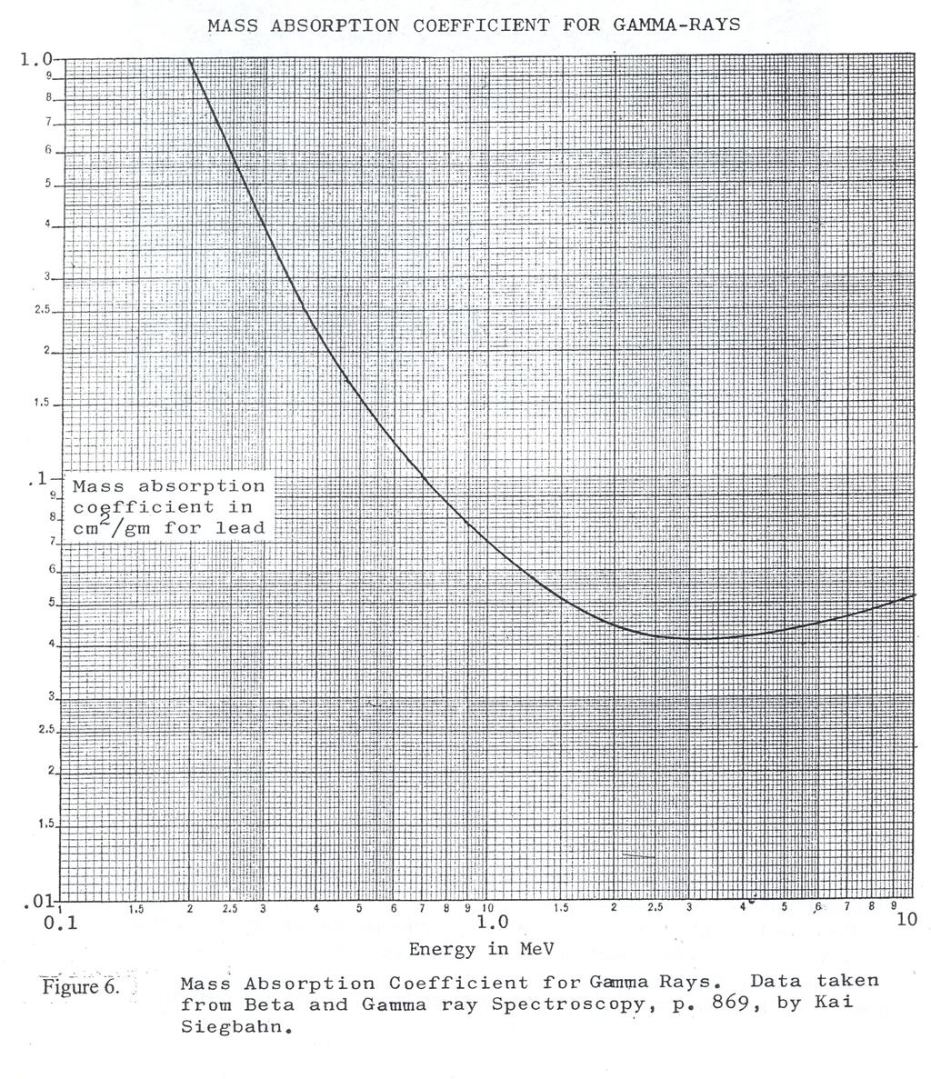
4 Experiment 6 4 The particular setup used in this experiment has been constructed in such a way that the two different scattering angles may be used: 69 and 90. These angles were arbitrarily chosen and there is no reason in principle why one could not construct scatterers for any chosen angle. The scatterers used are thin aluminum plates because aluminum has relatively high density of free electrons. Referring to Eq. (7) or (8), the constants h, m!, and c are known fundamental constants. Therefore, in order to verify the correctness of these predictions, we must measure the energies of both the unscattered (E) and the scattered (E! ) photons for one or more particular angles. Since the mass absorption coefficient of lead is a known function of energy, the energy measurement in this experiment is performed by measuring the mass absorption coefficient of the gamma rays in lead. For a given absorbing material, the absorption probability is proportional to the number of atoms of absorber per unit of cross sectional area. Therefore, it is convenient to measure absorber thickness in cm. A series of cylindrical lead absorbers is provided. These are constructed so that they may be placed in successively thicker layers around the detector. A beam of gamma rays of initial intensity I! is attenuated upon passing through a thickness t (in g/cm 2 ) of absorber according to the equation: I = I! exp( μt) (9) where μ is the mass absorption coefficient in cm 2 /g. The intensities I and I! are in terms of number of gamma rays per square centimeter per second. For a particular geometrical configuration of source and scatterer, the mass absorption coefficient is determined by measuring the counting rate (intensity) as a function of the thickness of the absorbers. This functional relationship may be then plotted by using a weighted linear- regression computer program such as Wlinfit or its equivalent. Wlinfit gives both the slope of the resulting straight line and its uncertainty. The energy may then be determined from the curve provided (Fig. 6). The sodium iodide (NaI) scintillation detector used in this experiment is an important and widely used device for detecting gamma rays. As shown in Fig. 4, it consists of a crystal of NaI to which a small amount of thallium has been added in order to make it an effective scintillator. The cylindrical NaI (2 ½ x 1 ¾ inch diameter) is hermetically encased in an aluminum cup with a glass window on one end. The glass window of the detector is mounted on the face of a 10- stage photo- multiplier tube. Gamma rays entering the NaI detector will interact by either the photoelectric or Compton process. Gamma rays with energy greater than 1.02 MeV can also produce an electron- position pair. The photoelectric process results in the gamma ray imparting essentially all of its energy to one of the bound electrons in the crystal. The Compton process is assumed to occur on a free electron, and as we have just seen, results in only a fraction of the gamma-
5 Experiment 6 5 ray energy being carried off by an electron. Electrons from either process will quickly lose their energy in the crystal by causing ionizing events with the atoms of the crystal with the net effect that photons in the visible region will be produced in the de- exitation of the atoms. The number of visible photons produced is directly proportional to the energy deposited by a gamma ray in the crystal. The photons are reflected by a coating of MgO surrounding the crystal except for the end covered by the glass window and many of them enter the photomultiplier. Figure 4:Gamma- ray detection and counting equipment, configured for parts one and two. Event (a) is a Compton scattering event and event (b) is a photoelectric process. The photons entering the photomultiplier will strike a photocathode surface present on the inside face of the phototube. Low energy electrons will be ejected from the photocathode and some of them will be incident on the first dynode of the tube. The dynode provides amplification of the number of electrons by the secondary emission process (more electrons are emitted than are incident on a given dynode). The number of electrons is increased by nice successive dynode stages and finally the electrons are collected on the anode where a reasonably large voltage pulse will result. The amplitude of the voltage pulse will again be directly proportional to the energy deposited in the NaI.
6 Experiment 6 6 After going through a preamplifier at the tube base, the voltage pulse is carried via a cable to the scaler on a table outside of the room where the Compton scattering experiment is located. Procedure: Part 1 1. Energy of the Unscattered Gamma Ray The radioactive source used in this part of the experiment is 137 Cs. It has a half life of about 30 years and emits a gamma ray with an energy of MeV. Before attempting to measure the energy of the Compton scattered gamma rays, an absorption measurement will be made of the direct or unscattered gamma ray in order to become familiar with the techniques. a. The AC and DC power should be turned on for the two power supplies in the room with the scattering experiment. The high voltage for the photomultiplier should be +850 volts. No adjustment of either power supply should be necessary. A small lead source holder and collimator is available and this should be placed 6 to 8 inches from the NaI detector with the collimation hole aimed directly at the detector. No cylindrical lead absorbers should be present around the detector for this part of the experiment. b. Go outside the room and turn on the AC power to the scaler (the switch is located on the upper left hand side on the back) and the oscilloscope. Oscilloscope control positions should be set as specified in the Addendum to the Compton Effect posted with the apparatus. Appropriate settings for the oscilloscope voltage display are obtained by rotating the gray CH 1 VOLTS/DIV knob to Volts/Div. This value should lie back opposite the notation 1X PROBE on the front panel. For best time display, the black brackets on the gray A and B TIME/DIV knob should straddle (select) the 2-5 μs value. It is instructive to observe the pulses coming from the NaI detector on channel one of the oscilloscope, prior to taking data. With correct settings, the pulses should appear on the oscilloscope as depicted in Fig. (5). The abundant, heavy trace, large pulses correspond to all of the energy of the gamma ray being left in the NaI crystal. Determine the amplitude (in volts) of these large pulses. The spread of smaller pulses beneath the full energy pulse corresponds to Compton processes where the scattered gamma ray escapes the crystal. c. First a background measurement should then be made using the scaler with the 137 Cs source in the lead storage pig on the floor. d. Remove the 137 Cs source from the lead brick on the floor where it is stored and insert it into the lead collimator. Care should be taken to minimize your

7 Experiment 6 7 exposure to the 137 Cs source. Always handle it by the opposite end of the rod to which it is attached. Do not stay in the room while you are taking data. None of the small rectangular lead absorbers are needed at this point. e. With no absorbers in place, use the scaler to determine the counting rate. Enough time should be allotted to accumulate at least 10,000 counts so that the statistical (random) error is no larger than about 1%. Because the counting rates are so high, 10,000 counts can be obtained in about one second. Measurements should then be carried out with 1, 2, 3 absorbers in the notches provided in the lead collimator, until finally all 10 absorbers are in place, to acquire a reasonable number of counts. Figure 5: Oscilloscope display of the photomultiplier pulses caused by incarceration of gamma rays with NaI crystal f. Correct the total counting rate by subtracting the average background count from each of the eleven total counts previously obtained. Then plot an absorption curve consisting of the natural log of the corrected counts per second plotted against absorber thickness t in g/cm 2 (see page 4 for the definition of t). t has a value of 1.79 g/cm 2 for each flat absorber. The plot can be made on the computer by use of the Mathematica Wlinfit weighted linear least squares fitting program. Wlinfit is available on the desktop of the lab PC s in the Scratch file. g. Extract half the half- thickness t 1/2 using your Wlinfit regression program summary that gives the slope and intercept values and their
8 Experiment 6 8 uncertainties. The half- thickness is the absorber thickness required to reduce the counting rate by ½ its initial value. Note that t!/! = ln(2)/m, where m is the slope from Wlinfit. Since μ = ln(2)/t!/!, then μ = m. h. With the use of Fig. 6 where the mass absorption coefficient for the lead is plotted as a function of gamma ray energy, determine the energy of the 137 Cs gamma ray from your value of μ. Compare your result with the known value of MeV. One can graphically determine the uncertainty in the value of μ (and therefore energy) using the Wlinfit Mathematica document.
9 Experiment 6 9
10 Experiment 6 10 Procedure: Part 2 1. Energy of the Scattered Gamma Rays For this part of the experiment, the 137 Cs source should be inserted in the source holder on the end of the Compton scattering table. The cylindrical lead absorbers that are to be used fit directly around the NaI scintillation detector. a. Take background readings for the 69 and 90 scattering measurements by having the source in place but the scatterers removed. This configuration will allow all radiation that reaches the detector but which does not comes from the scatterers to be subtracted as background. The best data is obtained if both background and total counts are taken for each of the five configurations of absorbers shown below. The suggested counting times are: Number of Absorbers 0 1 Counting Time 20 sec 20 sec 3 50 sec 5 60 sec 7 5 min b. Repeat the measurements discussed in Part 1a. first with the 69 and then the 90 scattering. By using Wlinfit or its equivalent, determine the absorption coefficient, μ, for each angle plotted, followed by their energies from Fig. 6 in the fashion described previously. Graphically estimate the uncertainty in your two energy measurements from your absorption curve. For both scattering angles, first 69 and then 90, and with no cylindrical absorbers in place, observe the pulses in the Compton band on the oscilloscope and try to estimate their height in volts. These consist of the most intense pulses of greatest amplitude. How do these maximum pulse heights compare to those observed with the unscattered gamma ray? Estimate the energies in MeV of the scattered radiation for both scattering angles, based on the maximum pulse height read for the 137 Cs gamma ray that corresponds to MeV. c. Comparison of experimental results with theory Using Eq. (8), calculate the energy of the scattered gamma ray E!, with E = MeV. Do this first with θ = 69, and the second with θ = 90. Compare the expected values of E! with your measured values and discuss any discrepancies. From a linear plot 1/E! versus (1 cos θ) for the three angles 0, 69, and 90, find the value of m e c 2 and compare it with the established value of MeV.
11 Experiment 6 11 Procedure: Part 3 1. Determination of Energy of the Scattered Gamma Rays Using a Pulse Height Analyzer A pulse height analyzer is a special- purpose computer that sorts and counts pulses. The amplitude of each incoming pulse is measured and is then converted to a digital value that is stored in a memory register location that is a part of a larger (16385 channel) memory. Since the voltage amplitude of each pulse is proportional to the energy of the photon that produced it, the PHA, in effect, assigns energy to channels in memory, in the order of ascending energy. This can be described algebraically by the equation E = mc + Eo, where E is energy and C is a corresponding channel number. It is in the general form y = mx + B, thus E0 is the intercept value of energy and m the slope of this linear relation between energy and channel location in the computer. In operation, pulses go from the NaI detector into a photo- multiplier tube, through an amplifier, and then into the PHA (see Fig. 7). The PHA then stores them over time and constructs a frequency distribution of the incoming pulses plotted against channel number. It then displays the distribution as a histogram on the computer screen with total counts per channel on the y- axis and channel numbers on the x- axis. If the pulses resulting from a 137 Cs gamma ray are sorted according to their height, a spectrum will be obtained similar to the one shown in Fig. 8. In Fig. 8, the peak on the right is the full energy peak and corresponds to the MeV gamma ray having a photoelectric event in the crystal or a Compton event where the scattered photon is also stopped in the crystal. This full energy or photoelectric peak is a useful calibration marker for measuring the energy of unknown gamma rays. The rather flat bump to the left of the full energy peak corresponds mostly to Compton events in the crystal where various amounts of energy escape from the crystal. The small peak at the far left is the photo- peak of a 32 kev x- ray that also comes from the 137 Cs radioactive decay.
12 Experiment 6 12 Figure 7: Gamma- ray detection and counting equipment, configured for Part 3. The event marked (a) is a Compton scattering event and the event marked (b) is a photoelectric process.
13 Experiment 6 13 Figure 8: A Pulse Height Spectrum for a 137 Cs Source 2. Energy Calibration of the Scintillation Detector a. The AC and DC power should be turned on for the two power supplies in the room with the scattering equipment. The high voltage for the photomultiplier should be set to +850 volts. On the table outside the room, turn on the power for the instruments contained in the PORTABLE BIN/POWER SUPPLY. This switch is located on the BIN s upper left- hand corner on the back. Your lab instructor or the lab coordinator will provide you with three small plastic calibration sources of 137 Cs, 22 Na, and 60 Co. They will also help you to set up the computer so it is ready for use in obtaining pulse height spectra. Place the small plastic 137 Cs source in the NaI detector. b. The oscilloscope should now show the pulses from the 137 Cs source as is shown in Fig. 5. The abundant large pulses correspond to the full energy peak (0.662 MeV) and the enhanced pulses near the baseline, the x- ray line at 32 kev. c. To display energy distributions on the computer screen, proceed as follows: i. Turn power on to both the Dell PC and its monitor.
14 Experiment 6 14 ii. When the Windows Logon window appears, type phys2150 in the password box and then press ENTER. The computer will boot to Windows XP desktop. iii. Now, click the following buttons in order: START à ALL PROGRAMS à GENIE 2000 à GAMMA ACQUISTION AND ANALYSIS. The Acquisition window will open. Click FILE à OPEN DATA SOURCE. Now click the DETECTOR button. A red and yellow radiation symbol labeled Detector MP2_MCA1 will appear in this window. Click CLEAR à START, and you will see that energy peaks begin developing across the bottom of black display screen. iv. The scroll bar on the right side of the display screen allows one to set the heights of peaks thus displayed. Move it up or down for best display of heights of energy histograms. d. The notations Channel (in memory) and Counts are displayed in blue at the top of the energy display window. To determine total counts in a given channel for a histogram, click anywhere in it with the mouse pointer. This will select a single memory location and a white marker will appear in the site selected. The channel number and total counts in this particular location will be given at the top. Pressing the left and right keys on the keyboard will cause the marker to scan across the screen to any of the memory locations. e. In general, to produce energy peak histograms, click STOP à CLEAR à START, and then wait as long as is reasonably possible for peaks to develop. Move the marker to the center of peaks of interest and record channel number and observed counts. Counting precision depends on having counts as high as possible. Place the 137 Cs source on top of the NaI detector. f. Follow the steps given previously to determine the appropriate channel numbers for the 137 Cs source. Then, remove the 137 Cs source from the NaI crystal detector and repeat this procedure for the 22 Na source, placed on top of the detector. Two prominent gamma energies are produced by the 22 Na source: a MeV line, caused by positron- electron annihilation. That produces two quanta each of MeV. The second line is a 1.28 MeV gamma. Remove the 22 Na source and replace it with the 60 Co source. This produces two high- energy peaks of interest in this experiment. They are 1.17 MeV and 1.33 MeV. Because each separate peak is hard to distinguish, their channel numbers can be determined and the channel numbers averaged, and the average 60 Co energy and corresponding average channel number can be used in calibration. g. Make a calibration graph of the energy (in MeV) of all the data you have taken. That is, plot energy in MeV against corresponding channel number for
15 Experiment 6 15 the three sources. Also use the Linfit or an equivalent liner least squares fitting program and your data to determine the slope and intercept of the line E = bc + E Determination of the Energy of the Scattered Gamma Ray a. Remove the strong 137 Cs source from the lead shielding blocks on the floor and insert it into the hole in the lead brick mounted on the left end of the scattering apparatus table. Put the 90 aluminum scatterers in their appropriate slots. Run the Acquisition program long enough to establish a well- defined peak and record its channel number. b. Repeat the process with the 69 scatterers in place. c. Using your calibration graph (from 1g. above) and the computer best- fit line, determine the energy of the Compton scattered gamma rays and estimate the uncertainty in their energies. d. Compare the expected values previously obtained using Eq. (8) with your experimental values and discuss any discrepancies.
THE COMPTON EFFECT Last Revised: January 5, 2007
 B2-1 THE COMPTON EFFECT Last Revised: January 5, 2007 QUESTION TO BE INVESTIGATED: How does the energy of a scattered photon change after an interaction with an electron? INTRODUCTION: When a photon is
SCINTILLATION DETECTORS & GAMMA SPECTROSCOPY: AN INTRODUCTION
 SCINTILLATION DETECTORS & GAMMA SPECTROSCOPY: AN INTRODUCTION OBJECTIVE The primary objective of this experiment is to use an NaI(Tl) detector, photomultiplier tube and multichannel analyzer software system
Copyright 2008, University of Chicago, Department of Physics. Experiment VI. Gamma Ray Spectroscopy
 Experiment VI Gamma Ray Spectroscopy 1. GAMMA RAY INTERACTIONS WITH MATTER In order for gammas to be detected, they must lose energy in the detector. Since gammas are electromagnetic radiation, we must
Scintillation Detector
 Scintillation Detector Introduction The detection of ionizing radiation by the scintillation light produced in certain materials is one of the oldest techniques on record. In Geiger and Marsden s famous
hν' Φ e - Gamma spectroscopy - Prelab questions 1. What characteristics distinguish x-rays from gamma rays? Is either more intrinsically dangerous?
 Gamma spectroscopy - Prelab questions 1. What characteristics distinguish x-rays from gamma rays? Is either more intrinsically dangerous? 2. Briefly discuss dead time in a detector. What factors are important
Gamma Ray Spectroscopy
 Gamma Ray Spectroscopy Uzair Latif, Imran Younus Department of Physics Lahore University of Management Sciences November 4, 2014 1 Objectives 1. To acquaint the students with some of the basic techniques
Analysis of γ spectrum
 IFM The Department of Physics, Chemistry and Biology LAB 26 Analysis of γ spectrum NAME PERSONAL NUMBER DATE APPROVED I. OBJECTIVES - To understand features of gamma spectrum and recall basic knowledge
The Compton Effect. Martha Buckley MIT Department of Physics, Cambridge, MA (Dated: November 26, 2002)
") The Compton Effect Martha Buckley MIT Department of Physics, Cambridge, MA 02139 marthab@mit.edu (Dated: November 26, 2002) We measured the angular dependence of the energies of 661.6 kev photons scattered
The Compton Effect Martha Buckley MIT Department of Physics, Cambridge, MA 02139 marthab@mit.edu (Dated: November 26, 2002) We measured the angular dependence of the energies of 661.6 kev photons scattered
Jazan University College of Science Physics Department. Lab Manual. Nuclear Physics (2) 462 Phys. 8 th Level. Academic Year: 1439/1440
 462 Phys. 8 th Level. Academic Year: 1439/1440") Jazan University College of Science Physics Department جاهعة جازان كلية العل وم قسن الفيزياء Lab Manual Nuclear Physics (2) 462 Phys 8 th Level Academic Year: 1439/1440 1 Contents No. Name of the Experiment
Jazan University College of Science Physics Department جاهعة جازان كلية العل وم قسن الفيزياء Lab Manual Nuclear Physics (2) 462 Phys 8 th Level Academic Year: 1439/1440 1 Contents No. Name of the Experiment
Gamma Spectroscopy. References: Objectives:
 Gamma Spectroscopy References: G.F. Knoll, Radiation Detection and Measurement (John Wiley & Sons, New York, 2000) W. R. Leo, Techniques for Nuclear and Particle Physics Experiments: A How-to Approach,
GAMMA RAY SPECTROSCOPY
 GAMMA RAY SPECTROSCOPY Gamma Ray Spectroscopy 1 In this experiment you will use a sodium iodide (NaI) detector along with a multichannel analyzer (MCA) to measure gamma ray energies from energy level transitions
PHYS 3650L - Modern Physics Laboratory
 PHYS 3650L - Modern Physics Laboratory Laboratory Advanced Sheet Photon Attenuation 1. Objectives. The objectives of this laboratory exercise are: a. To measure the mass attenuation coefficient at a gamma
ORTEC AN34 Experiment 10 Compton Scattering
 EQUIPMENT NEEDED FROM ORTEC 113 Preamplifier (2 ea.) TRUMP-PCI-2K MCA System including suitable PC operating Windows 98/2000/XP (other ORTEC MCAs may be used) 266 Photomultiplier Tube Base (2 ea.) 4001A/4002D
Physics 23 Fall 1989 Lab 5 - The Interaction of Gamma Rays with Matter
 Physics 23 Fall 1989 Lab 5 - The Interaction of Gamma Rays with Matter Theory The nuclei of radioactive atoms spontaneously decay in three ways known as alpha, beta, and gamma decay. Alpha decay occurs
Detection and measurement of gamma-radiation by gammaspectroscopy
 Detection and measurement of gamma-radiation by gammaspectroscopy Gamma-radiation is electromagnetic radiation having speed equal to the light in vacuum. As reaching a matter it interact with the different
Multi Channel Analyzer (MCA) Analyzing a Gamma spectrum
 Analyzing a Gamma spectrum") Multi Channel Analyzer (MCA) Analyzing a Gamma spectrum Objective: Using the MCA to acquire spectrums for different gamma sources and to identify an unknown source from its spectrum, furthermore to investigate
Multi Channel Analyzer (MCA) Analyzing a Gamma spectrum Objective: Using the MCA to acquire spectrums for different gamma sources and to identify an unknown source from its spectrum, furthermore to investigate
EXPERIMENT FOUR - RADIOACTIVITY This experiment has been largely adapted from an experiment from the United States Naval Academy, Annapolis MD
 EXPERIMENT FOUR - RADIOACTIVITY This experiment has been largely adapted from an experiment from the United States Naval Academy, Annapolis MD MATERIALS: (total amounts per lab) small bottle of KCl; isogenerator
Lecture 3 - Compton Scattering
 Lecture 3 - Compton Scattering E. Daw March 0, 01 1 Review of Lecture Last time we recalled that in special relativity, as in pre-relativistic dynamics, the total energy in an interaction or collision
Radionuclide Imaging MII Detection of Nuclear Emission
 Radionuclide Imaging MII 3073 Detection of Nuclear Emission Nuclear radiation detectors Detectors that are commonly used in nuclear medicine: 1. Gas-filled detectors 2. Scintillation detectors 3. Semiconductor
Copyright 2008, University of Chicago, Department of Physics. Gamma Cross-sections. NaI crystal (~2" dia) mounted on photo-multiplier tube
 mounted on photo-multiplier tube") Gamma Cross-sections 1. Goal We wish to measure absorption cross-sections for γ-rays for a range of gamma energies and absorber atomic number. 2. Equipment Pulse height analyzer Oscilloscope NaI crystal
Gamma Cross-sections 1. Goal We wish to measure absorption cross-sections for γ-rays for a range of gamma energies and absorber atomic number. 2. Equipment Pulse height analyzer Oscilloscope NaI crystal
EEE4106Z Radiation Interactions & Detection
 EEE4106Z Radiation Interactions & Detection 2. Radiation Detection Dr. Steve Peterson 5.14 RW James Department of Physics University of Cape Town steve.peterson@uct.ac.za May 06, 2015 EEE4106Z :: Radiation
1 The Cathode Rays experiment is associated. with: Millikan A B. Thomson. Townsend. Plank Compton
 1 The Cathode Rays experiment is associated with: A B C D E Millikan Thomson Townsend Plank Compton 1 2 The electron charge was measured the first time in: A B C D E Cathode ray experiment Photoelectric
Overview: In this experiment we will study the decay of a radioactive nucleus, Cesium. Figure 1: The Decay Modes of Cesium 137
 Radioactivity (Part I and Part II) Objectives: To measure the absorption of beta and gamma rays To understand the concept of half life and to measure the half life of Ba 137* Apparatus: Radioactive source,
Foundations of Modern Physics by Tipler, Theory: The dierential equation which describes the population N(t) is. dn(t) dt.
 is. dn(t) dt.") (Sept. 2007 revision) Physics 307 Laboratory Experiment #3 Probability Distributions and the Decay of Excited Quantum States Motivation: The purpose of this experiment is to introduce the student to counting
(Sept. 2007 revision) Physics 307 Laboratory Experiment #3 Probability Distributions and the Decay of Excited Quantum States Motivation: The purpose of this experiment is to introduce the student to counting
Experiment Radioactive Decay of 220 Rn and 232 Th Physics 2150 Experiment No. 10 University of Colorado
 Experiment 10 1 Introduction Radioactive Decay of 220 Rn and 232 Th Physics 2150 Experiment No. 10 University of Colorado Some radioactive isotopes formed billions of years ago have half- lives so long
COMPTON SCATTERING OF GAMMA RAYS
 COMPTON SCATTERING OF GAMMA RAYS v2.7 Last revised: R. A. Schumacher, January 2017 I. INTRODUCTION Compton scattering is the name given to the scattering of high-energy gamma rays from electrons. The gamma
Photoelectric Effect
 Photoelectric Effect The ejection of electrons from a surface by the action of light striking that surface is called the photoelectric effect. In this experiment, as you investigate the photoelectric effect,
Nuclear Lifetimes. = (Eq. 1) (Eq. 2)
 (Eq. 2)") Nuclear Lifetimes Theory The measurement of the lifetimes of excited nuclear states constitutes an important experimental technique in nuclear physics. The lifetime of a nuclear state is related to its
Nuclear Lifetimes Theory The measurement of the lifetimes of excited nuclear states constitutes an important experimental technique in nuclear physics. The lifetime of a nuclear state is related to its
dn(t) dt where λ is the constant decay probability per unit time. The solution is N(t) = N 0 exp( λt)
 dt where λ is the constant decay probability per unit time. The solution is N(t) = N 0 exp( λt)") (Aug. 2011 revision) Physics 307 Laboratory Experiment #3 Probability Distributions and the Decay of Excited Quantum States Motivation: The purpose of this experiment is to introduce the student to counting
(Aug. 2011 revision) Physics 307 Laboratory Experiment #3 Probability Distributions and the Decay of Excited Quantum States Motivation: The purpose of this experiment is to introduce the student to counting
Figure 1. Decay Scheme for 60Co
 Department of Physics The University of Hong Kong PHYS3851 Atomic and Nuclear Physics PHYS3851- Laboratory Manual A. AIMS 1. To learn the coincidence technique to study the gamma decay of 60 Co by using
GLOSSARY OF BASIC RADIATION PROTECTION TERMINOLOGY
 GLOSSARY OF BASIC RADIATION PROTECTION TERMINOLOGY ABSORBED DOSE: The amount of energy absorbed, as a result of radiation passing through a material, per unit mass of material. Measured in rads (1 rad
Mass of the electron m 0
 Mass of the electron m 0 1 Objective To determine the rest mass of the electron, m e, via γ-ray interactions (mainly Compton scattering and photoeffect) in a NaI scintillation detector. Based on the enclosed
Modern Physics. Laboratory Experiment. Compton Scattering. Boston University International Program. Technische Universität Dresden
 Modern Physics Laboratory xperiment Compton Scattering Boston University International Program Technische Universität Dresden Spring/Summer 009 1 COMPTO SCATTRIG Determination of the nergy γ of Scattered
MEASURING THE LIFETIME OF THE MUON
 B6-1 MEASURING THE LIFETIME OF THE MUON Last Revised September 19, 2006 QUESTION TO BE INVESTIGATED What is the lifetime τ of a muon? INTRODUCTION AND THEORY Muons are a member of a group of particles
AN34 Experiments in Nuclear Science Laboratory Manual Fourth Edition
 AN34 Experiments in Nuclear Science Laboratory Manual Fourth Edition Experiment IV-3 Gamma-Ray Spectroscopy Using NaI(Tl) Introduction The purpose of this experiment is to acquaint the student with some
The Photoelectric Effect and the Quantization of Light
 The Photoelectric Effect and the Quantization of Light INTRODUCTION When a light with a sufficiently high frequency shines on a metal plate, electrons are ejected from the plate. This effect is known as
Radioactivity APPARATUS INTRODUCTION PROCEDURE
 Radioactivity APPARATUS. Geiger Counter / Scaler. Cesium-7 sealed radioactive source. 0 pieces of paper. 8 aluminum plates. 0 lead plates 6. Graph paper - log-log and semi-log 7. Survey Meter ( unit for
RADIOACTIVITY MATERIALS: PURPOSE: LEARNING OBJECTIVES: DISCUSSION:
 RADIOACTIVITY This laboratory experiment was largely adapted from an experiment from the United States Naval Academy Chemistry Department MATERIALS: (total amounts per lab) small bottle of KCl; isogenerator
Gamma-ray spectroscopy with the scintillator/photomultiplierand with the high purity Ge detector: Compton scattering, photoeffect, and pair production
 Experiment N2: Gamma-ray spectroscopy with the scintillator/photomultiplierand with the high purity Ge detector: Compton scattering, photoeffect, and pair production References: 1. Experiments in Nuclear
"Neutron Flux Distribution"
 TECHNICAL UNIVERSITY DRESDEN Institute of Power Engineering Training Reactor Reactor Training Course Experiment "Neutron Flux Distribution" Instruction for Experiment Neutron Flux Distribution Content:
Atomic and nuclear physics
 Atomic and nuclear physics X-ray physics Physics of the atomic shell LEYBOLD Physics Leaflets Moseley s law and determination of the Rydberg constant P6.3.3.6 Objects of the experiment Measuring the K-absorption
Chapter 4 Scintillation Detectors
 Med Phys 4RA3, 4RB3/6R03 Radioisotopes and Radiation Methodology 4-1 4.1. Basic principle of the scintillator Chapter 4 Scintillation Detectors Scintillator Light sensor Ionizing radiation Light (visible,
The Mössbauer Effect
 Experimental Physics V85.0112/G85.2075 The Mössbauer Effect Spring, 2005 Tycho Sleator, David Windt, and Burton Budick Goals The main goal of this experiment is to exploit the Mössbauer effect to measure
Photoelectric effect
 Laboratory#3 Phys4480/5480 Dr. Cristian Bahrim Photoelectric effect In 1900, Planck postulated that light is emitted and absorbed in discrete but tiny bundles of energy, E = hν, called today photons. Here
Nuclear Physics Laboratory. Gamma spectroscopy with scintillation detectors. M. Makek Faculty of Science Department of Physics
 Nuclear Physics Laboratory Gamma spectroscopy with scintillation detectors M. Makek Faculty of Science Department of Physics Zagreb, 2015 1 1 Introduction The goal of this excercise is to familiarize with
Science of Nuclear Energy and Radiation a Comprehensive Course for Science Teachers June 22-25, 1998 McMaster University
 Science of Nuclear Energy and Radiation a Comprehensive Course for Science Teachers June 22-25, 1998 McMaster University Notes to accompany Lab demonstrations by Barry Diacon, Technician, Department of
Overview: In this experiment we study the decay of a radioactive nucleus, Cesium 137. Figure 1: The Decay Modes of Cesium 137
 Radioactivity (Part I and Part II) 7-MAC Objectives: To measure the absorption of beta and gamma rays To understand the concept of half life and to measure the half life of Ba 137* Apparatus: Radioactive
Applied Nuclear Physics (Fall 2006) Lecture 21 (11/29/06) Detection of Nuclear Radiation: Pulse Height Spectra
 Lecture 21 (11/29/06) Detection of Nuclear Radiation: Pulse Height Spectra") 22.101 Applied Nuclear Physics (Fall 2006) Lecture 21 (11/29/06) Detection of Nuclear Radiation: Pulse Height Spectra References: W. E. Meyerhof, Elements of Nuclear Physics (McGraw-Hill, New York, 1967),
22.101 Applied Nuclear Physics (Fall 2006) Lecture 21 (11/29/06) Detection of Nuclear Radiation: Pulse Height Spectra References: W. E. Meyerhof, Elements of Nuclear Physics (McGraw-Hill, New York, 1967),
Nuclear Physics and Astrophysics
 Nuclear Physics and Astrophysics PHY-30 Dr. E. Rizvi Lecture 4 - Detectors Binding Energy Nuclear mass MN less than sum of nucleon masses Shows nucleus is a bound (lower energy) state for this configuration
Radiation Detection and Measurement
 Radiation Detection and Measurement June 2008 Tom Lewellen Tkldog@u.washington.edu Types of radiation relevant to Nuclear Medicine Particle Symbol Mass (MeV/c 2 ) Charge Electron e-,! - 0.511-1 Positron
Compton Scattering. Aim
 Compton Scattering Aim The aim of this experiment is to look at how scattering angle is related to photon energy in Compton Scattering. We will then use these results to deduce the mass of an electron.
Gamma Ray Spectroscopy, Using NaI(Tl) Detectors. Department of Physics The University of Hong Kong
 Detectors. Department of Physics The University of Hong Kong") Count rate Modified by Data Ng Department of Physics The University of Hong Kong Physics Laboratory PHYS3851 Atomic and Nuclear Physics Experiment No. 3851-1 Gamma Ray Spectroscopy, Using NaI(Tl) Detector
Count rate Modified by Data Ng Department of Physics The University of Hong Kong Physics Laboratory PHYS3851 Atomic and Nuclear Physics Experiment No. 3851-1 Gamma Ray Spectroscopy, Using NaI(Tl) Detector
Radioactivity INTRODUCTION. Natural Radiation in the Background. Radioactive Decay
 Radioactivity INTRODUCTION The most common form of radiation is the electromagnetic wave. These waves include low energy radio waves, microwaves, visible light, x-rays, and high-energy gamma rays. Electromagnetic
Radioactive Decay of 220 Rn and 232 Th Physics 2150 Experiment No. 10 University of Colorado
 Experiment 10 1 Introduction Radioactive Decay of 220 Rn and 232 Th Physics 2150 Experiment No. 10 University of Colorado Some radioactive isotopes formed billions of years ago have half-lives so long
Attenuation of Radiation in Matter. Attenuation of gamma particles
 Attenuation of Radiation in Matter In this experiment we will examine how radiation decreases in intensity as it passes through a substance. Since radiation interacts with matter, its intensity will decrease
X-RAY SPECTRA. Theory:
 12 Oct 18 X-ray.1 X-RAY SPECTRA In this experiment, a number of measurements involving x-rays will be made. The spectrum of x-rays emitted from a molybdenum target will be measured, and the experimental
Particles and Waves Particles Waves
 Particles and Waves Particles Discrete and occupy space Exist in only one location at a time Position and velocity can be determined with infinite accuracy Interact by collisions, scattering. Waves Extended,
Gamma-ray spectroscopy with the scintillator/photomultiplierand with the high purity Ge detector: Compton scattering, photoeffect, and pair production
 Experiment N2: Gamma-ray spectroscopy with the scintillator/photomultiplierand with the high purity Ge detector: Compton scattering, photoeffect, and pair production References: 1. Experiments in Nuclear
PHY 192 Compton Effect Spring
 PHY 192 Compton Effect Spring 2010 1 The Compton Effect Introduction In this experiment we will study two aspects of the interaction of photons with electrons. The first of these is the Compton effect
Advanced lab course for Bachelor s students
 Advanced lab course for Bachelor s students Versuch T2 Gamma spectroscopy and Compton scattering February 2018 Prerequisites Interactions of photons and matter Working principle and usage of scintillation
MASS ATTENUATION COEFFICIENT OF LEAD
 OBJECTIVE MASS ATTENUATION COEFFICIENT OF LEAD The objective of this experiment is to measure the mass attenuation coefficient of lead by manipulating Beer-Lambert s law of attenuation. INTRODUCTION Background
4- Locate the channel number of the peak centroid with the software cursor and note the corresponding energy. Record these values.
 EXPERIMENT 2.1 GAMMA ENERGY CALIBRATION 1- Turn the power supply on to 900 V. Turn the NIM crate on to power the amplifiers. Turn the Oscilloscope on to check the gamma pulses. The main amplifier should
Relativistic Electrons
 Relativistic Electrons Physics 300 1 Introduction In this experiment you will make independent measurements of the momentum and kinetic energy of electrons emitted from a β source. You will use these data
Chapter 2 Problem Solutions
 Chapter Problem Solutions 1. If Planck's constant were smaller than it is, would quantum phenomena be more or less conspicuous than they are now? Planck s constant gives a measure of the energy at which
EQUIPMENT Beta spectrometer, vacuum pump, Cs-137 source, Geiger-Muller (G-M) tube, scalar
 tube, scalar") Modern Physics Laboratory Beta Spectroscopy Experiment In this experiment, electrons emitted as a result of the radioactive beta decay of Cs-137 are measured as a function of their momentum by deflecting
Modern Physics Laboratory Beta Spectroscopy Experiment In this experiment, electrons emitted as a result of the radioactive beta decay of Cs-137 are measured as a function of their momentum by deflecting
Experiment objectives: measure the ratio of Planck s constant to the electron charge h/e using the photoelectric effect.
 Chapter 1 Photoelectric Effect Experiment objectives: measure the ratio of Planck s constant to the electron charge h/e using the photoelectric effect. History The photoelectric effect and its understanding
Measuring Planck s Constant By Martin Hackworth
 Measuring Planck s Constant By Martin Hackworth Historical Perspective and Physics Theory Max Planck (1858-1947) was born in Kiel Germany and attended schools in Munich and Berlin. Planck was an early
Quantum and Atomic Physics - Multiple Choice
 PSI AP Physics 2 Name 1. The Cathode Ray Tube experiment is associated with: (A) J. J. Thomson (B) J. S. Townsend (C) M. Plank (D) A. H. Compton 2. The electron charge was measured the first time in: (A)
Andrew D. Kent. 1 Introduction. p 1
 Compton Effect Andrew D. Kent Introduction One of the most important experiments in the early days of quantum mechanics (93) studied the interaction between light and matter; it determined the change in
Warsaw University of Technology, Faculty of Physics. Laboratory of Nuclear Physics & Technology. Compton effect
 Warsaw University of Technology, Faculty of Physics Laboratory of Nuclear Physics & Technology Compton effect Author: MSc. Eng. Dariusz Aksamit, Dariusz.Aksamit@pw.edu.pl, Faculty of Physics on the basis
Modern Physics Laboratory Beta Spectroscopy Experiment
 Modern Physics Laboratory Beta Spectroscopy Experiment Josh Diamond and John Cummings Fall 2009 Abstract In this experiment, electrons emitted as a result of the radioactive beta decay of 137 55 Cs are
SCINTILLATION DETECTORS AND PM TUBES
 SCINTILLATION DETECTORS AND PM TUBES General Characteristics Introduction Luminescence Light emission without heat generation Scintillation Luminescence by radiation Scintillation detector Radiation detector
X-ray spectroscopy: Experimental studies of Moseley s law (K-line x-ray fluorescence) and x-ray material s composition determination
 and x-ray material s composition determination") Uppsala University Department of Physics and Astronomy Laboratory exercise X-ray spectroscopy: Experimental studies of Moseley s law (K-line x-ray fluorescence) and x-ray material s composition determination
Uppsala University Department of Physics and Astronomy Laboratory exercise X-ray spectroscopy: Experimental studies of Moseley s law (K-line x-ray fluorescence) and x-ray material s composition determination
Photoelectric Effect
 PC1144 Physics IV Photoelectric Effect 1 Purpose Demonstrate the different predictions of the classical wave and quantum model of light with respect to the photoelectric effect. Determine an experimental
Photoelectric Effect Experiment
 Experiment 1 Purpose The photoelectric effect is a key experiment in modern physics. In this experiment light is used to excite electrons that (given sufficient energy) can escape from a material producing
Activities at the Laboratory of the Nuclear Engineering Department of the Polytechnic University of Valencia
 7 th Workshop on European Collaboration for Higher Education and Research in Nuclear Engineering & Radiological Protection Bruxelles, Belgique 30 May - 1 June 2011 Activities at the Laboratory of the Nuclear
 P6.5.5.4 Atomic and nuclear physics Nuclear physics γ spectroscopy Identifying and determining the activity of radioactive samples Description from CASSY Lab 2 For loading examples and settings, please
Determination of Planck s constant and work function of metals using photoelectric effect
 Determination of Planck s constant and work function of metals using photoelectric effect Objective I. To determine Planck s constant h from the stopping voltages measured at different frequencies (wavelengths)
CHAPTER 3 The Experimental Basis of Quantum Theory
 CHAPTER 3 The Experimental Basis of Quantum Theory 3.1 3.2 3.3 3.4 3.5 3.6 3.7 3.8 3.9 Discovery of the X Ray and the Electron Determination of Electron Charge Line Spectra Quantization As far as I can
Neutrino Helicity Measurement
 PHYS 851 Introductory Nuclear Physics Instructor: Chary Rangacharyulu University of Saskatchewan Neutrino Helicity Measurement Stefan A. Gärtner stefan.gaertner@gmx.de December 9 th, 2005 2 1 Introduction
Ph 3504 Radioactive Decay
 Ph 3504 Radioactive Decay Required background reading Attached are several pages from an appendix on the web for Tipler. You do not have to read them all (unless you want to), but make sure you read the
FXA UNIT G485 Module X-Rays. Candidates should be able to : I = I 0 e -μx
 1 Candidates should be able to : HISTORY Describe the nature of X-rays. Describe in simple terms how X-rays are produced. X-rays were discovered by Wilhelm Röntgen in 1865, when he found that a fluorescent
K 40 activity and Detector Efficiency
 K 40 activity and Detector Efficiency Your goal in this experiment is to determine the activity of a salt substitute purchased in a local store. The salt subsitute is pure KCl. Most of the potassium found
Experiment 1: Photoelectric current verses light intensity. left/right 3. increases/decreases. 4. YES/NO. Conclusion: Answer: 6.
 Photoelectric Effect PPJOSHI 1/1/017 E:\Flash\QM-Oct07\PhotoEle\WS\StructurPhotoeleEffectDec08.doc Screen (Video) Text/Audio Remarks/Action History: The photoelectric effect discovered accidentally by
Alpha-Energies of different sources with Multi Channel Analyzer
 Physical Structure of Matter Radioactivity Alpha-Energies of different sources with Multi Channel Analyzer What you can learn about Decay series Radioactive equilibrium Isotopic properties Decay energy
Lab NUC. Determination of Half-Life with a Geiger-Müller Counter
 Lab NUC Determination of Half-Life with a Geiger-Müller Counter Object: Apparatus: To understand the concept of half-life; to become familiar with the use of a Geiger-Müller counter; to determine the half-lives
International Journal of Scientific & Engineering Research, Volume 5, Issue 3, March-2014 ISSN
 308 Angular dependence of 662 kev multiple backscattered gamma photons in Aluminium Ravindraswami K a, Kiran K U b, Eshwarappa K M b and Somashekarappa H M c* a St Aloysius College (Autonomous), Mangalore
There are three mechanisms by which gamma rays interact with absorber atoms from which two are important for nuclear medicine.
 Measurement of radioactivity. Radioactive decay is a random process and therefore fluctuations are expected in the radioactivity measurement. That is why measurement of radioactivity must be treated by
Lecture 15 Notes: 07 / 26. The photoelectric effect and the particle nature of light
 Lecture 15 Notes: 07 / 26 The photoelectric effect and the particle nature of light When diffraction of light was discovered, it was assumed that light was purely a wave phenomenon, since waves, but not
University of Massachusetts, Amherst
 PHYSICS 286: Modern Physics Laboratory SPRING 2010 (A. Dinsmore and K. Kumar) Feb 2009 Experiment 4: THE FRANCK HERTZ EXPERIMENT Electronic Excitations of a Gas, and Evidence for the Quantization of Atomic
Rb, which had been compressed to a density of 1013
 Modern Physics Study Questions for the Spring 2018 Departmental Exam December 3, 2017 1. An electron is initially at rest in a uniform electric field E in the negative y direction and a uniform magnetic
Absorption of Gamma Rays
 Introduction Absorption of Gamma Rays In this experiment, the absorption coefficient of gamma rays passing through several materials is studied. The materials will be compared to one another on their efficacy
Detecting high energy photons. Interactions of photons with matter Properties of detectors (with examples)
") Detecting high energy photons Interactions of photons with matter Properties of detectors (with examples) Interactions of high energy photons with matter Cross section/attenution length/optical depth Photoelectric
Detecting high energy photons Interactions of photons with matter Properties of detectors (with examples) Interactions of high energy photons with matter Cross section/attenution length/optical depth Photoelectric
ABSORPTION OF BETA AND GAMMA RADIATION
 ABSORPTION OF BETA AND GAMMA RADIATION The purpose of this experiment is to understand the interaction of radiation and matter, and the application to radiation detection and shielding Apparatus: 137 Cs
Atomic and nuclear physics
 Atomic and nuclear physics X-ray physics Attenuation of x-rays LEYBOLD Physics Leaflets P6.3.2.2 Investigating the wavelength dependency of the coefficient of attenuation Objects of the experiment To measure
LAB 4: Gamma-ray coincidence spectrometry (2018)
") LAB 4: Gamma-ray coincidence spectrometry (2018) As you have seen, in several of the radioactive sources we encountered so far, they typically emit more than one gamma photon per decay or even more than
LAB 4: Gamma-ray coincidence spectrometry (2018) As you have seen, in several of the radioactive sources we encountered so far, they typically emit more than one gamma photon per decay or even more than
Radiation (Particle) Detection and Measurement
 Detection and Measurement") Radiation (Particle) Detection and Measurement Radiation detection implies that the radiation interacts (e.g. leaves at least part of its energy) in the material. A specific material is chosen, because
Radiation (Particle) Detection and Measurement Radiation detection implies that the radiation interacts (e.g. leaves at least part of its energy) in the material. A specific material is chosen, because
DETECTORS. I. Charged Particle Detectors
 DETECTORS I. Charged Particle Detectors A. Scintillators B. Gas Detectors 1. Ionization Chambers 2. Proportional Counters 3. Avalanche detectors 4. Geiger-Muller counters 5. Spark detectors C. Solid State
Radiation Detection for the Beta- Delayed Alpha and Gamma Decay of 20 Na. Ellen Simmons
 Radiation Detection for the Beta- Delayed Alpha and Gamma Decay of 20 Na Ellen Simmons 1 Contents Introduction Review of the Types of Radiation Charged Particle Radiation Detection Review of Semiconductor
Experiments 30a and 30b INTERACTION OF GAMMA RAYS WITH MATTER
 30 i 3/31/2016 Experiments 30a and 30b INTERACTION OF GAMMA RAYS WITH MATTER INTRODUCTION: NUCLEAR DECAY AND GAMMA RAY PRODUCTION 1 SCATTERING AND ABSORPTION OF HIGH-ENERGY PHOTONS BY MATTER 2 Compton