Supplementary Information. Broad Spectrum Anti-Influenza Agents by Inhibiting Self- Association of Matrix Protein 1
|
|
|
- Rosalyn Johnston
- 5 years ago
- Views:
Transcription
1 Supplementary Information Broad Spectrum Anti-Influenza Agents by Inhibiting Self- Association of Matrix Protein 1 Philip D. Mosier 1, Meng-Jung Chiang 2, Zhengshi Lin 2, Yamei Gao 2, Bashayer Althufairi 1, Qibing Zhou 1,3, Faik Musayev 1, Martin K. Safo 1, Hang Xie 2 *, Umesh R. Desai 1 * 1 Department of Medicinal Chemistry and Institute for Structural Biology, Drug Discovery and Development, School of Pharmacy, Virginia Commonwealth University, Richmond, Virginia, United States of America 2 Division of Viral Products, Office of Vaccines Research and Review, Center for Biologics Evaluation and Research, United States Food and Drug Administration, Bethesda, Maryland, United States of America 3 Department of Nanomedicine & Biopharmaceuticals, National Engineering Research Center for Nanomedicine, Huazhong University of Science and Technology, Wuhan, Hubei, China Correspondence to: * urdesai@vcu.edu (URD); Hang.Xie@fda.hhs.gov (HX) TABLE OF CONTENTS Content Suppl. Fig. 1. ProfileGrid analysis of 742 IAV M1 sequences. Suppl. Fig. 2. Plot of overall sequence conservation among 742 IAV M1 sequences. Suppl. Fig. 3. ProfileGrid analysis of 1282 IAV M2 sequences. Suppl. Fig. 4. Plot of overall sequence conservation among 1282 IAV M2 sequences. Suppl. Fig. 5. Structures of LOPAC library virtual screening hits. Suppl. Fig. 6. Structures of Maybridge library virtual screening hits. Suppl. Table 1. Blind docking results for PHE. Suppl. Fig. 7. Concept of M1 disruption at potentially multiple sites. Suppl. Fig. 8. Multiple alternative binding sites for PHE. Suppl. Fig. 9. Disruption of M1 oligomerization by PHE via steric incompatibility. Suppl. Fig. 10. In vitro and in ovo toxicity of PHE. Suppl. Fig. 11. In ovo HA titer reduction by PHE for additional IAV strains. Page S2 S16 S18 S24 S25 S26 S27 S28 S29 S30 S31 S32
2 Supplementary Figure 1. JProfileGrid 2.0 analysis of 742 unique IAV M1 sequences from the NCBI Influenza Virus Resource Database. TH = Threshold cutoff values for cell coloring. SEQ = number of sequences. Posn Major M S L L T E V E T Y P1 M S F L T E V E T Y ala A A cys C 1 C asp D D glu E E phe F 2 F gly G 1 1 G his H 1 H ile I 1 I lys K 1 K leu L L met M 742 M asn N 1 1 N pro P 4 P arg R R ser S S thr T T val V V tyr Y 742 Y S2
3 Posn Major V L S I P S G P L P1 V L S I I P S G P L ala A 1 A cys C 1 C asp D 1 D glu E E phe F 1 3 F gly G 739 G his H H ile I I lys K K leu L L met M M asn N 1 N pro P P arg R R ser S S thr T T val V V tyr Y 1 Y Posn Major K A E I A Q R L E D P1 K A E I A Q K L E D ala A A asp D D glu E E phe F 1 F gly G 43 G his H 1 1 H ile I I lys K K leu L L met M M asn N 10 N pro P P gln Q 739 Q arg R 690 R ser S 54 S thr T 1 T val V V tyr Y 1 Y S3
4 Posn Major V F A G K N T D L E P1 V F A G K N T D L E ala A A asp D D glu E E phe F 742 F gly G G his H H ile I 4 7 I lys K K leu L 742 L met M 1 M asn N N pro P 1 P arg R 3 R ser S 5 13 S thr T 658 T val V V Posn Major A L M E W L K T R P P1 A L M E W L K T R P ala A 730 A asp D D glu E 742 E phe F 1 F gly G 1 G his H H ile I 2 87 I lys K K leu L L met M 739 M asn N N pro P 742 P gln Q 1 Q arg R 740 R ser S 4 S thr T T val V 7 1 V trp W 742 W S4
5 Posn Major I L S P L T K G I L P1 I L S P L T K G I L ala A 1 A asp D D glu E E phe F F gly G 742 G his H H ile I I lys K 741 K leu L L met M 22 M asn N N pro P P arg R 1 R ser S S thr T T val V 11 1 V Posn Major G F V F T L T V P S P1 G F V F T L T V P S ala A 1 A cys C 1 C asp D D glu E E phe F F gly G 742 G his H 1 H ile I 4 I lys K K leu L L met M M asn N 3 N pro P 738 P arg R R ser S S thr T T val V V tyr Y 2 Y S5
6 Posn Major E R G L Q R R R F V P1 E R G L Q R R R F V ala A A asp D D glu E 741 E phe F 741 F gly G G his H 1 H ile I 12 I lys K K leu L L met M M asn N N pro P 1 2 P gln Q Q arg R R ser S 1 1 S thr T T val V 730 V Posn Major Q N A L N G N G D P P1 Q N A L N G N G D P ala A A asp D D glu E E phe F F gly G G his H H ile I I lys K K leu L 742 L met M M asn N N pro P 741 P gln Q 742 Q arg R R ser S S thr T 6 2 T val V V S6
7 Posn Major N N M D A V K L Y P1 N N M D R A V K L Y ala A A asp D D glu E E phe F 1 F gly G 3 1 G his H H ile I I lys K K leu L 738 L met M M asn N N pro P P arg R R ser S 11 1 S thr T 1 1 T val V 737 V tyr Y 741 Y Posn Major K L K R E I T F H P1 K K L K R E I T F H ala A A asp D 1 D glu E 741 E phe F 740 F gly G 1 G his H 735 H ile I I lys K K leu L L met M 111 M asn N 3 N pro P P arg R R ser S 1 S thr T 742 T val V 9 V tyr Y 1 4 Y S7
8 Posn Major G A K E V A L S Y S P1 G A K E V A L S Y S ala A A cys C 1 C asp D D glu E E phe F 1 F gly G G his H 1 H ile I I lys K 738 K leu L 738 L met M 2 M asn N 1 N pro P 1 P arg R 4 R ser S S thr T 1 4 T val V 606 V tyr Y 741 Y Posn Major G A L A S C M G L P1 T G A L A S C M G L ala A A cys C C asp D D glu E E phe F F gly G G his H H ile I 2 3 I lys K K leu L L met M 739 M asn N N pro P 1 P arg R R ser S S thr T T val V V S8
9 Posn Major I Y N R M G T V T T P1 I Y N R M G T V T T ala A A asp D 1 D glu E E phe F F gly G 742 G his H H ile I I lys K 5 K leu L L met M M asn N N pro P P arg R 737 R ser S S thr T T val V 737 V tyr Y 742 Y Posn Major E A F G L V C A T P1 E V A F G L V C A T ala A A cys C 741 C asp D D glu E 742 E phe F F gly G G his H H ile I I lys K K leu L L met M 2 1 M asn N N pro P P arg R R ser S S thr T T val V V S9
10 Posn Major C E Q I A D S Q H R P1 C E Q I A D S Q H R ala A A cys C 741 C asp D 740 D glu E E phe F F gly G G his H H ile I 740 I lys K 13 K leu L L met M M asn N 1 N pro P P gln Q Q arg R 729 R ser S S thr T 1 T val V 1 V Posn Major S H R Q M T T T N P1 S H R Q M A T I T N ala A A asp D D glu E E phe F F gly G G his H 739 H ile I I lys K 1 K leu L 1 3 L met M M asn N N pro P 1 1 P gln Q Q arg R 741 R ser S S thr T T val V 442 V tyr Y 1 Y S10
11 Posn Major P L I R H E N R M V P1 P L I R H E N R M V ala A A asp D D glu E 742 E phe F F gly G G his H 742 H ile I I lys K 82 K leu L 742 L met M 742 M asn N 742 N pro P 742 P arg R R ser S S thr T T val V 741 V Posn Major L A S T T A K A M E P1 L A S T T A K A M E ala A A asp D 1 D glu E 742 E phe F F gly G G his H H ile I I lys K 739 K leu L 665 L met M M asn N N pro P 1 P arg R 3 R ser S 742 S thr T T val V 1 V S11
12 Posn Major Q M A G S S E Q A A P1 Q M A G S S E Q A A ala A A asp D 1 D glu E E phe F F gly G 741 G his H 7 H ile I 7 2 I lys K 1 K leu L L met M 719 M asn N 4 N pro P P gln Q Q arg R R ser S S thr T 3 4 T val V V Posn Major E A M E V A S Q A R P1 E A M E I A N Q A R ala A A cys C 1 C asp D 1 1 D glu E E phe F F gly G 1 8 G his H H ile I I lys K 6 K leu L 1 L met M 730 M asn N 126 N pro P P gln Q 728 Q arg R R ser S 605 S thr T T val V V S12
13 Posn Major Q M V Q A M R T I G P1 Q M V Q A M R T I G ala A A asp D D glu E E phe F F gly G 742 G his H H ile I I lys K 4 K leu L 1 L met M M asn N N pro P P gln Q Q arg R R ser S 2 S thr T 604 T val V V Posn Major T H P S S S G L K P1 T H P N S S A G L R ala A 451 A asp D D glu E E phe F F gly G 740 G his H H ile I I lys K 582 K leu L 742 L met M M asn N N pro P P gln Q 15 Q arg R R ser S S thr T T val V V S13
14 Posn Major D D L L E N L Q A Y P1 D N L L E N L Q A Y ala A A asp D D glu E E phe F 1 F gly G G his H 1 1 H ile I I lys K 3 4 K leu L L met M M asn N N pro P P gln Q 742 Q arg R R ser S 1 S thr T 110 T val V 2 1 V tyr Y 741 Y Posn Major Q K R M G V Q M Q R P1 Q K R M G V Q M Q R ala A A asp D 1 D glu E E phe F F gly G 742 G his H 4 H ile I 64 I lys K 658 K leu L L met M M asn N 80 N pro P P gln Q Q arg R R ser S S thr T 1 T val V V trp W 2 W S14
15 Posn TH SEQ Major F K P1 F K ala A A asp D D 0.9 glu E 1 E phe F 742 F gly G G his H H ile I I lys K 737 K leu L L met M M asn N N pro P P arg R 4 R ser S S thr T T val V V S15
16 Supplementary Figure 2. M1 sequence conservation across 742 unique IAV sequences from the NCBI Influenza Virus Resource Database. The M1 sequence is conserved near or at 100% at nearly all positions, except for a few (see Supplementary Figure 1). % Identity % Identity % Identity % Identity Amino Acid Position Amino Acid Position Amino Acid Position Amino Acid Position S16
17 % Identity % Identity % Identity Amino Acid Position Amino Acid Position Amino Acid Position S17
18 Supplementary Figure 3. JProfileGrid 2.0 analysis of 1282 unique IAV M2 sequences from the NCBI Influenza Virus Resource Database. TH = Threshold cutoff values for cell coloring. SEQ = number of sequences. Posn Major M S L L T E V E T P P1 M G L L T E V E T P ala A A cys C 1 C asp D D glu E E phe F 1 F gly G 1 1 G his H 56 H ile I 1 1 I lys K 1 K leu L L met M 1282 M asn N 1 1 N pro P P arg R 3 R ser S S thr T T val V 1280 V S18
19 Posn Major T R N W E C C P1 I R N E W G C R C N ala A 1 A cys C C asp D 2 1 D glu E E phe F 1 2 F gly G G his H 1 H ile I I lys K K leu L L met M M asn N N pro P P arg R R ser S S thr T T val V 2 42 V trp W 1282 W tyr Y Y Posn Major D S S D P L V A A P1 D S S D P L V V A A ala A A asp D D glu E 25 1 E phe F F gly G G his H 2 H ile I I lys K K leu L L met M 1 2 M asn N N pro P 1191 P gln Q 1 Q arg R R ser S S thr T T val V V tyr Y 4 Y S19
20 Posn Major I I G I L H L I L P1 N I I G I L H L I L ala A A asp D 2 D glu E 1 E phe F 1 1 F gly G G his H 1281 H ile I I lys K K leu L L met M 1 10 M asn N 598 N pro P 1 P arg R 1 1 R ser S 680 S thr T 2 18 T val V V Posn Major W I L D R L F F K C P1 W I L D R L F F K C ala A 5 A cys C C asp D 1276 D glu E E phe F F gly G G his H 7 H ile I I lys K 1281 K leu L L met M 1 M asn N 3 N pro P P gln Q 1 Q arg R R ser S S thr T 7 68 T val V 5 2 V trp W W tyr Y 1 81 Y S20
21 Posn Major I Y R R K Y G L K P1 V Y R L F K H G L K ala A 4 A cys C C asp D 4 6 D glu E E phe F F gly G 1260 G his H H ile I I lys K K leu L L met M 1 M asn N N pro P 1 P gln Q Q arg R R ser S S thr T 3 1 T val V V tyr Y Y Posn Major R G P S T E G V P E P1 R G P S T E G V P E ala A A asp D 4 D glu E E phe F 5 F gly G G his H H ile I I lys K K leu L 3 10 L met M M asn N N pro P P arg R R ser S S thr T T val V V tyr Y 1 Y S21
22 Posn Major S M R E E Y R Q E Q P1 S M R E E Y R K E Q ala A 1 A cys C 1 C asp D 1 D glu E E phe F 3 F gly G 1 3 G his H 2 H ile I 1 I lys K K leu L 1 L met M 1281 M asn N 2 N pro P P gln Q Q arg R R ser S 1278 S thr T T val V 1 V tyr Y Y Posn Major Q A V D V D D G H P1 Q N A V D A D D S H ala A A cys C 1 4 C asp D D glu E E phe F F gly G G his H H ile I 6 1 I lys K 1 K leu L L met M 1 M asn N N pro P 1 P gln Q Q arg R R ser S S thr T T val V V tyr Y 5 Y S22
23 Posn TH SEQ Major F V N I E L E P1 F V S I E L E ala A A asp D 6 1 D 0.9 glu E E phe F 1280 F gly G 5 7 G his H H ile I I lys K K leu L L met M M asn N 1060 N pro P 2 P arg R 2 1 R ser S S thr T T val V V tyr Y 2 Y S23
24 Supplementary Figure 4. M2 sequence conservation across 1282 unique IAV sequences from the NCBI Influenza Virus Resource Database. The overall degree of M2 conservation is less than in M1, and the fraction of M2 residues that are conserved at or near 100% is much less than in M1. % Identity % Identity % Identity Amino Acid Position Amino Acid Position Amino Acid Position S24


25 Supplementary Figure 5. The top ten hits from the LOPAC library virtual screen. S25

26 Supplementary Figure 6. The top four hits from the Maybridge virtual screen. S26
27 Supplementary Table 1. Blind docking results for PHE. Site Central residue ASP Rank Chemscore Rank Goldscore Rank ChemPLP Rank Total Consensus Rank 1 K T R27 6 T 14 T 8 5 T K35 11 T 7 T 3 T K R K57 7 R T R76 15 T T R T R T K T K T 14 T R T 41 9 T 14 K T T 15 K T R T T K R T L T T 20 L12 17 T I T I T Y119 4 T T 24 L T 25 I E E E23 6 T E T T 30 D30 11 T 7 T D38 11 T 24 3 T 5 T E40 11 T 14 T E E71 15 T T 41 9 T 35 D D94 4 T E T E T 39 E T E T 41 D T Cell colors denote chemical nature of the central residue side chain: blue = basic; green = hydrophobic; red = acidic. Ties are denoted with the letter T. Numbers in red correspond to the docked poses of PHE at the top-ranked site for each scoring function combined with the top six consensus-ranked sites (highest-ranked scoring function selected). These eight poses identify four additional potential binding sites (due to binding site overlap) and are shown in Supplementary Figures 8 and 9). S27
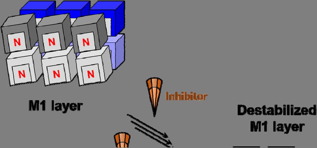
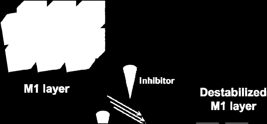

28 Supplementary Figure 7. Cartoon representation of M1 disruption at potentially multiple sites. S28


")


29 Supplementary Figure 8. Alternative binding sites for PHE. (a b) Location of probable PHE binding sites on M1 corresponding to the top-ranked sites from blind docking studies (see Supplementary Table 1) with either (a) electrostatic or (b) hydropathic potential mapped onto the M1 surface. Color scheme is the same as Figure 1 in the main text. PHE with yellow carbons, initial top-ranked PHE pose from VS; PHE with white carbons, top-ranked blind docking poses. S29


30 Supplementary Figure 9. The potential of PHE to disrupt M1 oligomerization at M1 M1 interfaces via steric incompatibility at multiple interaction sites, shown with top-ranked blind docking poses and within the context of the proposed oligomerization model described in Xie et al and depicted in the lower-right cartoon. The docked poses introduce obvious steric clashes with adjacent subunits that would disrupt oligomerization. Ribbons are colored by subunit, as shown in the cartoon. Yellow Connolly surfaces represent PHE at the initial topranked pose from VS; other surfaces are colored by subunit. S30
31 Supplementary Figure 10. In vitro and in ovo toxicity of PHE. A) PHE among six hit compounds had minimal cytotoxicity on MDCK cells (n=2 replicates). B) PHE at 740 ng/g had no toxicity on chicken embryos with or without the presence of viruses. S31
32 Supplementary Figure 11. PHE induction of a dose-dependent reduction in HA geometric mean of titer (GMT, black lines) of different IAV strains propagated in embryonic eggs, including A) H3N2 A/Fiji/2/2015 (Fiji/15) and B) H5N1 vaccine reassortant A/Egypt/N03072/2010XPR8 (EG/10XPR8). One-way ANOVA was performed to compare the differences between vehicle only and PHE treatments. HA titers were log-transformed before the analysis. * indicates P < 0.05; ** indicates P < 0.01; *** indicates P < A Fiji/15 (H3N2) in ovo replication B EG/10XPR8 (H5N1) in ovo replication HA titer (Log2) ** *** *** HA titer (Log2) * * * 0 Vehicle Vehicle PHE (ng/g) PHE (ng/g) S32
Proteins: Characteristics and Properties of Amino Acids
 SBI4U:Biochemistry Macromolecules Eachaminoacidhasatleastoneamineandoneacidfunctionalgroupasthe nameimplies.thedifferentpropertiesresultfromvariationsinthestructuresof differentrgroups.thergroupisoftenreferredtoastheaminoacidsidechain.
NMR study of complexes between low molecular mass inhibitors and the West Nile virus NS2B-NS3 protease
 University of Wollongong Research Online Faculty of Science - Papers (Archive) Faculty of Science, Medicine and Health 2009 NMR study of complexes between low molecular mass inhibitors and the West Nile
Packing of Secondary Structures
 7.88 Lecture Notes - 4 7.24/7.88J/5.48J The Protein Folding and Human Disease Professor Gossard Retrieving, Viewing Protein Structures from the Protein Data Base Helix helix packing Packing of Secondary
Secondary Structure. Bioch/BIMS 503 Lecture 2. Structure and Function of Proteins. Further Reading. Φ, Ψ angles alone determine protein structure
 Bioch/BIMS 503 Lecture 2 Structure and Function of Proteins August 28, 2008 Robert Nakamoto rkn3c@virginia.edu 2-0279 Secondary Structure Φ Ψ angles determine protein structure Φ Ψ angles are restricted
Structure and evolution of the spliceosomal peptidyl-prolyl cistrans isomerase Cwc27
 Acta Cryst. (2014). D70, doi:10.1107/s1399004714021695 Supporting information Volume 70 (2014) Supporting information for article: Structure and evolution of the spliceosomal peptidyl-prolyl cistrans isomerase
Amino Acid Side Chain Induced Selectivity in the Hydrolysis of Peptides Catalyzed by a Zr(IV)-Substituted Wells-Dawson Type Polyoxometalate
-Substituted Wells-Dawson Type Polyoxometalate") Amino Acid Side Chain Induced Selectivity in the Hydrolysis of Peptides Catalyzed by a Zr(IV)-Substituted Wells-Dawson Type Polyoxometalate Stef Vanhaecht, Gregory Absillis, Tatjana N. Parac-Vogt* Department
Amino Acid Side Chain Induced Selectivity in the Hydrolysis of Peptides Catalyzed by a Zr(IV)-Substituted Wells-Dawson Type Polyoxometalate Stef Vanhaecht, Gregory Absillis, Tatjana N. Parac-Vogt* Department
What makes a good graphene-binding peptide? Adsorption of amino acids and peptides at aqueous graphene interfaces: Electronic Supplementary
 Electronic Supplementary Material (ESI) for Journal of Materials Chemistry B. This journal is The Royal Society of Chemistry 21 What makes a good graphene-binding peptide? Adsorption of amino acids and
Supplementary Figure 3 a. Structural comparison between the two determined structures for the IL 23:MA12 complex. The overall RMSD between the two
 Supplementary Figure 1. Biopanningg and clone enrichment of Alphabody binders against human IL 23. Positive clones in i phage ELISA with optical density (OD) 3 times higher than background are shown for
Physiochemical Properties of Residues
 Physiochemical Properties of Residues Various Sources C N Cα R Slide 1 Conformational Propensities Conformational Propensity is the frequency in which a residue adopts a given conformation (in a polypeptide)
Other Methods for Generating Ions 1. MALDI matrix assisted laser desorption ionization MS 2. Spray ionization techniques 3. Fast atom bombardment 4.
 Other Methods for Generating Ions 1. MALDI matrix assisted laser desorption ionization MS 2. Spray ionization techniques 3. Fast atom bombardment 4. Field Desorption 5. MS MS techniques Matrix assisted
Supplementary Information Intrinsic Localized Modes in Proteins
 Supplementary Information Intrinsic Localized Modes in Proteins Adrien Nicolaï 1,, Patrice Delarue and Patrick Senet, 1 Department of Physics, Applied Physics and Astronomy, Rensselaer Polytechnic Institute,
Viewing and Analyzing Proteins, Ligands and their Complexes 2
 2 Viewing and Analyzing Proteins, Ligands and their Complexes 2 Overview Viewing the accessible surface Analyzing the properties of proteins containing thousands of atoms is best accomplished by representing
Using Higher Calculus to Study Biologically Important Molecules Julie C. Mitchell
 Using Higher Calculus to Study Biologically Important Molecules Julie C. Mitchell Mathematics and Biochemistry University of Wisconsin - Madison 0 There Are Many Kinds Of Proteins The word protein comes
Supplement information
 Electronic Supplementary Material (ESI) for Physil Chemistry Chemil Physics. This journal is the Owner Societies 216 Supplement information Fullerenol C 6 (OH) 16 prevents amyloid fibrillization of Aβ
7.012 Problem Set 1. i) What are two main differences between prokaryotic cells and eukaryotic cells?
 What are two main differences between prokaryotic cells and eukaryotic cells?") ame 7.01 Problem Set 1 Section Question 1 a) What are the four major types of biological molecules discussed in lecture? Give one important function of each type of biological molecule in the cell? b)
ame 7.01 Problem Set 1 Section Question 1 a) What are the four major types of biological molecules discussed in lecture? Give one important function of each type of biological molecule in the cell? b)
NMR Assignments using NMRView II: Sequential Assignments
 NMR Assignments using NMRView II: Sequential Assignments DO THE FOLLOWING, IF YOU HAVE NOT ALREADY DONE SO: For Mac OS X, you should have a subdirectory nmrview. At UGA this is /Users/bcmb8190/nmrview.
Electronic Supplementary Information
 Electronic Supplementary Information A Sensitive Phosphorescent Thiol Chemosensor Based on an Iridium(III) Complex with α,β-unsaturated Ketone Functionalized 2,2 -Bipyridyl Ligand Na Zhao, a Yu-Hui Wu,
Properties of amino acids in proteins
 Properties of amino acids in proteins one of the primary roles of DNA (but not the only one!) is to code for proteins A typical bacterium builds thousands types of proteins, all from ~20 amino acids repeated
Peptides And Proteins
 Kevin Burgess, May 3, 2017 1 Peptides And Proteins from chapter(s) in the recommended text A. Introduction B. omenclature And Conventions by amide bonds. on the left, right. 2 -terminal C-terminal triglycine
7.012 Problem Set 1 Solutions
 ame TA Section 7.012 Problem Set 1 Solutions Your answers to this problem set must be inserted into the large wooden box on wheels outside 68120 by 4:30 PM, Thursday, September 15. Problem sets will not
Ramachandran Plot. 4ysz Phi (degrees) Plot statistics
 Plot statistics") B Ramachandran Plot ~b b 135 b ~b ~l l Psi (degrees) 5-5 a A ~a L - -135 SER HIS (F) 59 (G) SER (B) ~b b LYS ASP ASP 315 13 13 (A) (F) (B) LYS ALA ALA 315 173 (E) 173 (E)(A) ~p p ~b - -135 - -5 5 135 (degrees)
B Ramachandran Plot ~b b 135 b ~b ~l l Psi (degrees) 5-5 a A ~a L - -135 SER HIS (F) 59 (G) SER (B) ~b b LYS ASP ASP 315 13 13 (A) (F) (B) LYS ALA ALA 315 173 (E) 173 (E)(A) ~p p ~b - -135 - -5 5 135 (degrees)
Supplementary figure 1. Comparison of unbound ogm-csf and ogm-csf as captured in the GIF:GM-CSF complex. Alignment of two copies of unbound ovine
 Supplementary figure 1. Comparison of unbound and as captured in the GIF:GM-CSF complex. Alignment of two copies of unbound ovine GM-CSF (slate) with bound GM-CSF in the GIF:GM-CSF complex (GIF: green,
Amino Acids and Proteins at ZnO-water Interfaces in Molecular Dynamics Simulations: Electronic Supplementary Information
 Amino Acids and Proteins at ZnO-water Interfaces in Molecular Dynamics Simulations: Electronic Supplementary Information Grzegorz Nawrocki and Marek Cieplak Institute of Physics, Polish Academy of Sciences,
Supporting information to: Time-resolved observation of protein allosteric communication. Sebastian Buchenberg, Florian Sittel and Gerhard Stock 1
 Supporting information to: Time-resolved observation of protein allosteric communication Sebastian Buchenberg, Florian Sittel and Gerhard Stock Biomolecular Dynamics, Institute of Physics, Albert Ludwigs
Exam I Answer Key: Summer 2006, Semester C
 1. Which of the following tripeptides would migrate most rapidly towards the negative electrode if electrophoresis is carried out at ph 3.0? a. gly-gly-gly b. glu-glu-asp c. lys-glu-lys d. val-asn-lys
Protein structure. Protein structure. Amino acid residue. Cell communication channel. Bioinformatics Methods
 Cell communication channel Bioinformatics Methods Iosif Vaisman Email: ivaisman@gmu.edu SEQUENCE STRUCTURE DNA Sequence Protein Sequence Protein Structure Protein structure ATGAAATTTGGAAACTTCCTTCTCACTTATCAGCCACCT...
FW 1 CDR 1 FW 2 CDR 2
 Supplementary Figure 1 Supplementary Figure 1: Interface of the E9:Fas structure. The two interfaces formed by V H and V L of E9 with Fas are shown in stereo. The Fas receptor is represented as a surface
Chapter 4: Amino Acids
 Chapter 4: Amino Acids All peptides and polypeptides are polymers of alpha-amino acids. lipid polysaccharide enzyme 1940s 1980s. Lipids membrane 1960s. Polysaccharide Are energy metabolites and many of
CHEM J-9 June 2014
 CEM1611 2014-J-9 June 2014 Alanine (ala) and lysine (lys) are two amino acids with the structures given below as Fischer projections. The pk a values of the conjugate acid forms of the different functional
Exam III. Please read through each question carefully, and make sure you provide all of the requested information.
 09-107 onors Chemistry ame Exam III Please read through each question carefully, and make sure you provide all of the requested information. 1. A series of octahedral metal compounds are made from 1 mol
Bacterial protease uses distinct thermodynamic signatures for substrate recognition
 Bacterial protease uses distinct thermodynamic signatures for substrate recognition Gustavo Arruda Bezerra, Yuko Ohara-Nemoto, Irina Cornaciu, Sofiya Fedosyuk, Guillaume Hoffmann, Adam Round, José A. Márquez,
Any protein that can be labelled by both procedures must be a transmembrane protein.
 1. What kind of experimental evidence would indicate that a protein crosses from one side of the membrane to the other? Regions of polypeptide part exposed on the outside of the membrane can be probed
C H E M I S T R Y N A T I O N A L Q U A L I F Y I N G E X A M I N A T I O N SOLUTIONS GUIDE
 C H E M I S T R Y 2 0 0 0 A T I A L Q U A L I F Y I G E X A M I A T I SLUTIS GUIDE Answers are a guide only and do not represent a preferred method of solving problems. Section A 1B, 2A, 3C, 4C, 5D, 6D,
Peptide Syntheses. Illustrative Protection: BOC/ t Bu. A. Introduction. do not acid
 Kevin Burgess, May 3, 2017 1 Peptide yntheses from chapter(s) in the recommended text A. Introduction do not acid -Met-e- -Met-Met- -e-e- -e-met- dipeptide dipeptide dipeptide dipeptide diketopiperazine
Model Mélange. Physical Models of Peptides and Proteins
 Model Mélange Physical Models of Peptides and Proteins In the Model Mélange activity, you will visit four different stations each featuring a variety of different physical models of peptides or proteins.
Clustering and Model Integration under the Wasserstein Metric. Jia Li Department of Statistics Penn State University
 Clustering and Model Integration under the Wasserstein Metric Jia Li Department of Statistics Penn State University Clustering Data represented by vectors or pairwise distances. Methods Top- down approaches
Oxygen Binding in Hemocyanin
 Supporting Information for Quantum Mechanics/Molecular Mechanics Study of Oxygen Binding in Hemocyanin Toru Saito and Walter Thiel* Max-Planck-Institut für Kohlenforschung, Kaiser-Wilhelm-Platz 1, D-45470
Protein Fragment Search Program ver Overview: Contents:
 Protein Fragment Search Program ver 1.1.1 Developed by: BioPhysics Laboratory, Faculty of Life and Environmental Science, Shimane University 1060 Nishikawatsu-cho, Matsue-shi, Shimane, 690-8504, Japan
Amino Acids and Peptides
 Amino Acids Amino Acids and Peptides Amino acid a compound that contains both an amino group and a carboxyl group α-amino acid an amino acid in which the amino group is on the carbon adjacent to the carboxyl
Read more about Pauling and more scientists at: Profiles in Science, The National Library of Medicine, profiles.nlm.nih.gov
 2018 Biochemistry 110 California Institute of Technology Lecture 2: Principles of Protein Structure Linus Pauling (1901-1994) began his studies at Caltech in 1922 and was directed by Arthur Amos oyes to
A Plausible Model Correlates Prebiotic Peptide Synthesis with. Primordial Genetic Code
 Electronic Supplementary Material (ESI) for ChemComm. This journal is The Royal Society of Chemistry 2018 A Plausible Model Correlates Prebiotic Peptide Synthesis with Primordial Genetic Code Jianxi Ying,
Sequence Alignments. Dynamic programming approaches, scoring, and significance. Lucy Skrabanek ICB, WMC January 31, 2013
 Sequence Alignments Dynamic programming approaches, scoring, and significance Lucy Skrabanek ICB, WMC January 31, 213 Sequence alignment Compare two (or more) sequences to: Find regions of conservation
Amino acids: Optimization in underivatized formula and fragment patterns in LC/ESI-MS analysis. Yoshinori Takano (JAMSTEC)
") (a) Positive ion mode (ph < 7) Amino acids: ptimization in underivatized formula and fragment patterns in L/ESI-MS analysis R 1 R 2 N + HA R 3 Base Acid R 1 R N H + 2 R 3 + A - Sample [M + H] + (b) Negative
(a) Positive ion mode (ph < 7) Amino acids: ptimization in underivatized formula and fragment patterns in L/ESI-MS analysis R 1 R 2 N + HA R 3 Base Acid R 1 R N H + 2 R 3 + A - Sample [M + H] + (b) Negative
CHMI 2227 EL. Biochemistry I. Test January Prof : Eric R. Gauthier, Ph.D.
 CHMI 2227 EL Biochemistry I Test 1 26 January 2007 Prof : Eric R. Gauthier, Ph.D. Guidelines: 1) Duration: 55 min 2) 14 questions, on 7 pages. For 70 marks (5 marks per question). Worth 15 % of the final
Supplemental Materials for. Structural Diversity of Protein Segments Follows a Power-law Distribution
 Supplemental Materials for Structural Diversity of Protein Segments Follows a Power-law Distribution Yoshito SAWADA and Shinya HONDA* National Institute of Advanced Industrial Science and Technology (AIST),
Nitrogenase MoFe protein from Clostridium pasteurianum at 1.08 Å resolution: comparison with the Azotobacter vinelandii MoFe protein
 Acta Cryst. (2015). D71, 274-282, doi:10.1107/s1399004714025243 Supporting information Volume 71 (2015) Supporting information for article: Nitrogenase MoFe protein from Clostridium pasteurianum at 1.08
Unraveling the degradation of artificial amide bonds in Nylon oligomer hydrolase: From induced-fit to acylation processes
 Electronic Supplementary Material (ESI) for Physical Chemistry Chemical Physics. This journal is the Owner Societies 2015 Supporting Information for Unraveling the degradation of artificial amide bonds
Solutions In each case, the chirality center has the R configuration
 CAPTER 25 669 Solutions 25.1. In each case, the chirality center has the R configuration. C C 2 2 C 3 C(C 3 ) 2 D-Alanine D-Valine 25.2. 2 2 S 2 d) 2 25.3. Pro,, Trp, Tyr, and is, Trp, Tyr, and is Arg,
Lecture 15: Realities of Genome Assembly Protein Sequencing
 Lecture 15: Realities of Genome Assembly Protein Sequencing Study Chapter 8.10-8.15 1 Euler s Theorems A graph is balanced if for every vertex the number of incoming edges equals to the number of outgoing
Background BIOCHEMISTRY LAB CHE-554. Experiment #1 Spectrophotometry
 BIOCHEMISTRY LAB Experiment #1 Spectrophotometry CHE-554 In day 1 we will use spectrophotometry as an analytical technique using a known extinction coefficient to assess the precision and accuracy of common
The translation machinery of the cell works with triples of types of RNA bases. Any triple of RNA bases is known as a codon. The set of codons is
 Relations Supplement to Chapter 2 of Steinhart, E. (2009) More Precisely: The Math You Need to Do Philosophy. Broadview Press. Copyright (C) 2009 Eric Steinhart. Non-commercial educational use encouraged!
Chemistry Chapter 22
 hemistry 2100 hapter 22 Proteins Proteins serve many functions, including the following. 1. Structure: ollagen and keratin are the chief constituents of skin, bone, hair, and nails. 2. atalysts: Virtually
Biological Macromolecules
 Introduction for Chem 493 Chemistry of Biological Macromolecules Dr. L. Luyt January 2008 Dr. L. Luyt Chem 493-2008 1 Biological macromolecules are the molecules of life allow for organization serve a
Deciphering the Rules for Amino Acid Co- Assembly Based on Interlayer Distances
 Supporting Information Deciphering the Rules for Amino Acid Co- Assembly Based on Interlayer Distances Santu Bera, Sudipta Mondal, Yiming Tang, Guy Jacoby, Elad Arad, ǁ Tom Guterman, Raz Jelinek, ǁ Roy
Molecular Structure Prediction by Global Optimization
 Molecular Structure Prediction by Global Optimization K.A. DILL Department of Pharmaceutical Chemistry, University of California at San Francisco, San Francisco, CA 94118 A.T. PHILLIPS Computer Science
Advanced Topics in RNA and DNA. DNA Microarrays Aptamers
 Quiz 1 Advanced Topics in RNA and DNA DNA Microarrays Aptamers 2 Quantifying mrna levels to asses protein expression 3 The DNA Microarray Experiment 4 Application of DNA Microarrays 5 Some applications
Diastereomeric resolution directed towards chirality. determination focussing on gas-phase energetics of coordinated. sodium dissociation
 Supplementary Information Diastereomeric resolution directed towards chirality determination focussing on gas-phase energetics of coordinated sodium dissociation Authors: samu Kanie*, Yuki Shioiri, Koji
Structure Investigation of Fam20C, a Golgi Casein Kinase
 Structure Investigation of Fam20C, a Golgi Casein Kinase Sharon Grubner National Taiwan University, Dr. Jung-Hsin Lin University of California San Diego, Dr. Rommie Amaro Abstract This research project
B O C 4 H 2 O O. NOTE: The reaction proceeds with a carbonium ion stabilized on the C 1 of sugar A.
 hbcse 33 rd International Page 101 hemistry lympiad Preparatory 05/02/01 Problems d. In the hydrolysis of the glycosidic bond, the glycosidic bridge oxygen goes with 4 of the sugar B. n cleavage, 18 from
Course Notes: Topics in Computational. Structural Biology.
 Course Notes: Topics in Computational Structural Biology. Bruce R. Donald June, 2010 Copyright c 2012 Contents 11 Computational Protein Design 1 11.1 Introduction.........................................
X-ray crystallography NMR Cryoelectron microscopy
 Molecular Graphics with PyMOL Overview of: Protein Data Bank Coordinates Jean-Yves Sgro PyMOL interface Hands-on! Experimental Methods 3 Main: X-ray crystallography NMR Cryoelectron microscopy X-ray source
Full wwpdb X-ray Structure Validation Report i
 Full wwpdb X-ray Structure Validation Report i Mar 14, 2018 02:00 pm GMT PDB ID : 3RRQ Title : Crystal structure of the extracellular domain of human PD-1 Authors : Lazar-Molnar, E.; Ramagopal, U.A.; Nathenson,
Introduction to the Ribosome Overview of protein synthesis on the ribosome Prof. Anders Liljas
 Introduction to the Ribosome Molecular Biophysics Lund University 1 A B C D E F G H I J Genome Protein aa1 aa2 aa3 aa4 aa5 aa6 aa7 aa10 aa9 aa8 aa11 aa12 aa13 a a 14 How is a polypeptide synthesized? 2
Introduction to Comparative Protein Modeling. Chapter 4 Part I
 Introduction to Comparative Protein Modeling Chapter 4 Part I 1 Information on Proteins Each modeling study depends on the quality of the known experimental data. Basis of the model Search in the literature
Protein Data Bank Contents Guide: Atomic Coordinate Entry Format Description. Version Document Published by the wwpdb
 Protein Data Bank Contents Guide: Atomic Coordinate Entry Format Description Version 3.30 Document Published by the wwpdb This format complies with the PDB Exchange Dictionary (PDBx) http://mmcif.pdb.org/dictionaries/mmcif_pdbx.dic/index/index.html.
Desorption/Ionization Efficiency of Common Amino Acids in. Surface-assisted Laser Desorption/ionization Mass Spectrometry
 SUPPORTING INFORMATION Desorption/Ionization Efficiency of Common Amino Acids in Surface-assisted Laser Desorption/ionization Mass Spectrometry (SALDI-MS) with Nanostructured Platinum Syuhei Nitta, Hideya
Ranjit P. Bahadur Assistant Professor Department of Biotechnology Indian Institute of Technology Kharagpur, India. 1 st November, 2013
 Hydration of protein-rna recognition sites Ranjit P. Bahadur Assistant Professor Department of Biotechnology Indian Institute of Technology Kharagpur, India 1 st November, 2013 Central Dogma of life DNA
12/6/12. Dr. Sanjeeva Srivastava IIT Bombay. Primary Structure. Secondary Structure. Tertiary Structure. Quaternary Structure.
 Dr. anjeeva rivastava Primary tructure econdary tructure Tertiary tructure Quaternary tructure Amino acid residues α Helix Polypeptide chain Assembled subunits 2 1 Amino acid sequence determines 3-D structure
Supporting information
 Electronic Supplementary Material (ESI) for New Journal of Chemistry. This journal is The Royal Society of Chemistry and the Centre National de la Recherche Scientifique 2015 Supporting information Influence
Major Types of Association of Proteins with Cell Membranes. From Alberts et al
 Major Types of Association of Proteins with Cell Membranes From Alberts et al Proteins Are Polymers of Amino Acids Peptide Bond Formation Amino Acid central carbon atom to which are attached amino group
PROTEIN SECONDARY STRUCTURE PREDICTION: AN APPLICATION OF CHOU-FASMAN ALGORITHM IN A HYPOTHETICAL PROTEIN OF SARS VIRUS
 Int. J. LifeSc. Bt & Pharm. Res. 2012 Kaladhar, 2012 Research Paper ISSN 2250-3137 www.ijlbpr.com Vol.1, Issue. 1, January 2012 2012 IJLBPR. All Rights Reserved PROTEIN SECONDARY STRUCTURE PREDICTION:
Bahnson Biochemistry Cume, April 8, 2006 The Structural Biology of Signal Transduction
 Name page 1 of 6 Bahnson Biochemistry Cume, April 8, 2006 The Structural Biology of Signal Transduction Part I. The ion Ca 2+ can function as a 2 nd messenger. Pick a specific signal transduction pathway
Snork Synthesis Lab Lab Directions
 Snork Synthesis Lab Lab Directions This activity, modified from the original at The Biology Corner, will help you practice your understanding of protein synthesis. Submit your lab answers according to
Supporting Information
 Supporting Information Micelle-Triggered b-hairpin to a-helix Transition in a 14-Residue Peptide from a Choline-Binding Repeat of the Pneumococcal Autolysin LytA HØctor Zamora-Carreras, [a] Beatriz Maestro,
A. Two of the common amino acids are analyzed. Amino acid X and amino acid Y both have an isoionic point in the range of
 Questions with Answers- Amino Acids & Peptides A. Two of the common amino acids are analyzed. Amino acid X and amino acid Y both have an isoionic point in the range of 5.0-6.5 (Questions 1-4) 1. Which
Table 1. Crystallographic data collection, phasing and refinement statistics. Native Hg soaked Mn soaked 1 Mn soaked 2
 Table 1. Crystallographic data collection, phasing and refinement statistics Native Hg soaked Mn soaked 1 Mn soaked 2 Data collection Space group P2 1 2 1 2 1 P2 1 2 1 2 1 P2 1 2 1 2 1 P2 1 2 1 2 1 Cell
Translation. A ribosome, mrna, and trna.
 Translation The basic processes of translation are conserved among prokaryotes and eukaryotes. Prokaryotic Translation A ribosome, mrna, and trna. In the initiation of translation in prokaryotes, the Shine-Dalgarno
Studies Leading to the Development of a Highly Selective. Colorimetric and Fluorescent Chemosensor for Lysine
 Supporting Information for Studies Leading to the Development of a Highly Selective Colorimetric and Fluorescent Chemosensor for Lysine Ying Zhou, a Jiyeon Won, c Jin Yong Lee, c * and Juyoung Yoon a,
Journal of Pharmacology and Experimental Therapy-JPET#172536
 A NEW NON-PEPTIDIC INHIBITOR OF THE 14-3-3 DOCKING SITE INDUCES APOPTOTIC CELL DEATH IN CHRONIC MYELOID LEUKEMIA SENSITIVE OR RESISTANT TO IMATINIB Manuela Mancini, Valentina Corradi, Sara Petta, Enza
Sequential resonance assignments in (small) proteins: homonuclear method 2º structure determination
 proteins: homonuclear method 2º structure determination") Lecture 9 M230 Feigon Sequential resonance assignments in (small) proteins: homonuclear method 2º structure determination Reading resources v Roberts NMR of Macromolecules, Chap 4 by Christina Redfield
Lecture 9 M230 Feigon Sequential resonance assignments in (small) proteins: homonuclear method 2º structure determination Reading resources v Roberts NMR of Macromolecules, Chap 4 by Christina Redfield
Geometrical Concept-reduction in conformational space.and his Φ-ψ Map. G. N. Ramachandran
 Geometrical Concept-reduction in conformational space.and his Φ-ψ Map G. N. Ramachandran Communication paths in trna-synthetase: Insights from protein structure networks and MD simulations Saraswathi Vishveshwara
Resonance assignments in proteins. Christina Redfield
 Resonance assignments in proteins Christina Redfield 1. Introduction The assignment of resonances in the complex NMR spectrum of a protein is the first step in any study of protein structure, function
Full wwpdb X-ray Structure Validation Report i
 Full wwpdb X-ray Structure Validation Report i Mar 8, 2018 06:13 pm GMT PDB ID : 5G5C Title : Structure of the Pyrococcus furiosus Esterase Pf2001 with space group C2221 Authors : Varejao, N.; Reverter,
BIOINF 4120 Bioinformatics 2 - Structures and Systems - Oliver Kohlbacher Summer Protein Structure Prediction I
 BIOINF 4120 Bioinformatics 2 - Structures and Systems - Oliver Kohlbacher Summer 2013 9. Protein Structure Prediction I Structure Prediction Overview Overview of problem variants Secondary structure prediction
Central Dogma. modifications genome transcriptome proteome
 entral Dogma DA ma protein post-translational modifications genome transcriptome proteome 83 ierarchy of Protein Structure 20 Amino Acids There are 20 n possible sequences for a protein of n residues!
Sequence Based Bioinformatics
 Structural and Functional Analysis of Inosine Monophosphate Dehydrogenase using Sequence-Based Bioinformatics Barry Sexton 1,2 and Troy Wymore 3 1 Bioengineering and Bioinformatics Summer Institute, Department
C CH 3 N C COOH. Write the structural formulas of all of the dipeptides that they could form with each other.
 hapter 25 Biochemistry oncept heck 25.1 Two common amino acids are 3 2 N alanine 3 2 N threonine Write the structural formulas of all of the dipeptides that they could form with each other. The carboxyl
April, The energy functions include:
 REDUX A collection of Python scripts for torsion angle Monte Carlo protein molecular simulations and analysis The program is based on unified residue peptide model and is designed for more efficient exploration
Protein Structure. Role of (bio)informatics in drug discovery. Bioinformatics
informatics in drug discovery. Bioinformatics") Bioinformatics Protein Structure Principles & Architecture Marjolein Thunnissen Dep. of Biochemistry & Structural Biology Lund University September 2011 Homology, pattern and 3D structure searches need
Bioinformatics Protein Structure Principles & Architecture Marjolein Thunnissen Dep. of Biochemistry & Structural Biology Lund University September 2011 Homology, pattern and 3D structure searches need
Section Week 3. Junaid Malek, M.D.
 Section Week 3 Junaid Malek, M.D. Biological Polymers DA 4 monomers (building blocks), limited structure (double-helix) RA 4 monomers, greater flexibility, multiple structures Proteins 20 Amino Acids,
What binds to Hb in addition to O 2?
 Reading: Ch5; 158-169, 162-166, 169-174 Problems: Ch5 (text); 3,7,8,10 Ch5 (study guide-facts); 1,2,3,4,5,8 Ch5 (study guide-apply); 2,3 Remember Today at 5:30 in CAS-522 is the second chance for the MB
Knowledge-based structure prediction of MHC class I bound peptides: a study of 23 complexes Ora Schueler-Furman 1,2, Ron Elber 2 and Hanah Margalit 1
 Research Paper 549 Knowledge-based structure prediction of MHC class I bound peptides: a study of 23 complexes Ora Schueler-Furman 1,2, Ron Elber 2 and Hanah Margalit 1 Background: The binding of T-cell
CSE 549: Computational Biology. Substitution Matrices
 CSE 9: Computational Biology Substitution Matrices How should we score alignments So far, we ve looked at arbitrary schemes for scoring mutations. How can we assign scores in a more meaningful way? Are
Biophysical Journal, Volume 96. Supporting Material
 Biophysical Journal, Volume 96 Supporting Material NMR dynamics of PSE-4 β-lactamase: an interplay of ps-ns order and μs-ms motions in the active site Sébastien Morin and Stéphane M. Gagné NMR dynamics
Supporting Information
 Theoretical examination of competitive -radical-induced cleavages of N-C and C -C bonds of peptides Wai-Kit Tang, Chun-Ping Leong, Qiang Hao, Chi-Kit Siu* Department of Biology and Chemistry, City University
Protein Struktur (optional, flexible)
") Protein Struktur (optional, flexible) 22/10/2009 [ 1 ] Andrew Torda, Wintersemester 2009 / 2010, AST nur für Informatiker, Mathematiker,.. 26 kt, 3 ov 2009 Proteins - who cares? 22/10/2009 [ 2 ] Most important
Protein Struktur (optional, flexible) 22/10/2009 [ 1 ] Andrew Torda, Wintersemester 2009 / 2010, AST nur für Informatiker, Mathematiker,.. 26 kt, 3 ov 2009 Proteins - who cares? 22/10/2009 [ 2 ] Most important
SEQUENCE ALIGNMENT BACKGROUND: BIOINFORMATICS. Prokaryotes and Eukaryotes. DNA and RNA
 SEQUENCE ALIGNMENT BACKGROUND: BIOINFORMATICS 1 Prokaryotes and Eukaryotes 2 DNA and RNA 3 4 Double helix structure Codons Codons are triplets of bases from the RNA sequence. Each triplet defines an amino-acid.
*Corresponding Author *K. F.: *T. H.:
 Theoretical Analysis of Activity Cliffs among Benzofuranone Class Pim1 Inhibitors Using the Fragment Molecular Orbital Method with Molecular Mechanics Poisson-Boltzmann Surface Area (FMO+MM-PBSA) Approach
Similarity or Identity? When are molecules similar?
 Similarity or Identity? When are molecules similar? Mapping Identity A -> A T -> T G -> G C -> C or Leu -> Leu Pro -> Pro Arg -> Arg Phe -> Phe etc If we map similarity using identity, how similar are
Supplementary Information. Affinity Selection and sequence-activity relationships of HIV-1 membrane fusion inhibitors
 Supplementary Information Affinity Selection and sequence-activity relationships of HIV-1 membrane fusion inhibitors directed at the drug-resistant variants Shinya Oishi,* a Kentaro Watanabe, a Saori Ito,