2D XRD Imaging by Projection-Type X-Ray Microscope
|
|
|
- Shavonne Kathlyn Singleton
- 6 years ago
- Views:
Transcription

1 0/25 National Institute for Materials Science,Tsukuba, Japan 2D XRD Imaging by Projection-Type X-Ray Microscope 1. Introduction - What s projection-type X-ray microscope? 2. Examples for inhomogeneous/patterned specimens 3. Application to stress imaging 4. Application to combinatorial imaging 5. Summary and future outlook Kenji SAKURAI
2 2θ Why 2D Imaging for X-Ray Diffraction? Because inhomogeneous system is usual for many sciences identification of materials - mixed crystal - polymorphism poly-crystalline materials - ceramics - metals - organic crystals 1/25 Microstructure - phase transition -diffusion Inhomogeneous mixture Grain boundaries Textures Macrostructure - grain growth - preferred orientation - dendrite I Average information? physical property - residual stress - heat transfer
 Types - 2/25 Scanning type")
 K.")

3 How to Obtain 2D Real Space Images? - Scanning vs. Projection (Reflection) Types - 2/25 Scanning type Projection type XY scan Synchrotron µ beam Sample T Wroblewski XRD (1995) K.Sakurai XRF movie (2003) X-Ray CCD camera with a collimator PC Fluorescent X-rays Diffracted X-rays Synchrotron wide beam detector High-resolution(~100nm) Sample Extremely quick(~100msec)
4 How Projection-type Microscope Works? Monochromator scan like EXAFS experiments 3/25 XRD imaging 2d n sinθ Bn = λ n Intensity E 0 E 1 E 2 E 3 X-Ray Energy E n = /λ n Normal XRD CCD Collimator SR Energy scan d 2 d 1 d 3 Monochromator Inhomogeneous sample

5 How Projection-type Microscope Works? 4/25 The experiment is just simple exposures without XY scan X-ray image Resolution: Δ= r/t d CCD BL-16A1 Photon Factory Collimator Primary X-rays r t d Sample

![Energy [kev] 6 7 8 9 10 11 12 13 1415 6100 ev](/docs-images/72/67094923/images/6-1.jpg "(XRF) 1 sec x 30 5/23 Intensity 60 40 20 0 2θ 64")

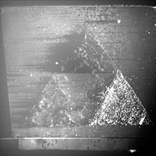
6 Cr K absorption edge Examples of 2D XRD Imaging Patterned metallic Cr thin films Energy [kev] ev (XRF) 1 sec x 30 5/23 Intensity θ 64 deg Camera- Sample 13 mm 5765 ev (XRD) 1 sec x d [A] 8mm 8mm
7 (220)(109)(1010)(300)(214)(018)(122)(116)(024)a-Al2O3Intensity[cps] (420)(131)(401)(232)(331)(114)(140)(123)(232)(231)(222)(230)(113)(312)(311)(203)(031)(222)(311)(310)(202)HfO2Intensity[cps] (833)(840)(662)(831)(660)(653)(822)(811)(800)(651)(642)(721)(640)(543)(444)(631)(622)(541)(620)(611)OCT0411Intensity[cps][ナ]HfO2 Y 2 O 3 Al 2 O 3 monoclinic HfO 2 (-401) Energy:6630eV (bragg angle: 45.31deg.) Cubic Y 2 O 3 (662) Energy:7160eV (bragg angle: 45.45deg.) alpha-al 2 O 3 (300) Energy:6345eV (bragg angle: 45.39deg.) Viewing Area 13mm 13mm Viewing Area 13mm 13mm 2d (Å) Examples of 2D XRD Imaging Different materials in the same view area 6/25
 Examples of 2D XRD Imaging Mapping of different phases of")
 13mm Intensity [Cps] Exposure Time 10 sec 10000 5000 (0 3")
![3) Monoclinic Cubic Energy [kev] 7 6.](/docs-images/72/67094923/images/8-2.jpg "5 6 (1 4 0) (1 1 4) 13mm (4 0 0) (4 0 0) (4 0 1) (2 2 3) (2 3 1) (0")
8 Monoclinic ZrO 2 (140) 6889eV(d=0.1266nm) Examples of 2D XRD Imaging Mapping of different phases of ZrO 2 7/25 Cubic ZrO 2 (400) 6787eV(d=0.1285nm) 13mm Intensity [Cps] Exposure Time 10 sec (0 3 3) Monoclinic Cubic Energy [kev] (1 4 0) (1 1 4) 13mm (4 0 0) (4 0 0) (4 0 1) (2 2 3) (2 3 1) (0 2 3) (2 3 0) (3 2 0) (3 1 2) (2 2 2) (1 1 3) XRD images Normal XRD d [A]


9 Examples of 2D XRD Imaging Mapping of different phases of TiO 2 8/ θ=76deg 2θ=84deg Viewing area 8 8mm A Intensity 50 anatase (200) rutile (210) R R Rutile Anatase 0 Incident beam:4900ev θ/2θ [deg]
= A 1 C A (x,y) +B 1 C B")
 = 2 A 1 I 1 A 2 A1 B 2")
10 Examples of 2D XRD Imaging Quantitative imaging for peak overlapping cases 9/23 Intensity B 1 A 1 B 2 A 2 I 1 (x,y)= A 1 C A (x,y) +B 1 C B (x,y) I 2 (x,y)= A 2 C A (x,y) +B 2 C B (x,y) I C A (x,y) = 1 B 2 I 2 B 1 A1 B 2 A 2 B 1 E 1 E X-ray Energy 2 I C B (x,y) = 2 A 1 I 1 A 2 A1 B 2 A 2 B 1
 (220) (311) RD RD Optical Photo Intensity [ 10 3 cps] 100 50 0 X-ray d=2.")
 (222) 7175 ev 1.7mm Line-pattern parallel to R.D. The strongest reflection is (311). d=1.")
11 Examples of 2D XRD Imaging 10/23 Orientation dependence of aluminum sheet (0.1mm t) (220) (311) RD RD Optical Photo Intensity [ 10 3 cps] X-ray d=2.338å 3751eV (111) (200) R.D. Normal XRD 6125eV (220) d=1.431å 6130eV θ[deg.] (311) (222) 7175 ev 1.7mm Line-pattern parallel to R.D. The strongest reflection is (311). d=1.221å 7140eV d=1.1690å 7504eV Viewing Area 8mm 8mm Exposure Time 1 sec
12 Application to Stress Imaging 11/25 Stress analysis has been done by conventional XRD Diffraction Angle, 2θ (deg.) Stress σ x X-ray ψ 1 ψ 2 ψ 3 2θ 2θ d+ ε 1 d d+ ε 2 d d+ ε 3 d larger ψ larger ε E Young s modulus ν Possion s ratio θ Bragg angle Ψ Orientation 2θ 1 + ν 2 v ε φψ = σ x sin ψ + ( σ 1 + σ 2 ) E E σ x Stress σ x σ x 2d sin θ=λ Δd ε = = cotθ Δθ d Strain Peak shift E (2θ φψ ) π = cotθ0 2 2(1 + ν ) sin ψ 180 = K M ( ε = E 1+ ν (sin ) φψ 2 ψ ) sin 2 Ψ Changing incidence angle
 E E CCD 12/25 X-rays σ x E ( ε = 1+ ν (sin E = 1+ ν = K M φψ ψ ) 1 λ 2 λ sin ψ 0 2 ) λ/λ 0 Sample 2 sin Ψ λ λ 0 plot sin 2 ψ CCD Sample is always fixed")
13 Application to Stress Imaging How to extend the method to 2D imaging? 1 + ν 2 v ε φψ = σ x sin ψ + ( σ 1 + σ 2 ) E E CCD 12/25 X-rays σ x E ( ε = 1+ ν (sin E = 1+ ν = K M φψ ψ ) 1 λ 2 λ sin ψ 0 2 ) λ/λ 0 Sample 2 sin Ψ λ λ 0 plot sin 2 ψ CCD Sample is always fixed Energy scan instead of 2θ scan Changing 2θ instead of incidence angle for ψ Sin 2 ψ λ/λo plot instead of sin 2 ψ 2θ plot Monochromator X-Rays ψ 2θ
14 Application to Stress Imaging Imaging of welded steel 13/25 Conventional XRD pattern (Cu Kα) Welded parts Blocks (low carbon steel)
 reflections at 90 deg 14/25")

15 Application to Stress Imaging 2D images for Fe(200) reflections at 90 deg 14/ eV 6128eV 6184eV Exposure time:5sec/image CCD steel block X-ray 90 Sample Welded part


16 Application to Stress Imaging 2D images for Fe(200) reflections for different ψ 15/25 X-ray CCD Sample X-ray CCD Sample X-ray CCD Sample ψ=39.0 ψ=45.0 ψ=52.5
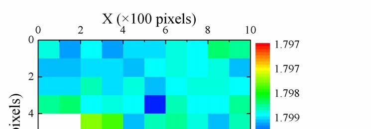
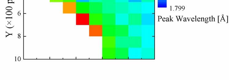
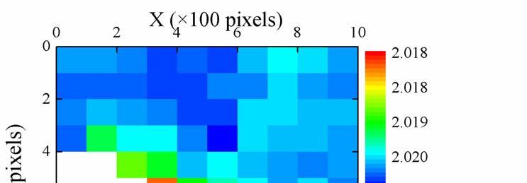
17 Application to Stress Imaging 16/25 2D images of peak shift amount (X-ray wavelength) ψ=39.0 ψ=52.5 steel block ψ=45.0 Welded part


18 Application to Stress Imaging Finally obtained stress image from sin 2 ψ λ/λo plots Strain images for different ψ 17/25 Intensity λ/λ 0 ψ=39.0 Wavelength sin 2 ψ ψ=45.0 Intensity Wavelength Stress image Slope values for each pixel compressive Intensity tensile ψ=52.5 Wavelength
19 Application to Combinatorial Imaging 18/25 Efficient analysis of arrayed samples on a single substrate E 1 E 2 E 3 E 4 Single-rod fishing Net- fishing Parameters Sample-1, Sample-2,. XRD intensity Absorption XAFS XRD composition, temperature, etc. E A E B E C E D
 Energy: 7160 ev α-al 2")

.")
20 Application to Combinatorial Imaging Y 2 O 3 Screening of high-k oxide candidates HfO 2 Cubic Y 2 O 3 (662) Energy: 7160 ev α-al 2 O 3 (300) Energy: 6345 ev 19/25 13mm Al 2 O 3 13mm Combinatorial library Graded ternary oxides sample prepared by pulsed laser deposition. A. Ahmet, Y.-Z. Yoo, K. Hasegawa, H. Koinuma, T. Chikyow Appl. Phys. A 79, (2004). - monoclinic HfO 2 (401) Energy: 6630 ev


21 Application to Combinatorial Imaging 20/25 XRF/XAFS imaging of CO 2 absorbing LiFeO 2 Synthesis temperature 100 o C 200 o C 300 o C 400 o C SQ substrate CO 2 Not exposed 8mm 100 o C 30 min 200 o C 30 min CO 2 exposure 350 o C 30 min 8mm Fe-K XRF image KeK-PF BL-16A1 Incident X-ray energy : 7130 ev (above the Fe-absorption edge) Imaging time : 3 sec Pixels : 1000 x 1000 Bulk Nano particles Chemical absorption of CO 2 2 LiFeO 2 + CO 2 Li 2 CO 3 + γ-fe 2 O 3

22 XRF Intensity (norm) Application to Combinatorial Imaging 21/25 XRF/XAFS imaging of CO 2 absorbing LiFeO 2 Synthesis temperature 100 o C 200 o C 300 o C 400 o C 0.5eV-step x 60 points Measuring time = 9 min!! CO 2 exposure Quick change at lower temperature absorption edge shift CO 2 exposure Not exposed 100 o C x 30 min 200 o C x 30 min 350 o C x 30 min X-Ray Energy (ev) 200 o C 400 o C
23 Summary 1M pixel imaging for ~ cm 2 area 2D XRD imaging by projection-type X-ray microscope Different phases 22/25 Energy-dispersive experiments with fixed geometry Glancing angle to the sample surface 1~3 deg Diffraction angle (CCD camera position) 60~120 deg Close distance between the sample and CCD device 0.5~15 mm Collimator plate inside the CCD camera 6 mrad Imaging of inhomogeneous/patterned polycrystalline specimens difference in materials, phases, orientations Application to stress imaging Application to combinatorial imaging Combining with XRF/XAFS imaging Different orientations
24 Summary 23/25 Both scanning and projection-types are necessary Geometry for the sample Scanning microscope Vertical arrangement (for most cases) Projection microscope (Non-Scanning) Horizontal arrangement Typical primary beam size µm x µm 8~12mm (H) x0.4mm(v) Necessity of focusing the primary beam Absolutely necessary Desirable for vertical direction Typical spatial resolution µm µm Typical observation area µm x µm 8-12mm x 8-12mm Ideal polarization in terms of S/B ratio Horizontally linear Vertically linear Typical pixel numbers ca. 100 x 100 More than 1000 x 1000 Typical measuring time for one image 3 24 h sec


25 Towards Future Projection-type microscope can be widely used Diffusion 24/25 Realtime Movie Deposition? composite materials 3D/4D Imaging Phase transition Chemical reaction Rapid Diagnostics Functionally graded materials Combinatorial screening American Chemical Society, Mitsubishi Electric Engineering Stress analysis We use a synchrotron but combination with X-ray tube is also promising..
26 Acknowledgement Thank you! Mari MIZUSAWA NIMS Stress imaging Hiromi EBA NIMS Masahiko Shoji NIMS Image processing Hiroshi SAWA Photon Factory, KEK Yusuke WAKABAYASHI Photon Factory, KEK Yoshinori UCHIDA Photon Factory, KEK Atsuo IIDA Photon Factory, KEK Combinatorial imaging BL-16A BL-16A BL-16A BL-4A 25/25 Active-Nano supported by MEXT, Japan government Photon Factory S-type Program References K.Sakurai, Spectrochimica Acta B54, 1497 (1999). K.Sakurai, Photon Factory Activity Report 2001 Part A, 33. K.Sakurai and H.Eba, Anal. Chem. 75, 355 (2003). M.Mizusawa and K.Sakurai, J. Synchrotron Rad. 11, 209 (2004). K.Sakurai and M.Mizusawa, AIP Conf. Proceedings (SRI-2003). (2004). K.Sakurai and M.Mizusawa, Nanotechnology, 15, S428 (2004). H.Eba and K.Sakurai, Materials Trans., 46, 665 (2005). H.Eba and K.Sakurai, Chemistry Letters, 34, 872 (2005). H.Eba and K.Sakurai, Appl. Surf. Sci., (in press). Japanese Patents , , , , , , ,
 Point Analysis 2 1 3 5 4 Stress 2D")

27 Measuring Point (No.1~5) Point Analysis Stress 2D image analysis region 2θ-sin 2 ψ plot σ=k M M is the slope in 2θ-sin 2 ψ plot K=-32.44kg/mm 2 /deg


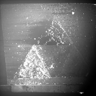
28 Quick X-Ray Fluorescence Imaging Metallic Cr thin film on glass substrate X-ray image Incident X-ray Energy 10.0 kev X-ray Fluorescence Exposure Time 1 sec. K L M 8 mm Optical microscope image 8 mm
DEVELOPMENT OF MEASURING SYSTEM FOR STRESS BY MEANS OF IMAGE PLATE FOR LABORATORY X-RAY EXPERIMENT
 Copyright JCPDS - International Centre for Diffraction Data 003, Advances in X-ray Analysis, Volume 46. 6 DEVELOPMENT OF MEASURING SYSTEM FOR STRESS BY MEANS OF IMAGE PLATE FOR LABORATORY X-RAY EXPERIMENT
Development of 2-Dimentional Imaging XAFS System at BL-4
 Development of 2-Dimentional Imaging XAFS System at BL-4 Koichi Sumiwaka 1, Misaki Katayama 2, Yasuhiro Inada 2 1) Department of Applied Chemistry, College of Science and Engineering, Ritsumeikan, University,
Fig. 2. Intersection between X-ray and diffracted beams. 2θ 1 (1) (2) (3) (4) method.
 (2) (3) (4) method.") 2017A-E10 Program Title An attempt to measure strains of coarse grains and micro area using double exposure method 1), 2) 3) 3) 4) Username : K. Suzuki 1), T. Shobu 2) R. Yasuda 3) A. Shiro 3) Y. Kiso
2017A-E10 Program Title An attempt to measure strains of coarse grains and micro area using double exposure method 1), 2) 3) 3) 4) Username : K. Suzuki 1), T. Shobu 2) R. Yasuda 3) A. Shiro 3) Y. Kiso
Early Development of Dispersive X-Ray Absorption Spectrometer and Recent Extension of Dispersive Optics to Quick X-ray Reflectometory
 Early Development of Dispersive X-Ray Absorption Spectrometer and Recent Extension of Dispersive Optics to Quick X-ray Reflectometory T. Matsushita, Photon Factory KEK Tsukuba, Ibaraki, Japan outline laboratory
Strain, Stress and Cracks Klaus Attenkofer PV Reliability Workshop (Orlando) April 7-8, 2015
 April 7-8, 2015") Strain, Stress and Cracks Klaus Attenkofer PV Reliability Workshop (Orlando) April 7-8, 2015 1 BROOKHAVEN SCIENCE ASSOCIATES Overview Material s response to applied forces or what to measure Definitions
Strain, Stress and Cracks Klaus Attenkofer PV Reliability Workshop (Orlando) April 7-8, 2015 1 BROOKHAVEN SCIENCE ASSOCIATES Overview Material s response to applied forces or what to measure Definitions
X-rays. X-ray Radiography - absorption is a function of Z and density. X-ray crystallography. X-ray spectrometry
 X-rays Wilhelm K. Roentgen (1845-1923) NP in Physics 1901 X-ray Radiography - absorption is a function of Z and density X-ray crystallography X-ray spectrometry X-rays Cu K α E = 8.05 kev λ = 1.541 Å Interaction
NEW CORRECTION PROCEDURE FOR X-RAY SPECTROSCOPIC FLUORESCENCE DATA: SIMULATIONS AND EXPERIMENT
 Copyright JCPDS - International Centre for Diffraction Data 2005, Advances in X-ray Analysis, Volume 48. 266 NEW CORRECTION PROCEDURE FOR X-RAY SPECTROSCOPIC FLUORESCENCE DATA: SIMULATIONS AND EXPERIMENT
Setting The motor that rotates the sample about an axis normal to the diffraction plane is called (or ).
.") X-Ray Diffraction X-ray diffraction geometry A simple X-ray diffraction (XRD) experiment might be set up as shown below. We need a parallel X-ray source, which is usually an X-ray tube in a fixed position
X-Ray Diffraction X-ray diffraction geometry A simple X-ray diffraction (XRD) experiment might be set up as shown below. We need a parallel X-ray source, which is usually an X-ray tube in a fixed position
Supplementary Information
 Supplementary Information Bismuth Sulfide Nanoflowers for Detection of X-rays in the Mammographic Energy Range Shruti Nambiar a,b, Ernest K. Osei a,c,d, John T.W.Yeow *,a,b a Department of Systems Design
Structure Report for J. Reibenspies
 X-ray Diffraction Laboratory Center for Chemical Characterization and Analysis Department of Chemistry Texas A & M University Structure Report for J. Reibenspies Project Name: Sucrose Date: January 29,
SYNCHROTRON X-RAY MICROBEAM CHARACTERIZATION OF SMECTIC A LIQUID CRYSTALS UNDER ELECTRIC FIELD
 73 SYNCHROTRON X-RAY MICROBEAM CHARACTERIZATION OF SMECTIC A LIQUID CRYSTALS UNDER ELECTRIC FIELD Atsuo Iida 1), Yoichi Takanishi 2) 1)Photon Factory, Institute of Materials Structure Science, High Energy
Scattering Techniques and Geometries How to choose a beamline. Christopher J. Tassone
 Scattering Techniques and Geometries How to choose a beamline Christopher J. Tassone Why Care About Geometries? How do you decide which beamline you want to use? Questions you should be asking Do I want
FUNDAMENTAL PARAMETER METHOD FOR THE LOW ENERGY REGION INCLUDING CASCADE EFFECT AND PHOTOELECTRON EXCITATION
 Copyright (c)jcpds-international Centre for Diffraction Data 2002, Advances in X-ray Analysis, Volume 45. 511 FUNDAMENTAL PARAMETER METHOD FOR THE LOW ENERGY REGION INCLUDING CASCADE EFFECT AND PHOTOELECTRON
Q. Shen 1,2) and T. Toyoda 1,2)
 and T. Toyoda 1,2)") Photosensitization of nanostructured TiO 2 electrodes with CdSe quntum dots: effects of microstructure in substrates Q. Shen 1,2) and T. Toyoda 1,2) Department of Applied Physics and Chemistry 1), and
Photosensitization of nanostructured TiO 2 electrodes with CdSe quntum dots: effects of microstructure in substrates Q. Shen 1,2) and T. Toyoda 1,2) Department of Applied Physics and Chemistry 1), and
Muffin-tin potentials in EXAFS analysis
 J. Synchrotron Rad. (5)., doi:.7/s6577555 Supporting information Volume (5) Supporting information for article: Muffin-tin potentials in EXAFS analysis B. Ravel Supplemental materials: Muffin tin potentials
Refinement of X-ray Fluorescence Holography for Determination of Local Atomic Environment
 Materials Transactions, Vol. 43, No. 7 (2002) pp. 1464 to 1468 Special Issue on Grain Boundaries, Interfaces, Defects and Localized Quantum Structure in Ceramics c 2002 The Japan Institute of Metals Refinement
Swanning about in Reciprocal Space. Kenneth, what is the wavevector?
 Swanning about in Reciprocal Space or, Kenneth, what is the wavevector? Stanford Synchrotron Radiation Laboratory Principles The relationship between the reciprocal lattice vector and the wave vector is
print first name print last name print student id grade
 print first name print last name print student id grade Experiment 2 X-ray fluorescence X-ray fluorescence (XRF) and X-ray diffraction (XRD) may be used to determine the constituent elements and the crystalline
MEASUREMENT OF TEMPORAL RESOLUTION AND DETECTION EFFICIENCY OF X-RAY STREAK CAMERA BY SINGLE PHOTON IMAGES
 Proceedings of IBIC212, Tsukuba, Japan MEASUREMENT OF TEMPORAL RESOLUTION AND DETECTION EFFICIENCY OF X-RAY STREAK CAMERA BY SINGLE PHOTON IMAGES A. Mochihashi, M. Masaki, S. Takano, K. Tamura, H. Ohkuma,
XRD RAPID SCREENING SYSTEM FOR COMBINATORIAL CHEMISTRY
 Copyright(c)JCPDS-International Centre for Diffraction Data 2001,Advances in X-ray Analysis,Vol.44 1 XRD RAPID SCREENING SYSTEM FOR COMBINATORIAL CHEMISTRY Bob B. He, John Anzelmo, Peter LaPuma, Uwe Preckwinkel,
Data collection Strategy. Apurva Mehta
 Data collection Strategy Apurva Mehta Outline Before.. Resolution, Aberrations and detectors During.. What is the scientific question? How will probing the structure help? Is there an alternative method?
MS482 Materials Characterization ( 재료분석 ) Lecture Note 4: XRF
 Lecture Note 4: XRF") 2016 Fall Semester MS482 Materials Characterization ( 재료분석 ) Lecture Note 4: XRF Byungha Shin Dept. of MSE, KAIST 1 Course Information Syllabus 1. Overview of various characterization techniques (1 lecture)
2016 Fall Semester MS482 Materials Characterization ( 재료분석 ) Lecture Note 4: XRF Byungha Shin Dept. of MSE, KAIST 1 Course Information Syllabus 1. Overview of various characterization techniques (1 lecture)
The design of an integrated XPS/Raman spectroscopy instrument for co-incident analysis
 The design of an integrated XPS/Raman spectroscopy instrument for co-incident analysis Tim Nunney The world leader in serving science 2 XPS Surface Analysis XPS +... UV Photoelectron Spectroscopy UPS He(I)
Röntgenpraktikum. M. Oehzelt. (based on the diploma thesis of T. Haber [1])
![Röntgenpraktikum. M. Oehzelt. (based on the diploma thesis of T. Haber [1])](/thumbs/91/106501237.jpg "Röntgenpraktikum. M. Oehzelt. (based on the diploma thesis of T. Haber [1])") Röntgenpraktikum M. Oehzelt (based on the diploma thesis of T. Haber [1]) October 21, 2004 Contents 1 Fundamentals 2 1.1 X-Ray Radiation......................... 2 1.1.1 Bremsstrahlung......................
Röntgenpraktikum M. Oehzelt (based on the diploma thesis of T. Haber [1]) October 21, 2004 Contents 1 Fundamentals 2 1.1 X-Ray Radiation......................... 2 1.1.1 Bremsstrahlung......................
NAME GEOL FORENSIC GEOLOGY X-RAY DIFFRACTION AND FORENSIC GEOLOGY
 NAME GEOL.2150 - FORENSIC GEOLOGY X-RAY DIFFRACTION AND FORENSIC GEOLOGY I. Introduction Minerals are crystalline solids. Individual minerals are distinguished on the basis of chemistry and the way in
Studying Metal to Insulator Transitions in Solids using Synchrotron Radiation-based Spectroscopies.
 PY482 Lecture. February 28 th, 2013 Studying Metal to Insulator Transitions in Solids using Synchrotron Radiation-based Spectroscopies. Kevin E. Smith Department of Physics Department of Chemistry Division
Portable type TXRF analyzer: Ourstex 200TX
 Excerpted from Adv. X-Ray. Chem. Anal., Japan: 42, pp. 115-123 (2011) H. Nagai, Y. Nakajima, S. Kunimura, J. Kawai Improvement in Sensitivity and Quantification by Using a Portable Total Reflection X-Ray
Strain-induced single-domain growth of epitaxial SrRuO 3 layers on SrTiO 3 : a high-temperature x-ray diffraction study
 Strain-induced single-domain growth of epitaxial SrRuO 3 layers on SrTiO 3 : a high-temperature x-ray diffraction study Arturas Vailionis 1, Wolter Siemons 1,2, Gertjan Koster 1 1 Geballe Laboratory for
Chapter 10. Nanometrology. Oxford University Press All rights reserved.
 Chapter 10 Nanometrology Oxford University Press 2013. All rights reserved. 1 Introduction Nanometrology is the science of measurement at the nanoscale level. Figure illustrates where nanoscale stands
X-ray diffraction geometry
 X-ray diffraction geometry Setting controls sample orientation in the diffraction plane. most important for single-crystal diffraction For any poly- (or nano-) crystalline specimen, we usually set: 1 X-ray
EXAFS. Extended X-ray Absorption Fine Structure
 AOFSRR Cheiron School 2010, SPring-8 EXAFS Oct. 14th, 2010 Extended X-ray Absorption Fine Structure Iwao Watanabe Ritsumeikan University EXAFS Theory Quantum Mechanics Models Approximations Experiment
AP5301/ Name the major parts of an optical microscope and state their functions.
 Review Problems on Optical Microscopy AP5301/8301-2015 1. Name the major parts of an optical microscope and state their functions. 2. Compare the focal lengths of two glass converging lenses, one with
X-ray, Neutron and e-beam scattering
 X-ray, Neutron and e-beam scattering Introduction Why scattering? Diffraction basics Neutrons and x-rays Techniques Direct and reciprocal space Single crystals Powders CaFe 2 As 2 an example What is the
X Rays & Crystals. Characterizing Mineral Chemistry & Structure. J.D. Price
 X Rays & Crystals Characterizing Mineral Chemistry & Structure J.D. Price Light - electromagnetic spectrum Wave behavior vs. particle behavior If atoms are on the 10-10 m scale, we need to use sufficiently
High-Resolution Neutron Diffraction Monochromators for Neutron Diffractometry
 High-Resolution Neutron Diffraction Monochromators for Neutron Diffractometry Pavol Mikula, Nuclear Physics Institute ASCR 25 68 Řež near Prague, Czech Republic NMI3-Meeting, Barcelona, 21 Motivation Backscattering
Imaging Methods: Scanning Force Microscopy (SFM / AFM)
") Imaging Methods: Scanning Force Microscopy (SFM / AFM) The atomic force microscope (AFM) probes the surface of a sample with a sharp tip, a couple of microns long and often less than 100 Å in diameter.
Imaging Methods: Scanning Force Microscopy (SFM / AFM) The atomic force microscope (AFM) probes the surface of a sample with a sharp tip, a couple of microns long and often less than 100 Å in diameter.
Visualization of Xe and Sn Atoms Generated from Laser-Produced Plasma for EUV Light Source
 3rd International EUVL Symposium NOVEMBER 1-4, 2004 Miyazaki, Japan Visualization of Xe and Sn Atoms Generated from Laser-Produced Plasma for EUV Light Source H. Tanaka, A. Matsumoto, K. Akinaga, A. Takahashi
Beamline practice at BL01B1 (XAFS) In-situ XAFS measurement of catalyst samples
 In-situ XAFS measurement of catalyst samples") Beamline practice at BL01B1 (XAFS) In-situ XAFS measurement of catalyst samples ver. 2015/09/18 T. Ina, K. Kato, T. Uruga (JASRI), P. Fons (AIST/JASRI) 1. Introduction The bending magnet beamline, BL01B1,
Beamline practice at BL01B1 (XAFS) In-situ XAFS measurement of catalyst samples ver. 2015/09/18 T. Ina, K. Kato, T. Uruga (JASRI), P. Fons (AIST/JASRI) 1. Introduction The bending magnet beamline, BL01B1,
Structure determination at high pressures: X-ray diffraction
 Structure determination at high pressures: X-ray diffraction Julio Pellicer Porres MALTA Consolider Team Dpto. de Física Aplicada ICMUV, Univ. València, Spain Julio.Pellicer@uv.es 1 Outline XRD at high
Ziessel a* Supporting Information (75 pages) Table of Contents. 1) General Methods S2
 Table of Contents. 1) General Methods S2") S1 Chemistry at Boron: Synthesis and Properties of Red to Near-IR Fluorescent Dyes based on Boron Substituted Diisoindolomethene Frameworks Gilles Ulrich, a, * Sebastien Goeb a, Antoinette De Nicola a,
S1 Chemistry at Boron: Synthesis and Properties of Red to Near-IR Fluorescent Dyes based on Boron Substituted Diisoindolomethene Frameworks Gilles Ulrich, a, * Sebastien Goeb a, Antoinette De Nicola a,
HOW TO APPROACH SCANNING ELECTRON MICROSCOPY AND ENERGY DISPERSIVE SPECTROSCOPY ANALYSIS. SCSAM Short Course Amir Avishai
 HOW TO APPROACH SCANNING ELECTRON MICROSCOPY AND ENERGY DISPERSIVE SPECTROSCOPY ANALYSIS SCSAM Short Course Amir Avishai RESEARCH QUESTIONS Sea Shell Cast Iron EDS+SE Fe Cr C Objective Ability to ask the
SAMANTHA GORHAM FRANK H. MORRELL CAMPUS ADVISOR: Prof. TREVOR A. TYSON (NJIT)
") SAMANTHA GORHAM FRANK H. MORRELL CAMPUS ADVISOR: Prof. TREVOR A. TYSON (NJIT) I WANT TO THANK PROFESSOR TREVOR A. TYSON FOR HIS HELP IN ASSISTING ME THROUGHOUT THE COURSE OF THIS PRJECT AND RESEARCH. I
SAMANTHA GORHAM FRANK H. MORRELL CAMPUS ADVISOR: Prof. TREVOR A. TYSON (NJIT) I WANT TO THANK PROFESSOR TREVOR A. TYSON FOR HIS HELP IN ASSISTING ME THROUGHOUT THE COURSE OF THIS PRJECT AND RESEARCH. I
X-RAY MICRODIFFRACTION STUDY OF THE HALF-V SHAPED SWITCHING LIQUID CRYSTAL
 Copyright JCPDS - International Centre for Diffraction Data 2004, Advances in X-ray Analysis, Volume 47. 321 X-RAY MICRODIFFRACTION STUDY OF THE HALF-V SHAPED SWITCHING LIQUID CRYSTAL Kazuhiro Takada 1,
Multilayer Interference Coating, Scattering, Diffraction, Reflectivity
 Multilayer Interference Coating, Scattering, Diffraction, Reflectivity mλ = 2d sin θ (W/C, T. Nguyen) Normal incidence reflectivity 1..5 1 nm MgF 2 /Al Si C Pt, Au 1 ev 1 ev Wavelength 1 nm 1 nm.1 nm Multilayer
IMPROVING THE ACCURACY OF RIETVELD-DERIVED LATTICE PARAMETERS BY AN ORDER OF MAGNITUDE
 Copyright (c)jcpds-international Centre for Diffraction Data 2002, Advances in X-ray Analysis, Volume 45. 158 IMPROVING THE ACCURACY OF RIETVELD-DERIVED LATTICE PARAMETERS BY AN ORDER OF MAGNITUDE B. H.
Two-dimensional homologous perovskites as light absorbing materials for solar cell applications
 Supporting Information for Two-dimensional homologous perovskites as light absorbing materials for solar cell applications Duyen H. Cao, Constantinos C. Stoumpos, Omar K. Farha,, Joseph T. Hupp, and Mercouri
MeV electron diffraction and microscopy
 UESDM, UCLA, Dec. 12 14, 2012 MeV electron diffraction and microscopy in Osaka University Jinfeng Yang Osaka University, Japan Collaborators: (RIKEN) Yoshie Murooka (Osaka Univ.) Y. Naruse, K. Kan, K.
X-Ray Fluorescence and Natural History
 X-Ray Fluorescence and Natural History How XRF Helps XRF can be used both quantitatively (homogenous samples) and quantitatively (heterogenous samples).! Trace elements in a fossil can help identify source,
Neutron Imaging at Spallation Neutron Sources
 Neutron Imaging at Spallation Neutron Sources E.H. LEHMANN, A. KAESTNER Paul Scherrer Institut, Deptm. Spallation Neutron Source, Switzerland OUTLINE 1. Introduction: Motivation for Neutron Imaging 2.
Soft X-ray Absorption Spectroscopy Kenta Amemiya (KEK-PF)
") Cheiron School 014 Soft X-ray Absorption Spectroscopy Kenta Amemiya (KEK-PF) 1 Atomic Number Absorption Edges in the Soft X-ray Region M edge L edge K edge. Li Absorption-edge Energy (ev) Studies using
Cheiron School 014 Soft X-ray Absorption Spectroscopy Kenta Amemiya (KEK-PF) 1 Atomic Number Absorption Edges in the Soft X-ray Region M edge L edge K edge. Li Absorption-edge Energy (ev) Studies using
Electrochemical Cell for in-situ XAFS Measurements
 Electrochemical Cell for in-situ XAFS Measurements Ryota Miyahara, Kazuhiro Hayashi, Misaki Katayama, and Yasuhiro Inada Applied Chemistry Course, Graduate School of Life Sciences, Ritsumeikan University,
Basics of Synchrotron Radiation Beamlines and Detectors. Basics of synchrotron radiation X-ray optics as they apply to EXAFS experiments Detectors
 Basics of Synchrotron Radiation Beamlines and Detectors Basics of synchrotron radiation X-ray optics as they apply to EXAFS experiments Detectors Important properties of Synchrotron Radiation Tunability
2008,, Jan 7 All-Paid US-Japan Winter School on New Functionalities in Glass. Controlling Light with Nonlinear Optical Glasses and Plasmonic Glasses
 2008,, Jan 7 All-Paid US-Japan Winter School on New Functionalities in Glass Photonic Glass Controlling Light with Nonlinear Optical Glasses and Plasmonic Glasses Takumi FUJIWARA Tohoku University Department
Combinatorial RF Magnetron Sputtering for Rapid Materials Discovery: Methodology and Applications
 Combinatorial RF Magnetron Sputtering for Rapid Materials Discovery: Methodology and Applications Philip D. Rack,, Jason D. Fowlkes,, and Yuepeng Deng Department of Materials Science and Engineering University
Laser matter interaction
 Laser matter interaction PH413 Lasers & Photonics Lecture 26 Why study laser matter interaction? Fundamental physics Chemical analysis Material processing Biomedical applications Deposition of novel structures
Chapter 12. Nanometrology. Oxford University Press All rights reserved.
 Chapter 12 Nanometrology Introduction Nanometrology is the science of measurement at the nanoscale level. Figure illustrates where nanoscale stands in relation to a meter and sub divisions of meter. Nanometrology
Lecture 23 X-Ray & UV Techniques
 Lecture 23 X-Ray & UV Techniques Schroder: Chapter 11.3 1/50 Announcements Homework 6/6: Will be online on later today. Due Wednesday June 6th at 10:00am. I will return it at the final exam (14 th June).
Diffractometer. Geometry Optics Detectors
 Diffractometer Geometry Optics Detectors Diffractometers Debye Scherrer Camera V.K. Pecharsky and P.Y. Zavalij Fundamentals of Powder Diffraction and Structural Characterization of Materials. Diffractometers
Time Resolved (Pump Probe) Experiment to watch structural dynamics by using the pulsed nature of synchrotron radiation
 Experiment to watch structural dynamics by using the pulsed nature of synchrotron radiation") SESAME-JSPS School November 14-16, 2011 Amman, Jordan Time Resolved (Pump Probe) Experiment to watch structural dynamics by using the pulsed nature of synchrotron radiation Shin-ichi Adachi (Photon Factory,
SESAME-JSPS School November 14-16, 2011 Amman, Jordan Time Resolved (Pump Probe) Experiment to watch structural dynamics by using the pulsed nature of synchrotron radiation Shin-ichi Adachi (Photon Factory,
Mechanical characterization of single crystal BaTiO 3 film and insitu. XRD observation of microstructure change due to
 76 Chapter 4 Mechanical characterization of single crystal BaTiO 3 film and insitu XRD observation of microstructure change due to mechanical loading 4.1 Introduction Ferroelectric materials have many
Measurement of EUV scattering from Mo/Si multilayer mirrors
 Measurement of EUV scattering from Mo/Si multilayer mirrors N. Kandaka, T. Kobayashi, T. Komiya, M. Shiraishi, T. Oshino and K. Murakami Nikon Corp. 3 rd EUVL Symposium Nov. 2-4 2004 (Miyazaki, JAPAN)
 Name : Roll No. :.. Invigilator s Signature :.. CS/B.Tech/SEM-2/PH-201/2010 2010 ENGINEERING PHYSICS Time Allotted : 3 Hours Full Marks : 70 The figures in the margin indicate full marks. Candidates are
Initial Results on the Feasibility of Hybrid X-Ray Microscopy
 CHINESE JOURNAL OF PHYSICS VOL. 43, NO. 5 OCTOBER 2005 Initial Results on the Feasibility of Hybrid X-Ray Microscopy P. K. Tseng, 1 W. F. Pong, 1 C. L. Chang, 1 C. P. Hsu, 1 F. Y. Lin, 2 C. S. Hwang, 2
An Introduction to Diffraction and Scattering. School of Chemistry The University of Sydney
 An Introduction to Diffraction and Scattering Brendan J. Kennedy School of Chemistry The University of Sydney 1) Strong forces 2) Weak forces Types of Forces 3) Electromagnetic forces 4) Gravity Types
Supporting Information
 Supporting Information Devlin et al. 10.1073/pnas.1611740113 Optical Characterization We deposit blanket TiO films via ALD onto silicon substrates to prepare samples for spectroscopic ellipsometry (SE)
Chapter 3 Chapter 4 Chapter 5
 Preamble In recent years bismuth-based, layer-structured perovskites such as SrBi 2 Nb 2 O 9 (SBN) and SrBi 2 Ta 2 O 9 (SBT) have been investigated extensively, because of their potential use in ferroelectric
THE IMPORTANCE OF THE SPECIMEN DISPLACEMENT CORRECTION IN RIETVELD PATTERN FITTING WITH SYMMETRIC REFLECTION-OPTICS DIFFRACTION DATA
 Copyright(c)JCPDS-International Centre for Diffraction Data 2001,Advances in X-ray Analysis,Vol.44 96 THE IMPORTANCE OF THE SPECIMEN DISPLACEMENT CORRECTION IN RIETVELD PATTERN FITTING WITH SYMMETRIC REFLECTION-OPTICS
Development of a Compact XAFS Measurement Chamber under Atmospheric Pressure in the Soft X-ray Region
 Development of a Compact XAFS Measurement Chamber under Atmospheric Pressure in the Soft X-ray Region Koji Nakanishi, Toshiaki Ohta Abstract We have developed a compact experimental set-up for X-ray absorption
Good Diffraction Practice Webinar Series
 Good Diffraction Practice Webinar Series High Resolution X-ray Diffractometry (1) Mar 24, 2011 www.bruker-webinars.com Welcome Heiko Ress Global Marketing Manager Bruker AXS Inc. Madison, Wisconsin, USA
Structural characterization. Part 1
 Structural characterization Part 1 Experimental methods X-ray diffraction Electron diffraction Neutron diffraction Light diffraction EXAFS-Extended X- ray absorption fine structure XANES-X-ray absorption
X-ray Polarimetry with gas proportional counters through rise-time
 X-ray Polarimetry with gas proportional counters through rise-time K. Hayashida, T. Horikawa, Y. Nakashima, H. Tsunemi (Osaka University, Japan) Acknowledge to Y. Namito (KEK, Japan) N.Tokanai (Yamagata
Structure analysis: Electron diffraction LEED TEM RHEED
 Structure analysis: Electron diffraction LEED: Low Energy Electron Diffraction SPA-LEED: Spot Profile Analysis Low Energy Electron diffraction RHEED: Reflection High Energy Electron Diffraction TEM: Transmission
PB I FEL Gas-Monitor Detectors for FEL Online Photon Beam Diagnostics BESSY
 FEL 2004 Gas-Monitor Detectors for FEL Online Photon Beam Diagnostics M. Richter S.V. Bobashev, J. Feldhaus A. Gottwald, U. Hahn A.A. Sorokin, K. Tiedtke BESSY PTB s Radiometry Laboratory at BESSY II 1
MT Electron microscopy Scanning electron microscopy and electron probe microanalysis
 MT-0.6026 Electron microscopy Scanning electron microscopy and electron probe microanalysis Eero Haimi Research Manager Outline 1. Introduction Basics of scanning electron microscopy (SEM) and electron
Chapter 1 X-ray Absorption Fine Structure (EXAFS)
") 1 Chapter 1 X-ray Absorption Fine Structure (EXAFS) 1.1 What is EXAFS? X-ray absorption fine structure (EXAFS, XAFS) is an oscillatory modulation in the X-ray absorption coefficient on the high-energy
1 Chapter 1 X-ray Absorption Fine Structure (EXAFS) 1.1 What is EXAFS? X-ray absorption fine structure (EXAFS, XAFS) is an oscillatory modulation in the X-ray absorption coefficient on the high-energy
Cauchois Johansson x-ray spectrograph for kev energy range
 REVIEW OF SCIENTIFIC INSTRUMENTS VOLUME 72, NUMBER 2 FEBRUARY 2001 Cauchois Johansson x-ray spectrograph for 1.5 400 kev energy range E. O. Baronova a) and M. M. Stepanenko RRC Kurchatov Institute, 123182,
Which of the following can be used to calculate the resistive force acting on the brick? D (Total for Question = 1 mark)
") 1 A brick of mass 5.0 kg falls through water with an acceleration of 0.90 m s 2. Which of the following can be used to calculate the resistive force acting on the brick? A 5.0 (0.90 9.81) B 5.0 (0.90 +
1 A brick of mass 5.0 kg falls through water with an acceleration of 0.90 m s 2. Which of the following can be used to calculate the resistive force acting on the brick? A 5.0 (0.90 9.81) B 5.0 (0.90 +
Light Source I. Takashi TANAKA (RIKEN SPring-8 Center) Cheiron 2012: Light Source I
 Cheiron 2012: Light Source I") Light Source I Takashi TANAKA (RIKEN SPring-8 Center) Light Source I Light Source II CONTENTS Introduction Fundamentals of Light and SR Overview of SR Light Source Characteristics of SR (1) Characteristics
Light Source I Takashi TANAKA (RIKEN SPring-8 Center) Light Source I Light Source II CONTENTS Introduction Fundamentals of Light and SR Overview of SR Light Source Characteristics of SR (1) Characteristics
EE 527 MICROFABRICATION. Lecture 5 Tai-Chang Chen University of Washington
 EE 527 MICROFABRICATION Lecture 5 Tai-Chang Chen University of Washington MICROSCOPY AND VISUALIZATION Electron microscope, transmission electron microscope Resolution: atomic imaging Use: lattice spacing.
Thermodynamic and Kinetic Investigations for Redox Reactions of Nickel Species Supported on Silica
 Thermodynamic and Kinetic Investigations for Redox Reactions of Nickel Species Supported on Silica Shohei Yamashita, Misaki Katayama, Yasuhiro Inada Graduate School of Life Sciences, Ritsumeikan University,
Large-Scale Synthesis of Transition-metal Doped TiO 2 Nanowires. with Controllable Overpotential
 Large-Scale Synthesis of Transition-metal Doped TiO 2 Nanowires with Controllable Overpotential Bin Liu 1, Hao Ming Chen, 1 Chong Liu 1,3, Sean C. Andrews 1,3, Chris Hahn 1, Peidong Yang 1,2,3,* 1 Department
Supporting Information
 Supporting Information Wiley-VCH 2013 69451 Weinheim, Germany 3D Honeycomb-Like Structured Graphene and Its High Efficiency as a Counter-Electrode Catalyst for Dye-Sensitized Solar Cells** Hui Wang, Kai
object objective lens eyepiece lens
 Advancing Physics G495 June 2015 SET #1 ANSWERS Field and Particle Pictures Seeing with electrons The compound optical microscope Q1. Before attempting this question it may be helpful to review ray diagram
Atomic Motion via Inelastic X-Ray Scattering
 Atomic Motion via Inelastic X-Ray Scattering Cheiron School Beamline Practical - Monday ONLY at BL35 Alfred Q.R. Baron & Satoshi Tsutsui We will introduce students to the use of inelastic x-ray scattering,
8. XAFS Measurements and Errors
 8. XAFS Measurements and rrors XAFS Measurements X-ray detectors: Ionization chamber detectors Solid state detectors lectron yield detectors Scintillation detectors µ ) = µ 1 + χ )) 0 Transmission Measurement
PHI 5000 Versaprobe-II Focus X-ray Photo-electron Spectroscopy
 PHI 5000 Versaprobe-II Focus X-ray Photo-electron Spectroscopy The very basic theory of XPS XPS theroy Surface Analysis Ultra High Vacuum (UHV) XPS Theory XPS = X-ray Photo-electron Spectroscopy X-ray
Solid State Science and Technology, Vol. 13, No 1 & 2 (2005) ISSN
 ISSN") FABRICATION OF Bi-Ti-O THIN FILM PRESSURE SENSOR PREPARED BY ELECTRON BEAM EVAPORATION METHOD Chong Cheong Wei, Muhammad Yahaya and Muhamad Mat Salleh Institue of Microengineering and Nanoelectronics,
FABRICATION OF Bi-Ti-O THIN FILM PRESSURE SENSOR PREPARED BY ELECTRON BEAM EVAPORATION METHOD Chong Cheong Wei, Muhammad Yahaya and Muhamad Mat Salleh Institue of Microengineering and Nanoelectronics,
with any accuracy. The parameter a of cubic substances is directly proportional to the spacing d of any particular set of lattice planes:
 The change in solute concentration or temperature produce only a small change in lattice parameter precise parameter measurements is needed to measure these quantities with any accuracy. The parameter
X-Ray Emission and Absorption
 X-Ray Emission and Absorption Author: Mike Nill Alex Bryant February 6, 20 Abstract X-rays were produced by two bench-top diffractometers using a copper target. Various nickel filters were placed in front
X-ray Absorption Spectroscopy
 X-ray Absorption Spectroscopy Matthew Newville Center for Advanced Radiation Sources University of Chicago 12-Sept-2014 SES VI SES VI 12-Sept-2014 SES VI What Is XAFS? X-ray Absorption Fine-Structure (XAFS)
Surface Sensitivity & Surface Specificity
 Surface Sensitivity & Surface Specificity The problems of sensitivity and detection limits are common to all forms of spectroscopy. In its simplest form, the question of sensitivity boils down to whether
X-Ray Diffraction as a key to the Structure of Materials Interpretation of scattering patterns in real and reciprocal space
 X-Ray Diffraction as a key to the Structure of Materials Interpretation of scattering patterns in real and reciprocal space Tobias U. Schülli, X-ray nanoprobe group ESRF OUTLINE 1 Internal structure of
Chemistry Instrumental Analysis Lecture 19 Chapter 12. Chem 4631
 Chemistry 4631 Instrumental Analysis Lecture 19 Chapter 12 There are three major techniques used for elemental analysis: Optical spectrometry Mass spectrometry X-ray spectrometry X-ray Techniques include:
ABC s of Electrochemistry series Materials Characterization techniques: SEM and EDS Ana María Valenzuela-Muñiz November 3, 2011
 ABC s of Electrochemistry series Materials Characterization techniques: SEM and EDS Ana María Valenzuela-Muñiz November 3, 2011 CEER, Department of Chemical and Biomolecular Engineering Outline Introduction
Chemistry 311: Instrumentation Analysis Topic 2: Atomic Spectroscopy. Chemistry 311: Instrumentation Analysis Topic 2: Atomic Spectroscopy
 Topic 2b: X-ray Fluorescence Spectrometry Text: Chapter 12 Rouessac (1 week) 4.0 X-ray Fluorescence Download, read and understand EPA method 6010C ICP-OES Winter 2009 Page 1 Atomic X-ray Spectrometry Fundamental
PHYSICS nd TERM Outline Notes (continued)
") PHYSICS 2800 2 nd TERM Outline Notes (continued) Section 6. Optical Properties (see also textbook, chapter 15) This section will be concerned with how electromagnetic radiation (visible light, in particular)
PHYSICS 2800 2 nd TERM Outline Notes (continued) Section 6. Optical Properties (see also textbook, chapter 15) This section will be concerned with how electromagnetic radiation (visible light, in particular)
Strong Facet-Induced and Light-Controlled Room-Temperature. Ferromagnetism in Semiconducting β-fesi 2 Nanocubes
 Supporting Information for Manuscript Strong Facet-Induced and Light-Controlled Room-Temperature Ferromagnetism in Semiconducting β-fesi 2 Nanocubes Zhiqiang He, Shijie Xiong, Shuyi Wu, Xiaobin Zhu, Ming
and Technology, Luoyu Road 1037, Wuhan, , P. R. China. *Corresponding author. ciac - Shanghai P. R.
 Electronic Supplementary Material (ESI) for Journal of Materials Chemistry A. This journal is The Royal Society of Chemistry Supplementary Information For Journal of Materials Chemistry A Perovskite- @BiVO
SUPPLEMENTARY INFORMATION
 doi: 10.1038/nature05669 SUPPLEMENTARY INFORMATION Reversible Shape Changes of Molecular Crystals by Photoirradiation Seiya Kobatake 1, Shizuka Takami, Hiroaki Muto, Tomoyuki Ishikawa 1 & Masahiro Irie
PLS-II s STXM and its application activities
 1 PLS-II s STXM and its application activities Hyun-Joon Shin Pohang Accelerator Laboratory, Pohang University of Science and Technology, Pohang, Korea shj001@postech.ac.kr Two accelerators for x-rays...