Supporting Online Material for
|
|
|
- Diane Arnold
- 6 years ago
- Views:
Transcription

1 Supporting Online Material for Remnants of the Early Solar System Water Enriched in Heavy Oxygen Isotopes Naoya Sakamoto, Yusuke Seto, Shoichi Itoh, Kiyoshi Kuramoto, Kiyoshi Fujino, Kazuhide Nagashima, Alexander N. Krot, Hisayoshi Yurimoto* *To whom correspondence should be addressed. Published 14 June 2007 on Science Express DOI: /science This PDF file includes: Materials and Methods Fig. S1 Tables S1 and S2 References
2 Supporting Online Material Materials and Methods A polished thin section of a primitive carbonaceous chondrite Acfer 094 has been prepared for this study. The thin section was coated by a carbon evaporation film of about 30 nm in order to reduce electrostatic charging during high-energy electron and ion bombardments for chemical and isotopic analyses. A field-emission type scanning electron microscope (FE-SEM, JEOL JSM-7000F) equipped with an energy dispersive X-ray spectrometer (EDS, Oxford INCAEnergy) has been used to analyze petrographical texture and chemical compositions. A Hokudai isotope microscope system (S1) (Cameca ims SCAPS; originally installed in Tokyo Institute of Technology and now in Hokkaido Univ. (Hokudai)) has been used to image precise isotope distribution (isotopography) (S2) in the chondrite matrices. A Cs + primary beam of 20 kev was homogeneously irradiated on the sample surface of approximately 80 µm in diameter with a beam current of ~0.3 na. A normal incident electron gun was used to compensate positive charging of the sputtered region due to the primary beam. We obtained secondary ion images of 12 C -, 13 C -, 12 C -, 27 Al -, 28 Si -, 16 O -, 18 O -, 16 O -, 17 O -, and 16 O - sequentially for one analytical sequence. The exposure time was 20 seconds for 12 C -, 1000 seconds for 13 C -, 100 seconds for 27 Al -, 200 seconds for 28 Si -, 20 seconds for 16 O -, 3200 seconds for 17 O - and 1600 seconds for 18 O - isotopography. A 50 µm contrast aperture was used and the secondary ion contributions except for
3 objective isotopes were cut by the exit slit. Beam irradiation time for the sequence was ~2 hour. The sputtering depth was less than 200 nm for the sequence. Typical spatial resolution under the condition of isotopography was ~0.3 µm. The width of a pixel of SCAPS corresponds to 0.2 µm on the sample surface. An image processing method of moving average with 3 3 pixels was applied to Figures 1 and 2 to reduce the statistical error due to small ion integration of 17 O and 18 O. As a result, a spatial resolution of Figures 1 and 2 is 0.6 µm, and the oxygen isotopic precision per the spatial resolution is ±25 (σ) for δ 17 O SMOW and ±10 (σ) for δ 18 O SMOW. On the other hand, the standard deviations of Table S1 were calculated from original image without the image processing. Other analytical methods for isotopography were described in detail elsewhere (S1, S2). A conventional point analysis of secondary ion mass spectrometry (SIMS) has been also applied to determine oxygen isotopic compositions using the Cameca ims An oval-shaped Cs + ion micro-probe ( µm 2 ) with 20 kev was used. Secondary ions of 16 O - -tail, 16 O -, 17 O -, 16 OH -, and 18 O - were measured by an electron multiplier. Terrestrial magnetite and olivine standards were used to normalize secondary ion-ratios to the δ 17,18 O SMOW -values for new-pcp and silicates, respectively. Other analytical procedures were described in detail elsewhere (S3). Dead time correction of the electron multiplier would introduce an analytical artifact of systematic shift of oxygen isotopic compositions along the slope-1 line of Fig. 3. The dead time is estimated to be 16.0±0.5 ns by measurements of Ti isotopes. Because the
4 dead time correction mainly modifies secondary ion intensities of the most dominant oxygen isotope, the count rates and the counting losses estimated by the dead time are shown in Table S2 for each measurement point. All measurements have been carried out similar conditions of secondary ion emission. Estimated counting losses of secondary ion intensities are smaller than oxygen isotopic variations between new-pcp and other phases. Such dead time corrections are not necessary to consider for the isotopography because the SCAPS is an integral type detector. An analytical transmission electron microscope (ATEM, JEOL JEM-2010) equipped with EDS (Thermo Electron Noran system SIX) has been used to analyze crystal structure, crystal size, texture and compositions. A sample for ATEM study was directly cut out from the thin section by focused ion beam (FIB) method using SII NanoTechnology SMI3050TB instrument. A film of ~8µm 8µm 50 nm was prepared and a 200 kev electron beam was used for the observation. References 1. H. Yurimoto, K. Nagashima, T. Kunihiro, Applied Surface Science , 793 (2003). 2. T. Kunihiro, K. Nagashima, H. Yurimoto, Geochim. Cosmochim. Acta 69, 763 (2005). 3. S. Itoh, H. Yurimoto, Nature 423, 728 (2003).
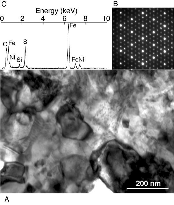
5 Fig. S1 (A) Transmission electron micrograph showing the texture of new-pcp. (B) a diffraction pattern and (C) an X-ray elemental spectrum from a single new-pcp grain. The main spots in the diffraction pattern are similar to those of magnetite (space group Fd3m); the weak extra spots indicate a 3-fold superstructure. This pattern is viewed along [111] of the Fd3m cell of the main spots.
6 Table S1 Table S1. Oxygen isotopic compositions ( ) of new-pcps, tochilinite, matrix and AOA of meteorites. Averaging areas: # of analysis areas (µm 2 ) used for averaging. Δ 17 O SMOW =δ 17 O SMOW -0.52δ 18 O SMOW. Object name Analysis area Averaging areas δ 17 O SMOW σ mean δ 18 O SMOW σ mean Δ 17 O Isotopography new-pcp of Acfer 094 # x # x # x # x # x # x Tochilinite of Murchison # x AOA of Acfer 094 # x # x Point SIMS new-pcp of Acfer 094 #17-s x #17-s x Matrix of Acfer 094 #s x #s x #s x #s x #s x #s x #s x #s x mean AOA of Acfer 094 #s x Isotopography: extracted from an isotope image by SCAPS using imaging SIMS. Point SIMS: measured by an electron multiplier using an oval-shaped ion micro-probe. AOA: ameoboid olivine aggregate.
7 Table S2 Table S2. Secondary ion count rate (cps) and calculated counting loss (%) of 16 O - secondary ion for each measurement piont. Object name Count rate Counting loss new-pcp of Acfer 094 #17-s29 5.3E #17-s47 3.4E Matrix of Acfer 094 #s39 4.1E #s40 3.6E #s41 3.6E #s43 4.0E #s44 3.6E #s45 3.2E #s46 4.1E #s49 4.4E AOA of Acfer 094 #s50 3.8E Standard Olivine 4.1E Magnetite 4.5E
8 Fig. S1.
SUPPLEMENTARY INFORMATION
 SUPPLEMENTARY INFORMATION Extreme oxygen isotope anomaly with a solar origin detected in meteoritic organics Ko Hashizume, Naoto Takahata, Hiroshi Naraoka & Yuji Sano Supplementary Discussions Basic descriptions
A promising method to obtain more accurate Mg isotope compositional data on presolar silicate particles
 A promising method to obtain more accurate Mg isotope compositional data on presolar silicate particles János Kodolányi 1 Max Planck Institute for Chemistry, Particle Chemistry Department Johann-Joachim-Becher-Weg
Silicate Stardust in Meteorites
 1 of 8 posted June 1, 2004 Silicate Stardust in Meteorites --- Silicates are the most abundant solids in disks around growing stars, but presolar silicates have not been found in even the most primitive
Secondary ion mass spectrometry (SIMS)
") Secondary ion mass spectrometry (SIMS) ELEC-L3211 Postgraduate Course in Micro and Nanosciences Department of Micro and Nanosciences Personal motivation and experience on SIMS Offers the possibility to
Secondary ion mass spectrometry (SIMS) ELEC-L3211 Postgraduate Course in Micro and Nanosciences Department of Micro and Nanosciences Personal motivation and experience on SIMS Offers the possibility to
ABNORMAL X-RAY EMISSION FROM INSULATORS BOMBARDED WITH LOW ENERGY IONS
 302 ABNORMAL X-RAY EMISSION FROM INSULATORS BOMBARDED WITH LOW ENERGY IONS M. Song 1, K. Mitsuishi 1, M. Takeguchi 1, K. Furuya 1, R. C. Birtcher 2 1 High Voltage Electron Microscopy Station, National
FIB - SIMS. Focussed Ion Beam Secondary Ion Mass Spectrometry.
 FIB - SIMS Focussed Ion Beam Secondary Ion Mass Spectrometry Outline Introduction to Hiden Analytical Introduction to SIMS FIB-SIMS - Introduction and key features FIB-SIMS - Applications data Hiden SIMS
Secondary Ion Mass Spectrometry (SIMS) Thomas Sky
 Thomas Sky") 1 Secondary Ion Mass Spectrometry (SIMS) Thomas Sky Depth (µm) 2 Characterization of solar cells 0,0 1E16 1E17 1E18 1E19 1E20 0,2 0,4 0,6 0,8 1,0 1,2 P Concentration (cm -3 ) Characterization Optimization
1 Secondary Ion Mass Spectrometry (SIMS) Thomas Sky Depth (µm) 2 Characterization of solar cells 0,0 1E16 1E17 1E18 1E19 1E20 0,2 0,4 0,6 0,8 1,0 1,2 P Concentration (cm -3 ) Characterization Optimization
Massachusetts Institute of Technology. Dr. Nilanjan Chatterjee
 Massachusetts Institute of Technology Dr. Nilanjan Chatterjee Electron Probe Micro-Analysis (EPMA) Imaging and micrometer-scale chemical compositional analysis of solids Signals produced in The Electron
Application of NanoSIMS on Organo Mineral Structures
 Application of NanoSIMS on Organo Mineral Structures Carmen Höschen*, Carsten W. Mueller, Katja Heister, Johann Lugmeier and Ingrid Kögel-Knabner Lehrstuhl für Bodenkunde, TU München, 85350 Freising-Weihenstephan,
Secondary ion mass spectrometry (SIMS)
") Secondary ion mass spectrometry (SIMS) Lasse Vines 1 Secondary ion mass spectrometry O Zn 10000 O 2 Counts/sec 1000 100 Li Na K Cr ZnO 10 ZnO 2 1 0 20 40 60 80 100 Mass (AMU) 10 21 10 20 Si 07 Ge 0.3 Atomic
Secondary ion mass spectrometry (SIMS) Lasse Vines 1 Secondary ion mass spectrometry O Zn 10000 O 2 Counts/sec 1000 100 Li Na K Cr ZnO 10 ZnO 2 1 0 20 40 60 80 100 Mass (AMU) 10 21 10 20 Si 07 Ge 0.3 Atomic
Original Paper Fission Gas Bubbles Characterisation in Irradiated UO 2 Fuel by SEM, EPMA and SIMS
 Microchim Acta 155, 183 187 (2006) DOI 10.1007/s00604-006-0540-y Original Paper Fission Gas Bubbles Characterisation in Irradiated UO 2 Fuel by SEM, EPMA and SIMS Jérôme Lamontagne, Lionel Desgranges,
Characterization of Secondary Emission Materials for Micro-Channel Plates. S. Jokela, I. Veryovkin, A. Zinovev
 Characterization of Secondary Emission Materials for Micro-Channel Plates S. Jokela, I. Veryovkin, A. Zinovev Secondary Electron Yield Testing Technique We have incorporated XPS, UPS, Ar-ion sputtering,
Supporting Information
 Supporting Information Bindi et al. 10.1073/pnas.1111115109 Fig. S1. Electron microprobe X-ray elemental maps for the grain reported in Fig. 1B. Experimental details are given in Experimental Methods.
APPENDICES. Appendix 1
 Corthouts, T.L., Lageson, D.R., and Shaw, C.A., 2016, Polyphase deformation, dynamic metamorphism and metasomatism of Mount Everest s summit limestone, east central Himalaya, Nepal/Tibet: Lithosphere,
Supplementary Materials for
 advances.sciencemag.org/cgi/content/full/2/3/e1501725/dc1 Supplementary Materials for Discovery of natural MgSiO3 tetragonal garnet in a shocked chondritic meteorite The PDF file includes: Naotaka Tomioka,
Characterization of presolar silicate and oxide grains in primitive. carbonaceous chondrites
 Characterization of presolar silicate and oxide grains in primitive carbonaceous chondrites ANN N. NGUYEN 1, *, FRANK J. STADERMANN 1, ERNST ZINNER 1, RHONDA M. STROUD 2, CONEL M. O D. ALEXANDER 3, LARRY
MSE 321 Structural Characterization
 Auger Spectroscopy Auger Electron Spectroscopy (AES) Scanning Auger Microscopy (SAM) Incident Electron Ejected Electron Auger Electron Initial State Intermediate State Final State Physical Electronics
3.2 Ga detrital uraninite in the Witwatersrand Basin, South. Africa: Evidence of a reducing Archean atmosphere
 GSA Data Repository 2018085 https://doi.org/10.1130/g39957.1 1 2 3 4 3.2 Ga detrital uraninite in the Witwatersrand Basin, South Africa: Evidence of a reducing Archean atmosphere Ian Burron 1, Giuliana
Auger Electron Spectroscopy Overview
 Auger Electron Spectroscopy Overview Also known as: AES, Auger, SAM 1 Auger Electron Spectroscopy E KLL = E K - E L - E L AES Spectra of Cu EdN(E)/dE Auger Electron E N(E) x 5 E KLL Cu MNN Cu LMM E f E
ToF-SIMS or XPS? Xinqi Chen Keck-II
 ToF-SIMS or XPS? Xinqi Chen Keck-II 1 Time of Flight Secondary Ion Mass Spectrometry (ToF-SIMS) Not ToF MS (laser, solution) X-ray Photoelectron Spectroscopy (XPS) 2 3 Modes of SIMS 4 Secondary Ion Sputtering
Supplementary Information for: Giant Kiruna-type deposits form by. efficient flotation of magmatic magnetite suspensions
 GSA DATA REPOSITORY 2015206 1 2 Supplementary Information for: Giant Kiruna-type deposits form by efficient flotation of magmatic magnetite suspensions 3 4 Jaayke L. Knipping, Laura D. Bilenker, Adam C.
TECHNIC A L WORK ING GROUP ITWG GUIDELINE ON SECONDARY ION MASS SPECTROMETRY (SIMS)
") NUCLE A R FORENSIC S INTERN ATION A L TECHNIC A L WORK ING GROUP ITWG GUIDELINE ON SECONDARY ION MASS SPECTROMETRY (SIMS) EXECUTIVE SUMMARY Secondary Ion Mass Spectrometry (SIMS) is used for elemental
NUCLE A R FORENSIC S INTERN ATION A L TECHNIC A L WORK ING GROUP ITWG GUIDELINE ON SECONDARY ION MASS SPECTROMETRY (SIMS) EXECUTIVE SUMMARY Secondary Ion Mass Spectrometry (SIMS) is used for elemental
Terrestrial and Extraterrestrial Applications of the Carnegie NanoSIMS
 Terrestrial and Extraterrestrial Applications of the Carnegie NanoSIMS Larry Nittler SIMS Group Department of Terrestrial Magnetism Carnegie Institution of Washington Erik Hauri Conel Alexander Jianhua
Record of ancient Martian hydrosphere preserved in zircon from a Martian meteoritie
 SUPPLEMENTARY INFORMATION DOI: 1.138/NGEO2231 Record of ancient Martian hydrosphere preserved in zircon from a Martian meteoritie A. A. Nemchin 1,2, M. Humayun 3, M. J. Whitehouse 1, R. H. Hewins 4,5,
Observations Regarding Automated SEM and SIMS Analysis of Minerals. Kristofor Ingeneri. April 22, 2009
 Observations Regarding Automated SEM and SIMS Analysis of Minerals Kristofor Ingeneri April 22, 2009 Forensic Geoscience A field of inquiry that utilizes techniques developed in the geosciences (geology,
Methods of surface analysis
 Methods of surface analysis Nanomaterials characterisation I RNDr. Věra Vodičková, PhD. Surface of solid matter: last monoatomic layer + absorbed monolayer physical properties are effected (crystal lattice
Towards SHRIMP SI: Developments in Stable Isotope Analysis with SHRIMP
 Towards SHRIMP SI: Developments in Stable Isotope Analysis with SHRIMP Trevor Ireland Research School of Earth Sciences The Australian National University Acknowledgments Ryan Ickert, Joe Hiess Peter Holden,
Particle Analysis of Environmental Swipe Samples
 IAEA-SM-367/10/07 Particle Analysis of Environmental Swipe Samples D. DONOHUE, S. VOGT, A. CIURAPINSKI, F. RUEDENAUER, M. HEDBERG Safeguards Analytical Laboratory International Atomic Energy Agency Vienna,
Gold nanothorns macroporous silicon hybrid structure: a simple and ultrasensitive platform for SERS
 Supporting Information Gold nanothorns macroporous silicon hybrid structure: a simple and ultrasensitive platform for SERS Kamran Khajehpour,* a Tim Williams, b,c Laure Bourgeois b,d and Sam Adeloju a
ANALYTICAL TECHNIQUES
 ANALYTICAL TECHNIQUES Raman Spectra Raman spectra were obtained by use of a T64000 (JY Horiba) triple-stage laser- Raman system having both macro-raman and confocal micro-raman capability which permits
A study of Mg and K isotopes in Allende CAIs: Implications to the time scale for the multiple heating processes
 Meteoritics & Planetary Science 41, Nr 12, 1871 1881 (2006) Abstract available online at http://meteoritics.org A study of Mg and K isotopes in Allende CAIs: Implications to the time scale for the multiple
Highly efficient SERS test strips
 Electronic Supplementary Information (ESI) for Highly efficient SERS test strips 5 Ran Zhang, a Bin-Bin Xu, a Xue-Qing Liu, a Yong-Lai Zhang, a Ying Xu, a Qi-Dai Chen, * a and Hong-Bo Sun* a,b 5 10 Experimental
GGR Cutting-Edge Review
 GGR Cutting-Edge Review NanoSIMS: Technical Aspects and Applications in Cosmochemistry and Biological Geochemistry Peter Hoppe (1)*, Stephanie Cohen (2) and Anders Meibom (2) (1) Particle Chemistry Department,
Secondary Ion Mass Spectrometry (SIMS)
") CHEM53200: Lecture 10 Secondary Ion Mass Spectrometry (SIMS) Major reference: Surface Analysis Edited by J. C. Vickerman (1997). 1 Primary particles may be: Secondary particles can be e s, neutral species
CHEM53200: Lecture 10 Secondary Ion Mass Spectrometry (SIMS) Major reference: Surface Analysis Edited by J. C. Vickerman (1997). 1 Primary particles may be: Secondary particles can be e s, neutral species
Water diffusion in silica glass through pathways formed by hydroxyls
 1 2 Revision 1 Water diffusion in silica glass through pathways formed by hydroxyls 3 4 Minami Kuroda 1, Shogo Tachibana 1,2, Naoya Sakamoto 3, Satoshi Okumura 4, Michihiko 5 Nakamura 4 and Hisayoshi Yurimoto
ABC s of Electrochemistry series Materials Characterization techniques: SEM and EDS Ana María Valenzuela-Muñiz November 3, 2011
 ABC s of Electrochemistry series Materials Characterization techniques: SEM and EDS Ana María Valenzuela-Muñiz November 3, 2011 CEER, Department of Chemical and Biomolecular Engineering Outline Introduction
Cosmochemical application of High Precision Multi-collector SIMS
 Cosmochemical application of High Precision Multi-collector SIMS Oxygen Three Isotopes in Chondrules Early Solar System Chronology of Refractory Inclusions Noriko Kita Wisc-SIMS Laboratory, University
Oxygen isotope systematics of chondrules in the Allende CV3 chondrite: High precision ion microprobe studies
 Available online at www.sciencedirect.com Geochimica et Cosmochimica Acta 75 (2011) 7596 7611 www.elsevier.com/locate/gca Oxygen isotope systematics of chondrules in the Allende CV3 chondrite: High precision
MSE 321 Structural Characterization
 Auger Spectroscopy Auger Electron Spectroscopy (AES) Scanning Auger Microscopy (SAM) Incident Electron Ejected Electron Auger Electron Initial State Intermediate State Final State Physical Electronics
MICRO-TOMOGRAPHY AND X-RAY ANALYSIS OF GEOLOGICAL SAMPLES
 THE PUBLISHING HOUSE PROCEEDINGS OF THE ROMANIAN ACADEMY, Series A, OF THE ROMANIAN ACADEMY Volume 18, Number 1/2017, pp. 42 49 MICRO-TOMOGRAPHY AND X-RAY ANALYSIS OF GEOLOGICAL SAMPLES Ion GRUIA University
Shock Effects in CML 0175: The Wow Stone
 PSU McNair Scholars Online Journal Volume 3 Issue 1 Identity, Communities, and Technology: On the Cusp of Change Article 17 2009 Shock Effects in CML 0175: The Wow Stone Kristy Hauver Portland State University
Metcalf and Buck. GSA Data Repository
 GSA Data Repository 2015035 Metcalf and Buck Figure DR1. Secondary ionization mass-spectrometry U-Pb zircon geochronology plots for data collected on two samples of Wilson Ridge plutonic rocks. Data presented
EPMA IMAGES. Figure 9. Energy-dispersive spectra of spot mineral analyses in sample 89GGR-33A for locations 1-5 in Figure 8.
 EPMA IMAGES The attached images and mineral data can be used to supplement an instrument-based lab, or serve as the basis for lab that can be completed without an instrument. Please provide credit for
Chapter 9. Electron mean free path Microscopy principles of SEM, TEM, LEEM
 Chapter 9 Electron mean free path Microscopy principles of SEM, TEM, LEEM 9.1 Electron Mean Free Path 9. Scanning Electron Microscopy (SEM) -SEM design; Secondary electron imaging; Backscattered electron
Analyse de la météorite "Paris" par faisceaux d'ions et d'agrégats. Manale NOUN 12 Février 2013
 Analyse de la météorite "Paris" par faisceaux d'ions et d'agrégats Manale NOUN 12 Février 213 Météorite «Paris» «Paris» : a CM chondrite (Blanchard et al., MetSoc 74, 211) 1.37 Kg 5 cm 5 mm 5 μm Détermination
LAACHER SEE REVISITED: HIGH SPATIAL RESOLUTION ZIRCON DATING IMPLIES RAPID FORMATION OF A ZONED MAGMA CHAMBER -
 LAACHER SEE REVISITED: HIGH SPATIAL RESOLUTION ZIRCON DATING IMPLIES RAPID FORMATION OF A ZONED MAGMA CHAMBER - DATA REPOSITORY ANALYTICAL PROCEDURES Ion microprobe U-Th measurements Th-U dating was performed
Secondary Ion Mass Spectroscopy (SIMS)
") Secondary Ion Mass Spectroscopy (SIMS) Analyzing Inorganic Solids * = under special conditions ** = semiconductors only + = limited number of elements or groups Analyzing Organic Solids * = under special
Secondary Ion Mass Spectroscopy (SIMS) Analyzing Inorganic Solids * = under special conditions ** = semiconductors only + = limited number of elements or groups Analyzing Organic Solids * = under special
CHARACTERIZATION OF PRESOLAR SILICATE AND OXIDE GRAINS IN PRIMITIVE CARBONACEOUS CHONDRITES
 The Astrophysical Journal, 656:1223 1240, 2007 February 20 # 2007. The American Astronomical Society. All rights reserved. Printed in U.S.A. CHARACTERIZATION OF PRESOLAR SILICATE AND OXIDE GRAINS IN PRIMITIVE
Th) ) dating of micro-baddeleyite
 ) dating of micro-baddeleyite") U-Pb (and U-ThU Th) ) dating of micro-baddeleyite 30 μm Axel K. Schmitt UCLA SIMS, NSF National Ion Microprobe Facility Collaborators: T. Mark Harrison (UCLA) Kevin Chamberlain (University of Wyoming)
U-Pb (and U-ThU Th) ) dating of micro-baddeleyite 30 μm Axel K. Schmitt UCLA SIMS, NSF National Ion Microprobe Facility Collaborators: T. Mark Harrison (UCLA) Kevin Chamberlain (University of Wyoming)
Applications of XPS, AES, and TOF-SIMS
 Applications of XPS, AES, and TOF-SIMS Scott R. Bryan Physical Electronics 1 Materials Characterization Techniques Microscopy Optical Microscope SEM TEM STM SPM AFM Spectroscopy Energy Dispersive X-ray
Dual Beam Helios Nanolab 600 and 650
 Dual Beam Helios Nanolab 600 and 650 In the Clean Room facilities of the INA LMA, several lithography facilities permit to pattern structures at the micro and nano meter scale and to create devices. In
Electron probe microanalysis - Electron microprobe analysis EPMA (EMPA) What s EPMA all about? What can you learn?
 What s EPMA all about? What can you learn?") Electron probe microanalysis - Electron microprobe analysis EPMA (EMPA) What s EPMA all about? What can you learn? EPMA - what is it? Precise and accurate quantitative chemical analyses of micron-size
Electron probe microanalysis - Electron microprobe analysis EPMA (EMPA) What s EPMA all about? What can you learn? EPMA - what is it? Precise and accurate quantitative chemical analyses of micron-size
Automated Isotopic Measurements of Micron-Sized Dust: Application to Meteoritic Presolar Silicon Carbide
 Automated Isotopic Measurements of Micron-Sized Dust: Application to Meteoritic Presolar Silicon Carbide Larry R. Nittler and Conel M. O D. Alexander Department of Terrestrial Magnetism, Carnegie Institution
Secondary Ion Mass Spectrometry (SIMS) for Surface Analysis
 for Surface Analysis") Secondary Ion Mass Spectrometry (SIMS) for Surface Analysis General overview of SIMS - principles, ionization, advantages & limitations SIMS as a surface analysis technique - operation modes, information
Secondary Ion Mass Spectrometry (SIMS) for Surface Analysis General overview of SIMS - principles, ionization, advantages & limitations SIMS as a surface analysis technique - operation modes, information
Electron Microprobe Analysis and Scanning Electron Microscopy
 Electron Microprobe Analysis and Scanning Electron Microscopy Electron microprobe analysis (EMPA) Analytical technique in which a beam of electrons is focused on a sample surface, producing X-rays from
X-Ray Photoelectron Spectroscopy (XPS) Prof. Paul K. Chu
 Prof. Paul K. Chu") X-Ray Photoelectron Spectroscopy (XPS) Prof. Paul K. Chu X-ray Photoelectron Spectroscopy Introduction Qualitative analysis Quantitative analysis Charging compensation Small area analysis and XPS imaging
X-Ray Photoelectron Spectroscopy (XPS) Prof. Paul K. Chu X-ray Photoelectron Spectroscopy Introduction Qualitative analysis Quantitative analysis Charging compensation Small area analysis and XPS imaging
HOW TO APPROACH SCANNING ELECTRON MICROSCOPY AND ENERGY DISPERSIVE SPECTROSCOPY ANALYSIS. SCSAM Short Course Amir Avishai
 HOW TO APPROACH SCANNING ELECTRON MICROSCOPY AND ENERGY DISPERSIVE SPECTROSCOPY ANALYSIS SCSAM Short Course Amir Avishai RESEARCH QUESTIONS Sea Shell Cast Iron EDS+SE Fe Cr C Objective Ability to ask the
Auger Electron Spectroscopy
 Auger Electron Spectroscopy Auger Electron Spectroscopy is an analytical technique that provides compositional information on the top few monolayers of material. Detect all elements above He Detection
MT Electron microscopy Scanning electron microscopy and electron probe microanalysis
 MT-0.6026 Electron microscopy Scanning electron microscopy and electron probe microanalysis Eero Haimi Research Manager Outline 1. Introduction Basics of scanning electron microscopy (SEM) and electron
MS482 Materials Characterization ( 재료분석 ) Lecture Note 4: XRF
 Lecture Note 4: XRF") 2016 Fall Semester MS482 Materials Characterization ( 재료분석 ) Lecture Note 4: XRF Byungha Shin Dept. of MSE, KAIST 1 Course Information Syllabus 1. Overview of various characterization techniques (1 lecture)
2016 Fall Semester MS482 Materials Characterization ( 재료분석 ) Lecture Note 4: XRF Byungha Shin Dept. of MSE, KAIST 1 Course Information Syllabus 1. Overview of various characterization techniques (1 lecture)
Hydrogen isotope evidence for loss of water from Mars through time
 GEOPHYSICAL RESEARCH LETTERS, VOL. 35, L05203, doi:10.1029/2007gl032721, 2008 Hydrogen isotope evidence for loss of water from Mars through time James P. Greenwood, 1 Shoichi Itoh, 2 Naoya Sakamoto, 3
Everhart-Thornley detector
 SEI Detector Everhart-Thornley detector Microscope chamber wall Faraday cage Scintillator Electrons in Light pipe Photomultiplier Electrical signal out Screen Quartz window +200 V +10 kv Always contains
Energy-Filtering. Transmission. Electron Microscopy
 Part 3 Energy-Filtering Transmission Electron Microscopy 92 Energy-Filtering TEM Principle of EFTEM expose specimen to mono-energetic electron radiation inelastic scattering in the specimen poly-energetic
Outlines 3/12/2011. Vacuum Chamber. Inside the sample chamber. Nano-manipulator. Focused ion beam instrument. 1. Other components of FIB instrument
 Focused ion beam instruments Outlines 1. Other components of FIB instrument 1.a Vacuum chamber 1.b Nanomanipulator 1.c Gas supply for deposition 1.d Detectors 2. Capabilities of FIB instrument Lee Chow
Chemical Geology 264 (2009) Contents lists available at ScienceDirect. Chemical Geology. journal homepage:
 Contents lists available at ScienceDirect. Chemical Geology. journal homepage:") Chemical Geology 264 (2009) 43 57 Contents lists available at ScienceDirect Chemical Geology journal homepage: www.elsevier.com/locate/chemgeo High precision SIMS oxygen isotope analysis and the effect
Chemical Geology 264 (2009) 43 57 Contents lists available at ScienceDirect Chemical Geology journal homepage: www.elsevier.com/locate/chemgeo High precision SIMS oxygen isotope analysis and the effect
Supporting Information s for
 Supporting Information s for # Self-assembling of DNA-templated Au Nanoparticles into Nanowires and their enhanced SERS and Catalytic Applications Subrata Kundu* and M. Jayachandran Electrochemical Materials
Detector Needs of Spectroscopy
 Detector Needs of Spectroscopy Klaus Attenkofer Inner Shell Spectroscopy Group (NSLS-2) 1 BROOKHAVEN SCIENCE ASSOCIATES RELEVANCE TO DOE MISSION Electro catalysis Environmental sciences: uptake of nutrition
Chemical Analysis in TEM: XEDS, EELS and EFTEM. HRTEM PhD course Lecture 5
 Chemical Analysis in TEM: XEDS, EELS and EFTEM HRTEM PhD course Lecture 5 1 Part IV Subject Chapter Prio x-ray spectrometry 32 1 Spectra and mapping 33 2 Qualitative XEDS 34 1 Quantitative XEDS 35.1-35.4
Supporting information. stereocomplex films. Kenta Kondo, Toshiyuki Kida, Yuji Ogawa, Yuuya Arikawa and Mitsuru Akashi*
 Supporting information Nanotube formation through the continuous one-dimensional fusion of hollow nanocapsules composed of layer-by-layer poly(lactic acd)s stereocomplex films Kenta Kondo, Toshiyuki Kida,
Nanoelectronics 09. Atsufumi Hirohata Department of Electronics. Quick Review over the Last Lecture
 Nanoelectronics 09 Atsufumi Hirohata Department of Electronics 13:00 Monday, 12/February/2018 (P/T 006) Quick Review over the Last Lecture ( Field effect transistor (FET) ): ( Drain ) current increases
Spatial Coherence Properties of Organic Molecules Coupled to Plasmonic Surface Lattice Resonances in the Weak and Strong Coupling Regimes
 Spatial Coherence Properties of Organic Molecules Coupled to Plasmonic Surface Lattice Resonances in the Weak and Strong Coupling Regimes Supplemental Material L. Shi, T. K. Hakala, H. T. Rekola, J. -P.
Lecture 22 Ion Beam Techniques
 Lecture 22 Ion Beam Techniques Schroder: Chapter 11.3 1/44 Announcements Homework 6/6: Will be online on later today. Due Wednesday June 6th at 10:00am. I will return it at the final exam (14 th June).
Supplementary Information
 Supplementary Information Time-dependent growth of zinc hydroxide nanostrands and their crystal structure Xinsheng Peng, ab Jian Jin, a Noriko Kobayashi, a Wolfgang Schmitt, c and Izumi Ichinose* a a Organic
Secondary Ion Mass Spectrometry (SIMS) for Surface Analysis
 for Surface Analysis") Secondary Ion Mass Spectrometry (SIMS) for Surface Analysis General overview of SIMS - principles, ionization, advantages & limitations SIMS as a surface analysis technique - operation modes, information
Secondary Ion Mass Spectrometry (SIMS) for Surface Analysis General overview of SIMS - principles, ionization, advantages & limitations SIMS as a surface analysis technique - operation modes, information
An Introduction to Auger Electron Spectroscopy
 An Introduction to Auger Electron Spectroscopy Spyros Diplas MENA3100 SINTEF Materials & Chemistry, Department of Materials Physics & Centre of Materials Science and Nanotechnology, Department of Chemistry,
photo-mineralization of 2-propanol under visible light irradiation
 Electronic Supplementary Information for WO 3 modified titanate network film: highly efficient photo-mineralization of 2-propanol under visible light irradiation Experimental Preparation of STN, and WO
Supporting Online Material for
 www.sciencemag.org/cgi/content/full/1128865/dc1 Supporting Online Material for Oxygen Isotope Variation in Stony-Iron Meteorites R. C. Greenwood,* I. A. Franchi, A. Jambon, J. A. Barrat, T. H. Burbine
Chapter 10. Nanometrology. Oxford University Press All rights reserved.
 Chapter 10 Nanometrology Oxford University Press 2013. All rights reserved. 1 Introduction Nanometrology is the science of measurement at the nanoscale level. Figure illustrates where nanoscale stands
Evidence for cavity-dwelling microbial life in 3.22 Ga tidal deposits
 GSA Data Repository 2016012 Evidence for cavity-dwelling microbial life in 3.22 Ga tidal deposits Homann, M., Heubeck, C., Bontognali, R.R., Bouvier A.-S., Baumgartner L.P., and Airo, A. Geology, 2015
Surface Analysis. Dr. Lynn Fuller Dr. Fuller s Webpage:
 ROCHESTER INSTITUTE OF TECHNOLOGY MICROELECTRONIC ENGINEERING Surface Analysis Dr. Lynn Fuller Dr. Fuller s Webpage: http://people.rit.edu/lffeee 82 Lomb Memorial Drive Rochester, NY 14623-5604 Tel (585)
Secondary Ion Mass Spectrometry (SIMS)
") Secondary Ion Mass Spectrometry (SIMS) SIMS: a desorption/ionization technique 1960s - A. Benninghoven, University of Münster, Germany (Benninghoven A., Rudenauer F.G., Werner H.W., Secondary Ion Mass
Secondary Ion Mass Spectrometry (SIMS) SIMS: a desorption/ionization technique 1960s - A. Benninghoven, University of Münster, Germany (Benninghoven A., Rudenauer F.G., Werner H.W., Secondary Ion Mass
Nanoscale Chemical Characterization: Moving to 3 Dimensions
 Nanoscale Chemical Characterization: Moving to 3 Dimensions Eric B. Steel Chemical Science & Technology Laboratory National Institute of Standards & Technology Outline What is and why do we need chemical
Supporting Online Material for
 www.sciencemag.org/cgi/content/full/332/6025/88/dc1 Supporting Online Material for Microtomography of Partially Molten Rocks: Three-Dimensional Melt Distribution in Mantle Peridotite Wenlu Zhu, * Glenn
Secondary Ion Mass Spectrometry (SIMS)
") OpenStax-CNX module: m50227 1 Secondary Ion Mass Spectrometry (SIMS) Kourtney Wright Andrew R. Barron This work is produced by OpenStax-CNX and licensed under the Creative Commons Attribution License 4.0
OpenStax-CNX module: m50227 1 Secondary Ion Mass Spectrometry (SIMS) Kourtney Wright Andrew R. Barron This work is produced by OpenStax-CNX and licensed under the Creative Commons Attribution License 4.0
MT Electron microscopy Scanning electron microscopy and electron probe microanalysis
 MT-0.6026 Electron microscopy Scanning electron microscopy and electron probe microanalysis Eero Haimi Research Manager Outline 1. Introduction Basics of scanning electron microscopy (SEM) and electron
Surface analysis techniques
 Experimental methods in physics Surface analysis techniques 3. Ion probes Elemental and molecular analysis Jean-Marc Bonard Academic year 10-11 3. Elemental and molecular analysis 3.1.!Secondary ion mass
PHI 5000 Versaprobe-II Focus X-ray Photo-electron Spectroscopy
 PHI 5000 Versaprobe-II Focus X-ray Photo-electron Spectroscopy The very basic theory of XPS XPS theroy Surface Analysis Ultra High Vacuum (UHV) XPS Theory XPS = X-ray Photo-electron Spectroscopy X-ray
CHARACTERIZATION of NANOMATERIALS KHP
 CHARACTERIZATION of NANOMATERIALS Overview of the most common nanocharacterization techniques MAIN CHARACTERIZATION TECHNIQUES: 1.Transmission Electron Microscope (TEM) 2. Scanning Electron Microscope
AP5301/ Name the major parts of an optical microscope and state their functions.
 Review Problems on Optical Microscopy AP5301/8301-2015 1. Name the major parts of an optical microscope and state their functions. 2. Compare the focal lengths of two glass converging lenses, one with
MS482 Materials Characterization ( 재료분석 ) Lecture Note 5: RBS
 Lecture Note 5: RBS") 2016 Fall Semester MS482 Materials Characterization ( 재료분석 ) Lecture Note 5: RBS Byungha Shin Dept. of MSE, KAIST 1 Course Information Syllabus 1. Overview of various characterization techniques (1 lecture)
2016 Fall Semester MS482 Materials Characterization ( 재료분석 ) Lecture Note 5: RBS Byungha Shin Dept. of MSE, KAIST 1 Course Information Syllabus 1. Overview of various characterization techniques (1 lecture)
Electron Emission Microscope. Michael J. Eller February 12 th 2013
 Electron Emission Microscope Michael J. Eller February 12 th 2013 Concept primary ions Individual mass spectrum n. Individual mass spectrum 2 EEM lenses x n,y n.. x 2, y 2 x 1, y 1 Position sensitive detector
Modern Optical Spectroscopy
 Modern Optical Spectroscopy X-Ray Microanalysis Shu-Ping Lin, Ph.D. Institute of Biomedical Engineering E-mail: splin@dragon.nchu.edu.tw Website: http://web.nchu.edu.tw/pweb/users/splin/ Backscattered
raw materials C V Mn Mg S Al Ca Ti Cr Si G H Nb Na Zn Ni K Co A B C D E F
 Today s advanced batteries require a range of specialized analytical tools to better understand the electrochemical processes that occur during battery cycling. Evans Analytical Group (EAG) offers a wide-range
A Sustainable Synthesis of Nitrogen-Doped Carbon Aerogels
 A Sustainable Synthesis of Nitrogen-Doped Carbon Aerogels Supporting Information By Robin J. White, a, * Noriko Yoshizawa, b Markus Antonietti, a and Maria-Magdalena Titirici. a * e-mail: robin.white@mpikg.mpg.de
Development and characterization of 3D semiconductor X-rays detectors for medical imaging
 Development and characterization of 3D semiconductor X-rays detectors for medical imaging Marie-Laure Avenel, Eric Gros d Aillon CEA-LETI, DETectors Laboratory marie-laure.avenel@cea.fr Outlines Problematic
Scanning Electron Microscopy & Ancillary Techniques
 Scanning Electron Microscopy & Ancillary Techniques By Pablo G. Caceres-Valencia The prototype of the first Stereoscan supplied by the Cambridge Instrument Company to the dupont Company, U.S.A. (1965)
EE 527 MICROFABRICATION. Lecture 5 Tai-Chang Chen University of Washington
 EE 527 MICROFABRICATION Lecture 5 Tai-Chang Chen University of Washington MICROSCOPY AND VISUALIZATION Electron microscope, transmission electron microscope Resolution: atomic imaging Use: lattice spacing.
The Controlled Evolution of a Polymer Single Crystal
 Supporting Online Material The Controlled Evolution of a Polymer Single Crystal Xiaogang Liu, 1 Yi Zhang, 1 Dipak K. Goswami, 2 John S. Okasinski, 2 Khalid Salaita, 1 Peng Sun, 1 Michael J. Bedzyk, 2 Chad
Contrasted strengths and weakness of EDS, WDS and AES for determining the composition of samples
 Contrasted strengths and weakness of EDS, WDS and AES for determining the composition of samples Ana-Marija Nedić Course 590B 12/07/2018 Iowa State University Contrasted strengths and weakness of EDS,
PHYS-E0541:Special Course in Physics Gas phase synthesis of carbon nanotubes for thin film application. Electron Microscopy. for
 PHYS-E0541:Special Course in Physics Gas phase synthesis of carbon nanotubes for thin film application Electron Microscopy for Introduction to Electron Microscopy Carbon Nanomaterials (nanotubes) Dr. Hua