Section III - Designing Models for 3D Printing
|
|
|
- Coleen Gilmore
- 6 years ago
- Views:
Transcription

1 Section III - Designing Models for 3D Printing In this section of the Jmol Training Guide, you will become familiar with the commands needed to design a model that will be built on a 3D Printer. As you become more comfortable using Jmol, this section will enable you to take the next step and design physical protein models. At the end of this section, you should be familiar with how to use Jmol to create a physical protein model on a rapid prototyping machine, including: Section 3: Designing Models for 3D Printing What is 3D Printing? Selecting Appropriate Display Formats and Colors Adding and Removing Hydrogen Bonds and Disulfide Bonds Adding and Removing Support Struts Coloring Hydrogen Bonds, SS Bonds, and Struts Adding Sidechains with a Clean Backbone" Additional Miscellaneous Jmol Commands 1


2 3-1 - What is 3D Printing? 3D Printing is a cutting-edge technology that can use stacked serial planes to build physical 3-dimensional models from any computer generated 3D file. Every physical protein model design goes through four key steps: X,Y,Z coordinates in the form of a PDB file are the starting point of every protein model. The model is then exported as a file that is recognized by a 3D printer Design software is then used to edit the features and format of the protein model Finally, the physical model is removed from the printer and completed in a post production process. Z-Corporation 510 and Although many varieties of 3D Printing and Rapid Prototyping machines exist, the MSOE Center for BioMolecular Modeling relies mainly on two Z- Corporation machines - the Z510 and the Z650. 2


3 How a 3D Printer Works - The inside of a 3D Printer has three main parts: the feed chamber that stores the plaster powder to be used, the build chamber where the printed model will be built, and the spreading and printing arm which applies a glue and pigment solution to the growing model in the build chamber. While printing, the spreading and printing arm moves from left to right pushing a thin layer of plaster powder into the build chamber. The arm then makes a second pass printing a glue pigment solution onto the new layer in the build chamber. The build chamber is then lowered as the feed chamber is raised so a new layer of plaster can be spread. This process continues until all layers of the model have been completed. The End Result - A designed and printed protein model will be an exact replica of the model created in the 3D visualization program. 2AVY.pdb Selecting Appropriate Display Formats and Colors What Display Should I Use? - This is a common question asked by model designers. And the answer to this question is another question: What is the story that you are telling with your model? o If your story is focused on a particular active site within the molecule, then perhaps the Alpha Carbon Backbone Format displaying key active site amino acid sidechains in Ball and Stick (Wireframe and Spacefill) Format would be best. If your story is focused 3


. If a display format is too thin, a protein model will be more likely to break during the 3D printing process.")
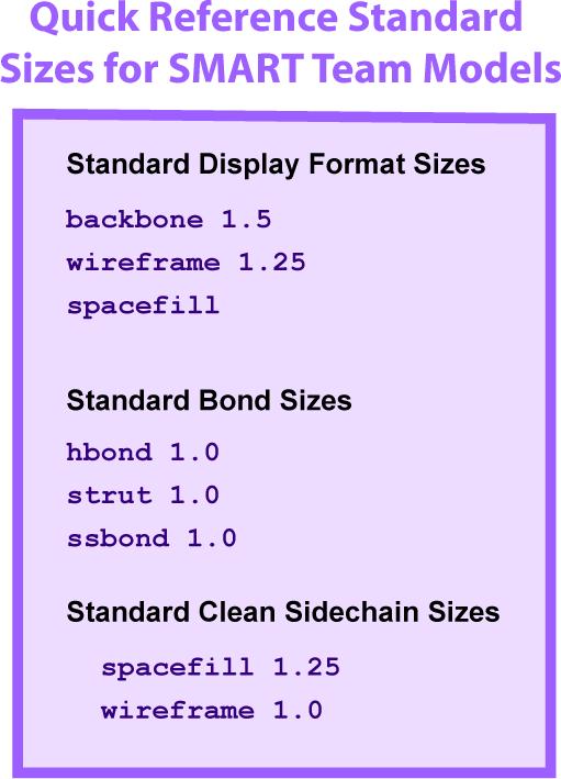
4 on how two subunits interface at the surface, then perhaps the Spacefill Format is the best choice. Ultimately the choice is yours. In Section I, there is a table highlighting the advantages and disadvantages of each display format. This will assist you in deciding which format is the best for telling your story. o The important point to remember is that no one model will tell every aspect of the story. Using Jmol in combination with a physical model will assist you in telling multiple aspects of your story. o The CBM will build models in the Spacefill Format and the Alpha Carbon Backbone Format. Models in the Alpha Carbon Backbone Format can have sidechains and heterologous groups displayed. We currently do not build models in Ribbons or Strands Format. 1A3N.pdb 1CLL.pdb DNA.pdb Format Sizes - Every display format in Jmol is able to be displayed with various thicknesses (see section 1.4 for more information on Display Formats). If a display format is too thin, a protein model will be more likely to break during the 3D printing process. If a format is too thick, the model will use excessive amounts of material and details will be less visible. Below is a list of suggested sizes for the different formats: Backbone 1.5 Wireframe

 a")

5 Spacefill (for sidechains only) Note: The default spacefill does not include Wireframe 1.25 a number. This is known as Full Spacefill. Spacefill 1.5 For all bonds and struts hbonds 1.0 ssbonds 1.0 Struts 1.0 Which Colors Should I Use? - Which colors you use, like which Display Format you choose, is largely influenced by the story you would like your model to show. o If the overall shape and fold of your protein is important to your story, you may want to color the secondary structures (alpha-helices and beta-sheets) a bright eye-catching color. o If there is a specific sidechain or active site area that is important to the function of your protein, you may want to highlight that area with a unique or bright color. o You can color a protein model in an almost unlimited variety of ways so it is very important to consider what you want to show and what colors would be best to highlight the key points to your protein's story. 5


 are essential to the stability of secondary structures in a protein.")
6 Colors Not to Use - There are a few colors or color combinations that you should avoid while designing a protein model to be build using 3D printing technologies. o Do not use any black or extremely dark colors. The 3D printers do not print well with dark colors and often leave stripes or unevenness in the final physical model. o Do not use colors that are too similar to highlight different features. These will often not be distinguishable in the final physical model. o Do not use too many extremely bright colors together on the same model. When used rarely, very bright colors are extremely good at drawing the eye to important areas of the protein model. Overuse of bright colors can distract from the model Adding and Removing Hydrogen Bonds and Disulfide Bonds Hydrogen Bonds - Hydrogen bonds (referred to as "hbonds" in Jmol) are essential to the stability of secondary structures in a protein. They form between the backbone oxygen of one amino acid and the backbone nitrogen in a second amino acid. To add hydrogen bonds to secondary structures within your model, use the calculate hbonds command. We typically do not add hydrogen bonds to alpha-helices, since they do not add stability to the model and actually clutter the view of the structure. Adding hydrogen bonds to beta-sheets provides additional support for the final model and is recommended. Use the following commands: o select sheet o calculate hbonds o After you have entered this command, you will notice that there are dotted lines that have appeared. 6


7 These are the hydrogen bonds. o The default size when you first add hydrogen bonds is the same as for wireframe which is very thin. Thickness - Hydrogen bonds, like wireframe, backbone, and spacefill, can be thickened by placing a number after the hbonds command. o hbonds 1.0 Solid hbonds - The default display for hydrogen bonds is a dashed line. You will need to change this into a solid cylinder for building a physical protein model using 3D printing. You can do this using the command: o set hbonds solid Setting hbonds to the Backbone - Notice that the hydrogen bonds are now thicker and solid, but that they appear to be floating in air. This appearance results from the fact that hydrogen bonds form between the atoms that make up the backbone of the amino acid (the nitrogen and the oxygen atoms), but since we have displayed only the alpha carbon atoms in Backbone Format, it appears as if the hydrogen bonds are floating in space. Therefore, we must set the hydrogen bonds to the backbone using the command: o set hbonds backbone Removing all hbonds - To turn off all the hydrogen bonds in a selected area, use the hbonds off command. o hbonds off Summary of Commands to Add hbonds: o select sheet o calculate hbonds o hbonds 1.0 o set hbonds solid o set hbonds backbone Adding or Removing Individual hbonds - At times you may want to add or remove a single hydrogen bond. First, you need to know the two amino acid residue numbers that the hydrogen bond connects. To do this, click on the two residues in the Display Window (see section 1.5 for more information on identifying atom numbers). Once you know the two 7

8 residue numbers, you must select only these two amino acids. Finally, use the hbonds 1.0 command to add an hbond or the hbonds off command to remove the bond between the two selected amino acids. o For example: select 167 or 73 For example: select 321 or 334 hbonds 1.0 hbonds off Disulfide Bonds - Some molecules will have disulfide bonds (referred to as "ssbonds" in Jmol) present within the structure. These bonds form between two cysteine amino acids that lie close to each other in 3D space. Disulfide bonds are added using the command ssbonds. o ssbonds Thickness - As we saw with the hydrogen bonds, simply typing ssbonds by itself will only produce thin lines. To give these bonds dimension, we must add a thickness (number) to the ssbonds. o ssbonds 1.0 Setting ssbonds to the Backbone - Notice that this command gives the disulfide bonds a thicker dimension, but as we saw with the hydrogen bond, the bonds are floating in space. This is because the disulfide bond is actually between the sulfur groups of the cysteine sidechains, and not the alpha carbons. To make the disulfide bond connect between the backbone units, we need to set the bonds to the backbone. Note that the disulfide bond is orange (the CPK color for sulfur). o set ssbonds backbone Note: You may wish to display the sidechains of the cysteines involved in the disulfide bond. If that is the case, then you will not need to set ssbonds to the backbone. Note: Not every PDB file has ssbonds. The Top 7 protein PDB file used in Section 1 does not have a disulfide bond. To practice adding disulfide bonds, try the PDB file 2hiu.pdb which contains the coordinates for one molecule of human insulin. Adding or Removing Individual ssbonds - At times you may want to add or remove a single disulfide bond. To do this, you first need to know the two amino acid residue numbers that the bond connects. To do this, click on the two residues in the Display Window (see section 1.5 for more information on identifying atom numbers). Once you know the two 8

9 residue numbers, you must select only these two amino acids. Finally, use the ssbonds 1.0 command to add an ssbond or the ssbonds off command to remove the bond between the two selected amino acids. o For example: select 167 or 73 For example: select 321 or 334 ssbonds 1.0 ssbonds off Adding and Removing Support Struts Support Struts - When a model is built on a 3D printer, it needs to be supported in order to withstand the additional pressure as powder builds up and as the model is removed and cured. Hence, support struts are added within the model for support. To do this, select your entire model and use the calculate struts command. o Calculate struts Thickness - Struts, like hydrogen bonds and ssbonds, can be set to different thicknesses by adding a number after the strut command. o Strut 1.0 Adding or Removing Individual Struts - At times you may want to remove a single strut (for example, if it goes through a sidechain you have represented). To do this, you first need to know the atom numbers of the two atoms the strut connects. To do this, click on the two atoms in the Display Window (see section 1.5 for more information on identifying atom numbers). Once you know the two atom numbers, you must select only those two atoms using the select atomno= command. Finally, use the struts 1.0 command to add a strut or the strut off command to remove the bond between the two selected amino acids. o For example: select atomno=167 or atomno=73 strut 1.0 o For example: select atomno=321 or atomno=334 strut off Note: The calculate strut command may not always provide enough structural support. Additional struts may be necessary. Struts will need to be added manually to attach any ligands (substrate, inhibitor, cofactors) to the protein. 9
.")

10 3.5 - Coloring Hydrogen Bonds, SS Bonds, and Struts Default Bond Colors - When bonds or struts initially appear, they will be the default Jmol colors (a translucent white for struts and the colors of the atoms that they connect for hbonds and ssbonds). o If the two atoms the bond is connecting are the same color, then the hbond or ssbond will be the same color throughout the entire length of the monitor line. o If the two atoms are different colors, then the hbond or ssbond will be half one color and half the other color. Changing All Bonds - To color all of a certain type of bond or strut, select all and then use the color command followed by the type of bond or strut you want to color and then the color you would like to color them. o For example: select all color hbond red or: select all color strut red Note: We recommend that you choose a light color for your struts, such as white or light gray. Struts are support structures and should not be the emphasis of your model. A vibrant or dark color will draw the user s eye to the struts, and that should not be the focus of your model. Changing A Single Bond Color - To change the color of an individual bond or strut, follow the same steps for removing an individual bond or strut. Identify the two atom numbers that the bond or strut connects. Then, select only those two atoms. Finally, use your color command to apply the new color. o For example: select atomno=333 or atomno=65 color hbond green 10
11 3.6 - Adding Sidechains to Create a "Clean Backbone" If your molecular story requires that you display specific amino acid sidechains in the model, we recommend that you do only display the sidechain atoms, rather than all of the atoms of the amino acid. This gives the model a more aesthetically pleasing appearance. In previous sections, we have simply selected the amino acid by its residue number and displayed all of the atoms in that amino acid. In this section, we are going to use Boolean operators to select just the atoms in the sidechain and display only these atoms. To select and display only the atoms of the sidechain of a specific amino acid, you want to use the select command followed by the amino acid name/number and end with the and (sidechain or alpha) text. o select cys30 and (sidechain or alpha) o This command selects the amino acid at residue 30, but limits the selected atoms to the sidechain atoms and the alpha carbon of that amino acid. Note: It is important to select the alpha carbon atom in addition to the sidechain atoms because this will attach the sidechain atoms to the alpha carbon. If you do not select the alpha carbon, the sidechain will build as a separate unit from the rest of the molecule. Once your sidechain is connected, give it wireframe and spacefill dimensions: wireframe 1.0 spacefill 1.25 Note: These two commands together will generate a ball and stick appearance. You can enter just the wireframe command and create a sticks appearance, but you cannot enter just the spacefill command. If you enter just the spacefill command, the atoms will be displayed as little spheres and the spheres may not be securely connected to one another. The image MAY look fine on the screen, but the model will come out of the 3D printer in pieces that can t be put back together! It is therefore imperative to add the wireframe command to connect the spheres together. If you do not use the and (sidechain or alpha) command when displaying sidechains, you will generate a bumpy backbone. Unless the backbone atoms of the amino acid (the amino nitrogen or the carbonyl oxygen) play a specific role in the story you are telling, we do not recommend displaying these atoms on the model. It is typically (although not always) the sidechain that has the specific chemical role within the molecule and plays the key role within your story. Therefore, this should be the part that is displayed within your molecule. 11

12 3.7 - Additional Miscellaneous Jmol Commands In Section I of the Jmol Training Guide, you were introduced to the basic commands of Jmol. We will now present additional commands to further develop your Jmol design skills. o The Center Command - This command allows you to center the rotation of the molecule on a certain portion of the molecule. o center Jmol will by default center the molecule at the center of the entire molecule. If you restrict your viewing to a certain subset, when you rotate the molecule around in space, the molecule will seem lopsided. This is because although you have restricted your viewing to just a part of the molecule, all of the atoms are still present and being used to determine the center of rotation; you just do not see them. Therefore, you need to use the center command to center the molecule on the restricted region. For example: center *c Note: Once again, we are using the * command to dictate to Jmol that we are centering the molecule on the all the atoms in Chain C. Identifying the Amino and Carboxy Termini - An important concept in protein structure is that each protein has an amino terminus and a carboxy terminus. Through Jmol, you can readily identify each of these termini. o Amino Terminus - The Amino Terminus is the first amino acid in the protein. When a protein is synthesized, it begins with the 5 end of the mrna and synthesizes in a 5 to 3 fashion. Therefore, the first amino acid in the protein will be the amino acid that is encoded at the 5 end of the mrna. 12
13 To determine the amino terminus of the protein in the PDB file, click on the atom at each end of the protein. The atom with the lowest amino acid number will be the amino terminus. Alternatively, you may search the PDB sequence information to identify the amino terminus amino acid and use the Jmol command line window to select the specific amino acid. o Carboxy Terminus - The Carboxy Terminus, on the other hand, will be the last amino acid in the protein. To determine the identity of the carboxy terminus, click on the atom at each end of the protein. The amino acid with the largest number will be the last amino acid in the protein. Note: Occasionally a PDB file will have gaps or missing sections in a chain because of incomplete experimental data in the file. This can easily be confused for the Carboxy Terminus. Be sure that you are really looking at the last amino acid in the chain and not just the beginning of a gap. Repeating a command - By pressing the up arrow key on the keyboard, the previous commands that you have entered into the command line window will be displayed, in reverse order. You can edit these commands before hitting enter. Undo Button - Near the bottom right of the Console Window is an undo button. This button can be used to undo recent commands and take your protein design back to a previous state. Be sure to save your work frequently! Use judiciously; sometimes you need to undo multiple steps to get to the command you want to undo. Selecting specific types of atoms - If you wish to select a subset of atoms, such as all of the carbon atoms, you can do so by using the select command followed by the type of atom you would like to select. o For example: select carbon 13
14 14
Bonds and Structural Supports
 Bonds and Structural Supports Part of the Jmol Training Guide from the MSOE Center for BioMolecular Modeling Interactive version available at http://cbm.msoe.edu/teachingresources/jmol/jmoltraining/struts.html
Part 7 Bonds and Structural Supports
 Part 7 Bonds and Structural Supports http://cbm.msoe.edu/newwebsite/learntomodel Introduction In addition to covalent bonds between atoms in a molecule, Jmol has the ability to render Hydrogen Bonds and
Contents. Acknowledgements. Section 1: Using Jmol as a Computer Visualization Tool
 1ZAA.pdb 1ZAA.pdb Contents Acknowledgements Section 1: Using Jmol as a Computer Visualization Tool 1-1 - Downloading Jmol 1-2 - Protein Data Bank (PDB) Files 1-3 - Launching Jmol and Opening a PDB File
Part 8 Working with Nucleic Acids
 Part 8 Working with Nucleic Acids http://cbm.msoe.edu/newwebsite/learntomodel Introduction Most Protein Databank files loaded into the CBM's Jmol Design Environment include protein structures and small
The Select Command and Boolean Operators
 The Select Command and Boolean Operators Part of the Jmol Training Guide from the MSOE Center for BioMolecular Modeling Interactive version available at http://cbm.msoe.edu/teachingresources/jmol/jmoltraining/boolean.html
Part 4 The Select Command and Boolean Operators
 Part 4 The Select Command and Boolean Operators http://cbm.msoe.edu/newwebsite/learntomodel Introduction By default, every command you enter into the Console affects the entire molecular structure. However,
Section II Understanding the Protein Data Bank
 Section II Understanding the Protein Data Bank The focus of Section II of the MSOE Center for BioMolecular Modeling Jmol Training Guide is to learn about the Protein Data Bank, the worldwide repository
Model Mélange. Physical Models of Peptides and Proteins
 Model Mélange Physical Models of Peptides and Proteins In the Model Mélange activity, you will visit four different stations each featuring a variety of different physical models of peptides or proteins.
Pymol Practial Guide
 Pymol Practial Guide Pymol is a powerful visualizor very convenient to work with protein molecules. Its interface may seem complex at first, but you will see that with a little practice is simple and powerful
Examples of Protein Modeling. Protein Modeling. Primary Structure. Protein Structure Description. Protein Sequence Sources. Importing Sequences to MOE
 Examples of Protein Modeling Protein Modeling Visualization Examination of an experimental structure to gain insight about a research question Dynamics To examine the dynamics of protein structures To
From Amino Acids to Proteins - in 4 Easy Steps
 From Amino Acids to Proteins - in 4 Easy Steps Although protein structure appears to be overwhelmingly complex, you can provide your students with a basic understanding of how proteins fold by focusing
SeeSAR 7.1 Beginners Guide. June 2017
 SeeSAR 7.1 Beginners Guide June 2017 Part 1: Basics 1 Type a pdb code and press return or Load your own protein or already existing project, or Just load molecules To begin, let s type 2zff and download
Preparing a PDB File
 Figure 1: Schematic view of the ligand-binding domain from the vitamin D receptor (PDB file 1IE9). The crystallographic waters are shown as small spheres and the bound ligand is shown as a CPK model. HO
Problem Set 1
 2006 7.012 Problem Set 1 Due before 5 PM on FRIDAY, September 15, 2006. Turn answers in to the box outside of 68-120. PLEASE WRITE YOUR ANSWERS ON THIS PRINTOUT. 1. For each of the following parts, pick
Introduction to Structure Preparation and Visualization
 Introduction to Structure Preparation and Visualization Created with: Release 2018-4 Prerequisites: Release 2018-2 or higher Access to the internet Categories: Molecular Visualization, Structure-Based
Experiment 1: The Same or Not The Same?
 Experiment 1: The Same or Not The Same? Learning Goals After you finish this lab, you will be able to: 1. Use Logger Pro to collect data and calculate statistics (mean and standard deviation). 2. Explain
Exercises for Windows
 Exercises for Windows CAChe User Interface for Windows Select tool Application window Document window (workspace) Style bar Tool palette Select entire molecule Select Similar Group Select Atom tool Rotate
Molecular modeling with InsightII
 Molecular modeling with InsightII Yuk Sham Computational Biology/Biochemistry Consultant Phone: (612) 624 7427 (Walter Library) Phone: (612) 624 0783 (VWL) Email: shamy@msi.umn.edu How to run InsightII
Molecular Modeling and Conformational Analysis with PC Spartan
 Molecular Modeling and Conformational Analysis with PC Spartan Introduction Molecular modeling can be done in a variety of ways, from using simple hand-held models to doing sophisticated calculations on
Conformational Geometry of Peptides and Proteins:
 Conformational Geometry of Peptides and Proteins: Before discussing secondary structure, it is important to appreciate the conformational plasticity of proteins. Each residue in a polypeptide has three
LS1a Fall 2014 Problem Set #2 Due Monday 10/6 at 6 pm in the drop boxes on the Science Center 2 nd Floor
 LS1a Fall 2014 Problem Set #2 Due Monday 10/6 at 6 pm in the drop boxes on the Science Center 2 nd Floor Note: Adequate space is given for each answer. Questions that require a brief explanation should
Molecular Modeling Lecture 7. Homology modeling insertions/deletions manual realignment
 Molecular Modeling 2018-- Lecture 7 Homology modeling insertions/deletions manual realignment Homology modeling also called comparative modeling Sequences that have similar sequence have similar structure.
APBS electrostatics in VMD - Software. APBS! >!Examples! >!Visualization! >! Contents
 Software Search this site Home Announcements An update on mailing lists APBS 1.2.0 released APBS 1.2.1 released APBS 1.3 released New APBS 1.3 Windows Installer PDB2PQR 1.7.1 released PDB2PQR 1.8 released
CHEM 463: Advanced Inorganic Chemistry Modeling Metalloproteins for Structural Analysis
 CHEM 463: Advanced Inorganic Chemistry Modeling Metalloproteins for Structural Analysis Purpose: The purpose of this laboratory is to introduce some of the basic visualization and modeling tools for viewing
Protein Bioinformatics Computer lab #1 Friday, April 11, 2008 Sean Prigge and Ingo Ruczinski
 Protein Bioinformatics 260.655 Computer lab #1 Friday, April 11, 2008 Sean Prigge and Ingo Ruczinski Goals: Approx. Time [1] Use the Protein Data Bank PDB website. 10 minutes [2] Use the WebMol Viewer.
BUILDING BASICS WITH HYPERCHEM LITE
 BUILDING BASICS WITH HYPERCHEM LITE LAB MOD1.COMP From Gannon University SIM INTRODUCTION A chemical bond is a link between atoms resulting from the mutual attraction of their nuclei for electrons. There
3D - Structure Graphics Capabilities with PDF-4 Database Products
 3D - Structure Graphics Capabilities with PDF-4 Database Products Atomic Structure Information in the PDF-4 Databases ICDD s PDF-4 databases contain atomic structure information for a significant number
Let s continue our discussion on the interaction between Fe(III) and 6,7-dihydroxynaphthalene-2- sulfonate.
 and 6,7-dihydroxynaphthalene-2- sulfonate.") Chemistry 5995(133)-8990(013) Bioinorganic Chemistry: The Good, the Bad, and the Potential of Metals Assignment 2- Aqueous Speciation, Magnetism, Redox, UV-Vis Spectroscopy, and Pymol Let s continue our
Chemistry 5995(133)-8990(013) Bioinorganic Chemistry: The Good, the Bad, and the Potential of Metals Assignment 2- Aqueous Speciation, Magnetism, Redox, UV-Vis Spectroscopy, and Pymol Let s continue our
NMR, X-ray Diffraction, Protein Structure, and RasMol
 NMR, X-ray Diffraction, Protein Structure, and RasMol Introduction So far we have been mostly concerned with the proteins themselves. The techniques (NMR or X-ray diffraction) used to determine a structure
ICM-Chemist-Pro How-To Guide. Version 3.6-1h Last Updated 12/29/2009
 ICM-Chemist-Pro How-To Guide Version 3.6-1h Last Updated 12/29/2009 ICM-Chemist-Pro ICM 3D LIGAND EDITOR: SETUP 1. Read in a ligand molecule or PDB file. How to setup the ligand in the ICM 3D Ligand Editor.
Supplementary Figure 1. Aligned sequences of yeast IDH1 (top) and IDH2 (bottom) with isocitrate
 and IDH2 (bottom) with isocitrate") SUPPLEMENTARY FIGURE LEGENDS Supplementary Figure 1. Aligned sequences of yeast IDH1 (top) and IDH2 (bottom) with isocitrate dehydrogenase from Escherichia coli [ICD, pdb 1PB1, Mesecar, A. D., and Koshland,
SUPPLEMENTARY FIGURE LEGENDS Supplementary Figure 1. Aligned sequences of yeast IDH1 (top) and IDH2 (bottom) with isocitrate dehydrogenase from Escherichia coli [ICD, pdb 1PB1, Mesecar, A. D., and Koshland,
Tutorial: Structural Analysis of a Protein-Protein Complex
 Molecular Modeling Section (MMS) Department of Pharmaceutical and Pharmacological Sciences University of Padova Via Marzolo 5-35131 Padova (IT) @contact: stefano.moro@unipd.it Tutorial: Structural Analysis
Chemistry 14CL. Worksheet for the Molecular Modeling Workshop. (Revised FULL Version 2012 J.W. Pang) (Modified A. A. Russell)
 (Modified A. A. Russell)") Chemistry 14CL Worksheet for the Molecular Modeling Workshop (Revised FULL Version 2012 J.W. Pang) (Modified A. A. Russell) Structure of the Molecular Modeling Assignment The molecular modeling assignment
Chemistry 14CL Worksheet for the Molecular Modeling Workshop (Revised FULL Version 2012 J.W. Pang) (Modified A. A. Russell) Structure of the Molecular Modeling Assignment The molecular modeling assignment
The Structure and Functions of Proteins
 Wright State University CORE Scholar Computer Science and Engineering Faculty Publications Computer Science and Engineering 2003 The Structure and Functions of Proteins Dan E. Krane Wright State University
Assignment 2: Conformation Searching (50 points)
") Chemistry 380.37 Fall 2015 Dr. Jean M. Standard September 16, 2015 Assignment 2: Conformation Searching (50 points) In this assignment, you will use the Spartan software package to investigate some conformation
Chemistry 380.37 Fall 2015 Dr. Jean M. Standard September 16, 2015 Assignment 2: Conformation Searching (50 points) In this assignment, you will use the Spartan software package to investigate some conformation
Computational Chemistry Lab Module: Conformational Analysis of Alkanes
 Introduction Computational Chemistry Lab Module: Conformational Analysis of Alkanes In this experiment, we will use CAChe software package to model the conformations of butane, 2-methylbutane, and substituted
Build_model v User Guide
 Build_model v.2.0.1 User Guide MolTech Build_model User Guide 2008-2011 Molecular Technologies Ltd. www.moltech.ru Please send your comments and suggestions to contact@moltech.ru. Table of Contents Input
Viewing and Analyzing Proteins, Ligands and their Complexes 2
 2 Viewing and Analyzing Proteins, Ligands and their Complexes 2 Overview Viewing the accessible surface Analyzing the properties of proteins containing thousands of atoms is best accomplished by representing
Chemical Kinetics I: The Dry Lab. Up until this point in our study of physical chemistry we have been interested in
 Chemical Kinetics I: The Dry Lab Up until this point in our study of physical chemistry we have been interested in equilibrium properties; now we will begin to investigate non-equilibrium properties and
Protein Structure Basics
 Protein Structure Basics Presented by Alison Fraser, Christine Lee, Pradhuman Jhala, Corban Rivera Importance of Proteins Muscle structure depends on protein-protein interactions Transport across membranes
and Environmental Science Centre
 1. Purpose The purpose of this document is to familiarize the user with the mode of function of the FT-Raman available at the facility, and to describe the sampling procedure. 2. Introduction Raman spectroscopy
To visualize the three-dimensional structures of some common molecules. To obtain bond angle, bond length, and hybridization data for molecules.
 Molecular Geometry PURPOSE A B C To explore some simple molecular structures. To explore the relationship between bond order and bond length. To explore resonance structures. GOALS To compare Lewis structures
NMR Assignments using NMRView II: Sequential Assignments
 NMR Assignments using NMRView II: Sequential Assignments DO THE FOLLOWING, IF YOU HAVE NOT ALREADY DONE SO: For Mac OS X, you should have a subdirectory nmrview. At UGA this is /Users/bcmb8190/nmrview.
ISIS/Draw "Quick Start"
 ISIS/Draw "Quick Start" Click to print, or click Drawing Molecules * Basic Strategy 5.1 * Drawing Structures with Template tools and template pages 5.2 * Drawing bonds and chains 5.3 * Drawing atoms 5.4
Geography 281 Map Making with GIS Project Four: Comparing Classification Methods
 Geography 281 Map Making with GIS Project Four: Comparing Classification Methods Thematic maps commonly deal with either of two kinds of data: Qualitative Data showing differences in kind or type (e.g.,
Introduction to Spark
 1 As you become familiar or continue to explore the Cresset technology and software applications, we encourage you to look through the user manual. This is accessible from the Help menu. However, don t
Introduction Molecular Structure Script Console External resources Advanced topics. JMol tutorial. Giovanni Morelli.
 Gen 19th, 2017 1 2 Create and edit Display and view Mesurament and labelling Surface and Orbitals 3 4 from Database Protein Enzyme Crystal Structure and Unit Cell 5 Symmetry Animation General information
AP Biology Unit 1, Chapters 2, 3, 4, 5
 Name Date AP Biology Unit 1, Chapters 2, 3, 4, 5 Research Question How are chemical structures visualized? Background You can represent a molecule with either a molecular formula or a structural formula.
Get familiar with PDBsum and the PDB Extract atomic coordinates from protein data files Compute bond angles and dihedral angles
 CS483 Assignment #2 Due date: Mar. 1 at the start of class. Protein Geometry Bedbug spit? Just say NO! Purpose of this assignment Get familiar with PDBsum and the PDB Extract atomic coordinates from protein
Introduction to Computer Tools and Uncertainties
 Experiment 1 Introduction to Computer Tools and Uncertainties 1.1 Objectives To become familiar with the computer programs and utilities that will be used throughout the semester. To become familiar with
How Do We Get Light from Matter: The Origin of Emission
 1 How Do We Get Light from Matter: The Origin of Emission Lines ORGANIZATION Pre-Lab: Origins of Lines Mode: inquiry, groups of 2 Grading: lab notes and post-lab questions Safety: no special requirements
Patrick: An Introduction to Medicinal Chemistry 5e MOLECULAR MODELLING EXERCISES CHAPTER 17
 MOLECULAR MODELLING EXERCISES CHAPTER 17 Exercise 17.6 Conformational analysis of n-butane Introduction Figure 1 Butane Me Me In this exercise, we will consider the possible stable conformations of butane
Ligand Scout Tutorials
 Ligand Scout Tutorials Step : Creating a pharmacophore from a protein-ligand complex. Type ke6 in the upper right area of the screen and press the button Download *+. The protein will be downloaded and
Assignment A02: Geometry Definition: File Formats, Redundant Coordinates, PES Scans
 Assignment A02: Geometry Definition: File Formats, Redundant Coordinates, PES Scans In Assignments A00 and A01, you familiarized yourself with GaussView and G09W, you learned the basics about input (GJF)
Electric Fields and Equipotentials
 OBJECTIVE Electric Fields and Equipotentials To study and describe the two-dimensional electric field. To map the location of the equipotential surfaces around charged electrodes. To study the relationship
What is the central dogma of biology?
 Bellringer What is the central dogma of biology? A. RNA DNA Protein B. DNA Protein Gene C. DNA Gene RNA D. DNA RNA Protein Review of DNA processes Replication (7.1) Transcription(7.2) Translation(7.3)
POC via CHEMnetBASE for Identifying Unknowns
 Table of Contents A red arrow is used to identify where buttons and functions are located in CHEMnetBASE. Figure Description Page Entering the Properties of Organic Compounds (POC) Database 1 CHEMnetBASE
Project 3: Molecular Orbital Calculations of Diatomic Molecules. This project is worth 30 points and is due on Wednesday, May 2, 2018.
 Chemistry 362 Spring 2018 Dr. Jean M. Standard April 20, 2018 Project 3: Molecular Orbital Calculations of Diatomic Molecules In this project, you will investigate the molecular orbitals and molecular
1. (5) Draw a diagram of an isomeric molecule to demonstrate a structural, geometric, and an enantiomer organization.
 Draw a diagram of an isomeric molecule to demonstrate a structural, geometric, and an enantiomer organization.") Organic Chemistry Assignment Score. Name Sec.. Date. Working by yourself or in a group, answer the following questions about the Organic Chemistry material. This assignment is worth 35 points with the
Organic Chemistry Assignment Score. Name Sec.. Date. Working by yourself or in a group, answer the following questions about the Organic Chemistry material. This assignment is worth 35 points with the
Application Note. U. Heat of Formation of Ethyl Alcohol and Dimethyl Ether. Introduction
 Application Note U. Introduction The molecular builder (Molecular Builder) is part of the MEDEA standard suite of building tools. This tutorial provides an overview of the Molecular Builder s basic functionality.
OECD QSAR Toolbox v.3.3. Step-by-step example of how to build a userdefined
 OECD QSAR Toolbox v.3.3 Step-by-step example of how to build a userdefined QSAR Background Objectives The exercise Workflow of the exercise Outlook 2 Background This is a step-by-step presentation designed
You w i ll f ol l ow these st eps : Before opening files, the S c e n e panel is active.
 You w i ll f ol l ow these st eps : A. O pen a n i m a g e s t a c k. B. Tr a c e t h e d e n d r i t e w i t h t h e user-guided m ode. C. D e t e c t t h e s p i n e s a u t o m a t i c a l l y. D. C
Creating a Pharmacophore Query from a Reference Molecule & Scaffold Hopping in CSD-CrossMiner
 Table of Contents Creating a Pharmacophore Query from a Reference Molecule & Scaffold Hopping in CSD-CrossMiner Introduction... 2 CSD-CrossMiner Terminology... 2 Overview of CSD-CrossMiner... 3 Features
CHAPTER 29 HW: AMINO ACIDS + PROTEINS
 CAPTER 29 W: AMI ACIDS + PRTEIS For all problems, consult the table of 20 Amino Acids provided in lecture if an amino acid structure is needed; these will be given on exams. Use natural amino acids (L)
Review Activity Module 1: Biological Chemistry
 Review Activity Module 1: Biological Chemistry Laroche: The picture above is of a molecule calle MC1R. Based on what you ve learned so far about the various biological macromolecules, what kind of macromolecule
Project Manual Bio3055. Apoptosis: Caspase-1
 Project Manual Bio3055 Apoptosis: Caspase-1 Bednarski 2003 Funded by HHMI Apoptosis: Caspase-1 Introduction: Apoptosis is another name for programmed cell death. It is a series of events in a cell that
Molecular Visualization. Introduction
 Molecular Visualization Jeffry D. Madura Department of Chemistry & Biochemistry Center for Computational Sciences Duquesne University Introduction Assessments of change, dynamics, and cause and effect
OECD QSAR Toolbox v.4.1. Tutorial illustrating new options for grouping with metabolism
 OECD QSAR Toolbox v.4.1 Tutorial illustrating new options for grouping with metabolism Outlook Background Objectives Specific Aims The exercise Workflow 2 Background Grouping with metabolism is a procedure
Basics of protein structure
 Today: 1. Projects a. Requirements: i. Critical review of one paper ii. At least one computational result b. Noon, Dec. 3 rd written report and oral presentation are due; submit via email to bphys101@fas.harvard.edu
Calculating Bond Enthalpies of the Hydrides
 Proposed Exercise for the General Chemistry Section of the Teaching with Cache Workbook: Calculating Bond Enthalpies of the Hydrides Contributed by James Foresman, Rachel Fogle, and Jeremy Beck, York College
Conformational search and QSAR (quantitative structure-activity relationships)
") Bio 5476 Lab Exercise 11.7.08 Instructor: Dan Kuster (d.kuster@gmail.com) Conformational search and QSAR (quantitative structure-activity relationships) Purpose: In this lab, you will use SYBYL to build,
Bio 5476 Lab Exercise 11.7.08 Instructor: Dan Kuster (d.kuster@gmail.com) Conformational search and QSAR (quantitative structure-activity relationships) Purpose: In this lab, you will use SYBYL to build,
CAP 5510 Lecture 3 Protein Structures
 CAP 5510 Lecture 3 Protein Structures Su-Shing Chen Bioinformatics CISE 8/19/2005 Su-Shing Chen, CISE 1 Protein Conformation 8/19/2005 Su-Shing Chen, CISE 2 Protein Conformational Structures Hydrophobicity
Identifying Interaction Hot Spots with SuperStar
 Identifying Interaction Hot Spots with SuperStar Version 1.0 November 2017 Table of Contents Identifying Interaction Hot Spots with SuperStar... 2 Case Study... 3 Introduction... 3 Generate SuperStar Maps
OECD QSAR Toolbox v.4.1. Tutorial on how to predict Skin sensitization potential taking into account alert performance
 OECD QSAR Toolbox v.4.1 Tutorial on how to predict Skin sensitization potential taking into account alert performance Outlook Background Objectives Specific Aims Read across and analogue approach The exercise
OECD QSAR Toolbox v.3.3. Step-by-step example of how to categorize an inventory by mechanistic behaviour of the chemicals which it consists
 OECD QSAR Toolbox v.3.3 Step-by-step example of how to categorize an inventory by mechanistic behaviour of the chemicals which it consists Background Objectives Specific Aims Trend analysis The exercise
Lewis Structures and Molecular Shapes
 Lewis Structures and Molecular Shapes Rules for Writing Lewis Structures 1. Determine the correct skeleton structure (connectivity of atoms). Usually, the most electronegative atoms go around the edges
M E R C E R W I N WA L K T H R O U G H
 H E A L T H W E A L T H C A R E E R WA L K T H R O U G H C L I E N T S O L U T I O N S T E A M T A B L E O F C O N T E N T 1. Login to the Tool 2 2. Published reports... 7 3. Select Results Criteria...
Silica surface - Materials Studio tutorial. CREATING SiO 2 SURFACE
 Silica surface - Materials Studio tutorial CREATING SiO 2 SURFACE Our goal surface of SiO2 6.948 Ǻ Import structure The XRD experiment gives us such parameters as: lattice parameters, symmetry group and
ON SITE SYSTEMS Chemical Safety Assistant
 ON SITE SYSTEMS Chemical Safety Assistant CS ASSISTANT WEB USERS MANUAL On Site Systems 23 N. Gore Ave. Suite 200 St. Louis, MO 63119 Phone 314-963-9934 Fax 314-963-9281 Table of Contents INTRODUCTION
Introduction to Hartree-Fock calculations in Spartan
 EE5 in 2008 Hannes Jónsson Introduction to Hartree-Fock calculations in Spartan In this exercise, you will get to use state of the art software for carrying out calculations of wavefunctions for molecues,
General Chemistry Lab Molecular Modeling
 PURPOSE The objectives of this experiment are PROCEDURE General Chemistry Lab Molecular Modeling To learn how to use molecular modeling software, a commonly used tool in chemical research and industry.
OECD QSAR Toolbox v.3.2. Step-by-step example of how to build and evaluate a category based on mechanism of action with protein and DNA binding
 OECD QSAR Toolbox v.3.2 Step-by-step example of how to build and evaluate a category based on mechanism of action with protein and DNA binding Outlook Background Objectives Specific Aims The exercise Workflow
Building small molecules
 Building small molecules Use the Builder (right panel) to build up molecules. Start building clicking a fragment/atom in the builder and it will appear to the workspace. Continue modifying the molecule
BOND LENGTH WITH HYPERCHEM LITE
 BOND LENGTH WITH HYPERCHEM LITE LAB MOD2.COMP From Gannon University SIM INTRODUCTION The electron cloud surrounding the nucleus of the atom determines the size of the atom. Since this distance is somewhat
Tutorial. Getting started. Sample to Insight. March 31, 2016
 Getting started March 31, 2016 Sample to Insight CLC bio, a QIAGEN Company Silkeborgvej 2 Prismet 8000 Aarhus C Denmark Telephone: +45 70 22 32 44 www.clcbio.com support-clcbio@qiagen.com Getting started
The Chemistry of Respiration and Photosynthesis
 The Chemistry of Respiration and Photosynthesis Objective- You should be able to write balanced equations for respiration and photosynthesis and explain how the two equations are related. Directions :
Biomolecules: lecture 10
 Biomolecules: lecture 10 - understanding in detail how protein 3D structures form - realize that protein molecules are not static wire models but instead dynamic, where in principle every atom moves (yet
3D Molecule Viewer of MOGADOC (JavaScript)
") 3D Molecule Viewer of MOGADOC (JavaScript) Movement of the Molecule Rotation of the molecule: Use left mouse button to drag. Translation of the molecule: Use right mouse button to drag. Resize the molecule:
3D Molecule Viewer of MOGADOC (JavaScript) Movement of the Molecule Rotation of the molecule: Use left mouse button to drag. Translation of the molecule: Use right mouse button to drag. Resize the molecule:
OECD QSAR Toolbox v.3.3. Step-by-step example of how to build and evaluate a category based on mechanism of action with protein and DNA binding
 OECD QSAR Toolbox v.3.3 Step-by-step example of how to build and evaluate a category based on mechanism of action with protein and DNA binding Outlook Background Objectives Specific Aims The exercise Workflow
Unit 2: Chemistry. Unit Overview:
 Unit 2: Chemistry Unit Overview: This unit will focus on basic chemistry and some of the major process of organic chemistry (dehydration synthesis, hydrolysis, and enzyme action) that help form carbon
Example: Identification
 Example: Identification In this structure we will explore Atom identification Working with maps Working with projections Eliminating diffusely diffracting solvent Disorder modeling Restrain application
VCell Tutorial. Building a Rule-Based Model
 VCell Tutorial Building a Rule-Based Model We will demonstrate how to create a rule-based model of EGFR receptor interaction with two adapter proteins Grb2 and Shc. A Receptor-monomer reversibly binds
Chem 253. Tutorial for Materials Studio
 Chem 253 Tutorial for Materials Studio This tutorial is designed to introduce Materials Studio 7.0, which is a program used for modeling and simulating materials for predicting and rationalizing structure
OECD QSAR Toolbox v.4.0. Tutorial on how to predict Skin sensitization potential taking into account alert performance
 OECD QSAR Toolbox v.4.0 Tutorial on how to predict Skin sensitization potential taking into account alert performance Outlook Background Objectives Specific Aims Read across and analogue approach The exercise
file:///biology Exploring Life/BiologyExploringLife04/
 Objectives Identify carbon skeletons and functional groups in organic molecules. Relate monomers and polymers. Describe the processes of building and breaking polymers. Key Terms organic molecule inorganic
EXPERIMENT 7: ANGULAR KINEMATICS AND TORQUE (V_3)
") TA name Lab section Date TA Initials (on completion) Name UW Student ID # Lab Partner(s) EXPERIMENT 7: ANGULAR KINEMATICS AND TORQUE (V_3) 121 Textbook Reference: Knight, Chapter 13.1-3, 6. SYNOPSIS In
TA name Lab section Date TA Initials (on completion) Name UW Student ID # Lab Partner(s) EXPERIMENT 7: ANGULAR KINEMATICS AND TORQUE (V_3) 121 Textbook Reference: Knight, Chapter 13.1-3, 6. SYNOPSIS In
This experiment is a continuation of the earlier experiment on molecular
 Molecular Modeling: Experiment 2 Page 115 Bonding and Molecular Structure Experiment 2 This experiment is a continuation of the earlier experiment on molecular structure. In that experiment you used a
Outline. Levels of Protein Structure. Primary (1 ) Structure. Lecture 6:Protein Architecture II: Secondary Structure or From peptides to proteins
 Structure. Lecture 6:Protein Architecture II: Secondary Structure or From peptides to proteins") Lecture 6:Protein Architecture II: Secondary Structure or From peptides to proteins Margaret Daugherty Fall 2004 Outline Four levels of structure are used to describe proteins; Alpha helices and beta sheets
Lecture 6:Protein Architecture II: Secondary Structure or From peptides to proteins Margaret Daugherty Fall 2004 Outline Four levels of structure are used to describe proteins; Alpha helices and beta sheets
2 Electric Field Mapping Rev1/05
 2 Electric Field Mapping Rev1/05 Theory: An electric field is a vector field that is produced by an electric charge. The source of the field may be a single charge or many charges. To visualize an electric
Physics 476LW Advanced Physics Laboratory Atomic Spectroscopy
 Physics 476LW Atomic Spectroscopy 1 Introduction The description of atomic spectra and the Rutherford-Geiger-Marsden experiment were the most significant precursors of the so-called Bohr planetary model