Maestro 8.5. Tutorial. Schrödinger Press
|
|
|
- Isabel Spencer
- 6 years ago
- Views:
Transcription
1 Maestro Tutorial Maestro 8.5 Tutorial Schrödinger Press
2 Maestro Tutorial Copyright 2008 Schrödinger, LLC. All rights reserved. While care has been taken in the preparation of this publication, Schrödinger assumes no responsibility for errors or omissions, or for damages resulting from the use of the information contained herein. Canvas, CombiGlide, ConfGen, Epik, Glide, Impact, Jaguar, Liaison, LigPrep, Maestro, Phase, Prime, PrimeX, QikProp, QikFit, QikSim, QSite, SiteMap, Strike, and WaterMap are trademarks of Schrödinger, LLC. Schrödinger and MacroModel are registered trademarks of Schrödinger, LLC. MCPRO is a trademark of William L. Jorgensen. Desmond is a trademark of D. E. Shaw Research. Desmond is used with the permission of D. E. Shaw Research. All rights reserved. This publication may contain the trademarks of other companies. Schrödinger software includes software and libraries provided by third parties. For details of the copyrights, and terms and conditions associated with such included third party software, see the Legal Notices for Third-Party Software in your product installation at $SCHRODINGER/docs/html/third_party_legal.html (Linux OS) or %SCHRODINGER%\docs\html\third_party_legal.html (Windows OS). This publication may refer to other third party software not included in or with Schrödinger software ("such other third party software"), and provide links to third party Web sites ("linked sites"). References to such other third party software or linked sites do not constitute an endorsement by Schrödinger, LLC. Use of such other third party software and linked sites may be subject to third party license agreements and fees. Schrödinger, LLC and its affiliates have no responsibility or liability, directly or indirectly, for such other third party software and linked sites, or for damage resulting from the use thereof. Any warranties that we make regarding Schrödinger products and services do not apply to such other third party software or linked sites, or to the interaction between, or interoperability of, Schrödinger products and services and such other third party software. June 2008
3 Contents Document Conventions... v Chapter 1: About This Tutorial... 1 Chapter 2: Starting Maestro and Viewing Molecules Copying the Files You Need To Run this Tutorial Starting Maestro Features of the Maestro Main Window Importing a Maestro File Viewing a Structure in the Workspace Including and Excluding Entries in the Project Table Saving the Maestro Project Chapter 3: Building Molecules Using Maestro Opening the Build Panel Clearing the Workspace and Building with Fragments Adjusting Angles Deleting and Labeling Atoms and Adjusting Formal Charge Saving Your Structure to the Project Table Copying and Renaming a Project Entry Deleting Bonds and Reattaching Fragments Adding Hydrogen Atoms to a Structure Substituting Functional Groups in Place Mode Replacing Functional Groups in Grow Mode Adjusting a Dihedral Angle Duplicating a Project Entry Changing Bond Orders Maestro 8.5 Tutorial iii
4 Contents 3.14 Changing Elements Adding Atoms by Freehand Drawing Moving Atoms Cleaning Up Structures Changing the Chirality Exporting Structures to a File Chapter 4: Changing the Appearance of Structures Getting Ready for the Exercises in this Chapter Displaying and Undisplaying Atoms Changing the Atom Coloring Scheme Changing the Appearance of Atoms and Bonds Displaying the Protein Backbone as a Ribbon Creating and Displaying a Molecular Surface Chapter 5: Measuring, Analyzing, and Superimposing Structures Getting Ready for the Exercises in this Chapter Measuring Distances Measuring Angles Measuring Dihedrals Superimposing Multiple Structures Displaying Hydrogen Bonds Chapter 6: Beyond the Basics Advanced Maestro Features Programs Accessible from Maestro Getting Help iv Maestro 8.5 Tutorial
5 Document Conventions In addition to the use of italics for names of documents, the font conventions that are used in this document are summarized in the table below. Font Example Use Sans serif Project Table Names of GUI features, such as panels, menus, menu items, buttons, and labels Monospace $SCHRODINGER/maestro File names, directory names, commands, environment variables, and screen output Italic filename Text that the user must replace with a value Sans serif uppercase CTRL+H Keyboard keys Links to other locations in the current document or to other PDF documents are colored like this: Document Conventions. In descriptions of command syntax, the following UNIX conventions are used: braces { } enclose a choice of required items, square brackets [ ] enclose optional items, and the bar symbol separates items in a list from which one item must be chosen. Lines of command syntax that wrap should be interpreted as a single command. File name, path, and environment variable syntax is generally given with the UNIX conventions. To obtain the Windows conventions, replace the forward slash / with the backslash \ in path or directory names, and replace the $ at the beginning of an environment variable with a % at each end. For example, $SCHRODINGER/maestro becomes %SCHRODINGER%\maestro. In this document, to type text means to type the required text in the specified location, and to enter text means to type the required text, then press the ENTER key. References to literature sources are given in square brackets, like this: [10]. Maestro 8.5 Tutorial v
6 vi Maestro 8.5 Tutorial
7 Maestro Tutorial Chapter 1 Chapter 1: About This Tutorial This document is designed to show you how to use the basic capabilities of Maestro, in easy steps. In the early parts of the book, we will explain everything in great detail what to do with the mouse, where to click, where buttons are, and so on so you aren t missing any part of the procedure. At the same time, we ll provide a more compact notation for explaining what to do. Later in the book, we ll use the compact notation, with references back to the explanation, and still later, we ll just use the compact notation. By that time, you ll have learnt what it means and how to perform the actions, and won t need the detailed explanation. But it ll always be there to refer back to. This tutorial is designed for you to work through from the beginning, but you can start at any of the chapters if you so desire. However, the explanations in the later chapter are more compact, so you might have to go back to the earlier chapters to find out how to perform an action. If you are reading this book online, you may notice that there are words that are marked in an indigo color, like this: Contents. These are links to other parts of the book. If you click on the word in your PDF reader, it takes you to the other part of the book. To go back to the place you came from, click the Back button in your PDF reader. Try it with the word Contents, above. It will take you to the table of contents for this book. There are also indigo-colored links to various sections of the Maestro User Manual. These links are provided so you can read more about the feature you are working with, if you are interested. Clicking on these links takes you to the section referred to. Try clicking the words Maestro User Manual in the first sentence of this paragraph, then click the Back button. Maestro 8.5 Tutorial 1
8 2 Maestro 8.5 Tutorial
9 Maestro Tutorial Chapter 2 Chapter 2: Starting Maestro and Viewing Molecules Maestro is a freely available, full-featured molecular visualization environment that also serves as the interface to all of Schrödinger s computational chemistry software. When coupled with Schrödinger software such as Glide, Prime, or Phase, Maestro is a powerful tool for interpreting, managing, and sharing the results of computational experiments. As a standalone program, Maestro is an easy-to-use tool for building, visualizing, and sharing 3-dimensional chemical models. In this tutorial, you ll learn how to perform these simple tasks using Maestro s intuitively designed interface. Working through the exercises in this chapter, you ll start Maestro, import structure files, and learn how to view them in the Maestro Workspace. Maestro is designed to run on Linux, Windows, and SGI machines, but can be displayed over a network to many commonly used platforms. With a little effort, Windows users can also install one of many Linux distributions on their PCs. Not only do these distributions include easy-tooperate graphical user interfaces, but they can be installed on a computer without removing the Windows operating system. 2.1 Copying the Files You Need To Run this Tutorial To perform the exercises in this tutorial, you ll need to make sure that you have the structures easily accessible. These structures can be copied from the Maestro tutorial subdirectory in the installation to your own directory. To copy the files under Windows: 1. Click Start, then click My Computer. An explorer window opens. 2. Move to the Schrödinger software installation. This installation is not in Programs and Settings, because the path to the software cannot have spaces in it. It s likely to be on your C drive, so start looking there if you don t know exactly where it is. 3. When you get to the installation, open the maestro-vversion folder. Here, version is the 5-digit Maestro version number, for example, This folder should contain a folder labeled tutorial. Maestro 8.5 Tutorial 3
10 Chapter 2: Starting Maestro and Viewing Molecules 4. Open another explorer window and move to the place you want to keep the tutorial files. For example, click Start, then click My Documents. You can create a folder to keep the files in if you want to. 5. Drag the tutorial folder from the first window to the second. To copy the files under UNIX: 1. Open a terminal window so that you have a command-line prompt. The steps to open a terminal window vary slightly between operating systems, but it is generally possible to right-click in your desktop and select an option like New Terminal from the menu that appears. 2. Next, set the SCHRODINGER environment variable. This environment variable contains the path to your installation of Maestro. How you set the SCHRODINGER environment variable depends on the shell used in the terminal window. If you are using either the bash or ksh shell, enter the following command: export SCHRODINGER=installation-directory If you are using either the csh or the tcsh shell, enter the following command: setenv SCHRODINGER installation-directory In the commands above, installation-directory should be replaced with the path to the directory where Maestro is installed. If you re not sure which shell you re using you can type the command which $SHELL, which will return the location and name of the shell that you re using. 3. Change to a directory for which you have write permission. cd my-directory 4. If you like, create a subdirectory for the tutorial, and change to that subdirectory. mkdir -p my-subdirectory cd my-subdirectory 5. Copy the tutorial files to your current working directory with the following command: cp $SCHRODINGER/maestro-vversion/tutorial/*.*. Here, version is the 5-digit Maestro version number, for example, Many UNIX systems have a graphical file manager that operates much like a Windows system. You can use this file manager to copy the files instead of typing the commands. 4 Maestro 8.5 Tutorial
11 Chapter 2: Starting Maestro and Viewing Molecules 2.2 Starting Maestro After you ve installed Maestro, starting the program is a simple matter. To start Maestro under Windows: Simply double-click the Maestro icon on your desktop. The Maestro interface should appear after a short delay. To start Maestro under UNIX: 1. Make sure that you ve set the SCHRODINGER environment variable and moved to the directory you want to work in If you haven t done this, follow the instructions under To copy the files under UNIX: in the previous section. 2. Start Maestro by entering the following command: $SCHRODINGER/maestro & The Maestro interface should appear after a short delay. The & at the end of the line means that Maestro is run in the background, and you can continue to use the terminal window. Without it, you would have to wait until Maestro has finished running to type any other commands. (This has no effect on how well Maestro runs.) Here, version is the 5-digit Maestro version number, for example From here on, the terminal and the command line are no longer used all tasks can be performed within the Maestro interface. 2.3 Features of the Maestro Main Window Once you ve started Maestro, you ll want to take note of various features in the Maestro main window, as shown in Figure 2.1. At the center of the interface is the Maestro Workspace, which remains empty until you import structures, build molecules, or open a project. At the top of the Maestro interface is the Maestro menu bar, which gives you access to all kinds of options, features, and programs. On the left-hand side of the interface is the main Maestro toolbar all of the frequently used Maestro tasks can be performed using the toolbar. Maestro 8.5 Tutorial 5
12 Chapter 2: Starting Maestro and Viewing Molecules Title bar Menu bar Workspace Toolbar Auto-Help text area Status bar Figure 2.1. The Maestro main window. The status bar near the bottom of the window gives information about the number of atoms and molecules in the Workspace, and also displays more specific structural information when you pause the pointer over an atom in the Workspace. At the bottom of the window is the Auto-Help text area. This area automatically displays a hint about the task you re doing. There are other features that can be added to the main window, that aren t displayed by default. You can display these features using the Display menu. For more information, see Section 2.2 of the Maestro User Manual. 6 Maestro 8.5 Tutorial
13 Chapter 2: Starting Maestro and Viewing Molecules In this tutorial, you ll be using the toolbar a lot, so it s useful to know a bit about it beforehand. Notice that in Figure 2.1, most of the buttons are dimmed, which means they can t be used. That s because there s nothing in the Workspace. The toolbar is shown to the left with all the buttons active. Many of the toolbar buttons have a little triangle in the lower right corner we ll discuss those shortly. The ones that don t are buttons that perform a simple action, like opening a panel or changing the view of what s in the Workspace. All you need to do is click the button to perform the action. The buttons that have little triangles in the lower right corner are menu buttons. If you click and hold on any of these buttons, a menu appears to the lower right. This menu is called a button menu. You can choose an item on the menu by continuing to hold the mouse button down, moving the mouse until the item you want is highlighted, then releasing the mouse button. The menu disappears and the item you highlighted is selected. We ll call this action choosing an item from a button menu. It s just like choosing an item from any other menu. If you accidentally click on the wrong button, just move the pointer out of the menu and release the mouse button. If you chose an item that represents a group of atoms or a structural feature Atoms, Bonds, Molecules, Residues, and so on the toolbar button is indented after you release the mouse button. This indicates that the button s action will be applied to the group of atoms you pick. When you move the pointer into the Workspace, it turns into a small square with a letter beside it. The letter tells you what you chose on the menu. The button indentation and the appearance of the pointer tells you that Maestro expects you to click on an atom ( pick an atom) or bond in the Workspace to select some atoms. The action represented by the button is then performed on these atoms. To get out of this picking mode, just click on the button again. The button is no longer indented, and the pointer has its normal appearance in the Workspace. When you display the button menu again, you ll see that the item you chose has a red diamond beside it. Other items on the button menus can be groups of atoms to which the action is applied immediately, options that are set, or properties that can be applied to structures. Some items open dialog boxes in which you can perform an action or select atoms to apply the button s action to. Some examples of button menus are shown in Figure 2.2. One of the useful features of the Maestro interface is the availability of help features. In addition to the Auto-Help text area, in the upper right-hand corner of the menu bar, you ll see a Help menu that can be used to find information about Maestro features. You may have also noticed that when you pause the pointer over one of the buttons in the toolbar, a pop-up Maestro 8.5 Tutorial 7
14 Chapter 2: Starting Maestro and Viewing Molecules tooltip appears, with text that describes the function of the button. We ll use this text to refer to the buttons. (When you move the mouse again, it disappears.) All Maestro windows, called panels, have a Help button at the bottom that can be used to display relevant information. If you can t find the information you need, you can also help@schrodinger.com to get an answer to your questions. 2.4 Importing a Maestro File The most common operation within Maestro is the simple manipulation of structures in the Workspace they can be rotated, translated, and made to appear larger or smaller. But before you can experiment with these manipulations, you ll need to import some structures. To do so: 1. Open the Import panel by clicking on the Import Structures button on the toolbar. (You can also open the panel with the keyboard shortcut CTRL+I.) Take a moment to study the layout of the Import panel. In the top is the Path text box, which displays the location of the directory that you ll be importing structures from. By default, the path is first set to your current working directory, but you can change this setting by navigating through the Directories list. To the right is the Files list, which displays all of the files of the selected Format that exist in the selected directory. If you correctly copied the tutorial structure files to your current working directory, you should see the file small_molecule.mae in the Files list. 2. To import the file small_molecule.mae, either double-click on the file name, or click the file name, and click Import. Figure 2.2. Display only selected atoms and Display only button menus. 8 Maestro 8.5 Tutorial

15 Chapter 2: Starting Maestro and Viewing Molecules Figure 2.3. The Import panel. You ll see that a structure appears in the Workspace. Take a moment to observe the color scheme used. Carbon atoms are colored gray, nitrogen atoms are colored blue, oxygen atoms are colored red, and hydrogen atoms are colored white. This commonly used color scheme makes it easy to identify the elements in the Workspace. 3. Close the Import panel by clicking the Close button in the lower right corner of the panel. If you re curious about some of the options in the Import panel that weren t used, you can either click on the Help button in the Import panel, or see Section 3.1 of the Maestro User Manual. Maestro 8.5 Tutorial 9
16 Chapter 2: Starting Maestro and Viewing Molecules 2.5 Viewing a Structure in the Workspace After importing a molecule into Maestro, you re ready to manipulate the structure in the Workspace. In this exercise, you ll learn how to translate, rotate, and zoom in or out on a molecule, You ll also want to note that these manipulations require the use of a three-button mouse. If you don t have a three-button mouse (or a two-button mouse capable of three-button emulation), you may want to consider obtaining one. But first, a word about the axis system used by Maestro. The x axis is the horizontal axis, and the y axis is the vertical axis. The z axis is the axis coming out of the screen towards you (this is the positive z direction). Now, let s try some manipulations. And, by the way, when you do these manipulations, you aren t really changing the position of the molecule. It s as if you were operating a video camera, and you are changing the camera angle to get a different view of the molecule. Rotating the molecule: To rotate the molecule left or right (around the y axis), place the pointer anywhere in the Workspace, hold down the middle mouse button, and move the mouse to the left or right. The pointer changes to an arrow going around in a circle, with the letters xy above it, to show that you are rotating around the x (horizontal) or y (vertical) axis. To rotate the molecule up or down, (around the x axis) repeat the above step, but move the mouse up or down. You can rotate in both directions simultaneously by moving the mouse diagonally. If you want to make sure you rotate only in the x direction or the y direction, hold down the SHIFT key along with the middle mouse button. To rotate the molecule in the plane of the screen (around the z axis), place the pointer anywhere in the Workspace, hold down the CTRL key and the middle mouse button, and move the mouse to the left or right. The pointer changes to an arrow going around in a circle, with the letter z above it, to show that you are rotating around the z axis (the one perpendicular to the screen). Translating the molecule: To translate the molecule in the Workspace (change its position in the Workspace), place the pointer anywhere in the Workspace, hold down the right mouse button, and move the mouse left, right, up, or down. The pointer changes to a pair of double-headed arrows perpendicular to each other, to show that you are translating the Workspace structure. 10 Maestro 8.5 Tutorial
17 Chapter 2: Starting Maestro and Viewing Molecules Zooming in or out: To zoom in on the structure, place the pointer anywhere in the Workspace, hold down the middle and right mouse buttons, and move the mouse to the right. The pointer changes to a magnifying glass, to indicate that you are zooming. To zoom out on the structure, repeat the above step, but move the mouse to the left instead of to the right. It s also possible to automatically fit the structure to the Workspace, so that it fills the screen. To do so, simply click the Fit to screen button on the toolbar. Saving the view: You can also save the orientation of the Workspace, and return to it later. Here s an example. 1. First, click the Save view button on the toolbar. 2. Next, rotate the structure toward you by clicking on the Rotate around X axis by 90 degrees button. 3. Rotate the structure 90 degrees to the right by clicking on the Rotate around Y axis by 90 degrees button. 4. Click on the Restore view button to return to the view that you saved in Step 1. Congratulations, you re now manipulating structures in the Workspace. Feel free to practise these operations until you are familiar with them. You can always restore the original view of the molecule by clicking the Reset Workspace button: Maestro 8.5 Tutorial 11
18 Chapter 2: Starting Maestro and Viewing Molecules 2.6 Including and Excluding Entries in the Project Table Now that you know how to manipulate structures in the Workspace, you re ready to learn how to include and exclude structures from the Workspace using Maestro s Project Table. The Project Table is where Maestro stores structures that you ve imported. It can also be used to manage the results of computations, and store structure-associated data. For now, though, we ll just use the Project Table to control which molecules are displayed in the Workspace. For more information about the Project Table, see Chapter 8 of the Maestro User Manual. 1. Open the Import panel by clicking the Import structures button on the toolbar. 2. From the Format menu, choose SD. This action causes all files in SD format (with the extension.sdf) to appear in the Files list. There are many file formats used to store molecular structures, and Maestro can read most of the common ones. 3. Double-click on multi-structure_file.sdf to import it. 4. Click the Close button in the bottom right-hand corner of the Import panel to close it. 5. Click the Open/Close Project table button on the toolbar to open the Project Table panel. In the Project Table, you ll notice that several entries, represented by various rows with text in them, are highlighted in yellow. These highlighted entries are the structures that you just imported. You ll also notice a tan row with a in the In column. This is the label row for an entry group. When you imported the structures, they were added as a group, named after the file they came from. You can open and close the group by clicking its In column. You can include the structures in the Workspace, or exclude them from the Workspace, by clicking on the diamonds to the left of the title text (in the In column).there is no limit to the number of entries that can be included in the Workspace. An X through the diamond indicates that the structure is currently included in the Workspace. By default, only the first new entry (in this case sd_molecule_01) is included in the Workspace when you import multiple structures. 6. Include sd_molecule_02 in the Workspace by clicking the diamond next to its title. (Later on, we ll just say Include entry in the Workspace.) 12 Maestro 8.5 Tutorial
19 Chapter 2: Starting Maestro and Viewing Molecules When you click, the molecule that was originally in the Workspace, sd_molecule_01, is excluded from the Workspace, and sd_molecule_02 is included in the Workspace. This is the normal behavior: including one entry in the Workspace excludes all others. 7. To include both sd_molecule_02 and sd_molecule_03 in the Workspace, controlclick the diamond next to the entry for sd_molecule_03. Holding down the control key when including an entry just affects that entry: other included entries remain included. 8. To clear the Workspace, control-click the diamond next to sd_molecule_02, and then control-click the diamond next to sd_molecule_03. Control-clicking the diamond for an entry that is already included in the Workspace excludes it from the Workspace. This only affects whether or not the structure appears in the Workspace no atoms are deleted when excluding an entry. 9. To include sd_molecule_01, sd_molecule_02, and sd_molecule_03 in the Workspace, click the diamond next to sd_molecule_01, then shift-click the diamond next to sd_molecule_03. Shift-clicking includes all entries between the two most recently clicked entries in the Workspace. 10. Clear the Workspace by clicking the Clear Workspace button on the main toolbar. This automatically excludes all entries from the Workspace. (Later on, we ll just say Clear the Workspace.) Figure 2.4. The Project Table panel. Maestro 8.5 Tutorial 13

20 Chapter 2: Starting Maestro and Viewing Molecules Figure 2.5. The Save As Project dialog box. 2.7 Saving the Maestro Project Maestro projects are collections of structures and data that you can save in a designated location and then return to later on. Because structures that you work with in Maestro are copies that are saved to disk in a designated directory, structural changes that you make in Maestro will not affect the actual files that you imported. If you ve saved your Maestro project, these copies are saved in the project directory that you specified. Otherwise, the copies are saved in a temporary directory. In this exercise, you ll save your project so that you can return to it later. 1. Click the Save as button in the main toolbar. The Save Project As dialog box appears. 2. In the Project text box, type a name for your project, such as tutorial. 3. Click the Save button. Changes you make in the project are automatically saved, so there s no need to save the project again later. 14 Maestro 8.5 Tutorial
21 Chapter 2: Starting Maestro and Viewing Molecules If you quit Maestro and want to re-open this project later, you can use the Open project button on the Maestro toolbar. This button opens a file chooser in which you can navigate to the project and select it. Maestro 8.5 Tutorial 15
22 16 Maestro 8.5 Tutorial
23 Maestro Tutorial Chapter 3 Chapter 3: Building Molecules Using Maestro Once you ve mastered the process of starting Maestro and importing structures, you re ready to begin building structures of your own. Maestro s Build panel supports a wide variety of 3D building operations that can be used to easily create any reasonably sized chemical structure that you re interested in. You can even build polypeptides and DNA sequences, although experimentally resolved structures of proteins and other macromolecules are freely available online at (For protein sequences without experimentally resolved structures, Schrödinger s Prime program can be used to predict the naturally occurring low-energy protein structure.) In this chapter, you ll work through the process of creating three familiar analgesic drugs aspirin, tylenol, and ibuprofen using Maestro s Build panel. For a full description of the Build panel and all its various features, see Chapter 4 of the Maestro User Manual. If you d like to learn about using Schrödinger software to prepare experimentally resolved protein structures for calculations, see Chapter 3 of the Glide User Manual. Building 3D structures is a multi-step process, and for any given structure there are many different ways to create it. This tutorial suggests just one way of building these structures, and is designed to introduce most of the useful facets of the Build panel rather than demonstrate the quickest possible way to build this particular molecule. After you complete the exercises in this chapter, feel free to rebuild these molecules without the aid of the tutorial, and see how the process can differ. O O OH OH O OH O N Figure 3.1. Aspirin, tylenol, and ibuprofen. O Maestro 8.5 Tutorial 17
24 Chapter 3: Building Molecules Using Maestro If you make an error while working through the exercises in this chapter, keep in mind that you can always undo your last action by clicking on the Undo button in Maestro s toolbar: 3.1 Opening the Build Panel To begin the exercises in this chapter, we ll display the Build toolbar, then open the Build panel and survey its contents. 1. Click on the Show/Hide the Build toolbar button on the main toolbar. A row of buttons is displayed across the top of the Workspace. Some of them have icons, some of them have chemical structures on them. The ones with chemical structures can be used to build molecules but you can also build molecules with a much larger variety of fragments in the Build panel, which we ll now open. 2. Either choose Fragments from the Edit menu, or use the CTRL+B keyboard shortcut to open the panel. You ll see the Build Panel open along with the main toolbar and the Build toolbar, it contains all the tools you need to perform the structure-building exercises in this chapter. You can also use the menu on the Show/Hide the Build toolbar button to open the panel. The Build panel has three tabs, Fragments, Atom Properties, and Residue Properties. The Fragments tab allows you to simply build common, predefined substructures in the Workspace with one click anything from simple organic fragments to more complicated heterocycles. The Atom Properties tab allows you to set element types, formal charges, and so on, although much of this behavior can be performed using the toolbar. The Residue Properties tab can be useful when building polypeptides or refining protein structures, but is generally not needed when building small molecules. 18 Maestro 8.5 Tutorial

25 Chapter 3: Building Molecules Using Maestro Phenyl fragment Acid fragments Figure 3.2. The Build panel. 3.2 Clearing the Workspace and Building with Fragments Now that you ve opened the Build panel, you can begin building the structure using Maestro s fragment libraries. These pre-defined groups of substructures and functional groups can be placed in the Workspace or attached to existing structures, eliminating the need to manually add every individual atom in a structure. 1. Begin by clicking the Clear workspace button on the main toolbar. This simply excludes any structures that may currently be in the Workspace no atoms or molecules in existing structures are deleted. This button is dimmed if there s nothing in the Workspace, so you don t need to click it. Maestro 8.5 Tutorial 19
26 Chapter 3: Building Molecules Using Maestro 2. Click the Fragments tab to display a selection of fragment libraries. 3. Choose Organic from the menu labeled Fragments. 4. Click on the phenyl fragment in the library in the center of the panel (see Figure 3.2). 5. To place the fragment, click near the center of the Workspace. You ll see the complete phenyl ring appear in wire frame representation. Carbon atoms are colored gray, and hydrogen atoms are colored white. Next, we ll add an acid group to the ring. There are two acid fragments in the Organic fragment library, one for adding at the C H bond, and the other for adding at the O H bond. The bond that is replaced is indicated by a curved line. 6. Click on the carboxylic acid fragment whose C H bond is divided by a curved line. 7. To place this fragment, click on any of the white hydrogen atoms in the Workspace. When you move the pointer into the Workspace, it turns into a yellow square with an A B beside it. The square is the target area when an atom is inside the square and you click, that atom is selected for the operation you are doing. Once you click, the hydrogen atom is replaced with a carboxylic acid group. Now you have a benzoic acid molecule in the Workspace. To complete the structure of aspirin, we need to add an acetate group. We can do this by adding the other carboxylic acid group and a methyl group, in two steps. 8. Click on the other carboxylic acid fragment, whose O H bond is divided by a curved line. 20 Maestro 8.5 Tutorial


27 Chapter 3: Building Molecules Using Maestro 9. Click on the ring hydrogen atom nearest to the OH of the acid group, as indicated in the figure to the right. The acid is added to form an ester, but it is a formate ester, not an acetate ester. The last step is to add a methyl group in place of the formate hydrogen. 10. Click on the methyl fragment. 11. Now click on the hydrogen of the formate group, as indicated in the figure to the right. 12. The formate group is converted into acetate. You should now have a structure that looks like the one in Figure 3.3, below. Congratulations! You ve just successfully build aspirin (acetylsalicylic acid) in a few easy steps using Maestro s fragment library. In the following exercises, you ll see how to adjust the positions of the atoms in a molecule and transform this structure into other molecules. Figure 3.3. The completed aspirin structure. Maestro 8.5 Tutorial 21
28 Chapter 3: Building Molecules Using Maestro 3.3 Adjusting Angles Some of the angles in the structure you ve just built don t look quite right. In this exercise, we ll adjust the C O C angle of the ester to make the structure look a bit better. 1. Click and hold on the Adjust button on the main Maestro toolbar (not the Build toolbar), and choose Angle from the menu that appears. (If you don t remember how to do this, see the explanation on page 7.) Notice that the pointer changes to a yellow square with an A beside it, to indicate that you are picking atoms, in this case to define the angle. 2. Click on the carbon atom in the ring, then the oxygen atom, then the carbon atom of the carboxylate (see Figure 3.4). At each click, a purple box appears around the atom. When you have clicked the third atom, the boxes disappear and the angle is marked with a green line over the bonds and an orange arrow, with the value of the angle next to it (see Figure 3.4). 3. Hold down the left mouse button and move the mouse left or right until the angle is about 120 (this is called dragging horizontally with the left mouse button ). You ll see the angle and its value change as you move the mouse. The atoms that move are the ones attached to the last atom you picked in Step 2. Figure 3.4. Selecting an angle. 22 Maestro 8.5 Tutorial
 to leave adjustment mode. 3.")
29 Chapter 3: Building Molecules Using Maestro 4. Click and hold on the Adjust button on the main toolbar, and choose Delete Adjustments from the button s menu. The lines and the arrow that marked the angles disappear from the Workspace. 5. Finally, click the Adjust button (click and release immediately, not click and hold) to leave adjustment mode. 3.4 Deleting and Labeling Atoms and Adjusting Formal Charge Acetylsalicylic acid is partially ionized in solution. In this exercise, you ll delete the acidic hydrogen, label atoms with structural information, and adjust the formal charge of an atom. 1. Choose Atoms from the Delete button menu on the main toolbar. (If you don t remember how to do this, see the explanation on page 7.) When you move the pointer into the Workspace, it turns into a square with an A beside it, to show that you are picking atoms, which will be deleted. 2. Click on the acidic hydrogen atom in the Workspace. The hydrogen atom is removed. If you make a mistake when you delete an atom, you can always click the Undo button to restore it. 3. Next, choose Formal charge from the Label atoms button menu on the main toolbar. You ll notice that no new information appears in the Workspace that s because the formal charge of all the atoms is still zero. The oxygen atom of the acidic group needs to have a formal charge of 1 for a carboxylate anion. Maestro 8.5 Tutorial 23

30 Chapter 3: Building Molecules Using Maestro Figure 3.5. The ionized aspirin structure showing the formal charge. 4. Click the Decrement formal charge button on the Build toolbar. The button is indented. When you move the pointer into the Workspace, it turns into a square with an A beside it, to show that you are picking atoms, which will have their formal charge decremented. 5. Click on the oxygen atom that the hydrogen atom was attached to. You ll see that the atom is then labeled with formal charge information (see Figure 3.5). 6. To undisplay the atom labels, choose Delete Labels from the Label atoms button menu. Congratulations, you ve just adjusted the geometry and formal charge of a structure. In the next exercise, you ll place the structure in the Maestro Project Table. 3.5 Saving Your Structure to the Project Table Once you ve created your structure, it s time to place it in the Project Table. Placing your structure in the Project Table allows you to include and exclude the structure from the Workspace, save it to disk, copy it, and add properties to it. First, we ll open the Project Table (if it s not already open from the last chapter), then add the structure to it. 1. If the Project Table panel isn t open, click the Open/Close project table button on the main toolbar. 24 Maestro 8.5 Tutorial


31 Chapter 3: Building Molecules Using Maestro Figure 3.6. The Create Entry from Workspace dialog box. 2. Click the Create entry from Workspace button on the main toolbar. The Create entry from Workspace dialog box opens. 3. Set the the Title to ionized aspirin. 4. Click Create to add the structure to the Project Table. The structure is added to the Project Table as an entry in the second row. 3.6 Copying and Renaming a Project Entry For the next few exercises, you ll need a copy of the aspirin structure that you just created. We ll create a copy by duplicating the aspirin entry and renaming it. 1. Make sure the aspirin entry is selected by clicking on its row number. The entry should be highlighted in yellow, as in Figure 3.7, to show that it is selected. Figure 3.7. The Project Table panel with the aspirin entry. Maestro 8.5 Tutorial 25
32 Chapter 3: Building Molecules Using Maestro Figure 3.8. The Project Table panel with the title highlighted in the duplicated entry. 2. From the Entry menu on the Project Table menu bar, choose Duplicate, then choose In Place. Another row appears in the Project Table, with the same title. This isn t a problem for Maestro, because the entry also has an entry ID which is unique, and is used to identify the entry. (It also has an entry name which is normally hidden. If you want to see it, choose Show Entry Name from the Property menu.) Now we ll change the title, so we can tell which entry is which. (There s no need to change the entry name.) 3. Click in the Title column of the third row of the Project Table. The table cell is outlined in black, and a cursor appears where you clicked in the text, to indicate that you can edit the contents of the table cell. 4. Change the title to tylenol and press ENTER. The outline and the cursor disappears. The title is now changed. 3.7 Deleting Bonds and Reattaching Fragments Now that you have duplicated and renamed the aspirin entry, you can modify the new entry to create another molecule. This process can be useful when you want to make a series of molecules that have a similar core structure but with different groups attached. In this exercise, you ll move the acetate group from the ortho position to the para position. The new entry should already be in the Workspace, because when you duplicate a single entry, it is automatically included. If there isn t an X in the diamond for the tylenol entry, click in the diamond. Now you re ready to continue. 26 Maestro 8.5 Tutorial
33 Chapter 3: Building Molecules Using Maestro Step 2 Step 9 Step 6 Figure 3.9. Detaching, moving and reattaching the acetate fragment. 1. Choose Bonds from the Delete button menu on the main toolbar. (If you don t remember how to do this, see the explanation on page 7.) When you move the pointer into the Workspace, it turns into a square with an B beside it, to show that you are picking bonds, which will be deleted. 2. Click on the bond between the ring and the acetate oxygen (see Figure 3.9). The bond disappears, and there are two fragments in the Workspace. 3. Click the Delete toolbar button again to exit Delete mode. Next, we want to move the acetate fragment. Normally, when you move something in the Workspace, everything moves. To move this fragment and leave everything else where it is, we must perform a local transformation. 4. Choose Molecules from the Local transformation button menu on the main toolbar. Note that the button is indented, to show that we are doing a local transformation. 5. Click an atom in the acetate fragment. This selects the acetate molecule for a local transformation. You can now move the fragment without moving the rest of the structure. It s marked in yellow with a magenta circle with crossed lines in it. This circle marks the center around which you can rotate the frag- Maestro 8.5 Tutorial 27

34 Chapter 3: Building Molecules Using Maestro ment, if you want. If you don t want to see these markers, deselect Show Markers on the Local transformation button menu. For now we are only going to translate the fragment. 6. Drag the fragment with the right mouse button so that the oxygen atom that was attached to the ring is near the para hydrogen of the ring (see Figure 3.9). If you see a menu appear in the Workspace, it means that you were a bit too slow in dragging the fragment. Just release the right mouse button and press it again to drag. If you pressed it while the pointer was over an atom, a yellow dot appears on the atom. To get rid of the dot, click the Workspace selection toolbar button, then click in the Workspace; if you didn t finish moving the fragment, click again on the Local transformation button. These menus are shortcuts to various commands that you can apply to the Workspace or to the selected atoms. We won t be using them, but feel free to explore them. Now we ll reconnect the acetate fragment to the ring. 7. From the Edit menu, choose Connect & Fuse. The Connect & Fuse panel opens. The markers disappear and the Local transformation button is no longer indented the mode has been turned off by this panel. 8. Make sure that Pick to define atom pairs and Show markers are both selected. 9. Click on the ring carbon atom in the para (or 4) position, then on the singly-bonded oxygen atom of the acetate fragment (see Figure 3.9). A purple box appears around the carbon atom on the first click, then on the second click, a green dotted line appears between the two atoms (see Figure 3.9). In the Connect or fuse atoms list in the Connect & Fuse panel, the atom numbers of the two atoms are displayed. Figure The Connect & Fuse panel. 28 Maestro 8.5 Tutorial
35 Chapter 3: Building Molecules Using Maestro 10. Click Connect. A bond appears between the acetate oxygen and the ring carbon, and the hydrogen that was attached to the ring is deleted. The methyl group of the acetate is very close to the ring. We will adjust the structure later. If you want to connect two molecules with more than one bond, you can select more atom pairs. When you click Connect, a bond is created for each atom pair. 3.8 Adding Hydrogen Atoms to a Structure Now that the acetate group is joined back on, we need to add a hydrogen atom to the place where it was removed. Although you can add hydrogen atoms one-by-one to a structure, this is unnecessarily tedious. Maestro allows you to automatically add hydrogen atoms wherever they re needed with just a few clicks of the mouse. In this exercise, we ll add the missing hydrogen atom to the ring, where we cut off the acetate group. Missing hydrogen atoms can be added to any structure in the Workspace by double-clicking the Add hydrogens button on the main Maestro toolbar. Double-click the Add hydrogens button to build the necessary hydrogen atoms. The ring carbon atom now has a hydrogen atom attached to it. For more information on the Add Hydrogens feature, see Section 4.9 of the Maestro User Manual. 3.9 Substituting Functional Groups in Place Mode When you built the aspirin structure, you added functional groups by clicking on hydrogen atoms. The new group replaced the hydrogen atom. You can also substitute functional groups in the middle of a molecule, provided they have similar sorts of connections. For example, you can convert a hydrocarbon into an ether by replacing a CH2 group with an O group. In this exercise, you ll convert the acetate group into an acetamide group. This time, you ll use the fragment on the Build toolbar. These fragments can be placed in Place mode. 1. On the Build toolbar, click on the amine fragment. Maestro 8.5 Tutorial 29
36 Chapter 3: Building Molecules Using Maestro Figure The structure after conversion of acetate to acetamide. 2. Click on the oxygen atom that connects the acetate group to the ring. The oxygen atom is replaced with an NH group: the acetate group is now an acetamide group Replacing Functional Groups in Grow Mode Maestro has two modes for adding or replacing fragments in a molecule, Place mode and Grow mode. So far, you have been using Place mode, in which you click to place a fragment in the desired location. In Grow mode, the location of the fragment is determined by the grow bond, which is displayed in the Workspace as a green arrow. This mode is very useful for rapidly adding fragments to a molecule. All you need to do is to click on the fragment you want to add next, and it s added to the structure. You can also use Grow mode to replace fragments, which is what you ll do in this exercise. 1. In the Build panel, select Grow. A green arrow appears on the C H bond next to the carboxylate group (see Figure 3.12). This isn t the place we want to add a fragment, so we ll move the grow bond. (It s possible that the green arrow might be somewhere else, but it doesn t matter if it is.) 2. Make sure that Pick is selected in the Define grow bond section, and that Atoms is selected on the Pick menu. 3. Click on the ring carbon that s attached to the carboxylate group, then on the carboxylate carbon (see Figure 3.12). The green arrow moves to the bond you just picked, and is pointing towards the carboxylate. If you had picked the atoms in the other order, the bond would be pointing in the opposite direction. The atom that the arrow head points to is the one that is replaced. 30 Maestro 8.5 Tutorial

37 Chapter 3: Building Molecules Using Maestro Figure Moving the grow bond. 4. Click on the hydroxyl fragment in the Build panel. The carboxylate is replaced with a hydroxyl group. 5. Deselect Grow in the Build panel Adjusting a Dihedral Angle Finally, the acetyl group needs to be rotated around the C N bond because the methyl group is too close to the ring hydrogen. We ll change a dihedral angle to do this. 1. Choose Dihedral from the Adjust button menu on the main toolbar. 2. Click on the carbon atom in the ring that the nitrogen atom is attached to, then the nitrogen atom, then the carbonyl carbon atom, and finally, the oxygen atom (see Figure 3.13). At each click, a purple box appears around the atom. When you have clicked the fourth atom, the boxes disappear and the angle is marked with a red line over the bonds and a turquoise arrow, with the value of the angle next to it. The angle should be about 180 (it might be plus or minus). Maestro 8.5 Tutorial 31


38 Chapter 3: Building Molecules Using Maestro Figure Selecting a dihedral angle. 3. Hold down the left mouse button and move the mouse horizontally until the angle is about 0. You should see the acetyl group rotate around the C N bond as you move the mouse. 4. Choose Delete Adjustments from the Adjust button menu on the main toolbar. The lines and the arrow that marked the angles disappear from the Workspace. 5. Finally, click the button again to leave adjustment mode. Congratulations! You have just turned aspirin into tylenol. You ve also learned how to use Grow mode, replace functional groups in both Grow and Place modes, and detach and reattach fragments. In the remaining exercises, you ll convert tylenol into ibuprofen Duplicating a Project Entry Before we go ahead with the structural changes, we need to duplicate the tylenol entry and rename it. This time we ll use the keyboard shortcut for duplication. 1. Make sure the tylenol entry is selected in the Project Table by clicking on its row number. The entry should be highlighted in yellow. 32 Maestro 8.5 Tutorial

39 Chapter 3: Building Molecules Using Maestro Figure The completed tylenol structure. 2. Type CTRL+D. Another row appears in the Project Table, with the same Title. 3. Now change the title to ibuprofen. If you aren t sure how to do this, see Section 3.6 on page Finally, click the In column of the new entry to place it in the Workspace. We now have a new entry, and we will convert the structure in it to ibuprofen Changing Bond Orders So far, we haven t had to change any of the bond orders. The functional groups we added already had the correct bond orders. In this exercise, the first thing to do is to change some bond orders. Maestro has a very easy way of changing bond orders. We re going to experiment with adjusting bond orders while we prepare the structure for the next step. 1. On the Build toolbar, click the Decrement bond order toolbar button. 2. In the Workspace, click on the carbonyl C=O bond. The double bond turns into a single bond. 3. On the Build toolbar, click the Increment bond order toolbar button. Maestro 8.5 Tutorial 33
40 Chapter 3: Building Molecules Using Maestro Figure The incremented C N bond showing the valence violation. 4. In the Workspace, click on the C N bond between the carbonyl carbon and the nitrogen. You ll see the bond order increase, and the number of hydrogen atoms is automatically adjusted. 5. Click again on the carbon-nitrogen bond. The bond is now a triple bond, but the bonds to the other atoms aren t changed. Maestro lets you violate the valences of atoms when adjusting bonds, because it s sometimes more convenient to increase the bond order of one bond before decreasing the bond order of other bonds. Before you finish changing bond orders, don t forget to make sure that the valences are correct. Now we ll set the bond orders the way they need to be. 6. On the Build toolbar, click the Decrement bond order toolbar button. 7. In the Workspace, click twice on the carbon-nitrogen bond. The triple bond turns back into a single bond. The hydrogen atoms aren t added back again, so the nitrogen is missing a hydrogen atom. We ll add hydrogens later, when we have finished changing the rest of the structure. 34 Maestro 8.5 Tutorial
41 Chapter 3: Building Molecules Using Maestro Figure The structure after conversion of N and O to C Changing Elements Once you ve built a molecule, it s possible to change the atoms in the molecule to atoms of a different element without deleting and then rebuilding a substructure. In this exercise, you ll see how to change elements using the Build toolbar. This is the next step in converting tylenol to ibuprofen. 1. Choose C from the Set element button menu on the Build toolbar. 2. Click on the nitrogen atom, then on the two oxygen atoms. The blue nitrogen atom and the red oxygen atoms are replaced with gray carbon atoms, changing the side chains into hydrocarbon chains. No hydrogen atoms were added to the carbons we ll add these in a later exercise. For less commonly modeled elements that aren t available from the toolbar, you can open the Atom Properties tab in the Build panel, choose Element from the Property option menu, and then select an element in the periodic table by clicking on it. Clicking on an atom in the Workspace then changes it to the selected element. Maestro 8.5 Tutorial 35

42 Chapter 3: Building Molecules Using Maestro 3.15 Adding Atoms by Freehand Drawing Although the Build panel contains a variety of common fragments that can be combined to form complex molecules, it is sometimes useful to build parts of your structure manually. In freehand drawing mode, Maestro allows you to do just that. This exercise will introduce you to the freehand drawing tool as you continue to convert tylenol to ibuprofen. For more information on freehand drawing in Maestro, see Section 4.4 of the Maestro User Manual. 1. Click and hold the Draw structures button on the Build toolbar, and choose C from the menu that appears. This action sets the element type to carbon. The menu allows you to choose from a variety of common elements, including hydrogen, oxygen, nitrogen, sulfur, and phosphorous. 2. Click on the carbon atom attached to the ring that only has a hydrogen attached to it (see Figure 3.17). A purple box appears around the atom, indicating that you ve selected this atom (the active atom) as the starting point for the next drawing operation. 3. Click near the hydrogen atom to place a new carbon atom (see Figure 3.17). You ll see that a new carbon atom is placed and bonded to the first carbon atom. It s only a carbon atom it doesn t have any hydrogens attached to it. We ll add those later. Step 2 Step 3 Step 4 Figure Adding a carbon atom by drawing. 36 Maestro 8.5 Tutorial

43 Chapter 3: Building Molecules Using Maestro 4. Click again on the new carbon atom. The purple box disappears, to show that you ve finished drawing at this location. We need to add a carboxylic acid group to the carbon atom. We could do this with the fragments, but this time we ll do it by drawing. 5. Click on the same carbon atom as you clicked on in Step 2 (see Figure 3.18). The purple box around the atom reappears. 6. Click near this carbon atom, but away from the carbon and hydrogen atoms (see Figure 3.18). Another new carbon atom is placed and bonded to the first carbon atom. Now, we ll change the element we re drawing with to oxygen, so we can draw the acid group. 7. Choose O from the Draw structures button menu on the Build toolbar. Notice that the purple box disappeared from around the carbon atom when you changed elements. This indicates that drawing is no longer active, and you have to pick an atom to start drawing again. Step 5 Step 8 Step 10 Step 6 Step 9 Figure Completing the carboxylate group by drawing. Maestro 8.5 Tutorial 37
44 Chapter 3: Building Molecules Using Maestro 8. Click on the carbon atom you just placed, then click near the active carbon atom (see Figure 3.18). A red oxygen atom appears, bonded to the carbon atom, but the bond is only a single bond. We could use the toolbar buttons to increment the bond, but this time we ll draw in the second bond. 9. Click back on the carbon atom that s bonded to the oxygen (see Figure 3.18). The bond changes from a single bond to a double bond, and the carbon atom becomes the active atom. Notice that, even though we are drawing with oxygen, the carbon atom didn t turn into an oxygen atom. If you click on an atom that s already there, it doesn t change to a different element. That s very useful for drawing in bonds you don t have to worry about which element you have selected. 10. Click again near the active carbon atom, away from the other atoms (see Figure 3.18). Another oxygen atom is placed. New atoms are always atoms of the selected element; old atoms keep their elemental identity. You can also join one atom to another in Draw mode. We ll do this, and then undo the operation. 11. Click on the carbon atom you placed first. A bond is created, to form a 4-membered ring. 12. Click the Undo button on the main toolbar. The bond disappears, and the purple cube is now on the oxygen atom. 13. Click the active oxygen atom to finish drawing. Finally, we need to add hydrogen atoms. We could draw in the hydrogen atoms on this structure (and feel free to do so if you like), but as we saw in Section 3.8, Maestro provides a convenient way of automatically adding hydrogen atoms wherever they re needed. 14. Double-click the Add hydrogens button to add the necessary hydrogen atoms. Hydrogen atoms are added to all the carbon atoms that are missing them, and to the singly bonded oxygen in the carboxylate group. 38 Maestro 8.5 Tutorial

45 Chapter 3: Building Molecules Using Maestro Step 4 Step Moving Atoms Figure The hydrogen and carbon atoms to move. When you ve been drawing atoms, it s usually the case that they aren t in the optimum location the bond lengths and angles can be far from what they are in the real structure. Also, when you are drawing atoms, they are always placed in the same plane. If you want to place atoms in a different plane, you can rotate the structure and start placing them again, but if they are already in place, you might want to move them. The Build panel provides a tool for moving atoms that have already been placed. In this exercise, you ll move some of the atoms so that they are in a more realistic location. In the next exercise, you ll use another tool to get much better structures. First, let s move one of the CH hydrogens out of the plane. 1. Choose Z from the Move button menu on the Build toolbar. 2. Click on the CH hydrogen atom in the CH(CH 3 )COOH group (see Figure 3.19). Although it looks like nothing happened, the hydrogen moved 0.5 Å away from you (in the z direction) each click. You can rotate the structure to see what happened. Click the Reset Workspace button when you have finished looking at the rotated structure. Next, we ll move one of the methyl group out of the plane. Maestro 8.5 Tutorial 39
46 Chapter 3: Building Molecules Using Maestro 3. Choose +Z from the Move button menu on the Build toolbar. 4. Click twice on the methyl carbon atom in the CH(CH 3 )COOH group. 5. Now rotate the structure (middle mouse button) to see what happened. Notice that the methyl hydrogens moved with the carbon atoms. Groups that are attached to an atom move with the atom. 6. Click the Reset Workspace button after you have looked at the rotated structure. Finally, we ll move one of the CH hydrogen atoms in plane. 7. Choose XY from the Move button menu on the Build toolbar. 8. Click on the CH hydrogen atom in the CH 2 CH(CH 3 ) 2 group (see Figure 3.20). A purple cube appears around the atom, to show that it has been selected for movement in the xy plane. 9. Click somewhere between the two methyl groups. The hydrogen atom moves to the new location. Figure MMoving the CH hydrogen in the xy plane. 40 Maestro 8.5 Tutorial

47 Chapter 3: Building Molecules Using Maestro Moving atoms with these tools is useful when you are drawing a structure, so that you get approximately the right geometry, but it doesn t give structures that are close to the real structure. In the next exercise, you ll see how to improve the structure Cleaning Up Structures Maestro s fragment libraries allow you to build molecules out of fragments with optimized geometries, and the structures you create usually have reasonable geometries. When you have been using Draw mode, the structures usually need cleaning up. The Build panel allows you to clean up the structure using the UFF (universal force field) minimizer. Click the Clean up geometry button on the Build toolbar. As the structure is refined, a dialog box appears that says Cleanup in progress, and you can see the structure changing in the Workspace. When cleanup is finished, notice how the functional groups have moved (see Figure 3.21). The UFF minimizer is suitable for refining manually built structures, but it offers less accuracy and flexibility than the minimizers and force fields present in software such as MacroModel, and it sometimes doesn t give good geometries for groups that should be planar. Always check your structures after using the UFF minimizer. Figure The ibuprofen structure before (left) and after (right) cleanup. Maestro 8.5 Tutorial 41
COOH group) is S. We need to check the chirality of this atom, and change it if it isn t correct.")

48 Chapter 3: Building Molecules Using Maestro 3.18 Changing the Chirality Ibuprofen is a chiral molecule. The configuration at the chiral atom of the actual molecule (the CH carbon atom in the CH(CH 3 )COOH group) is S. We need to check the chirality of this atom, and change it if it isn t correct. First, we ll label the atoms with their chirality labels. 1. Choose Chirality from the Label atoms button menu on the main toolbar. A label appears on the chiral atom. For the structure that you built, it should be R. If you can t see the label clearly, rotate the molecule. Now we ll change the chirality from R to S. 2. Choose Chirality from the Adjust button menu on the main toolbar (not the Build panel toolbar). To change the chirality, you have to pick three atoms: the chiral atom, then the two that you want to stay where they are. The other two atoms are swapped, with their attached groups. 3. Click on the chiral carbon atom, then click on the ring carbon and the carboxylate carbon (see Figure 3.22). Figure Changing the configuration of ibuprofen from R to S. 42 Maestro 8.5 Tutorial

49 Chapter 3: Building Molecules Using Maestro Figure The Project Table panel with the three entries selected. On the first and second clicks, a purple cube appears around the picked atoms. On the third click, the groups are moved, and the chirality label changes from R to S. Congratulations! You have completed the transformation of aspirin to tylenol to ibuprofen, and have learnt how to use most of the tools in the Build panel. In the next few chapters, you ll see how to use some of the other capabilities of Maestro Exporting Structures to a File Now that you ve created these three molecules, you can share them by exporting them to a disk file. SD format is used by a number of other programs, so we ll export them in this format. 1. In the Project Table, click the aspirin entry, then shift-click the ibuprofen entry. The three structures should be highlighted in yellow, which means they are selected. 2. From the Table menu in the Project Table panel choose Export, then choose Structures, to open the Export panel. 3. Choose SD from the Format option menu. 4. If you want to save the file in place other than your tutorial directory, navigate to the place you want to save the structure. 5. Enter a name in the File text box, such as analgesics.sdf. 6. Click Export. The file should now be saved to disk. 7. Click Close to close the Export panel. To learn more about to exporting your structures, see Section 3.2 of the Maestro User Manual. Maestro 8.5 Tutorial 43

50 Chapter 3: Building Molecules Using Maestro Figure The Export panel. 44 Maestro 8.5 Tutorial
51 Maestro Tutorial Chapter 4 Chapter 4: Changing the Appearance of Structures Now that you know how to import and build structures using Maestro, you re ready to change the way structures appear in the Workspace. Using Maestro s visualization tools, you can color atoms, change the three-dimensional representation of atoms and bonds, and view molecular surfaces. This section of the tutorial will demonstrate how to make such changes, allowing you to draw attention to important structural features and create presentation-quality graphics. 4.1 Getting Ready for the Exercises in this Chapter To perform the exercises in this chapter, you ll need to import the structure file visualization_exercises.mae. This file includes a protein-ligand complex originally taken from the PDB (Protein Data Bank) and prepared using Schrödinger s protein preparation tools. 1 This protein-ligand complex contains a protein structure, a ligand, and several active site water molecules. If you haven t already copied this file from the installation subdirectory maestro-vversion/tutorial, copy it now from this directory to your working directory. To import the file into Maestro: 1. Click the Import structures toolbar button to open the Import panel. 2. Ensure that the file format is set to Maestro. 3. Select the file visualization_exercises.mae from the Files list. 4. Click Import to import the multi-structure Maestro file. 5. Close the Import panel by clicking the Close button in the bottom right-hand corner. You re now ready to begin the exercises in this chapter. 1. PDB structures are experimental protein structures. The data in these structures can be incomplete: missing hydrogen atoms, formal charges, and so on. The protein preparation process ensures that all atoms are present and have the correct charges and bonding patterns, and can also do other tasks to clean up the structures. Maestro 8.5 Tutorial 45
52 Chapter 4: Changing the Appearance of Structures 4.2 Displaying and Undisplaying Atoms One of the most common changes you might want to make to the molecular representation is to display or undisplay structural features. In order to draw attention to certain parts of a molecular structure, it is often helpful to display only a portion of the entire structure. Rather than doing so by deleting other parts of the structure, this can be done using Maestro s undisplay and display tools. These tools allow you to make visible parts of the structure invisible, and to make invisible parts of the structure visible again. The Maestro toolbar includes several buttons for displaying and undisplaying atoms, shown below. This section of the tutorial will demonstrate how to use these tools. You will use them again in a later part of this chapter. Display only selected atoms button Display only button Also display button Undisplay button Display atoms within N angstroms of currently displayed atoms button Using the Display Only Buttons There are two buttons in the toolbar that allow you to display only a subset of the atoms in the Workspace. These buttons are the Display only selected atoms button, which allows you to select individual groups of atoms for display, and the Display only button, which allows you to choose predefined structural feature types for display. This exercise demonstrates how to use these toolbar buttons. When you imported the file in the previous exercise, it should have been automatically included in the Workspace. If it wasn t, open the Project Table panel and include the entry Protein-ligand complex. See Section 2.6 on page 12 for more information on including and excluding entries. 1. Choose Atoms from the Display only selected atoms menu button (see Figure 4.1). (If you don t remember how to do this, see the explanation on page 7.) When you move the pointer into the Workspace, you should see a yellow square with the letter A next to it, indicating that you can select atoms in the Workspace. 46 Maestro 8.5 Tutorial

, and let go of the left mouse")

.")
53 Chapter 4: Changing the Appearance of Structures Figure 4.1. Display only selected atoms and Display only button menus. 2. Hold down the left mouse button, drag the pointer over any set of atoms in the Workspace (creating a dotted yellow box), and let go of the left mouse button. The atoms that were not within the yellow box are undisplayed only the atoms that you selected remain visible. 3. Choose All from the Display only button menu (see Figure 4.1). All atoms in the Workspace are displayed again. Figure 4.2. Atoms selected for display (left) and the result (right). Maestro 8.5 Tutorial 47

54 Chapter 4: Changing the Appearance of Structures Figure 4.3. Residue displayed (left) and protein backbone displayed (right). 4. Choose Residues from the Display only selected atoms button menu. In a protein, a residue typically refers to a single amino acid subunit, a ligand, a water molecule, or a cofactor. When you move the pointer into the Workspace, you should see a yellow square with the letter R next to it, indicating that you can select residues in the Workspace. 5. Click on any one atom in the Workspace. Maestro displays only the residue that contains the atom you clicked on. All other atoms are undisplayed. 6. Choose Protein Backbone from the Display only button menu. Now only the protein backbone is visible, and all other atoms are invisible. 7. Choose All from the Display only button menu, to redisplay all atoms. The toolbar buttons behave similarly when other modes are selected from the button menus. For example, if you choose Molecules from the Display only selected atoms button menu, you can click on any molecule in the Workspace, and all other molecules will be undisplayed Using the Other Display Buttons In the previous section we saw how to display only a certain set of atoms in a single operation. However, Maestro also allows you to display subsets of invisible atoms without undisplaying any currently visible atoms, and to selectively undisplay a subset of currently visible atoms. These actions can be performed using the Also display and Undisplay toolbar buttons. 48 Maestro 8.5 Tutorial
55 Chapter 4: Changing the Appearance of Structures Figure 4.4. Protein side chains undisplayed (left) and entire protein undisplayed (right). Maestro also allows you to display atoms within a given distance of any currently displayed atoms. This can be done using the Display residues within N angstroms of currently displayed atoms toolbar button (which we ll shorten to Display residues within N angstroms). This exercise demonstrates how the Also display, Undisplay, and Display residues within N angstroms toolbar buttons can be used to selectively display the structural features that you are interested in. 1. Choose All from the Also display button menu. This is to make sure that all atoms are displayed. Note that this operation will have no apparent effect if you displayed all atoms at the end of the previous step. 2. Choose Protein Side Chains from the Undisplay button menu. Observe that the protein backbone, ligand, and water molecules are now visible, but the side chains have disappeared. 3. Choose Protein Backbone from the Undisplay button menu. The protein backbone has disappeared, and only the water molecules and the ligand remain visible in the Workspace. Maestro 8.5 Tutorial 49
56 Chapter 4: Changing the Appearance of Structures Figure 4.5. Ligand only (left) and ligand with residues within 4Å (right). 4. Choose Waters from the Undisplay button menu, to undisplay the water molecules. Only the ligand remains visible in the Workspace. Now that the ligand is the only molecule displayed in the Workspace, we can use the Display residues within N angstroms toolbar button to display the active site features. 5. Choose +4 Angstroms from the Display residues within N angstroms button menu Residues that have any atoms within 4 Å of the currently displayed ligand are now visible. These residues are all part of the active site, and include some water molecules. 6. To display the remaining water molecules without undisplaying any of the currently visible atoms, choose Waters from the Also display button menu. Observe that the ligand, active site residues, and waters are all visible. If you want to enlarge the view of the structure, click the Fit to screen button. 7. To display the protein backbone without undisplaying any of the currently visible atoms, choose Protein Backbone from the Also display button menu. Observe that the protein backbone, ligand, active site residues, and water molecules are all visible. To view the entire structure, click the Fit to screen button again. 50 Maestro 8.5 Tutorial

57 Chapter 4: Changing the Appearance of Structures Now that you know how to operate the various tools that control atom visibility, you can apply the same basic steps when working with structures of your own. Keep in mind that there may be more than one way to display or undisplay any given set of atoms feel free to use whatever method works best for you. We ll be using many of these controls again in a later exercise. You may have noticed that most of the menus for the toolbar buttons described in the above exercises all had an option called Select. When you choose this option, Maestro opens the Atom Selection dialog box. This tool allows you to select combinations of atoms that are a bit more complicated than those in the tutorial examples. To learn more about the Atom Selection dialog box, see Section 5.3 of the Maestro User Manual. 4.3 Changing the Atom Coloring Scheme When working in a three-dimensional modeling environment, you can use color to show information about a structure. In most cases, Maestro uses an element-based coloring scheme by default. In this exercise, we ll use atom coloring schemes to visualize information about our protein structure. Take a moment to look over the protein structure in the Workspace. Right now, the color scheme represents various elements in the Workspace, but it can also tell you about calculated and experimental structural properties. 1. Choose Atom Partial Charge from the Color Scheme button menu. The atoms are colored by their partial charge. The correspondence of the colors to the partial charges is shown in the table below. The partial charge is an estimate of the real charge on an atom, as opposed to the formal charge, which is always an integer and is derived from valency rules. These partial charges came from a MacroModel calculation. Color Charge Range blue aquamarine to green to white 0.05 to yellow 0.15 to 0.05 orange 0.25 to red 0.25 Maestro 8.5 Tutorial 51

58 Chapter 4: Changing the Appearance of Structures Another property that can be visualized using color is the B factor that was experimentally determined when the protein structure was resolved. The B factor is a measure of how mobile a particular region of a protein is. Side chains and loops, for example, tend to have higher B factors while the relatively rigid helix and sheet regions of the protein backbone tend to have lower B factors. 2. To view the B factors, choose Atom PDB B Factor (Temperature Factor) from the Color Scheme button menu. The color scheme used by Maestro ranges from blue for low values through white at values in the mid 30s, to red for high values, above about 60. Color schemes can be useful for identifying parts of the displayed structure. In a big molecule like this protein-ligand complex, it s hard to tell what s the protein, what s the ligand, and what s something else. Each of these pieces is usually a separate molecule, so you can color them by molecule number to tell which is which. 3. Choose Molecule Number from the Color Scheme button menu. 4. To return to the element-based coloring scheme, choose Element from the Color Scheme button menu. Using the Atom Coloring panel, it s possible to apply custom color schemes to structures. For more information on using the Atom Coloring panel, see Section 6.1 of the Maestro User Manual. 52 Maestro 8.5 Tutorial
59 Chapter 4: Changing the Appearance of Structures 4.4 Changing the Appearance of Atoms and Bonds Prior to this section, the tutorial operations were all performed using a simple wire frame representation for molecular structures. In this section, you ll learn how to represent atoms and bonds as spheres and tubes. Not only can you make it easier to view the structure by changing the way that atoms and bonds appear, but you can also use various representation modes to differentiate substructures in the Maestro Workspace. The Maestro toolbar includes several buttons used to control the representation of atoms and bonds in the Workspace. Structures can be represented as wires, space-filling CPK atoms, balls and sticks, or tubes. Draw bonds in wire button Draw atoms in CPK button Draw atoms in Ball & Stick button Draw bonds in tube button To begin this exercise, you will need to ensure that you can see the ligand and the water molecules clearly. Follow the instructions below to set up the Workspace for this exercise. 1. Zoom in on the ligand until it occupies a large part of the Workspace. (Drag horizontally with the middle and right mouse buttons.) 2. Right-click on an atom in the middle of the ligand, to center the ligand in the Workspace. Now we ll start changing the representation. First, we ll display the structure in Ball & Stick representation instead of wire frame, and then convert the protein back to wire frame. 3. Double-click the Draw atoms in Ball & Stick button menu (page 54, upper left). Double-clicking applies the action of the button to all atoms in the Workspace. You can use a double-click for all four representation buttons. 4. Choose Molecule from the Draw bonds in wire button menu, and click on a protein atom. Observe that both the protein backbone and active site residues are displayed as wires, but the ligand and the water molecule remain unchanged (page 54, upper right). The ligand and water molecules are now much easier to distinguish from the protein. Next, we ll change the representation of the ligand and the residues in the active site. 5. To represent the ligand using space-filling CPK atoms, choose Molecules from the Draw atoms in CPK button menu, and click on any atom in the ligand (page 54, lower left). 6. To represent active site residues as tubes, choose Residues from the Draw bonds in tube button menu, and click on an atom in each active site residue (page 54, lower right). Maestro 8.5 Tutorial 53


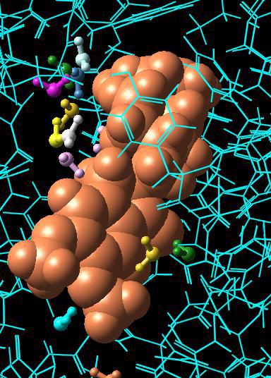
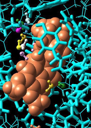
60 Chapter 4: Changing the Appearance of Structures 54 Maestro 8.5 Tutorial
61 Chapter 4: Changing the Appearance of Structures Beyond the options in the Maestro toolbar, it is also possible to customize settings such as the width of tubes, the size of spheres, and so on. For more information on changing these settings, refer to Section 6.2 of the Maestro User Manual. 4.5 Displaying the Protein Backbone as a Ribbon In addition to representing atoms and bonds using 3D features, Maestro can also represent protein backbones using three-dimensional ribbons. Ribbons can be turned on or off using the Show, hide, or color ribbons button on the Maestro toolbar: In this exercise, you ll represent the protein backbone using a ribbon. 1. Click the Fit to screen toolbar button, so you can see all of the protein. 2. From the Show, hide, or color ribbons button menu, choose Show Ribbons for All Residues. The protein is now represented by ribbons that trace the protein backbone instead of atoms, but the ligand and the water molecules remain in their previous representation. 3. Choose Display Atoms from the Show, hide, or color ribbons button menu. The protein atoms reappear, with the ribbons displayed as well. Observe that the ribbon follows the protein backbone. 4. Choose Delete Ribbons from the Show, hide, or color ribbons button menu. The ribbons disappear, leaving the atoms on display. The default coloring scheme for ribbons is to color according to residue position. As you follow the ribbon along, you ll see the colors progress through the spectrum as the residue number increases. The menu on the ribbons button includes alternate coloring schemes that you can select. Once you have experience with Maestro s ribbon rendering options, you may wish to further customize the look and feel of protein ribbons. For more information on doing so, see Section of the Maestro User Manual. Maestro 8.5 Tutorial 55

 and")
62 Chapter 4: Changing the Appearance of Structures Color schemes for ribbons Figure 4.6. Show, hide, or color ribbons button menu. Figure 4.7. Ribbon representation of protein with atoms (right) and without (left). 56 Maestro 8.5 Tutorial

63 Chapter 4: Changing the Appearance of Structures 4.6 Creating and Displaying a Molecular Surface In addition to custom rendering for higher-level structural features like protein backbones, Maestro can also represent the surfaces that are created by groups of molecules. Surfaces can represent the volume of a molecule as approximated by van der Waals radii. Surfaces can also display properties such as the electron density created by programs like Jaguar. In this example, we ll create and visualize a molecular surface, which is a smooth, contiguous surface that represents the solvent-accessible area presented by a structure. We ll use the protein from the previous exercise, so leave the structure in the Workspace. 1. Choose Molecular Surface from the Surfaces submenu of the Display menu on the Maestro menu bar. The Molecular Surface panel opens. 2. Delete the text in the Name text box at the top of the panel and type Tutorial Surface. This text box contains the text Molecular Surface by default. Figure 4.8. The Molecular Surface panel. Maestro 8.5 Tutorial 57

64 Chapter 4: Changing the Appearance of Structures Below the Name text box is an area titled Atoms for surface display. This is used to define the structure for which Maestro creates a surface. For this exercise we ll select the protein structure only: 3. Choose Molecules from the Pick option menu. 4. Click on any atom in the protein to select the entire protein. The wire bonds in the protein are temporarily colored a purple color, to show that they have been selected for the surface. Note that the text in the ASL text box is automatically filled in once you click on the protein. ASL stands for Atom Specification Language, and is a way of describing which atoms have been selected. If you want to be more selective in the atoms you choose, you can click the Select button, which opens the Atom Selection dialog box. For more information, see Section 5.3 of the Maestro User Manual. The rest of the settings can be left at their default values. For large molecules such as proteins, the default image quality setting of Low is usually appropriate. For small molecules, higher quality settings may be useful. 5. Click Create Surface to generate the molecular surface. Once the surface is created, the Surface Table panel automatically appears. Notice that you can see inside the surface where the ligand atoms are. This is because the surface doesn t cover these atoms: we chose to put a surface on only the protein. 6. Click Close to close the Molecular Surface panel. Figure 4.9. The Surface Table panel. 58 Maestro 8.5 Tutorial

65 Chapter 4: Changing the Appearance of Structures Figure Molecular surface, green at 100% transparency (left) and light blue at 50% transparency (right). Next, we ll experiment with the color and transparency of this surface. These properties can be changed in the Display Options dialog box. 7. Click the Display Options button near the bottom of the Surface Table panel. The Display Options dialog box opens. 8. Use the Transparency slider to set the transparency to 50 (fifty percent transparent). 9. Select Light Blue from the Color option menu. 10. Click OK to accept and view the changes (see Figure 4.10). The dialog box closes and the changes are applied. If you want to experiment more with the display options, click Apply instead. The dialog box remains open, and you can make whatever other changes you would like to make. 11. Once you ve viewed the surface, control-click the diamond in the leftmost column of the Surface Table to undisplay the surface. 12. Close the Surface Table panel by clicking the Close button. Maestro 8.5 Tutorial 59

66 Chapter 4: Changing the Appearance of Structures Figure The Surface Table - Display Options dialog box. Maestro is also capable of generating several other surface types. The interfaces used to create these surfaces are generally similar to the Molecular Surfaces panel, and all settings for the generated surfaces are controlled via the Surface Table panel. For more information on creating and viewing surfaces using Maestro, see Chapter 11 of the Maestro User Manual. 60 Maestro 8.5 Tutorial
67 Maestro Tutorial Chapter 5 Chapter 5: Measuring, Analyzing, and Superimposing Structures Once you re satisfied with the way that your structures appear in the Workspace, you can go on to examine the structures and their properties. For example, you can examine the geometric differences between two conformations of a molecule by superimposing them, or you can have Maestro display the hydrogen-bonding interactions between a ligand and a receptor. In this chapter, we ll start with basic operations like measuring distances and angles before performing more complicated structural analyses. For more information on the full extent of Maestro s analysis capabilities, see Chapter 9 of the Maestro User Manual. 5.1 Getting Ready for the Exercises in this Chapter To perform the exercises in this chapter, you ll need to import the structure file analysis_exercises.mae. This is a multi-structure file that includes several small molecules, a set of conformations created by MacroModel, and a ligand that has been docked to a protein structure using Glide. If you haven t already copied this file from $SCHRODINGER/ maestro-vversion/tutorial, copy it now from this directory to your working directory. To import the file into Maestro: 1. Click the Import structures toolbar button to open the Import panel. 2. Ensure that the file format is set to Maestro. 3. Select the file analysis_exercises.mae from the Files list. 4. Click Import to import the multi-structure Maestro file. 5. Close the Import panel by clicking the Close button in the bottom right-hand corner. 6. If the Project Table panel isn t open, open it by clicking the Open/Close Project Table toolbar button. Maestro 8.5 Tutorial 61

68 Chapter 5: Measuring, Analyzing, and Superimposing Structures Figure 5.1. The Project Table panel with the new entries. In the Project Table panel, you ll notice that several new rows have been added. These rows represent new entries in the Project Table. Some of these entries contain only one molecule, while other entries contain multiple molecules. The Project Table has an easy-to-use set of controls that allow you to record structure-related properties, present the results of calculations, and include and exclude entries in the Workspace. You ve already done some of these operations in earlier chapters of this tutorial. The exercises in this chapter will walk you through any necessary Project Table manipulations, but for more information on the Project Table, see Chapter 8 of the Maestro User Manual. We ll be using the Project Table quite a bit in these exercises, so we re going to leave it open. If it gets in the way, you can move it, or you can close it and re-open it when you need to use it again. To close the Project Table panel, click the Open/Close Project Table button in the toolbar (it should be indented), or click the Close button in the bottom right-hand corner of the Project Table panel. You re now ready to begin the exercises in this chapter. 5.2 Measuring Distances One simple analysis task is to determine the distance between two atoms. These can be any two atoms, bonded or non-bonded. In this example, we ll use the Measure distances, angles, or dihedrals button on Maestro s toolbar to determine bonded and non-bonded interatomic distances on a structure that has been optimized using Jaguar. To begin, we ll make sure that the appropriate Project Table entry is included in the Workspace. 1. In the Project Table, find the entry named Small Molecule Maestro 8.5 Tutorial

69 Chapter 5: Measuring, Analyzing, and Superimposing Structures 2. Click the diamond in the In column for the entry to ensure that this entry is included in the Workspace and all others are excluded. 3. Choose Distance from the Measure distances, angles, or dihedrals button menu in the main toolbar. (If you don t remember how to do this or are starting with this chapter, see the explanation of how to use button menus on page 7.) 4. Click on any atom in the Workspace. A purple cube appears over the atom, to indicate that you have selected it as the starting point for your measurement. 5. Click on an atom that is bonded to the atom you ve just selected. A dashed marker appears between the atoms, and the interatomic distance (measured in angstroms) is displayed over the marker. If you rotate the molecule in the Workspace (by holding down the middle mouse button and moving the mouse), you ll see that the measurement marker moves with the structure. Next, we ll measure a distance between two non-bonded atoms. Because the measurements tool is still active (the Measure distances, angles, or dihedrals button is indented to show this), with Distance selected, you don t need to repeat the process of selecting Distance from the toolbar button menu. 6. Click on the same atom that you clicked on in Step 4. Figure 5.2. Distance measurements. Maestro 8.5 Tutorial 63
70 Chapter 5: Measuring, Analyzing, and Superimposing Structures Note that the purple box appears again, indicating that you ve selected this atom as the starting point for your measurement. 7. Now click on any atom in the Workspace that s not bonded to the atom that you clicked on in the previous step. You ll see that a new distance marker appears between the two atoms that you just clicked on. You may also have noticed that the yellow lines disappeared from the original measurement when you made the new measurement. This is because the yellow marker on both sides of the dashed line simply indicates that it was the most recently measured distance. Finally, after measuring the distances, we can remove the markers from the Workspace. 8. Choose Delete Measurements from the Measure distances, angles, or dihedrals button menu. The structure remains in the Workspace unchanged, and the distance markers disappear. 5.3 Measuring Angles Another easily performed analysis task is angle measurement. In this example, we ll use the structure from the previous exercise to see that angles can be measured in much the same way that distances can be measured. 1. Choose Angle from the Measure distances, angles, or dihedrals button menu. 2. Click the oxygen of the carbonyl near the middle of the molecule. You ll notice that a purple cube appears to indicate that you ve selected an atom to begin an angle measurement. 3. Click the carbonyl carbon atom. You ll see a second purple cube appear, indicating that you ve selected the clicked atom to be part of your angle measurement. This second atom clicked will always be the vertex of the angle that is measured. 4. Finally, click on a carbon atom that is bonded to the atom you picked in the last step. After you click on the third atom, the purple cubes vanish, and a green angle marker appears. 64 Maestro 8.5 Tutorial

71 Chapter 5: Measuring, Analyzing, and Superimposing Structures Figure 5.3. Measuring an angle. If you like, you can define several other angle measurements on the structure. Just like distance measurements, multiple angle measurements can appear in the Workspace at any given time, and you can measure angles between non-bonded atoms. 5. Finally, delete the angle markers from the Workspace by choosing Delete Measurements from the Measure distances, angles, or dihedrals button menu. The angle markers disappear, and the structure remains unchanged in the Workspace. 5.4 Measuring Dihedrals Once you know how to measure distances and angles, it s a simple matter to measure a dihedral angle, the torsion defined by any four atoms. For this example, we ll measure the dihedral angles in two different conformations of n-propanol. We ll begin this exercise by placing one of the n-propanol structures in the Workspace. 1. Locate the entry n-propanol anti in the Project Table. 2. Include the n-propanol anti entry in the Workspace and exclude all other entries, by clicking the diamond to the left of the title. 3. Choose Dihedral from the Measure distances, angles, or dihedrals button menu. 4. Locate the terminal methyl group in the molecule and click on the carbon atom in that group. A purple cube appears, indicating that you ve selected this atom to be used as part of the dihedral measurement. Maestro 8.5 Tutorial 65

72 Chapter 5: Measuring, Analyzing, and Superimposing Structures Figure 5.4. Measuring a dihedral angle. 5. Next, click on the carbon atom that is bonded to the one you clicked on in the previous step. After doing so, another purple cube appears. 6. Click on the one remaining carbon atom in the structure. A third purple cube appears. 7. Finally, click on the red oxygen atom. A marker is superimposed over the structure, and the measurement (180 ) is displayed over the marker. After measuring the dihedral angle of the anti conformation, we ll include the staggered conformation in the Workspace and measure the corresponding dihedral angle. 8. Locate the entry n-propanol staggered in the Project Table. 9. Control-click (click while holding down the CTRL key) the diamond next to the entry name to include this entry in the Workspace. You ll notice that control-clicking makes the staggered entry appear in the Workspace next to the anti entry rather than replacing it. This is because control-clicking has no effect on the inclusion of other entries: it only affects the entry you are working with. Because the measurement tool is still active (indicated by the indented appearance of the Measure distances, angles, or dihedrals button in the toolbar), with Dihedral selected, you don t need to click the button again to measure another dihedral angle. To measure the dihedral, you click on four atoms, just as for the anti conformation. 10. Click the carbon atom in the terminal methyl group of the entry that you just added to in the Workspace, then on the carbon atom that s bonded to it, then on the remaining carbon atom, and finally on the oxygen atom. 66 Maestro 8.5 Tutorial
73 Chapter 5: Measuring, Analyzing, and Superimposing Structures A purple cube appears for each of the first three atoms. When you click on the fourth atom, the purple cubes vanish, and a dihedral marker appears. The dihedral measurement ( 63.9 ) is visible near the marker. 11. After you ve examined both dihedral measurements, delete the dihedral markers from the Workspace by choosing Delete Measurements from the Measure distances, angles, or dihedrals button menu. As you saw, it s possible to view measurements on multiple Project Table entries at the same time. You can display as many different measurements at the same time as you like, and include angle and distance measurements along with dihedral measurements. 5.5 Superimposing Multiple Structures Since we ve already placed multiple conformations of a molecule in the Workspace, we re ready to superimpose the two structures. Maestro s Superposition panel allows you to easily align two or more structures and calculate the root-mean-square deviation (RMSD) between atom pairs. This can be useful when examining the results of MacroModel conformation searches, comparing computational results to experimentally determined structures, or any other time you might want to gauge geometric differences between multiple structures. In this exercise, we ll open the Superposition panel and then go on to align the conformations used in the previous exercise. 1. If the two Project Table entries used in the previous exercise are not present in the Workspace, locate the entries in the project table, click on the diamond for n-propanol staggered, and then control-click on the diamond for n-propanol anti. 2. Open the Superposition panel by choosing Superposition from the Tools menu of the Maestro menu bar. When the Superposition panel appears, you ll see that it has a number of options for defining molecules and substructures that you can align. Near the top of the panel are controls that allow you to pick individual atom pairs that you can align. However, we ll use the Superimpose by ASL section in this example. 3. Select the Create property option. 4. Click All to perform the RMSD calculation for all atoms. You ll notice that one of the conformations is aligned on top of the other, and the RMSD value is again printed in the table at the bottom of the panel. If you scroll the Project Table to the right, you ll see that a property has been added that contains the RMSD value for the staggered conformation. Maestro 8.5 Tutorial 67
 for structures without aligning them.")
 option.")
74 Chapter 5: Measuring, Analyzing, and Superimposing Structures Figure 5.5. The Superposition panel. Maestro can also calculate a root-mean-square deviation (RMSD) for structures without aligning them. This capability is useful when you want to compare two structures in the context of another structure, such as two ligands docked to a receptor. To calculate the RMSD without aligning the structures, select the Calculate in place (no transformation) option. Figure 5.6. The superimposed propanol conformers. 68 Maestro 8.5 Tutorial
75 Chapter 5: Measuring, Analyzing, and Superimposing Structures Figure 5.7. The Project Table panel with the RMSD property added. In addition to the abovementioned tools for superimposing using atom pairs, you may have noticed that the Superposition panel contained a blue button labeled Select. This button opens the Atom Selection dialog box, an interface that allows you to select groups of atoms that can be used as input for most Maestro operations. For more information on the Atom Selection dialog box, see Section 5.3 of the Maestro User Manual. For a more thorough description of the Superposition panel, see Section 9.3 of the Maestro User Manual. 5.6 Displaying Hydrogen Bonds In this exercise, we ll use the results of a Glide docking job to demonstrate one of Maestro s useful analysis tools, the ability to display hydrogen bonding interactions. By displaying hydrogen bonding interactions, it s possible to visualize the interactions that make ligand binding energetically favorable. We ll begin this exercise by including the protein-ligand complex in the Maestro Workspace, and then use the toolbar to display and undisplay hydrogen bonding interactions. 1. Clear the Workspace. (Click the Clear Workspace button on the toolbar.) 2. In the Project Table, click on the diamond next to the entry named Protein with Glidedocked ligand. The protein complex appears in the Workspace. The protein is colored blue and the ligand is colored orange. You might want to zoom in on the ligand and center the ligand in the Workspace. To do this, drag horizontally with the middle and right mouse buttons, then right-click on a ligand atom. Maestro 8.5 Tutorial 69
76 Chapter 5: Measuring, Analyzing, and Superimposing Structures 3. To display hydrogen-bonding interactions between the ligand and the receptor, choose Inter H-bonds from the Display H-bonds button menu. 4. Click on any atom in the ligand (colored orange), to display intermolecular hydrogen bonds between that molecule and all other molecules in the Workspace. You ll see that hydrogen-bonding interactions are marked with dashed yellow lines. 5. To display intramolecular hydrogen bonding interactions, choose Intra H-bonds from the Display H-bonds button menu. 6. Click on any atom in the protein structure (colored blue) to visualize intramolecular hydrogen bonds. You ll see many dashed yellow markers appear among these are the hydrogen-bonding interactions that form helices and sheets, resulting in the overall structure of the protein. 7. Undisplay the hydrogen bonds by choosing Delete H-bonds from the Display H-bonds button menu. 8. Finally, close the project table by clicking on the Open/Close Project Table button in the toolbar (it should be indented), or by clicking the Close button in the bottom right-hand corner of the Project Table panel. Congratulations, you ve just completed a quick survey of Maestro s analysis capabilities. In addition to the analysis tasks covered in this chapter, Maestro can also display intermolecular steric contacts, add H-bond counts to the project table, and align protein structures. For more information on Maestro s analysis tasks, see Chapter 9 of the Maestro User Manual. In the next chapter, you ll see an overview of some of Maestro s more advanced visualization and analysis capabilities, and a brief description of other Schrödinger products. 70 Maestro 8.5 Tutorial

 and internal protein")
77 Chapter 5: Measuring, Analyzing, and Superimposing Structures Figure 5.8. Protein-ligand complex showing hydrogen bonds between the ligand and the protein (left) and internal protein hydrogen bonds (right). Maestro 8.5 Tutorial 71
CombiGlide 2.8. User Manual. Schrödinger Press
 CombiGlide User Manual CombiGlide 2.8 User Manual Schrödinger Press CombiGlide User Manual Copyright 2012 Schrödinger, LLC. All rights reserved. While care has been taken in the preparation of this publication,
Jaguar User Manual. Jaguar 6.0. User Manual. Schrödinger Press
 Jaguar User Manual Jaguar 6.0 User Manual Schrödinger Press Copyright 2005 Schrödinger, LLC. All rights reserved. Schrödinger, FirstDiscovery, Glide, Impact, Jaguar, Liaison, LigPrep, Maestro, Phase, Prime,
Exercises for Windows
 Exercises for Windows CAChe User Interface for Windows Select tool Application window Document window (workspace) Style bar Tool palette Select entire molecule Select Similar Group Select Atom tool Rotate
ISIS/Draw "Quick Start"
 ISIS/Draw "Quick Start" Click to print, or click Drawing Molecules * Basic Strategy 5.1 * Drawing Structures with Template tools and template pages 5.2 * Drawing bonds and chains 5.3 * Drawing atoms 5.4
Covalent Docking. Schrödinger Software Release Schrödinger Press
 Covalent Docking Covalent Docking Schrödinger Software Release 2015-2 Schrödinger Press Covalent Docking Copyright 2015 Schrödinger, LLC. All rights reserved. While care has been taken in the preparation
Chemistry 14CL. Worksheet for the Molecular Modeling Workshop. (Revised FULL Version 2012 J.W. Pang) (Modified A. A. Russell)
 (Modified A. A. Russell)") Chemistry 14CL Worksheet for the Molecular Modeling Workshop (Revised FULL Version 2012 J.W. Pang) (Modified A. A. Russell) Structure of the Molecular Modeling Assignment The molecular modeling assignment
Chemistry 14CL Worksheet for the Molecular Modeling Workshop (Revised FULL Version 2012 J.W. Pang) (Modified A. A. Russell) Structure of the Molecular Modeling Assignment The molecular modeling assignment
Assignment #0 Using Stellarium
 Name: Class: Date: Assignment #0 Using Stellarium The purpose of this exercise is to familiarize yourself with the Stellarium program and its many capabilities and features. Stellarium is a visually beautiful
How to Make or Plot a Graph or Chart in Excel
 This is a complete video tutorial on How to Make or Plot a Graph or Chart in Excel. To make complex chart like Gantt Chart, you have know the basic principles of making a chart. Though I have used Excel
NMR Predictor. Introduction
 NMR Predictor This manual gives a walk-through on how to use the NMR Predictor: Introduction NMR Predictor QuickHelp NMR Predictor Overview Chemical features GUI features Usage Menu system File menu Edit
Introduction to Structure Preparation and Visualization
 Introduction to Structure Preparation and Visualization Created with: Release 2018-4 Prerequisites: Release 2018-2 or higher Access to the internet Categories: Molecular Visualization, Structure-Based
Molecular Modeling and Conformational Analysis with PC Spartan
 Molecular Modeling and Conformational Analysis with PC Spartan Introduction Molecular modeling can be done in a variety of ways, from using simple hand-held models to doing sophisticated calculations on
Application Note. U. Heat of Formation of Ethyl Alcohol and Dimethyl Ether. Introduction
 Application Note U. Introduction The molecular builder (Molecular Builder) is part of the MEDEA standard suite of building tools. This tutorial provides an overview of the Molecular Builder s basic functionality.
Preparing a PDB File
 Figure 1: Schematic view of the ligand-binding domain from the vitamin D receptor (PDB file 1IE9). The crystallographic waters are shown as small spheres and the bound ligand is shown as a CPK model. HO
Comparing whole genomes
 BioNumerics Tutorial: Comparing whole genomes 1 Aim The Chromosome Comparison window in BioNumerics has been designed for large-scale comparison of sequences of unlimited length. In this tutorial you will
Create Satellite Image, Draw Maps
 Create Satellite Image, Draw Maps 1. The goal Using Google Earth, we want to create and import a background file into our Adviser program. From there, we will be creating paddock boundaries. The accuracy
Chem 253. Tutorial for Materials Studio
 Chem 253 Tutorial for Materials Studio This tutorial is designed to introduce Materials Studio 7.0, which is a program used for modeling and simulating materials for predicting and rationalizing structure
Computational Chemistry Lab Module: Conformational Analysis of Alkanes
 Introduction Computational Chemistry Lab Module: Conformational Analysis of Alkanes In this experiment, we will use CAChe software package to model the conformations of butane, 2-methylbutane, and substituted
Jaguar DFT Optimizations and Transition State Searches
 Jaguar DFT Optimizations and Transition State Searches Density Functional Theory (DFT) is a quantum mechanical (QM) method that gives results superior to Hartree Fock (HF) in less computational time. A
The Geodatabase Working with Spatial Analyst. Calculating Elevation and Slope Values for Forested Roads, Streams, and Stands.
 GIS LAB 7 The Geodatabase Working with Spatial Analyst. Calculating Elevation and Slope Values for Forested Roads, Streams, and Stands. This lab will ask you to work with the Spatial Analyst extension.
Calculating Bond Enthalpies of the Hydrides
 Proposed Exercise for the General Chemistry Section of the Teaching with Cache Workbook: Calculating Bond Enthalpies of the Hydrides Contributed by James Foresman, Rachel Fogle, and Jeremy Beck, York College
ArcGIS 9 ArcGIS StreetMap Tutorial
 ArcGIS 9 ArcGIS StreetMap Tutorial Copyright 2001 2008 ESRI All Rights Reserved. Printed in the United States of America. The information contained in this document is the exclusive property of ESRI. This
Tutorial. Getting started. Sample to Insight. March 31, 2016
 Getting started March 31, 2016 Sample to Insight CLC bio, a QIAGEN Company Silkeborgvej 2 Prismet 8000 Aarhus C Denmark Telephone: +45 70 22 32 44 www.clcbio.com support-clcbio@qiagen.com Getting started
Data Structures & Database Queries in GIS
 Data Structures & Database Queries in GIS Objective In this lab we will show you how to use ArcGIS for analysis of digital elevation models (DEM s), in relationship to Rocky Mountain bighorn sheep (Ovis
POC via CHEMnetBASE for Identifying Unknowns
 Table of Contents A red arrow was used to identify where buttons and functions are located in CHEMnetBASE. Figure Description Page Entering the Properties of Organic Compounds (POC) Database 1 Swain Home
Electric Fields and Equipotentials
 OBJECTIVE Electric Fields and Equipotentials To study and describe the two-dimensional electric field. To map the location of the equipotential surfaces around charged electrodes. To study the relationship
POC via CHEMnetBASE for Identifying Unknowns
 Table of Contents A red arrow is used to identify where buttons and functions are located in CHEMnetBASE. Figure Description Page Entering the Properties of Organic Compounds (POC) Database 1 CHEMnetBASE
Assignment 1: Molecular Mechanics (PART 1 25 points)
") Chemistry 380.37 Fall 2015 Dr. Jean M. Standard August 19, 2015 Assignment 1: Molecular Mechanics (PART 1 25 points) In this assignment, you will perform some molecular mechanics calculations using the
Chemistry 380.37 Fall 2015 Dr. Jean M. Standard August 19, 2015 Assignment 1: Molecular Mechanics (PART 1 25 points) In this assignment, you will perform some molecular mechanics calculations using the
General Chemistry Lab Molecular Modeling
 PURPOSE The objectives of this experiment are PROCEDURE General Chemistry Lab Molecular Modeling To learn how to use molecular modeling software, a commonly used tool in chemical research and industry.
Astronomy 101 Lab: Stellarium Tutorial
 Name: Astronomy 101 Lab: Stellarium Tutorial Please install the Stellarium software on your computer using the instructions in the procedure. If you own a laptop, please bring it to class. You will submit
Watershed Modeling Orange County Hydrology Using GIS Data
 v. 10.0 WMS 10.0 Tutorial Watershed Modeling Orange County Hydrology Using GIS Data Learn how to delineate sub-basins and compute soil losses for Orange County (California) hydrologic modeling Objectives
Working with ArcGIS: Classification
 Working with ArcGIS: Classification 2 Abbreviations D-click R-click TOC Double Click Right Click Table of Content Introduction The benefit from the use of geographic information system (GIS) software is
Using Microsoft Excel
 Using Microsoft Excel Objective: Students will gain familiarity with using Excel to record data, display data properly, use built-in formulae to do calculations, and plot and fit data with linear functions.
(THIS IS AN OPTIONAL BUT WORTHWHILE EXERCISE)
") PART 2: Analysis in ArcGIS (THIS IS AN OPTIONAL BUT WORTHWHILE EXERCISE) Step 1: Start ArcCatalog and open a geodatabase If you have a shortcut icon for ArcCatalog on your desktop, double-click it to start
PART 2: Analysis in ArcGIS (THIS IS AN OPTIONAL BUT WORTHWHILE EXERCISE) Step 1: Start ArcCatalog and open a geodatabase If you have a shortcut icon for ArcCatalog on your desktop, double-click it to start
GIS Workshop UCLS_Fall Forum 2014 Sowmya Selvarajan, PhD TABLE OF CONTENTS
 TABLE OF CONTENTS TITLE PAGE NO. 1. ArcGIS Basics I 2 a. Open and Save a Map Document 2 b. Work with Map Layers 2 c. Navigate in a Map Document 4 d. Measure Distances 4 2. ArcGIS Basics II 5 a. Work with
Molecular Visualization. Introduction
 Molecular Visualization Jeffry D. Madura Department of Chemistry & Biochemistry Center for Computational Sciences Duquesne University Introduction Assessments of change, dynamics, and cause and effect
EOS 102: Dynamic Oceans Exercise 1: Navigating Planet Earth
 EOS 102: Dynamic Oceans Exercise 1: Navigating Planet Earth YOU MUST READ THROUGH THIS CAREFULLY! This exercise is designed to familiarize yourself with Google Earth and some of its basic functions while
Mnova Software for Analyzing Reaction Monitoring NMR Spectra
 Mnova Software for Analyzing Reaction Monitoring NMR Spectra Version 10 Chen Peng, PhD, VP of Business Development, US & China Mestrelab Research SL San Diego, CA, USA chen.peng@mestrelab.com 858.736.4563
1. Double-click the ArcMap icon on your computer s desktop. 2. When the ArcMap start-up dialog box appears, click An existing map and click OK.
 Module 2, Lesson 1 The earth moves In this activity, you will observe worldwide patterns of seismic activity (earthquakes) and volcanic activity (volcanoes). You will analyze the relationships of those
SeeSAR 7.1 Beginners Guide. June 2017
 SeeSAR 7.1 Beginners Guide June 2017 Part 1: Basics 1 Type a pdb code and press return or Load your own protein or already existing project, or Just load molecules To begin, let s type 2zff and download
PQSMol. User s Manual
 PQSMol User s Manual c Parallel Quantum Solutions, 2006 www.pqs-chem.com Conventions used in this manual Filenames, commands typed in at the command prompt are written in typewriter font: pqsview aspirin.out
CHEMDRAW ULTRA ITEC107 - Introduction to Computing for Pharmacy. ITEC107 - Introduction to Computing for Pharmacy 1
 CHEMDRAW ULTRA 12.0 ITEC107 - Introduction to Computing for Pharmacy 1 Objectives Basic drawing skills with ChemDraw Bonds, captions, hotkeys, chains, arrows Checking and cleaning up structures Chemical
How many states. Record high temperature
 Record high temperature How many states Class Midpoint Label 94.5 99.5 94.5-99.5 0 97 99.5 104.5 99.5-104.5 2 102 102 104.5 109.5 104.5-109.5 8 107 107 109.5 114.5 109.5-114.5 18 112 112 114.5 119.5 114.5-119.5
Tutorial 8 Raster Data Analysis
 Objectives Tutorial 8 Raster Data Analysis This tutorial is designed to introduce you to a basic set of raster-based analyses including: 1. Displaying Digital Elevation Model (DEM) 2. Slope calculations
Conformational Analysis of n-butane
 Conformational Analysis of n-butane In this exercise you will calculate the Molecular Mechanics (MM) single point energy of butane in various conformations with respect to internal rotation around the
LAB 2 - ONE DIMENSIONAL MOTION
 Name Date Partners L02-1 LAB 2 - ONE DIMENSIONAL MOTION OBJECTIVES Slow and steady wins the race. Aesop s fable: The Hare and the Tortoise To learn how to use a motion detector and gain more familiarity
CE 365K Exercise 1: GIS Basemap for Design Project Spring 2014 Hydraulic Engineering Design
 CE 365K Exercise 1: GIS Basemap for Design Project Spring 2014 Hydraulic Engineering Design The purpose of this exercise is for you to construct a basemap in ArcGIS for your design project. You may execute
Computer simulation of radioactive decay
 Computer simulation of radioactive decay y now you should have worked your way through the introduction to Maple, as well as the introduction to data analysis using Excel Now we will explore radioactive
Lab 2: Projecting Geographic Data
 Lab 2: Projecting Geographic Data What you ll Learn: Basic methods for map projections in ArcMap. What You ll Produce: A map of Minnesota in three different statewide projections, a map of reprojected
Dock Ligands from a 2D Molecule Sketch
 Dock Ligands from a 2D Molecule Sketch March 31, 2016 Sample to Insight CLC bio, a QIAGEN Company Silkeborgvej 2 Prismet 8000 Aarhus C Denmark Telephone: +45 70 22 32 44 www.clcbio.com support-clcbio@qiagen.com
BUILDING BASICS WITH HYPERCHEM LITE
 BUILDING BASICS WITH HYPERCHEM LITE LAB MOD1.COMP From Gannon University SIM INTRODUCTION A chemical bond is a link between atoms resulting from the mutual attraction of their nuclei for electrons. There
3D Molecule Viewer of MOGADOC (JavaScript)
") 3D Molecule Viewer of MOGADOC (JavaScript) Movement of the Molecule Rotation of the molecule: Use left mouse button to drag. Translation of the molecule: Use right mouse button to drag. Resize the molecule:
3D Molecule Viewer of MOGADOC (JavaScript) Movement of the Molecule Rotation of the molecule: Use left mouse button to drag. Translation of the molecule: Use right mouse button to drag. Resize the molecule:
Experiment 0 ~ Introduction to Statistics and Excel Tutorial. Introduction to Statistics, Error and Measurement
 Experiment 0 ~ Introduction to Statistics and Excel Tutorial Many of you already went through the introduction to laboratory practice and excel tutorial in Physics 1011. For that reason, we aren t going
Space Objects. Section. When you finish this section, you should understand the following:
 GOLDMC02_132283433X 8/24/06 2:21 PM Page 97 Section 2 Space Objects When you finish this section, you should understand the following: How to create a 2D Space Object and label it with a Space Tag. How
OCEAN/ESS 410 Lab 4. Earthquake location
 Lab 4. Earthquake location To complete this exercise you will need to (a) Complete the table on page 2. (b) Identify phases on the seismograms on pages 3-6 as requested on page 11. (c) Locate the earthquake
3D - Structure Graphics Capabilities with PDF-4 Database Products
 3D - Structure Graphics Capabilities with PDF-4 Database Products Atomic Structure Information in the PDF-4 Databases ICDD s PDF-4 databases contain atomic structure information for a significant number
Learning to Use Scigress Wagner, Eugene P. (revised May 15, 2018)
") Learning to Use Scigress Wagner, Eugene P. (revised May 15, 2018) Abstract Students are introduced to basic features of Scigress by building molecules and performing calculations on them using semi-empirical
Learning to Use Scigress Wagner, Eugene P. (revised May 15, 2018) Abstract Students are introduced to basic features of Scigress by building molecules and performing calculations on them using semi-empirical
Experiment: Oscillations of a Mass on a Spring
 Physics NYC F17 Objective: Theory: Experiment: Oscillations of a Mass on a Spring A: to verify Hooke s law for a spring and measure its elasticity constant. B: to check the relationship between the period
VCell Tutorial. Building a Rule-Based Model
 VCell Tutorial Building a Rule-Based Model We will demonstrate how to create a rule-based model of EGFR receptor interaction with two adapter proteins Grb2 and Shc. A Receptor-monomer reversibly binds
Lesson Plan 2 - Middle and High School Land Use and Land Cover Introduction. Understanding Land Use and Land Cover using Google Earth
 Understanding Land Use and Land Cover using Google Earth Image an image is a representation of reality. It can be a sketch, a painting, a photograph, or some other graphic representation such as satellite
Using the Budget Features in Quicken 2008
 Using the Budget Features in Quicken 2008 Quicken budgets can be used to summarize expected income and expenses for planning purposes. The budget can later be used in comparisons to actual income and expenses
Introduction to Computer Tools and Uncertainties
 Experiment 1 Introduction to Computer Tools and Uncertainties 1.1 Objectives To become familiar with the computer programs and utilities that will be used throughout the semester. To become familiar with
Moving into the information age: From records to Google Earth
 Moving into the information age: From records to Google Earth David R. R. Smith Psychology, School of Life Sciences, University of Hull e-mail: davidsmith.butterflies@gmail.com Introduction Many of us
Introduction to Coastal GIS
 Introduction to Coastal GIS Event was held on Tues, 1/8/13 - Thurs, 1/10/13 Time: 9:00 am to 5:00 pm Location: Roger Williams University, Bristol, RI Audience: The intended audiences for this course are
Chemistry 112 Laboratory Experiment 5: Visualizing Molecular Orbitals: A MacSpartan Pro Experience (This experiment will be conducted in OR341)
") Chemistry 112 Laboratory Experiment 5: Visualizing Molecular Orbitals: A MacSpartan Pro Experience (This experiment will be conducted in OR341) Introduction In class we have discussed Lewis structures,
Chemistry 112 Laboratory Experiment 5: Visualizing Molecular Orbitals: A MacSpartan Pro Experience (This experiment will be conducted in OR341) Introduction In class we have discussed Lewis structures,
BCMB/CHEM 8190 Lab Exercise Using Maple for NMR Data Processing and Pulse Sequence Design March 2012
 BCMB/CHEM 8190 Lab Exercise Using Maple for NMR Data Processing and Pulse Sequence Design March 2012 Introduction Maple is a powerful collection of routines to aid in the solution of mathematical problems
Lewis Structures and Molecular Shapes
 Lewis Structures and Molecular Shapes Rules for Writing Lewis Structures 1. Determine the correct skeleton structure (connectivity of atoms). Usually, the most electronegative atoms go around the edges
McIDAS-V Tutorial Displaying Point Observations from ADDE Datasets updated July 2016 (software version 1.6)
") McIDAS-V Tutorial Displaying Point Observations from ADDE Datasets updated July 2016 (software version 1.6) McIDAS-V is a free, open source, visualization and data analysis software package that is the
McIDAS-V Tutorial Displaying Point Observations from ADDE Datasets updated July 2016 (software version 1.6) McIDAS-V is a free, open source, visualization and data analysis software package that is the
ScienceWord and PagePlayer Chemical formulae. Dr Emile C. B. COMLAN Novoasoft Representative in Africa
 ScienceWord and PagePlayer Chemical formulae Dr Emile C. B. CMLAN Novoasoft Representative in Africa Emails: ecomlan@yahoo.com ecomlan@scienceoffice.com Web site: www.scienceoffice.com Chemical formulae
Molecular modeling with InsightII
 Molecular modeling with InsightII Yuk Sham Computational Biology/Biochemistry Consultant Phone: (612) 624 7427 (Walter Library) Phone: (612) 624 0783 (VWL) Email: shamy@msi.umn.edu How to run InsightII
You w i ll f ol l ow these st eps : Before opening files, the S c e n e panel is active.
 You w i ll f ol l ow these st eps : A. O pen a n i m a g e s t a c k. B. Tr a c e t h e d e n d r i t e w i t h t h e user-guided m ode. C. D e t e c t t h e s p i n e s a u t o m a t i c a l l y. D. C
Senior astrophysics Lab 2: Evolution of a 1 M star
 Senior astrophysics Lab 2: Evolution of a 1 M star Name: Checkpoints due: Friday 13 April 2018 1 Introduction This is the rst of two computer labs using existing software to investigate the internal structure
Physical Chemistry Lab: The Hydrolysis of Aspirin. Each collaborative team determines the rate constant for the hydrolysis of aspirin.
 CH 348 Physical Chemistry Spring 2009 MK Bowman Adapted from: Marrs, Peter S. J. Chem. Educ. 2004 81 870. Physical Chemistry Lab: The Hydrolysis of Aspirin Each collaborative team determines the rate constant
Application Note. Capillary Zone Electrophoresis
 Application Note Capillary Zone Electrophoresis i Application Note: Capillary Zone Electrophoresis Version 8/PC Part Number 30-090-101 September 2008 Copyright IntelliSense Software Corporation 2004, 2005,
Login -the operator screen should be in view when you first sit down at the spectrometer console:
 Lab #2 1D 1 H Double Resonance (Selective Decoupling) operation of the 400 MHz instrument using automated sample insertion (robot) and automated locking and shimming collection of 1D 1 H spectra retrieving
Lab 1 Uniform Motion - Graphing and Analyzing Motion
 Lab 1 Uniform Motion - Graphing and Analyzing Motion Objectives: < To observe the distance-time relation for motion at constant velocity. < To make a straight line fit to the distance-time data. < To interpret
1 Introduction to Computational Chemistry (Spartan)
") 1 Introduction to Computational Chemistry (Spartan) Start Spartan by clicking Start / Programs / Spartan Then click File / New Exercise 1 Study of H-X-H Bond Angles (Suitable for general chemistry) Structure
1 Introduction to Computational Chemistry (Spartan) Start Spartan by clicking Start / Programs / Spartan Then click File / New Exercise 1 Study of H-X-H Bond Angles (Suitable for general chemistry) Structure
The CSC Interface to Sky in Google Earth
 The CSC Interface to Sky in Google Earth CSC Threads The CSC Interface to Sky in Google Earth 1 Table of Contents The CSC Interface to Sky in Google Earth - CSC Introduction How to access CSC data with
AMS 132: Discussion Section 2
 Prof. David Draper Department of Applied Mathematics and Statistics University of California, Santa Cruz AMS 132: Discussion Section 2 All computer operations in this course will be described for the Windows
ADF-GUI Tutorial. ADF Program System Release
 ADF-GUI Tutorial ADF Program System Release 2009.01 Scientific Computing & Modelling NV Vrije Universiteit, Theoretical Chemistry De Boelelaan 1083; 1081 HV Amsterdam; The Netherlands E-mail: support@scm.com
Relative Photometry with data from the Peter van de Kamp Observatory D. Cohen and E. Jensen (v.1.0 October 19, 2014)
") Relative Photometry with data from the Peter van de Kamp Observatory D. Cohen and E. Jensen (v.1.0 October 19, 2014) Context This document assumes familiarity with Image reduction and analysis at the Peter
Relative Photometry with data from the Peter van de Kamp Observatory D. Cohen and E. Jensen (v.1.0 October 19, 2014) Context This document assumes familiarity with Image reduction and analysis at the Peter
ST-Links. SpatialKit. Version 3.0.x. For ArcMap. ArcMap Extension for Directly Connecting to Spatial Databases. ST-Links Corporation.
 ST-Links SpatialKit For ArcMap Version 3.0.x ArcMap Extension for Directly Connecting to Spatial Databases ST-Links Corporation www.st-links.com 2012 Contents Introduction... 3 Installation... 3 Database
Automatic Watershed Delineation using ArcSWAT/Arc GIS
 Automatic Watershed Delineation using ArcSWAT/Arc GIS By: - Endager G. and Yalelet.F 1. Watershed Delineation This tool allows the user to delineate sub watersheds based on an automatic procedure using
Standards-Based Quantification in DTSA-II Part II
 Standards-Based Quantification in DTSA-II Part II Nicholas W.M. Ritchie National Institute of Standards and Technology, Gaithersburg, MD 20899-8371 nicholas.ritchie@nist.gov Introduction This article is
v Prerequisite Tutorials GSSHA WMS Basics Watershed Delineation using DEMs and 2D Grid Generation Time minutes
 v. 10.1 WMS 10.1 Tutorial GSSHA WMS Basics Creating Feature Objects and Mapping Attributes to the 2D Grid Populate hydrologic parameters in a GSSHA model using land use and soil data Objectives This tutorial
An Introduction to Alice. By Jenna Hayes under the direction of Professor Susan Rodger Duke University, June 2009
 An Introduction to Alice By Jenna Hayes under the direction of Professor Susan Rodger Duke University, June 2009 Hello! I m Alice, and I m going to teach you how to use the Alice program. With Alice, you
Figure 2.1 The Inclined Plane
 PHYS-101 LAB-02 One and Two Dimensional Motion 1. Objectives The objectives of this experiment are: to measure the acceleration due to gravity using one-dimensional motion, i.e. the motion of an object
Looking hard at algebraic identities.
 Looking hard at algebraic identities. Written by Alastair Lupton and Anthony Harradine. Seeing Double Version 1.00 April 007. Written by Anthony Harradine and Alastair Lupton. Copyright Harradine and Lupton
Viewing and Analyzing Proteins, Ligands and their Complexes 2
 2 Viewing and Analyzing Proteins, Ligands and their Complexes 2 Overview Viewing the accessible surface Analyzing the properties of proteins containing thousands of atoms is best accomplished by representing
Chem 1 Kinetics. Objectives. Concepts
 Chem 1 Kinetics Objectives 1. Learn some basic ideas in chemical kinetics. 2. Understand how the computer visualizations can be used to benefit the learning process. 3. Understand how the computer models
Interacting Galaxies
 Interacting Galaxies Contents Introduction... 1 Downloads... 1 Selecting Interacting Galaxies to Observe... 2 Measuring the sizes of the Galaxies... 5 Making a Colour Image in IRIS... 8 External Resources...
Ligand Scout Tutorials
 Ligand Scout Tutorials Step : Creating a pharmacophore from a protein-ligand complex. Type ke6 in the upper right area of the screen and press the button Download *+. The protein will be downloaded and
Task 1: Open ArcMap and activate the Spatial Analyst extension.
 Exercise 10 Spatial Analyst The following steps describe the general process that you will follow to complete the exercise. Specific steps will be provided later in the step-by-step instructions component
Version 1.2 October 2017 CSD v5.39
 Mogul Geometry Check Table of Contents Introduction... 2 Example 1. Using Mogul to assess intramolecular geometry... 3 Example 2. Using Mogul to explain activity data... 5 Conclusions... 8 Further Exercises...
Star Cluster Photometry and the H-R Diagram
 Star Cluster Photometry and the H-R Diagram Contents Introduction Star Cluster Photometry... 1 Downloads... 1 Part 1: Measuring Star Magnitudes... 2 Part 2: Plotting the Stars on a Colour-Magnitude (H-R)
Geology Wilson Computer lab Pitfalls II
 Geology 554 - Wilson Computer lab Pitfalls II Today we ll explore a subtle pitfall that can have significant influence on the interpretation of both simple and complex structures alike (see example 10
BOND LENGTH WITH HYPERCHEM LITE
 BOND LENGTH WITH HYPERCHEM LITE LAB MOD2.COMP From Gannon University SIM INTRODUCTION The electron cloud surrounding the nucleus of the atom determines the size of the atom. Since this distance is somewhat
Tutorial 23 Back Analysis of Material Properties
 Tutorial 23 Back Analysis of Material Properties slope with known failure surface sensitivity analysis probabilistic analysis back analysis of material strength Introduction Model This tutorial will demonstrate
Students will explore Stellarium, an open-source planetarium and astronomical visualization software.
 page 22 STELLARIUM* OBJECTIVE: Students will explore, an open-source planetarium and astronomical visualization software. BACKGROUND & ACKNOWLEDGEMENTS This lab was generously provided by the Red Rocks
This tutorial is intended to familiarize you with the Geomatica Toolbar and describe the basics of viewing data using Geomatica Focus.
 PCI GEOMATICS GEOMATICA QUICKSTART 1. Introduction This tutorial is intended to familiarize you with the Geomatica Toolbar and describe the basics of viewing data using Geomatica Focus. All data used in
Lab 2: Projecting Geographic Data
 Lab 2: Projecting Geographic Data What you ll Learn: Basic methods for map projections in ArcGIS Pro. What You ll Produce: A map of Minnesota in three different statewide projections, a map of reprojected