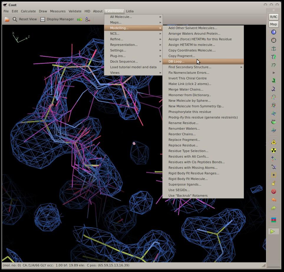
Modelling Macromolecules with Coot
|
|
|
- Rosamond Harris
- 6 years ago
- Views:
Transcription
1 Modelling Macromolecules with Coot Overview Real Space Refinement A Sample of Tools Tools for Cryo-EM Tools for Ligands [Carbohydrates] Paul Emsley MRC Laboratory of Molecular Biology


2 Acknowldegments, Collaborators Bernhard Lohkamp Kevin Cowtan Eugene Krissinel Stuart McNicholas Martin Noble Alexei Vagin
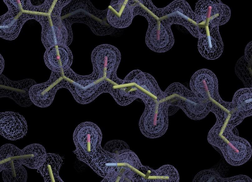
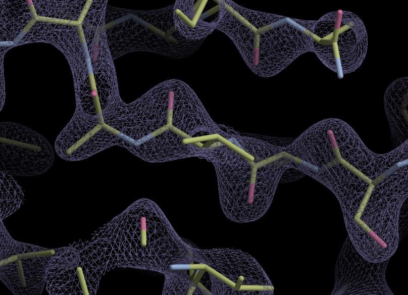
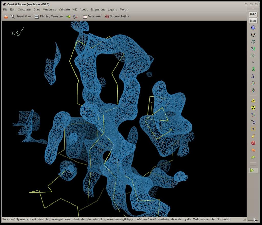
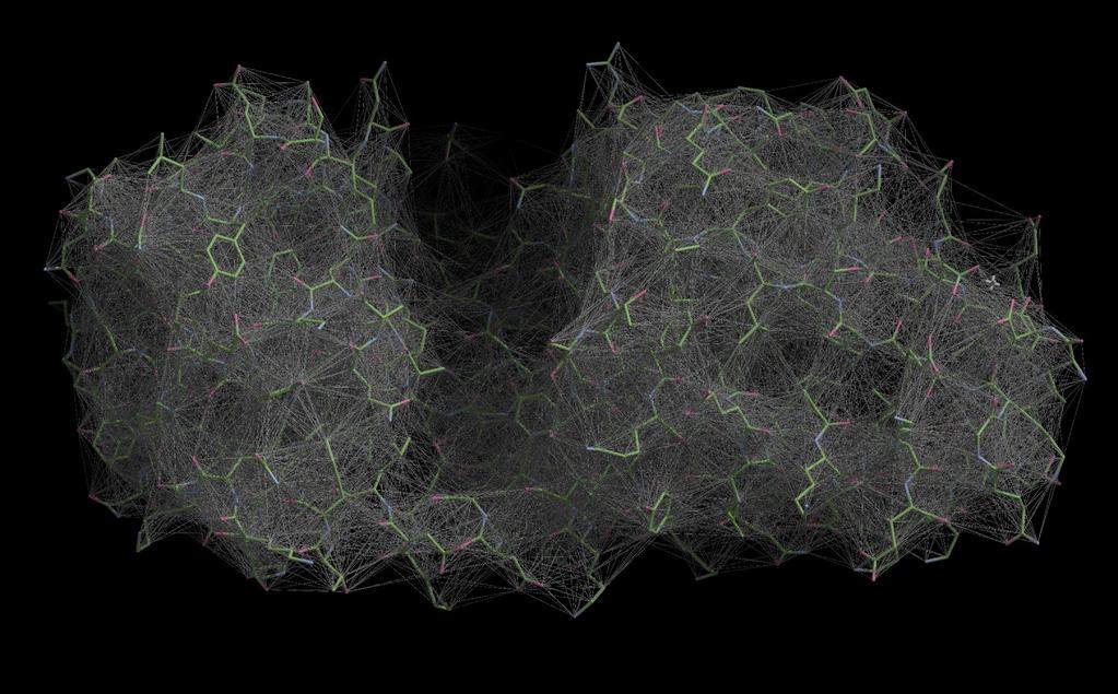
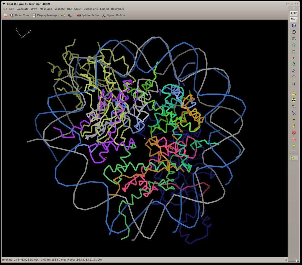
3 Coot Crystallographic Object-Oriented Tool-kit Primarily a tool for the interpretation of electron density generated from X-ray data with tools for modelling: rotate/translate, rotamers, refinement & regularization add, delete ligand fitting and analysis to be used post-automation A workhorse, not a show-pony
4 Why bother? Automated (complete) model-building still impractical Extremely demanding It takes a brain to validate Concerted motion of atoms connected by geometric restraints is difficult Coot is built with Novice users in mind (but not exclusively) because using the key-bindings will turn you from Noob Pro
5 Feature Integration Refinement External Internal e.g. REFMAC Validation Internal External e.g. MolProbity Validation, Model Building and Refinement should be used together
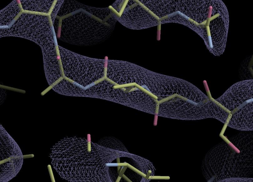
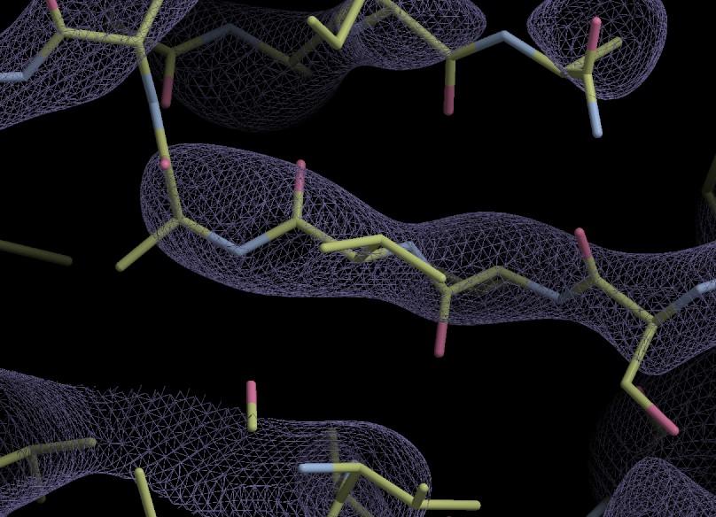
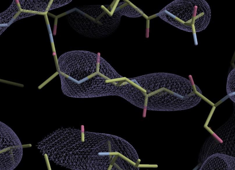
6 1.0Å
7 1.2Å
8 1.4Å
9 1.6Å
10 1.8Å
11 2.0Å
12 2.2Å
13 2.4Å
14 2.6Å
15 2.8Å
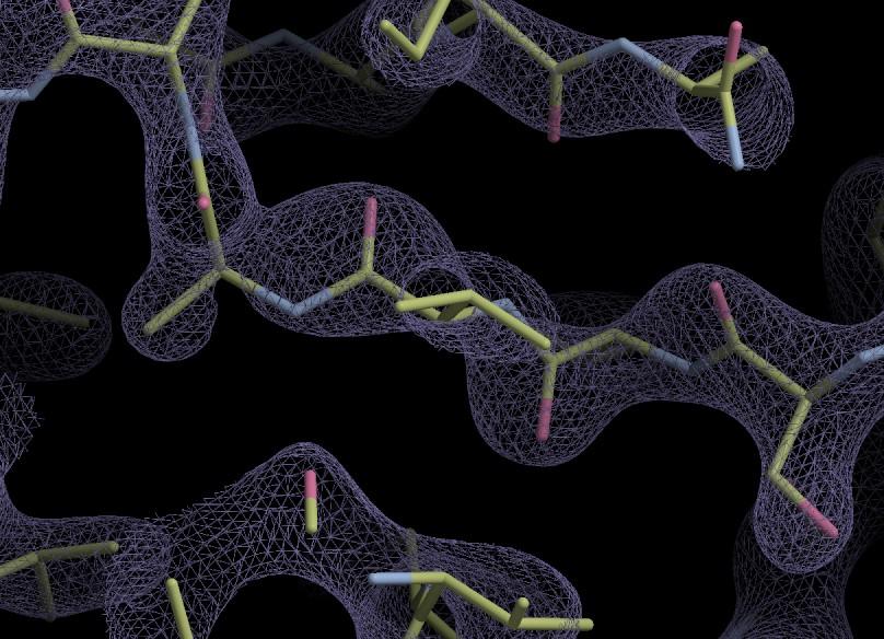
16 3.0Å
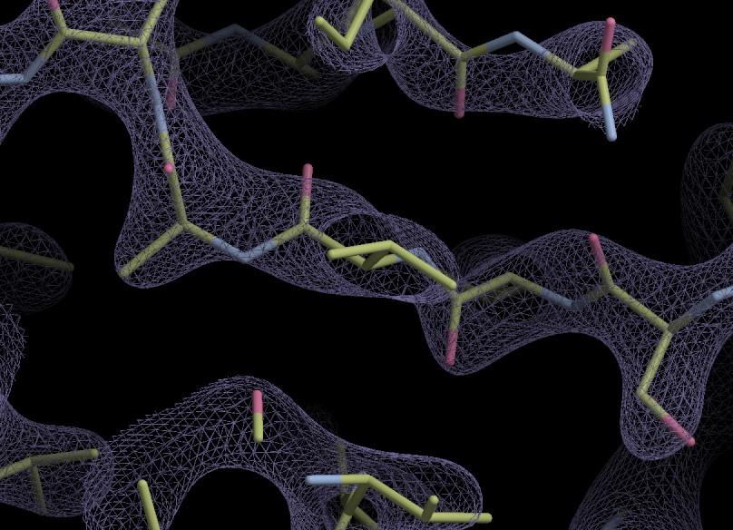
17 3.2Å
18 3.4Å
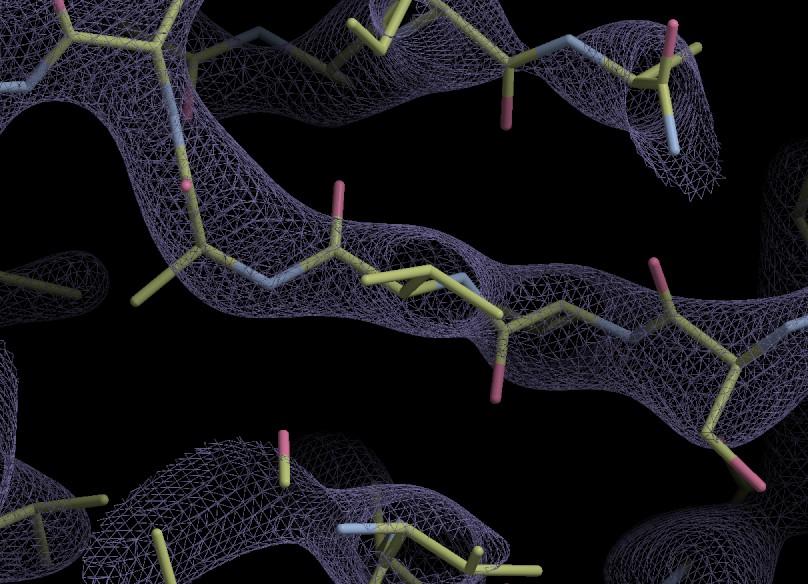
19 3.6Å
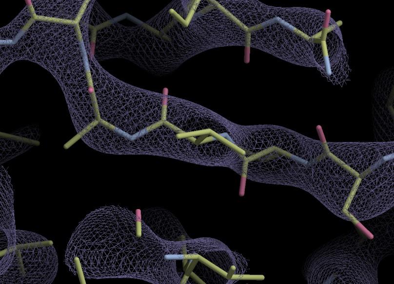
20 3.8Å
21 4.0Å
22 4.2Å
23 4.4Å
24 4.6Å
25 4.8Å
26 What is Refinement? The adjustment of model parameters (co-ordinates) so that the calculated structure factors match the observations as nearly as possible In one-shot real-space refinement, such as in Coot, this translates to: move the atoms into as high density as possible while minimizing geometrical distortions
27 Real Space Refinement Major Feature of Coot Gradient-based minimiser (BFGS derivative) Geometry library is the standard CIF-based Refmac dictionary Minimise deviations in bond length, angles, torsions, planes, chiral volume, non-bonded contacts Including links and modifications Provides interactive refinement Subject to substantial extension e.g. Sphere Refine
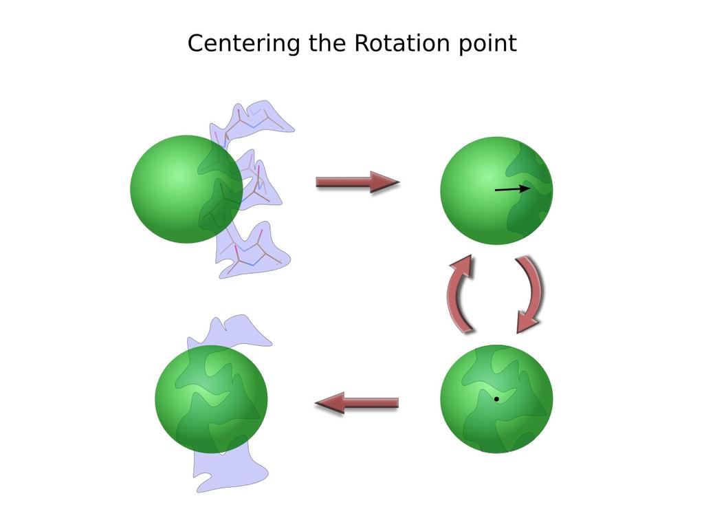
28 Distorted Geometry Pre-Refinement
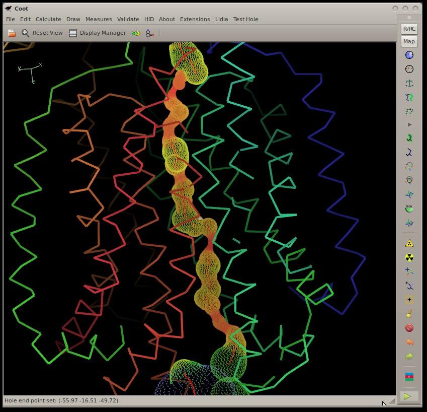
29 Refinement Gradients
30 Refinement: Cycle 3

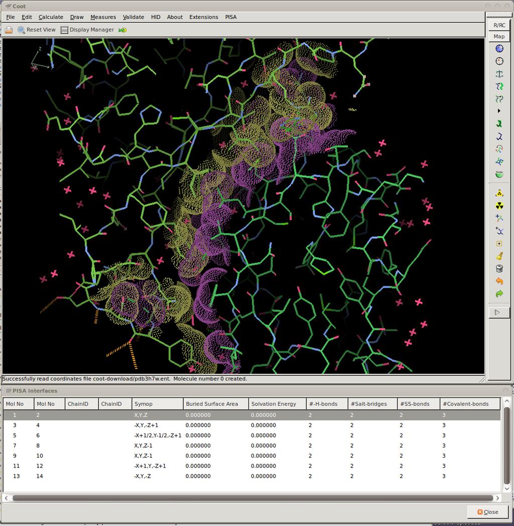
31 Refinement Cycle 200: Minimized

32 Representation of Results: The first attempt Student Reaction: Oh, I don't look at that window...

33 Representation of Results: Second attempt... Student Reaction: Oh, box of meaningless numbers. Go away 30/130

34 Representation of Results: Traffic Lights Traffic Lights represent the RMSd values for each of the refined geometry types Good refinement Bad refinement 31/120
35 Refinement Techniques Dragging an atom with Sphere Refine... too much moves, so use: Single-Atom Drag Over-dragging Key-bindings: Triple Refine Single Residue Refine with Auto-accept Ramachandran Refinement Best done with hydrogens Parallel Plane Restraints
36 Finding Holes An implementation of Smart, Goodfellow & Wallace (1993) Biophysics Journal 65, 2455 Atomic radii from AMBER I used radii from CCP4 monomer library sans simulated annealing
37
38 Interfaces and Assemblies: Interface to PISA
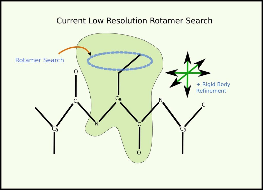
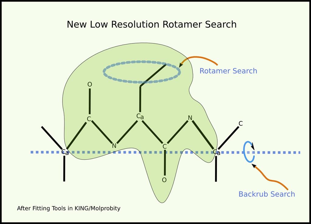
39

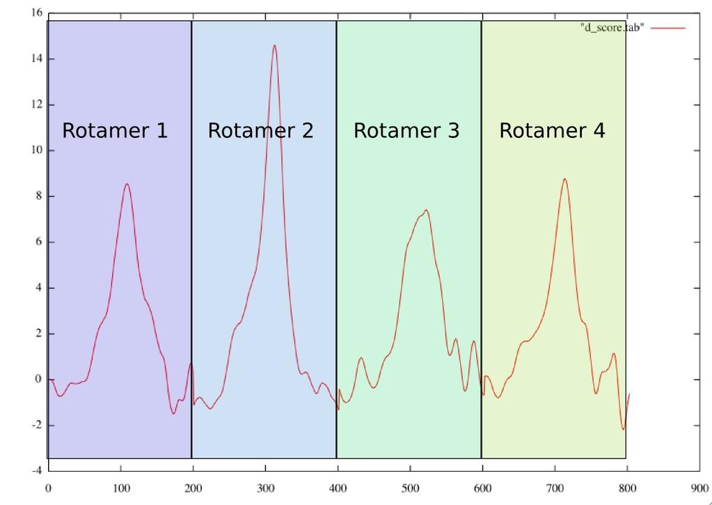
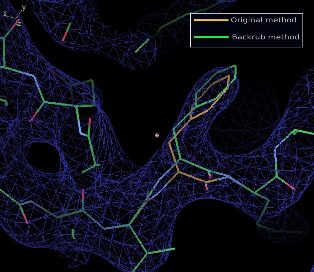
40 Some Representation Tools Gruber & Noble (2007)
41
42

43 Other Things Surfaces that use dictionary partial charges
44 Some Representation Tools

45 Other Things Molprobity dots for ligands Highlight interesting site
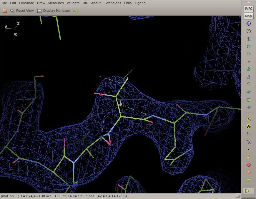
46 Tools for Cryo-EM and Low-Resolution
47 Backrub Rotamers High probability models with low resolution data
48 Previous
49
50
51
52
53
54
55
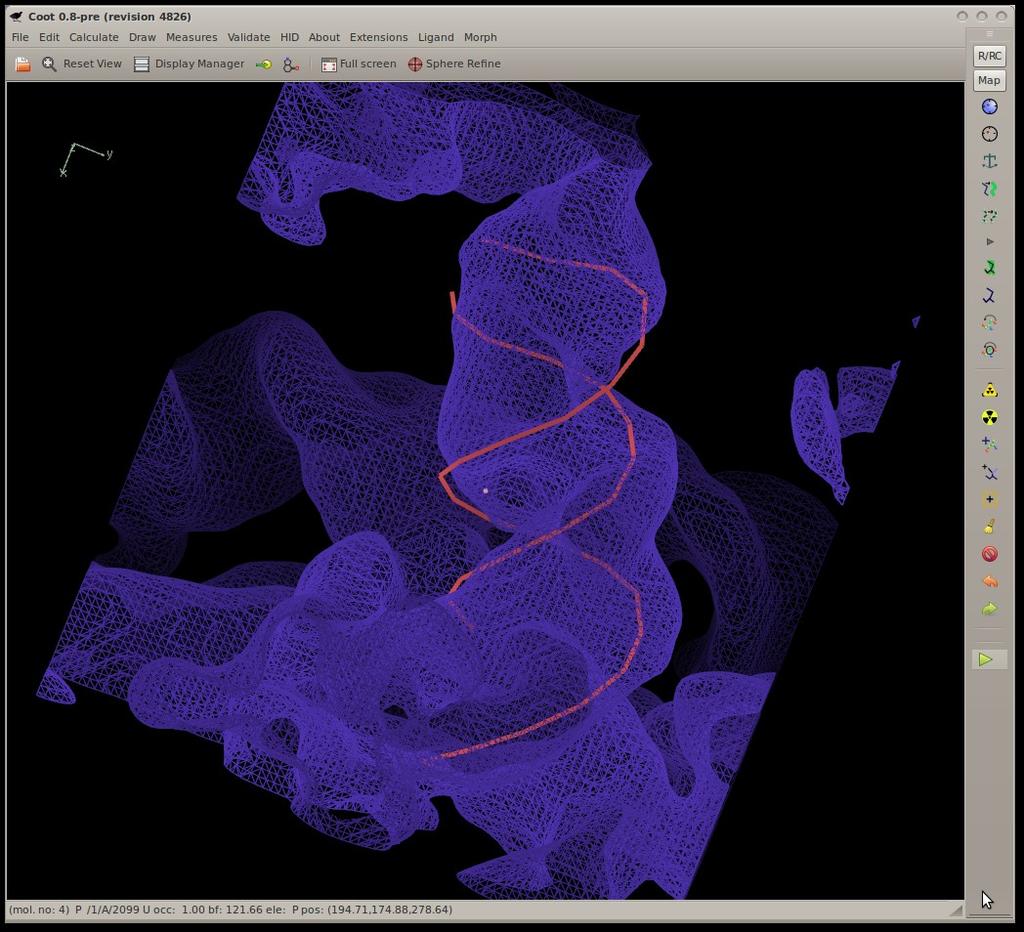
56
57 To turn it on... (ROTAMERSEARCHLOWRES)
58 Jiggle Fit How do I rotate and translate these atoms to fit the density? 6-dimensional problem Originally used to fit simple ligands/solvent molecules to blobs of density Now extended to fit arbitrary atom selections e.g. by Chain
59 Jiggle Fit: How it Works Loop n (say 1000) times: Generate random angles and translations Transform atom selection by these rotations and translation Score and store the fit to density Rank density fit scores, Pick top 20 solution, for each of them Rigid body fit and score solutions Pick the highest scoring solution if it's better than the starting model) Radius of Convergence is larger when using a low-pass map
60
61
62
63 Model Morphing: How it Works For each residue in a chain, we ask: where does a small fragment centred on this residue want to go? (Robust) average the transformations and apply them on a per-residue basis Repeat

64 Model Morphing: Generating the Raw RTs

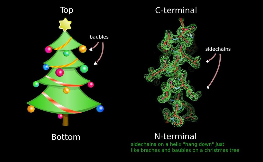
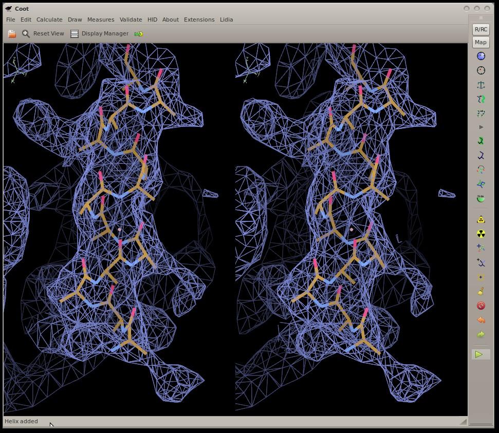
65 Model Morphing: Example
66 Model Morphing: Robust Averaging What are the residues in the environment of a residue? What are their RTs? Create a metric 'distance', sort on that Discard the top and bottom 25% Use remaining RTs to generate average...which is then applied to central residue Repeat for all residues Larger environment radii make the shifts smaller/more conservative More cycles needed
67
68
69
70
71
72 Alpha Helix Placement Scenario: Looking at a new map, not built with automatic tools: I can see that there s a helix here - build it for me! From a given point: Move to local averaged maximum Do a 2D MR-style orientation search on a cylinder of electron density Build a helix (both directions) 1D Rotation search to find best fit Score based on density at CB positions Trim n Grow 72/120
73


74 Cylinder Search Pick the orientation that encapsulates the most electron density Using 2 rotation axes

75 2 x 1-D Helix orientation searches
76
77
78
79
80 Additional Restraints
81 ProSMART integration ProSMART generates distance restraints from homologous structures to be applied to current model for refinement now available in Coot
82 ProSMART Restraints
83 Tools for RNA

84 Plane Restraints Derivativaties are an eigenvector scaled by out-ofplane distance
2 + (c1- c2)2 Not easy to use in")
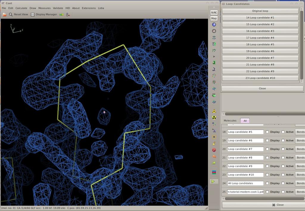
85 Parallel Planes Restraints S = (a1- a2)2 + (b1- b2)2 + (c1- c2)2 Not easy to use in Coot
86 Parallel Planes Restraints Also, we have considered parallel-planes distance restraints More tricky still to implement Not implemented yet (not in Coot, anyway)

87 Parallel Planes Restraints

88 Parallel Plane Restraints Shift to Origin
89 Parallel Planes Restraints Shift to Origin Move Back to Molecule
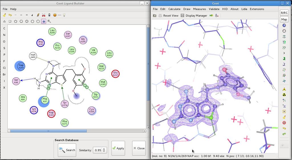
90
91
92
93 Loop Fitting Tool: (sloop) Kevin Cowtan
94
95
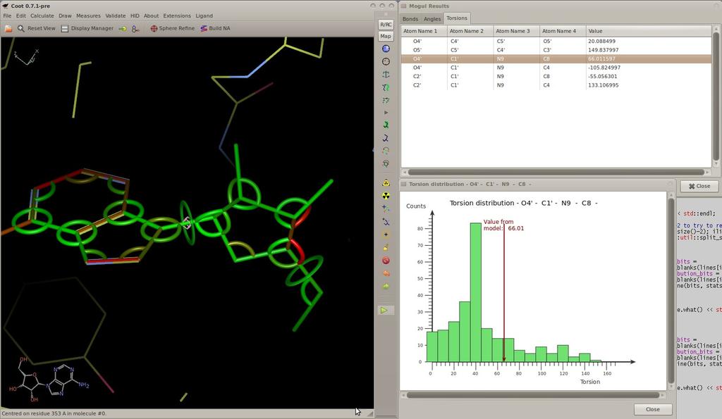
96
97

98 2D Ligand Builder Free sketch SBase search

99 2D Sketcher Structural Alerts Check vs. vector of SMARTS (from Biscu-it)

")
100 QED Score Quantitative Evaluation of Drug-likeness Bickerton et al (2012) Nature Chemistry

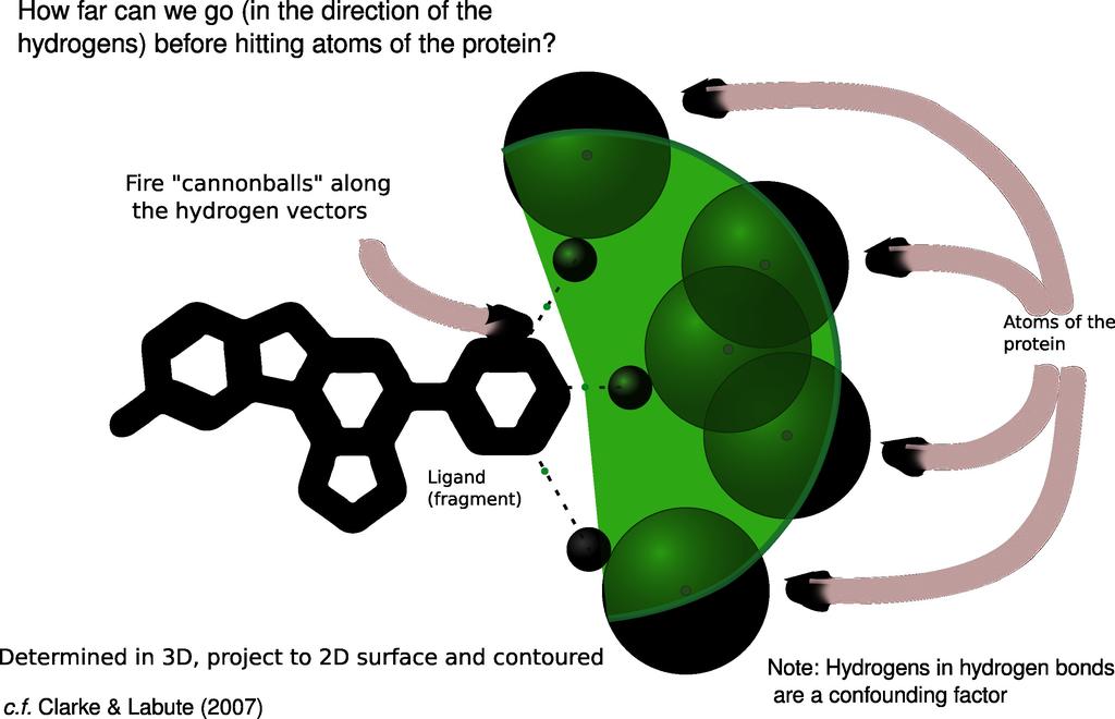
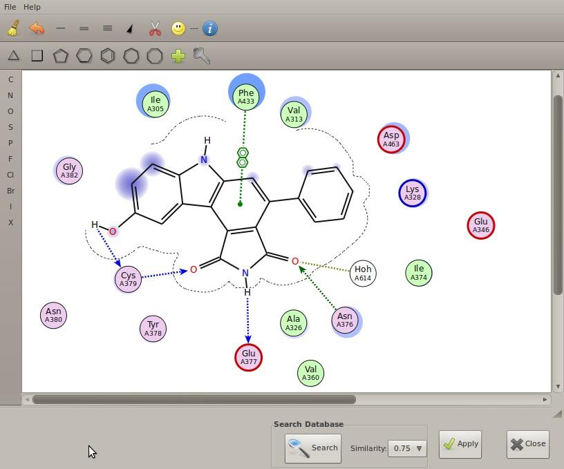
101 2D Sketcher QED score Silicos-it's Biscu-it
102 Ligand Utils Get Molcule Uses network connection to Wikipedia Get comp-id ligand-description from PDBe downloads and reads (e.g.) AAA.cif (extracted from chemical component library) Drag and drop Uses network connection to get URLs or file-system files pyrogen, acedrg restraints generation
 Perfect refinement with incorrect parameters distorted structure CSD's Mogul Knowledge-base of geometric parameters based")
103 Parmatisation issues... (what if they are wrong?) Perfect refinement with incorrect parameters distorted structure CSD's Mogul Knowledge-base of geometric parameters based on the CSD Can be run as a batch job Mean, median, mode, quartiles, Z-scores.
104 Ligand Validation Mogul plugin in Coot Run mogul, graphical display of results Update restraints (target and esds for bonds and angles) CSD data not so great for plane, chiral and torsion restraints (not by me, anyway) 105/120
105 Mogul Results Representation
106 New Software for Restraints Generation
")
107 COD Atom Types COD H1B: C9: 2nd order-based H(CHHO) C[5,5,6](C[5,5]CHH)(C[5,6]CHH)(C[5,6]CHO)(H)

108 Ligand Represenation Bond orders (from dictionary restraints)

109 Chemical Features...and on the fly thumbnailing
 Can we do better?")
110 Ligand Environment Layout 2d Ligand pocket layout (ligplot, poseview) Can we do better? - Interactivity?
111 Ligand Environment Layout Binding pocket residues Interactions Substitution contour Solvent accessibility halos Solvent exclusion by ligand


112 Solvent Exposure Identification of solvent accessible atoms
113 Ligand Enviroment Layout Considerations 2D placement and distances should reflect 3D metrics (as much as possible) H-bonded residues should be close the atoms to which they are bonded Residues should not overlap the ligand Residues should not overlap each other c.f. Clark & Labute (2007)

114 Layout Energy Terms Residues match 3D Distances Residues don't overlay each other Residues are close to H-bonding ligand atoms Residues don't overlap ligand

115 Don't overlap the ligand

116 Ligand Environment Layout Residue position minimisation

117 Determination of the Substitution Contour
118 118/120
119 119/120
120 Acknowledgements Group Murshudov Fei Long, Andrea Thorn & Rob Nicholls Kevin Cowtan Bernhard Lohkamp Libraries, Dictionaries Alexei Vagin Eugene Krissinel Richardsons (Duke)
121 Modelling Carbohydrates Validation, Model-building, Refinement
122 Problematic Glycoproteins Crispin, Stuart & Jones (2007) NSB Correspondence one third of entries contain significant errors in carbohydrate stereochemistry... carbohydrate-specific building and validation tools capable of guiding and construction of biologically relevant stereochemically accurate models should be integrated into popular crystallographic software. Rigorous treatment of the structural biology of glycosylation can only enhance the analysis of glycoproteins and our understanding of their function PDB curators concur
123 Modelling Carbohydrates Validation, Model-building, Refinement
124 Problematic Glycoproteins Crispin, Stuart & Jones (2007) NSB Correspondence one third of entries contain significant errors in carbohydrate stereochemistry... carbohydrate-specific building and validation tools capable of guiding and construction of biologically relevant stereochemically accurate models should be integrated into popular crystallographic software. Rigorous treatment of the structural biology of glycosylation can only enhance the analysis of glycoproteins and our understanding of their function PDB curators concur

125 Validate the Tree: N-linked carbohydrates
126 Carbohydrate Links Thomas Lütteke (2007)
127 Linking Oligsaccharides/Carbohydrates: LO/Carb Complex carbohydrate structure from a dictionary of standard links and monomers torsion-angle refinement
128
129
130
131
")
132 Refinement Trials (NAG-ASN example)
133 Acknowledgements Group Murshudov Fei Long, Andrea Thorn & Rob Nicholls Kevin Cowtan Bernhard Lohkamp Libraries, Dictionaries Alexei Vagin Eugene Krissinel Richardsons (Duke)
Manipulating Ligands Using Coot. Paul Emsley May 2013
 Manipulating Ligands Using Coot Paul Emsley May 2013 Ligand and Density... Ligand and Density... Ligand and Density... Protein-ligand complex models are often a result of subjective interpretation Scoring
Tools for Cryo-EM Map Fitting. Paul Emsley MRC Laboratory of Molecular Biology
 Tools for Cryo-EM Map Fitting Paul Emsley MRC Laboratory of Molecular Biology April 2017 Cryo-EM model-building typically need to move more atoms that one does for crystallography the maps are lower resolution
Garib N Murshudov MRC-LMB, Cambridge
 Garib N Murshudov MRC-LMB, Cambridge Contents Introduction AceDRG: two functions Validation of entries in the DB and derived data Generation of new ligand description Jligand for link description Conclusions
This is an author produced version of Privateer: : software for the conformational validation of carbohydrate structures.
 This is an author produced version of Privateer: : software for the conformational validation of carbohydrate structures. White Rose Research Online URL for this paper: http://eprints.whiterose.ac.uk/95794/
Coot Updates. Paul Emsley Sept 2016
 Coot Updates Paul Emsley Sept 2016 Coot 0.8.4 Released Patterson from intensities Fill-partial-residue uses Backrub-rotamers Sequence dialog is now dynamically updated Outliers Only in Ramachanran Plot
Coot, pyrogen & CCP4 SRS. Paul Emsley MRC Laboratory of Molecular Biology, Cambridge, UK June 2015
 Coot, pyrogen & CCP4 SRS Paul Emsley MRC Laboratory of Molecular Biology, Cambridge, UK June 2015 Coot Updates JED-Flip (as it's currently known) added Torsion-spec-dependent rotation of ligand fragment
Full wwpdb X-ray Structure Validation Report i
 Full wwpdb X-ray Structure Validation Report i Mar 13, 2018 04:03 pm GMT PDB ID : 5NMJ Title : Chicken GRIFIN (crystallisation ph: 6.5) Authors : Ruiz, F.M.; Romero, A. Deposited on : 2017-04-06 Resolution
wwpdb X-ray Structure Validation Summary Report
 wwpdb X-ray Structure Validation Summary Report io Jan 31, 2016 06:45 PM GMT PDB ID : 1CBS Title : CRYSTAL STRUCTURE OF CELLULAR RETINOIC-ACID-BINDING PROTEINS I AND II IN COMPLEX WITH ALL-TRANS-RETINOIC
1.b What are current best practices for selecting an initial target ligand atomic model(s) for structure refinement from X-ray diffraction data?!
 for structure refinement from X-ray diffraction data?!") 1.b What are current best practices for selecting an initial target ligand atomic model(s) for structure refinement from X-ray diffraction data?! Visual analysis: Identification of ligand density from
1.b What are current best practices for selecting an initial target ligand atomic model(s) for structure refinement from X-ray diffraction data?! Visual analysis: Identification of ligand density from
Full wwpdb X-ray Structure Validation Report i
 Full wwpdb X-ray Structure Validation Report i Mar 8, 2018 08:34 pm GMT PDB ID : 1RUT Title : Complex of LMO4 LIM domains 1 and 2 with the ldb1 LID domain Authors : Deane, J.E.; Ryan, D.P.; Maher, M.J.;
Pipelining Ligands in PHENIX: elbow and REEL
 Pipelining Ligands in PHENIX: elbow and REEL Nigel W. Moriarty Lawrence Berkeley National Laboratory Physical Biosciences Division Ligands in Crystallography Drug design Biological function studies Generate
Dictionary of ligands
 Dictionary of ligands Some of the web and other resources Small molecules DrugBank: http://www.drugbank.ca/ ZINC: http://zinc.docking.org/index.shtml PRODRUG: http://www.compbio.dundee.ac.uk/web_servers/prodrg_down.html
Electronic Supplementary Information (ESI) for Chem. Commun. Unveiling the three- dimensional structure of the green pigment of nitrite- cured meat
 for Chem. Commun. Unveiling the three- dimensional structure of the green pigment of nitrite- cured meat") Electronic Supplementary Information (ESI) for Chem. Commun. Unveiling the three- dimensional structure of the green pigment of nitrite- cured meat Jun Yi* and George B. Richter- Addo* Department of Chemistry
Electronic Supplementary Information (ESI) for Chem. Commun. Unveiling the three- dimensional structure of the green pigment of nitrite- cured meat Jun Yi* and George B. Richter- Addo* Department of Chemistry
Full wwpdb/emdatabank EM Map/Model Validation Report i
 Full wwpdb/emdatabank EM Map/Model Validation Report i Sep 25, 2018 07:01 PM EDT PDB ID : 6C0V EMDB ID: : EMD-7325 Title : Molecular structure of human P-glycoprotein in the ATP-bound, outwardfacing conformation
Full wwpdb X-ray Structure Validation Report i
 Full wwpdb X-ray Structure Validation Report i Jan 28, 2019 11:10 AM EST PDB ID : 6A5H Title : The structure of [4+2] and [6+4] cyclase in the biosynthetic pathway of unidentified natural product Authors
Full wwpdb X-ray Structure Validation Report i
 Full wwpdb X-ray Structure Validation Report i Jan 17, 2019 09:42 AM EST PDB ID : 6D3Z Title : Protease SFTI complex Authors : Law, R.H.P.; Wu, G. Deposited on : 2018-04-17 Resolution : 2.00 Å(reported)
Full wwpdb X-ray Structure Validation Report i
 Full wwpdb X-ray Structure Validation Report i Mar 8, 2018 06:13 pm GMT PDB ID : 5G5C Title : Structure of the Pyrococcus furiosus Esterase Pf2001 with space group C2221 Authors : Varejao, N.; Reverter,
Full wwpdb X-ray Structure Validation Report i
 Full wwpdb X-ray Structure Validation Report i Mar 8, 2018 10:24 pm GMT PDB ID : 1A30 Title : HIV-1 PROTEASE COMPLEXED WITH A TRIPEPTIDE INHIBITOR Authors : Louis, J.M.; Dyda, F.; Nashed, N.T.; Kimmel,
Generating Small Molecule Conformations from Structural Data
 Generating Small Molecule Conformations from Structural Data Jason Cole cole@ccdc.cam.ac.uk Cambridge Crystallographic Data Centre 1 The Cambridge Crystallographic Data Centre About us A not-for-profit,
Full wwpdb X-ray Structure Validation Report i
 Full wwpdb X-ray Structure Validation Report i Mar 10, 2018 01:44 am GMT PDB ID : 1MWP Title : N-TERMINAL DOMAIN OF THE AMYLOID PRECURSOR PROTEIN Authors : Rossjohn, J.; Cappai, R.; Feil, S.C.; Henry,
Joana Pereira Lamzin Group EMBL Hamburg, Germany. Small molecules How to identify and build them (with ARP/wARP)
") Joana Pereira Lamzin Group EMBL Hamburg, Germany Small molecules How to identify and build them (with ARP/wARP) The task at hand To find ligand density and build it! Fitting a ligand We have: electron
Joana Pereira Lamzin Group EMBL Hamburg, Germany Small molecules How to identify and build them (with ARP/wARP) The task at hand To find ligand density and build it! Fitting a ligand We have: electron
Full wwpdb X-ray Structure Validation Report i
 Full wwpdb X-ray Structure Validation Report i Mar 14, 2018 02:00 pm GMT PDB ID : 3RRQ Title : Crystal structure of the extracellular domain of human PD-1 Authors : Lazar-Molnar, E.; Ramagopal, U.A.; Nathenson,
Full wwpdb X-ray Structure Validation Report i
 Full wwpdb X-ray Structure Validation Report i Jan 14, 2019 11:10 AM EST PDB ID : 6GYW Title : Crystal structure of DacA from Staphylococcus aureus Authors : Tosi, T.; Freemont, P.S.; Grundling, A. Deposited
Full wwpdb NMR Structure Validation Report i
 Full wwpdb NMR Structure Validation Report i Feb 17, 2018 06:22 am GMT PDB ID : 141D Title : SOLUTION STRUCTURE OF A CONSERVED DNA SEQUENCE FROM THE HIV-1 GENOME: RESTRAINED MOLECULAR DYNAMICS SIMU- LATION
MR model selection, preparation and assessing the solution
 Ronan Keegan CCP4 Group MR model selection, preparation and assessing the solution DLS-CCP4 Data Collection and Structure Solution Workshop 2018 Overview Introduction Step-by-step guide to performing Molecular
Full wwpdb X-ray Structure Validation Report i
 Full wwpdb X-ray Structure Validation Report i Feb 17, 2018 01:16 am GMT PDB ID : 1IFT Title : RICIN A-CHAIN (RECOMBINANT) Authors : Weston, S.A.; Tucker, A.D.; Thatcher, D.R.; Derbyshire, D.J.; Pauptit,
Creating a Pharmacophore Query from a Reference Molecule & Scaffold Hopping in CSD-CrossMiner
 Table of Contents Creating a Pharmacophore Query from a Reference Molecule & Scaffold Hopping in CSD-CrossMiner Introduction... 2 CSD-CrossMiner Terminology... 2 Overview of CSD-CrossMiner... 3 Features
Introduction to Structure Preparation and Visualization
 Introduction to Structure Preparation and Visualization Created with: Release 2018-4 Prerequisites: Release 2018-2 or higher Access to the internet Categories: Molecular Visualization, Structure-Based
Analyzing Molecular Conformations Using the Cambridge Structural Database. Jason Cole Cambridge Crystallographic Data Centre
 Analyzing Molecular Conformations Using the Cambridge Structural Database Jason Cole Cambridge Crystallographic Data Centre 1 The Cambridge Structural Database (CSD) 905,284* USOPEZ a natural product intermediate,
Tutorial. Getting started. Sample to Insight. March 31, 2016
 Getting started March 31, 2016 Sample to Insight CLC bio, a QIAGEN Company Silkeborgvej 2 Prismet 8000 Aarhus C Denmark Telephone: +45 70 22 32 44 www.clcbio.com support-clcbio@qiagen.com Getting started
Direct Method. Very few protein diffraction data meet the 2nd condition
 Direct Method Two conditions: -atoms in the structure are equal-weighted -resolution of data are higher than the distance between the atoms in the structure Very few protein diffraction data meet the 2nd
ICM-Chemist-Pro How-To Guide. Version 3.6-1h Last Updated 12/29/2009
 ICM-Chemist-Pro How-To Guide Version 3.6-1h Last Updated 12/29/2009 ICM-Chemist-Pro ICM 3D LIGAND EDITOR: SETUP 1. Read in a ligand molecule or PDB file. How to setup the ligand in the ICM 3D Ligand Editor.
User Guide for LeDock
 User Guide for LeDock Hongtao Zhao, PhD Email: htzhao@lephar.com Website: www.lephar.com Copyright 2017 Hongtao Zhao. All rights reserved. Introduction LeDock is flexible small-molecule docking software,
Molecular Visualization. Introduction
 Molecular Visualization Jeffry D. Madura Department of Chemistry & Biochemistry Center for Computational Sciences Duquesne University Introduction Assessments of change, dynamics, and cause and effect
Acta Crystallographica Section F
 Supporting information Acta Crystallographica Section F Volume 70 (2014) Supporting information for article: Chemical conversion of cisplatin and carboplatin with histidine in a model protein crystallised
Version 1.2 October 2017 CSD v5.39
 Mogul Geometry Check Table of Contents Introduction... 2 Example 1. Using Mogul to assess intramolecular geometry... 3 Example 2. Using Mogul to explain activity data... 5 Conclusions... 8 Further Exercises...
MD Simulations of Proteins: Practical Hints and Pitfalls to Avoid
 MD Simulations of Proteins: Practical ints and Pitfalls to Avoid Stefan Boresch stefan@mdy.univie.ac.at Department of Computational Biological Chemistry Faculty of Chemistry, University of Vienna Vienna
APBS electrostatics in VMD - Software. APBS! >!Examples! >!Visualization! >! Contents
 Software Search this site Home Announcements An update on mailing lists APBS 1.2.0 released APBS 1.2.1 released APBS 1.3 released New APBS 1.3 Windows Installer PDB2PQR 1.7.1 released PDB2PQR 1.8 released
Molecular Modeling Lecture 7. Homology modeling insertions/deletions manual realignment
 Molecular Modeling 2018-- Lecture 7 Homology modeling insertions/deletions manual realignment Homology modeling also called comparative modeling Sequences that have similar sequence have similar structure.
PDBe TUTORIAL. PDBePISA (Protein Interfaces, Surfaces and Assemblies)
") PDBe TUTORIAL PDBePISA (Protein Interfaces, Surfaces and Assemblies) http://pdbe.org/pisa/ This tutorial introduces the PDBePISA (PISA for short) service, which is a webbased interactive tool offered by
PDBe TUTORIAL PDBePISA (Protein Interfaces, Surfaces and Assemblies) http://pdbe.org/pisa/ This tutorial introduces the PDBePISA (PISA for short) service, which is a webbased interactive tool offered by
Introduction to Spark
 1 As you become familiar or continue to explore the Cresset technology and software applications, we encourage you to look through the user manual. This is accessible from the Help menu. However, don t
Performing a Pharmacophore Search using CSD-CrossMiner
 Table of Contents Introduction... 2 CSD-CrossMiner Terminology... 2 Overview of CSD-CrossMiner... 3 Searching with a Pharmacophore... 4 Performing a Pharmacophore Search using CSD-CrossMiner Version 2.0
Macromolecular Crystallography Part II
 Molecular Biology Course 2009 Macromolecular Crystallography Part II Tim Grüne University of Göttingen Dept. of Structural Chemistry November 2009 http://shelx.uni-ac.gwdg.de tg@shelx.uni-ac.gwdg.de From
Docking with Water in the Binding Site using GOLD
 Docking with Water in the Binding Site using GOLD Version 2.0 November 2017 GOLD v5.6 Table of Contents Docking with Water in the Binding Site... 2 Case Study... 3 Introduction... 3 Provided Input Files...
Visualization of Macromolecular Structures
 Visualization of Macromolecular Structures Present by: Qihang Li orig. author: O Donoghue, et al. Structural biology is rapidly accumulating a wealth of detailed information. Over 60,000 high-resolution
Pymol Practial Guide
 Pymol Practial Guide Pymol is a powerful visualizor very convenient to work with protein molecules. Its interface may seem complex at first, but you will see that with a little practice is simple and powerful
Large Scale Evaluation of Chemical Structure Recognition 4 th Text Mining Symposium in Life Sciences October 10, Dr.
 Large Scale Evaluation of Chemical Structure Recognition 4 th Text Mining Symposium in Life Sciences October 10, 2006 Dr. Overview Brief introduction Chemical Structure Recognition (chemocr) Manual conversion
Protein structure prediction. CS/CME/BioE/Biophys/BMI 279 Oct. 10 and 12, 2017 Ron Dror
 Protein structure prediction CS/CME/BioE/Biophys/BMI 279 Oct. 10 and 12, 2017 Ron Dror 1 Outline Why predict protein structure? Can we use (pure) physics-based methods? Knowledge-based methods Two major
Introduction to Comparative Protein Modeling. Chapter 4 Part I
 Introduction to Comparative Protein Modeling Chapter 4 Part I 1 Information on Proteins Each modeling study depends on the quality of the known experimental data. Basis of the model Search in the literature
CCP4 Diamond 2014 SHELXC/D/E. Andrea Thorn
 CCP4 Diamond 2014 SHELXC/D/E Andrea Thorn SHELXC/D/E workflow SHELXC: α calculation, file preparation SHELXD: Marker atom search = substructure search SHELXE: density modification Maps and coordinate files
Homology Modeling (Comparative Structure Modeling) GBCB 5874: Problem Solving in GBCB
 GBCB 5874: Problem Solving in GBCB") Homology Modeling (Comparative Structure Modeling) Aims of Structural Genomics High-throughput 3D structure determination and analysis To determine or predict the 3D structures of all the proteins encoded
Homology Modeling (Comparative Structure Modeling) Aims of Structural Genomics High-throughput 3D structure determination and analysis To determine or predict the 3D structures of all the proteins encoded
Catalytic Mechanism of the Glycyl Radical Enzyme 4-Hydroxyphenylacetate Decarboxylase from Continuum Electrostatic and QC/MM Calculations
 Catalytic Mechanism of the Glycyl Radical Enzyme 4-Hydroxyphenylacetate Decarboxylase from Continuum Electrostatic and QC/MM Calculations Supplementary Materials Mikolaj Feliks, 1 Berta M. Martins, 2 G.
Prediction and refinement of NMR structures from sparse experimental data
 Prediction and refinement of NMR structures from sparse experimental data Jeff Skolnick Director Center for the Study of Systems Biology School of Biology Georgia Institute of Technology Overview of talk
Fondamenti di Chimica Farmaceutica. Computer Chemistry in Drug Research: Introduction
 Fondamenti di Chimica Farmaceutica Computer Chemistry in Drug Research: Introduction Introduction Introduction Introduction Computer Chemistry in Drug Design Drug Discovery: Target identification Lead
Application Note. U. Heat of Formation of Ethyl Alcohol and Dimethyl Ether. Introduction
 Application Note U. Introduction The molecular builder (Molecular Builder) is part of the MEDEA standard suite of building tools. This tutorial provides an overview of the Molecular Builder s basic functionality.
Molecular modeling with InsightII
 Molecular modeling with InsightII Yuk Sham Computational Biology/Biochemistry Consultant Phone: (612) 624 7427 (Walter Library) Phone: (612) 624 0783 (VWL) Email: shamy@msi.umn.edu How to run InsightII
CSD. Unlock value from crystal structure information in the CSD
 CSD CSD-System Unlock value from crystal structure information in the CSD The Cambridge Structural Database (CSD) is the world s most comprehensive and up-todate knowledge base of crystal structure data,
Ultra-high resolution structures in validation
 Ultra-high resolution structures in validation (and not only...) Mariusz Jaskolski Department of Crystallography,, A. Mickiewicz University Center for Biocrystallographic Research, Polish Academy of Sciences,
Macromolecular Crystallography Part II
 Molecular Biology Course 2010 Macromolecular Crystallography Part II University of Göttingen Dept. of Structural Chemistry November 2010 http://shelx.uni-ac.gwdg.de tg@shelx.uni-ac.gwdg.de Crystallography
Preparing a PDB File
 Figure 1: Schematic view of the ligand-binding domain from the vitamin D receptor (PDB file 1IE9). The crystallographic waters are shown as small spheres and the bound ligand is shown as a CPK model. HO
DOCKING TUTORIAL. A. The docking Workflow
 2 nd Strasbourg Summer School on Chemoinformatics VVF Obernai, France, 20-24 June 2010 E. Kellenberger DOCKING TUTORIAL A. The docking Workflow 1. Ligand preparation It consists in the standardization
HOMOLOGY MODELING. The sequence alignment and template structure are then used to produce a structural model of the target.
 HOMOLOGY MODELING Homology modeling, also known as comparative modeling of protein refers to constructing an atomic-resolution model of the "target" protein from its amino acid sequence and an experimental
Rational Design of Thermodynamic and Kinetic Binding Profiles by. Optimizing Surface Water Networks Coating Protein Bound Ligands
 SUPPORTING INFORMATION Rational Design of Thermodynamic and Kinetic Binding Profiles by Optimizing Surface Water Networks Coating Protein Bound Ligands Stefan G. Krimmer,, Jonathan Cramer,, Michael Betz,
Dock Ligands from a 2D Molecule Sketch
 Dock Ligands from a 2D Molecule Sketch March 31, 2016 Sample to Insight CLC bio, a QIAGEN Company Silkeborgvej 2 Prismet 8000 Aarhus C Denmark Telephone: +45 70 22 32 44 www.clcbio.com support-clcbio@qiagen.com
Kd = koff/kon = [R][L]/[RL]
![Kd = koff/kon = [R][L]/[RL]](/thumbs/96/127564193.jpg "Kd = koff/kon = [R][L]/[RL]") Taller de docking y cribado virtual: Uso de herramientas computacionales en el diseño de fármacos Docking program GLIDE El programa de docking GLIDE Sonsoles Martín-Santamaría Shrödinger is a scientific
Taller de docking y cribado virtual: Uso de herramientas computacionales en el diseño de fármacos Docking program GLIDE El programa de docking GLIDE Sonsoles Martín-Santamaría Shrödinger is a scientific
Protein structure analysis. Risto Laakso 10th January 2005
 Protein structure analysis Risto Laakso risto.laakso@hut.fi 10th January 2005 1 1 Summary Various methods of protein structure analysis were examined. Two proteins, 1HLB (Sea cucumber hemoglobin) and 1HLM
Examples of Protein Modeling. Protein Modeling. Primary Structure. Protein Structure Description. Protein Sequence Sources. Importing Sequences to MOE
 Examples of Protein Modeling Protein Modeling Visualization Examination of an experimental structure to gain insight about a research question Dynamics To examine the dynamics of protein structures To
G L. Achieving high quality protein-ligand X-ray structures for drug design. Oliver Smart Global Phasing Ltd
 Achieving high quality protein-ligand X-ray structures for drug design Oliver Smart Global Phasing Ltd G L Structural Basis of Pharmacology: Deeper Understanding of Drug Discovery through Crystallography
Full wwpdb/emdatabank EM Map/Model Validation Report io
 Full wwpdb/emdatabank EM Map/Model Validation Report io Apr 11, 218 7:1 PM EDT PDB ID : 6CV EMDB ID: : EMD-7631 Title : Cryo-electron microscopy structure of infectious bronchitis coronavirus spike protein
CSD Conformer Generator User Guide
 CSD Conformer Generator User Guide 2018 CSD Release Copyright 2017 Cambridge Crystallographic Data Centre Registered Charity No 800579 Conditions of Use The CSD Conformer Generator is copyright work belonging
Supporting Information. Synthesis of Aspartame by Thermolysin : An X-ray Structural Study
 Supporting Information Synthesis of Aspartame by Thermolysin : An X-ray Structural Study Gabriel Birrane, Balaji Bhyravbhatla, and Manuel A. Navia METHODS Crystallization. Thermolysin (TLN) from Calbiochem
CSD. CSD-Enterprise. Access the CSD and ALL CCDC application software
 CSD CSD-Enterprise Access the CSD and ALL CCDC application software CSD-Enterprise brings it all: access to the Cambridge Structural Database (CSD), the world s comprehensive and up-to-date database of
NGF - twenty years a-growing
 NGF - twenty years a-growing A molecule vital to brain growth It is twenty years since the structure of nerve growth factor (NGF) was determined [ref. 1]. This molecule is more than 'quite interesting'
Softwares for Molecular Docking. Lokesh P. Tripathi NCBS 17 December 2007
 Softwares for Molecular Docking Lokesh P. Tripathi NCBS 17 December 2007 Molecular Docking Attempt to predict structures of an intermolecular complex between two or more molecules Receptor-ligand (or drug)
The Schrödinger KNIME extensions
 The Schrödinger KNIME extensions Computational Chemistry and Cheminformatics in a workflow environment Jean-Christophe Mozziconacci Volker Eyrich Topics What are the Schrödinger extensions? Workflow application
X-ray Crystallography
 2009/11/25 [ 1 ] X-ray Crystallography Andrew Torda, wintersemester 2009 / 2010 X-ray numerically most important more than 4/5 structures Goal a set of x, y, z coordinates different properties to NMR History
Better Bond Angles in the Protein Data Bank
 Better Bond Angles in the Protein Data Bank C.J. Robinson and D.B. Skillicorn School of Computing Queen s University {robinson,skill}@cs.queensu.ca Abstract The Protein Data Bank (PDB) contains, at least
Protein Structures: Experiments and Modeling. Patrice Koehl
 Protein Structures: Experiments and Modeling Patrice Koehl Structural Bioinformatics: Proteins Proteins: Sources of Structure Information Proteins: Homology Modeling Proteins: Ab initio prediction Proteins:
Supporting Online Material for
 www.sciencemag.org/cgi/content/full/309/5742/1868/dc1 Supporting Online Material for Toward High-Resolution de Novo Structure Prediction for Small Proteins Philip Bradley, Kira M. S. Misura, David Baker*
Protein Structure Prediction
 Page 1 Protein Structure Prediction Russ B. Altman BMI 214 CS 274 Protein Folding is different from structure prediction --Folding is concerned with the process of taking the 3D shape, usually based on
Supplementary figure 1. Comparison of unbound ogm-csf and ogm-csf as captured in the GIF:GM-CSF complex. Alignment of two copies of unbound ovine
 Supplementary figure 1. Comparison of unbound and as captured in the GIF:GM-CSF complex. Alignment of two copies of unbound ovine GM-CSF (slate) with bound GM-CSF in the GIF:GM-CSF complex (GIF: green,
The structure of Aquifex aeolicus FtsH in the ADP-bound state reveals a C2-symmetric hexamer
 Volume 71 (2015) Supporting information for article: The structure of Aquifex aeolicus FtsH in the ADP-bound state reveals a C2-symmetric hexamer Marina Vostrukhina, Alexander Popov, Elena Brunstein, Martin
4. Constraints and Hydrogen Atoms
 4. Constraints and ydrogen Atoms 4.1 Constraints versus restraints In crystal structure refinement, there is an important distinction between a constraint and a restraint. A constraint is an exact mathematical
Automated Protein Model Building with ARP/wARP
 Joana Pereira Lamzin Group EMBL Hamburg, Germany Automated Protein Model Building with ARP/wARP (and nucleic acids too) Electron density map: the ultimate result of a crystallography experiment How would
SUPPLEMENTARY FIGURES
 SUPPLEMENTARY FIGURES Supplementary Figure 1 Protein sequence alignment of Vibrionaceae with either a 40-residue insertion or a 44-residue insertion. Identical residues are indicated by red background.
HTCondor and macromolecular structure validation
 HTCondor and macromolecular structure validation Vincent Chen John Markley/Eldon Ulrich, NMRFAM/BMRB, UW@Madison David & Jane Richardson, Duke University Macromolecules David S. Goodsell 1999 Two questions
Protein Crystallography Part II
 Molecular Biology Course 2007 Protein Crystallography Part II Tim Grüne University of Göttingen Dept. of Structural Chemistry November 2007 http://shelx.uni-ac.gwdg.de tg@shelx.uni-ac.gwdg.de Overview
CSD Conformer Generator User Guide
 CSD Conformer Generator User Guide 2017 CSD Release Copyright 2016 Cambridge Crystallographic Data Centre Registered Charity No 800579 Conditions of Use The CSD Conformer Generator is copyright work belonging
Data File Formats. There are dozens of file formats for chemical data.
 1 Introduction There are dozens of file formats for chemical data. We will do an overview of a few that are often used in structural bioinformatics. 2 1 PDB File Format (1) The PDB file format specification
Maximum Likelihood. Maximum Likelihood in X-ray Crystallography. Kevin Cowtan Kevin Cowtan,
 Maximum Likelihood Maximum Likelihood in X-ray Crystallography Kevin Cowtan cowtan@ysbl.york.ac.uk Maximum Likelihood Inspired by Airlie McCoy's lectures. http://www-structmed.cimr.cam.ac.uk/phaser/publications.html
Exploiting Protein Conformational Change to Optimize Adenosine-Derived Inhibitors of HSP70
 SUPPRTIG IFRMATI Exploiting Protein Conformational Change to ptimize Adenosine-Derived Inhibitors of HSP70 Matthew D. Cheeseman, 1 Isaac M. Westwood, 1,2 livier Barbeau, 1 Martin Rowlands, 1 Sarah Dobson,
Structural Bioinformatics (C3210) Molecular Docking
 Molecular Docking") Structural Bioinformatics (C3210) Molecular Docking Molecular Recognition, Molecular Docking Molecular recognition is the ability of biomolecules to recognize other biomolecules and selectively interact
Structural Bioinformatics (C3210) Molecular Docking Molecular Recognition, Molecular Docking Molecular recognition is the ability of biomolecules to recognize other biomolecules and selectively interact
Rietveld Structure Refinement of Protein Powder Diffraction Data using GSAS
 Rietveld Structure Refinement of Protein Powder Diffraction Data using GSAS Jon Wright ESRF, Grenoble, France Plan This is a users perspective Cover the protein specific aspects (assuming knowledge of
Automated ligand fitting by core-fragment fitting and extension into density
 Acta Crystallographica Section D Biological Crystallography ISSN 0907-4449 Editors: E. N. Baker and Z. Dauter Automated ligand fitting by core-fragment fitting and extension into density Thomas C. Terwilliger,
Protein Structure Determination from Pseudocontact Shifts Using ROSETTA
 Supporting Information Protein Structure Determination from Pseudocontact Shifts Using ROSETTA Christophe Schmitz, Robert Vernon, Gottfried Otting, David Baker and Thomas Huber Table S0. Biological Magnetic
Conformational Searching using MacroModel and ConfGen. John Shelley Schrödinger Fellow
 Conformational Searching using MacroModel and ConfGen John Shelley Schrödinger Fellow Overview Types of conformational searching applications MacroModel s conformation generation procedure General features
Figure 1. Molecules geometries of 5021 and Each neutral group in CHARMM topology was grouped in dash circle.
 Project I Chemistry 8021, Spring 2005/2/23 This document was turned in by a student as a homework paper. 1. Methods First, the cartesian coordinates of 5021 and 8021 molecules (Fig. 1) are generated, in
Sequence analysis and comparison
 The aim with sequence identification: Sequence analysis and comparison Marjolein Thunnissen Lund September 2012 Is there any known protein sequence that is homologous to mine? Are there any other species
Exercises for Windows
 Exercises for Windows CAChe User Interface for Windows Select tool Application window Document window (workspace) Style bar Tool palette Select entire molecule Select Similar Group Select Atom tool Rotate
Programme Last week s quiz results + Summary Fold recognition Break Exercise: Modelling remote homologues
 Programme 8.00-8.20 Last week s quiz results + Summary 8.20-9.00 Fold recognition 9.00-9.15 Break 9.15-11.20 Exercise: Modelling remote homologues 11.20-11.40 Summary & discussion 11.40-12.00 Quiz 1 Feedback