A taxonomic evaluation of the stipitate Licea species
|
|
|
- Earl Nash
- 6 years ago
- Views:
Transcription
1 A taxonomic evaluation of the stipitate Licea species Fungal Diversity D. Wrigley de Basanta and C. Lado Real Jardín Botánico de Madrid, CSIC. Plaza de Murillo, 2, Madrid, Spain Wrigley de Basanta, D. and Lado, C. (2005). A taxonomic evaluation of the stipitate Licea species. Fungal Diversity 20: A taxonomic study was made of the type specimens of 21 stipitate Licea species. Relevant characters were examined by light microscope and by SEM. Evidence of synonymy was found in five of the taxa, Licea tropica with L. bulbosa, L. cristallifera with L. eleanorae, L. longa and L. capitata with L. floriformis var. aureospora, and L. tanzanica with L. scyphoides. One taxon Licea capitatoides var. fujiokana is recombined as a variety of a different species, Licea rugosa var. fujiokana. A new name Licea verrucospora, and a new status was given to Licea scyphoides var. reticulata. One species, Licea takahashii was excluded as it is an immature form of another genus. Detailed standardized descriptions are made of each species examined, with comments on the most relevant taxonomic characters. Light and scanning electron micrographs of relevant morphological details are included. Nomenclatural information is given for each taxon. A key to the stipitate Licea species is proposed. Key words: distribution, Liceales, Myxomycetes, nomenclature, taxonomy, type collections. Introduction The genus Licea (order Liceales, Myxomycetes) currently encompasses more than 65 species of worldwide distribution (Lado, 2001). The original description of the genus Licea was by Schrader at the end of the 18 th century. In his Nova Genera Plantarum, Schrader (1797: 16) established the character essentialis and the character generalis of the genus and included descriptions of and comments on four species, L. variabilis Schrad. and L. pusilla Schrad., now accepted as autonomous species, and L. tubulina Schrad. and L. clavata Schrad., which are now considered synonyms of Tubulifera arachnoidea Jacq. [= Tubifera ferruginosa (Batsch) J.F. Gmel.] (Lado, 2001). Since then the genus has been maintained, although it is an unnatural genus (Gilert, 1994) since it is defined mainly by the lack of one of the most informative characters of myxomycetes, the capillitium. The taxonomic treatment of the genus has been changed over the years. The type of dehiscence of the peridium and the presence or absence of a stalk Corresponding author: C. Lado; lado@ma-rjb.csic.es 261
2 have been used in the past as the most relevant characters, and served to segregate the genus Orcadella Wingate (1889), based on sporocarps which were stalked and dehisced by a membranous lid (Lister, 1925), and the genus Hymenobolina Zukal (1893), based on sessile sporocarps opening by a membranous lid (Lister, 1925). Even these characters, are of questionable taxonomic relevance, as Lado and Pando (1997: 109) recognized, and characters which are important at first glance, and used occasionally to distinguish species, can be unstable within taxa or even within fructifications. Nannenga-Bremekamp (1965), in a revision of the genus also emphasized the type of dehiscence and recognized within the genus Licea three subgenera, the subgen. Orcadella (Wingate) Nann.-Bremek., which dehisces by means of a circular or almost circular lid. The subgen. Pleiomorpha Nann.- Bremek., cracks open irregularly or by a split, and the subgen. Licea, opens along ridges that divide the peridium in platelets. With the descriptions of recent new species, the limits between these subgenera are unclear, and the proposal of Nannenga-Bremekamp is now of doubtful application. Martin and Alexopoulos (1969) recognized 19 species in the genus and this total was increased eight years later by Keller and Brooks (1977) to 30. In the latter classic contribution towards a monograph of the genus the authors established the bases for a more rational study of the group, and elaborated a detailed key of the species that the authors recognised. In this paper all the species were placed in a single genus Licea, the subgenera proposed by Nannenga-Bremekamp were not recognised, and only 5 truly stipitate species were included. Since then Lakhanpal et al. (1990) added three new stipitate species in their paper Notes on Licea (Myxomycetes) from India, and at present, among the 67 recognized species belonging to this genus (Lado, 2001), a large number (27) of stipitate Licea have been described in the literature (Table 1). The current concept of the genus (Lado and Pando, 1997) includes species with plasmodiocarpic to sporocarpic, sessile or stipitate minute sporophores, with a peridium that is membranous or coriaceous, consisting of one or two layers. A columella, capillitium and pseudocapillitium are always absent. On account of this, species and specimens that should be placed under the genus Didymium (order Physarales) or Perichaena (order Trichiales) are sometimes erroneously identified as belonging to Licea, if they lack capillitium when the specimens develop under unfavourable conditions. The number of recent descriptions of stipitate taxa (Table 1), some of them practically simultaneously in press (Ing, 1999; Flatau, 2000), coupled with the simplicity of the sporophores and lack of taxonomic characters, made the systematics of these minute myxomycetes somewhat confused. The fact 262
3 Fungal Diversity Table 1. Stipitate Licea described in the literature in chronological order of their description. Name Reference Licea operculata (Wingate) G.W. Martin, described as Wingate (1889); Martin (1942) Orcadella operculata Wingate, 1889 Licea pedicellata (H.C. Gilbert) H.C. Gilbert, described as Gilbert (1934); Martin (1942) Hymenobolina pedicellata H.C. Gilbert, 1934 Licea erecta K.S. Thind & Dhillon Thind and Dhillon (1967) Licea scyphoides T.E. Brooks & H.W. Keller Keller and Brooks (1977) Licea perexigua T.E. Brooks & H.W. Keller Keller and Brooks (1977) Licea lucens Nann.-Bremek. Nannenga-Bremekamp (1981) Licea capitata Ing & McHugh Ing (1982) Licea atricapilla Nann.-Bremek. & Y. Yamam. Nannenga-Bremekamp and Yamamoto (1983) Licea erectoides Nann.-Bremek. & Y. Yamam. = Licea erecta var. erectoides (Nann.-Bremek. & Y. Yamam.) Y. Yamam. Nannenga-Bremekamp and Yamamoto (1983); Yamamoto (1998) Licea bulbosa Nann.-Bremek. & Y. Yamam. Nannenga-Bremekamp and Yamamoto (1987) Licea rugosa Nann.-Bremek. & Y. Yamam. Nannenga-Bremekamp and Yamamoto (1987) Licea capitatoides Nann.-Bremek. & Y. Yamam. Nannenga-Bremekamp and Yamamoto (1990) Licea lilacina Nann.-Bremek., T.N. Lakh. & R.K. Chopra Lakhanpal et al. (1990) Licea scyphoides var. reticulata T.N. Lakh., Nann.- Lakhanpal et al. (1990) Bremek. & R.K. Chopra Licea floriformis T.N. Lakh. & R.K. Chopra Lakhanpal et al. (1990) Licea floriformis var. aureospora M.T.M. Willemse & Nann.-Bremek. Willemse and Nannenga- Bremekamp (1994) Licea tropica Chao H. Chung & C.H. Liu Chung and Liu (1996) Licea tanzanica Ukkola, Härk. & Gilert Ukkola et al. (1996) Licea poculiformis Ukkola Ukkola (1998) Licea erddigensis Ing Ing (1999) Licea eleanorae Ing Ing (1999) Licea crateriformis Ing Ing (1999) Licea cristallifera Flatau Flatau (2000) Licea longa Flatau Flatau (2000) Licea capitatoides var. fujiokana Y. Yamam. Yamamoto (2000) Licea parvicapitata Y. Yamam. Yamamoto (2000) Licea takahashii Y. Yamam. Yamamoto (2000) 263
4 that the species of Licea are very small (about 0.1 to 1 mm in height) and mainly the result of moist chamber culture, which means that there are often few specimens to work with, and many aberrant forms, have further complicated the picture. Gilert (1994) did ultrastructure studies on some members of the genus, which clarified the relationships between some species, but she studied mainly sessile species. She commented on the heterogeneous nature of the genus as a whole and expressed doubt over the taxonomic significance of the presence or absence of a capillitium, concluding that a protoplasmodium with certain fructification morphotypes was more important. The only stipitate species in her study was Licea operculata. No comparative study of type material of the stipitate species, had been done when this paper was begun, there were no keys to the numerous new species recently described, some of the descriptions did not allow for easy species identification, and so a taxonomic revision of these members of the genus was deemed necessary. The contribution of Keller and Brooks (1977) detailed most of the sessile species, of which 17 more have been described since. The present paper attempts to clarify the similarities and differences between the stipitate species, as a step towards further analysis of the genus Licea as a whole. Materials and methods This revision is based on the study of almost 100 herbarium collections. Type specimens of all the stipitate species in the literature (Table 1) were requested, and 21 of the 27 listed in the table were examined simultaneously. The remainder were requested, but were not sent. Type material was loaned by the following herbaria BPI, BR, H, TNS and B, and material conserved in MA- Fungi, our own collections (dwb, Lado), and those kindly lent to us by Dr. L. Flatau (LF), Dr. B. Ing (Ing), Dr. F. Pando (Pando), Dr. M. Schnittler (sc), Ms. M. de Haan (MdH) and Mr. A. Varela-García (VGA), were all studied in the same manner. The same optical instruments, a Nikon SMZ-1000 stereomicroscope and a Nikon Eclipse E-600 microscope with a Nomarski system, were used to examine the specimens. The same magnifications and the same light intensity were used, to allow valid comparisons of such taxonomic characters as spore and peridial colour, and ornamentation by transmitted light, and in order to unify the terminology used for their description. Slides of most of the type material were not included in the samples sent from the different herbaria. Consequently slides were made by us in a standardized way by mounting a whole sporocarp in PVA (polyvynil alcohol) and squashing it very gently with a cover slip, just sufficient to encourage dehiscence while 264
5 Fungal Diversity maintaining the sporocarp whole when possible. Other diagnostic techniques such as PCR were not possible due to the scant type material of some species and the regulations of some herbaria. Light micrographs were made with Nomarski optics, and SEM photographs were taken using the critical point technique. The age and treatment of some herbarium specimens meant they did not respond well to the critical point technique used for the SEM observations, leaving some spores collapsed (Fig. 32). Colour notations in parentheses are from the ISCC-NBS Color-Name Charts Illustrated with Centroid Colors (Anonymous, 1976). The terminology used follows Lado and Pando (1997). Results and discussion As described by Martin and Alexopoulos (1969) and Martin et al. (1983: 41) the family Liceaceae, has a single genus, Licea. This genus includes species with plasmodiocarpic to sporocarpic, sessile or stipitate sporophores, with a peridium membranous or coriaceous, consisting of one or two layers, then the external layer gelatinous when wet, drying horny, the inner always membranous, the external surface frequently with deposits of granular material, the peridium dehiscence can be irregular, by platelets or by lids; the columella, the capillitium and the pseudocapillitium are always absent, and the spores are free, globose, subglobose or ovoid, of variable colour but usually pale, decorated or smooth, and with spore wall of uniform thickness or with a thinner area. The nomenclatural treatment of the genus in this paper is according to Lado (2001) and Hernández-Crespo and Lado (2005). Licea Schrad., Nov. Gen. Pl.: Lectotype, Licea pusilla Schrad. (designated by Martin, 1942: 700) = Cylichnium Wallr., Fl. Crypt. Germ. 2: Type: Cylichnium operculatum Wallr. = Protoderma Rostaf., Sluzowce Monogr.: [Nom. illeg., non Protoderma Kütz., 1854]. Protodermium Rostaf. ex Berl. in Saccardo, Syll. Fung. 7: [Nom. nov., based on Protoderma Rostaf.]. Protodermodium Kuntze, Revis. Gen. Pl. 2: [Nom. nov., based on Protoderma Rostaf.].- Type: Protoderma pusillum (Schrad.) Rostaf. (= Licea pusilla Schrad.). = Orcadella Wingate, Proc. Acad. Nat. Sci. Philadelphia 41: Licea subgen. Orcadella (Wingate) Nann.-Bremek., Acta Bot. Neerl. 14: Type: Orcadella operculata Wingate. = Hymenobolus Zukal, Oesterr. Bot. 43: [Nom. illeg., non Hymenobolus Durieu & Mont., 1845]. Hymenobolina Zukal, Oesterr. Bot. 43: [Nom. subst., based on Hymenobolus Zukal].- Type: Hymenobolus parasiticus Zukal. = Kleistobolus C. Lippert, Verh. Zool.-Bot. Ges. Wien 44: Abh Type: Kleistobolus pusillus C. Lippert. 265
6 = Licea subgen. Pleiomorpha Nann.-Bremek., Acta Bot. Neerl. 14: [as Pleismorpha ].- Type: Licea variabilis Schrad. The genus Licea has a worldwide distribution, and a total of 67 species are now recognised (Lado, 2001). The species described in this genus as stipitate were studied and are treated here in alphabetical order. 1. Licea bulbosa Nann.-Bremek. & Y. Yamam., Proc. Kon. Ned. Akad. Wetensch., C 90(3): TYPE: JAPAN, Kochi Pref., Aki-shi, Nabika, developed on the bark of a living tree in a moist chamber, 28 VI 16 V 1986, Y.Y (holotype: TNS; isotype NENB now at BR!). (Figs. 1-7) = Licea tropica Chao H. Chung & C.H. Liu, Proc. Natl. Sci. Council Republ. China, B 20(4): , syn. nov. TYPE: TAIWAN, Pingtung, Manchou Hsiang, Wan-li-te-shan, about 120º50 E 22º2 N, tropical rain forest, on unidentified dead leaves of angiosperms collected in 2 II 1996, cultured in a moist chamber from 4 II III 1996, fruiting bodies appeared since 18 III 1996, C.-h. Chung M1000 (holotype: TAI). Sporocarps scattered, stipitate, (-340) µm in height. Sporotheca brown, subglobose, up to 160 µm diam. Hypothallus inconspicuous. Stalk up to 180 µm in height, straight, subcylindrical, tapering slightly towards the apex, opaque, nearly black by reflected light, especially at the base, mid-width less than one third of the height, by transmitted light (TL) pale brown and filled with granular material. Peridium double, except for the equatorial area where it is single, in this area a clear ring of dehiscence is visible by reflected light (Figs. 1-2), the top half to two thirds of the peridium lifts off as a lid with very prominent protuberances on the inner edge, the outer layer gelatinous continuous with the stalk, transparent in the lower half showing the spores inside, brown by reflected light in the upper half with granular refuse material, the inner layer membranous, smooth except for the area of dehiscence, where there are some warts; dehiscence circumcissile and more or less equatorial. Spores free, pale yellow or greyish green to almost hyaline (121. p. Y G-122. gy. Y G) by TL, globose, µm diam., smooth; spore wall of uniform thickness (Fig. 6). Material examined: JAPAN, Kochi Pref.: Aki-shi, Nabika, on bark of living tree in moist chamber, 28-IV to 19-V-1986, NENB (BR, isotype). CUBA, Sancti Spiritus, Alturas de Banao, N W, on Cyathea woodwardiodes petioles in moist chamber, 8-II-2003, dwb Habitat: bark of living trees, angiosperm leaf litter, tree-fern petioles, ground litter. Distribution: Australia, Cuba, Japan, Taiwan, Tanzania. Illustrations: Nannenga-Bremekamp and Yamamoto (1987: Fig. 9); Chung and Liu (1996: 141, Figs. 2-4) as L. tropica; Ukkola et al. (1996: 56, Figs. 2-4); Yamamoto (1998: 149); McHugh et al. (2003: 492, Fig. 2) The sporocarps of the isotype of this species had a height and diam. somewhat larger than the original description. The height was up to 280 µm vs. 266
. 4.")
7 Fungal Diversity Figs Licea bulbosa Habit. Note line of circumcissile dehiscence (isotype NENB BR). 3. Dehiscing sporocarp with transparent base to sporotheca and upper half lifting as a lid (NENB 15055). 4. Whole sporocarp with transparent base to sporotheca containing spores and band of refuse below the line of dehiscence (dwb 2256). 5. Dehisced sporocarp by SEM (NENB 15055). 6. Spore by TL (NENB 15055). 7. Detail of outer edge of base of sporotheca with warted edge (NENB 15055) µm in the original description, and the diam. of the sporotheca was up to 160 µm in the largest specimens (40-60 µm in the original description). The stalk was light brown by TL with refuse deposits inside (Figs. 3-4). There was a definite separation between the stalk and the sporotheca. The inner peridium was smooth and transparent with an adhering outer layer containing refuse particles, some of which can be crystals visible with Nomarski. It is difficult to 267
8 distinguish the two layers. The upper part of the peridium lifts off in one piece almost like a lid (Figs. 3-4) with very prominent protuberances on the edge (Fig. 7). The cup that is left has few refuse particles included (Fig. 5) and appeared totally transparent. We were not able to examine the type of Licea tropica, which was found on unidentified dead angiosperm leaves in Wan-li-te-shan, Manchou Hsiang, Pingtung, Taiwan (Chung and Liu, 1996). The material was requested, but not sent. However according to the description, comments and illustrations by Chung and Liu (1996: 141), the constant and very clear characters of the transparent base to the sporotheca and the darkened brownish band below the area of dehiscence (Fig. 4), were observed in material recently isolated from moist chamber cultures of tree fern petioles from Cuba. The separation between the outer peridial layer of the top half and the bottom half of the sporotheca are other clear features. All these characters, except the brown stalk, concur with the isotype of L. bulbosa, and so we agree with McHugh et al. (2003: 491) and believe these to be the same species. These authors made no comments in their synonymy of the species, but their photograph of Australian material shows all the above-mentioned characters. In the description of L. tropica, Chung and Liu (1996: 142) recognized the similarity of their species with L. bulbosa, but found height and diam. differences of the sporocarps. We found, in our examination of the isotype of L. bulbosa, that there is an overlap in the measurements. In the Cuban material in moist chamber, the equatorial thinner band was observed as a light line in developing sporocarps as soon as the sporotheca formed as a transparent bulb at the top of the stalk, before spore formation and darkening of the upper half. It was as if the developing outer peridium is stretched forming two halves, which are separated in the middle. This species is also like L. scyphoides and L. eleanorae in its circumcissile dehiscence, but differs from the former in its almost hyaline smaller spores (10-11 µm vs µm in L. scyphoides), and from both in the smooth spores, smooth inner peridium, and smooth transparent base to the sporotheca. 2. Licea capitatoides Nann.-Bremek. & Y. Yamam., Proc. Kon. Ned. Akad. Wetensch. 93(3): TYPE: JAPAN, Tokushima Pref., Tokushima-shi, Kamo, developed in a moist chamber on bark of a living Aphananthe aspera, V 1988, Y. Yamamoto 6387 (holotype: TNS!, isotype NENB , now at BR!). (Figs. 8-11) Sporocarps scattered, stipitate, µm in height. Sporothecae dark grey-brown, globose or subglobose, µm diam. Stalk (-280) 268

9 Fungal Diversity Figs Licea capitatoides (NENB ). 8. Habit. 9. Inner surface of peridium stippled with dispersed warts. 10. Dehisced sporocarp with v-shaped calyculus. 11. Spore by SEM with dense even warts. µm in height, concolourous and continuous with sporotheca, cylindrical, expanded at the base, full of refuse, mid-width less than half the height. Peridium double, outer gelatinous layer with granular refuse in patches, the inner layer membranous yellowish grey (93. y. Gray) by TL, inner surface stippled with very obvious warts; dehiscence by irregular fracture, into wavyedged fragments of the upper sporotheca, leaving a calyculus with a clear inner separation from stalk (Fig. 10). Spores free, greyish yellow to pale yellow (90. gy. Y-89. p. Y) by TL, 8-10 µm diam., smooth by TL, closely warted by SEM (Fig. 11); spore wall with an obvious paler area. Material examined: JAPAN, Tokushima Pref.: Tokushima-shi, Kamo, on bark of living Aphananthe aspera in moist chamber, 11 to 28-V-1988, Y. Yamamoto 6387 (TNS, holotype) as Licea capitatoides; also NENB (BR, isotype) Habitat: bark of living Aphananthe aspera. Distribution: Known only from the type locality. 269
10 Illustrations: Nannenga-Bremekamp and Yamamoto (1990: 269, Figs. 3A-C); Yamamoto (1998: 139). This species is known only from the type locality. It is most similar in habit to L. pedicellata, but differs from it in the ornamentation and smaller size of the spores. By transmitted light the spores appear to be smooth, but by SEM (Fig. 11) very dense close warts are visible which are smaller and denser than the warts in L. pedicellata. The L. capitatoides also has paler spores which are slightly smaller than those of L. pedicellata (8-10 vs µm diam.). Another difference is that in L. capitatoides only the top part of the sporotheca, not the whole sporotheca, breaks into platelets on dehiscence. This leaves a large v-shaped calyculus showing the separation of the inner peridium from the stalk. The separation visible in the L. pedicellata is a small flat disc shape. The densely stippled inner surface of the peridium however, is very similar by SEM (Fig. 9) in the two species (Fig. 68). Licea capitatoides can be distinguished from L. scyphoides and L. tanzanica on the basis of the mode of dehiscence (platelets vs. circumcissile), the different spore ornamentation and darker smaller spores of L. capitatoides. 3. Licea eleanorae Ing, Myxomycetes Britain and Ireland: TYPE: SWITZERLAND, Ticino, Lugano, Cantine di Gandria, in cortice vivo Platanus hispanicae in camera humida, September 1997, B. Ing (holotype: Hb. Ing No !) (Figs ) = Licea cristallifera Flatau, Stapfia 73: , syn. nov. TYPE: GERMANY, Hessen, Kassel, Borke, liegender Fagus-Stamm (F. sylvatica), (2), 170 m über NN., , LF 3148 (holotype: B!). Sporocarps scattered, solitary stipitate, µm in height. Sporotheca shiny golden, µm diam. Stalk µm in height, dark brown by reflected light, straight, sub-cylindrical, full of crystals and refuse matter, continuous with outer layer of sporotheca, mid-width one third of the height. Peridium double, outer layer dark and full of refuse, birefringent crystals on the outer upper surface (Fig. 13), inner layer translucent and finely warted (Fig. 19); dehiscence by means of a circular apical split (Figs ). Spores free, pale yellow (89. p. Y) by TL, subglobose 8-11 µm diam., smooth by TL (Fig. 14), with square or rhomboid ornamentation by SEM (Fig. 20); spore wall of uniform thickness (Fig. 18). Figs Licea eleanorae. 12. Habit (B. Ing 98136). 13. Dehiscing sporocarp showing crystals on upper surface of sporotheca (B. Ing 97097). 14. Spores by TL with wall of uniform diam. (B. Ing 97097). 15. Whole sporocarp (LF 3148 as Licea cristallifera Flatau). 16. Dehiscing sporocarp showing inner peridium separate from stalk (LF 3148). 17. Dehisced sporocarp and spores by SEM (LF 3148). 18. Spores by TL with wall of uniform diam. (LF 3242 Licea cristallifera Flatau). 19. Inner surface of peridium (LF 3148). 20. Spore ornamentation by SEM showing rhomboid granules (LF 3148). 270

11 Fungal Diversity 271
12 Material examined: SWITZERLAND, Ticino: Lugano, Cantine di Gandria, on bark of living Platanus hispanicae in moist chamber, IX-1997, Ing (slide of the holotype). ENGLAND, London: Holland Park, bark of Tilia in moist chamber, XII-1998, Ing GERMANY, Hessen, Kassel, Fuldatal, Nähe Kragenhofbrücke, on bark of Fagus sylvatica, 3 to 27-VII-1998, LF 3148 (1) (B, isotype of L. cristallifera); LF 3148 (18) (isotype of L. cristallifera); 8 to 20-VII-1999 LF 3242 (all specimens as L. cristallifera). Habitat: bark of living trees. Distribution: England, Germany, Scotland, Switzerland. Illustrations: Ing (1999: 49, Fig. 24); Flatau (2000: 65, Fig. 1A-E as L. cristallifera) The type material shows shiny sporocarps somewhat vase shaped with a slightly flattened top to the sporotheca (Fig. 12). The most distinguishing character apart from the crystals in the peridium and stalk is the inner peridium holding the spores. By TL this looks like a clear bubble full of spores (Figs. 13, 16), surrounded by the outer crystalline layer which is continuous with the stalk (Fig. 17). The appearance and characters of the type material of Licea cristallifera, as well as the abundant material kindly sent to us by L. Flatau, are identical to those of L. eleanorae. A range of sizes was seen in the sporocarps of L. cristallifera. The former species was being described while the description of L. eleanorae was in press which may account for the double description. We synonymize these two species here. Licea eleanorae is similar in its method of dehiscence to L. bulbosa, L. tanzanica and L. scyphoides. It differs from them and from all the other stipitate Licea species in having the upper half of the peridium covered with birefringent crystals and the tiny rhomboid or square ornamentation of the spores by SEM (Fig. 20). 4. Licea erddigensis Ing, Myxomycetes Britain and Ireland: TYPE: UNITED KINGDOM, Gallia septentrionalis, Wrexham, Erddig Park, in cortice vivo Aceris pseudoplatanis in camera humida, March 1999, B. Ing (holotype: Hb. B. Ing No ). (Figs ) Sporocarps solitary scattered, stipitate, µm in height. Sporotheca subglobose, brown, µm diam. Hypothallus inconspicuous. Stalk black by refelected light, furrowed, straight, cylindrical but narrower at the apex, full of debris, mid-width one third of height, continuous with the sporotheca. Peridium single, dark greyish yellow to mid yellow brown (91. d. gy. Y-77. m. y Br), stippled with very faint flattened warts on the inner surface (Fig. 24); dehiscence by irregular apical split. Spores free, very pale yellow to almost colourless (89. p. Y-92. y White) by TL, subglobose, smooth, µm diam.; spore wall of uniform thickness (Fig. 25). Material examined: UNITED KINGDOM, Wales: Wrexham, Erddig Park, on bark of living Acer pseudoplatanus in moist chamber, II-1999, Ing (paratype). Habitat: bark of living sycamore tree. Distribution: Known only from the type locality. 272

13 Fungal Diversity Figs Licea erddigensis (B. Ing 99005). 21. Habit (photo D.W. Mitchell with permission). 22. Sporotheca by TL showing irregular dehiscence. 23. Thin transparent peridium. 24. Detail peridial surface with faint flat warts. 25. Spores with uniform thick wall inside peridium. Illustrations: Ing (1999: 49, Fig. 23) This species has a very wide sporotheca and the stalk is gathered below it (Fig. 22). This, the membranous peridium (Fig. 23), which tends to roll back on itself, with very faint warts on the inner surface, and the large size of the sporocarp make it very easy to distinguish this taxon from the other stipitate species examined. The reddish-brown colour of the spores, described by Ing (1999: 49) was not visible in the paratype examined, which had pale yellow to colourless spores. The author suggests that this is a sign of immaturity. This species looks very similar to the description of L. floriformis var. floriformis (see below), which we requested, but were not able to obtain. In comparison to the original description of the latter species (Lakhanpal et al., 1990), it is different in its persistent dark peridium and mode of dehiscence. Ing (1999) also distinguishes it from other stipitate species by the orange colour of the sporocarp during development. 273
14 5. Licea erecta var. erectoides (Nann.-Bremek. & Y. Yamam.) Y. Yamam., Myxomycete Biota Japan: Licea erectoides Nann.-Bremek. & Y. Yamam., Proc. Kon. Ned. Akad. Wetensch., C 86(2): TYPE: JAPAN, Kochi Pref., Motoyama-cho, cult. on bark of an unidentified tree, 1-19 VII 1980 (holotype: NENB now at BR!, isotype Y.Y. 640 p.p.) (Figs ) Sporocarps scattered, stipitate, µm in height. Sporothecae dark brown, ovoid µm diam. Hypothallus inconspicuous. Stalk up to 700 µm long, concolourous with sporotheca, brownish orange (54. br O), straight, sub-cylindrical, tapering towards the top, furrowed, mid-width one tenth of the height. Peridium double, although appearing single, inner layer yellowish grey to colourless (93. y Gray) by TL, covered with a gelatinous layer with granular refuse material forming longitudinal striations continuous with the outer layer of the stalk, the inner surface prominently warted by TL (Fig. 29); dehiscence into elongated platelets in upper sporotheca leaving a short lobed calyculus. Spores free, whitish in mass, almost hyaline to pale yellow green (121. p. Y G) by TL, subglobose, µm diam., smooth by TL, evenly and densely warted by SEM (Fig. 32); spore wall of uniform thickness. Material examined: JAPAN, Kochi Pref.: Motoyama-cho, bark of unidentified tree, in moist chamber, 1 to 19-VII-1980, NENB (BR, holotype). COSTA RICA, Puntarenas Prov: Monteverde, nr St. Elena village, N W, on a decayed frond of Chamaedorea tepijiote, field collection. 11-VI-1999, M. Schnittler & Y. Novozhilov, sc CUBA, Sancti Spiritus: Alturas de Banao, N W, on dead liana in moist chamber, 6-II-2003, dwb Habitat: bark of trees and vines. Distribution: Costa Rica, Cuba, Japan, Illustrations: Nannenga-Bremekamp and Yamamoto (1983: 210, Fig. 2a-d); Yamamoto (1998: 131). The large closed tulip-shaped sporocarps (Figs ) were over 800 µm tall. When viewed with TL the sporotheca can be seen as almost transparent and distinct from the brown stalk. The stalk has longitudinal striations of granular material within an outer covering giving the appearance of a stalk within a stalk. The inner surface of the peridium is covered with large warts (Fig. 31) especially in the upper half. Between these warts or baculae by SEM, at very high magnification, smaller flatter warts are visible (Fig. 30). Dehiscence is by means of platelets which break away and open leaving a lobed cup. The spores are whitish in mass, almost hyaline by TL, smooth and thin-walled, µm diam. (Fig. 29). The neotropical specimens (sc and dwb 2258) have shorter peridial warts. This species was published as Licea erectoides, but later combined as a variety of L. erecta by one of the authors (Yamamoto, 1998), who stated that the only difference from L. erecta was the spore size. We requested the type of 274
. 29 Warted inner peridial surface and spores with wall of uniform diam. by TL (dwb 2258). 30. Detail of inner peridial ornamentation with smallest surface warts x9000 by SEM. 31.")
15 Fungal Diversity Figs Licea erecta var. erectoides. 26. Habit (dwb 2258). 27. Tulip-shaped sporotheca by SEM with granular striations (NENB as Licea erectoides). 28. Habit (NENB ). 29 Warted inner peridial surface and spores with wall of uniform diam. by TL (dwb 2258). 30. Detail of inner peridial ornamentation with smallest surface warts x9000 by SEM. 31. Larger warts of inner peridial surface x6000 by SEM. 32. Detail of spore surface by SEM. L. erecta var. erecta but had no reply and so have been unable to study it. De Haan (2002) SEM photographs shows a spore of L. erecta var. erecta from the collection of Nannenga- Bremekamp, showing it is smooth. We studied this material [BR-Myc ,61 from the collection Nannenga- Bremekamp 275
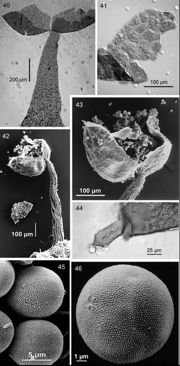
16 14807 (ex Yamamoto Y. 2912)] and doubt the rather small sporocarps belong to L. erecta var. erecta. The sporocarps measured 288 µm in height, and the diam. of the sporotheca was 115 µm. The box is labeled Licea erecta Thind? & Dhillon. It did not fit the description of L. erecta var. erecta (see below), which exactly matches our observations of the holotype of L. erecta var. erectoides, with the exception of the spore size and ornamentation. Our SEM and microscopic examination of L. floriformis var. aureospora (see below) shows similarities between these two species. The inner surface of the peridium is warted in both, they have almost colourless spores by TL of similar size range, and the ornamentation of the spores by SEM is also similar. They are different, however, when viewed by TL, as the inner layer of the peridium of Licea erecta var. erectoides appears as a flat base to the sporotheca and the outer layer of refuse appears as ribs outside along the length of the sporotheca, continuous with the ribbed stalk. The inner layer of the peridium of L. floriformis var. aureospora ends in a pointed calyculus, protruding down into the stalk and the outer refuse appears as a thin layer covering only the basal portion of the sporotheca. Macroscopically they are also different, with L. floriformis var. aureospora having a rounded sporotheca with a yellow spore mass showing through the top half, which fractures into small irregular platelets. The sporotheca of Licea erecta var. erectoides is tulip shaped, denser and all one colour, since none of the whitish spore mass shows through. The platelets are also slightly elongated. Nannenga-Bremekamp and Yamamoto (1983) and de Haan (2002) both commented on specimen YY 101 (NENB 13044), as being different from the type of L. erecta var. erectoides. The spores by SEM are very similar to those of L. floriformis var. aureospora, and it seems probable that it belongs to that taxon. 6. Licea floriformis var. aureospora M.T.M. Willemse & Nann.-Bremek., Proc. Kon. Ned. Akad. Wetensch. 97(1): TYPE: THE NETHERLANDS, in the rural estate of Hoekelum near Ede, on bark of living Sambucus nigra, on 16 XII 1986 (holotype: 104 L in the collection of M.T.M. Willemse 104a, isotype NENB now at BR!). (Figs ) = Licea longa Flatau, Stapfia 73: , syn. nov. TYPE: GERMANY, Kassel, Borke von Populus spec., (1), 150 m über NN, , LF 3180 (holotype: B!). Figs Licea floriformis var. aureospora Habit (NENB ). 35. Whole sporocarp by SEM. Dehiscing sporotheca and stalk with multiple strands (M. de Haan ). 36. Dehisced sporocarp by TL (M. de Haan ). 37. Detail of spores by TL (M. de Haan ). 38. Inner peridial surface with warts (M.de Haan ). 39. Spore by SEM (Lado 11715). 276

17 Fungal Diversity 277
18 = Licea capitata Ing & McHugh in Ing, Trans. Brit. Mycol. Soc. 78(3): , syn. nov. TYPE: UNITED KINGDOM, Scotia, Dumbarton, Balloch Park, reperta in vitro in cortice ablato ab arboris vivae Aceris pseudoplatani, 4 Oct. 1980, B. Ing (holotype: K!). Sporocarps scattered, stipitate, µm in height. Sporotheca light olive brown, subglobose, (-700) µm diam. Stalk olive brown (94. l. Ol Br) concolourous with the sporotheca, µm long, straight, subcylindrical, tapering towards the top, plicate, filled with algae by TL, continuous with the sporotheca, mid-width a quarter of the height. Peridium double, covered below with gelatinous layer with granular refuse material forming patches or longitudinal striations continuous with the outer layer of the stalk, the inner layer membranous, greyish yellow (90. gy. Y- 91. d. gy. Y) with no inclusions, its inner surface warted to almost spiny by TL and by SEM (Fig. 38); dehiscence irregular in upper sporotheca into platelets, which are spiny at edge, leaving a short calyculus. Spores free, yellow gold in mass, pale yellow green (121. p. Y G) to colourless by TL, smooth, densely warted by SEM, 10-12,5 µm diam.; spore wall of uniform thickness (Fig. 37). Material examined: THE NETHERLANDS, Ede: Hoekelum, on bark of living Sambucus nigra in moist chamber, 16-XII-1986, NENB (BR, isotype); BELGIUM, Zillebeke: Palingbeek, on logs of broad-leaved trees, 28-X-2001, MdH ; MEXICO, Veracruz: Catemaco, Los Tuxtlas Biological Reserve, N W, 200 m, on bark of living tree and bryophytes, 2-XII-1999, Lado GERMANY, Hessen, Kassel- Wolfsanger, bark of living Populus sp. in moist chamber, 6 to 26-X-1998, LF 3180 (B, holotype of Licea longa); 7 to 26-X-1998, Hessen, Kassel, Fuldatal, bark of living Populus sp. in moist chamber, 17-IX to 26-X-1998, LF 3169; 10-XII-1998, LF 3194 as Licea longa; Hessen, Kassel-Wolfsanger, bark of living Populus sp. in moist chamber, 21-XII-1999 to 3-II- 2000, LF 3277 as Licea longa. UNITED KINGDOM, Scotland, Dumbartonshire, Dumbarton, Balloch Park, on bark of Acer pseudoplatanus, 4-X-1980, B. Ing [K(M): holotype of Licea capitata]. Wales, Coed Aber, SH , on bark of Quercus petraea in moist chamber, 2-III-1993, dwb Habitat: bark of living trees. Distribution: Belgium, Germany, Ireland, Japan, Mexico, The Netherlands, United Kingdom. Illustrations: Willemse and Nannenga Bremekamp (1994: 138, 139, Figs. 1-2); Flatau (2000: 67, Fig. 3 as Licea longa); de Haan (2002: 30, Plate 1; 32, Figs. 3-4) The type specimens were very big with a habit that is more like a Trichia than a Licea (Figs ) but without capillitium. The sporocarps were up to 1200 µm in height with the largest sporotheca 700 µm in diam. The peridium was dull pale brown by reflected light with a mottled appearance at the top with the yellow spore mass showing through the thinner outer peridial layer in places. The inner layer adhered to the outer layer and was densely warted on the inner surface (Fig. 38). The dark, obviously plicate stalk was made of overlapping strands visible by SEM (Figs. 35, 42) and filled with algae. The stalk was continuous with the sporotheca. Dehiscence was by platelets which had few patchy areas with refuse inclusions from the outer layer showing 278
19 Fungal Diversity through, and were warted to almost spiny and irregularly shaped (Figs ). An irregular very shallow calyculus was left. The spores were yellow gold in mass, hyaline to pale yellow green (121. p. Y G) and smooth by TL but evenly and densely warted with some fused warts by SEM (Fig. 39). We also examined several collections of L. longa Flatau, kindly sent to us by the author, and the type material of this species (LF 3180). The habit, height ( µm) and diam. ( µm) of the specimens match those of the isotype of L. floriformis var. aureospora (Figs. 35, 42). The dehiscence was by irregular platelets which had plates with zip-like junctions and spines on the edge (Fig. 44) and similar patches of refuse to L. floriformis var. aureospora and which we think may be the remains of the outer peridium. The inner peridium surface has the same ornamentation of closely packed warts and the stalk was continuous with sporotheca SEM (Fig. 43). The spores were µm yellow gold and smooth by TL but evenly and densely warted with some fused warts by SEM (Figs ). In the description Flatau (2000) commented that these species were differentiated by the apparently single (L. longa) versus double peridium, and the opaque stalk of L. floriformis var. aureospora. We found the stalks and the peridium to be identical and we believe these to be the same species, on the basis of the detailed comparisons of the morphological features made of the type material of each and the SEM photographs. We therefore synonymize them here. Comparison by SEM of this species with material from Mexico published under Licea sp. 1 by Lado et al. (2003) has allowed us to include this specimen in L. floriformis var. aureospora. De Haan (2002), in her SEM photographs shows the spores of the collection YY 101 (NENB 13044), tentatively identified as L. erecta var. erectoides, to be very similar to those of L. floriformis var. aureospora, and we agree that the specimen should be included in this species. Spore ornamentation viewed by SEM has been shown by Gilert (1994) to be a stable and important taxonomic character. Ing (1999: 55) included under Licea operculata inoperculate forms previously described as L. capitata by Ing (1982), which he stated were the result of conditions of moisture during fructification. We requested the type specimen of L. capitata from Kew and compared it with the isotype of L. floriformis var. aureospora, and found that the size and ornamentation of the spores were the same, as were the characters of the stalk and the peridium. There were also comments on inoperculate sporocarps placed in L. operculata by Mitchell and McHugh (2000), but the photograph they publish with refuse material from the stalk continuing up the sides of the sporotheca to the middle, and the description of its irregular dehiscence at the apex, suggest that these specimens also belong to L. floriformis var. aureospora. 279
20 280
21 Fungal Diversity This variety was also recently reported from Asahi Town, Fukui Prefecture, Japan (Yamamoto et al., 2004), extending its geographical distribution, previously limited to Europe and America. The authors state that L. floriformis var. aureospora differs from L. floriformis var. floriformis in having a longer stalk and the colour of the spores in mass are yellow not black and colourless by TL not pale pink. As we were unable to study material of the latter variety we can not confirm this. 7. Licea lucens Nann-Bremek., Proc. Kon. Ned. Akad. Wetecsch., C 84(3): TYPE: FRANCE, Dept. Doubs, near Gigot, on moses and liverworts on bark taken from a living Acer, developed in a moist chamber: bark collected on 10 VI 1978, moistened 12 II 1979, the sporangia ripened from 15 III to 30 IV 1979 (holotype: NENB , now at BR!). (Figs ) Sporocarps scattered or gregarious, stipitate, up to 150 µm in height. Sporotheca red brown when moist, rosy when dry, elliptical, µm width, µm height. Hypothallus inconspicuous. Stalk very short up to 30 µm long, hyaline by transmitted light, straight, cylindrical, with little included granular material, mid-width approximately equal to the height. Peridium single, transparent, hyaline, smooth and glossy on both sides; dehiscence by irregular fracture of the top, leaving a deep cup. Spores free, red brown to rosy in mass, pale yellow to light yellow by TL (89. p. Y-86. l. Y), subglobose, 8-10 µm diam., warted by TL, very prominent warts by SEM, spore wall uniformly thin. Material examined: FRANCE, Dept. Doubs, Nr Gigot, on mosses and liverworts on bark of living Acer sp. in moist chamber, 15-III-1979 to 30-IV-1979, NENB (BR, holotype) Habitat: on mosses and liverworts on bark of living Acer. Distribution: Known only from the type locality. Illustrations: Nannenga-Bremekamp (1981: 286, Fig. 1). The type material shows many beautiful tiny sporocarps. They look like miniature glass beads full of rosy golden spores (Fig. 47). They appear to be sessile but close examination shows they are on a very short (30 µm) stalk which is just an extension of the peridium (Fig. 48). The peridium is completely hyaline and smooth outside and inside by transmitted light. By SEM however the inner surface is very sparsely and irregularly warted (Fig. Figs Licea floriformis var. aureospora (as Licea longa Flatau). 40. Dehiscing sporocarp and spores by TL (LF 3169). 41. Fragments of peridium by TL (LF 3180). 42. Whole sporocarp by SEM. Dehiscing sporotheca and stalk with multiple strands (LF 3169). 43. Detail of sporotheca by SEM (LF 3169). 44. Detail of inner peridial surface and edge of platelet by TL (LF 3180). 45. Group of spores by SEM (LF 3169). 46. Surface detail of spore by SEM (LF 3169). 281
. The spores show very clear dense ornamentation by TL (Fig.")
22 Figs Licea lucens (NENB ). 47. Habit. 48. Intact sporocarp with spores by TL. 49. Group of spores by TL. 50. Smooth inner surface of peridium by SEM. 51. Two spores showing verrucate ornamentation. 50). The spores show very clear dense ornamentation by TL (Fig. 49) which is prominent, and of the echinulate to verrucate type (Rammeloo, 1974, 1975) by SEM (Fig. 51). This combination of characters is completely unique among the stipitate Licea species we have examined. The size and initial habit of shiny sporocarps is similar to L. perexigua (see below), but the shape, spore colour and ornamentation, and the mode of dehiscence are all different. This species is only known from the type locality, and its minute dimensions have probably caused it to be overlooked, or confused with algae or other bark epiphytes. 8. Licea operculata (Wingate) G.W. Martin, Mycologia 34(6): Orcadella operculata Wingate, Proc. Acad. Nat. Sci. Philadelphia 41: TYPE: U.S.A., Pennsylvania: Philadelphia, Fairmount Park or 282
23 Fungal Diversity Chestnut Hill, on trunks of living Quercus rubra L. (holotype: PH, now at BPI!). (Figs ) Sporocarps scattered to loosely gregarious, stipitate, µm in total height. Sporotheca dull brown, usually urn-shaped, but rarely ovoid or almost globose, µm diam. Stalk µm in height, nearly black, slightly tapering towards the apex, furrowed, mid-width less than one fifth of height, filled with granular refuse material. Peridium single, thin, warted on the inner surface (Fig. 54) except for the lid, with ornamentation particularly visible at the edge of dehiscence (Fig. 55); dehiscence by a distinct shiny gold lid with a smooth inner surface by TL; the lid minutely punctate by SEM. Spores free, almost colourless to pale greenish yellow (104. p. g Y-121. p. Y G), smooth by TL (Fig. 54), 8-11µm diam., minutely punctate by SEM (Fig. 58); spore wall of uniform thickness. Material examined: USA, Philadelphia: Fairmount Park, [On bark living red-oak trees (Quercus rubra) Wingate 1889] (Orcadella Wingate 1275) ex state coll. (BPI , type). ECUADOR, Prov. Orellana: Amazonian basin, Yasuni National Park, Lago Agrio, S W ± 600 m, bark of deciduous tree in moist chamber, sc17779; S W bark of Cedrelinga catenaeformis in moist chamber 28-V-2000, dwb 1833; S W, bark of deciduous tree in moist chamber, sc Pichincha Prov.: Western Andean Slopes, Macquipucuna Reserve; Calacalí, N W, bark of dead liana in moist chamber, sc PERU, Mazan: 3 28'S 74 55'W, on dead liana in moist chamber, 22-VI-2002, dwb PUERTO RICO, Fajardo: El Verde, N W bark of living deciduous trees in moist chamber, sc 17091; sc Fajardo: on a ca. 1 km ESE San Vicente, N W, bark of a living deciduous tree in moist chamber, sc MEXICO, Veracruz: Catemaco, Los Tuxtlas Biological Reserve, N W, on bark of Porteroni viridescens in moist chamber, 31-V-2000, dwb Queretaro: Sta María del Mexicano, N W, on bark of Yucca sp. in moist chamber, 17-II-2004, dwb Habitat: Bark of living trees. Distribution: Ecuador, Mexico, Peru, Puerto Rico, Tanzania, USA. Illustrations: Wingate (1889: 280); Lister (1925: pl. 149d-f, as Orcadella operculata); Nannenga-Bremekamp (1965: 134, 1974: 72); Martin and Alexopoulos (1969: Fig. 9); Emoto (1977: pl. 7, Figs. 5-8); Neubert et al. (1993: pl. 6, Figs. 3-4); Gilert (1994: Figs ); Lado and Pando (1997: 124, Fig. 24); Yamamoto (1998: 152). The presence of variable morphotypes in this species was commented on by Wingate, in his original description (Wingate, 1889). He gave the range in height from µm. The sporocarps of the type material examined were > 800 µm in total height. The sporotheca were µm in diam., urnshaped, dull brown, and with a distinct shiny gold lid (Fig. 52) with a smooth inner surface by TL. The nearly black furrowed stipe had 3 or 4 intertwined strands inside it visible by TL. The type material had only a few (6) sporocarps left so we were not able to do SEMs of it. Our SEM pictures are of Schnittler s collection (sc17103) from Puerto Rico, and there are further SEM pictures in Gilert (1994). 283
. 56. Urn-shaped sporotheca by TL (dwb 1833). 57. Outer surface of sporotheca by SEM (sc 17103). 58. Spore by SEM (sc 17103).")
24 Figs Licea operculata. 52. Habit (BPI ). 53. Urn-shaped sporotheca by TL (sc 17103). 54. Inner peridial surface and spores by TL (sc 17103). 55. Detail of rim of sporotheca by SEM (sc 17103). 56. Urn-shaped sporotheca by TL (dwb 1833). 57. Outer surface of sporotheca by SEM (sc 17103). 58. Spore by SEM (sc 17103). Typical large urn-shaped sporothecae with a distinct lid like the type specimens are unmistakable (Figs. 53, 56). Many of the smaller particularly tropical forms from moist chamber cultures we have examined are not as easily determined. They are variable in size from µm in total height and the stalk is sometimes light brown and not furrowed. In addition in some collections the lid appears almost smooth on the inner surface, whereas in others some ornamentation (baculate processes see Gilert, 1994) is easily visible at 400x by TL. We have included in this taxon any specimens larger than 500 µm with a distinct lid, tapering stalk and smooth spores by TL within the size range and colour of this species. 284
25 Fungal Diversity This species is similar in habit and spore ornamentation to L. floriformis var. aureospora and L. erecta var. erectoides (see above), but it can be distinguished easily by its mode of dehiscence with a lid, and the edge of the sporotheca where the lid separates which has clear warts (Fig. 56). The spores of L. operculata are also smaller although there is overlap in the extremes of the sizes. The differences between this species and L. poculiformis Ukkola, the only other operculate stipitate Licea, are commented upon under that species below. After comparing some specimens from Mexico published as Licea sp. 1 by Lado et al. (2003), with a visible lid, similar spores and peridium, we also include them in L. operculata, although the sporocarps are smaller. Ing (1999: 55) included inoperculate forms previously described as L. capitata but we have found that the type of L. capitata belongs to L. floriformis var. aureospora (see comments under this taxon). Licea operculata was also reported from Tanzania (Ukkola, 1998) on Araucaria bark in moist chamber culture. The author mentions that some specimens had poorly developed lids. The brief description of Orcadella operculata var. sessile G. Lister (Monogr. Mycetozoa, ed. 3: ) suggests a completely different sessile species. 9. Licea parvicapitata Y. Yamam., Bull. Natl. Sci. Mus., Tokyo, B 26(3): TYPE: JAPAN, Chiba Pref., Sawara-shi, Sawara-ho, Atago-jinja, on bark of living Quercus acuta, in culture, 18 V 1982, MT-2664 (holotype: TNS!). (Figs ) Sporocarps scattered, solitary, stipitate, µm in height. Sporotheca golden, globose, 160 µm diam., with large warts on the outer surface held up by a distinct thin darker stalk. Stalk µm in height, black (Fig. 59), straight, subcylindrical, tapering slightly towards the apex, filled with refuse matter (Fig. 61), opaque, mid-width up to a quarter of its height. Peridium single membranous, almost hyaline to pale grey, warted on its inner surface (Figs ), covered with refuse material discontinuous with the stalk, and irregular darker warts scattered on outer surface (Fig. 62); dehiscence circumcissile by a line very close to the base of the sporotheca leaving a very small calyculus, the remainder of the peridium breaks into small irregular platelets. Spores free, pale greenish yellow (121. p. Y G-104. p. g Y) to colourless, subglobose, smooth (Fig. 65), 7-8 µm diam.; spore wall thin, with thickened area on one side. Material examined: JAPAN, Chiba Pref.: Sawara-shi, Sawara-ho, Atago-jinja, on bark of living Quercus acuta, 18-V-1982, MT 2664 (TNS, holotype) Habitat: bark of Quercus acuta. Distribution: Known only from the type locality. 285

26 Figs Licea parvicapitata (MT 2664). 59. Habit. 60. Whole sporocarp showing line of dehiscence and spotted surface. 61. dehisced sporocarp by TL. 62. Outer surface of peridium with refuse and darker irregular spots Inner surface of hyaline peridium with warts. 65. Spores by TL. Illustrations: Yamamoto (2000: 113) This species is only known from type material which showed stipitate sporocarps, most with a spotty appearance due to the peridial warts, although these were not evident on all sporocarps. The stalk was very thin and fragile and fractured on mounting. The combination of the dark thin stalk, the spotted golden brown sporotheca and the small spores make this species easily distinguished from the others. There were crystals among the refuse material of some specimens when viewed with Nomarski, but the longer thinner stalk, larger sporocarp size ( µm vs in L. eleanorae), dehiscence near the base not the apex, and lighter spores (121. p. Y G-104. p. g Y vs. 89. p. Y) of L. parvicapitata differentiate it from L. eleanorae. 286
27 Fungal Diversity 10. Licea pedicellata (H.C. Gilbert) H.C. Gilbert in Martin, Mycologia 34(6): Hymenobolina pedicellata H.C. Gilbert, Stud. Nat. Hist. Iowa Univ. 16(2): TYPE: U.S.A., Iowa, Milford, on bark of living Ulmus, 16 July, 1932, H.C. Gilbert 2117 (holotype: BPI!). (Figs ) Sporocarps scattered to gregarious, stipitate, µm in height. Sporotheca dark brown or black, subglobose, rounded even when dry, µm diam. Stalk µm in height, thick, straight, cylindrical, furrowed, continuous with outer peridium, filled with granular refuse material, mid-width less than half the height. Peridium double, but apparently single, with an adhering outer layer of refuse material, membranous, dull brown; dehiscence into irregular platelets µm wide, inner layer warted (Fig. 68). Spores free, dark brown in mass, medium yellow to greyish yellow (87. m. Y-90. gy. Y) by TL, subglobose, (10-)11-13(-14) µm diam., minutely warted by TL (Fig. 70) and densely warted by SEM (Fig. 72); spore wall with a paler thinner area. Material examined: USA, Iowa, Milford [coll W. Okoboji acc. to Gilberts letter Dec. 7, 1934 on box] on bark of elm (in moist chamber), 16-VII-1932, H.C. Gilbert 2117 (BPI , holotype) as Hymenobolina pedicellata; (BPI , isotype) as Licea pedicellata. Iowa: Milford [on bark of living Ulmus, see Gilbert (1934: 154)], 16-VII-1932, coll. W. Okaboji [HC Gilbert 2117] (BPI , isotype) as Licea pedicellata. Iowa city on dead Ulmus sp., 00-XII- 1961, coll. G.W. Martin (BPI ). Virginia, Mt. Lake Biol. Station, on bark of Quercus sp. in moist chamber, 12-VIII-1969, leg. & det. C.J. Alexopoulos (BPI ). Texas: Austin, Hancock Center, on bark of living Ulmus crassifolia in moist chamber, 5-VIII-1974, leg. & det. C.J. Alexopoulos (BPI ). Habitat: Bark of living trees. Distribution: Zaire?, USA. Illustrations: Gilbert (1934: 154, Fig. 1); Martin and Alexopoulos (1969: 481, pl. 1 Fig. 11a-c); Gilert (1994: 17, Fig. 47); Yamamoto (1998: 142). In the original description of the species, Gilbert (1934) states that the spores are smooth or faintly and finely warted. Examination of all the BPI collections of type material, both by TL and SEM, showed all the spores to be warted, and none were smooth as Gilbert suggested. This is the main distinguishing character of this species from other similar species like L. rugosa (Table 2). The faint warts are discernible by TL at 400x (Fig. 70), and SEM examination revealed dispersed warts (Fig. 72), which were irregular in size. At great magnification (Fig. 74) they were seen to have a warted surface themselves. Another clear characteristic visible by TL is that the interior of the peridium is warted, but the warts are dispersed and separate (Fig. 73). In addition almost the entire peridium breaks into platelets leaving a very shallow structure attached to the stalk (Figs. 69, 71). We were not able to see clear ridges or lines of dehiscence in any of the specimens, and all the specimens retained their rounded shape even when dry (Fig. 67). Martin and Alexopoulos (1969: 48) also commented that they rarely observed platelets in the 287
. 70. Spore by TL (BPI 826385). 71. Dehisced sporocarp with flat remains of peridium by SEM (BPI 657294). 72.")
28 Figs Licea pedicellata. 66. Habit (BPI ). 67. Whole sporocarp by SEM (BPI ). 68. Irregular platelets with warted inner surface (BPI ). 69. Dehisced sporocarp by TL with shallow flat remains of peridium (BPI ). 70. Spore by TL (BPI ). 71. Dehisced sporocarp with flat remains of peridium by SEM (BPI ). 72. Spore by SEM (BPI ). 73. Group of spores and detail of inner peridial surface by SEM (BPI ). 74. Highly magnified ( 20,000) detail of spore ornamentation by SEM (BPI ). 288
29 Fungal Diversity Table 2. Characters differentiating L. pedicellata, L. rugosa var. rugosa and L. rugosa var. fujiokana. Name Dehiscence Peridium Spore Other L. pedicellata Irregular fragments; flat base of Dispersed warts on inner surface Yellowish brown; warted; (10-)11-13(-14) µm diam. Sporotheca round when dry L. rugosa var. rugosa L. rugosa var. fujiokana sporotheca left Irregular fragments Irregular fragments Prominent dense warts on inner surface Prominent dense warts on inner surface Olive-brown; smooth; µm diam. Greyish yellow brown; smooth; µm diam. Sporotheca wrinkled when dry Sporotheca wrinkled when dry peridium. We did a SEM study of material cited by Pando and Oltra (2001) from Mogente, Spain [MA-Fungi (Oltra 2519)] but found it to have smooth spores and a different ornamentation of the inner peridium and we conclude that it does not belong to this taxon. Of the other material examined in our herbarium that had been assigned to this species (DWM 3313 from Mexico on red cedar bark in moist chamber, and TEB 3704 from Virginia- Baskerville cemetery, USA, cedar tree #3, 28-VIII-1970), none had warted spores except another sample (DWM 4478) from Zaire, but the single slide of this was insufficient for us to positively confirm its identity. The SEM picture in Gilert (1994) shows a rugose sporocarp with apparent platelets, which casts doubt on its identity as L. pedicellata. It is possible that this species has been confused with L. rugosa, and that its distribution is not as widespread as Martin and Alexopoulos (1969: 48) and other authors (Yamamoto, 1998; Ing, 1999) believed, since they mention also Australia, Austria, Great Britain, Greece, India, Japan, Mexico, Tunisia and Turkey. The similarities between L. pedicellata, L. rugosa and the varieties of each are discussed below. It is worth noting that the three boxes of dried type material lent to us by the BPI herbarium have slight differences in the labels. One of them (BPI ) lists the locality as Milford Iowa, as does Gilbert s description (1934), with a note coll. W. Okoboji acc. to Gilberts letter Dec. 7, 1934, another (BPI ) lists Okoboji, Iowa, both in script. Collection BPI lists Milford and coll. W. Okaboji (sic). We note for clarification that West Okoboji is a town slightly West of Milford, both in Dickinson County Iowa. It seems that coll. here refers to the place of collection, not who it was collected by, the accepted meaning of this abbreviation (Stearn, 1996). 289
. 78. Inner surface of peridium densely papillate by TL (HWK 1166). 79-80.")
30 Figs Licea perexigua. 75. Habit (dwb 1936). 76. Inner surface of peridium densely papillate by TL (HWK 1166). 77. Inner surface of dehisced sporotheca with spores by SEM (dwb 1936). 78. Inner surface of peridium densely papillate by TL (HWK 1166) Detail of spore surface ornamentation by SEM (dwb1936). 290
31 Fungal Diversity 11. Licea perexigua T.E. Brooks & H.W. Keller in Keller and Brooks, Mycologia 69(4): TYPE: U.S.A., Arkansas, Crawford County, on the bark of living Juniperus virginiana, October 27, 1964, T.E. Brooks 2747 (holotype: BPI!). (Figs ) Sporocarps scattered to gregarious, stipitate or sessile on a narrowed base, up to 100 µm in height. Sporotheca golden, iridescent, subglobose, µm diam. Hypothallus inconspicuous. The stalk when present pale yellow by TL, less than half the total height of sporocarp, filled with granular refuse material. Peridium single, membranous, colourless by TL, with scant granular refuse matter evenly distributed over the surface, inner surface closely papillate; dehiscence circumcissile low down at the base of the sporotheca leaving a calyculus. Spores free, pale to greyish green yellow (121. p. Y G gy. Y G), from µm diam., smooth by TL, minutely ornamented with unevenly scattered warts by SEM (Fig. 79); spore wall thin, with a paler thinner area. Material examined: USA, Arkansas: Crawford Co., [on bark of living Juniperus virginiana in moist chamber (Keller and Brooks, 1977)], 27-X-1964, coll. TEB 2747 (BPI , holotype). Mississippi: Mississippi co., moist chamber, coll. HWK 1166 (slide). MEXICO, Tlaxcala: Cuapiaxtla, N W, on bark of living Juniperus deppeana in moist chamber, 18-I-2001, dwb Hidalgo: Progreso, La Cruz, N W, on bark of living tree Opuntia sp. in moist chamber, 26-III-2001, dwb SPAIN, Cuenca: Saceda-Trassierra, on bark of Quercus ilex in moist chamber, Pando 1185, 21-III-1990, MA-Fungi Habitat: bark of living trees. Distribution: Mexico, Spain, USA. Illustrations: Keller and Brooks (1977: 675, Figs ); Lado and Pando (1997: 127, Fig. 35); Novozhilov et al. (2003) (as L. cf. perexigua). The type material showed very tiny, almost sessile, golden irridescent sporocarps. The specific epithet gives the most obvious character of exceedingly small and with a stalk so short that even the stipitate sporocarps appear sessile. Many calyculi of dehisced sporocarps were visible on the bark surface in the type specimen, the stalk remaining with a tiny cup-shaped sporothecal base on top. The single peridium in some specimens is completely hyaline, and refuse material is very sparce even when present. The ornamentation on the inner surface is visible at 400x (Figs. 76, 78), appearing to be in a reticulate pattern as described by the authors (Keller and Brooks, 1977), but by SEM (Fig. 77) it can be seen that this pattern is not a true reticulum but is the result of the uneven distribution of the papillae and there are some smooth areas without ornamentation. By TL the spores seem smooth and have thin walls with a paler thinner area at which they have a tendency to fold (Fig. 78), but by SEM irregularly distributed rhomboid warts are visible (Figs ) with some so close together as to appear fussed, and others with spaces between them. 291
. 85. Spores by TL (dwb 1759). 86. Inner surface of peridium by TL (dwb 1759).")
32 Figs Licea poculiformis. 81. Habit (Ukkola 319A). 82. Whole sporocarp showing goblet-shaped sporotheca and pale lid (Ukkola 319A). 83. Dehisced sporocarp (dwb 1759). 84. Deep cup of dehisced sporotheca by TL (dwb 1759). 85. Spores by TL (dwb 1759). 86. Inner surface of peridium by TL (dwb 1759). The unique pattern of ornamentation on the spores distinguishes this species from the other stipitate species. In addition it is smaller than all of them except L. lucens (see above). The transparent peridium is another clear character. Keller and Brooks (1977) differentiate L. perexigua from L. tenera by the broad base of the sessile sporocarps in the latter species, its different 292
33 Fungal Diversity peridial colour and ornamentation, and its larger (12 µm vs µm diam.) spores. The authors also differentiate it from L. scyphoides by its almost sessile habit, its smaller [ (-14) µm diam. in the latter species] slightly darker spores (121. p. Y G-122. gy. Y G vs. 90. gy. Y-104. p. g Y), and its manner of dehiscence (irregular vs. circumcissile in L. scyphoides). Our observations concur with theirs. 12. Licea poculiformis Ukkola, Acta Bot. Fennica 160: TYPE: TANZANIA, Tanga Province, Lushoto District, East Usambara Mts., on mosses growing on bark of Cupressus sp., in moist chamber culture, 12.XII.1995, Ukkola 319A (holotype: H!). (Figs ) Sporocarps scattered, stipitate, (-400) µm in height. Sporotheca treacle-brown, goblet-shaped, µm diam., with a shiny pale lid (Fig. 81). Stalk µm in height, furrowed, mid-width half the stalk height. Peridium double, outer layer coriaceous, shiny, inner layer membranous; dehiscence by a pale shiny lid with a clear edge and warts on the margin, leaving a deep cup (Fig. 84). Spores free, pale greenish to greyish yellow (90. gy. Y p. g Y) by TL, µm diam., smooth by TL (Figs ) but warted by SEM (Ukkola, 1998: 6, Fig. 7); spore wall with a slightly thinner paler area. Material examined: TANZANIA, Tanga Province, Lushoto District, East Usambra Mountains, Amani Forest Reserve, submontane forest, alt. 900 m, on mosses growing (sec. Ukkola, 1998) on bark of living Cupressus sp. in moist chamber culture, 13-IX-1995 (on the label), Ukkola 319A (H, holotype). MEXICO, Quintana Roo, El Eden, bark of living Hematoxylon campechianum in moist chamber, 10-II-2000, dwb 1759 [Lado et al., 2003: 88]. Habitat: moss and bark of living trees. Distribution: Mexico, Tanzania. Illustrations: Ukkola (1998: 6, Figs. 2-7). The sporotheca and stalk of this small species are concolourous, treaclebrown. The abundant sporocarps in the type specimen were mostly projecting from the tips of moss leaflets. The Mexican material is much larger (up to 400 µm see Fig. 83) but otherwise agrees with the type. This species has been reported recently from Okayama Prefecture, Japan on the bark of living Prunus sp. (Yamamoto and Fujioka, 2004) and the Japanese specimens described are also larger than the type. This species is closest to L. operculata, and shows similarity in the habit, and also the inner surface of the peridium by SEM, but the type specimens are different in their deep brown colour, have a large difference in size ( µm in height vs µm in L. operculata), and a double peridium vs. single in L. operculata. The lid of L. poculiformis is at the broadest diam. of the sporotheca, and wider than that of L. operculata, has a clear edge and is distinctly papillose by TL, whereas the lid of the L. operculata appears smooth by TL and is at the narrowing top portion of its 293
34 more globose sporotheca. Finally, the ratio of stalk width to height is different with L. poculiformis much shorter and squatter and with a straight wide stalk (Fig. 82) and with the outer peridium continuous with the sporotheca, not long and tapered and separate from the sporotheca like L. operculata. 13. Licea rugosa Nann.-Bremek. & Y. Yamam., Proc. Kon. Ned. Akad. Wetensch., C 90(3): var. rugosa. TYPE: JAPAN, Kochi Pref., Agawa-mura, Mnt. Nakatsu, on the bark of a living Aesculus turbinate Blume, VII 1984, Y.Y (holotype: TNS, isotype NENB now at BR!). (Figs ) Sporocarps scattered, stipitate, up to µm in height. Sporotheca dark brown to almost black, subglobose up to 150 µm diam. Stalk up to 180 µm in height, straight, cylindrical, furrowed (Fig. 88), stout, continuous with the outer peridium, mid-width half of the stalk height. Peridium single, membranous, wrinkled with darkened ridges visible when dry, prominently warted on the inner surface; dehiscence irregular fracture into fragments in the upper part leaving a calyculus (Figs. 88, 92) Spores free, dark brown in mass, olive brown (94. l. Ol Br) by TL, subglobose, µm diam., totally smooth; spore wall with a thinner area (Fig. 89). Material examined: JAPAN, Kochi Pref.: Agawa-mura, Mnt. Nakatsu, bark of living Aesculus turbinate, VII 1984, coll. NENB [ex YY 2300] (BR, isotype). MEXICO, Hidalgo: Progreso, La Cruz, N W, on bark of living Prosopis laevigata in moist chamber, 22-III-2001, dwb 2005; on bark of living Schinus molle in moist chamber, 16- III-2001, dwb Puebla: Emilio Portes Gil, N W, on bark of living Yucca filifera in moist chamber, 25-I-2001, dwb Zapotitlán de las Salinas, N W, on bark of living Beaucarnea gracilis in moist chamber, 11-IX-2003, dwb 2311; ibidem, 13-IX-2003, dwb Tehuacán, Santiago Nopala, N W, on bark of living Yucca periculosa in moist chamber, 11-XI-2003, dwb 2332; ibidem, bark of living Beaucarnea gracilis in moist chamber, 22-XI-2003, dwb Queretaro: Peña Miller, Camargo, N W, on bark of living Prosopis laevigata in moist chamber, 15-II-2002, dwb Habitat: bark of living trees. Distribution: Japan, Mexico. Illustrations: Nannenga-Bremekamp and Yamamoto (1987: 326); Yamamoto (1998: 144). Figs Licea rugosa var. rugosa. 87. Habit (NENB ). 88. Dehisced sporocarp by SEM showing calyculus and furrowed stalk (NENB ). 89. Dark spores with wall thinner in one area by TL (dwb 2006). 90. Inner surface of peridium with prominent dense warts (NENB ). 91. Smooth spore by SEM (NENB ). 92. Calyculus of dehisced sporocarp containing spores (dwb 2006). 93. Group of smooth spores by SEM (dwb 2319). 294

35 Fungal Diversity 295
36 The abundant Mexican material was identical to the type both by microscopic examination and by SEM. The most obvious differentiating features of this species are the size and rather dark colour of the spores. The sporocarps examined were of varied height from almost sessile to 500 µm. The sporotheca are globose when wet but very wrinled when dry, with prominent ridges. The spores by TL look like the spores of L. parasitica (Zukal) Martin [11-13(-16) µm diam.], but the thinner area is less pronounced, and the spores were completely smooth even by SEM (Figs. 91, 93). The wrinkled sporotheca breaking into platelets instead of the lid of L. parasitica easily avoids any confusion even with the sessile specimens. The plasmodium, visible in moist chamber cultures, is thick dull brown and appears as a continuous sludge on the bark surface like many large protoplasmodia combined. In the original description of L. rugosa Nannenga Bremekamp & Yamamoto (1987) differentiated it from L. pedicellata on the basis of the spore colour being dark brown and the smooth spores. The spores are also slightly bigger in L. rugosa and the smooth surface vs. the warted surface of L. pedicellata spores (Figs ) is confirmed by SEM. The peridial ornamentation is also different in density and distribution (Figs. 64, 90) as revealed by SEM examination, and some of the warts are fused in L. rugosa (Fig. 90). However, L. rugosa and its var. fujiokana (see below) are very like L. pedicellata but with the definable differences listed (Table 2). DNA sequencing techniques may demonstrate that they are in fact a continuum of the same species, but at present pending further studies, we prefer to maintain them as separate taxa. 14. Licea rugosa var. fujiokana (Y. Yamam.) D. Wrigley & Lado, comb. nov. (Figs ) Licea capitatoides var. fujiokana Y. Yamam., Bull. Natl. Sci. Mus., Tokyo, B 26(3): TYPE: JAPAN, Tokyo Pref., Chiyoda-ku, on the premises of Imperial Palace, on bark of living Acer buergerianum, 3 VI 1999, Y. Yamamoto, 99TK-29 (holotype: TNS!; isotype 99TK-26 in TNS). Sporocarps scattered, stipitate, µm in height. Sporothecae dark brown, globose or subglobose, µm diam. Stalk µm in height, slightly tapering towards the top, filled with refuse material, mid-width half the height. Peridium double, wrinkled, outer layer gelatinous with granular refuse material, inner layer membranous, yellow-grey (93. y Gray) with a densely warted surface; dehiscence by irregular fracture into fragments in upper peridium leaving a calyculus. Spores free, greyish yellow brown (80. gy. Y Br), subglobose, (9-)10-11 µm diam., smooth by TL and SEM; spore wall with an obvious paler area about half the diam. of the spore (Figs ). 296

37 Fungal Diversity Figs Licea rugosa var. fujiokana (99TK29 as Licea capitatoides var. fujiokana Y. Yamam.). 94. Habit. 95. Dehiscing sporocarp by TL. 96. Spores by TL with paler area. 97. Spore by SEM. 98. Inner surface of peridium by TL. 99. Group of spores and inner surface of peridium by SEM. Material examined: JAPAN, Tokyo Pref.: Chiyoda-ku, bark of living Acer buergerianum in moist chamber, 3-VI-1999, leg. Y. Yamamoto, 99TK-29 (TNS, holotype). MEXICO, Tlaxcala: Calpulalpan, El Peñón, on Abies religiosa in moist chamber, 19-II-1998, VGA 671. Hidalgo: El Cardonal, N W, on bark of Prosopis juliflora in moist chamber, 9-X1-2000, dwb Metzquititlan, N W, on bark of Acacia sp. in moist chamber, 5-X-2001, dwb 2076, idem, 23-XI-2001, dwb Puebla: Emilio Portes Gil, N W, on bark of living Nolina parviflora in moist chamber, 18-I-2001, dwb 1932; on bark of living Yucca filifera in moist chamber, 25-I-2001, dwb 1955; 3-II-2001, dwb Zapotitlán de las Salinas, N W, on bark of living Beaucarnea gracilis in moist chamber, 1-X-2001, dwb S. Martin Esperilla, N W, on bark of living Yucca periculosa in moist chamber, 22-X1-2003, dwb 2348; idem, 29-X1-2003, dwb Veracruz: Totalco, N W, on bark of living Juniperus deppeana in moist chamber, 18-I-2001, dwb San Luis Potosi: 297
38 Xilitla, Reten, N W, on bark of living Liquidamber styraciflua in moist chamber, 26-X1-2001, dwb Oaxaca: Tepelmeme, La Unión, N W, on bark of living Prosopis laevigata in moist chamber, 15-XI-2003, dwb 2316; Tepelmeme, Mex-135 highway, Km. 109, N W, on bark of living Yucca periculosa in moist chamber, 29-X1-2003, dwb Habitat: bark of living trees. Distribution: Japan, Mexico. Illustrations: Yamamoto (2000: 112, Fig. 6). When this species was originally described by Yamamoto as L. capitatoides var. fujiokana he stated that it differed from L. capitatoides by the colour and size of the spores (greyish yellow and µm diam. in the former, and pale yellow, 8-10 µm diam. in the latter). However comparison of the types showed additional differences. Licea rugosa var. fujiokana has a roughened sporotheca when dry, a lighter stalk by TL and darker spores (Fig. 95). SEM examination showed the spores to be totally smooth (Fig. 97) like L. rugosa var. rugosa, although smaller. The peridium fractured into fragments of approximately 30 µm in diam. (Fig. 98), and the inner peridium by SEM (Fig. 99) was covered with warts with some fused warts. This ornamentation is the same as that of the inner peridium of L. rugosa var. rugosa, and different in both density and distribution from L. capitatoides. There are also patches of refuse material on the outer surface of the peridium, which suggest the remains of a gelatinous layer. The Mexican material was examined by SEM, and is identical to the type. The type of var. fujiokana is therefore exactly the same as the type of var. rugosa in all other characters except its double peridium, spore size (10-12 µm diam. in var. fujiokana and µm diam. in var. rugosa) and the yellowish brown spore colour of var. fujiokana (80. gy. y Br) instead of olive brown in var. rugosa (94. l. Ol Br). For this reason the new combination as a variety of L. rugosa is justifiable. The two taxa can be separated under the light microscope on the basis of the darker, larger spores of L. rugosa var. rugosa (Table 2). 15. Licea scyphoides T.E. Brooks & H.W. Keller in Keller & Brooks, Mycologia 69(4): TYPE: U.S.A., Ohio, Greene County, next to the swimming pool at John Bryan State Park, from the bark of living Juniperus virginiana, August 18, 1976, H.W. Keller 1945 (holotype: BPI!).(Figs ) = Licea tanzanica Ukkola, Härk. & Gilert in Ukkola, Härkönen & Saarimäki, Karstenia 36(2): , syn. nov. TYPE: TANZANIA, Tanga Prov., Lushoto Distr., W Usambara Mts., on Azadirachta indica in moist chamber cultura, 12.XII.1988, Härkönen 3693 (holotype: H!). Sporocarps scattered to gregarious, stipitate, (-400) µm in height. Sporotheca globose, golden brown, shiny, (-220) µm diam. Stalk

39 Fungal Diversity Figs Licea scyphoides (HWK 1945) Habit Base of sporotheca from above containing spores by SEM Inner surface of peridium by SEM Upper half of sporotheca showing clear area of dehiscence by TL Spore with closely punctate ornamentation by SEM Group of spores appearing roughened. 299

40 Figs Licea scyphoides (MH 3693 as Licea tanzanica) Habit Dehisced sporocarp leaving calyculus Inner surface of peridium Spore by SEM with closely punctate ornamentation. 180 µm in height, dark brown by reflected light, 150 µm in height, thick, erect, furrowed, straight, cylindrical, mid-width half of the height. Peridium single, membranous, with refuse matter on outside, inner surface of the peridium clearly punctate (Fig. 102); dehiscence circumcissile at a transparent equatorial band, visible by TL, leaving a basal calyculus (Fig. 101). Spores free, yellowish (90. gy. Y-104. p. g Y) by TL, globose, (-14) µm diam., roughened to smooth by TL and low magnification SEM (Figs. 101, 105), and closely punctate by at higher magnification SEM (Fig. 104); spore wall with a paler thinner area. Material examined: USA, Ohio: Greene county, John Bryan State Park near swimming pool, on bark surface of red cedar, 18-XIII-1976, coll. Keller 1945 (BPI , holotype). CUBA, Sancti Spiritus: Alturas de Banao, N W, on dead liana in moist 300
41 Fungal Diversity chamber, 6-II-2003, dwb MEXICO, Hidalgo: El Cardonal, N W, bark of living Prosopis juliflora in moist chamber, 9-X1-2000, dwb Progreso, La Cruz, N W, bark of living Schinus molle in moist chamber, 13-III-2001, dwb 1989; ibidem, bark of living Prosopis laevigata in moist chamber, 13-III-2001, dwb 1992, dwb 1993; ibidem, bark of living Opuntia sp., 16-III-2001, dwb PERU. Mazan: 3 28 S W, dead liana in moist chamber, 27-V-2002, dwb 2193; ibidem, 4-VI-2002, dwb 2198; ibidem, 3-VI-2002, dwb PUERTO RICO, Fajardo, Dry Coastal Forest on rocks, Las Cabezas de San Juan Nature Reserve, near lands end, N W, bark of living Tamarindus indica in moist chamber, sc SPAIN. Cuenca: Saceda-Trasierra, bark of living Quercus ilex, in moist chamber, 21-III-1990, Pando, MA-Fungi UNITED KINGDOM, Wales: Aber, bark of living Quercus petraea, in moist chamber, 31-I- 1993, dwb 1130a. TANZANIA, Tanga Province: Lusotho District, West Usambra Mts., Mombo, town centre, yard of the restaurant, 0438CD, on Azadirachta indica in moist chamber, 12-XII-1988, MH 3693 (H holotype); idem, MH 3591, MH Arusha (Northern) Province: Moshi District. Moshi International school park, 0337AD, on Jacaranda in moist chamber, 23- V-1988, MH 3576 as Licea tanzanica. Habitat: Bark of living trees and living and dead vines. Distribution: Mexico, Peru, Spain, Tanzania, USA, Wales. Illustrations: Keller and Brooks (1977: 680, Figs ); Ukkola et al. (1996: 56, 58); Lado and Pando (1997: 132, Fig. 38); Flatau (2000: 70, Fig. 5); Yamamoto (1998: 145); de Haan (2001: 16, pl. 1). The sporocarps examined were from (-450) µm in total height and (-200) µm diam., with globose, golden brown shiny sporotheca. The peridium contained refuse matter which thins towards the middle of the sporotheca leaving a transparent equatorial band, very obvious on both sides of the line of dehiscence when specimens are mounted and viewed by TL (Fig. 103). The warts of the inner suface of the peridium are visible in the transparent area of dehiscence by TL at 400x. In this species as with several other stipitate Licea, some sporocarps in dry material appear sessile if they are fallen against the substrate and the stalk blends with the substrate refuse material. We examined the type material and several other specimens of L. tanzanica, which we found to be very similar to this species. The SEM examination showed the spore ornamentation (Figs. 104, 109) and the ornamentation of the inner peridium (Figs. 102, 108) of the two species to be identical. There are no differences in the spore sizes (L. scyphoides µm diam. and L. tanzanica µm diam.). The types show L. tanzanica somewhat larger, reaching 400 µm in height and 200 µm in diam., but the sizes overlap. The ratio of stalk width to height is also different with L. scyphoides shorter and squatter. In L. scyphoides a cup is left after dehiscence but L. tanzanica dehisces lower down and leaves only the base of the sporotheca. The spore colour is also subtly different. Licea tanzanica spores are olivaceous and L. scyphoides are pale yellow without the darker tints. The transparent border visible at the area of circumcissile dehiscence is a constant feature of both and 301
42 measures up to 70 µm. The authors of each species recognized that they are very close, but believed them to be different (Ukkola et al., 1996). We feel the combination of small differences may represent geographic differences, but are not sufficient to maintain these as separate species, and we synonymize them here. Perhaps these small differences indicate a continuum of a variable species. Licea bulbosa (see above) is similar to L. scyphoides in habit, but its smaller (10-11 µm diam.) almost hyaline smooth spores, its longer thinner stalk, and the smooth inner peridium with a totally transparent basal half differentiate it. 16. Licea verrucospora (T.N. Lakh., Nann.-Bremek. & R.K. Chopra) D. Wrigley & Lado, nom. nov. et status nov. (Figs ) Licea scyphoides var. reticulata T.N. Lakh., Nann.-Bremek. & R.K. Chopra, Proc. Kon. Ned. Akad. Wetensch. 93(3): [basion.], non. L. reticulata Berk. & Broome, TYPE: INDIA, Himalchal Pradesh, North Western Himalaya mountains, Shimla, Summer Hill area, 2067 m, developed in a moist chamber on bark of Quercus incana from 0-9 m above the ground, 6 IV 1981 (holotype HPUB 15405, paratype NENB now at BR!). Sporocarps scattered to gregarious, stipitate, (-700) µm in height. Sporothecae dark ochraceous-brown, shiny, subglobose, µm diam. Stalk (-500) µm in height, long, dark, straight, subcylindrical, rugose with several strands visible by TL within the main stalk (Fig. 110), mid-width a quarter of stalk height. Peridium single, membranous, covered with refuse matter, smoky grey, densely and prominently warted; dehiscence irregular into platelets formed by fragmentation of the whole peridium. Spores free, pale yellow to greyish yellow (89. p. Y gy. Y) by TL, globose, 10-12(-13) µm diam., very minutely warted but warts in irregular patches on the spore surface, visible by TL and also by SEM (Fig. 115); spore wall with a pale thinner area (Fig. 111). Material examined: INDIA, Himalchal Pradesh: Shimla, Summer Hill, on bark of living Quercus incana in moist chamber, 14-IV-1981, NENB (BR, paratype of Licea scyphoides var. reticulata) Habitat: bark of living Quercus incana. Distribution: Known only from the type locality. Illustrations: Lakhanpal et al. (1990: 261, Fig. 7). We raise its rank here to species level because on comparison of the material with L. scyphoides we noticed many differences, sufficient to consider it to be a totally different species. The original description of L. scyphoides var. reticulata notes that dehiscence takes place above an equatorial line to leave a cup with a smooth margin. We found no such line, and in fact the platelet margins are visible extending to the base of the sporotheca (Figs. 110, 113). The base, when remaining after dehiscence, is a thin vestige of peridium more 302
. 113. Fragmented peridium by SEM. 114. Stalk and fragmented remains of sporotheca. 115.")
43 Fungal Diversity Figs Licea verrucospora (NENB as L. scyphoides var. reticulata) Habit Dehisced sporocarp by TL Detail of inner surface of peridium by SEM with flattened reticulate markings (arrow) Fragmented peridium by SEM Stalk and fragmented remains of sporotheca Spore showing irregular verrucose ornamentation. 303
44 like a collar than a cup (Fig. 114), whereas in L. scyphoides a clear cup is left after dehiscence. Licea scyphoides has a transparent equatorial band at the area of dehiscence which is not present in L. verrucospora. The inner surface of the peridium in L. scyphoides is minutely warted (Fig. 102), but the inner surface of L. verrucospora is densely marked with protruding warts of various sizes, and the lines of the outer reticulate pattern show through (Fig. 112). The spores in L. verrucospora have a unique ornamentation of rounded raised warts of different sizes, dispersed irregularly over the spore surface, different from L. scyphoides even by TL, and more so by SEM (Fig. 115). This pattern is also different from any other stipitate Licea described so far. These differences are in addition to those noted by the authors of this species which were the larger size of the sporocarps but they overlap, and its reticulately-ridged peridium. The surface of the sporothecae of the type material is crossed with a network of shiny lines on ridges, marking the surface, but not the platelets by which it dehisces (Fig. 112). The epithet verrucospora was chosen to denote the character of the spore surface, since the variety name has already been used for a different species (Lado, 2001). Licea verrucospora is different from L. tanzanica in spore size (10-12 µm diam. vs µm diam.), ornamentation, and in its mode of dehiscence (platelets vs. circumcissile). Its yellowish, irregularly ornamented spores, also separate it from L. pedicellata with regular ornamentation and L. rugosa which has completely smooth spores. Excluded species: (6) Licea atricapilla Nann.-Bremek. & Y. Yamam., Proc. Kon. Ned. Akad. Wetensch., C 86(2): TYPE: JAPAN, Kochi Pref., Susaki-shi, Hachimangu, cult. on bark of living Cinnamomum camphora (L.) Presl, 7-31 X 1981 (holotype: Y.Y. 1277, isotype NENB ). We were unable to examine this species. The following description is adapted from Nannenga-Bremekamp and Yamamoto (1983). Sporocarps scattered, stipitate, 260 µm in total height. Sporotheca silvery irridescent with a dark apical operculum, subglobose, 160 µm diam. Stalk nearly black, furrowed, filled with granular refuse material. Peridium membranous, separate from the stalk, densely and minutely warted on the inner surface. Dehiscence circumscissile along edge of the apical disc. Spores, olivaceous brown with a large paler area and a thinner wall, µm diam., smooth by TL. Habitat: bark of living Cinnamomum camphora. Distribution: only known from type locality. 304
45 Fungal Diversity Illustrations: Nannenga-Bremekamp and Yamamoto (1983: 208); Yamamoto (1998: 136). The authors place this species in the subgenus Orcadella on account of its black operculum. They differentiate it from L. operculata which has a glossy operculum, and by its larger darker spores (olivaceous brown and µm diam. vs µm diam. and almost colourless in L. operculata). They also comment that L. scyphoides differs from it by spore colour (greyish yellow in the latter species), a more densely warted peridium and a larger lid, which occupies half of the sporangium. The description of this species is unlike any we have examined. Licea crateriformis B. Ing, Myxomycetes Britain and Ireland TYPE: UNITED KINGDOM, Scotland, Isle of Arran, Kings Cross Point, in thallo vivo Radulae complanatae in rame vivo Corylus avellanae, 2 September 1998, B. Ing (holotype: Hb. B. Ing 98068). The following description is adapted from the original publication by Ing (1999) since we were unable to obtain the type material of this species for examination. Sporocarps short stipitate, 800 µm in height. Sporothecae µm diam. shaped like a Craterium. Stalk dark, furrowed, from a discoid hypothallus. Peridium single, dark brown to chestnut with a distinct goldenbronze lid; dehiscence by lid. Spores colourless by TL, µm diam., subglobose, smooth; spore wall of uniform thickness. Habitat: on liverworts on living branches. Distribution: Known only from the type locality. Illustrations: Ing (1999: 47). According to the author s description, the shape and dehiscence of this species are similar to Licea poculiformis, but that species is much smaller [ (-400) µm in height] and the spores have a wall with a thinner area. Licea erecta var. erecta K.S. Thind & Dhillon, Mycologia 59(3): TYPE: INDIA, Eastern Himalayas, Darjeeling, Tiger Hill, on decaying bamboo twigs, Septembar 23, 1964, No. 451 (holotype: PAN). We were unable to obtain the type material of this species for examination. The following is adapted from the original description by Thind and Dhillon (1967). Sporocarps scattered, stipitate, up to 1500 µm in height. Sporothecae dark brown, oblong or ovate, up to µm. Stalk up to 900 µm in height, 200 µm wide, long, erect, ridged, concolourous. Peridium tough, thick, with dark granular inclusions, marked with ridges from the top of the stalk and 305
46 wrinkles between the segments; dehiscence irregular from above. Spores free, hyaline by TL, µm diam., minutely verrucose to almost smooth. Habitat: decaying bamboo twig. Distribution: India; Thailand. Illustrations: Thind and Dhillon (1967: 464). Licea erecta var. erecta, according to Martin and Alexopoulos (1969) is somewhat similar to L. operculata, from which it differs in its more robust habit, complete lack of an operculum, and particularly in the netted peridium which approaches that of a Cribraria, although there is no evidence that the peridium falls away from the interstices of the net and there is no Cribraria which approaches it in appearance or has such large spores. The L. erecta var. erectoides is described above. As both de Haan (2002) and Yamamoto (1999) point out, the main difference between L. erecta var. erecta and the var. erectoides is the spore size. One of de Haan s SEM photographs shows a spore of the var. erecta from the collection of Nannenga- Bremekamp (14807). The material illustrated by de Haan (2002), and examined by us, is a collection labeled by Nannenga-Bremekamp as a questionable identification (Licea erecta Thind? & Dhillon). It is much smaller than the described L. erecta and has a round sporotheca and short stalk. Without the type to examine, it is impossible to evaluate this species. Licea floriformis var. floriformis T.N. Lakh. & R.K. Chopra in Lakhanpal, Nannenga-Bremekamp and Chopra, Proc. Kon. Ned. Akad. Wetensch. 93(3): TYPE: INDIA, Himalchal Pradesh, North Western Himalaya mountains, Shimla, Summer Hill area, 2067 m, developed in a moist chamber on bark of Quercus incana taken 3-6 m the ground, harvested on 4 VIII 1981 (holotype: HPUB 76423). The following description is adapted from Lakhanpal et al. (1990) since we were unable to examine the type material of this species. Sporocarps scattered, stipitate, up to 700 µm in height. Sporothecae globose or subglobose, red-brown to nearly black, smooth or wrinkled with minute ridges, µm diam. Stalk µm in height, sturdy, subconical, nearly black. Peridium single, membranous, thin, inner surface bearing long papillae; dehiscence by irregular fracture in the upper sporotheca and lobes below. Spores free, smooth, smooth, black in mass, light pale pink to colorless by TL, 11-12(-14) µm diam. Habitat: bark of living Quercus incana. Distribution: Known only from the type locality. Illustrations: Lakhanpal et al. (1990: 255) [a drawing by Nannenga-Bremekamp from a photograph]. In the description of L. floriformis var. floriformis, Lakhanpal et al. (1990) note that it differs from L. erecta var. erecta K.S. Thind & Dhillon only 306
47 Fungal Diversity in its slightly smaller spores, but that they overlap in size. The spores in both are smooth. Perhaps these are the same species, but without being able to examine the type specimens of either species, it is impossible to be sure. Licea lilacina Nann.-Bremek., T.N. Lakh. & R.K. Chopra in Lakhanpal, Nannenga-Bremekamp and Chopra, Proc. Kon. Ned. Akad. Wetensch. 93(3): TYPE: INDIA, Himalchal Pradesh, North Western Himalaya mountains, Shimla, Summer Hill area, 2067 m, developed in a moist chamber on bark from a living Quercus incana, 3 m above the ground (holotype: HPUB 11,001; isotype NENB now at BR). We were unable to obtain the type material of this species. The following description is adapted from Lakhanpal et al. (1990). Sporocarps scattered, or in pairs, stipitate or sessile, µm in height. Sporothecae globose or subglobose µm diam., violet-brown or irridescent blue/violet, sometimes sprinkled with white particles. Stalk if present up to 300 µm long, (the description in English has an errata, reporting the stalk up to 3 mm long. In the Latin description it is 0.3 mm which is in keeping with other measurements) thick, opaque or ochraceous with refuse matter, slightly tapering towards the top, width about half of the height at base. Peridium double, the outer layer gelatinous with little granular refuse at the base, the inner layer membranous ochraceous, smooth or minutely wrinkled; dehiscence by irregular fracture, sometimes lobately from above leaving a cup with an irregular margin. Spores pale lilaceous in mass, paler rosy-lilac by TL, (8-)9-10(-12.5) µm diam., densely and minutely spinulose; spore wall uniformely thin. Habitat: bark of living Quercus incana and Pinus wallichiana. Distribution: Known only from the type locality. Illustrations: Lakhanpal et al. (1990: 256) The authors point out that this species has lilac spores which, suggests other orders and not the Liceales. They include it in the genus Licea because of the absence of capillitium and a double peridium. Their reference to a sprinkling on the peridium of white particles also suggests another group, which is an opinion we share. Apparently plentiful material was collected (38 collections), but the second author (Nannenga-Bremekamp) states that she saw only two slides. Licea takahashii Y. Yamam., Bull. Natl. Sci. Mus., Tokyo, B 26(3): TYPE: JAPAN, Gifu Pref., Osaka-machi, Nigorigo-onsen, ca m alt., on dead wood, 8 X 1994, K. Takahashi, YY (holotype: TNS!). (Figs ) 307
.")
48 Figs Licea takahashii (YY 14468) Habit Spiny thick-walled spores and immature elaters (arrows). We have examined the type material YY and we have detected some rudimentary capillitium, made up of small, immature poorly formed elaters (Fig. 117). It appears to be an immature Trichia instead of a Licea. This is supported by the fact that it was found on dead wood. It also has very large mottled sporocarps (up to 2 mm tall and 500 µm diam.), with a yellow spore mass and spiny thick-walled yellow spores, µm diam. We thus exclude this material from the genus Licea. The authors of the species mentioned its similarity in habit to Trichia or Hemitrichia but they said it obviously lacks capillitium. Discussion More than ninety collections were examined for this evaluation. The simultaneous study of 21 type specimens of these Licea species has enabled us to make detailed comparisons and come to the conclusion that in five cases, L. capitata, L. longa, L. cristallifera, L. tanzanica and L. tropica, the newly described species actually belonged to an existing taxon. They have been synonymized with L. floriformis var. aureospora (the first two), L. eleanorae, L. scyphoides and L. bulbosa, respectively. In the case of L. capitatoides var. fujiokana, it is recombined as a variety of a different species, Licea rugosa var. fujiokana. A new name Licea verrucospora, and a new status was given to Licea scyphoides var. reticulata, since it was found to be so different from L. 308
Recent discoveries of corticolous Licea species from the British Isles with descriptions of two new species
 Karstenia 40: 103-109, 2000 Recent discoveries of corticolous Licea species from the British Isles with descriptions of two new species DAVID W. MITCHELL and ROLAND McHUGH MITCHELL, D.W. & McHUGH, R. 2000:
A NEW SPECIES OF TRICHIA (MYXOMYCETES) FROM SINGAPORE
 FROM SINGAPORE") A NEW SPECIES OF TRICHIA (MYXOMYCETES) FROM SINGAPORE 1 G. MORENO, 2 D.W. MITCHELL, 3 W.C. ROSING & 4 S.L. STEPHENSON 1 Departamento Biología Vegetal (Botánica), Universidad de Alcalá, 28871 Alcalá de
A NEW SPECIES OF TRICHIA (MYXOMYCETES) FROM SINGAPORE 1 G. MORENO, 2 D.W. MITCHELL, 3 W.C. ROSING & 4 S.L. STEPHENSON 1 Departamento Biología Vegetal (Botánica), Universidad de Alcalá, 28871 Alcalá de
A New Locality of Fossombronia mylioides (Fossombroniaceae, Marchantiophyta)
") Bull. Natl. Mus. Nat. Sci., Ser. B, 42(1), pp. 19 23, February 22, 2016 A New Locality of Fossombronia mylioides (Fossombroniaceae, Marchantiophyta) Masanobu Higuchi Department of Botany, National Museum
Bull. Natl. Mus. Nat. Sci., Ser. B, 42(1), pp. 19 23, February 22, 2016 A New Locality of Fossombronia mylioides (Fossombroniaceae, Marchantiophyta) Masanobu Higuchi Department of Botany, National Museum
Phaeocalicium populneum
 Phaeocalicium populneum markpowell222@btinternet.com After conducting a survey of the RHS garden at Wisley on 18 th August 2018, Fay Newbery kindly showed me the colony of P. populneum at Esher Common.
Department of Botany, University of Dhaka, Dhaka 1000, Bangladesh. Key words: Seaweeds, Marine algae, Kallymenia spp., St. Martin's Is.
 Bangladesh J. Bot. 37(2): 173-178, 2008 (December) MARINE ALGAE OF THE ST. MARTIN S ISLAND, BANGLADESH. VI. NEW RECORDS OF SPECIES OF THE GENUS KALLYMENIA J. AG. (RHODOPHYTA) ABDUL AZIZ, A.K.M. NURUL ISLAM
Licea eremophila, a new myxomycete from arid areas of South America
 Mycologia, 102(5), 2010, pp. 000 000. DOI: 10.3852/09-309 # 2010 by The Mycological Society of America, Lawrence, KS 66044-8897 Licea eremophila, a new myxomycete from arid areas of South America D. Wrigley
SPORE-FORMS IN SPOROPHORES OF GANODERMA LUCIDUM (LEYSS.) KARST.
 KARST.") SPORE-FORMS IN SPOROPHORES OF GANODERMA LUCIDUM (LEYSS.) KARST. BY SACHINDRANATH BANERJEE AND ANJALI SARKAR (Department of Botany, University o[ Calcutta) Received September 12, 1958 (Communicated by Dr.
SPORE-FORMS IN SPOROPHORES OF GANODERMA LUCIDUM (LEYSS.) KARST. BY SACHINDRANATH BANERJEE AND ANJALI SARKAR (Department of Botany, University o[ Calcutta) Received September 12, 1958 (Communicated by Dr.
Ultrastructure of Barbeyella minutissima (Myxomycetes)
") Karstenia 40: 159-166, 2000 Ultrastructure of Barbeyella minutissima (Myxomycetes) MARTIN SCHNITILER, STEVEN L. STEPHENSON and YURI K. NOVOZHILOV SCHNITTLER, M., STEPHENSON, S.L., & NOVOZHILOV, Y.K. 2000:
Karstenia 40: 159-166, 2000 Ultrastructure of Barbeyella minutissima (Myxomycetes) MARTIN SCHNITILER, STEVEN L. STEPHENSON and YURI K. NOVOZHILOV SCHNITTLER, M., STEPHENSON, S.L., & NOVOZHILOV, Y.K. 2000:
Identification of Balsam poplars - 1
 Identification of Balsam poplars - 1 Poplars (Populus spp.) can be split into four groups: 1. Populus tremula (Aspen) 2. Populus alba (White Poplar) and its hybrid with P. tremula, P. x canescens (Grey
SYMPHYTOCARPUS TRECHISPORUS (MYXOGASTREA) IN POLAND. Agnieszka Salamaga 1, Barbara Grzesiak, Magdalena Kochanowska & Janusz Kochanowski.
 IN POLAND. Agnieszka Salamaga 1, Barbara Grzesiak, Magdalena Kochanowska & Janusz Kochanowski.") Polish Botanical Journal 59(2): 279 283, 2014 DOI: 10.2478/pbj-2014-0038 SYMPHYTOCARPUS TRECHISPORUS (MYXOGASTREA) IN POLAND Agnieszka Salamaga 1, Barbara Grzesiak, Magdalena Kochanowska & Janusz Kochanowski
Polish Botanical Journal 59(2): 279 283, 2014 DOI: 10.2478/pbj-2014-0038 SYMPHYTOCARPUS TRECHISPORUS (MYXOGASTREA) IN POLAND Agnieszka Salamaga 1, Barbara Grzesiak, Magdalena Kochanowska & Janusz Kochanowski
CALLISPHENUS GRACILIS, N. GEN., N. SP. A FOSSIL ALGA FROM THE WENLOCK OF THE OSLO REGION
 CALLISPHENUS GRACILIS, N. GEN., N. SP. A FOSSIL ALGA FROM THE WENLOCK OF THE OSLO REGION BY OVE HØEG WITH 2 PLATES The specimen described below was found in August, 191 O, on the small island of Kommersøy
S. SANDHYA RANI, M. SOWGHANDIKA,
 S. SANDHYA RANI, M. SOWGHANDIKA, 76 Habitat: Rare, grows on red rocks over soil. Specimen examined: Coffee plantations in Galikonda (VSKP), MS 33959. India : Darjeeling, Sikkim, Khasi hills. World : East
Plant Crib VERONICA. 1. Veronica serpyllifolia
 VERONICA 1. Veronica serpyllifolia Illustrations reproduced, with permission, from M. McC. Webster (1978). Flora of Moray, Nairn & East Inverness. Aberdeen. Subsp. humifusa (Dicks.) Syme Subsp. serpyllifolia
Two new species in Hippotis (Rubiaceae) from Ecuador and Peru. Melissa Calderón University of Puerto Rico- Río Piedras Mentor: Charlotte M.
 from Ecuador and Peru. Melissa Calderón University of Puerto Rico- Río Piedras Mentor: Charlotte M.") Two new species in Hippotis (Rubiaceae) from Ecuador and Peru Melissa Calderón University of Puerto Rico- Río Piedras Mentor: Charlotte M. Taylor Neotropics Large plant and ecosystem diversity. Hot and
Two new species in Hippotis (Rubiaceae) from Ecuador and Peru Melissa Calderón University of Puerto Rico- Río Piedras Mentor: Charlotte M. Taylor Neotropics Large plant and ecosystem diversity. Hot and
Literature. Morphology. Morphology of the mycorrhizal system. Morphology of the unramified ends
 Literature references Müller WR, Rauscher T, Agerer R, Chevalier G (1996) Tuber aestivum Vitt. + Corylus avellana L.Descr Ectomyc 1: 167-172. Rauscher T, Müller WR, Chevalier G, Agerer R (1996) Tuber aestivum.
Description of a new species of Perichaena (Myxomycetes) from arid areas of Argentina
 from arid areas of Argentina") Anales del Jardín Botánico de Madrid Vol. 66S1: 63-70, 2009 ISSN: 0211-1322 doi: 10.3989/ajbm.2229 Description of a new species of Perichaena (Myxomycetes) from arid areas of Argentina by 1 Carlos Lado,
Anales del Jardín Botánico de Madrid Vol. 66S1: 63-70, 2009 ISSN: 0211-1322 doi: 10.3989/ajbm.2229 Description of a new species of Perichaena (Myxomycetes) from arid areas of Argentina by 1 Carlos Lado,
PTERIS REPTANS (PTERIDACEAE) - A NEW RECORD FOR INDIA
 - A NEW RECORD FOR INDIA") FERN GAZ. 19(1):25-29. 2012 25 PTERIS REPTANS (PTERIDACEAE) - A NEW RECORD FOR INDIA V.K. SREENIVAS 1 & P.V. MADHUSOODANAN 2 1 Department of Botany, University of Calicut, Kerala, India - 673635 (Email:
FERN GAZ. 19(1):25-29. 2012 25 PTERIS REPTANS (PTERIDACEAE) - A NEW RECORD FOR INDIA V.K. SREENIVAS 1 & P.V. MADHUSOODANAN 2 1 Department of Botany, University of Calicut, Kerala, India - 673635 (Email:
Lecture Almost Fungi: Oomycota. - Eumycetozoans (slime molds) Almost Fungi : Oomycota
 Almost Fungi : Oomycota") Lecture 20 - Almost Fungi: Oomycota - Eumycetozoans (slime molds) Almost Fungi : Oomycota - about 700 species in 95 genera and several families and Orders - morphologically similar to Fungi but always
Lecture 20 - Almost Fungi: Oomycota - Eumycetozoans (slime molds) Almost Fungi : Oomycota - about 700 species in 95 genera and several families and Orders - morphologically similar to Fungi but always
OBSERVATIONS ON AMOEBA FEEDING ON THE CILIATE FRONTONIA.*
 OBSERVATIONS ON AMOEBA FEEDING ON THE CILIATE FRONTONIA.* BY C. DALE BEERS. (The Zoological Laboratory of the Johns Hopkins University.) THE manner of capture of rotifers and Paramecia by Amoeba has been
BOTANY LAB #1 MITOSIS AND PLANT TISSUES
 Mitosis and cytokinesis in plants BOTANY LAB #1 MITOSIS AND PLANT TISSUES In plants the formation of new cells takes place in specialized regions of meristematic tissue. Meristematic tissues contain immature,
A handful of primary features are useful for distinguishing water primrose (Ludwigia) from other plants. Understand what to look for, such as leaf
 from other plants. Understand what to look for, such as leaf") A handful of primary features are useful for distinguishing water primrose (Ludwigia) from other plants. Understand what to look for, such as leaf arrangement and number of petals. Pairing morphological
A handful of primary features are useful for distinguishing water primrose (Ludwigia) from other plants. Understand what to look for, such as leaf arrangement and number of petals. Pairing morphological
Revista Mexicana de Micología ISSN: Sociedad Mexicana de Micología México
 Revista Mexicana de Micología ISSN: 0187-3180 gerardo.mata@inecol.edu.mx Sociedad Mexicana de Micología México Andrade Bezerra, Maria de Fatima de; Teófilo da Silva, Wendell Medrado; Holanda Cavalcanti,
Thanatephorus ochraceus: a saprotrophic and orchid endomycorrhizal species
 Thanatephorus ochraceus: a saprotrophic and orchid endomycorrhizal species Peter Roberts The Herbarium, Royal Botanic Gardens, Kew, Surrey TW9 3AE, England* Roberts, P. (1998). Thanatephorus ochraceus:
Vesicular-arbuscular mycorrhizal fungal sporocarps associated with Pennisetum pedicillatum
 Proc. lndian Acad. Sci. (Plant Sci.), Vol. 96, No. 2, June 1986, pp. 153--158. 9 Printed in India. Vesicular-arbuscular mycorrhizal fungal sporocarps associated with Pennisetum pedicillatum K AMMANI, K
Overview. Revised through 30 June Initial Groups ("naked-eye" characters)
") Overview Revised through 30 June 2010 Initial Groups ("naked-eye" characters) Plants essentially leafless, consisting of strongly inclined, highly asymmetric capsules on a stout papillose seta; the "bug-on-a-stick"
Overview Revised through 30 June 2010 Initial Groups ("naked-eye" characters) Plants essentially leafless, consisting of strongly inclined, highly asymmetric capsules on a stout papillose seta; the "bug-on-a-stick"
(Pl. VI Fig. 36) Ramaria maculatipes sp. nov.
 Ramaria maculatipes sp. nov.") 103 Ramaria maculatipes sp. nov. (Pl. VI Fig. 36) Basidiocarpia terrestria 10 cm alta 6 cm crassa, stipite simplici 2.0-4.0 x 1.5-2.0 cm, e basi usque ad septuplo sursum ramificantia, apicibus polynodulosis,
103 Ramaria maculatipes sp. nov. (Pl. VI Fig. 36) Basidiocarpia terrestria 10 cm alta 6 cm crassa, stipite simplici 2.0-4.0 x 1.5-2.0 cm, e basi usque ad septuplo sursum ramificantia, apicibus polynodulosis,
Useful Terms. Bryophyte Habitats
 Common Mosses and Liverworts of the Chicago Region Kalman Strauss, Jerry Jenkins, Gary Merrill, Charles DeLavoi, Juan Larraín, Laura Briscoe & Matt von Konrat The Field Museum, The Northern Forest Atlas
Mosquito Systematics Vol. 6(Z) June 1974
 June 1974") Mosquito Systematics Vol. 6(Z) June 1974 93 Research on the Mosquitoes of Angola. VII - Redescription of the Larva of Aedes durbanensis durbanensis (Theo., 1903) and Description of Aedes durbanensis angozae
Mosquito Systematics Vol. 6(Z) June 1974 93 Research on the Mosquitoes of Angola. VII - Redescription of the Larva of Aedes durbanensis durbanensis (Theo., 1903) and Description of Aedes durbanensis angozae
Microthyriales of Tierra del Fuego I: The Genus Parasterinella SPEGAZZINI
 Verlag Ferdinand Berger & Söhne Ges.m.b.H., Horn, Austria, download unter www.biologiezentrum. Sydowia, Annales Mycologici Ser. II. Vol. 38: 1-5 (1985) Verlag Ferdinand Berger & Söhne Gesellschaft m.b.h.,
Arbuscular mycorrhizal fungi from northeast India
 Journal of Agricultural Technology 2009 Vol.5(2): 291-298 Journal of Agricultural Available online Technology http://www.ijat-rmutto.com 2009, Vol.5(2): 291-298 ISSN 1686-9141 Arbuscular mycorrhizal fungi
Morphological comparison and key to Juniperus deltoides and J. oxycedrus
 58 Morphological comparison and key to Juniperus deltoides and J. oxycedrus Robert P. Adams Baylor University, Biology Department, One Bear Place, #97388, Waco, TX 76798, USA, email Robert_Adams@baylor.edu
Two remarkable xylariaceous ascomycetes associated with elephant dung
 Two remarkable xylariaceous ascomycetes associated with elephant dung Deepna Latha KP and Manimohan P* Department of Botany, University of Calicut, Kerala, 673 635, India Deepna Latha KP, Manimohan P 2012
Amphigomphus somnuki n. sp. from North Thailand (Odonata: Gomphidae) MATTI HAMALAINEN
 MATTI HAMALAINEN") Entomol. Z., 106(5), 1996 177 Amphigomphus somnuki n. sp. from North Thailand (Odonata: Gomphidae) MATTI HAMALAINEN With 8 figures Abstract: A new dragonfly species, Amphigomphus somnuki n. sp. (holotype
Entomol. Z., 106(5), 1996 177 Amphigomphus somnuki n. sp. from North Thailand (Odonata: Gomphidae) MATTI HAMALAINEN With 8 figures Abstract: A new dragonfly species, Amphigomphus somnuki n. sp. (holotype
THREE NEW SPECIES OF HESPERIIDAE FROM MEXICO. H. A. FREEMANl Lewis Dr., Carland, Texas
 1967 Journal of the L epidopterists' Society 115 THREE NEW SPECIES OF HESPERIIDAE FROM MEXICO H. A. FREEMANl 1605 Lewis Dr., Carland, Texas Since starting a systematic study of the Hesperiidae of Mexico
Wood Anatomy Lab What is wood? The lateral meristems Cell types Vessels Tracheids
 Wood Anatomy Lab Objectives of the Lab: 1) Learn to recognize major cell types and features of wood including: tracheids vessels rays axial parenchyma pits tyloses resin canals 2) Look at wood in three
Global Patterns Gaston, K.J Nature 405. Benefit Diversity. Threats to Biodiversity
 Biodiversity Definitions the variability among living organisms from all sources, including, 'inter alia', terrestrial, marine, and other aquatic ecosystems, and the ecological complexes of which they
APPLICATIONS UNDER EXAMINATION. MAGNOLIA (Magnolia) Proposed denomination: Cleopatra Application number: Application date: 2011/02/25
 Proposed denomination: Cleopatra Application number: Application date: 2011/02/25") (Magnolia) Proposed denomination: Cleopatra Application number: 11-7201 Application date: 2011/02/25 Applicant: Agent in Canada: BioFlora Inc., St. Thomas, Ontario Breeder: Description: PLANT: weak vigour,
(Magnolia) Proposed denomination: Cleopatra Application number: 11-7201 Application date: 2011/02/25 Applicant: Agent in Canada: BioFlora Inc., St. Thomas, Ontario Breeder: Description: PLANT: weak vigour,
HIOLOGX
 HIOLOGX FIELDIANA: BOTANY A Contimiation of the BOTANICAL SERIES of FIELD MUSEUM OF NATURAL HISTORY VOLUME 32 FIELD MUSEUM OF NATURAL HISTORY CHICAGO, U.S.A. *rx ~j TABLE OF CONTENTS 1. A New Guatemalan
Post-doc fellowships to non-eu researchers FINAL REPORT. Home Institute: Centro de Investigaciones Marinas, Universidad de La Habana, CUBA
 Recipient: Maickel Armenteros Almanza. Post-doc fellowships to non-eu researchers FINAL REPORT Home Institute: Centro de Investigaciones Marinas, Universidad de La Habana, CUBA Promoter: Prof. Dr. Wilfrida
Appendix A: Core descriptions
 Appendix A: Core descriptions Core: Tan0706 1 Water Depth: 2550m Gear: Piston Date Collected: 6/5/07 Date Described: 7/12/10 Description and Comments: 0-23 (2.5Y 6/4), light yellowish brown. Sandy silt.
PROTOCOL FOR DISTINCTNESS, UNIFORMITY AND STABILITY TESTS
 PROTOCOL FOR DISTINCTNESS, UNIFORMITY AND STABILITY TESTS Gaura L. GAURA UPOV Species Code: GAURA Adopted on 21/03/2012 Entry into force on 21/03/2012 1 I SUBJECT OF THE PROTOCOL The protocol describes
The endemic West African GLYCYMERIDIDAE species. summary and presentation of two new species
 The endemic West African GLYCYMERIDIDAE species summary and presentation of two new species The family GLYCYMERIDIDAE in West Africa - limited to the genus Glycymeris; - a very difficult mind-breaking
MYCOTAXON. Volume 110, pp October December A phylogeny of Cribrariaceae among Myxomycetes derived from morphological characters
 MYCOTAXON Volume 110, pp. 331 355 October December 2009 A phylogeny of Cribrariaceae among Myxomycetes derived from morphological characters José Martín Ramírez-Ortega 1, Efraín De Luna 2 & Arturo Estrada-Torres
European Union Community Plant Variety Office
 European Union Community Plant Variety Office PROTOCOL FOR DISTINCTNESS, UNIFORMITY AND STABILITY TESTS ELATIOR BEGONIA Begonia xhiemalis Fotsch UPOV Species Code: BEGON_HIE Adopted on 14 th November 2007
Plant Names and Classification
 Plant Names and Classification Science of Taxonomy Identification (necessary!!) Classification (order out of chaos!) Nomenclature (why not use common names?) Reasons NOT to use common names Theophrastus
Weighing in at fourteen pounds,
 Book Review: Conifers Around the World Peter Del Tredici Conifers Around the World Zsolt Debreczy and István Rácz, edited by Kathy Musial Budapest: DendroPress Ltd., 2011. Two volumes 1,089 pages, 474
The tissue which furnish the characters are especially the epidermis, the stomatal complex, the hypodermis, the crystal cells, the chlorenchymatous
 GENERAL DISCUSSION Since a large number of morphological characters is known for Freycinetia and Pandanus species, it appears useful to consider their use in identifying species of Pandanaceae from Java.
Mycological Notes 7: Resupinatus species in New Zealand
 Mycological Notes 7: Resupinatus species in New Zealand Jerry Cooper, June 2012 Resupinatus is a genus of small pleurotoid or cupulate dark coloured fungi on wood. Stevenson had a fairly broad concept
TUBES FORMED BY LIGHTNING
 TUBES FORMED BY LIGHTNING by CHARLES DARWIN ORIGINALLY PUBLISHED IN JOURNAL OF RESEARCHES INTO THE NATURAL HISTORY AND GEOLOGY OF THE VARIOUS COUNTRIES VISITED DURING THE VOYAGE ROUND THE WORLD OF HMS
Myxomycetes (Myxogastria) of Nampo Shoto (Bonin & Volcano Islands) (3)
 of Nampo Shoto (Bonin & Volcano Islands) (3)") Bull. Natl. Mus. Nat. Sci., Ser. B, 43(3), pp. 63 68, August 22, 2017 Myxomycetes (Myxogastria) of Nampo Shoto (Bonin & Volcano Islands) (3) Tsuyoshi Hosoya 1, *, Kentaro Hosaka 1 and Yukinori Yamamoto
Bull. Natl. Mus. Nat. Sci., Ser. B, 43(3), pp. 63 68, August 22, 2017 Myxomycetes (Myxogastria) of Nampo Shoto (Bonin & Volcano Islands) (3) Tsuyoshi Hosoya 1, *, Kentaro Hosaka 1 and Yukinori Yamamoto
Feet CLAY; silty, greenish gray and clayey fine sand; Color: 5Y 3/1
 -. CLAY; silty, greenish gray and clayey fine sand; Color: Y /. -. SAND; fine-medium, clayey, with sandy clay layers; very abundant broken thin, tiny shells; shell hash at several horizons, heavily burrowed;
Monitoring and modelling hydrological fluxes in support of nutrient cycling studies in Amazonian rain forest ecosystems Tobon-Marin, C.
 UvA-DARE (Digital Academic Repository) Monitoring and modelling hydrological fluxes in support of nutrient cycling studies in Amazonian rain forest ecosystems Tobon-Marin, C. Link to publication Citation
The Transfer of Two Japanese Synedra Species (Bacillariophyceae) to the Genus Ulnaria
 to the Genus Ulnaria") Bull. Natl. Mus. Nat. Sci., Ser. B, 35(1), pp. 11 16, March 22, 2009 The Transfer of Two Japanese Synedra Species (Bacillariophyceae) to the Genus Ulnaria Akihiro Tuji Department of Botany, National Museum
Bull. Natl. Mus. Nat. Sci., Ser. B, 35(1), pp. 11 16, March 22, 2009 The Transfer of Two Japanese Synedra Species (Bacillariophyceae) to the Genus Ulnaria Akihiro Tuji Department of Botany, National Museum
 TP-1 N61E 0 DARK BROWN SANDY SILT (ML) stiff, wet with roots (Disturbed Surficial Soil) DEPTH (FEET) 5 REDDISH BROWN SANDSTONE intensely fractured, weak to friable, deeply weathered, tight (Franciscan
The Petiolar Structure of Christella dentata (Forssk.) Brownsey & Jermy (Thelypteridaceae, Pteridophyta)
 Brownsey & Jermy (Thelypteridaceae, Pteridophyta)") Ethnobotanical Leaflets 12: 96-102. 2008. The Petiolar Structure of Christella dentata (Forssk.) Brownsey & Jermy (Thelypteridaceae, Pteridophyta) KAMINI SRIVASTAVA, M.Sc, D.Phil Department of Botany,
Ethnobotanical Leaflets 12: 96-102. 2008. The Petiolar Structure of Christella dentata (Forssk.) Brownsey & Jermy (Thelypteridaceae, Pteridophyta) KAMINI SRIVASTAVA, M.Sc, D.Phil Department of Botany,
Caloplaca aurantia. Caloplaca flavescens
 Caloplaca aurantia Overall appearance: A rounded crustose lichen, up to 12 cm across; pale egg-yellow to golden-yellow in colour, with a lighter yellow zone set back from the outer margin. The centre is
Plant Structure and Function Extension
 Plant Structure and Function Extension NGSSS: SC.912.L.14.7 Relate the structure of each of the major plant organs and tissues to physiological processes. (AA) Part 1A: Leaves The leaf of a plant serves
SEED COAT AND FRUIT SURFACE MICROMORPHOLOGY OF SOME CYNOGLOSSUM L. (BORAGINACEAE) SPECIES OZNUR ERGEN AKCIN
 SPECIES OZNUR ERGEN AKCIN") Bangladesh J. Bot. 37(2): 115-119, 2008 (December) SEED COAT AND FRUIT SURFACE MICROMORPHOLOGY OF SOME CYNOGLOSSUM L. (BORAGINACEAE) SPECIES OZNUR ERGEN AKCIN Department of Biology, Faculty of Sciences
Bangladesh J. Bot. 37(2): 115-119, 2008 (December) SEED COAT AND FRUIT SURFACE MICROMORPHOLOGY OF SOME CYNOGLOSSUM L. (BORAGINACEAE) SPECIES OZNUR ERGEN AKCIN Department of Biology, Faculty of Sciences
Urban fieldwork the stories from materials, colours, lines and shapes Find out the stories told by materials used in building and for decoration
 Urban fieldwork the stories from materials, colours, lines and shapes Find out the stories told by materials used in building and for decoration When you look at the stones used for buildings and in parks
A MEGASECOPTERON FROM UPPER CARBONIFEROUS BY F. M. CARPENTER. In I962 Professor F. Stockmans, of the Institut Royal des Sciences STRATA IN SPAIN
 A MEGASECOPTERON FROM UPPER CARBONIFEROUS STRATA IN SPAIN BY F. M. CARPENTER Harvard University In I962 Professor F. Stockmans, of the Institut Royal des Sciences Naturelles de. Belgique, kindly sent me
a p p a l a c h i a n m u s h r o o m s
 a p p a l a c h i a n m u s h r o o m s c o n t e n t s Cautionary NotE PrefacE AcknowlEDgments Introduction Mushroom identification How to Use This BooK Gilled Mushrooms Non-Gilled Fungi vii ix xi xiii
LENTIBULARIACEAE BLADDERWORT FAMILY
 LENTIBULARIACEAE BLADDERWORT FAMILY Barry Rice Center for Plant Diversity, Department of Plant Sciences, University of California, 1 Shields Avenue, Davis CA 95616 Perennial and annual herbs, carnivorous,
Common Name: GEORGIA QUILLWORT. Scientific Name: Isoetes georgiana Luebke. Other Commonly Used Names: none. Previously Used Scientific Names: none
 Common Name: GEORGIA QUILLWORT Scientific Name: Isoetes georgiana Luebke Other Commonly Used Names: none Previously Used Scientific Names: none Family: Isoetaceae (quillwort) Rarity Ranks: G2G3/S2S3 State
Title. Author(s)Lewvanich, Angoon. Issue Date Doc URL. Type. File Information SCHOENOBIINAE)
Lewvanich, Angoon. Issue Date Doc URL. Type. File Information SCHOENOBIINAE)") Title TWO NEW SPECIES OF THE GENUS SCIRPOPHAGA FROM NEPAL SCHOENOBIINAE) Author(s)Lewvanich, Angoon Insecta matsumurana. New series : journal of the Fac Citation17-27 Issue Date 1981-07 Doc URL http://hdl.handle.net/2115/9815
Title TWO NEW SPECIES OF THE GENUS SCIRPOPHAGA FROM NEPAL SCHOENOBIINAE) Author(s)Lewvanich, Angoon Insecta matsumurana. New series : journal of the Fac Citation17-27 Issue Date 1981-07 Doc URL http://hdl.handle.net/2115/9815
The Fungi. Introduction. Introduction. Introduction
 The Fungi Instructor: George Wong Office: St. John, 612B Phone: X63940 Email: gwong@hawaii hawaii.edu Office Hour: Wednesdays,3:30 Introduction Once upon a time fungi, algae and bacteria were classified
My favourite Haworthia
 My favourite Haworthia Gerhard Marx My wife likes playing the game with me by asking without thinking, tell me what image comes up immediately when I say. and then she would name something. The technique
Science and Health EFFECT OF EROSION ON THE CONDITION OF SOIL AND LAND SHAPE
 4 Module 51 Science and Health EFFECT OF EROSION ON THE CONDITION OF SOIL AND LAND SHAPE A DepEd-BEAM Distance Learning Program supported by the Australian Agency for International Development To the Learner
The Living World. AIIMS,CBSE,AIPMT, AFMC,Bio.Tech & PMT, Contact : , Mail at :- by AKB
 The Living World Very Short Answer Questions 1. What does ICBN stand for? A: International Code for Botanical Nomenclature. 2. What is flora? A: It is a publication containing actual account of habitat,
Prismatic cleavage and steep rhombohedral form in s-quartz.
 259 Prismatic cleavage and steep rhombohedral form in s-quartz. By JULIEN DRUGM*~, M.Sc., PhD. [Read November 3, 1938.] (l) AN EXAMPLE OF ~-QUARTZ SHOWING GOOD :PRISMATIC CLEAVAGES. S is well known, quartz
Purpose (1 point) Investigate differences to cell size and shape across various kingdoms
 Investigate differences to cell size and shape across various kingdoms") Living Cells Lab 61 points total As will be seen through this lab, there is no such thing as a typical cell. Though both prokaryotic and eukaryotic cells are often shown as general cells (p. 206), rarely
Living Cells Lab 61 points total As will be seen through this lab, there is no such thing as a typical cell. Though both prokaryotic and eukaryotic cells are often shown as general cells (p. 206), rarely
ARE YOU READY TO THINK? Look at the first slide THINK PAIR SHARE!
 ARE YOU READY TO THINK? Look at the first slide THINK PAIR SHARE! WHAT PROMINENT FEATURE CAN YOU IDENTIFY IN THIS PICTURE? What do you think the different colors represent? Who might find such a picture
Other Commonly Used Names: wintergreen quillwort, evergreen quillwort
 Common Name: WINTER QUILLWORT Scientific Name: Isoetes hyemalis D.F. Brunton Other Commonly Used Names: wintergreen quillwort, evergreen quillwort Previously Used Scientific Names: none Family: Isoetaceae
Parenchyma Cell. Magnification 2375X
 Parenchyma Cell The large size of parenchyma cells is due in part to their relatively large vacuole (V) and in part also to the large number of chloroplasts (Cp) they contain. From a crimson clover, Trifolium
A SYNOPTIC GALERINA KEY
 Galerina. This synoptic key by David Savage, 2008, is intended as an alternative approach to fitting Galerina specimens to the descriptions in British Fungus Flora Vol. 7 (Watling & Gregory). Most of the
" Vascellum 1I0ydianum nov sp. (Fig. 2-3)
") - 410-1'adicata, the long spines soonhreaking" away leaving the scurf and short spines but finally these too ± disappearing; endoperidium papery and delicate, breaking up over apex but persistent on sides.
- 410-1'adicata, the long spines soonhreaking" away leaving the scurf and short spines but finally these too ± disappearing; endoperidium papery and delicate, breaking up over apex but persistent on sides.
ON SOLIDAGO MACVAUGHII (ASTERACEAE: ASTEREAE), A RARE MEXICAN ENDEMIC OF THE TORTIFOLIAE GROUP OF SOLIDAGO. SUBSECT. TRIPLINERVIAE
, A RARE MEXICAN ENDEMIC OF THE TORTIFOLIAE GROUP OF SOLIDAGO. SUBSECT. TRIPLINERVIAE") Semple, J.C. 2018. On Solidago macvaughii (Asteraceae: Astereae), a rare Mexican endemic in the Tortifoliae group of Solidago subsect. Triplinerviae. Phytoneuron 2018-35: 1 6. Published 30 May 2018. ISSN
Semple, J.C. 2018. On Solidago macvaughii (Asteraceae: Astereae), a rare Mexican endemic in the Tortifoliae group of Solidago subsect. Triplinerviae. Phytoneuron 2018-35: 1 6. Published 30 May 2018. ISSN
NEWS FROM RESEARCH. Observations of Surface Residue Features Found on Andesine from China
 NEWS FROM RESEARCH Observations of Surface Residue Features Found on Andesine from China Shane F. McClure Abstract Unusual glassy materials filling crevasses and low areas in rough samples of andesine
Angiosperms: Dicotyledons
 Angiosperms: Dicotyledons This section contains anatomical descriptions of stem and twig xylem, as well as the bark and pith regions of 244 dicotyledonous species belonging to 61 families. Angiosperms:
Hyphae excluding ascogenous hyphae/ascus
 SEPTUM / PORE CAP Characters and Character States Hyphae excluding ascogenous hyphae/ascus Character location: 1N, haploid mycelium; NN, dikaryotic mycelium; 2N, diploid mycelium; XN, mycelium of unknown
(%,& ) " METEORITE NEWSLETTER '+ ) +# + ()#+ * ) " '+ ) +#(' % '*+#+,+ (! (% ) * ) " ($-(
 METEORITE NEWSLETTER '+ ) +# + ()#+ * ) '+ ) +#(' % '*+#+,+ (! (% ) * ) ($-(") METEORITE NEWSLETTER INTRODUCTION Classification and Description of Antarctic Meteorites This newsletter reports 352 Yamato98 and Yamato00 meteorites. They include 7 CM2 chondrites, 1 EH3 chondrite, 1
METEORITE NEWSLETTER INTRODUCTION Classification and Description of Antarctic Meteorites This newsletter reports 352 Yamato98 and Yamato00 meteorites. They include 7 CM2 chondrites, 1 EH3 chondrite, 1
THE FORMATION OF 'H-PIECES' IN THE WALLS OF ULOTHRIX AND HORMIDIUM
 THE FORMATION OF 'H-PIECES' IN THE WALLS OF ULOTHRIX AND HORMIDIUM BY FRANK W. JANE University College, Eondon AND N. WOODHEAD University College of North Wales (With 3 figures in the text) THERE would
Billie L. Turner Plant Resources Center The University of Texas, Austin, TX ABSTRACT
 Phytologia (December 2011) 93(3) 341 TAXONOMY AND DISTRIBUTION OF SENECIO PARRYI (ASTERACEAE) Billie L. Turner Plant Resources Center The University of Texas, Austin, TX 78712 billie@uts.cc.utexas.edu
On the Validity of the Name teyahalee as Applied to a Member of the Plethodon glutinosus Complex (Caudata: Plethodontidae): A New Name
: A New Name") On the Validity of the Name teyahalee as Applied to a Member of the Plethodon glutinosus Complex (Caudata: Plethodontidae): A New Name NELSON G. HAIRSTON, SR. Department of Biology, University of North
On the Validity of the Name teyahalee as Applied to a Member of the Plethodon glutinosus Complex (Caudata: Plethodontidae): A New Name NELSON G. HAIRSTON, SR. Department of Biology, University of North
The Aesculus-Feeding Species of Exartema with Description of a New Species (Lepidoptera, Eucosmidae)
") The Ohio State University Knowledge Bank kb.osu.edu Ohio Journal of Science (Ohio Academy of Science) Ohio Journal of Science: Volume 51, Issue 6 (November, 1951) 1951-11 The Aesculus-Feeding Species of
The Ohio State University Knowledge Bank kb.osu.edu Ohio Journal of Science (Ohio Academy of Science) Ohio Journal of Science: Volume 51, Issue 6 (November, 1951) 1951-11 The Aesculus-Feeding Species of
A SEM-study of some types of nivicolous Physarales
 Östcrr. Z. Pilzk. 13(2004) 75 A SEM-study of some types of nivicolous Physarales H SINGER G. MORENO C. 1LLANA Departamento Biologia Vegetal L'niversidad de Alcalä E- 28871 Alcalä de Henares. Madrid, Spain
Tree morphology and identification. TreeKeepers March 18, 2017
 Tree morphology and identification TreeKeepers March 18, 2017 Tree identification keys What if you don t know what kind of tree it is? Dichotomous keys work you through it. Ask a series of yes or no questions
This thesis is an outcome of efforts made on studies on the diversity, ecology and
 Chapter V SUMMARY This thesis is an outcome of efforts made on studies on the diversity, ecology and activities of microfungi associated with some medicinal plants of the forests of Western Ghats in Goa,
Common Name: GLADE MEADOW-PARSNIP. Scientific Name: Thaspium pinnatifidum (Buckley) Gray. Other Commonly Used Names: cutleaf meadow-parsnip
 Gray. Other Commonly Used Names: cutleaf meadow-parsnip") Common Name: GLADE MEADOW-PARSNIP Scientific Name: Thaspium pinnatifidum (Buckley) Gray Other Commonly Used Names: cutleaf meadow-parsnip Previously Used Scientific Names: none Family: Apiaceae/Umbelliferae
Common Name: GLADE MEADOW-PARSNIP Scientific Name: Thaspium pinnatifidum (Buckley) Gray Other Commonly Used Names: cutleaf meadow-parsnip Previously Used Scientific Names: none Family: Apiaceae/Umbelliferae
Note on an Amoeba-like Parasite from Clavellina.
 AN AMOEBA-LIKE PARASITE FROM CLAVEM/INA. 413 Note on an Amoeba-like Parasite from Clavellina. By.... Julian S. Huxley, Fellow of New College and Senior Demonstrator in the Department of Comparative Anatomy,
MYCOTAXON REEXAMINATION OF THE NOMENCLATURAL TYPES OF POLYPORUS RIMOSUS BERK. AND P. BADIUS BERK. MICHAEL J. LARSEN
 MYCOTAXON Vol. XXXVII, pp. 353-361 April-June 1990 REEXAMINATION OF THE NOMENCLATURAL TYPES OF POLYPORUS RIMOSUS BERK. AND P. BADIUS BERK. MICHAEL J. LARSEN Center for Forest Mycology Research USDA Forest
28. Genus Xoanon Semenov Fig. C28.1 (female dorsal habitus) Fig. C28.2 (female lateral habitus) Fig. C28.3 (male dorsal habitus)
 Fig. C28.2 (female lateral habitus) Fig. C28.3 (male dorsal habitus)") 28. Genus Xoanon Semenov Fig. C28.1 (female dorsal habitus) Fig. C28.2 (female lateral habitus) Fig. C28.3 (male dorsal habitus) Xoanon Semenov, 1921: 87. Type species: Xoanon mysta Semenov, by original
28. Genus Xoanon Semenov Fig. C28.1 (female dorsal habitus) Fig. C28.2 (female lateral habitus) Fig. C28.3 (male dorsal habitus) Xoanon Semenov, 1921: 87. Type species: Xoanon mysta Semenov, by original
SPECTROSCOPIC STUDY OF LUMINESCENCE PATTERNS IN DIAMOND BY ANNA MANI. Received August 21, 1944 (Communicated by Sir C. V. Raman, Kt., F.R.S., N.L.
 SPECTROSCOPIC STUDY OF LUMINESCENCE PATTERNS IN DIAMOND BY ANNA MANI (From the Department of Physics, Indian Institute of Science, Bangalore) Received August 21, 1944 (Communicated by Sir C. V. Raman,
Asian Bush Honeysuckles. Lonicera morrowii, L. tatarica, L. x bella, L. maackii
 Asian Bush Honeysuckles Lonicera morrowii, L. tatarica, L. x bella, L. maackii Table comparing nonnative shrubby Lonicera spp. Table adapted from: A guide to Nonnative Invasive Plants Inventoried in the
ALGAE AQUAE DULCIS JAPONICAE EXSICCATAE III
 ALGAE AQUAE DULCIS JAPONICAE EXSICCATAE III 28 Dec. 2010 This exsiccata set, made from Japanese freshwater micro-algal specimens, is issued by the National Museum of Nature and Science. This third fascicle
DESCRIPTIONS OF HORSEFLIES FROM MIDDLE AMERICA. I.*
 DESCRIPTIONS OF HORSEFLIES FROM MIDDLE AMERICA. I.* JAMES S, HINE. During the continuance of my studies of North American Tabanidae more or less material has come to hand from southern localities. Panama
Recommended Resources: The following resources may be useful in teaching this lesson:
 Unit A: Understanding Horticulture Lesson 4: Classifying Ornamental Plants Student Learning Objectives: Instruction in this lesson should result in students achieving the following objectives: 1. Describe
PSYCHE A NEW GENUS AND SPECIES OF. Vertex with vestiges of ocelli. Antennae with first segment very
 PSYCHE Vol. 70 December, 963 No. 4 A NEW GENUS AND SPECIES OF NEOTROPICAL HORSEFLY (DIPTERA: TABANIDAE) * BY G. B. FAIRCHILD Gorgas Memorial Laboratory, Panama, R. de P. The Neotropical Tabanidae are remarkable
Acrobotrys tritubus Riedel
 151 Acrobotrys tritubus Riedel Acrobotrys tritubus Riedel, 1957, p.80, pl.1, fig.5 DESCRIPTION Cephalis trilobate, with large subglobular [antecephalic] lobe, smaller globular [cephalic] lobe, and inflated-conical
Myxomycetes associated with dryland ecosystems of the Tehuacán-Cuicatlán Valley Biosphere Reserve, Mexico
 Fungal Diversity Myxomycetes associated with dryland ecosystems of the Tehuacán-Cuicatlán Valley Biosphere Reserve, Mexico Estrada-Torres, A. 1, Wrigley de Basanta, D. 2, Conde, E. 1 and Lado, C. 2* 1
NOTES ON THE GENUS PTILOGYNA (Diptera: Tipulidae)
") Pacific Insects 14 (4) : 697-701 20 December 1972 NOTES ON THE GENUS PTILOGYNA (Diptera: Tipulidae) WESTWOOD By N. V. Dobrotworsky 1 Abstract: The genus Phacelodocera Enderlein, 1912 is relegated to subgeneric
Pacific Insects 14 (4) : 697-701 20 December 1972 NOTES ON THE GENUS PTILOGYNA (Diptera: Tipulidae) WESTWOOD By N. V. Dobrotworsky 1 Abstract: The genus Phacelodocera Enderlein, 1912 is relegated to subgeneric