Number sequence representation of protein structures based on the second derivative of a folded tetrahedron sequence
|
|
|
- Michael Shelton
- 5 years ago
- Views:
Transcription
1 Number sequence representation of protein structures based on the second derivative of a folded tetrahedron sequence Naoto Morikawa (nmorika@genocript.com) October 7, Abstract A protein is a sequence of amino-acids of length typically less than 1, 000, where there are 20 kinds of amino-acids. In nature, each protein is folded into a well-defined three-dimensional structure, the native structure, and its functional properties are largely determined by the structure. Since many important cellular functions are carried out by proteins, understanding the native structure of proteins is the key to understanding biology at the molecule level. This paper proposes a new mathematical approach to characterize native protein structures based on the discrete differential geometry of tetrahedron tiles. In the approach, local structure of proteins is classified into finite types according to shape. And one would obtain a number sequence representation of protein structures automatically. As a result, it would become possible to quantify structural preference of amino-acids objectively. And one could use the wide variety of sequence alignment programs to study protein structures since the number sequence has no internal structure. The programs are available from Keywords: protein structure; discrete differential geometry; one-dimensional profile; classification; secondary structure assignment. 1 Introduction A protein is a sequence of amino-acids of length typically less than 1, 000, where there are 20 kinds of amino-acids. In nature, each protein is folded into a well-defined three-dimensional structure, the native structure, and its functional properties are largely determined by the structure. Since many important cellular functions are carried out by proteins, understanding the native structure of proteins is the key to understanding biology at the molecule level. Currently protein structures are characterized by classifications based on structural similarity. But classification is to some extent subjective and there 1
2 exists a number of classification databases with different organization, such as CATH [3] and SCOP [12]. For example, protein structures are usually described using intermediate structure, such as α-helix, β-sheet, and turn, which are formed by hydrogen-bonding between distant amino-acids. But there exists no consensus about the assignment of secondary structure, particularly their exact boundaries. (In protein science the intermediate structure is referred to as secondary structure, whereas the amino-acid sequence is primary and the spatial organization of secondary structure elements is tertiary.) This paper proposes a new mathematical approach to characterize native protein structures based on the discrete differential geometry of tetrahedron tiles [7]. In the approach, local structure of proteins is classified into finite types according to shape. And one would obtain a number sequence representation of protein structures automatically. As a result, it would become possible to quantify structural preference of amino-acids objectively. And one could use the wide variety of sequence alignment programs to study protein structures since the number sequence has no internal structure. The programs are available from 2 Previous works An amino-acid sequence has only two rotational freedom per each amino-acid and protein structures was often represented by the two rotational angles, referred as the Ramachandran plot. The torsion angle between Cα atoms were also used to define second structure [6]. [11] proposed a representation of protein structures based on differential geometry. In their method, a protein is represented as broken lines and they defined the curvature and torsion at each point. And [9] described the topology of a protein by 30 numbers inspired by knot theory. Finally, [10] defined an imaginary cylinder to describe helices, whose axis is approximated by calculating the mean three-dimensional coordinate of a window of four consecutive Cα atoms. And [4] extended the result to define secondary structure based on a number of geometric parameters. For more information, see [14] which reviews various geometric methods for non-(protein) specialists. As for one-dimensional profile of protein structures, [1] described every amino acid position in terms of its solvent accessibility, the polarity of its environment and its secondary structure location. And [5] used energy potential to describe amino acid positions. On the other hand, [13] proposed a periodic table constructed from idealized stick-figures as a classification tool, which shifts the classification from a clustering problem to that of finding the best set of ideal structures. And [15] proposed a set of short structural prototypes, structural alphabet, to encode protein structures. 2
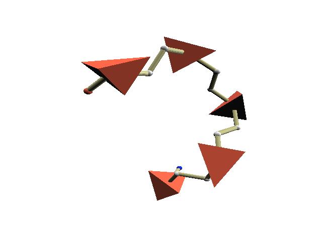
3 Figure 1: Tetrahedron sequence. (a): Folding. (b): Four directions of a tile (gray). (c): Coding rule (Assignment of the second derivative ). 3 Encoding method 3.1 Differential structure of a tetrahedron sequence We approximate native protein structures by a particular kind of tetrahedron sequence which satisfies the following conditions (Fig.1(a)): (i) each tetrahedron consists of four short edges and two long edges, where the ratio of the length is 3/2 and (ii) successive tetrahedrons are connected via a long edge and have the rotational freedom around the edge. Each tetrahedron of a folded sequence assumes one of the four directions of its short edges, which is determined by the configuration of the previous and next tiles (Fig.1(b)). Then, we can describe change of the direction of tiles, i.e. the second derivative, by a binary sequence, where either U or D is assigned to each tile. The coding rule is simple: change the value if the direction changes. For example, suppose that a protein backbone has been approximated up to the previous atom A0 by a tetrahedron sequence up to tetrahedron T 0 (Fig.1(c)). That is, the direction of T 0 and the position of T 1 has been determined by the position of the current atom A1 (left). Then, there are two candidates T 2a and T 2b for the position of the next tile T 2. If the next atom is A2a, then T 2a is closer to the atom. Thus the next tile assumes the position of T 2a and the direction of tetrahedrons changes (middle). In this case assign U to T 1 if the value of T 0 is D and assign D to T 1 otherwise. On the other hand, if the next atom is A2b, then the next tile assumes the position of T 2b and the direction of tetrahedrons does not change (right). In this case assign D to T 1 if the value of T 0 is D and assign U to T 1 otherwise. At endpoints, choose the tile which is closer to A1. (See [7] for the mathematical foundation.) tile code of proteins Upon approximation, we consider the position of the centre of amino-acids of proteins. In other words, we identify an amino-acid with the α-carbon atom 3
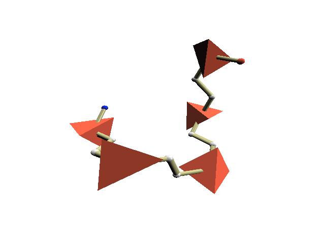
4 Figure 2: Approximation of transferase, PDB ID 1rkl, and others. Broken lines show protein backbones. (a): Folding only. (b): Folding with rotation. (c): Folding with rotation and translation. (d): α-helix (from the 142-th to 154-th amino-acids of protein 1be3). (e): β-sheet (from the 826-th to 838-th aminoacids of protein 1jz7). (Cα) located in its centre. And we allow rotation and translation of tetrahedrons during the folding process to absorb irregularity of actual protein structures. That is, once the direction of tile T 1 is determined (Fig.1(c)), we rotate T 1 to make its direction parallel with the direction from A0 to A2 and translate T 1 to the position of A1. Fig.2(a), (b), and (c) show approximation of a protein (transferase), whose PDB (Protein Data Bank) ID is 1rkl, by folding only, by folding with rotation, and by folding with rotation and translation respectively. To study protein structures, we consider every fragment of an bodd number of amino-acids, say n, contained in a given protein. We start encoding from the middle point amino-acid, say A, and call the obtained binary sequence n- tile code of amino-acid A, where A is always assigned value D. (Note that encoding depends on choice of the initial amino-acid.) For example, the middle point amino-acid of the α-helix shown in Fig.1(d) is encoded into 13-tile code DDDDDU DU DDDDD and the middle point amino-acid of the β-sheet shown in Fig.1(e) is encoded into DDDDDDDDDDDDD (all Ds). Since it is enough to consider 5-tile codes to detect α-helix, we restrict ourselves on 5-tile codes below. Using 5-tile codes, we obtain 16-valued sequence for each protein. For example, the α-helix of Fig.1(d) is encoded into a 5-tile code sequence of length nine, HHHHHHHHH, and the β-sheet of Fig.1(e) is encoded into SSST SSSSS, where H stands for DUDUD, S for DDDDD, and T for UUDUU. See appendix A for the list of 5-tile codes and their examples. 4 Results tile code assignment of superfolds It is well known that there exist highly populated families of second structure arrangements, called superfolds [8], shared by a diverse range of amino-acid sequences. And we consider nine superfolds, from which we extract 1, 215 fragments of 5 amino-acids, to study the correspondence between 5-tile codes and 4
5 Table 1: Frequency of 5-tile codes and their DSSP assignments (superfolds). secondary structure of proteins, such as helix, sheet, and turn. We use DSSP (Dictionary of Secondary Structure of Proteins) program [2] to assign secondary structure elements to each amino-acid, which calculates energies of hydrogen bonds using a classical electrostatic function. Table 1(a) shows the frequency distribution of 5-tile codes and their DSSP assignments. This shows that code DDDDD mainly corresponds to sheet, DUDUD mainly to helix, and UUDUU mainly to turn. On the other hand, most of helixes are covered by three codes DUDUD, UUDUD, and DUDUU, most of sheets are covered by DDDDD, and most of turns are covered by UUDUU, UUDUD, and DUDUU. Appendix B shows the spacial distribution of four 5-tile code groups, mainlyhelix {DUDUD}, mainly-sheet {DDDDD}, mainly-turn {UUDUU, UUDUD, DUDUU}, and else. 4.2 Frequency distribution of 5-tile codes Currently protein structures are classified in a hierarchical fashion. For example, the SCOP (structural classification of proteins) database classifies more than 25, 000 proteins into about 3, 000 families manually. To study the frequency distribution of 5-tile codes, we took one protein for each SCOP family (1.69 release), from which we extracted 1, 591, 608 fragments of 5 amino-acids. They corresponds to 612, 232 types of amino-acid sequences, where 26, 014 types are occurred more than nine times. As table 2(a) shows, 93% of the fragments are covered by top five 5-tile codes. Table 2(b) is concerned with the number of the codes related to one amino-acid sequence, where sequences which occurred more than nine times only are considered. For example, more than 60% of the amino-acid sequences 5
6 Table 2: Statistics of 5-tile codes. (a): Frequency of 5-tile codes. (b): Frequency of amino-acid sequences which relate to a given number of the top five 5-tile codes. (Sequences which occurred more than nine times only are considered) (c): 5-tile code assignments of the amino-acid sequences which relate to all of the top five 5-tile codes. Sequences are given by one-letter code of amino-acids and the figures show the frequency of DDDDD/DUDUD/UUDUU/DUDUU/UUDUD/Else. are encoded uniquely and there exist twelve sequences which relate to all of the five codes. Table 2(c) shows their 5-tile code assignments. Note that most of them have a preference for a specific code. Finally, appendix C gives the preference of amino-acids for 5-tile codes. As you see, amino-acids are grouped into three categories: DDDDD-oriented, DU DU D-oriented, and others. 5 Discussion Recently more than a few secondary structure assignment programs are available. But assignments differ from one program to another because of the arbitrariness of the definition of secondary structure, particularly its exact boundaries. According to [4], the degree of disagreement between two different assignments could reach almost 20%. On the other hand, our approach dose not require any pre-defined secondary structure. Instead, it classifies local structure according to shape. For example, in the case of 5-tile coding, local structure is grouped into 16 types of elements (See appendix A). Though it can neither distinguish three types of helixes from 6
7 one another nor describe global features directly, such as hydrogen bonding, it can detect secondary structure to same extent (See appendix B). That is, it allows a comprehensive description of, not only specific (secondary) structure, but also arbitrary local structure. And it becomes possible to quantify structural preference of amino-acids objectively (See appendix C). It could be used to characterize binding sites of drugs and the active site of enzymes, too. Moreover, since our method encodes a protein into a number sequence without any structure, it is easy to analyse the (number sequence) profile and we could use the wide variety of sequence alignment programs to compare protein structures. And the incorporation of structural information should lead to more powerful protein structure predictions because structure is much more strongly conserved than sequence during evolution. For example, in the case of 5-tile coding, more than 60% of amino-acid sequences are uniquely encoded. Thus, they can be substituted with 5-tile codes when one considers plausible structures of a given amino-acid sequence. 6 Conclusions This paper proposes a new mathematical approach to characterize native protein structures based on the discrete differential geometry of tetrahedron tiles [7]. In the approach, local structure of proteins is classified into finite types according to shape. And we have obtained a comprehensive description of, not only specific (secondary) structure, but also arbitrary local structure elements. As a result, one could quantify structural preference of amino-acids objectively. And one could use the wide variety of sequence alignment programs to study protein structures since the one-dimensional profile of the assignment is given as a simple number sequence. References [1] Bowie, J.U., Luthy, R. & Eisenberg, D A method to identify protein sequences that fold into a known three-dimensional structure. Science 253, [2] Kabsch, W. & Sander, C Dictionary of protein secondary structure: pattern recognition of hydrogen-bonded and geometrical features. Biopolymers 22, [3] Orengo, C.A., Michie, A.D., Jones, S., Jones, D.T., Swindells, M.B., & Thornton, J.M CATH- A Hierarchic Classification of Protein Domain Structures. Structure. 5, [4] Cubellis, M.V., Caulliez, F., & Lovell, S.C Secondary structure assignment that accurately reflects physical and evolutionary characteristics. BMC Bioinformatics 6 (Suppl 4), S8. 7
8 [5] Jones, D.T., Taylor, W.R. & Thornton, J.M A new approach to protein fold recognition. Nature 358, [6] Levitt, M Protein folding by restrained energy minimization and molecular dynamics. J. Mol. Biol. 170, [7] Morikawa, N Discrete differential geometry of triangle tiles and algebra of closed trajectories. ArXiv: math.co/ [8] Orengo, C.A., Jones, D.T. & Thornton, J.M Protein superfamilies and domain superfolds. Nature 372, [9] Rogen, P & Fain, B Automatic classification of protein structure by using Gauss integrals. Proc. Natl. Acad. Sci. 100, [10] Richardson, J.S. & Richardson, D.C Amino acid preferences for specific locations at the ends of alpha helices. Science 240, [11] Rackovsky, S. & Scheraga, H.A Differential Geometry and Polymer Conformation. 1. Macromolecules 11, [12] Murzin A. G., Brenner S. E., Hubbard T., & Chothia C SCOP: a structural classification of proteins database for the investigation of sequences and structures. J. Mol. Biol. 247, [13] Tayler, W.R A periodic table for protein structre. Nature 416, [14] Taylor, W.R. & Aszodi, A Protein Geometry, Classification, Topology and Symmetry: A computational analysis of structure. IOP Publishing Ltd., London. [15] Tyagi, M., Sharma, P., Swamy, C.S., Cadet, F., Srinivasan, N., de Brevern, A.G., Offmann, B Protein Block Expert (PBE): a web-based protein structure analysis server using a structural alphabet. Nucleic Acids. Res. 34, W119-W123 (Web server issue). 8


9 A 5-tile code list Table 3: 5-tile code list. Broken lines show protein backbones. 9


10 B Spacial distributions of 5-tile codes # $%&' ( '$ )* +% Figure 3: 5-tile code distributions of nine superfolds. Broken lines show protein backbones and Red balls are the Cα atoms of the amino-acids of mainlyhelix code DU DU D, yellow balls mainly-turn codes {U U DU U, DU DU U, UUDUD}, and small blue balls mainly-sheet code DDDDD. 10
11 C 5-tile code preference of amino-acids # # $ % $ Table 4: Preference of amino-acids for 5-tile codes. 11
Protein Structure: Data Bases and Classification Ingo Ruczinski
 Protein Structure: Data Bases and Classification Ingo Ruczinski Department of Biostatistics, Johns Hopkins University Reference Bourne and Weissig Structural Bioinformatics Wiley, 2003 More References
Supporting Online Material for
 www.sciencemag.org/cgi/content/full/309/5742/1868/dc1 Supporting Online Material for Toward High-Resolution de Novo Structure Prediction for Small Proteins Philip Bradley, Kira M. S. Misura, David Baker*
A General Model for Amino Acid Interaction Networks
 Author manuscript, published in "N/P" A General Model for Amino Acid Interaction Networks Omar GACI and Stefan BALEV hal-43269, version - Nov 29 Abstract In this paper we introduce the notion of protein
Analysis and Prediction of Protein Structure (I)
") Analysis and Prediction of Protein Structure (I) Jianlin Cheng, PhD School of Electrical Engineering and Computer Science University of Central Florida 2006 Free for academic use. Copyright @ Jianlin Cheng
Analysis and Prediction of Protein Structure (I) Jianlin Cheng, PhD School of Electrical Engineering and Computer Science University of Central Florida 2006 Free for academic use. Copyright @ Jianlin Cheng
Identification of Representative Protein Sequence and Secondary Structure Prediction Using SVM Approach
 Identification of Representative Protein Sequence and Secondary Structure Prediction Using SVM Approach Prof. Dr. M. A. Mottalib, Md. Rahat Hossain Department of Computer Science and Information Technology
Protein Science (1997), 6: Cambridge University Press. Printed in the USA. Copyright 1997 The Protein Society
, 6: Cambridge University Press. Printed in the USA. Copyright 1997 The Protein Society") 1 of 5 1/30/00 8:08 PM Protein Science (1997), 6: 246-248. Cambridge University Press. Printed in the USA. Copyright 1997 The Protein Society FOR THE RECORD LPFC: An Internet library of protein family
1 of 5 1/30/00 8:08 PM Protein Science (1997), 6: 246-248. Cambridge University Press. Printed in the USA. Copyright 1997 The Protein Society FOR THE RECORD LPFC: An Internet library of protein family
Protein structure alignments
 Protein structure alignments Proteins that fold in the same way, i.e. have the same fold are often homologs. Structure evolves slower than sequence Sequence is less conserved than structure If BLAST gives
Procheck output. Bond angles (Procheck) Structure verification and validation Bond lengths (Procheck) Introduction to Bioinformatics.
 Structure verification and validation Bond lengths (Procheck) Introduction to Bioinformatics.") Structure verification and validation Bond lengths (Procheck) Introduction to Bioinformatics Iosif Vaisman Email: ivaisman@gmu.edu ----------------------------------------------------------------- Bond
Structure verification and validation Bond lengths (Procheck) Introduction to Bioinformatics Iosif Vaisman Email: ivaisman@gmu.edu ----------------------------------------------------------------- Bond
1. Protein Data Bank (PDB) 1. Protein Data Bank (PDB)
 1. Protein Data Bank (PDB)") Protein structure databases; visualization; and classifications 1. Introduction to Protein Data Bank (PDB) 2. Free graphic software for 3D structure visualization 3. Hierarchical classification of protein
Protein structure databases; visualization; and classifications 1. Introduction to Protein Data Bank (PDB) 2. Free graphic software for 3D structure visualization 3. Hierarchical classification of protein
Protein structure analysis. Risto Laakso 10th January 2005
 Protein structure analysis Risto Laakso risto.laakso@hut.fi 10th January 2005 1 1 Summary Various methods of protein structure analysis were examined. Two proteins, 1HLB (Sea cucumber hemoglobin) and 1HLM
CS612 - Algorithms in Bioinformatics
 Fall 2017 Databases and Protein Structure Representation October 2, 2017 Molecular Biology as Information Science > 12, 000 genomes sequenced, mostly bacterial (2013) > 5x10 6 unique sequences available
Molecular modeling. A fragment sequence of 24 residues encompassing the region of interest of WT-
 SUPPLEMENTARY DATA Molecular dynamics Molecular modeling. A fragment sequence of 24 residues encompassing the region of interest of WT- KISS1R, i.e. the last intracellular domain (Figure S1a), has been
Protein Structure & Motifs
 & Motifs Biochemistry 201 Molecular Biology January 12, 2000 Doug Brutlag Introduction Proteins are more flexible than nucleic acids in structure because of both the larger number of types of residues
CAP 5510 Lecture 3 Protein Structures
 CAP 5510 Lecture 3 Protein Structures Su-Shing Chen Bioinformatics CISE 8/19/2005 Su-Shing Chen, CISE 1 Protein Conformation 8/19/2005 Su-Shing Chen, CISE 2 Protein Conformational Structures Hydrophobicity
Secondary Structure. Bioch/BIMS 503 Lecture 2. Structure and Function of Proteins. Further Reading. Φ, Ψ angles alone determine protein structure
 Bioch/BIMS 503 Lecture 2 Structure and Function of Proteins August 28, 2008 Robert Nakamoto rkn3c@virginia.edu 2-0279 Secondary Structure Φ Ψ angles determine protein structure Φ Ψ angles are restricted
SCOP. all-β class. all-α class, 3 different folds. T4 endonuclease V. 4-helical cytokines. Globin-like
 SCOP all-β class 4-helical cytokines T4 endonuclease V all-α class, 3 different folds Globin-like TIM-barrel fold α/β class Profilin-like fold α+β class http://scop.mrc-lmb.cam.ac.uk/scop CATH Class, Architecture,
Genome Databases The CATH database
 Genome Databases The CATH database Michael Knudsen 1 and Carsten Wiuf 1,2* 1 Bioinformatics Research Centre, Aarhus University, DK-8000 Aarhus C, Denmark 2 Centre for Membrane Pumps in Cells and Disease
STRUCTURAL BIOLOGY AND PATTERN RECOGNITION
 STRUCTURAL BIOLOGY AND PATTERN RECOGNITION V. Cantoni, 1 A. Ferone, 2 O. Ozbudak, 3 and A. Petrosino 2 1 University of Pavia, Department of Electrical and Computer Engineering, Via A. Ferrata, 1, 27, Pavia,
Analysis on sliding helices and strands in protein structural comparisons: A case study with protein kinases
 Sliding helices and strands in structural comparisons 921 Analysis on sliding helices and strands in protein structural comparisons: A case study with protein kinases V S GOWRI, K ANAMIKA, S GORE 1 and
CMPS 6630: Introduction to Computational Biology and Bioinformatics. Structure Comparison
 CMPS 6630: Introduction to Computational Biology and Bioinformatics Structure Comparison Protein Structure Comparison Motivation Understand sequence and structure variability Understand Domain architecture
CMPS 6630: Introduction to Computational Biology and Bioinformatics. Tertiary Structure Prediction
 CMPS 6630: Introduction to Computational Biology and Bioinformatics Tertiary Structure Prediction Tertiary Structure Prediction Why Should Tertiary Structure Prediction Be Possible? Molecules obey the
ALL LECTURES IN SB Introduction
 1. Introduction 2. Molecular Architecture I 3. Molecular Architecture II 4. Molecular Simulation I 5. Molecular Simulation II 6. Bioinformatics I 7. Bioinformatics II 8. Prediction I 9. Prediction II ALL
The CATH Database provides insights into protein structure/function relationships
 1999 Oxford University Press Nucleic Acids Research, 1999, Vol. 27, No. 1 275 279 The CATH Database provides insights into protein structure/function relationships C. A. Orengo, F. M. G. Pearl, J. E. Bray,
HMM applications. Applications of HMMs. Gene finding with HMMs. Using the gene finder
 HMM applications Applications of HMMs Gene finding Pairwise alignment (pair HMMs) Characterizing protein families (profile HMMs) Predicting membrane proteins, and membrane protein topology Gene finding
Protein Structure Prediction II Lecturer: Serafim Batzoglou Scribe: Samy Hamdouche
 Protein Structure Prediction II Lecturer: Serafim Batzoglou Scribe: Samy Hamdouche The molecular structure of a protein can be broken down hierarchically. The primary structure of a protein is simply its
Alpha-helical Topology and Tertiary Structure Prediction of Globular Proteins Scott R. McAllister Christodoulos A. Floudas Princeton University
 Alpha-helical Topology and Tertiary Structure Prediction of Globular Proteins Scott R. McAllister Christodoulos A. Floudas Princeton University Department of Chemical Engineering Program of Applied and
CMPS 3110: Bioinformatics. Tertiary Structure Prediction
 CMPS 3110: Bioinformatics Tertiary Structure Prediction Tertiary Structure Prediction Why Should Tertiary Structure Prediction Be Possible? Molecules obey the laws of physics! Conformation space is finite
MSAT a Multiple Sequence Alignment tool based on TOPS
 MSAT a Multiple Sequence Alignment tool based on TOPS Te Ren, Mallika Veeramalai, Aik Choon Tan and David Gilbert Bioinformatics Research Centre Department of Computer Science University of Glasgow Glasgow,
2MHR. Protein structure classification is important because it organizes the protein structure universe that is independent of sequence similarity.
 Protein structure classification is important because it organizes the protein structure universe that is independent of sequence similarity. A global picture of the protein universe will help us to understand
Statistical Machine Learning Methods for Bioinformatics IV. Neural Network & Deep Learning Applications in Bioinformatics
 Statistical Machine Learning Methods for Bioinformatics IV. Neural Network & Deep Learning Applications in Bioinformatics Jianlin Cheng, PhD Department of Computer Science University of Missouri, Columbia
Comparing Protein Structures. Why?
 7.91 Amy Keating Comparing Protein Structures Why? detect evolutionary relationships identify recurring motifs detect structure/function relationships predict function assess predicted structures classify
2 Dean C. Adams and Gavin J. P. Naylor the best three-dimensional ordination of the structure space is found through an eigen-decomposition (correspon
 A Comparison of Methods for Assessing the Structural Similarity of Proteins Dean C. Adams and Gavin J. P. Naylor? Dept. Zoology and Genetics, Iowa State University, Ames, IA 50011, U.S.A. 1 Introduction
Protein quality assessment
 Protein quality assessment Speaker: Renzhi Cao Advisor: Dr. Jianlin Cheng Major: Computer Science May 17 th, 2013 1 Outline Introduction Paper1 Paper2 Paper3 Discussion and research plan Acknowledgement
Motif Prediction in Amino Acid Interaction Networks
 Motif Prediction in Amino Acid Interaction Networks Omar GACI and Stefan BALEV Abstract In this paper we represent a protein as a graph where the vertices are amino acids and the edges are interactions
Heteropolymer. Mostly in regular secondary structure
 Heteropolymer - + + - Mostly in regular secondary structure 1 2 3 4 C >N trace how you go around the helix C >N C2 >N6 C1 >N5 What s the pattern? Ci>Ni+? 5 6 move around not quite 120 "#$%&'!()*(+2!3/'!4#5'!1/,#64!#6!,6!
Orientational degeneracy in the presence of one alignment tensor.
 Orientational degeneracy in the presence of one alignment tensor. Rotation about the x, y and z axes can be performed in the aligned mode of the program to examine the four degenerate orientations of two
proteins Comprehensive description of protein structures using protein folding shape code Jiaan Yang* INTRODUCTION
 proteins STRUCTURE O FUNCTION O BIOINFORMATICS Comprehensive description of protein structures using protein folding shape code Jiaan Yang* MicrotechNano, LLC, Indianapolis, Indiana 46234 ABSTRACT Understanding
Physiochemical Properties of Residues
 Physiochemical Properties of Residues Various Sources C N Cα R Slide 1 Conformational Propensities Conformational Propensity is the frequency in which a residue adopts a given conformation (in a polypeptide)
Bioinformatics. Macromolecular structure
 Bioinformatics Macromolecular structure Contents Determination of protein structure Structure databases Secondary structure elements (SSE) Tertiary structure Structure analysis Structure alignment Domain
COMP 598 Advanced Computational Biology Methods & Research. Introduction. Jérôme Waldispühl School of Computer Science McGill University
 COMP 598 Advanced Computational Biology Methods & Research Introduction Jérôme Waldispühl School of Computer Science McGill University General informations (1) Office hours: by appointment Office: TR3018
Design of a Novel Globular Protein Fold with Atomic-Level Accuracy
 Design of a Novel Globular Protein Fold with Atomic-Level Accuracy Brian Kuhlman, Gautam Dantas, Gregory C. Ireton, Gabriele Varani, Barry L. Stoddard, David Baker Presented by Kate Stafford 4 May 05 Protein
THE TANGO ALGORITHM: SECONDARY STRUCTURE PROPENSITIES, STATISTICAL MECHANICS APPROXIMATION
 THE TANGO ALGORITHM: SECONDARY STRUCTURE PROPENSITIES, STATISTICAL MECHANICS APPROXIMATION AND CALIBRATION Calculation of turn and beta intrinsic propensities. A statistical analysis of a protein structure
Template Free Protein Structure Modeling Jianlin Cheng, PhD
 Template Free Protein Structure Modeling Jianlin Cheng, PhD Associate Professor Computer Science Department Informatics Institute University of Missouri, Columbia 2013 Protein Energy Landscape & Free Sampling
Analysis of MD trajectories in GROMACS David van der Spoel
 Analysis of MD trajectories in GROMACS David van der Spoel What does MD produce? Energy terms E(t) Coordinates x(t) Velocities v(t) Forces f(t) Managing your files trjcat - merging trajectories concatenating
The sequences of naturally occurring proteins are defined by
 Protein topology and stability define the space of allowed sequences Patrice Koehl* and Michael Levitt Department of Structural Biology, Fairchild Building, D109, Stanford University, Stanford, CA 94305
Computer simulations of protein folding with a small number of distance restraints
 Vol. 49 No. 3/2002 683 692 QUARTERLY Computer simulations of protein folding with a small number of distance restraints Andrzej Sikorski 1, Andrzej Kolinski 1,2 and Jeffrey Skolnick 2 1 Department of Chemistry,
Computational Molecular Modeling
 Computational Molecular Modeling Lecture 1: Structure Models, Properties Chandrajit Bajaj Today s Outline Intro to atoms, bonds, structure, biomolecules, Geometry of Proteins, Nucleic Acids, Ribosomes,
Bioinformatics: Secondary Structure Prediction
 Bioinformatics: Secondary Structure Prediction Prof. David Jones d.t.jones@ucl.ac.uk Possibly the greatest unsolved problem in molecular biology: The Protein Folding Problem MWMPPRPEEVARK LRRLGFVERMAKG
Presentation Outline. Prediction of Protein Secondary Structure using Neural Networks at Better than 70% Accuracy
 Prediction of Protein Secondary Structure using Neural Networks at Better than 70% Accuracy Burkhard Rost and Chris Sander By Kalyan C. Gopavarapu 1 Presentation Outline Major Terminology Problem Method
The typical end scenario for those who try to predict protein
 A method for evaluating the structural quality of protein models by using higher-order pairs scoring Gregory E. Sims and Sung-Hou Kim Berkeley Structural Genomics Center, Lawrence Berkeley National Laboratory,
Grouping of amino acids and recognition of protein structurally conserved regions by reduced alphabets of amino acids
 Science in China Series C: Life Sciences 2007 Science in China Press Springer-Verlag Grouping of amino acids and recognition of protein structurally conserved regions by reduced alphabets of amino acids
Hidden symmetries in primary sequences of small α proteins
 Hidden symmetries in primary sequences of small α proteins Ruizhen Xu, Yanzhao Huang, Mingfen Li, Hanlin Chen, and Yi Xiao * Biomolecular Physics and Modeling Group, Department of Physics, Huazhong University
Modeling for 3D structure prediction
 Modeling for 3D structure prediction What is a predicted structure? A structure that is constructed using as the sole source of information data obtained from computer based data-mining. However, mixing
Gauge symmetries and structure of proteins
 Gauge symmetries and structure of proteins Alexander Molochkov 1,a, Alexander Begun 1, and Antti Niemi 234 1 Laboratory of Physics of Living Matter, Far Eastern Federal University, 690950, Sukhanova 8,
Protein Structure Prediction
 Page 1 Protein Structure Prediction Russ B. Altman BMI 214 CS 274 Protein Folding is different from structure prediction --Folding is concerned with the process of taking the 3D shape, usually based on
IT og Sundhed 2010/11
 IT og Sundhed 2010/11 Sequence based predictors. Secondary structure and surface accessibility Bent Petersen 13 January 2011 1 NetSurfP Real Value Solvent Accessibility predictions with amino acid associated
Introduction to Comparative Protein Modeling. Chapter 4 Part I
 Introduction to Comparative Protein Modeling Chapter 4 Part I 1 Information on Proteins Each modeling study depends on the quality of the known experimental data. Basis of the model Search in the literature
Ranjit P. Bahadur Assistant Professor Department of Biotechnology Indian Institute of Technology Kharagpur, India. 1 st November, 2013
 Hydration of protein-rna recognition sites Ranjit P. Bahadur Assistant Professor Department of Biotechnology Indian Institute of Technology Kharagpur, India 1 st November, 2013 Central Dogma of life DNA
Giri Narasimhan. CAP 5510: Introduction to Bioinformatics. ECS 254; Phone: x3748
 CAP 5510: Introduction to Bioinformatics Giri Narasimhan ECS 254; Phone: x3748 giri@cis.fiu.edu www.cis.fiu.edu/~giri/teach/bioinfs07.html 2/15/07 CAP5510 1 EM Algorithm Goal: Find θ, Z that maximize Pr
Structural Alignment of Proteins
 Goal Align protein structures Structural Alignment of Proteins 1 2 3 4 5 6 7 8 9 10 11 12 13 14 PHE ASP ILE CYS ARG LEU PRO GLY SER ALA GLU ALA VAL CYS PHE ASN VAL CYS ARG THR PRO --- --- --- GLU ALA ILE
Towards De Novo Folding of Protein Structures from Cryo-EM 3D Images at Medium Resolutions
 Towards De Novo Folding of Protein Structures from Cryo-EM 3D Images at Medium Resolutions Jing He * and Dong Si Department of Computer Science Old Dominion University Norfolk, VA * jhe@cs.odu.edu Abstract
Bioinformatics III Structural Bioinformatics and Genome Analysis Part Protein Secondary Structure Prediction. Sepp Hochreiter
 Bioinformatics III Structural Bioinformatics and Genome Analysis Part Protein Secondary Structure Prediction Institute of Bioinformatics Johannes Kepler University, Linz, Austria Chapter 4 Protein Secondary
Study of Mining Protein Structural Properties and its Application
 Study of Mining Protein Structural Properties and its Application A Dissertation Proposal Presented to the Department of Computer Science and Information Engineering College of Electrical Engineering and
Protein structure similarity based on multi-view images generated from 3D molecular visualization
 Protein structure similarity based on multi-view images generated from 3D molecular visualization Chendra Hadi Suryanto, Shukun Jiang, Kazuhiro Fukui Graduate School of Systems and Information Engineering,
Protein structure (and biomolecular structure more generally) CS/CME/BioE/Biophys/BMI 279 Sept. 28 and Oct. 3, 2017 Ron Dror
 CS/CME/BioE/Biophys/BMI 279 Sept. 28 and Oct. 3, 2017 Ron Dror") Protein structure (and biomolecular structure more generally) CS/CME/BioE/Biophys/BMI 279 Sept. 28 and Oct. 3, 2017 Ron Dror Please interrupt if you have questions, and especially if you re confused! Assignment
Protein structure (and biomolecular structure more generally) CS/CME/BioE/Biophys/BMI 279 Sept. 28 and Oct. 3, 2017 Ron Dror Please interrupt if you have questions, and especially if you re confused! Assignment
Improving Protein 3D Structure Prediction Accuracy using Dense Regions Areas of Secondary Structures in the Contact Map
 American Journal of Biochemistry and Biotechnology 4 (4): 375-384, 8 ISSN 553-3468 8 Science Publications Improving Protein 3D Structure Prediction Accuracy using Dense Regions Areas of Secondary Structures
09/06/25. Computergestützte Strukturbiologie (Strukturelle Bioinformatik) Non-uniform distribution of folds. Scheme of protein structure predicition
 Non-uniform distribution of folds. Scheme of protein structure predicition") Sequence identity Structural similarity Computergestützte Strukturbiologie (Strukturelle Bioinformatik) Fold recognition Sommersemester 2009 Peter Güntert Structural similarity X Sequence identity Non-uniform
Sequence identity Structural similarity Computergestützte Strukturbiologie (Strukturelle Bioinformatik) Fold recognition Sommersemester 2009 Peter Güntert Structural similarity X Sequence identity Non-uniform
Protein Structures: Experiments and Modeling. Patrice Koehl
 Protein Structures: Experiments and Modeling Patrice Koehl Structural Bioinformatics: Proteins Proteins: Sources of Structure Information Proteins: Homology Modeling Proteins: Ab initio prediction Proteins:
Can protein model accuracy be. identified? NO! CBS, BioCentrum, Morten Nielsen, DTU
 Can protein model accuracy be identified? Morten Nielsen, CBS, BioCentrum, DTU NO! Identification of Protein-model accuracy Why is it important? What is accuracy RMSD, fraction correct, Protein model correctness/quality
7 Protein secondary structure
 78 Grundlagen der Bioinformatik, SS 1, D. Huson, June 17, 21 7 Protein secondary structure Sources for this chapter, which are all recommended reading: Introduction to Protein Structure, Branden & Tooze,
8 Protein secondary structure
 Grundlagen der Bioinformatik, SoSe 11, D. Huson, June 6, 211 13 8 Protein secondary structure Sources for this chapter, which are all recommended reading: Introduction to Protein Structure, Branden & Tooze,
The PDB is a Covering Set of Small Protein Structures
 doi:10.1016/j.jmb.2003.10.027 J. Mol. Biol. (2003) 334, 793 802 The PDB is a Covering Set of Small Protein Structures Daisuke Kihara and Jeffrey Skolnick* Center of Excellence in Bioinformatics, University
Part I: Introduction to Protein Structure Similarity
 Lecture 8: Finding structural similarities among proteins (I) 24-1 CS5238 Combinatorial methods in bioinformatics 2004/2005 Semester 1 Lecture 8: Finding structural similarities among proteins (I) Lecturer:
DATE A DAtabase of TIM Barrel Enzymes
 DATE A DAtabase of TIM Barrel Enzymes 2 2.1 Introduction.. 2.2 Objective and salient features of the database 2.2.1 Choice of the dataset.. 2.3 Statistical information on the database.. 2.4 Features....
Useful background reading
 Overview of lecture * General comment on peptide bond * Discussion of backbone dihedral angles * Discussion of Ramachandran plots * Description of helix types. * Description of structures * NMR patterns
A profile-based protein sequence alignment algorithm for a domain clustering database
 A profile-based protein sequence alignment algorithm for a domain clustering database Lin Xu,2 Fa Zhang and Zhiyong Liu 3, Key Laboratory of Computer System and architecture, the Institute of Computing
DesignabilityofProteinStructures:ALattice-ModelStudy usingthemiyazawa-jerniganmatrix
 PROTEINS: Structure, Function, and Genetics 49:403 412 (2002) DesignabilityofProteinStructures:ALattice-ModelStudy usingthemiyazawa-jerniganmatrix HaoLi,ChaoTang,* andneds.wingreen NECResearchInstitute,Princeton,NewJersey
A Tool for Structure Alignment of Molecules
 A Tool for Structure Alignment of Molecules Pei-Ken Chang, Chien-Cheng Chen and Ming Ouhyoung Department of Computer Science and Information Engineering, National Taiwan University {zick, ccchen}@cmlab.csie.ntu.edu.tw,
Ab-initio protein structure prediction
 Ab-initio protein structure prediction Jaroslaw Pillardy Computational Biology Service Unit Cornell Theory Center, Cornell University Ithaca, NY USA Methods for predicting protein structure 1. Homology
Molecular dynamics simulation of Aquaporin-1. 4 nm
 Molecular dynamics simulation of Aquaporin-1 4 nm Molecular Dynamics Simulations Schrödinger equation i~@ t (r, R) =H (r, R) Born-Oppenheimer approximation H e e(r; R) =E e (R) e(r; R) Nucleic motion described
Introducing Hippy: A visualization tool for understanding the α-helix pair interface
 Introducing Hippy: A visualization tool for understanding the α-helix pair interface Robert Fraser and Janice Glasgow School of Computing, Queen s University, Kingston ON, Canada, K7L3N6 {robert,janice}@cs.queensu.ca
Introduction to" Protein Structure
 Introduction to" Protein Structure Function, evolution & experimental methods Thomas Blicher, Center for Biological Sequence Analysis Learning Objectives Outline the basic levels of protein structure.
Protein Secondary Structure Prediction using Feed-Forward Neural Network
 COPYRIGHT 2010 JCIT, ISSN 2078-5828 (PRINT), ISSN 2218-5224 (ONLINE), VOLUME 01, ISSUE 01, MANUSCRIPT CODE: 100713 Protein Secondary Structure Prediction using Feed-Forward Neural Network M. A. Mottalib,
Protein tertiary structure prediction with new machine learning approaches
 Protein tertiary structure prediction with new machine learning approaches Rui Kuang Department of Computer Science Columbia University Supervisor: Jason Weston(NEC) and Christina Leslie(Columbia) NEC
Getting To Know Your Protein
 Getting To Know Your Protein Comparative Protein Analysis: Part III. Protein Structure Prediction and Comparison Robert Latek, PhD Sr. Bioinformatics Scientist Whitehead Institute for Biomedical Research
Packing of Secondary Structures
 7.88 Lecture Notes - 4 7.24/7.88J/5.48J The Protein Folding and Human Disease Professor Gossard Retrieving, Viewing Protein Structures from the Protein Data Base Helix helix packing Packing of Secondary
Molecular Modelling. part of Bioinformatik von RNA- und Proteinstrukturen. Sonja Prohaska. Leipzig, SS Computational EvoDevo University Leipzig
 part of Bioinformatik von RNA- und Proteinstrukturen Computational EvoDevo University Leipzig Leipzig, SS 2011 Protein Structure levels or organization Primary structure: sequence of amino acids (from
Protein dynamics. Folding/unfolding dynamics. Fluctuations near the folded state
 Protein dynamics Folding/unfolding dynamics Passage over one or more energy barriers Transitions between infinitely many conformations B. Ozkan, K.A. Dill & I. Bahar, Protein Sci. 11, 1958-1970, 2002 Fluctuations
12 Protein secondary structure
 Grundlagen der Bioinformatik, SoSe 14, D. Huson, July 2, 214 147 12 Protein secondary structure Sources for this chapter, which are all recommended reading: Introduction to Protein Structure, Branden &
Protein Threading Based on Multiple Protein Structure Alignment
 Protein Threading Based on Multiple Protein Structure lignment Tatsuya kutsu Kim Lan Sim takutsu@ims.u-tokyo.ac.jp klsim@ims.u-tokyo.ac.jp Human Genome Center, Institute of Medical Science, University
Advanced Certificate in Principles in Protein Structure. You will be given a start time with your exam instructions
 BIRKBECK COLLEGE (University of London) Advanced Certificate in Principles in Protein Structure MSc Structural Molecular Biology Date: Thursday, 1st September 2011 Time: 3 hours You will be given a start
BIOCHEMISTRY Unit 2 Part 4 ACTIVITY #6 (Chapter 5) PROTEINS
 PROTEINS") BIOLOGY BIOCHEMISTRY Unit 2 Part 4 ACTIVITY #6 (Chapter 5) NAME NAME PERIOD PROTEINS GENERAL CHARACTERISTICS AND IMPORTANCES: Polymers of amino acids Each has unique 3-D shape Vary in sequence of amino
BIOLOGY BIOCHEMISTRY Unit 2 Part 4 ACTIVITY #6 (Chapter 5) NAME NAME PERIOD PROTEINS GENERAL CHARACTERISTICS AND IMPORTANCES: Polymers of amino acids Each has unique 3-D shape Vary in sequence of amino
Optimization of the Sliding Window Size for Protein Structure Prediction
 Optimization of the Sliding Window Size for Protein Structure Prediction Ke Chen* 1, Lukasz Kurgan 1 and Jishou Ruan 2 1 University of Alberta, Department of Electrical and Computer Engineering, Edmonton,
A Study of the Protein Folding Dynamic
 A Study of the Protein Folding Dynamic Omar GACI Abstract In this paper, we propose two means to study the protein folding dynamic. We rely on the HP model to study the protein folding problem in a continuous
Protein Structure and Function Prediction using Kernel Methods.
 Protein Structure and Function Prediction using Kernel Methods. A THESIS SUBMITTED TO THE FACULTY OF THE GRADUATE SCHOOL OF THE UNIVERSITY OF MINNESOTA BY Huzefa Rangwala IN PARTIAL FULFILLMENT OF THE
arxiv:cond-mat/ v1 2 Feb 94
 cond-mat/9402010 Properties and Origins of Protein Secondary Structure Nicholas D. Socci (1), William S. Bialek (2), and José Nelson Onuchic (1) (1) Department of Physics, University of California at San
1. (5) Draw a diagram of an isomeric molecule to demonstrate a structural, geometric, and an enantiomer organization.
 Draw a diagram of an isomeric molecule to demonstrate a structural, geometric, and an enantiomer organization.") Organic Chemistry Assignment Score. Name Sec.. Date. Working by yourself or in a group, answer the following questions about the Organic Chemistry material. This assignment is worth 35 points with the
Organic Chemistry Assignment Score. Name Sec.. Date. Working by yourself or in a group, answer the following questions about the Organic Chemistry material. This assignment is worth 35 points with the
Protein folding. α-helix. Lecture 21. An α-helix is a simple helix having on average 10 residues (3 turns of the helix)
") Computat onal Biology Lecture 21 Protein folding The goal is to determine the three-dimensional structure of a protein based on its amino acid sequence Assumption: amino acid sequence completely and uniquely
Computat onal Biology Lecture 21 Protein folding The goal is to determine the three-dimensional structure of a protein based on its amino acid sequence Assumption: amino acid sequence completely and uniquely
Homology Modeling. Roberto Lins EPFL - summer semester 2005
 Homology Modeling Roberto Lins EPFL - summer semester 2005 Disclaimer: course material is mainly taken from: P.E. Bourne & H Weissig, Structural Bioinformatics; C.A. Orengo, D.T. Jones & J.M. Thornton,
CAP 5510: Introduction to Bioinformatics CGS 5166: Bioinformatics Tools. Giri Narasimhan
 CAP 5510: Introduction to Bioinformatics CGS 5166: Bioinformatics Tools Giri Narasimhan ECS 254; Phone: x3748 giri@cis.fiu.edu www.cis.fiu.edu/~giri/teach/bioinff18.html Proteins and Protein Structure