Transformation of head structures during the metamorphosis of Chrysomela populi (Coleoptera: Chrysomelidae)
|
|
|
- Carmella Walters
- 5 years ago
- Views:
Transcription
1 73 (1): Senckenberg Gesellschaft für Naturforschung, Transformation of head structures during the metamorphosis of Chrysomela populi (Coleoptera: Chrysomelidae) Si-Qin Ge *, 1, Yi Hua 1, 2, Jing Ren 1, 2, Adam Ślipiński 3, Bruce Heming 4, Rolf Georg Beutel 5, Xing-Ke Yang *, 1 & Benjamin Wipfler 1 1 Key Laboratory of Zoological Systematics and Evolution, Institute of Zoology, Chinese Academy of Sciences, Beijing, China; Si-Qin Ge [gesq@ioz.ac.cn]; Xing-Ke Yang [yangxk@ioz.ac.cn] 2 University of Chinese Academy of Sciences, Beijing, China 3 CSIRO Ecosystem Sciences, Australian National Insect Collection, Canberra, Australia 4 University of Alberta, Department of Biological Sciences, Edmonton, Canada 5 Entomology Group, Institut für Spezielle Zoologie and Evolutionsbiologie mit Phyletischem Museum, Friedrich-Schiller- Universität Jena, Jena, Germany * Corresponding authors Accepted 11.iii Published online at on 17.iv Abstract External and internal head structures of last instar (3 rd ) larvae, 4 th day pupae and adults of Chrysomela populi were examined using a combination of traditional and modern morphological techniques, especially µ-computed tomography and computer-based 3D reconstruction. Morphological differences and similarities between the stages were assessed. In addition to long known transformations such as the appearance of compound eyes and the reorientation of the head capsule, the adult differs from the larva by having elongated antennae and palps, a gula and anterior and dorsal tentorial arms. Additionally, several changes in the muscular system occur. Most of these cephalic transformations are related to different tasks playing a predominant role in the developmental stages: feeding in the larvae, and dispersal and mating in the adults, the latter requiring improved sensory perception. The 4 th day pupal head shows a combination of adult and larval characters. Almost all adult cephalic elements are already present but the internal anatomy shows several larval traits, especially in the musculature. The central nervous system is intermediate with enlarged optic lobes but still identifiable individual nerves cords. A short historical review on the application of non-invasive methods to study morphological details of metamorphosis is provided and the advantages and limitations of these approaches are discussed. Key words Metamorphosis, pupa, larva, morphology, Chrysomela populi Linnaeus, X-ray computed tomography, 3D-computer reconstruction. 1. Introduction Holometabola (= Endopterygota) are an exceptional group of organisms and their origin was referred to as a nodal point in insect evolution (Hinton 1977; but see Kristensen 1999). With approximately 800,000 described species they comprise about 2/3s of the known total diversity of animals (Grimaldi & Engel 2005). Bursts of diversification took place in different megadiverse subgroups and different factors played a role, such as an improved flight apparatus or their co-evolution with angiosperms (Grimaldi & Engel 2005; Beutel et al. 2011). Nevertheless, it is likely that complete metamorphosis with larvae differing profoundly from adults and a pupal stage was a crucial feature in the evolution of this megadiverse lineage (e.g., Beutel & Pohl 2006). This includes ontogenetic specializations including diet, reduced intraspecific competition between juveniles and adults, and more efficient control of development (e.g., Grimaldi & Engel 2005). ISSN (print) eissn (online) 129
, lateral view; C: adult, lateral view. Scale bar: 2 mm.")

2 GE et al.: Metamorphosis of Chrysomela populi A B C Fig. 1. Chrysomela populi Linnaeus, photographs: A: larva, lateral view; B: pupa (pupal sheath with pharate adult inside), lateral view; C: adult, lateral view. Scale bar: 2 mm. Until now, most studies on the metamorphosis of holometabolous insects have been limited to selected model organisms such as the fruit fly Drosophila melanogaster (e.g., robertson 1936; Hartenstein, 1993; WHite 1999; see also review in HeminG 2003), the mealworm Tenebrio molitor (BreidBacH 1987; see also BreidBacH 1988), or blowflies (Calliphora; crossley 1965). Even though the external habitus of pupae of a comparatively large number of beetle species have been described and pupal features have occasionally been used in phylogenetic analyses (e.g., archangelsky 1997, 1998), very few detailed studies have been carried out on metamorphosis in the extremely successful Coleoptera. A relatively detailed treatment of the pupa of Dytiscus marginalis l. was presented in the remarkable two volumes on this species by KorscHelt (1923, 1924) and some related earlier studies (e.g., Bauer 1910). Polilov & Beutel (2009, 2010) compared changes in the hooded beetle Sericoderus lateralis (Coleoptera: Corylophidae) and Mikado sp. (Coleoptera: Ptiliidae). While their studies covered the larvae, pupae and adults, their main focus was on the effects of miniaturization and phylogeny. Moreover, they omitted detailed information on the anatomy of the pupa. In other endopterygote groups comparative morphological data are equally scarce. oertel (1930) provided a detailed account of the transformations of the skeleton, digestiveand muscular systems in the honeybee Apis mellifera, but omitted detailed information on cephalic musculature. It is apparent that the pupal stage, an instar characterized by major reconstruction of most body parts and organs, is crucial to better understanding complete metamorphosis. The morphological transformations occurring during meta morphosis are presently still very insufficiently known. In recent studies (deans et al. 2012; lowe et al. 2013; friedrich et al. 2014) it was shown that computed tomography (µ-ct) is a valuable tool to study insect anatomy. Experiments carried out at the Institute of Zoology of the Chinese Academy of Sciences confirm that it also greatly facilitates the study of the pharate adult within the pupal sheath. This and the conspicuous lack of information on metamorphosis in beetles induced us to execute this comparative study of the skeleto-muscular system of the head. It covers crucial stages of the life cycle of Chrysomela populi (Coleoptera, Chrysomelidae): the final larval instar, the pupal stage (day four), and the adult (Fig. 1A C). For th is investigation we chose this species for several reasons. First, it belongs to the species rich phytophagan superfamily Chrysomeloidea (ca. 67,000 known spp.). Kristensen (1999) emphasized that Phytophaga, i.e. Chrysomeloidea and Curculionoidea, constitute one of the truly megadiverse holometabolan lineages. Chrysomelid beetles exclusively feed on plant materials, usually on green parts, but members of some subgroups secondarily switched to pollen, flowers, or seeds (Jolivet & verma 2002). farrell (1998) hypothesized that angiosperm feeding may have been the principal cause for the success of this group, an evolutionary correlation that probably also applies to glossatan Lepidoptera (Kristensen 1999). Therefore herbivory and the successful co-evolution with angiosperms may have been critical to megadiversification in these lineages of Holometabola. Aside from their great economic importance (Jolivet & verma 2002) chrysomelids are widely recognised as a model for plantherbivore co-evolution (ehrlich & raven 1964; mitter & farrell 1991; Becerra 1997; Xue et al. 2008, 2009). Another point in favour of examining Chrysomelidae and Chrysomela populi is the well-established leaf beetle research group at the Institute of Zoology of the Chinese Academy of Sciences, with taxonomic and morphological expertise, rich preserved material, and with well-established rearing facilities. Finally, C. populi was chosen to honour the outstanding 18 th century biologist Carolus Linnaeus, who described it as the first chrysomelid species, the type species for the entire family. Morphological studies on chrysomelid beetles and their immatures are surprisingly scarce. Structural features of larvae and adults were examined by rivnay (1928), maulik (1926), mann & crowson (1981, 1983, 1984, 1996), cox (1981, 1988, 1996), reid (1995, 2000), suzuki (1996), samuelson (1996), schmit t (1998), HeidenreicH et al. (2009), Klass et al. (2011) and HüBler & Klass (2013). However, the information provided on internal structures such as the musculature remained scarce, and virtually no information was available on the anatomy of pupae and on the changes undergone by 130
3 ARTHROPOD SYSTEMATICS & PHYLOGENY 73 (1) 2015 internal structures during metamorphosis. The primary goal of this work is to provide a detailed description of the skeleto-muscular system of the head of the last (3 rd ) instar larvae, pupae and adults of a well-known species. Other character systems such as nervous system, digestive system, sensilla or glands were only treated incompletely. Additionally, the potential and limitations of noninvasive methods in the study of metamorphosis are evaluated. A broad description of all events occurring during the pupal stage is presently in preparation, but is beyond the scope of this study and will be presented separately. 2. Materials and methods Examined specimens. Mature larvae and adults of Chrysomela populi Linnaeus (Fig. 1) were preserved in 75% ethanol. They were collected in May 2012 in Shuiquangou, Dazhuangke, Yanqing, Beijing, People s Republic of China. Larvae from the same locality and date were reared in glass jars ( cm) with sand on the bottom and a sufficient supply of host plants. Fresh plant leaves were added every day and wilted leaves removed. The containers were kept closed except for feeding and removing old material, at a constant temperature of 25ºC, 16 hours of light and 8 hours of darkness, and a humidity of around 80%. The pupae were collected 4 days after the mature larvae pupated and fixed for morphological study. The adult specimens were killed of day after hatching. All collected specimens were identified by the first author. X-ray computer tomography. Species used for x-ray micro-computed tomography (µ-ct) were dehydrated with ethanol (75 100%) and acetone and dried at the critical point (Hitachi hcp-2). One fourth day pupa and one adult were scanned with a Skyscan 2010 µ-ct at the Beijing Institute of Technology (beam strength: 40 kev, absorption contrast), while the larva was scanned with an X-radia 200 at Shanghai Jiaotong University (beam strength: 40 kev, absorption contrast). Three-dimensional reconstruction (3D). Micro-CT images were used for 3-dimensional reconstructions. Based on the obtained image stacks, structures of the larva, pupa and adult were reconstructed with Amira 5.4 for Figs. 4, 7 & 11. The data files were then transferred to Maya 2013 (Autodesk) in order to use the smoothing function, and the specific display, and rendering options implemented in this software. Material separation for figure 6 was done with Amira 5.4 (Visage Imaging) and subsequent rendering was performed with VG Studiomax 2.1 (Volume Graphics). Final figures were prepared with Photoshop and Illustrator (CS5, Adobe). Photography. Habit photos of the larvae, pupa and adult were taken with a D300s Nikon camera connected to the stereoscope (Zeiss Stereo Discovery V12). Final figures were prepared with Photoshop and Illustrator (CS5, Adobe). Scanning electronic microscopy (SEM). Specimens were transferred to 100% ethanol, then dried at the critical point (critical point dryer: Leica EM CPD 300) and subsequently sputter-coated (Leica EM SCD 050). Microscopy was performed on a FEI Quanta 450. Final figures were prepared with Photoshop and Illustrator (CS5, Adobe). Histological sections. One adult head was embedded in Araldite (Huntsman Advanced Materials, Bergkamen, Germany) for semi-thin sectioning (1 µm) with a glass knife on a microtom HM 360 (Microm, Walldorf, Germany). The sections were stained with toluidin-blue. Images of sections were taken with a Zeiss Axioplan (Zeiss, Göttingen, Germany). Terminology. We used the general terminology of Beutel et al. (2014). The muscles are subsequently numbered with a prefixed letter indicating the developmental stage: an l in the larva, a p in the pupa and an a in the adult. In the discussion (Table 4), muscles are named according to the terminology of Wipfler et al. (2011). 3. Results In the following sections external features and internal organ systems including the central nervous system, the musculature and the cephalic digestive tract are described for final instar larvae, 4 th day pupae and adults of Chrysomela populi Mature larva Figs. 1 4 General body shape similar to that of pupa and adult (Fig. 1A). Moderately elongate, not compressed dorsoventrally; abdomen evenly narrowing towards apex, lacking urogomphi. Head, legs and protonotum strongly sclerotized and very darkly pigmented; remaining body surface with a conspicuous pattern of membranous pink integument armored with numerous sclerotized and darkly pigmented setiferous plates. Head capsule. (Figs. 2, 4) Orthognathous, angle between longitudinal body axis and longitudinal axis of head (between top of head and mandibular apex) 88. Head capsule heavily sclerotized. Coloration black except for few less sclerotized parts. Six stemmata (ste; Figs. 2A,E, 4A,C) present on each side of head, four (ste1 4; Fig. 131
 of Chrysomela populi, SEM micrographs.")


. Epicranial suture distinct, Y-shaped; coronal suture (cs; Fig.")
4 GE et al.: Metamorphosis of Chrysomela populi Fig. 2. Mature larva (3rd instar) of Chrysomela populi, SEM micrographs. A: head, dorsal view; B: head, lateral view; C: head, frontal view; D: mouthparts, frontal view; E: antenna and stemmata, lateral view; F: apex of antenna. Abbreviations: ant: antenna; as1, as2, as3: antennal segment 1, 2, 3; cd: cardo; cl: clypeus; cs: coronal suture; fr: frons; fs: frontal suture; lb: labrum; lbr: clypeo-labral ridge; lig: ligula; lp: labial palp; ma: mala; md: mandible; mx: maxilla; pm: prementum; ptm: postmentum; st: stipes; ste: stemmata; ste1, 2, 3, 4, 5, 6: stemmata 1, 2, 3, 4, 5, 6. Scale bar: A D: 1 mm, E: 0.3 mm, F: 0.1 mm. 2E) forming a group behind the antenna and two below the antennal base (ste5 6; Fig. 2E). Epicranial suture distinct, Y-shaped; coronal suture (cs; Fig. 2A) about 1/3 132 as long as length of dorsal head capsule; frontal sutures (fs; Fig. 2A) forming a wide angle with each other, with very slight irregularities, reaching antennal sockets an-

. Frontoclypeal strengthening ridge distinctly recognizable externally, slightly concave.")
. Hypostomal rods and ventral epicranial ridges absent.")
; Anterior and dorsal arms missing. Labrum and epipharynx.")
5 ARTHROPOD SYSTEMATICS & PHYLOGENY 73 (1) 2015 Fig. 3. Mature larva (3 rd instar) of Chrysomela populi, SEM micrographs. A: mandible, ventral view; B: mandible, lateral view; C: mandible, dorsal view; D: distal part of mandible, ventral view; E: maxillary palp; F: distal part of maxillary palp. Abbreviations: co: condyle; mx: maxilla; mxp: maxillary palp; pf: palpifer. Scale bar: A D: 1 mm, E: 0.1 mm, F: 0.03 mm. terolaterally; median endocarina extending from base of frontal sutures to clypeus. Frons broad (fr; Fig. 2A). Frontoclypeal strengthening ridge distinctly recognizable externally, slightly concave. Clypeus (cl; Figs. 2A, 4G) roughly trapezoid, nearly four times as wide as long, with rounded lateral edges. Foramen occipitale wide, enclosed by well-developed postocciptal ridge dorsally and laterally (por; Fig. 4G). Hypostomal rods and ventral epicranial ridges absent. Gula or hypostomal bridge absent, posterior labial margin contiguous with ventral cervical membrane. Internal surface of frons with Y-shaped sclerotized structure with short basal part. Cephalic endoskeleton. Tentorium represented only by pos terior arms, which are connected internally by a corpo ten torium (pta; Fig. 4G); Anterior and dorsal arms missing. Labrum and epipharynx. Labrum (lb; Figs. 2A,D, 4G) movably connected with anterior clypeal margin by internal membranous fold; orientation almost vertical. Transverse, sclerotized, with rounded lateral edges and deep antero-median notch. Labral lumen subtrapezoid in cross section. Anterior epipharynx slightly convex, with shallow median edge and strongly developed but relatively short paramedian sclerotized tormae. Antennae. Antenna (ant; Fig. 2A,C) short and 3-segmented (as1 3; Fig. 2F). Insertion areas located at lateral ends of frontal sutures; bulged, round articulatory membrane broader posteriorly than anteriorly (Fig. 2E,F). Proximal antennomere short, ring-shaped; second antennomere twice as long as first, with conical sensory papilla and six sensilla of different size; distal antennomere elongate, narrow, with six long sensilla on its apical surface. Mandibles. Mandibles symmetrical and palmate (md; Figs. 2D, 3A D, 4A,D,F), strongly sclerotized, with typical dicondylic articulation (i.e., with dorsal acetabulum/socket and ventral condyle). Distally with five teeth arranged in a nearly vertical row, three of them more prominent, apically pointed and serrated along their lateral edges; two proximal teeth less prominent and blunt; indistinct serration only recognizable on distal one. Penicillus, prostheca and mola absent. Maxillae. Maxilla retracted, inserted in deep maxillary articulating area between postmentum and ventrolateral wall of head capsule. Narrow triangular section of articulatory membrane visible externally, laterad of submentum and posterior to mentum. Cardo large, broadly triangular. Stipes almost two times as long as cardo, slightly narrower, undivided; with long seta on ventral side and additional seta on broad lateral surface. Mala distinct, sclerotized, shaped like a large, apical palpomere. Palpifer present, large, appearing palpomere-like in ventral 133


6 GE et al.: Metamorphosis of Chrysomela populi 134
7 ARTHROPOD SYSTEMATICS & PHYLOGENY 73 (1) 2015 Fig. 4. Mature larva (3 rd instar) of Chrysomela populi, 3D reconstructions of internal structures. A: frontal view, cuticle rendered transparent; B: lateral view, cuticle rendered transparent, muscles removed; C: posterior view, cuticle rendered transparent, right half of labium and right maxilla rendered transparent; D: lateral view, cuticle made transparent; E: mid-sagittal section, pharynx; F: mid-sagittal section, pharynx, labium and parts of musculature removed, maxilla rendered transparent; G: mid-sagittal section, head capsule only. Abbreviations: br: brain; cl: clypeus; l1 l19: musculature; lb: labrum; lig: ligula; lp: labial palp; md: mandible; mx: maxilla: mxp: maxillary palp; ph: pharynx; por: postoccipital ridge; pta: posterior tentorial arms; sm: submentum; sog: suboesophageal ganglion: ste: stemmata. Scale bar: 1 mm. Table 1. Musculature of the larva of Chrysomela populi. Number Origin Insertion Function l1 dorsal wall of head capsule postero-dorsal margin of scape levator of the antenna l2 frons medially on ventral labral wall depressor of the labrum l3 broadly on dorsal and lateral wall of head capsule with a tendon on median mandibular wall adductor of the mandible l4 lateral wall of head capsule with a tendon on lateral mandibular wall abductor of the mandible l5 tentorium with several bundles along entire wall of cardo promotor of the maxilla l6 anterior side of tentorial bridge, directly anterad l5 stipes, very close to stipitocardinal ridge promotor of the maxilla l7 oral arm of hypopharynx stipes, anterad l6 promotor of the maxilla? l8 lateral wall of head capsule, directly ventrad of 0md1 basal edge of mala adductor of the mala l9 stipital wall basal edge of mala adductor of the mala l10 lateral stipital wall, close to l9 ventro-lateral edge of palpomere 1 flexor of the maxillary palp l11 anterio-ventral area of tentorial bar latero-basal edge of prementum retractor of the prementum l12 anterior side of tentorium, slightly dorsad of l11 area in between labium and ligula retractor of the prementum l13 frons, directly posterad l2 oral arms of hypopharnynx elevator of the hypopharynx l14 ventral frons, slightly dorsad clypeofrontal ridge epipharyngeal wall, anterad frontal ganglion dilator of the cibarium l15 frons, laterad l12 dorsal side of pharynx dilator of the pharynx l16 frons, posterad l15 dorsal side of pharynx, directly anterad brain dilator of the pharynx l17 anterior region of tentorial bridge ventral side of pharynx dilator of the pharynx l18 anterior region of tentorial bridge, laterad l17 ventral side of pharynx, posterad l17 dilator of the pharynx l19 area between posterior tentorial arms and head capsule ventral pharyngeal wall, close to insertion of l17 dilator of the pharynx l20 ring muscle layer around pharynx constrictor of the pharynx l21 longitudinal muscle layer along pharynx contractor of the pharynx view but not closed mesally; longer seta inserted ventrally and short seta close to mesal edge. Maxillary palp (mxp; Figs. 3E, 4A) 3-segmented; basal palpomere short, ring-shaped; palpomere 2 slightly narrower but longer than basal one; apical palpomere about as long as proximal ones, conical; apical concavity with 11 sensilla (Fig. 3F); all palpomeres without digitiform sensilla. Labium. Anterior labium and hypopharynx fused, forming a labiohypopharyngeal complex or prelabium. Postmentum (ptm; Figs. 2B,D, 4E) broad and trapezoid, largely undivided; border between submentum and mentum only recognizable close to lateral margin. Broad posterior postmental margin adjacent to cervical membrane (see above). Anterior region of postmentum subparallel, connected with posterior premental margin by internal membrane. Prementum well developed, subparallel. Palpiger absent. Palp 2-segmented, slightly smaller than two distal maxillary palpomeres combined; proximal palpomere short but wider than apical one; apical palpomere conical, similar to apical maxillary palpomere, with apical concavity with 10 sensilla. Ligula (lig) prominent, continuous with premental wall, enclosed by malae and reaching their apices; weakly sclerotized; apex pointed and almost reaching mandibular teeth. Paired tube-like glands (usually present in cucujiform larvae) not recognizable in µ-ct images, apparently absent. Pharynx. Posterior prelabiohypopharyngeal complex and posterior epipharynx not fused, closed prepharyngeal tube absent. Pharynx (ph; Fig. 4B,C E) very wide, diameter at least 52% of diameter of head capsule, filling out a large parts of central and posterior lumen of head; shape almost quadrangular in cross section, without distinct longitudinal folds for attachment of dilators. Cerebrum and suboesophageal ganglion. Brain (br; Fig. 4A) located in anterior head region, small in relation to entire cephalic lumen. Two hemispheres rather compact. Recognizable external boundaries between proto-, deuto- and tritocerebrum including tritocerebral commisure not visible. Moderately thick optic tracts arise from lateral protocerebral region; shortly before reaching lateral wall of head capsule dividing into six separate optic nerves, arranged according to position of stemmata (Fig. 4A C). Suboesophageal ganglion (sog; Fig. 4B,C) small in relation to head size; larger part located in postero-ventral part of head capsule, partly shifted to prothorax. Musculature. The musculature of the larva is illustrated in Fig. 4 and described in Table

.")
 strongly inflected, hypognathous, not visible from above. Coloration mostly black, but posterior cephalic parts yellowish brown.")


8 GE et al.: Metamorphosis of Chrysomela populi Fig. 5. Pupa of Chrysomela populi, SEM micrographs. A: head, dorsal view; B: head, lateral view; C: head, frontal view; D: distal part of antenna. Abbreviations: ant: antenna; cl: clypeus; lb: labrum: lp: labial palp; md: mandible; mx: maxilla; mxp: maxilla palp. Scale bars: A C: 0.5 mm, D: 0.1 mm th day pupa Figs. 5 7 Pupa immobile and of adecticous exarate type, lacking urogomphi (Fig. 1B). Body oval, convex dorsally. Coloration pale yellow with many large black dots (Fig. 1B). Pharate adult well developed within pupal sheath (4 th day pupa) (consequently all internal structures are treated below under Pharate adult within pupa) Pupal sheath Head capsule. He ad (Figs. 5A,B, 6A C) strongly inflected, hypognathous, not visible from above. Coloration mostly black, but posterior cephalic parts yellowish brown. Evenly rounded laterally and posterolaterally, distinctly narrowing towards foramen occipitale. Compound eyes recognizable as indistinct kidney-shaped swelling posterior to basal antennomeres. Shallow convexities above antennal bases separated by flat median frontal region; paired moderately convex halves of vertex separated by indistinct coronal suture. Sheath smooth and largely glabrous. Frontal suture not recognizable. Clypeofrontal ridge very indistinctly recognizable externally. Clypeus transverse. Articulation of labrum and antenna (ant; Fig. 5A D) indistinct. Mouthparts distinctly developed and exposed, but individual elements such as endite lobes or palpomeres not clearly defined. Labrum. Labrum (lb; Fig. 5A C) with strongly converging lateral margins, smoothly rounded anterolateral edges, and median emargination; length ca. 0.4 basal width; clypeolabral suture indistinct laterally, not recognizable medially. Antennae. Antenna widening towards apex. Subdivision into antennomeres scarcely recognizable proximally, slightly more distinct between antennomeres Distal five antennomeres with distinct tubercles; mesal and dorsal surface flattened; apex of terminal antennomere with papillae (Fig. 5C,D). 136

 almost globular, with two small pointed tubercles apically. Maxillae.")

 and mouthparts 128, separated from pupal sheath by fairly wide space filled with")

9 ARTHROPOD SYSTEMATICS & PHYLOGENY 73 (1) 2015 Fig. 6. Pupa and pharate adult of Chrysomela populi, 3D reconstructions, pupal sheath rendered transparent. A: anterior view; B: lateral view; C: posterior view. Abbreviations: ant: antenna; ce: compound eye; gu: gula; la: labium; lb: labrum; lp: labial palp; md: mandible; mx: maxilla; mxp: maxillary palp; por: postoccipital ridge; pup: pupal sheath. Scale bar: 1 mm. Mandibles. Mandibles well developed and largely exposed. Visible part (md; Fig. 5A C) almost globular, with two small pointed tubercles apically. Maxillae. Individual elements not distinctly separated and closely adjacent with labium mesally. Segmentation of maxillary palp (mxp; Fig. 5A,C) only vaguely indicated; setae, tubercles or papillae absent. Endite lobes undifferentiated, together placed between lower surface of mandibles, maxillary and labial palps. Labium. Labial elements not distinctly separated. Palps (lp; Fig. 5A C) similar to those of maxillae but shorter Pharate adult within pupal sheath Head capsule. In between orthognathous and subpro gnathous, angle between longitudinal body axis (indicated by foramen occipitale) and mouthparts 128, separated from pupal sheath by fairly wide space filled with liquid, distinctly smaller than pupal sheath. Head much more rounded than in adult laterally and posteriorly. Cephalic sutures indistinct. Compound eyes kidney-shaped and distinctly visible. Antennal insertion more dorsad. Suture separating posterior submentum from ventrolateral wall of head capsule not visible. Gula present, transversely extended, short and broad, anteriorly fused with submentum (gu; Fig. 6C); internal gular ridge and posterior tentorial groove not distinguishable. Foramen occipitale large dorsally but distinctly less wide than maximum width of head capsule; dorsally and laterally enclosed by well-developed postocciptal ridge. Cephalic endoskeleton. Long and slender tentorial bars (tb; Fig. 7G) arising on each side from lateral sides of gula. Anterior arms (ata; Fig. 7G) short and massive, attached to head capsule nearly along their entire length. 137


10 GE et al.: Metamorphosis of Chrysomela populi 138
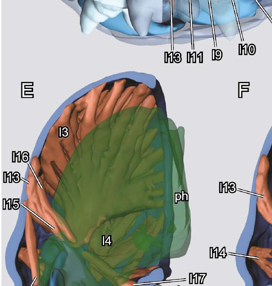
11 ARTHROPOD SYSTEMATICS & PHYLOGENY 73 (1) 2015 Fig. 7. Four day pharate adult of Chrysomela populi, 3D reconstructions of internal structures. A: frontal view, cuticle rendered transparent; B: lateral view, cuticle rendered transparent, muscles removed; C: posterior view, cuticle rendered transparent, right half of labium and right maxilla rendered transparent; D: lateral view, cuticle rendered transparent; E: midsagittal section, pharynx made transparent; F: midsagittal section, pharynx, labium and parts of musculature removed, maxilla rendered transparent; G: midsagitttal section, head capsule only. Abbreviations: ata: anterior tentorial arm; br: brain; cl: clypeus; dta: dorsal tentorial arms; fcr: frontoclypeal; gu: gula; lb: labrum; md: mandible; mxp: maxillary palp; ph: p1 p16: musculature; pharynx; por: postocciptal ridge; pta: posterior tentorial arm; sc: scapus; sog: suboesophageal ganglion; tb: tentorial bar. Scale bar: 1 mm. Table 2. Musculature of the 4 day pharate adult of Chrysomela populi. Number Origin Insertion p1 anterior and dorsal tentorial arm ventral base of antenna p2 dorsal tentorial arm dorsal base of antenna p3 dorsal scapal wall lateral edge of pedicel p4 dorsal scapal wall mesal edge of pedicel p5 mesally on posterior frons antero-lateral frons, in between anterior and dorsal tentorial arms p6 lateral and dorsal head capsule with a tendon on median mandibular wall p7 lateral head capsule, ventrad p6 with a tendon on lateral mandibular wall p8 in 2 distinct bundles on latero-ventral head capsule, partly on tentorial bar cardinal process, bundle 1 on edge of the process, bundle 2 on its distal margin p9 ventro-lateral head capsule, close origin of tentorial bar basal edge of lacinia p10 mesal stipital wall mesally on palpomere 1 p11 base of tentorial bar basal edge of prementum p12 gula latero-basal edge of prementum p13 base of tentorial bar, close to p11 dorsal labial area, border to ligula p14 frons inner epipharyngeal wall p15 mesally along frons along dorsal pharynx, anterad brain p16 base of tentorial bar ventral side of pharynx Dorsal arms (dta; Fig. 7G) longer than anterior ones and also massive. Also attached to head capsule along almost their entire length. Corpotentorium and posterior arms absent. Labrum and epipharynx. Labrum (lb; Figs. 6A B, 7G) sclerotized, short, approximately trapezoid, with strongly rounded anterolateral edges and median emargination. Epipharynx membranous, with well-developed median longitudinal bulge. Antennae. Inserted below oblique anterolateral edge of head capsule (ant; Fig. 6A C), anterior to compound eyes; antennifer not present. Moderately long, posteriorly almost reaching bases of elytra, composed of scapus, pedicellus, and nine distinct flagellomeres. Scapus (sc; Fig. 7A) longer than all other antennomeres except apical one; articulatory part separated from larger club-shaped distal portion by indistinct impression; anterior edge strongly rounded, posterior edge almost straight; surface of distal part smooth. Pedicellus smaller than scapus, shorter than following antennomeres; with narrower basal part and broader, larger distal portion. Flagellomere 1 similar to pedicellus but longer apically, distinct ringshaped basal bulge invisible; flagellomere 2 similar but slightly shorter; flagellomere 3 similar in shape to 2; flagellomere 4 shaped like elongate cup; with smooth basal part and widened distal portion; flagellomeres 5 8 similar, but increasing in size and basal portion shorter; apical antennomere as large as scapus; with short basal part; middle region cylinder-shaped and distal part conical. Pubescence of antennae invisible. Mandibles. Slightly asymmetric mandibles (md; Figs. 6A C, 7A,B,D,F) strongly sclerotized, stout, roughly triangular in dorsal view, with rounded lateral margins. External side broad at base and narrowing anteriorly. Distal part of mandible divided into three well-developed, roughly triangular teeth arranged in a vertical row (Fig. 7B,F); middle tooth larger than others, with oblique, slightly irregular edge; dorsal tooth about equally long, more acuminate; ventral tooth quite distant from mandibular apex and blunt. Maxillae. Maxilla (mx; Fig. 7C,F) composed of cardo, stipes, galea and lacinia and palp. Borders between subelements indistinct. Cardo small, subtriangular, with oblique anterior edge. Stipes with triangular lateral bulge, subdivided into basi- and mediostipes by basally obliterated not recognizable suture. Lacinia and galea present. Proximal galeomere parallelogram-shaped in ventral view; distal galeomere inserted in distal articulation area of galeomere 1; apically rounded. Palpifer weakly developed, laterally adjacent to stipes and proximal galeomere; comparatively small but distinctly visible in ventral view and well-sclerotized. Palp (mxp; Figs. 6A C, 7A,B) composed of four relatively thick segments, longer than distance from cardinal base to distal margin of galea; palpomere 1 short, inserted on palpifer; palpomere 2 about twice as long, distally widening, with strongly 139
12 GE et al.: Metamorphosis of Chrysomela populi convex posterior margin, thus appearing curved inwards; palpomere 3 similarly shaped but slightly shorter; palpomere 4 about as long as 2, spindle-shaped; widening after shallow constriction in proximal third; evenly narrowing towards apex; apical region rounded, with anteriorly oriented. Labium. Labium composed of submentum, mentum and prementum. Submentum large, fused with adjacent regions of head capsule laterally; posterior part moderately converging anteriorly, laterally delimited by rather indistinct furrow; anterior part adjacent to maxillary articulating area, lacking defined lateral border. Mentum small, transverse; connected with anterior submental margin; anterior and posterior margins almost straight. Prementum well-developed, about as large as mentum; cranial edge round (ligula). Palpiger not cleary distinguishable. Labial palp (lp; Fig. 6A C, 7A,B) similar to maxillary palp, but 3-segmented and shorter; palpomere 1 short and stout, cylindrical; palpomere 2 similar to maxillary palpomere 2 but slightly smaller; apical palpomere 3 very similar to terminal maxillary palpomere. Pharynx. Pharynx of pharate adult round to oval in cross section, its diameter at least 19% of head capsule. Cerebrum and suboesophageal ganglion. Brain of pharate adult in central head region, occupying only a small part of entire cephalic lumen. Pars intercerebralis of brain narrow. Protocerebral hemispheres continuous with compact proximal parts of developing optic lobes; lobes laterally branching repeatedly before reaching developing compound eye, thus forming a bush-like pattern (Fig. 7A B). Recognizable external boundaries between proto- and deutocerebrum not distinguishable. Tritocerebral commissure present. Circumoesophageal connectives broader than in adult. Suboesophageal ganglion small, located in posteroventral head region. Musculature. The musculature of the pupa (within the pharate adult, see above) is illustrated in Figure 7 and described in Table 2. In some cases muscle bundles and fibers not well developed and generally muscles appear frayed. Additionally some muscles do not attach to the cuticle Adult Figs Fully sclerotized, without exposed membranes (Fig. 1C). Intensive coloration, with red elytra and other exposed body parts black. Distinctly convex, especially on dorsal side. Head capsule. Subprognathous, slightly inclined, angle between longitudinal body axis (indicated by foramen occipitale) and mouthparts 154, as wide as anterior pronotal emargination. Posterior half retracted into prothorax (Fig. 1C). Shape almost square, about as wide as long, with parallel lateral margins behind compound eyes; posterior edges rounded; anterior part in front of compound eyes, short and broad, trapezoid (Fig. 8A). Strongly sclerotized, with thick cuticle. Setation as shown in Figure 8; longer setae absent except for clypeus; anterior half with fairly dense pattern of punctures; these less dense and less distinct on smooth surface of retracted posterior half of head capsule (Fig. 8A). Circumocular ridge scarcely recognizable externally but well developed internally; supraorbital suture absent. Compound eyes (ce; Figs. 8A,B, 11A D) moderately sized, kidney shaped, rather elongate in vertical direction; with nearly round, distinctly convex, cuticular lenses; longitudinal central region more prominent than marginal areas. Frons (fr; Fig. 8A) short, triangular, enclosed by distinct, short and straight frontal sutures (fs; Fig. 8A); frontal sutures meet posterololateral clypeal margin anteriorly, distinctly separated from oblique anterolateral antennal articulatory areas; frontal tubercles absent. Coronal suture present, fairly distinct on anterior half of head capsule, obliterated posteriorly (cs; Fig. 8A). Vertex (vx; Fig. 8A) not separated from genal region; rather shallow rounded prominence on posterior margin flanked by shallow paired emarginations. Genal region posterior to compound eyes largely smooth, with indistinct, irregular vertical furrows. Frontoclypeal ridge (fcr; Fig. 11E) well developed internally; externally visible as a shallow furrow. Clypeus (cl; Figs. 8A, 11E) undivided, very short, about 10 as wide as long; lateral margin distinctly separated from anterolateral edge of head capsule; anterior edge very slightly convex. Anterior tentorial groove not visible externally. Subgenal ridge absent. Antennal insertions broadly separated, insertion area covered by oblique edge in dorsal view; anteriorly, ventrally and posteriorly enclosed by deep and very distinct furrow. Distinct bulge present between lateral mandibular articulating area and compound eye, and posterior to shallow maxillary groove; latter section separated from adjacent parts of ventrolateral head capsule by deep furrow. Gula present, transversely extended, short and broad, enclosed by very deep posterior tentorial pits; anteriorly completely fused with large submentum; internal gular ridge short, connected with base of posterior tentorial groove. Foramen occipitale large dorsally but distinctly less wide than maximum width of head capsule; dorsally and laterally enclosed by well-developed postocciptal ridge. Internal surface of frons with a Y-shaped sclerotized structure with short basal part. Cephalic endoskeleton. Nearly straight and simple tentorial bars (tb; Fig. 11E) arise from massive posterior base of tentorium, which is continuous with short gular ridge mesally and with well-developed postoccipital ridge laterally (por; Fig. 11E). Short anterior arms form nearly 90 angle with tentorial bars, attached to frontocly peal ridge near anterior mandibular articulation. Posterodorsally oriented dorsal arms (dta; Fig. 11E) arise from anterior arms; apical part attached to posterior frons. Corpotentorium absent. Labrum and epipharynx. Labrum (l b; Figs. 8, 11E) sclerotized, short, approximately trapezoid, with strongly rounded anterolateral edges and median emargination; movably attached to anterior clypeal margin by internal membranous fold; strongly developed, elongate tormae 140


, anterior to upper part of compound eyes; antennifer not present.")
 longer than all other antennomeres except for apical one; globular and glabrous articulatory part separated form larger club-shaped distal portion by distinct impression;")
 much smaller than scapus, shorter than following antennomeres; with narrower basal part and distinctly widening, larger distal portion; vestiture similar to that of distal")
13 ARTHROPOD SYSTEMATICS & PHYLOGENY 73 (1) 2015 Fig. 8. Adult of Chrysomela populi, SEM micrographs. A: head, dorsal view; B: head, lateral view; C: head, frontal view; D: mouthparts, frontal view. Abbreviations: cd: cardo; ce: compound eye; cl: clypeus; cs: coronal suture; fr: frons; fs: frontal suture; gu: gula; lb: labrum; lbr: labrum; lig: ligula; lp: labial palp; md: mandible; mt: mentum; mx: maxilla; mxp: maxillary palp; pe: pedicellus; pf: palpifer; pm: prementum; sc: scapus; smt: submentum; st: stipes; vx: vertex. Scale bar: 1 mm. (to; Fig. 11E) protrude deeply into lumen of head capsule from posterolateral labral edges. Numerous very short sensilla inserted medially on anterior margin; upper surface with six symmetrically placed setiferous punctures. Lateral and anterolateral margin without bristles or spines. Epipharynx membranous, with well-developed median longitudinal bulge. Antennae. Inserted below oblique anterolateral edge of head capsule (Fig. 9A), anterior to upper part of compound eyes; antennifer not present. Moderately long, posteriorly almost reaching bases of elytra, composed of scapus, pedicellus, and nine flagellomeres. Scapus (sc; Figs. 8A C, 9A) longer than all other antennomeres except for apical one; globular and glabrous articulatory part separated form larger club-shaped distal portion by distinct impression; anterior edge strongly rounded, posterior edge almost straight; surface of distal part smooth, with relatively scarce vestiture of punctures. Pedicellus (pe; Figs. 8B, 9A) much smaller than scapus, shorter than following antennomeres; with narrower basal part and distinctly widening, larger distal portion; vestiture similar to that of distal scapus and next three antennomeres. Flagellomere 1 similarly shaped as pedicellus but distinctly longer and less broad apically; with distinct ring shaped basal bulge (fl1; Fig. 9A B); flagellomere 2 similar but slightly shorter and lacking bulge (fl2; Fig. 9A); flagellomere 3 similar in shape to 2; flagellomere 4 shaped like elongate cup; with smooth basal part and widened and densely pubescent distal portion; following four flagellomeres similar, but increasing in size and 141

 with typical dicondylic articulation with strongly developed ventral condyle. Strongly sclerotized, stout, roughly triangular in dorsal view, with rounded lateral margins.")

. Maxillae. Maxilla (mx; Figs.")

14 GE et al.: Metamorphosis of Chrysomela populi Fig. 9. Adult of Chrysomela populi, SEM micrographs. A: antenna; B: 1 st flagellomere; C: 8 th flagellomere; D: 9 th flagellomere. Abbreviations: fl1, 2, 3, 4, 5, 6, 7, 8, 9: flagellomeres; pe: pedicellus; sc: scapus. Scale bar: 0.5 mm. glabrous basal portions progressively shorter (Fig. 9C); apical antennomere about as large as scapus; with short glabrous basal part; middle region cylinder-shaped (Fig. 9D). Mandibles. Slightly asymmetric mandibles (md; Figs. 8B,D, 10A C, 11C,F G) with typical dicondylic articulation with strongly developed ventral condyle. Strongly sclerotized, stout, roughly triangular in dorsal view, with rounded lateral margins. External side broad at base and narrowing anteriorly, basally shallowly concave, with punctures; additional oblique and parallel-sided concavity present on middle region of external side. Distal part of mandible divided into three strongly developed, roughly triangular teeth arranged in a vertical row (Fig. 10A C); middle tooth larger than others, with oblique, slightly irregular edge; dorsal tooth about equally long, more acuminate; ventral tooth quite distant from mandibular apex and blunt. Mesal side of mandible extensive and slightly concave, with large, concave membranous lobe densely covered with microtrichia (ml; Fig. 10B). Maxillae. Maxilla (mx; Figs. 8D, 11B) composed of cardo (ca; Fig. 10D), subdivided stipes (ca; Fig. 10D), galea (ga; Fig. 10D), lacinia (la; Fig. 10D) and palp. Cardo small, subtriangular, with oblique anterior edge; surface smooth, lacking spines or punctures. Stipes divided into basi- and mediostipes by basally obliterated suture. Basistipes shaped like elongate triangle; shortest side connected with anterior cardinal edge. Shape of mediostipes similar but with reversed orientation. Lacinia articulates with distinct mesal prominence of basistipes; curved; ventral surface of distal part with vestiture of extremely small, fine microtrichia. Proximal galeomere parallelogram-shaped in ventral view, completely fused with distal mediostipes; distal galeomere inserted into distal articulation area of galeomere 1; apically rounded. Palpifer laterally adjacent with distal basistipes and proximal galeomere; comparatively small but distinctly visible in ventral view and well-sclerotized. Palp (mxp; Figs. 8B,D, 10D, 11B D,F) composed of four relatively broad segments, longer than distance from cardinal base to distal margin of galea; palpomere 1 short, inserted on palpifer; palpomere 2 about twice as long, distally widening, with strongly convex posterior margin, thus appearing curved inwards; with one long seta and several shorter ones; palpomere 3 similarly shaped but slightly shorter; palpomere 4 about as long as 2, spindle-shaped; 142


; connected with anterior submental margin by internal membrane; anterior and posterior margins almost straight.")
, enclosed by paired extensions formed anterior to insertion areas of palps.")
15 ARTHROPOD SYSTEMATICS & PHYLOGENY 73 (1) 2015 Fig. 10. Adult of Chrysomela populi, SEM micrographs. A: mandible, dorsal view; B: mandible, ventral view; C: mandible, lateral view; D: maxilla; E: labium. Abbreviations: ca: cardo; co: condyle; ga: galea; la: lacinia; lig: ligula; lp: labium palp; mnt: mentum; mxp: maxilla palp; ml: membranous lobe. Scale bar: 0.2 mm widening after shallow constriction in proximal third; evenly narrowing towards apex; surface with microreticulation; apical region rounded, with anteriorly oriented, sharply delimited oblique sensorial area with numerous minute sensilla; apex with digitiform sensilla. Labium. Labium composed of submentum, mentum and prementum. Submentum large, fused with adjacent regions of head capsule laterally; posterior part moderately converging anteriorly, laterally delimited by rather indistinct furrow; anterior part adjacent to maxillary articulating area, lacking defined lateral border. Mentum small, transverse, inserted between stipites (mnt; Figs. 8B,D, 10D); connected with anterior submental margin by internal membrane; anterior and posterior margins almost straight. Anterior labium fused with hypopharynx, both forming a labiohypopharyngeal complex or prelabium; salivarial duct appeares dorsoventrally compressed in cross section, with outer edges slightly bent downwards. Prementum well-developed, about as large as mentum; ventral surface glabrous; cranial edge bilobed (ligula), enclosed by paired extensions formed anterior to insertion areas of palps. Palpiger not present as separate, defined sclerite. Labial palp (lp; Figs. 8B,D, 10E) similar to maxillary palp, but 3-segmented and shorter; palpomere 1 short and stout, cylindrical; palpomere 2 similar to maxillary palpomere 2 but slightly smaller; apical palpomere 3 very similar to terminal maxillary palpomere, also with oblique area with numerous sensilla, short, fine setae and microreticulation. Pharynx. Prelabium and epipharynx separate. Pharynx (ph; Fig. 11A B,F) round in cross section, its diameter approximately 15% of diameter of head capsule. Internal surface with bulges to increase surface area. Oesophagus extremely widened in posterior region of head capsule. 143


16 GE et al.: Metamorphosis of Chrysomela populi 144
17 ARTHROPOD SYSTEMATICS & PHYLOGENY 73 (1) 2015 Fig. 11. Adult of Chrysomela populi, 3D reconstructions of internal structures. A: dorsal view, cuticle rendered transparent; B: lateral view, cuticle rendered transparent, muscles removed; C: ventral view, cuticle rendered transparent, right half of labium and right maxilla rendered transparent; D: lateral view, cuticle rendered transparent; E: midsagittal section, head capsule only; F: midsagittal section, pharynx and labium rendered transparent; G: midsagittal section, pharynx, labium and parts of musculature removed, maxilla rendered transparent. Abbreviations: a1 a27: musculature; br: brain; ce: compound eye; cl: clypeus; dta: dorsal tentorial arm; fcr: frontoclypeal ridge; la: labium; lb: labrum; md: mandible; mx: maxilla; mxp: maxillary palp; ph: pharynx; por: postocciptal ridge; sog: suboesophageal ganglion; tb: tentorial bar; to: torma. Scale bar: 1 mm. Table 3. Musculature of the adult of Chrysomela populi. Number Origin Insertion Function a1 along entire dorsal tentorial arm ventrally on basal edge of scape depressor of the antenna a2 dorsal area of corpotentorium dorsally on basal edge of scape levator of the antenna a3 dorsal tentorial arm laterally on basal edge of scape depressor and rotator of the antenna a4 mesal frons, directly posterad epistomal ridge dorso-mesally on basal edge of scape levator and rotator of the antenna a5 dorsal scapal wall lateral edge of pedicel extensor of the flagellum a6 dorsal scapal wall, mesad a5 ventro-mesal edge of pedicel flexor of the flagellum a7 dorsal scapal wall, mesad a6 dorso-mesal edge of pedicel flexor of the flagellum a8 dorsal head capsule distal tip of elongate tormae depressor of the labrum a9 epistomal ridge distal tip of elongate tormae, near a8 levator of the labrum? a10 anterior torma anterior torma of opposite side, thus overspanning anterior pharynx? a11 labral wall dorsal epipharyngeal wall dilator of the epipharynx a12 entire lateral head capsule with a tendon on median mandibular wall adductor of the mandible a13 latero-ventral head capsule with a tendon on lateral mandibular wall abductor of the mandible a14 distal edge of torma postero-mesal edge of mandible adductor of the mandible? a15 in 2 distinct bundles on ventral head capsule, partly on the posterior tentorial arms cardinal process, bundle 1 on edge of the process, bundle 2 on its distal margin promotor of the maxilla a16 base of dorsal tentorial arms basal edge of lacinia adductor of the lacinia a17 mesal stipital wall basal edge of first maxillary palpomere flexor of the maxillary palp a18 basal edge of palpomere 1 basal edge of palpomere 2 flexor of the 2 nd palpomere a19 basal edge of palpomere 2 basal edge of palpomere 3 flexor of the 3 rd palpomere a20 basal edge of palpomere 3 basal edge of palpomere 4 flexor of the 4 th palpomere a21 anterior edge of postmentum posterior premental rim retractor of the prementum a22 basal prementum mesal edge of labial palp adductor of the labial palp a23 basal prementum, close to a22 lateral edge of labial palp abductor of the labial palp a24 base of tentorial bar between prementum and hypopharynx on the ligula retractor of the labium and ligula a25 frons dorsal epipharyngeal wall dilator of the cibarium a26 anterior half of frons dorsal pharyngeal wall dilator of the pharynx a27 mesally on frons, far behind a26 and anterad brain along the entire dorsal side of pharynx, reaching below brain dilator of the pharynx a28 base of tentorial bar ventro-lateral pharynx dilator of the pharynx a29 ring muscle layer around pharynx constrictor of the pharynx a30 longitudinal muscle layer along pharynx contractor of the pharynx Nervous system. Central pars intercerebralis of brain (br; Fig. 11A) small in relation to head size, with slightly protruding protocerebral hemispheres. Optic lobes fully developed, forming large proportion of cephalic part of central nervous system, with extensive laminae ganglionares adjacent to compound eyes. Deutocerebrum not recognisable as separate unit externally. Tritocerebral commissure present. Circumoesophageal connectives fairly elongate. Suboesophageal ganglion (sog; Fig. 11C) about as wide as submentum, located in posteroventral head region. Musculature. The musculature of the adult is illustrated in Fig. 11 and described in Table Discussion 4.1. Methodological aspects and implications Insect metamorphosis is a complex and fascinating process. From a morphological point of view its study and documentation is a great challenge as it is highly dynamic and covertly taking place within a pupal sheath. The traditional techniques for studying internal struc- 145
18 Ge et al.: Metamorphosis of Chrysomela populi tures are dissections or histological sections. The former has narrow limitations in terms of size, the latter is time-consuming, and both are invasive (Friedrich et al. 2014). Moreover, sections are technically demanding to produce and prone to artifacts such as compression and loss or folding of individual slices. Thus it is not surprising that several new and innovative methods have been developed and optimized in the last decade. They greatly increase the efficiency of morphological investigations and also facilitate the observation of insects during metamorphosis. Mapelli et al. (1997) investigated metamorphosis for the first time without relying on histological methods or dissections. They used magnetic resonance imaging (MRI) to document the development of an individual of Bombyx mori from a young larva to an adult moth with a focus on the silk glands. MRI uses nuclear magnetic resonance to visualize internal features (see Hart et al for a review of its application in entomology). This technique does not have a recognizable harmful effect on live specimens, but has a rather low spatial resolution and a weak contrast between different tissues (Hart et al. 2003). Michaelis et al. (2005) followed this approach and studied the brain of the moth Manduca sexta. Pelling et al. (2009) combined MRI with optical beam deflection to study the heartbeat of a monarch butterfly during metamorphosis. Micro-computed tomography (µ-ct) yields superior resolution but involves the application of potentially harmful X-rays. It has been used in insect morphology for more than a decade and is established as a standard technique today (Deans et al. 2012; Friedrich et al. 2014). However, it was not used to investigate metamorphosis until very recently when Lowe et al. (2013) studied the tracheal and digestive systems of live specimens of the butterfly Vanessa cardui. One of the reasons this approach was not used earlier is the presence of water in the live insect, which greatly reduces the contrast between different tissues thus making their distinction almost impossible. Thus Lowe et al. (2013) could only study the trachea and parts of the gut, tissues with a distinctly different density. To differentiate between soft tissues like muscles, nerves or glands, samples have to be dried at the critical point or dehydrated with suitable chemicals. Even though this is obviously not possible with live material, we followed this approach in the present study to document skeleto-muscular changes during metamorphosis for the first time using µ-ct. As a result, we could not follow the development of single individuals over several stages as in the studies on Bombyx mori, Manduca sexta and Vanessa cardui (Mapelli et al. 1997; Michaelis et al. 2005; Loew et al. 2013). Nevertheless, this approach is highly efficient for documenting internal structures of different developmental stages, allowing a detailed comparison between them. In future studies it will be a valuable tool to investigate a series of sequential pupal stages, which will eventually lead to a much more detailed description of the dynamic process of metamorphosis than was previously possible Developmental transformations of cephalic structures The main focus of this study was the detailed documentation of transformations in the skeleto-muscular system between different developmental stages of the flower beetle Chrysomela populi. The head morphology of the larva and adult differ extensively and nearly all character systems are affected by metamorphosis. In Table 4 the muscles are homologized between the stages and Table 5 summarizes all observed differences. The specialization of larvae and adults for life in different habitats and microhabitats and on diverse food substrates results in a reduced intraspecific competition between these stages. This was addressed as one factor contributing to the unparalleled evolutionary success of Holometabola (e.g., Grimaldi & Engel 2005; Beutel et al. 2011). It is also conceivable that a division of labor between developmental stages may have resulted in selection for reduced equipment in larvae, which do not need elaborate sense organs such as those on the antennae of adults or the compound eyes and no organs for dispersal over long distances. In Chrysomelidae (and most other endopterygotes) the main function of the phytophagous larva is feeding and accumulation of energy-rich substances in its fat body, whereas the principal functions of the adults are dispersal and reproduction (Chen 1964). Our study reveals how these divergent functions affect the metamorphosis of different structural elements of the head. In the larva the digestive system is greatly enlarged, and the diameter of the pharynx is at least 52% that of the entire head capsule, compared to only 15% in the adult. Due to the tremendously enlarged larval cephalic foregut and the resulting limitation of space in the remaining parts of the head, portions of the cephalic nervous system are shifted into the prothorax. The larval mandibles are equipped with 5 distal teeth with serrate lateral edges, which allow them to cut leaf surfaces efficiently. Chrysomela populi feeds on Chinese white poplar (Populus tomentosa) in all four active stages (note: the pupa doesn t feed so only the 3 larval and the adult stage consume food) (Yu et al. 1996). However the larvae prefer fresh and thus softer leaves while adults usually feed on older ones with a thicker cuticle and cell walls (pers. obs. S.-Q. Ge). The role of the membranous basal lobe of the adult mandible (Fig. 10B,C) in this context remains unclear. As an unsclerotized structure it is probably not suitable for grinding more solid substrates (Yu et al. 1996). The most conspicuous change affects the orientation of the head: the larva is orthognathous while the adult is subprognathous. The larval legs, especially the prothoracic ones are very short (Fig. 1A). Thus the body is almost adjacent to the surface of the leaf and the larva feeds directly on the portion below its ventrally directed mouthparts (pers. obs. S.-Q. Ge). There is no easy functional explanation for the subprognathous head of adults. The body with its distinctly longer legs is held well above 146
19 ARTHROPOD SYSTEMATICS & PHYLOGENY 73 (1) 2015 Table 4. Homologisation of the musculature of Chrysomela populi with the terminology of Wipfler et al. (2011). Number Name Larva Pupa Adult 0an1 M. tentorioscapalis anterior p1 a1 0an2 M. tentorioscapalis posterior p2 a2 l1 0an3 M. tentorioscapalis lateralis a3 0an4 M. tentorioscapalis medialis a4 0an6 M. scapopedicellaris lateralis p3 a5 0an7 M. scapopedicellaris medialis p4 a6 + a7 0lb2 M. frontoepipharyngalis l2 p5 a8 + a9? 0lb4 M. labralis transversalis a10 0lb5 M. labroepipharyngealis a11 0md1 M. craniomandibularis internus l3 p6 a12 0md3 M. craniomandibularis externus posterior l4 p7 a13? 0md6 M. tentoriomandibularis lateralis inferior a14? 0mx1 M. craniocardinalis p8 a15 0mx2 M. craniolacinialis l8 p9 a16 0mx3 M. tentoriocardinalis l5 0mx4 M. tentoriostipitalis anterior l7? 0mx5 M. tentoriostipitalis posterior l6 0mx6 M. stipitolacinialis l9 0mx8 10 M. stipitopalpalis l10 p10 a17 0mx12 M. palpopalpalis maxillae primus a18 0mx13 M. palpopalpalis maxillae secundus a19 0mx14 M. palpopalpalis maxillae tertius a20 0la5 M. tentoriopraementalis l11 p12 0la8 M. submentopraementalis p11 a21 0la13 M. praementopalpalis internus a22 0la14 M. praementopalpalis externus a23 0hy1 M. frontooralis l12 0hy2 M. tentoriooralis l13 p13 a24 0ci1 M. clypeopalatalis l14 p14 a25 0bu2 M. frontobuccalis anterior l15 p15 a26 0bu3 M. frontobuccalis posterior l16 p15 a27 0bu5 M. tentoriobuccalis anterior l17 0bu6 M. tentoriobuccalis posterior l18 0ph2 M. tentoriopharyngealis l19 p16 a28 0st1 M. annularis stomodaei l20 a29 0st2 M. longitudinalis stomodaei l21 a30 the plant surface (Fig. 1C). Thus the beetles have to bend their heads downward to feed on the leaf they stand on (pers. obs. S.-Q. Ge) although other chrysomelid adults usually feed on the leaf margins. It is likely that the subprognathous orientation is simply a relatively slight modification of the prognathous condition, which is a groundplan feature of adult Coleoptera. The change in head orientation has profound consequences for the cephalic morphology: with the lifting of the mouthparts resulting in an almost horizontal orientation of the head capsule, a posteroventral gap between the posterior margin of the ventral mouthparts and the foramen occipitale has to be closed by an additional structure. This is the gula of adults, which not only closes the head ventrally but also increases mechanical stability in the ventral head area between the right and left hemispheres. Thus it functionally replaces the corpotentorium, which provides this support in the larva, but which is reduced in adults, along with the muscles originating on it (M. tentoriobuccalis anterior and posterior, M. tentoriocardinalis, M. tentoriostipitalis anterior and posterior, M. tentoriopraementalis). These muscles are partly replaced by others: M. submentopraementalis of adults functions as a premental retractor instead of the larval M. tentoriopraementalis. M. craniocardinalis of adults is present instead of the larval M. tentoriocardinalis, but in this case both muscles have a different function. The distinct subdivision of the postmentum into a well-defined mentum and submentum in adults is possibly also related to the modified orientation of the mouthparts. Flight capacity and the necessity to find a potential mating partner requires a far more complicated sensory system in the adult than is present in larvae: instead of simple stemmata, complex compound eyes are present. This change in the visual system also requires strong modifications in the brain, notably in the optic lobes which greatly increase in size. The antennae are greatly elongated in adults and the number of antennomeres is increased from three to eleven. To ensure controlled movements of adult antennae a more complex muscle system is required. Four extrinsic and two intrinsic muscles are present in antennae of adults, whereas only one extrinsic bundle is present in those of larvae. It is conceivable that the absence of the anterior and dorsal tentorial arms in larvae is related to the greatly reduced condition of the extrinsic antennal muscles as the only larval muscle originates on head capsule cuticle. The labial and maxillary palps bear one additional segment each and their movability is increased. Intrinsic muscles are present in the adult maxillary palp and the labial palp is controlled by two extrinsic muscles absent in larvae. In addition to modifications more or less closely related to the different functional requirements of the developmental stages, the adults exhibit some differences which cannot be easily explained in such context. The adult labrum is characterized by strongly elongate tormae and a more complex musculature obviously resulting in increased movability. The comparatively restricted movability of the labrum of larvae is seemingly in conflict with their function as the main feeding stages. This possibly relates to the reduced complexity of most larval cephalic and postcephalic body parts, which is likely a derived groundplan features of Holometabola (e.g., Beutel et al. 2011). Holometabolan larvae display a minimum of structural complexity, which allows them to fulfill their necessary function of feeding, but reduces investment in developing complicated structures only useful in adults. Such also applies to characters of the maxillary and labial palps and the posterior labium. The mouthparts differ in several aspects between larvae and adults. Most changes are likely associated with the different tasks of these life stages and have been discussed above. The undivided larval mala is replaced with separate endite lobes in the adults, the laciniae and galeae. These larval features apparently have a historical background and the functional significance of the fusion is difficult to assess. In the larval groundplan of Coleoptera and Polyphaga the lacinia and galea are clearly 147
20 Ge et al.: Metamorphosis of Chrysomela populi separate (Beutel 1997), whereas an undivided mala is almost universally present in larvae of the extremely diverse Cucujiformia (e.g., Leschen et al. 2010; Leschen & Beutel 2014), to which the Chrysomeloidea belong. This is probably an apomorphy of a very large subunit of Cucujiformia and this derived condition is preserved in Chrysomelidae (Crowson & Crowson 1996). In addition to muscular changes associated with reduction of the corpotentorium (see above), M. stipitolacinialis is present in larvae but not in the adults. This is surprising as one would assume that separate endite lobes in the adult would require more muscular control than in the larva with its undivided mala. Morphological changes occurring in beetles during metamorphosis are insufficiently known: the larva and adult of the ptiliid Mikado sp. were compared by Polilov & Beutel (2009) and those of the corylophid Sericoderus lateralis by Polilov & Beutel (2010). These species differ in several ways from Chrysomela populi: both the larvae and adults are prognathous, and consequently several changes associated with the reorientation of the head capsule in Chrysomela do not occur: the gula is absent in adults of Mikado, whereas it is present and fused with the head capsule in adults and larvae of Sericoderus. The tentorial bridge is present in all stages of both species. Larval anterior tentorial arms are present in Mikado while they are absent in Seridocerus and Chrysomela. The larval antennal muscles of Mikado originate on the anterior tentorial arm in contrast to Chrysomela, and only the dorsal tentorial arm is missing (no information provided for Seridocerus; Polilov & Beutel 2009). This confirms that the presence of anterior tentorial arms in larvae is related to their function as area of origin of antennal muscles. Other features are similar in all three species: in adults the number of antennomeres increases to 11 (versus three in the larvae), a 4 th maxillary palpomere is present, the volume of the digestive system strongly decreases, and the volume of the elements of the central nervous system increases. The functional forces driving morphological change between larvae and adults of Chrysomela mainly feeding in the larval stages and dispersal and mating in adults, associated with greatly improved sensory (and locomotor) organs is similar in Mikado and Seridocerus. However, miniaturization and the resulting limitation of space also play an essential role in these two very small species, especially in the minute first instar larvae (Polilov & Beutel 2009, 2010) The pupa Within the 4 th day pupal sheath the pharate adult shows an interesting combination of larval and adult characters. Table 5 provides an overview of similarities and differences between these stages. In general the pharate adult resembles the adult in almost all skeletal elements including sclerotized elements of the compound eyes, the 11-segmented antennae, the mandible, the maxilla with separate endite lobes and a 4-segmented palp, the presence of a gula, the distinctly subdivided postmentum, and the presence of anterior and dorsal tentorial arms. The only difference found in a skeletal element is the shorter tormae. Aside from these skeletal features, a major transformation takes place in the orientation of the head. The larva is clearly orthognathous with an angle of approximately 87 between the longitudinal body axis and the longitudinal axis of the mouthparts. The adult is subprogna thous with an angle of 154. After 4 days of pupation the head and mouthparts of the pharate adult form an angle of 128 with the longitudinal body axis, a position intermediate between the other two. We therefore assume that the anterior orientation of the mouthparts is a continuous shift rather than a sudden reorientation, and takes place more or less continuously during the six days of pupal metamorphosis. The gula of the pharate adult is not as broad as in the adult. This is possibly related to the incomplete anterior shift of the mouthparts. To confirm these hypotheses, a more detailed study of a sequence of pupal stages would be required. The advanced skeletal modifications of the pupa stand in sharp contrast to the condition of most internal soft parts. The very large larval foregut is already strongly reduced in size: the pharynx occupies at least 50 % of the diameter of the larval head capsule but only 19% in the pupa (day 4) and 15% in the adult. In contrast to this, the musculature and nervous system lag behind in their development. Even though ommatidia of the compound eyes are already present in the pharate adult, the optic lobes are still largely undeveloped and individual nerve cords can still be distinguished, in contrast to the adult where they are fused into a single compact structure. The musculature of the pharate adult appears also intermediate between those of the last instar larva and the adult. Most pupal muscles are much smaller than the corresponding larval or adult ones (e.g., M. craniomandibularis internus and externus) and they appear frayed or do not attach to the head capsule cuticle. In general, five categories of muscle transformations during metamorphosis can be distinguished (Heming 2003: fig ). In addition to muscles which only exist either in the larva or adult, three types are present in both life stages: those which pass unchanged in function and form from larva to adult, those which are respecified in function and those which disintegrate completely and are replaced by a newly formed muscles (Heming 2003: ). As we studied only a single, 4 day pharate adult we were unable to follow the individual fate of each muscle and to distinguish between these types. The only exceptions are M. annularis stomadei and M. longitudinalis stomodei. As they are absent in the studied pharate adult but present in both larva and adult they apparently belong to the last type. This is consistent with a sequence described for the honey bee, where these foregut muscles begin to disintegrated after 45 hours and are replaced by adult muscles after 70 hours (Oertel 1930). In contrast to this, certain unidentified muscles in the head partly disappear and reform according to Oertel (1930). 148

21 Table 5. Observed morphological differences and similarities between the studied life stages of Chrysomela populi. ARTHROPOD SYSTEMATICS & PHYLOGENY 73 (1)
Arthropod Systematics & Phylogeny 70 (1) 3 42 Senckenberg Gesellschaft für Naturforschung, eissn ,
 3 42 Senckenberg Gesellschaft für Naturforschung, eissn ,") Arthropod Systematics & Phylogeny 3 70 (1) 3 42 Senckenberg Gesellschaft für Naturforschung, eissn 1864-8312, 05.04.2012 The adult head morphology of Dascillus (L.) (Dascilloidea: Dascillidae) and Glaresis
Arthropod Systematics & Phylogeny 3 70 (1) 3 42 Senckenberg Gesellschaft für Naturforschung, eissn 1864-8312, 05.04.2012 The adult head morphology of Dascillus (L.) (Dascilloidea: Dascillidae) and Glaresis
Morphology and chaetotaxy of the fìrst-instar larva of Hydraena {Phothydraena) hernandoi FRESNEDA & LAGAR (Coleoptera: Hydraenidae)
 hernandoi FRESNEDA & LAGAR (Coleoptera: Hydraenidae)") Koleopterologische Rundschau 66 147-154 Wien, Juni 1996 Morphology and chaetotaxy of the fìrst-instar larva of Hydraena {Phothydraena) hernandoi FRESNEDA & LAGAR (Coleoptera: Hydraenidae) J.A. DELGADO
Koleopterologische Rundschau 66 147-154 Wien, Juni 1996 Morphology and chaetotaxy of the fìrst-instar larva of Hydraena {Phothydraena) hernandoi FRESNEDA & LAGAR (Coleoptera: Hydraenidae) J.A. DELGADO
Department of Entomology University of New Hampshire Durham, New Hampshire 03824
 LARVAE OF WRACK COLEOPTERA IN THE FAMILIES CORYLOPHIDAE, RHIZOPHAGIDAE, AND LATHRIDIIDAE* BY DONALD S. CHANDLER Department of Entomology University of New Hampshire Durham, New Hampshire 03824 Wrack studies
The First Instar Larva of Hydrobius pauper SHARP (Coleoptera, Hydrophilidae)
") Elytra, Tokyo, New Series, 2 (2): 279 284 December 31, 2012 title 279 The First Instar Larva of Hydrobius pauper SHARP (Coleoptera, Hydrophilidae) Yûsuke MINOSHIMA 1) & Masakazu HAYASHI 2) 1) Systematic
Elytra, Tokyo, New Series, 2 (2): 279 284 December 31, 2012 title 279 The First Instar Larva of Hydrobius pauper SHARP (Coleoptera, Hydrophilidae) Yûsuke MINOSHIMA 1) & Masakazu HAYASHI 2) 1) Systematic
The cuticular structure
 The Exoskeleton = key contributor to insect success External : exoskeleton, apodemes, wings => barrier to outside environment Internal: tracheal tubes, glands, fore and hindgut Ranges from rigid/armor
The head morphology of Micromalthus debilis (Coleoptera: Micromalthidae) an archostematan beetle with an unusual morphology and a unique life cycle
 an archostematan beetle with an unusual morphology and a unique life cycle") 76 (3): 475 486 11.12.2018 Senckenberg Gesellschaft für Naturforschung, 2018. The head morphology of Micromalthus debilis (Coleoptera: Micromalthidae) an archostematan beetle with an unusual morphology
76 (3): 475 486 11.12.2018 Senckenberg Gesellschaft für Naturforschung, 2018. The head morphology of Micromalthus debilis (Coleoptera: Micromalthidae) an archostematan beetle with an unusual morphology
Head anatomy of Xyelidae (Hexapoda: Hymenoptera) and phylogenetic implications
 and phylogenetic implications") Organisms, Diversity & Evolution 7 (2007) 207 230 www.elsevier.de/ode Head anatomy of Xyelidae (Hexapoda: Hymenoptera) and phylogenetic implications Rolf Georg Beutel a,, Lars Vilhelmsen b a Institut für
Organisms, Diversity & Evolution 7 (2007) 207 230 www.elsevier.de/ode Head anatomy of Xyelidae (Hexapoda: Hymenoptera) and phylogenetic implications Rolf Georg Beutel a,, Lars Vilhelmsen b a Institut für
Parasitology Research
 Parasitology Research Electronic Supplementary Material for: Sarcophaga (Liosarcophaga) tibialis Macquart 1851 (Diptera: Sarcophagidae): micromorphology of preimaginal stages of a fly of medical and veterinary
HYDRAENIDAE: III. Description of the third instar larva of Ochthebius (s.str.) gonggashanensis JÄCH. (Coleoptera)
 gonggashanensis JÄCH. (Coleoptera)") JÄCI1 & Jl (eels.): Water Beetles of China Vol. Ill 371-377 Wien, April 2003 HYDRAENIDAE: III. Description of the third instar larva of Ochthebius (s.str.) gonggashanensis JÄCH (Coleoptera) J.A. DELGADO
JÄCI1 & Jl (eels.): Water Beetles of China Vol. Ill 371-377 Wien, April 2003 HYDRAENIDAE: III. Description of the third instar larva of Ochthebius (s.str.) gonggashanensis JÄCH (Coleoptera) J.A. DELGADO
A NEW GENUS, SPECIES, AND SUBTRIBE OF TERMITOPHILOUS STAPHYLINIDAE FROM AUSTRALIA (Coleoptera)
") Pacific Insects 12 (3): 499-596 30 October 1970 A NEW GENUS, SPECIES, AND SUBTRIBE OF TERMITOPHILOUS STAPHYLINIDAE FROM AUSTRALIA (Coleoptera) With a Description of its Glands 1 By David H. Kistner 2 Abstract:
Pacific Insects 12 (3): 499-596 30 October 1970 A NEW GENUS, SPECIES, AND SUBTRIBE OF TERMITOPHILOUS STAPHYLINIDAE FROM AUSTRALIA (Coleoptera) With a Description of its Glands 1 By David H. Kistner 2 Abstract:
Description and identification of white grubs (Coleoptera: Scarabaeidae) that attack pineapple crops in South Africa
 that attack pineapple crops in South Africa") Description and identification of white grubs (Coleoptera: Scarabaeidae) that attack pineapple crops in South Africa T.J. Smith' G.J. Petty2 & M.H. Villet1 Department of Zoology and Entomology, Rhodes
Description and identification of white grubs (Coleoptera: Scarabaeidae) that attack pineapple crops in South Africa T.J. Smith' G.J. Petty2 & M.H. Villet1 Department of Zoology and Entomology, Rhodes
Morphology of the first instar larva of Tenomerga mucida (Chevrolat, 1829) (Coleoptera: Archostemata: Cupedidae)
 (Coleoptera: Archostemata: Cupedidae)") 73 (2): 239 258 20.8.2015 Senckenberg Gesellschaft für Naturforschung, 2015. Morphology of the first instar larva of Tenomerga mucida (Chevrolat, 1829) (Coleoptera: Archostemata: Cupedidae) Margarita I.
73 (2): 239 258 20.8.2015 Senckenberg Gesellschaft für Naturforschung, 2015. Morphology of the first instar larva of Tenomerga mucida (Chevrolat, 1829) (Coleoptera: Archostemata: Cupedidae) Margarita I.
Introduction to Animals
 Introduction to Animals Characteristics of Animals multicellular Except for sponges, animal cells are arranged into tissues. Tissues are necessary to produce organs and organ systems. Tissues, organs,
Mosquito Systematics Vol. 6(Z) June 1974
 June 1974") Mosquito Systematics Vol. 6(Z) June 1974 93 Research on the Mosquitoes of Angola. VII - Redescription of the Larva of Aedes durbanensis durbanensis (Theo., 1903) and Description of Aedes durbanensis angozae
Mosquito Systematics Vol. 6(Z) June 1974 93 Research on the Mosquitoes of Angola. VII - Redescription of the Larva of Aedes durbanensis durbanensis (Theo., 1903) and Description of Aedes durbanensis angozae
GHABBOUR, M.W. Plant Protection Research Institute, Agricultural Research Center, Ministry of Agriculture, Dokki, Egypt.
 Entomologica, Bari, 33, (1999): 73-83 GHABBOUR, M.W. Plant Protection Research Institute, Agricultural Research Center, Ministry of Agriculture, Dokki, Egypt. DESCRIPTIONS OF THE FIRST-INSTAR NYMPHS OF
STRUCTURAL FEATURES OF COCKROACH EGG CAPSULES. II. THE OOTHECA OF CARIBLATTA LUTEA LUTEA (ORTHOPTERA: BLATTIDAE) 1
 1") STRUCTURAL FEATURES OF COCKROACH EGG CAPSULES. II. THE OOTHECA OF CARIBLATTA LUTEA LUTEA (ORTHOPTERA: BLATTIDAE) 1 FRED A. LAWSON Department of Zoology and Entomology, The University of Tennessee, Knoxville
STRUCTURAL FEATURES OF COCKROACH EGG CAPSULES. II. THE OOTHECA OF CARIBLATTA LUTEA LUTEA (ORTHOPTERA: BLATTIDAE) 1 FRED A. LAWSON Department of Zoology and Entomology, The University of Tennessee, Knoxville
Journal of Entomological Society of Iran 2008, 27(2), 1-11
, 1-11") Journal of Entomological Society of Iran 2008, 27(2), 1-11 1 Morphology and ultrastructure of chemosensory sensilla of labiomaxillary complex in the Colorado potato beetle, Leptinotarsa decemlineata (Col.:
Journal of Entomological Society of Iran 2008, 27(2), 1-11 1 Morphology and ultrastructure of chemosensory sensilla of labiomaxillary complex in the Colorado potato beetle, Leptinotarsa decemlineata (Col.:
A new species of the genus Oxyomus Dejean, 1833 from Thailand (Coleoptera: Scarabaeidae: Aphodiinae) Łukasz MINKINA INTRODUCTION MATERIAL AND METHODS
 Łukasz MINKINA INTRODUCTION MATERIAL AND METHODS") Studies and Reports Taxonomical Series 14 (1): 145-149, 2018 A new species of the genus Oxyomus Dejean, 1833 from Thailand (Coleoptera: Scarabaeidae: Aphodiinae) Łukasz MINKINA os. Polana Szaflarska 4/39,
Studies and Reports Taxonomical Series 14 (1): 145-149, 2018 A new species of the genus Oxyomus Dejean, 1833 from Thailand (Coleoptera: Scarabaeidae: Aphodiinae) Łukasz MINKINA os. Polana Szaflarska 4/39,
Amphigomphus somnuki n. sp. from North Thailand (Odonata: Gomphidae) MATTI HAMALAINEN
 MATTI HAMALAINEN") Entomol. Z., 106(5), 1996 177 Amphigomphus somnuki n. sp. from North Thailand (Odonata: Gomphidae) MATTI HAMALAINEN With 8 figures Abstract: A new dragonfly species, Amphigomphus somnuki n. sp. (holotype
Entomol. Z., 106(5), 1996 177 Amphigomphus somnuki n. sp. from North Thailand (Odonata: Gomphidae) MATTI HAMALAINEN With 8 figures Abstract: A new dragonfly species, Amphigomphus somnuki n. sp. (holotype
28. Genus Xoanon Semenov Fig. C28.1 (female dorsal habitus) Fig. C28.2 (female lateral habitus) Fig. C28.3 (male dorsal habitus)
 Fig. C28.2 (female lateral habitus) Fig. C28.3 (male dorsal habitus)") 28. Genus Xoanon Semenov Fig. C28.1 (female dorsal habitus) Fig. C28.2 (female lateral habitus) Fig. C28.3 (male dorsal habitus) Xoanon Semenov, 1921: 87. Type species: Xoanon mysta Semenov, by original
28. Genus Xoanon Semenov Fig. C28.1 (female dorsal habitus) Fig. C28.2 (female lateral habitus) Fig. C28.3 (male dorsal habitus) Xoanon Semenov, 1921: 87. Type species: Xoanon mysta Semenov, by original
The morphology of galerucine and alticine larvae (Coleoptera: Chrysomelidae) and its phylogenetic implications
 and its phylogenetic implications") 72 (2): 75 94 25.7.204 Senckenberg Gesellschaft für Naturforschung, 204. The morphology of galerucine and alticine larvae (Coleoptera: Chrysomelidae) and its phylogenetic implications Yi Hua, 2, 3, Rolf
72 (2): 75 94 25.7.204 Senckenberg Gesellschaft für Naturforschung, 204. The morphology of galerucine and alticine larvae (Coleoptera: Chrysomelidae) and its phylogenetic implications Yi Hua, 2, 3, Rolf
This work is licensed under the Creative Commons Attribution-Noncommercial-Share Alike 3.0 United States License. To view a copy of this license,
 This work is licensed under the Creative Commons Attribution-Noncommercial-Share Alike 3.0 United States License. To view a copy of this license, visit http://creativecommons.org/licenses/by-nc-sa/3.0/us/
Objectives. RECEPTION & INTEGRATION: The Nervous System. Human Olfaction
 RECEPTION & INTEGRATION: The Nervous System Objectives 1. Describe the origin of the insect nervous system. 2. Identify the major structures of the insect nervous system and describe their function. 3.
The head morphology of Pyrrhosoma nymphula larvae (Odonata: Zygoptera) focusing on functional aspects of the mouthparts
 focusing on functional aspects of the mouthparts") Büsse et al. Frontiers in Zoology (2017) 14:25 DOI 10.1186/s12983-017-0209-x RESEARCH Open Access The head morphology of Pyrrhosoma nymphula larvae (Odonata: Zygoptera) focusing on functional aspects of
Büsse et al. Frontiers in Zoology (2017) 14:25 DOI 10.1186/s12983-017-0209-x RESEARCH Open Access The head morphology of Pyrrhosoma nymphula larvae (Odonata: Zygoptera) focusing on functional aspects of
Description of a new species of Hydraena KUGELANN (Coleoptera: Hydraenidae) from the southeastern USA
 from the southeastern USA") Linzer biol. Beitr. 30/1 299-304 31.7.1998 Description of a new species of Hydraena KUGELANN (Coleoptera: Hydraenidae) from the southeastern USA M.A. JÄCH & J. A. DIAZ Abstract: Hydraena cornelli n.sp.
Linzer biol. Beitr. 30/1 299-304 31.7.1998 Description of a new species of Hydraena KUGELANN (Coleoptera: Hydraenidae) from the southeastern USA M.A. JÄCH & J. A. DIAZ Abstract: Hydraena cornelli n.sp.
Ceratophysella michalinae, a new species from Poland (Collembola: Hypogastruridae)
") Genus Vol. 16 (1): 1-5 Wroc³aw, 31 III 2005 Ceratophysella michalinae, a new species from Poland (Collembola: Hypogastruridae) DARIUSZ SKAR YÑSKI Zoological Institute, Wroc³aw University, Przybyszewskiego
Genus Vol. 16 (1): 1-5 Wroc³aw, 31 III 2005 Ceratophysella michalinae, a new species from Poland (Collembola: Hypogastruridae) DARIUSZ SKAR YÑSKI Zoological Institute, Wroc³aw University, Przybyszewskiego
^ ^ LIBRARY Division of Crustace;
 /re/// ^ ^ LIBRARY Division of Crustace; Larval Development of Helice tridens wuana Rathbun ^Y RTEBRAT and H. tridens tridens de Haan (Crustacea, Brachyura) ZOOLOGY Reared in the Laboratory Crustacea By
OF THE LEMNA FROND MORPHOLOGY
 MORPHOLOGY OF THE LEMNA FROND FREDERICK H. BLODGETT (WITH PLATE XIV AND ONE FIGURE) In the case of structure simplified by reduction, it is sometimes necessary to trace the development of the parts through
New long-proboscid lacewings of the mid-cretaceous provide insights into ancient. Gorges Entomological Museum, P.O. Box 4680, Chongqing , China.
 Supplementary Materials. New long-proboscid lacewings of the mid-cretaceous provide insights into ancient plant-pollinator interactions Xiu-Mei Lu a, Wei-Wei Zhang b, Xing-Yue Liu a * a Department of Entomology,
Morphology of the Immature Stages of Hydrochara libera (SHARP) (Coleoptera, Hydrophilidae)
 (Coleoptera, Hydrophilidae)") Elytra, Tokyo, New Series, 2 (2): 285 302 December 31, 2012 Immature Stages of title Hydrochara libera 285 Morphology of the Immature Stages of Hydrochara libera (SHARP) (Coleoptera, Hydrophilidae) Yûsuke
Elytra, Tokyo, New Series, 2 (2): 285 302 December 31, 2012 Immature Stages of title Hydrochara libera 285 Morphology of the Immature Stages of Hydrochara libera (SHARP) (Coleoptera, Hydrophilidae) Yûsuke
Comparative Mouthpart Morphology and Evolution of the Carnivorous Heptageniidae (Ephemeroptera) 1
 1") l Aquatic Insects, Vol. 8 (1986). No. 2, pp. 83-89 0165-0424/86/0802-0083 $3.00 Swets & Zeitlinger Comparative Mouthpart Morphology and Evolution of the Carnivorous Heptageniidae (Ephemeroptera) 1 by W.
l Aquatic Insects, Vol. 8 (1986). No. 2, pp. 83-89 0165-0424/86/0802-0083 $3.00 Swets & Zeitlinger Comparative Mouthpart Morphology and Evolution of the Carnivorous Heptageniidae (Ephemeroptera) 1 by W.
NOTES ON THE BIOLOGY OF EUPHORIASPIS HIRTIPES (HORN) AND DESCRIPTIONS OF THE LARVA AND PUPA (COLEOPTERA: SCARABAEIDAE)
 AND DESCRIPTIONS OF THE LARVA AND PUPA (COLEOPTERA: SCARABAEIDAE)") University of Nebraska - Lincoln DigitalCommons@University of Nebraska - Lincoln Papers in Entomology Museum, University of Nebraska State February 1976 NOTES ON THE BIOLOGY OF EUPHORIASPIS HIRTIPES (HORN)
University of Nebraska - Lincoln DigitalCommons@University of Nebraska - Lincoln Papers in Entomology Museum, University of Nebraska State February 1976 NOTES ON THE BIOLOGY OF EUPHORIASPIS HIRTIPES (HORN)
A Strange New Genus and Species of Mesosini from North Thailand (Coleoptera, Cerambycidae, Lamiinae) [Studies on Asian Mesosini, VII]
![A Strange New Genus and Species of Mesosini from North Thailand (Coleoptera, Cerambycidae, Lamiinae) [Studies on Asian Mesosini, VII]](/thumbs/94/118924602.jpg "A Strange New Genus and Species of Mesosini from North Thailand (Coleoptera, Cerambycidae, Lamiinae) [Studies on Asian Mesosini, VII]") Elytra, Tokyo, New Series, 1(1): 119 123 July 31, 2011 A Strange New Genus and Species of Mesosini from North Thailand (Coleoptera, Cerambycidae, Lamiinae) [Studies on Asian Mesosini, VII] Junsuke YAMASAKO
Elytra, Tokyo, New Series, 1(1): 119 123 July 31, 2011 A Strange New Genus and Species of Mesosini from North Thailand (Coleoptera, Cerambycidae, Lamiinae) [Studies on Asian Mesosini, VII] Junsuke YAMASAKO
A new asellote isopod of the genus Microjanira Schiecke & Fresi, 1970 (Crustacea: Isopoda: Asellota: Janiridae) from Japan
 from Japan") Bull. Kitakyushu Mus. Nat. Hist. Hum. Hist., Ser. A, 6: 13-18, March 31, 2008 A new asellote isopod of the genus Microjanira Schiecke & Fresi, 1970 (Crustacea: Isopoda: Asellota: Janiridae) from Japan
Bull. Kitakyushu Mus. Nat. Hist. Hum. Hist., Ser. A, 6: 13-18, March 31, 2008 A new asellote isopod of the genus Microjanira Schiecke & Fresi, 1970 (Crustacea: Isopoda: Asellota: Janiridae) from Japan
NORTH AMERICAN SCIARIDAE (Diptera). 1. A NEW SPECIES OF SCATOPSCIARA AND LECTOTYPE DESIGNATION OF EUGNORISTE OCCIDENTALIS COQUILLETT 1
. 1. A NEW SPECIES OF SCATOPSCIARA AND LECTOTYPE DESIGNATION OF EUGNORISTE OCCIDENTALIS COQUILLETT 1") Pacific Insects 10 (1) : 37-41 10 May 1968 NORTH AMERICAN SCIARIDAE (Diptera). 1. A NEW SPECIES OF SCATOPSCIARA AND LECTOTYPE DESIGNATION OF EUGNORISTE OCCIDENTALIS COQUILLETT 1 By Wallace A. Steffan BISHOP
Pacific Insects 10 (1) : 37-41 10 May 1968 NORTH AMERICAN SCIARIDAE (Diptera). 1. A NEW SPECIES OF SCATOPSCIARA AND LECTOTYPE DESIGNATION OF EUGNORISTE OCCIDENTALIS COQUILLETT 1 By Wallace A. Steffan BISHOP
Helicopsyche agnetae, new species (Trichoptera, Helicopsychidae) described from Hong Kong
 described from Hong Kong") Zootaxa 1854: 63 68 (2008) www.mapress.com/zootaxa/ Copyright 2008 Magnolia Press ISSN 1175-5326 (print edition) ZOOTAXA ISSN 1175-5334 (online edition) Helicopsyche agnetae, new species (Trichoptera,
Zootaxa 1854: 63 68 (2008) www.mapress.com/zootaxa/ Copyright 2008 Magnolia Press ISSN 1175-5326 (print edition) ZOOTAXA ISSN 1175-5334 (online edition) Helicopsyche agnetae, new species (Trichoptera,
Two new Thopeutica species from the Philippines
 Mitt. internat. entomol. Ver. Band 40 'Heft ll2 Frankfurt a.m. Seiten I - 8 rssn 1019-2808 30.1 1.2015 Two new Thopeutica species from the Philippines (Coleoptera : Carabidae: Cicindelinae) r) Jürgen WTESNER
Introduction to Lepidoptera
 Introduction to Lepidoptera Taxonomic Workshop for Early Detection of Important Tortricidae and Other Lepidopteran Agricultural and Silvicultural Pests UMass Amherst 15-17 July 2013 Todd M. Gilligan, Ph.D.
Arthropoda ARTHRO JOINTED PODA FEET
 Arthropoda ARTHRO JOINTED PODA FEET The arthropods are a group of animals which has attained the greatest biological success largest number of species and individuals and occupy the greatest number of
ENIGMADIPLOSIS AGAPANTHI, A NEW GENUS AND SPECIES OF GALL MIDGE (DIPTERA, CECIDOMYIIDAE) DAMAGING
 DAMAGING") ENIGMADIPLOSIS AGAPANTHI, A NEW GENUS AND SPECIES OF GALL MIDGE (DIPTERA, CECIDOMYIIDAE) DAMAGING AGAPANTHUS FLOWERS IN ENGLAND I Keith M. Harris, 81 Linden Way, Ripley, Woking, Surrey, GU23 6LP, and Andrew
ENIGMADIPLOSIS AGAPANTHI, A NEW GENUS AND SPECIES OF GALL MIDGE (DIPTERA, CECIDOMYIIDAE) DAMAGING AGAPANTHUS FLOWERS IN ENGLAND I Keith M. Harris, 81 Linden Way, Ripley, Woking, Surrey, GU23 6LP, and Andrew
Integument: Structure & Function. 1 February 2010
 Integument: Structure & Function 1 February 2010 Terrestriality Insects are one of the few lineages to have successfully colonized terrestrial environments. What are some of the other major lineages? Terrestriality
Two new species of Cryptophagus HERBST, 1792 (Coleoptera: Cryptophagidae) from New Mexico (United States of America)
 from New Mexico (United States of America)") Linzer biol. Beitr. 49/1 571-576 28.7.2017 Two new species of Cryptophagus HERBST, 1792 (Coleoptera: Cryptophagidae) from New Mexico (United States of America) Jens ESSER A b s t r a c t : Two new species
Linzer biol. Beitr. 49/1 571-576 28.7.2017 Two new species of Cryptophagus HERBST, 1792 (Coleoptera: Cryptophagidae) from New Mexico (United States of America) Jens ESSER A b s t r a c t : Two new species
Leica EZ4D Scope Training
 Leica EZ4D Scope Training By: Ryan S. Davis Arthropod Diagnostician UPPDL Fine tuning your photographic skills, and taking pictures of what s important Lecture Preparing insects for photographing Ways
Description of the Immature Stages of Trioza uniqua (Caldwell) (Homoptera: Psyllidae)1
 (Homoptera: Psyllidae)1") Vol. 31, December 31,1992 219 Description of the Immature Stages of Trioza uniqua (Caldwell) (Homoptera: Psyllidae)1 GRANT K. UCHIDA2 and JOHN W. BEARDSLEY3 ABSTRACT. The laxonomic position of Triout uniqua
Vol. 31, December 31,1992 219 Description of the Immature Stages of Trioza uniqua (Caldwell) (Homoptera: Psyllidae)1 GRANT K. UCHIDA2 and JOHN W. BEARDSLEY3 ABSTRACT. The laxonomic position of Triout uniqua
Description of Haliplus larvae from Lebanon (Coleoptera: Haliplidae)
") Koleopterologische Rundschau 81 41 54 Wien, September 2011 Description of Haliplus larvae from Lebanon (Coleoptera: Haliplidae) B.J. van VONDEL Abstract The third instar larvae of Haliplus kulleri VONDEL
Koleopterologische Rundschau 81 41 54 Wien, September 2011 Description of Haliplus larvae from Lebanon (Coleoptera: Haliplidae) B.J. van VONDEL Abstract The third instar larvae of Haliplus kulleri VONDEL
On the systematic of the water mite Piona annulata (Thor, 1900) (Acari, Hydrachnidia: Pionidae)
 (Acari, Hydrachnidia: Pionidae)") ISSN 2336-9744 (online) ISSN 2337-0173 (print) The journal is available on line at www.biotaxa.org/em Correspondence On the systematic of the water mite Piona annulata (Thor, 1900) (Acari, Hydrachnidia:
ISSN 2336-9744 (online) ISSN 2337-0173 (print) The journal is available on line at www.biotaxa.org/em Correspondence On the systematic of the water mite Piona annulata (Thor, 1900) (Acari, Hydrachnidia:
The Helotidae is a small cucujoid family containing
 Zoological Studies 46(6): 760-769 (2007) Morphology and Ontogeny of Immature Stages of Helotidae Based on Descriptions of Helota thoracica Ritsema and H. gemmata Gorham (Insecta: Coleoptera: Cucujoidea)
ZOOLOGISCHE MEDEDELINGEN
 ZOOLOGISCHE MEDEDELINGEN UITGEGEVEN DOOR HET RIJKSMUSEUM VAN NATUURLIJKE HISTORIE TE LEIDEN (MINISTERIE VAN CULTUUR, RECREATIE EN MAATSCHAPPELIJK WERK) Deel 49 no. 3 4 april 1975 TELOCHILUS FREYI, A NEW
Evidence from Mouthpart Structure on Interordinal Relationships in Endopterygota?
 Arthropod Systematics & Phylogeny 7 65 (1) 7 14 Museum für Tierkunde Dresden, eissn 1864-8312 Evidence from Mouthpart Structure on Interordinal Relationships in Endopterygota? HARALD W. KRENN Department
Male reproductive system. Spicule
 Lecture 06 - Male Reproductive System The production of sperms takes place in testis. In nematodes, whenever the number of testis is one, it is known as monarchic conditions and when they are tow in number,
Primitively there is a pair of ganglia per body segment but there has been progressive fusion of ganglia both within and between segments.
 Multicellular organisms contain systems of organs that carry out specialised functions that enable them to survive and reproduce examining the specialised cells and tissues involved in structure and function
Kingdom Animalia. Zoology the study of animals
 Kingdom Animalia Zoology the study of animals Summary Animals are multicellular and eukaryotic. consume and digest organic materials thereby being heterotrophs. Most are motile at some time in their lives.
Citation 熱帯医学 Tropical medicine 15(3). p173-
. p173-") NAOSITE: Nagasaki University's Ac Title Author(s) A New Species of Nocticanace from D Canaceidae) Miyagi, Ichiro Citation 熱帯医学 Tropical medicine 15(3). p173- Issue Date 1973-10-20 URL http://hdl.handle.net/10069/4146
NAOSITE: Nagasaki University's Ac Title Author(s) A New Species of Nocticanace from D Canaceidae) Miyagi, Ichiro Citation 熱帯医学 Tropical medicine 15(3). p173- Issue Date 1973-10-20 URL http://hdl.handle.net/10069/4146
EXTERNAL ANATOMY OF INSECTS
 External Anatomy of Insects 1 The insect s exoskeleton is made up of a series of plates EXTERNAL ANATOMY OF INSECTS These plates make up the insect s exoskeleton. These plates are connected by joints or
Kenny Jandausch*, 1, Hans Pohl 1, Ulrike Aspöck 2, Shaun L. Winterton 3 & Rolf G. Beutel Introduction 76 (3):
:") 76 (3): 529 560 11.12.2018 Senckenberg Gesellschaft für Naturforschung, 2018. Morphology of the primary larva of Mantispa aphavexelte Aspöck & Aspöck, 1994 (Neuro ptera: Mantispidae) and phylogenetic implications
76 (3): 529 560 11.12.2018 Senckenberg Gesellschaft für Naturforschung, 2018. Morphology of the primary larva of Mantispa aphavexelte Aspöck & Aspöck, 1994 (Neuro ptera: Mantispidae) and phylogenetic implications
Insects physiology. Lecture 1
 Insects physiology Lecture 1 1 Introduction The components that constitute the exoskeleton make an overwhelming contribution to the terrestrial success that arthropods can claim. Like the skin of vertebrates,
Acrobotrys tritubus Riedel
 151 Acrobotrys tritubus Riedel Acrobotrys tritubus Riedel, 1957, p.80, pl.1, fig.5 DESCRIPTION Cephalis trilobate, with large subglobular [antecephalic] lobe, smaller globular [cephalic] lobe, and inflated-conical
28 3 Insects Slide 1 of 44
 1 of 44 Class Insecta contains more species than any other group of animals. 2 of 44 What Is an Insect? What Is an Insect? Insects have a body divided into three parts head, thorax, and abdomen. Three
SPECIES FROM THAILAND, MEXICO AND BRAZIL (Diptera: Nycteribiidae)!
!") Pacific Insects 10 (1): 25-32 10 May 1968 NEW BASILIA SPECIES FROM THAILAND, MEXICO AND BRAZIL (Diptera: Nycteribiidae)! By T. C. Maa 2 Abstract: Females of Basilia benkingi, B. traubi and B. producta
Pacific Insects 10 (1): 25-32 10 May 1968 NEW BASILIA SPECIES FROM THAILAND, MEXICO AND BRAZIL (Diptera: Nycteribiidae)! By T. C. Maa 2 Abstract: Females of Basilia benkingi, B. traubi and B. producta
UNIVERSITY OF MICHIGAN PRESS
 OCCASIONAL PAPERS OF THE MUSEUM OF ZOOLOGY UNIVERSITY OF MICHIGAN ANN ARBOR, MICHIQAN UNIVERSITY OF MICHIGAN PRESS A NEW SPECIES OF ARCISILESTES FROM MEXICO (ODONATA : LESTINAE ) IN the fall of 1938, H.
Bulletin Zoölogisch Museum
 Bulletin Zoölogisch Museum UNIVERSITEIT VAN AMSTERDAM Vol.11 No. 15 1988 Redescription of johanna Monod, 1926 Virgin Isls (Isopoda) from St. John, Hans Georg Müller Summary Based on the type material,
Angelique Paulk. André Karwath aka Aka Wikimedia Commons
 Angelique Paulk André Karwath aka Aka Wikimedia Commons Objectives What is the structure of the wing? How can insects fly? What is the neural wiring allowing insects to fly? The evolution of the wing
Arthropod Structure & Development
 Arthropod Structure & Development 39 (2010) 52 69 Contents lists available at ScienceDirect Arthropod Structure & Development journal homepage: www.elsevier.com/locate/asd Developmental stages of the hooded
WITH DESCRIPTIONS OF THE LARVA AND PUPA
 BIOLOGICAL NOTES ON OCYPU80LENS, A PREDATOR OF BROWN GARDEN SNAIL, WITH DESCRIPTIONS OF THE LARVA AND PUPA (COLEOPTERA: STAPHYLINIDAE) x By R. E. ORTH, IAN MOORE, T. W. FISHER AND E. F. LEGNER Division
ABSTRACT. less modified bee larvae. A taxonomic key to these. species is presented as are diagnoses and descriptions
 AMERICAN MUSEUM Novltates PUBLISHED BY THE AMERICAN MUSEUM OF NATURAL HISTORY CENTRAL PARK WEST AT 79TH STREET, NEW YORK, N.Y. 10024 Number 2957, 19 pp., 40 figs. October 5, 1989 Morphology and Systematic
PSYCHE A NEW GENUS AND SPECIES OF. Vertex with vestiges of ocelli. Antennae with first segment very
 PSYCHE Vol. 70 December, 963 No. 4 A NEW GENUS AND SPECIES OF NEOTROPICAL HORSEFLY (DIPTERA: TABANIDAE) * BY G. B. FAIRCHILD Gorgas Memorial Laboratory, Panama, R. de P. The Neotropical Tabanidae are remarkable
STRATEGUS SYPHAX (FABR.):A DESCRIPTION OF THE THIRD INSTAR LARVA AND PUPA (COLEOPTERA: SCARABAEIDAE : DYNASTINAE)
:A DESCRIPTION OF THE THIRD INSTAR LARVA AND PUPA (COLEOPTERA: SCARABAEIDAE : DYNASTINAE)") University of Nebraska - Lincoln DigitalCommons@University of Nebraska - Lincoln Papers in Entomology Museum, University of Nebraska State January 1980 STRATEGUS SYPHAX (FABR.):A DESCRIPTION OF THE THIRD
University of Nebraska - Lincoln DigitalCommons@University of Nebraska - Lincoln Papers in Entomology Museum, University of Nebraska State January 1980 STRATEGUS SYPHAX (FABR.):A DESCRIPTION OF THE THIRD
Primary chaetotaxy and larval morphometry of Phaenonotum exstriatum and Dactylosternum cacti (Coleoptera: Hydrophilidae)
") ACTA ENTOMOLOGICA MUSEI NATIONALIS PRAGAE Published 15.vii.2016 Volume 56(1), pp. 167 193 ISSN 0374-1036 http://zoobank.org/urn:lsid:zoobank.org:pub:272a8c9d-6190-4e24-a4e2-2981985266b2 Primary chaetotaxy
ACTA ENTOMOLOGICA MUSEI NATIONALIS PRAGAE Published 15.vii.2016 Volume 56(1), pp. 167 193 ISSN 0374-1036 http://zoobank.org/urn:lsid:zoobank.org:pub:272a8c9d-6190-4e24-a4e2-2981985266b2 Primary chaetotaxy
LEARN 10 Insect Orders of the Wenatchee Watershed
 LEARN 10 Insect Orders of the Wenatchee Watershed Text and photos by Susan Ballinger. Photos of specimens from the collection of Dr. Robert Gillespie, Wenatchee Valley College Order Odonata dragonflies
Morphology of the developmental stages of Pella (=Zyras) laticollis (Märkell, 1844) with remarks on its biology (Coleoptera: Staphylinidae)
 laticollis (Märkell, 1844) with remarks on its biology (Coleoptera: Staphylinidae)") Genus Vol. 20(2): 225-242 Wrocław, 15 VII 2009 Morphology of the developmental stages of Pella (=Zyras) laticollis (Märkell, 1844) with remarks on its biology (Coleoptera: Staphylinidae) 1 Bernard Staniec,
Genus Vol. 20(2): 225-242 Wrocław, 15 VII 2009 Morphology of the developmental stages of Pella (=Zyras) laticollis (Märkell, 1844) with remarks on its biology (Coleoptera: Staphylinidae) 1 Bernard Staniec,
AUSTRALIAN TERMITOPHILES ASSOCIATED WITH
 Pacific Insects 12 (1): 9-15 20 May 1970 AUSTRALIAN TERMITOPHILES ASSOCIATED WITH MICROCEROTERMES (Isoptera: Amitermitinae) I. A new Subtribe, genus, and species (Coleoptera, Staphylinidae) with notes
PR1VATE LIBRARY OE WILLIAM L P.EIER_.S
 PR1VATE LIBRARY OE WILLIAM L P.EIER_.S PENTAGENllDAE: A NEW FAMILY OF EPHEMEROIDEA (EPHEMEROPTERA) 1 2 W. P. McCAFFERTY 3 Department of Entomology University of Georgia Athens, Georgia 30601 ABSTRACT On
Overview. Revised through 30 June Initial Groups ("naked-eye" characters)
") Overview Revised through 30 June 2010 Initial Groups ("naked-eye" characters) Plants essentially leafless, consisting of strongly inclined, highly asymmetric capsules on a stout papillose seta; the "bug-on-a-stick"
Overview Revised through 30 June 2010 Initial Groups ("naked-eye" characters) Plants essentially leafless, consisting of strongly inclined, highly asymmetric capsules on a stout papillose seta; the "bug-on-a-stick"
Description of the egg, larva and pupa of Platystethus alutaceus (THOMSON, 1861) (Coleoptera: Staphylinidae)
 (Coleoptera: Staphylinidae)") Genus Vol. 14 (1): 27-41 Wroc³aw, 15 IV 2003 Description of the egg, larva and pupa of Platystethus alutaceus (THOMSON, 1861) (Coleoptera: Staphylinidae) BERNARD STANIEC Department of Zoology, Maria-Curie
Genus Vol. 14 (1): 27-41 Wroc³aw, 15 IV 2003 Description of the egg, larva and pupa of Platystethus alutaceus (THOMSON, 1861) (Coleoptera: Staphylinidae) BERNARD STANIEC Department of Zoology, Maria-Curie
LARVAL DESCRIPTIONS OF EIGHT SPECIES OF MEGASOMA KIRBY (COLEOPTERA: SCARABAEIDAE: DYNASTINAE) WITH A KEY FOR IDENTIFICATION AND NOTES ON BIOLOGY
 WITH A KEY FOR IDENTIFICATION AND NOTES ON BIOLOGY") University of Nebraska - Lincoln DigitalCommons@University of Nebraska - Lincoln Papers in Entomology Museum, University of Nebraska State January 2005 LARVAL DESCRIPTIONS OF EIGHT SPECIES OF MEGASOMA
University of Nebraska - Lincoln DigitalCommons@University of Nebraska - Lincoln Papers in Entomology Museum, University of Nebraska State January 2005 LARVAL DESCRIPTIONS OF EIGHT SPECIES OF MEGASOMA
The Sesiidae (Lepidoptera) of Poland. Marek Bąkowski
 of Poland. Marek Bąkowski") The Sesiidae (Lepidoptera) of Poland Marek Bąkowski Contents. Abstract.... Introduction.... Acknowledgements.... Material and methods.... Morphology of imago..... Adult appearance..... Male genitalia.....
The Sesiidae (Lepidoptera) of Poland Marek Bąkowski Contents. Abstract.... Introduction.... Acknowledgements.... Material and methods.... Morphology of imago..... Adult appearance..... Male genitalia.....
Plant Crib VERONICA. 1. Veronica serpyllifolia
 VERONICA 1. Veronica serpyllifolia Illustrations reproduced, with permission, from M. McC. Webster (1978). Flora of Moray, Nairn & East Inverness. Aberdeen. Subsp. humifusa (Dicks.) Syme Subsp. serpyllifolia
Chapter 1: Introduction to Anatomy and Physiology
 Chapter 1: Introduction to Anatomy and Physiology MULTIPLE CHOICE 1. The anatomic term means toward the midline. a. anterior b. posterior c. medial d. cranial The term medial indicates an anatomic direction
Immature stages of Taenioderini (Coleoptera: Scarabaeidae: Cetoniinae): a report of hidden morphological diversity
: a report of hidden morphological diversity") ACTA ENTOMOLOGICA MUSEI NATIONALIS PRAGAE Published 15.xii.2014 Volume 54(2), pp. 571 604 ISSN 0374-1036 http://zoobank.org/urn:lsid:zoobank.org:pub:402537ad-557b-4f95-8eb1-ec67eaee0ddb Immature stages
ACTA ENTOMOLOGICA MUSEI NATIONALIS PRAGAE Published 15.xii.2014 Volume 54(2), pp. 571 604 ISSN 0374-1036 http://zoobank.org/urn:lsid:zoobank.org:pub:402537ad-557b-4f95-8eb1-ec67eaee0ddb Immature stages
Developmental Zoology. Ectodermal derivatives (ZOO ) Developmental Stages. Developmental Stages
 Developmental Stages. Developmental Stages") Developmental Zoology (ZOO 228.1.0) Ectodermal derivatives 1 Developmental Stages Ø Early Development Fertilization Cleavage Gastrulation Neurulation Ø Later Development Organogenesis Larval molts Metamorphosis
Developmental Zoology (ZOO 228.1.0) Ectodermal derivatives 1 Developmental Stages Ø Early Development Fertilization Cleavage Gastrulation Neurulation Ø Later Development Organogenesis Larval molts Metamorphosis
Larvae of the supertribe Siagonitae: genera Siagona LATREILLE and Enceladus BONELLI (Coleoptera: Carabidae)
") Koleopterologische Rundschau 69 1-10 Wien, Juni 1999 Larvae of the supertribe Siagonitae: genera Siagona LATREILLE and Enceladus BONELLI (Coleoptera: Carabidae) V.V. GREBENNKOV Abstract Larvae of Siagona
Koleopterologische Rundschau 69 1-10 Wien, Juni 1999 Larvae of the supertribe Siagonitae: genera Siagona LATREILLE and Enceladus BONELLI (Coleoptera: Carabidae) V.V. GREBENNKOV Abstract Larvae of Siagona
Cockroach. Segmentation:
 ARTHROPODA EXTERNAL CHARACTERS Phlyum Arthropoda was established by Von Sicbold. Arthropoda constitute about 80% of the known animal species. Insecta constitutes the largest class in the animal kingdom.
Screening Aid. Six-toothed Bark Beetle Ips sexdentatus (Börner) Joseph Benzel
 Joseph Benzel") Screening Aid Joseph Benzel 1) Identification Technology Program (ITP) / Colorado State University, USDA-APHIS-PPQ-Science & Technology (S&T), 2301 Research Boulevard, Suite 108, Fort Collins, Colorado
Screening Aid Joseph Benzel 1) Identification Technology Program (ITP) / Colorado State University, USDA-APHIS-PPQ-Science & Technology (S&T), 2301 Research Boulevard, Suite 108, Fort Collins, Colorado
Genetic Lab 3. Drosophila Fly
 Genetic Lab 3 Drosophila Fly An Introduction to fruit or vinegar fly Drosophila Melanogaster Is a small (about 3mm long), common fly found near unripe and rotted fruit, so that it called fruit or vinegar
ANTENNAL MORPHOLOGY AND SENSILLUM DISTRIBUTION IN PISSODES VALIDIROSTRIS (COLEOPTERA: CURCULIONIDAE)
") Indian J. Agric. Res.., 46 (1) : 7-74, 212 AGRICULTURAL RESEARCH COMMUNICATION CENTRE www.arccjournals.com / indianjournals.com ANTENNAL MORPHOLOGY AND SENSILLUM DISTRIBUTION IN PISSODES VALIDIROSTRIS
Indian J. Agric. Res.., 46 (1) : 7-74, 212 AGRICULTURAL RESEARCH COMMUNICATION CENTRE www.arccjournals.com / indianjournals.com ANTENNAL MORPHOLOGY AND SENSILLUM DISTRIBUTION IN PISSODES VALIDIROSTRIS
Morphology and biology of Malachius prolongatus (Coleoptera: Melyridae: Malachiinae)
") ACTA ENTOMOLOGICA MUSEI NATIONALIS PRAGAE Published 31.xii.2017 Volume 57(2), pp. 603 615 ISSN 0374-1036 http://zoobank.org/urn:lsid:zoobank.org:pub:108ef06d-9958-436e-90da-2133685f84e8 https://doi.org/10.1515/aemnp-2017-0092
ACTA ENTOMOLOGICA MUSEI NATIONALIS PRAGAE Published 31.xii.2017 Volume 57(2), pp. 603 615 ISSN 0374-1036 http://zoobank.org/urn:lsid:zoobank.org:pub:108ef06d-9958-436e-90da-2133685f84e8 https://doi.org/10.1515/aemnp-2017-0092
Observations on first and second-instar larvae of Megaselia scalaris (Loew) (Diptera: Phoridae)
 (Diptera: Phoridae)") June, 2004 Journal of Vector Ecology 79 Observations on first and second-instar larvae of Megaselia scalaris (Loew) (Diptera: Phoridae) Noppawan Boonchu, Kom Sukontason 1, Kabkaew L. Sukontason, Tarinee
June, 2004 Journal of Vector Ecology 79 Observations on first and second-instar larvae of Megaselia scalaris (Loew) (Diptera: Phoridae) Noppawan Boonchu, Kom Sukontason 1, Kabkaew L. Sukontason, Tarinee
Musk thistle and Canada thistle
 Musk thistle and Canada thistle Musk thistle, Carduus nutans Identification & origins Eurasian origin Sometimes called the nodding thistle : long slender stems bear heavy flowers Flowers are broader at
PSYCHE. However, on the basis of the labium and the horizontal A TERRESTRIAL DAMSELFLY NYMPH (MEGAPODAGRIONIDAE) FROM NEW CALEDONIA
 FROM NEW CALEDONIA") PSYCHE V.ol. 62 December, 1955 No. 4 A TERRESTRIAL DAMSELFLY NYMPH (MEGAPODAGRIONIDAE) FROM NEW CALEDONIA BY RUTH LIPPITT WILLEY Biological Laboratories, Harvard University Recently, a damselfly nymph
PSYCHE V.ol. 62 December, 1955 No. 4 A TERRESTRIAL DAMSELFLY NYMPH (MEGAPODAGRIONIDAE) FROM NEW CALEDONIA BY RUTH LIPPITT WILLEY Biological Laboratories, Harvard University Recently, a damselfly nymph
Dermestid Beetles: A Self-Tutorial for PPQ Identifiers (Part 2)
") Dermestid Beetles: A Self-Tutorial for PPQ Identifiers (Part 2) Charles F. Brodel, Ph.D. Collateral National Coleoptera Specialist USDA APHIS - PPQ Miami Inspection Station Miami, Florida August, 2008
Dermestid Beetles: A Self-Tutorial for PPQ Identifiers (Part 2) Charles F. Brodel, Ph.D. Collateral National Coleoptera Specialist USDA APHIS - PPQ Miami Inspection Station Miami, Florida August, 2008
Canthon (Pseudepilissus) Muticus Muticus Harold, 1868 (Coleoptera: Scarabaeidae): Descriptions of the Third Instar and Pupa
 Muticus Muticus Harold, 1868 (Coleoptera: Scarabaeidae): Descriptions of the Third Instar and Pupa") Canthon (Pseudepilissus) Muticus Muticus Harold, 1868 (Coleoptera: Scarabaeidae): Descriptions of the Third Instar and Pupa Author(s): Enrique Morelli and Patricia González-Vainer Source: The Coleopterists
Canthon (Pseudepilissus) Muticus Muticus Harold, 1868 (Coleoptera: Scarabaeidae): Descriptions of the Third Instar and Pupa Author(s): Enrique Morelli and Patricia González-Vainer Source: The Coleopterists
FIELD IDENTIFICATION CARDS FOR INVASIVE NON-NATIVE PLANT SPECIES KNOWN TO THREATEN ARIZONA WILDLANDS.
 FIELD IDENTIFICATION CARDS FOR INVASIVE NON-NATIVE PLANT SPECIES KNOWN TO THREATEN ARIZONA WILDLANDS. PACKET CONTAINS INFORMATION ON 74 INVASIVE NON-NATIVE PLANTS INCLUDING: morphologic descriptions photos
A new species of Parochodaeus Nikolajev from the southwestern United States (Coleoptera: Scarabaeoidea: Ochodaeidae)
") Insecta Mundi A Journal of World Insect Systematics 0184 A new species of Parochodaeus Nikolajev from the southwestern United States (Coleoptera: Scarabaeoidea: Ochodaeidae) M.J. Paulsen Systematics Research
Insecta Mundi A Journal of World Insect Systematics 0184 A new species of Parochodaeus Nikolajev from the southwestern United States (Coleoptera: Scarabaeoidea: Ochodaeidae) M.J. Paulsen Systematics Research
*Add to Science Notebook Name 1
 *Add to Science Notebook Name 1 Arthropods, Ch. 13, pg. 374-382 Characteristics of Arthropods *Arthropods are the largest group of animals. *Arthropods have jointed and include,,, and. *Arthropod appendages
STUDIES OF THE GENUS EMPOASCA (HOMOPTERA, CICADELLIDAE)
") STUDIES OF THE GENUS EMPOASCA (HOMOPTERA, CICADELLIDAE) PART VI TWENTY-TWO NEW SPECIES OF EMPOASCA FROM NORTH AMERICA RALPH H. DAVIDSON AND DWIGHT M. DELONG Ohio State University Empoasca necyla n. sp.
STUDIES OF THE GENUS EMPOASCA (HOMOPTERA, CICADELLIDAE) PART VI TWENTY-TWO NEW SPECIES OF EMPOASCA FROM NORTH AMERICA RALPH H. DAVIDSON AND DWIGHT M. DELONG Ohio State University Empoasca necyla n. sp.
Arthropods. Ch. 13, pg
 Arthropods Ch. 13, pg. 374-382 382 Arthropods Insects Arachnids Centipedes and Millipedes Crustaceans Characteristics of Arthropods Arthropods have jointed appendages and include legs, antennae, claws,
Lesson 16. References: Chapter: 9: Reading for Next Lesson: Chapter: 9:
 Lesson 16 Lesson Outline: The Skull o Neurocranium, Form and Function o Dermatocranium, Form and Function o Splanchnocranium, Form and Function Evolution and Design of Jaws Fate of the Splanchnocranium
Forty. Annelids. The. group of in humid. elongate, worm-like. bodies with
 WEEK 2: INSECT MACROEVOLUTION Forty million years ago some insects were trapped in tree resin and preserved in what became amber. These trapped insects look almost exactly the same as insects around us
A NEW SPECIES OF OPHIOMEGISTUS (ACARI: PARAMEGISTIDAE) FROM A MALAYSIAN KUKRI SNAKE 1
 FROM A MALAYSIAN KUKRI SNAKE 1") Pacific Insects Vol. 22, no. 3-4: 380-384 17 December 1980 1980 by the Bishop Museum A NEW SPECIES OF OPHIOMEGISTUS (ACARI: PARAMEGISTIDAE) FROM A MALAYSIAN KUKRI SNAKE 1 By M. Lee Goff 2 Abstract. Ophiomegistus
Pacific Insects Vol. 22, no. 3-4: 380-384 17 December 1980 1980 by the Bishop Museum A NEW SPECIES OF OPHIOMEGISTUS (ACARI: PARAMEGISTIDAE) FROM A MALAYSIAN KUKRI SNAKE 1 By M. Lee Goff 2 Abstract. Ophiomegistus
Proceedings of Meetings on Acoustics
 Proceedings of Meetings on Acoustics Volume 19, 2013 http://acousticalsociety.org/ ICA 2013 Montreal Montreal, Canada 2-7 June 2013 Speech Communication Session 5aSCb: Production and Perception II: The