MOLECULAR AND MORPHOLOGICAL CHARACTERIZATION OF SEGMENTATION IN ARTEMIA FRANCISCANA. Beata J. Blachuta. A Dissertation Submitted to the Faculty of the
|
|
|
- Christine Wilkerson
- 6 years ago
- Views:
Transcription
1 1 MOLECULAR AND MORPHOLOGICAL CHARACTERIZATION OF SEGMENTATION IN ARTEMIA FRANCISCANA By Beata J. Blachuta A Dissertation Submitted to the Faculty of the DEPARTMENT OF MOLECULAR AND CELLULAR BIOLOGY In Partial Fulfillment of the Requirements For the Degree of DOCTOR OF PHILOSOPHY In the Graduate College THE UNIVERSITY OF ARIZONA 2009
2 2 THE UNIVERSITY OF ARIZONA GRADUATE COLLEGE As members of the Dissertation Committee, we certify that we have read the dissertation prepared by Beata J. Blachuta entitled Molecular and Morphological Characterization of Segmentation in Artemia franciscana and recommend that it be accepted as fulfilling the dissertation requirement for the Degree of Doctor of Philosophy Date: 12/18/08 Dr. Lisa Nagy Date: 12/18/08 Dr. Gail Burd Date: 12/18/08 Dr. Ted Weinert Final approval and acceptance of this dissertation is contingent upon the candidate's submission of the final copies of the dissertation to the Graduate College. I hereby certify that I have read this dissertation prepared under my direction and recommend that it be accepted as fulfilling the dissertation requirement. Date: 12/05/08 Dissertation Director: Dr. Lisa Nagy
3 3 STATEMENT BY AUTHOR This dissertation has been submitted in partial fulfillment of requirements for an advanced degree at the University of Arizona and is deposited in the University Library to be made available to borrowers under rules of the Library. Brief quotations from this dissertation are allowable without special permission, provided that accurate acknowledgment of source is made. Requests for permission for extended quotation from or reproduction of this manuscript in whole or in part may be granted by the head of the major department or the Dean of the Graduate College when in his or her judgment the proposed use of the material is in the interests of scholarship. In all other instances, however, permission must be obtained from the author. SIGNED: Beata J. Blachuta
4 4 ACKNOWLEDGEMENTS I am thrilled to put this chapter of my life behind me. However, I can not close the book on my time in Tucson without thanking the wonderful people that kept me sane while I struggled and fought the great battle. I am eternally grateful to the two most wonderful professors with whom I had the pleasure to teach. Both Dr. Thomas Lindell and Dr. Jonathan Flax were both a pleasure to work with. I enjoyed our many wonderful philosophical conversations, and will be forever grateful for the constant advice and moral support. You were my adoptive mentors and kindred spirits. Thank you. If this chapter has a cheering section, it surely is led by Barb Johnson. Thank you Barb for your constant support financial, logistical, and most importantly, emotional. Your unfailing support and kindness kept me going in the darkest moments. Thanks to the lab folk who I had the pleasure of working with, namely Will Sewell, Maey Gharbiah, Bob Reed, Jessica Crance, Cristen Kern Hays, Jessy Wandelt, Matthew Terry, Ayaki Nakamoto, Julia Bowsher. You made me laugh, took my stuff out of the incubator when necessary, and were great lunch and coffee buddies. Most importantly, you were my mentors, my advisors and my scientific peers all rolled into one wacky package. I leave Tucson with a slew of new friends I will treasure forever. I will never forget our time together. Thanks to Jessica Crance, Drew Erickson, Lynda Hu-Donie, Ben Donie, Suzy Kim, Matt Knatz, Meghan McChesney, Jennifer Morris, Juliette Moore, William Sewell, Rebecca Spokony, Kelley Stanko, Susanne Stringfield, Andi Wardinsky, and Nicole Washington. You were a great support system and I treasure our friendships. I also thank Lori Pro for her friendship and for opening her doors to me in time of need. Thank you for taking in the strays, we enjoyed our time with you immensely. I was fortunate to have a Tucson family the Garners. Thanks to Norm, Krista, Karly, and Karrin. You were the best adoptive family a girl can have. Thanks for all the great times, for feeding me, for giving me shelter, for giving me dogs, and for being the most supportive and coolest people ever. I cannot believe how lucky I was to have met you all. Finally, I would like to thank my family. My sisters, Eva and Anna have always been my musketeers one for all and all for one. And as far as supportive parents go, I definitely got won the lottery. My parents, Danuta and Miroslav have not only supported me financially through this endeavor, but have also always encouraged me to keep fighting until the battle ends. So as I finally come to the end of this journey, I thank you for your unfailing support and dedicate this thesis to both of you.
5 5 TABLE OF CONTENTS LIST OF FIGURES AND TABLES.. 6 ABSTRACT...7 CHAPTER 1: INTRODUCTION...8 Overview...8 What is a segment? Evolution of Segmentation..9 Chordate Segmentation...11 Annelid Segmentation Concluding Remarks and Rationale for Study...38 CHAPTER 2: GAMMA SECRETASE INHIBITION ARRESTS SEGMENTATION IN THE BRANCHIOPOD CRUSTACEANS THAMNOCEPHALUS PLATYURUS AND ARTEMIA FRANCISCANA...46 Abstract Introduction Methods Results Discussion CHAPTER 3: SUMMARY OF CONCLUSIONS AND FUTURE DIRECTIONS APPENDIX A: MORPHOLOGY OF SEGMENTATION.. 84 APPENDIX B: MORPHOLOGY OF DAPT MUTANTS...91 APPENDIX C: PARTIAL SEQUENCE OF ARTEMIA NOTCH APPENDIX D: WINDOW TREATMENTS OF ARTEMIA LARVAE WITH DAPT...97 WORKS CITED
6 6 LIST OF FIGURES AND TABLES FIGURE: 1.1 Hypotheses for the evolution of segmentation Artemia development and body plan overview Summary of the Delta/Notch signaling pathway Artemia development Timing of the addition of Artemia segments DAPT affects Artemia segmentation in a dose dependent manner DApT treatment does not affect larval growth Ubx/abdA expression in the posterior growth zone of DAPT treated larvae..76 A1.1 Newly hatched larva has no evidence of trunk segments 87 A1.2 Segment five and six morphology...89 A2.1 Cellular organization in the posterior of DAPT treated larvae 92 A3.2 Partial Artemia Notch amino acid sequence 95 A hour DAPT treatment window at hours after hatching 98 TABLE: 2.1 Loss of Notch signaling on segmentation in vertebrates and arthropods...78
7 7 ABSTRACT In the animal kingdom, only the annelids, arthropods and chordates are segmented. Whether the common bilateran ancestor to these three phyla was segmented, remains debated. One way to address the origins of the evolution of segmentation is to compare the molecular mechanisms underlying this complex process between the phyla and across each phylum. This thesis first examines what we already know about segmentation in each of the three phyla, and compares the models of segmentation in each phylum as well as between the three. Then, the role of γ-secretase mediated signaling in segmentation was examined in the branchiopod crustacean, Artemia franciscana. These findings were further compared to another crustacean Thamnocephalus platyurus. Both of these species develop their thoracic segments sequentially from anterior to posterior, and exposure to a γ-secretase inhibitor slows segmentation in a dose dependent manner, but does not affect the overall growth. My results suggest that Delta/Notch signaling is an essential for segment patterning in these two species, although it may not function as a molecular oscillator, as is the case in vertebrates. Similar findings in other arthropods suggest that the role of Notch in segmentation is not as unique to vertebrates as once thought. Finding such similarities in the molecular pathways that pattern segments across segmented phyla suggests that the Urbilaterian may have indeed been segmented.
8 8 CHAPTER 1: INTRODUCTION Overview segmented organisms make up about a third of the described phyla (Nielsen, 2001), They are very diverse and successful in a large variety of habitats. The way organisms develop segments, or functional body units, is as interesting as it is complex. This dynamic and complex process requires a high degree of molecular coordination, such as specific temporal and spatial regulation of gene expression, and targeted degradation of regulatory proteins. As researchers sample a wide variety of species and compare how segments are patterned, they can begin to ask questions about the evolutionary origins of segmentation, and whether it speaks to the relatedness of all segmented organisms. What is a segment? Simply, a segment is one of a number of repetitive units into which something can be divided. Animal segments are a series of repeated structures along the anterior-posterior axis of the body that share several morphological and functional characteristics (see Scholtz, 2002 for review). Units that define true segments are comprised of both ectoderm and mesoderm, and include muscles, nerves, coeloms (mesodermal hollow
9 9 spaces), blood vessels, excretory glands, and a pair of appendages (Scholtz, 2002; Seaver, 2003; Tautz, 2004; Wilmer, 1990). Although there are many examples of animals with serial repetition, such as flatworms with serially repeated gut structures and nematodes with cuticular rings, true segments are unique to only annelids, arthropods and chordates, each part of a different clade of bilaterans (Figure 1.1A) (Balavoine, 1998; De Robertis and Sasai, 1996; Valentine and Collins, 2000). Evolution of Segmentation We do not know whether the last common bilateran ancestor to the annelids, arthropods and chordates, known as the Urbilaterian, was segmented (De Robertis and Sasai, 1996; Valentine and Collins, 2000). Based on the fossil record, the Urbilaterian is thought to have been worm-like (Valentine, 1994). If this vermiform animal was segmented, then the process of segmentation may have evolved only once, giving rise to segmentation in all three phyla (Figure 1.1A) (Kimmel, 1996). This plausible hypothesis was the basis for grouping the annelids, arthropods and chordates as closely related clades, until the protostome-deuterostome distinction asserted that most bilaterans are more related to either the arthropods/annelid group or the chordate group, than these groups were related to each other (Grobben, 1908). This separation of the arthropods and annelids from the chordates, led to the belief that segmentation may have evolved twice and the Urbilaterian may not have been segmented (Figure 1.1A) (Brusca, 1990; Clark, 1981). In
10 10 accordance with this view, segmentation was used to unite the annelids and arthropods in a clade, which excluded unsegmented phyla (Eernisse, 1992). However, both molecular and morphological data suggest that arthropods and annelids are more closely related to unsegmented phyla than they are to each other (Adoutte et al., 1999; de Rosa et al., 1999; Eernisse, 1992). This finding supports the hypothesis that segmentation evolved three separate times during the course of evolution (Figure 1.1A) (Wilmer, 1990). Understanding the molecular regulation that governs the patterning of segments, and identifying parallels, or the lack thereof, among these three phyla can be used to distinguish between the three models for the evolutionary origins of the segmentation process. However, comparisons to regulatory networks made between species have to keep in mind the potential qualification that after segmentation evolved, the program was likely modified differently in different species, without changing the outcome of a segmented organism (Salazar-Ciudad et al., 2001). As such, some differences are likely, and the degree to which the same molecular program is similar between species becomes significant. Perhaps the most well known example of how useful molecular information can be in our understanding of whether certain structures are related across phyla is that of eye development. Vertebrate and insect eyes were not though of as homologous structures until the discovery that eyeless and pax6 genes program eye development in both flies and mice (Quiring et al., 1994). Indeed, if we find that common pathways govern
11 11 segmentation in annelids, arthropods and chordates, then it is likely that the Urbilaterian was segmented and segmentation evolved only once. However, it is also possible that we find similarities in the molecular program for segmentation between the arthropods and annelids, and a completely different program in the chordates, supporting the hypothesis that segmentation evolved independently in protostomes and deuterostomes. Finally, finding mostly differences among the regulatory networks that pattern segments in arthropods, annelids and chordates, lends itself to the possibility that segmentation evolved separately in these three phyla (reviewed in Davis and Patel, 1999). Most studies aimed at understanding regulatory networks governing segmentation focus on a single species. Since different species are more amenable to different experimental protocols than others, information obtained from different species is put together to obtain a general model of the process in all organisms in the same phylum. This is certainly the case in chordates, where understanding of the regulation of segmentation is much richer when information about the process from studies in chick, mouse and zebrafish is combined into one model. Chordate Segmentation In the vertebrates, segments along the anterior-posterior axis of the body develop as blocks of tissues called somites. Somites form from the pre-somitic mesoderm (PSM), on either side of the dorsal midline of the animal. Somites form at very regular time
12 12 intervals in an anterior to posterior direction, suggesting the existence of an internal clock which regulated this process (Dubrulle and Pourquie, 2004a; Pourquie, 2001; Pourquie, 2003). The clock and wavefront model is the most widely accepted model explaining the periodic nature of vertebrate segmentation (Cooke and Zeeman, 1976). The clock component is a molecular oscillator which consists of waves of gene expression along the PSM from posterior to anterior. Each wave is initiated in the posterior of the PSM, progresses anterior, and is narrowed to a band that marks the location of the next somite to form (Palmeirim et al., 1997). The wavefront component of the model is the anterior to posterior gradient of FGF expression and the posterior to anterior gradient of Retinoic Acid, which together control the spacing mechanism of somite boundaries (Delfini et al., 2005; Dubrulle et al., 2001; Dubrulle and Pourquie, 2004b; Goldbeter et al., 2007). The next two sections review the mechanisms by which the wavefront and clock models are thought to establish segment boundaries. The Wavefront Component of Vertebrate Segmentation The wavefront component of vertebrate segmentation refers to the polarization of the PSM, where molecular gradients establish positional information along the A-P axis (Dubrulle et al., 2001; Maroto et al., 2005; Palmeirim et al., 1997). Highest in the posterior of the PSM, a gradient of FGF creates a permissive environment for oscillation of the downstream targets of Notch/Wnt signaling (Dubrulle et al., 2001; Sawada et al., 2001). As the axis elongates and cells move anteriorly, they experience gradually
13 13 decreasing levels of FGF levels along the portion of the PSM where segments are being patterned, called the determination front. They ultimately reach a region along the PSM where FGF levels drop below threshold and the cell can no longer respond to the oscillating signals (see (Dubrulle and Pourquie, 2004a) for review). The number of cells in the determination front during the length of one cycle of oscillations thus regulates the size of each segment. Recent studies suggest that Wnt signaling also acts to establish a posterior to anterior gradient in the PSM (Aulehla et al., 2003; Aulehla et al., 2008). Furthermore, the PSM is also polarized from anterior to posterior by a gradient of retinoic acid (RA), which acts to arrest oscillations in the anterior portion of the PSM, stopping the signal at the location where the next segment will form (Dubrulle et al., 2001; Dubrulle and Pourquie, 2004b; Sawada et al., 2001). The Clock Component of Vertebrate Segmentation The first molecular evidence for the theoretical model of a segmentation clock was the discovery in chick that the mrna expression of the basic helix-loop-helix (bhlh) gene, c-hairy1 oscillated at regular intervals along the PSM, and the timing of each oscillation corresponded to the length of time necessary for the addition of one somite (Palmeirim et al., 1997). Since then, other hairy/ Enhancer of split related bhlh genes have been found to cycle in chick, mouse and zebrafish (Bessho et al., 2003; Holley et al., 2000; Oates and Ho, 2002) suggesting that the role these genes play in the timing of somitogenesis is conserved in vertebrates. These molecules cycle within individual PSM cells, and these single cell oscillations are then coordinated between neighboring cells to
14 14 form a dynamic wave-like domain of expression (Maroto et al., 2005; Palmeirim et al., 1997). As a broad wave of gene expression moves anterior, it narrows, responding to the FGF and Retinoic Acid gradients, until a band of expression is formed just posterior to the already patterned somite and signals the formation of the next somite (Dubrulle et al., 2001; Maroto et al., 2005; Palmeirim et al., 1997). Hairy is a downstream target of the Delta/Notch signaling pathway (see Figure 1.3 for a summary of the Delta/Notch signaling pathway), which has been shown to be required for proper segment formation in vertebrates (Conlon et al., 1995; Hrabé de Angelis, 1997; Huppert et al., 2005). However, the mechanism by which Notch and Delta regulate hairy oscillation may vary between species. In zebrafish, deltac expression is cyclic; suggesting that this Notch ligand is regulated hairy expression, leading to Notch mediated signaling in the neighboring cell. The activation of Notch then leads to the expression of hairy, and subsequently deltac in that cell, hence causing a wave of hairy expression from once cell to the next (Jiang et al., 2000; Oates et al., 2005). Directionality of hairy oscillation is ensured by the fact that hairy inhibits its own expression, whereby cells that expressed hairy quickly shut it down and can not express more until it is degraded, limiting the signal only to neighboring cells that have not yet expressed the gene (Lewis, 2003; Oates and Ho, 2002). In chick on the other hand, there is no evidence of oscillation of Notch or Delta genes. Furthermore, the only evidence for oscillation of these genes in mouse is the fact that Delta-like 1 oscillations were detected with a riboprobe to the intronic region of the gene. (Maruhashi et al., 2005; Shifley et al.,
15 ). However, in both of these systems the expression of the glycosyl-transferase lunatic fringe, a modulator of Notch signaling, has been shown to oscillate (Forsberg et al., 1998; McGrew et al., 1998; Morales et al., 2002; Prince et al., 2001). No oscillating lunatic fringe homologue has been identified in zebrafish. Interestingly, loss of Notch signaling in vertebrates does not result in the loss of segments, instead affecting the symmetry of the somite pairs and the integrity of segment borders between adjacent somites along the anterior-posterior axis (Conlon et al., 1995; Takke and Campos-Ortega, 1999). Taken together, these data suggest that even though components of the Notch pathway oscillate and are therefore part of the clock program, the fact that these oscillations can be maintained independently of Notch activity and that a loss of Notch activity does not result in a loss of segments means that Notch is not the pacemaker of the clock it was once thought to be (Holley et al., 2002; Jouve et al., 2000; Morales et al., 2002). Other hypotheses regarding the possible roles of Notch signaling in the PSM include: (1) establishment of boundaries between adjacent somites (Barrantes et al., 1999; Saga, 2007; Takahashi et al., 2000), and (2) mediating communication between neighboring cells to synchronize oscillations (Horikawa et al., 2006; Jiang et al., 2000) (Ozbudak and Lewis, 2008). In addition to components of the Notch pathway, members of the Wnt pathway also oscillate in mouse (Aulehla et al., 2003; Ishikawa et al., 2004). These Wnt pathway oscillations are out of phase with and slightly ahead of the Notch pathway oscillators. Since disruption of the Wnt signaling leads to a disruption of oscillating Notch pathway
16 16 components, and Wnt pathway targets have been shown to oscillate even when Notch signaling is impaired, the Wnt pathway likely acts upstream of the Notch pathway (Aulehla et al., 2003; Satoh et al., 2006). Furthermore, a microarray analysis of the PSM transcriptome in mouse revealed a large network of cyclic genes in the Notch, Wnt and also FGF signaling pathways (Dequeant et al, 2006). FGF pathway oscillators are in synch with Notch pathway oscillators, and therefore slightly behind oscillations of Wnt pathway components (Dequeant et al, 2006). Mutations that disrupt the Wnt pathway stop oscillations of FGF signaling molecules (Dunty et al., 2008). However, in this mutants, Axin2, a member of the Wnt signaling pathway, and members of the Notch signaling pathway continue to oscillate, suggesting that Wnt signaling may play a permissive rather than instructive role in the segmentation clock (Aulehla et al., 2008; Dunty et al., 2008). Since the components of FGF signaling that have been shown to oscillate are predominantly negative regulators of the FGF pathway, it has been postulated that this signaling pathway may be involved in driving the molecular clock (Dale et al., 2006; Dequeant et al., 2006; Niwa et al., 2007). In sum, the current model of the molecular clock regulating vertebrate segmentation segmentation relies on a network of molecular signals from three signaling pathways (Dequeant et al, 2006) Although there are differences between species, proper timing and formation of trunk segments involves oscillatory signaling of the Notch, Wnt, and FGF pathways. Since mutations in components of these pathways do not result in a complete arrest in segmentation, it is likely that these pathways compensate for each
17 17 other offering robustness to the process of segmentation (Dequeant et al., 2006; Dequeant and Pourquie, 2008; Riedel-Kruse et al., 2007). Though these failsafe mechanisms allow for a greater degree of reliability for proper segment development, the underlying complexity has made deciphering the roles of all of the components of the molecular clock difficult (Dequeant et al, 2006). Segment Counting The number of segments that an organism forms is species specific but variable among vertebrates (Richardson et al., 1998). If all vertebrates use a similar clock-wavefront mechanism to pattern their segments, then how can there be differences in segment number between species? The wavefront of FGF and Wnt gradients, which provides a permissive determination front for segmentation, travels caudally as segments are added and the axis elongates (Aulehla et al., 2003; Sawada et al., 2001). However, as the final segments are added, the PSM shrinks due to a gradual extinction of the signals that maintain the permissive nature of these cells to the segmentation process (Cambray and Wilson, 2007). In chick and mouse embryos, this shrinking process is aided by extensive cell death in the tail bud, and provides a spatial boundary for the addition of further segments (Sanders et al, 1986). Interestingly, a recent study of segmentation in snakes, which have a greater number of segments than the chick, mouse or zebrafish, showed that the rate at which somites are added does not account for the larger number of segments (Gomez et al., 2008). However, the average cell generation time, and more significantly, the development rate of the snake embryo were much higher (Gomez et al, 2008). Taken
18 18 together, these data show that the segmentation clock is faster over developmental time in snakes than in the other vertebrates (Gomez et al, 2008). Since the segmentation clock is comprised of a complex network of molecular regulators that act in concert to pattern both the spatial and temporal addition of segments, it is not surprising that this process is highly conserved among vertebrates. The species specific variability therefore depends on changes of other processes associated with segmentation, such as the overall rate of development in the snake (Gomez et al, 2008), variability of which may be under the control of differential expression of just a few growth factors, and thus easily manipulated during the course of evolution. With this degree of conservation in the vertebrate segmentation program, a large amount of similarity to this molecular program is expected in the signaling pathways that govern segmentation in arthropods and annelids if the Urbilaterian was indeed segmented. Arthropod Segmentation Drosophila Segmentation The fruit fly, Drosophila melanogaster, has served as the primary model for arthropod segmentation since the 1980s, when many of the transcription factors involved in the regulatory cascade that patterns segments were identified in through a genetic screen (Nusslein-Volhard et al., 1980; Nusslein-Volhard and Wieschaus, 1980). The mechanism
19 19 of segmentation in this insect still remains the best described arthropod segmentation program. This hierarchic gene cascade systematically subdivides the Drosophila embryo into increasingly smaller domains, until each unit is the width of one embryonic segment (parasegment) along the anterior-posterior axis of the embryo (Deutsch, 2004). This cascade begins with maternally provided coordinate gene products, including bicoid and nanos, which establish the anterior and posterior poles of the embryo, respectively (Driever et al., 1989; Driever and Nusslein-Volhard, 1989; Wang and Lehmann, 1991). These transcription factors diffuse to form gradients along the embryo, and their combined regulatory activity (both positive and negative) regulates the expression of the gap genes, including tailless, orthodenticle, giant, Kruppel and knirps (Eldon and Pirrotta, 1991; Hader et al., 1998; Knipple et al., 1985; Lehmann, 1984; Liaw and Lengyel, 1993; Nauber et al., 1988; Wieschaus et al., 1984). Gap genes are expressed in several segment-wide bands and act to not only narrow their expression domains, but also coordinate the expression of pair-rule genes (Carroll and Scott, 1986; Frigerio et al., 1986; Gergen and Butler, 1988; Goto et al., 1989; Hafen et al., 1984; Harding et al., 1989; Howard et al., 1988; Kilchherr, 1986). The pair-rules are expressed in seven stripes along the anterior-posterior axis, and are the first genes in the cascade to delineate segment-wide boundaries by either the presence or absence of pair-rule gene expression. The primary pair-rule genes, including hairy, runt and even-skipped, are expressed in direct response to gap and maternal coordinate gene signaling (Pankratz, 1993). These genes in turn help to define the expression domains of the secondary pair-rules including fushi tarazu and paired (Pankratz, 1993). The pair rule genes then act to activate the
20 20 segment polarity genes engrailed and wingless, which give anterior and posterior identity to each segment (Baker, 1987; DiNardo et al., 1985; DiNardo and O'Farrell, 1987; Kornberg et al., 1985). One of the key features of Drsophila development which allows for the nearly simultaneous patterning of segments is the fact that while segments are patterned the embryo is a syncytium (reviewed in (Bate, 1993; Campos-Ortega, 1985). During this syncitial blastoderm stage, which follows fertilization, the embryo undergoes 13 synchronous nuclear divisions. In this stage, there are no cell membranes separating the nuclei, allowing for transcription factor mediated signaling without the use of cell surface receptors. Comparisons between the segmentation program in chordates and Drosophila would deduce that the common ancestor to these two phyla was most likely not segmented, since they do not share any segmentation mechanisms. However, the fly is a highly derived hexapod, and hence not be the best model for generalizations of the mode of segmentation in the arthropods (see Figure 1.1B for the evolutionary relationship between arthropod groups) (Averof, 1995). Unlike the nearly simultaneous development of segments in the Drosophila embryo, most arthropods are sequentially segmenting developers and develop at least the posterior segments in a sequential pattern from anterior to posterior from the growth zone located in the posterior region of the unsegmented trunk. This is similar to the progression of vertebrate segmentation. It is
21 21 most likely that the ancestral arthropod was sequentially segmenting (Davis and Patel, 2002; Stern, 1990; Tautz, 1994). Nevertheless, what we know about Drosophila segmentation can be used as a stepping stone to learning more about segmentation in other arthropods, through a candidate gene approach. An important factor to consider when comparing the regulation of segment patterning in sequentially segmenting developers is that the patterning of segments occurs in a cellularized environment, and hence can not rely solely on transcription factor gradients (Peel et al., 2005). This leads us to two equally interesting questions: (1) How much of the molecular cascade underlying Drosophila segmentation is involved in patterning segments in sequentially segmenting Arthropods? (2) What cell-cell communication pathway(s) are necessary for proper sequential segment patterning? Candidate approaches based on the Drosophila model have attempted to ascertain the level of conservation in the segmentation patterning mechanisms of sequentially segmenting arthropods. The following sections summarize what has been learned about the conservation of the Drosophila pathway in sequentially segmenting arthropods. Comparing Segmentation in Sequentially Segmenting Arthropods to Drosophila Maternal Coordinate Genes One of the first maternal signals that establish the anterior of the embryo is bicoid. Although bicoid genes have been isolated in several lower dipteran species (Sommer and Tautz, 1991; Stauber et al., 2000), attempts at isolating this gene from more distantly
22 22 related species have been unsuccessful (Brown et al., 2001; Stauber et al., 2002). In fact, it has been suggested that bicoid may be specific to Diptera (Stauber et al., 1999). In the flour beetle Tribolium castaneum the gene products of hunchback, orthodenticle-1, pangolin, and eagle are localized in the anterior of the embryo and there is functional data that the first two are indeed required for anterior patterning (Bucher et al., 2005; Schroder, 2003). Furthermore, knockdown of orhthodenticle-1 and hunchback in Tribolium results in larvae missing the complete head, a phenotype reminiscent of bicoid loss of function mutations in Drosophila (Schinko et al., 2008). As such, it appears that these two genes, which serve as gap genes in the Drosophila segmentation hierarchy, organize anterior patterning. This anterior patterning role for orthodenticle in nondipteran insects is supported by the finding that it is required for head formation in the wasp Nasonia vitripennis (Lynch et al., 2006). The role of orthodenticle in patterning the anterior of the embryo remains to be determined in non-insect arthropods. In fact, it has been suggested that sequentially segmenting arthropods may not require anterior signaling to properly pattern segments (Lall et al., 2003). However, it is just as likely that the anterior signal is not one found in Drosophila. I propose several alternative hypotheses to explain anterior coordination of the sequentially developing arthropod. First, with the recent findings that some of the signaling components involved in patterning vertebrate segments may be conserved in arthropods, it is possible that the anterior signal is retinoic acid. Second, it is possible the anterior of the unsegmented region in the sequentially segmenting arthropod embryo, the growth zone, is specified by the posterior-most segment. Third, the anterior can be specified by the absence of a
23 23 signal rather than the presence of one, possibly creating a permissive zone for segment formation. Establishment of the posterior pole seems to be more highly conserved among the arthropods. Caudal expression is required for proper posterior signaling during early Drosophila segmentation, and the requirement for this gene product in the posterior of the embryo is even more pronounced in other arthropods studied to date. In Drosophila, caudal is required for the formation of only a few posterior segments (Macdonald and Struhl, 1986). When caudal is knocked down in Tribolium and the cricket Gryllus bimaculatus there is a loss of the posterior portion of the embryo and although the phenotypes range in severity, the most extreme RNAi phenotype is a loss of all but the anterior-most head segments (Copf et al., 2004; Shinmyo et al., 2005). As expected, caudal is expressed in the posterior regions of both Tribolium and Gryllus embryos (Schulz et al., 1998; Shinmyo et al., 2005). Similarly, knock down of caudal in the crustacean Artemia franciscana resulted in the loss of posterior segments, in the most severe cases including some of the posterior trunk segments (Copf et al., 2004). Presumably, caudal is also necessary for the posterior patterning of the grasshopper Schistocerca gregaria (insect) (Dearden and Akam, 2001), the barnacle Sacculina carcini (crustacean) (Rabet et al., 2001), and the centipede Strigamia maritima (myriapod) (Chipman et al., 2004) since it is expressed in the posterior region of the developing embryo in both these species; however there is no functional data to support that conclusion. Another signal required for the proper posterior patterning in Drosophila is
24 24 nanos. Unlike caudal, nanos expression has not been characterized in many other arthropods, and its involvement in posterior patterning has not been functionally tested. However, nanos is expressed in the posterior of the embryo in the grasshopper Schistocerca americana (Lall et al., 2003), and three different species of mosquitoes (Calvo et al., 2005). Comparing Segmentation in Sequentially Segmenting Arthropods to Drosophila Gap Genes Drosopihla gap genes are under direct regulation by the maternal coordinate genes. Their expression spans several segments of the embryo, and loss of gap gene function results in a loss of several neighboring segments creating a gap along the anterior-posterior axis of the embryo. Hunchback, Kruppel, and giant orthologs have been found in Tribolium (Bucher and Klingler, 2004; Cerny et al., 2005; Marques-Souza et al., 2008; Schroder, 2003; Sommer and Tautz, 1993; Wolff et al., 1995), and as the Drosophila model would predict, they are expressed in domains that span several segments. However, their loss of function does not result in a gap in the segment pattern as in flies. Instead, it results in a loss of posterior segments and homeotic transformation of some of the remaining segments. These findings are parallel to what has been found in Gryllus (Mito et al., 2006; Mito et al., 2005), Schistocerca (Patel et al., 2001) and Oncopeltus (Liu and Kaufman, 2004a; Liu and Kaufman, 2004b). Thus, gap genes serve a different function in sequentially segmenting arthropods than they do in Drosophila. In the fly, gap genes act upstream of signals that initiate the patterning of each segment, where as in sequentially
25 25 segmenting arthropods, these genes appear to play a role in patterning the identity of already patterned segments. It appears that their spatial expression domains made them an ideal candidate for being co-opted for segment patterning in Drosophila. In Drosophila, gap genes directly regulate the primary pair rule genes hairy, runt and even-skipped. Kruppel loss of function results in a loss of even-skipped expression both in the domain of Kruppel expression, and in more posterior segments in both Tribolium (Cerny et al., 2005) and Gryllus (Mito et al., 2006), indicating that its regulation must be transduced to neighboring posterior segments. Furthermore, even-skipped has been shown to have gap gene function in both Oncopeltus (Liu and Kaufman, 2005) and Gryllus (Mito et al., 2007), was also unexpected based on the Drosophila model and suggests that perhaps even-skipped lost its gap gene role in Drosophila development. There have been very few investigations of gap genes in arthropods other than insects. In the myriapod (Strigamia) the expression of both hunchback and Kruppel has been looked at, but only in relation to neural development (Chipman and Stollewerk, 2006). In the crustacean (Artemia), expression studies suggest that hunchback function is restricted to neurogenesis and mesodermal patterning, but has no role in the patterning of segments (Kontarakis et al., 2006). All in all, gap gene function in Drosophila segmentation appears to be highly derived and limited to simultaneously segmenting arthropods. By contrast the involvement of Hox genes in segmentation of sequentially developing arthropods appears to be more conserved.
26 26 Comparing Segmentation in Sequentially Segmenting Arthropods to Drosophila Pairrule Genes Based on the Drosophila model, one would predict that pair-rule genes would function in alternating segment-wide domains, mirroring their role in the fly genetic hierarchy. Interestingly, Drosophila pair-rule genes can be subdivided into two categories (Pankratz, 1993). The primary pair-rule genes, even-skipped, hairy, and runt, respond directly to information from the gap genes and regulate secondary pair-rule genes, including fushitarazu, odd-skipped, paired, odd-paired, and sloppy-paired, which then play a more direct role in regulating segment polarity genes. Expression of these genes in Tribolium suggests that pair-rule gene function is similar in short germ developers (Choe et al., 2006; Patel et al., 1994; Sommer and Tautz, 1993). However, RNAi experiments have led to a pair-rule function model that is different from that of Drosophila. Choe and colleagues (2006) have found that even-skipped, runt and odd-skipped act as primary pair-rule genes, while the functions of paired and sloppy-paired are secondary. However, only the loss of even-skipped, odd-paired, and sloppy-paired leads to a loss of alternating segments. Unlike in Drosophila, Tribolium primary pair-rules act in a cascade, whereby even-skipped is required to activate runt, which is then required to activate odd-skipped. Odd-skipped in turn inhibits even-skipped expression in the neighboring segment. This lack of simultaneous signaling likely reflects the absence of a syncytial environment and therefore the absence of simultaneous signaling by transcription factors in neighboring nuclei.
27 27 The analysis of the function of pair-rule gene in other insects is much less complete,. The expression patterns of paired orthologs have been described in Oncopeltus, Gryllus, Schistocerca gregaria, and the honey bee Apis mellifera (Davis et al., 2001; Mito et al., 2007; Osborne and Dearden, 2005). In all of these species, paired is expressed in every segment of the developing animal and not in alternating segments as it is in Drosophila. Even-skipped expression is also segmentally iterated in Oncopeltus where it has been shown to have a pair-rule function, but it is expressed in partially single segment and partially double-segment iterations in Gryllus (Liu and Kaufman, 2005; Mito et al., 2007). Interestingly, fushi tarazu is expressed in stripes in the firebrat Thermobia domestica, suggesting it plays a role in segment patterning in this organism, but there are not stripes of fushi tarazu expression in Schistocerca gregaria (Dawes et al., 1994; Hughes et al., 2004). In Artemia, the only crustacean where pair-rule gene expression has been investigated, even-skipped is expressed in a broad posterior domain only (Patel et al., 1992). There are segmentally iterated stripes of paired expression in the anterior portion of the Artemia larvae, and there is a dynamic expression pattern in the posterior region suggesting the presence of a segmentation clock similar to that regulating vertebrate segmentation (Davis et al., 2005). The double segmentally iterated pattern of Strigamia odd-paired suggests it has a pairrule role in segmentation in this myriapod (Chipman et al., 2004). In the centipede Lithobius atkinsoni, even skipped is expressed in a broad posterior domain and only a few
28 28 stripes between the newly formed posterior segments, similarly to Artemia suggesting it plays a role in segmentation (Hughes and Kaufman, 2002b). Similarly, Lithobius fushi tarazu expression suggests it too is involved in segment patterning of this centipede, even though it does not have a pair-rule function as it does in Drosophila (Hughes and Kaufman, 2002c). The majority of pair-rule information for the chelicerates comes from expression data in Cupiennius, where both primary and secondary pair rule genes are expressed in a stripe in every segment (Damen et al., 2005; Damen et al., 2000; Schoppmeier and Damen, 2005a). Since some pair-rule genes are expressed more posterior in the growth zone than others, it is likely that a hierarchy of regulation among pair-rules is conserved between spiders, flies and beetles (Damen et al., 2005). Furthermore, in the spider mite Tetranychus urticae paired is expressed in alternating segments similar to Drosophila (Dearden et al., 2002). The little data available for pair-rule genes in arthropods suggests that they are for the most part involved in segment patterning in not only Drosophila, but the short germ developers as well. However, the spotty sampling of expression patterns and even more importantly, analysis of function of these genes are not sufficient to determine the exact role these genes play, and their interactions not only with other pair-rules, but also with gap genes and ultimately segment polarity genes. Apart from work done in Tribolium, very little can be concluded from the expression data in other sequentially segmenting
29 29 arthropods, but it is apparent that pair-rule expression, and possibly function, is not as highly conserved among this group as that of the segment polarity genes. Therefore, as we shift our focus to the differences in segmentation mechanisms among arthropods as opposed to the similarities, pair-rule genes are a prime target for those studies. Comparing Segmentation in Sequentially Segmenting Arthropods to Drosophila Segment Polarity Genes Unlike pair-rule genes, the expression of segment polarity genes is highly conserved among all arthropods looked at to date, including insects, crustaceans, myriapods and chelicerates (Damen, 2002; Damen et al., 2005; Dearden and Akam, 2001; Duman- Scheel et al., 2002; Hughes and Kaufman, 2002d; Janssen et al., 2008; Janssen and Damen, 2006; Manzanares et al., 1993; Nagy and Carroll, 1994; Nulsen and Nagy, 1999; Patel et al., 1989; Peterson et al., 1998; Scholtz et al., 1994; Simonnet et al., 2004). Their consistent expression in stripes in either the anterior (i.e.: wingless) or posterior (i.e.: engrailed) of each embryonic segment suggests that just like in Drosophila segment polarity genes specify anterior-posterior polarity in each segments. This is not surprising, considering that in Drosophila segment polarity genes are active after cellularization of syncytial nuclei has taken place. This suggests that the changes necessary to evolve the Drosophila patterning system lie upstream of the segment polarity genes. Comparing Segmentation in Sequentially Segmenting Arthropods to Drosophila Conclusions
30 30 With the notable exception of bicoid, the genes involved in the Drosophila segmentation patterning network are also involved in the segmentation of sequentially segmenting arthropods, even if their function is not conserved. This is remarkable, given that in Drosophila these transcription factors can diffuse between closely neighboring nuclei in the syncitium, whereas most arthropods, including crustaceans pattern their segments in a cellularized environment. As such, in sequentially segmenting arthropods the signaling between transcription factors patterning segmentation has to be transduced, for example by receptor-ligand interactions. Indeed, the fact that segments form sequentially from anterior to posterior in a cellularized environment makes short germ segmentation a prime candidate for requiring signaling pathways that parallel those governing vertebrate segmentation. This is especially true since the Notch/Delta signaling pathway mediates a myriad of functions that involve cell-cell communication in all metazoanss (Lai, 2004). An approach to determining the regulatory networks governing segmentation in sequentially segmenting arthropods is to look at cell-cell signaling pathways. The most obvious signaling pathway candidate is the Notch pathway, since its involvement in vertebrate segmentation is well described, and the components of this signaling pathway were the first molecular oscillators identified. Other signaling pathways that may be transducing information in neighboring cells during segmentation are Wnt and FGF, both of which are necessary for proper patterning of vertebrate segments. Finding that these pathways coordinate segmentation in sequentially segmenting arthropods may support the theory that the closest common relative to arthropds and vertebrates was segmented.
31 31 Cell-Cell Communication Pathway(s) in Sequentially Developing Arthropods: Delta/Notch Signaling Studies of components of the Notch signaling pathway in insects found no evidence that they were involved in segment patterning in insects even though hairy and fringe are expressed in segmentally iterated stripes in Tribolium and Schistocerca gregaria respectively (Aranda et al., 2008; Dearden and Akam, 2000; Eckert et al., 2004; Sommer and Tautz, 1993; Tautz, 2004). However, a very recent study in the cockroach Periplaneta americana, where Notch signaling was knocked out with maternal RNAi, found that Notch is required for the proper development of thoracic segments by establishing both segmental borders and segment primorida, as well as being involved in segment growth (Pueyo et al., 2008). Furthermore, studies of the roles of Delta/Notch signaling in Cupiennius development uncovered that this pathway is also involved in segment patterning in the spider (Stollewerk, 2002; Stollewerk et al., 2003). Notch is ubiquitously expressed in the posterior growth zone in the developing spider, and Delta, hairy, Suppressor of Hairless and Presenillin are expressed in stripes that are added from anterior to posterior along the growth zone and appear before there is morphological evidence of the corresponding segment (Schoppmeier and Damen, 2005b; Stollewerk et al., 2003). Furthermore, interruption of Delta/Notch signaling in spider by maternal RNAi of Notch leads to both a loss of organization of hairy stripe expression in the growth zone and defects in the
32 32 size, shape and width of segments (Stollewerk et al, 2003; Schoppmeier and Damen, 2005b). In the spider Achaearanea tepidariorum, loss of Delta, Notch, or Suppressor of Hairless activity by maternal RNAi also results in segmentation defects, the most severe of which is an almost complete loss of segments, showing that Delta/Notch signaling is involved in segment patterning in this spider species as well (Oda et al., 2007). Mutations of Notch signaling components in spider show that the pathway is involved in both patterning segments, and growth of the posterior growth zone, as mutants resulted in a decrease in the size of the growth zone (Oda et al., 2007; Stollewerk et al., 2003). Several studies report that myriapods too may use the Delta/Notch signaling pathway in segment patterning. In the centipede Lithobius forficatus and the millipede Glomeris marginata, Delta is expressed in stripes that are added in the posterior growth zone during the course of development as it is in the spider (Damen et al., 2005; Kadner and Stollewerk, 2004). Similarly, Delta and Notch expression in the centipede Sttrigamia maritime also suggest a role for this pathway in segmentation (Chipman and Akam, 2008). To date, there is no functional data in myriapods to support Delta/Notch signaling involvement in segmentation. Within the crustacean, the only data which supports Delta/Notch signaling in segmentation is from work done in Paryhale hawainesis. Notch is expressed in a broad domain and Delta is expressed in stripes in the posterior growth zone of the animal. Furthermore, treatment with the γ-secretase inhibitor DAPT which has been shown to
33 33 inhibit Notch cleavage, and hence disrupt Delta/Notch signaling in other organisms, results in both a loss of segments and a decrease in the size of the growth zone in Paryhale (O'Day, 2006). Considering the fact that in short germ arthropods segments are added consecutively from anterior to posterior in a cellularized environment, it is not surprising to find that a receptor/ligand cell-cell communication pathway is involved in patterning these segments. Several studies in arthropods show that Delta/Notch signaling components are expressed in a diffuse band of expression in the posterior region of the growth zone. Furthermore, stripes of expression of these signaling molecules, such as hairy and Delta, originate from this posterior expression domain, and migrate anteriorly, eventually forming a band of expression in each segment (Chipman and Akam, 2008; Pueyo et al., 2008; Schoppmeier and Damen, 2005b; Stollewerk et al., 2003). Although this expression pattern is reminiscent of the oscillations of these genes in the vertebrate PSM, it is unlikely that these messages actually oscillate within individual cells in sequentially segmenting arthropods. Instead, these expression patterns suggest that the expression of these genes originates in cells in the posterior of the growth zone, and then these cells move anteriorly to form segments. Perhaps instead of oscillatory expression patterns along the anterior-posterior axis of the growth zone, these signaling molecules are turned on or off as cells exit the posterior determining zone of the growth zone.
34 34 Cell-Cell Communication Pathway(s) in Sequentially Developing Arthropods: Wnt and FGF Signaling Vertebrate segmentation also requires Wnt and FGF signaling. There are no reports of investigation into the potential involvement of FGF in arthropod segmentation. Recently, Wnt8 has been shown to control the formation of the posterior growth zone in Achaearanea (McGregor et al., 2008). Loss of Wnt8 function results by RNAi results in the absence of not only the posterior segments, but the posterior of the embryo. Furthermore, loss of Wnt8 function in the spider affects the expression of Delta, hairy and caudal in the posterior of the embryo. As such, Wnt8 not only establishes the growth zone, but may play a permissive role for segmentation in the growth zone. This data is supported by functional studies in Oncopeltus, and Tribolium (Angelini and Kaufman, 2005; Bolognesi et al., 2008). In both cases, wingless RNAi results in a complete loss of several posterior segments. This is quite different from what is seen in Drosophila, where wingless is a segment polarity gene, and its loss does not halt segmentation results in the loss of a portion of each segment (Bejsovec and Martinez Arias, 1991). Furthermore, wingless expression data the crustacean Triops longicaudatus, the myriapod Lithobius, and the chelicerate Cupiennius, where wingless is not only expressed in a segmentally iterated pattern, but also in a ring at the posterior of the growth zone, supports this model (Damen, 2002; Hughes and Kaufman, 2002b; Nulsen and Nagy, 1999). These data suggest that future studies of arthropod segmentation shift from looking for segmentation candidates from the Drosophila model to those in vertebrates.
35 35 Cell-Cell Communication Pathway(s) in Sequentially Developing Arthropods: Conclusions The use of the Delta/Notch and Wnt signaling in segmentation in both arthropods and vertebrates suggests homology of segmentation between these two phyla, and that their last common ancestor may have been segmented. The fact that both phyla use the Notch signaling pathway to pattern segments may not necessarily be surprising, considering the many processes that rely on this pathway. Since both Delta/Notch signaling is upstream to many regulatory components in segment patterning may support the theory that there is a common origin for the involvement of this pathway in segmentation, and suggest that the common ancestor to vertebrates and arthropods shared this regulatory network (Damen, 2007). However, the abundance of Delta/Notch signaling in development makes the possibility that this pathway was co-opted to mediate cell-cell communication in the segmentation process on two or more separate evolutionary occasions impossible to completely disregard. The fact that both arthropods and vertebrates also use the Wnt signaling pathway does add support to the theory of a common segmented ancestor. This is especially true in light of recent findings that Wnt may play a permissive role in the patterning of segments in both phyla (Aulehla et al., 2008; Dunty et al., 2008; McGregor et al., 2008). The conclusion that the common ancestor to arthropods and chordates was segmented would imply that segmentation is conserved among all three segmented phyla. As such, this theory would be strengthened if Annelids shared common mechanism of segmentation with arthropods and vertebrates.
36 36 Annelid Segmentation Annelid segmentation stems from five teloblast cells on either side of the embryo. Soon after their birth, teloblast cells undergo repeated asymmetrical divisions to produce a bandlet of smaller daughter cells which are together referred to as primary blast cells. Four of these five bands of cells join to form an ectodermal germ band and the remaining forms a mesodermal germ band. As the pair of germ bands elongate, they gradually curve and eventually meet to form the germinal plate, which is the site of segmentation. Segments develop from anterior to posterior. Each segment is comprised of progeny from one daughter cell of each of the teloblasts through a series of divisions of the blast cells. As such, it is likely that molecular programs that govern the patterning of segments are linked to the cell cycle (Rivera et al., 2005). This is a very different mechanism for segmentation than in vertebrates, where cells in the PSM undergo very little division during the segmentation process (see Pourquie, 2001 for review). However, the growth zone of the sequentially segmenting arthropod continues to elongate in the posterior as segments are added in the posterior, and segments may be prepatterned in the proliferative posterior region (McGregor et al, 2008). Unfortunately, our understanding of the molecular basis of segmentation in annelids is very limited. In the marine annelid Platynereis dumerilii, expression data suggests that caudal and even-skipped are involved in the posterior addition of segments as both are expressed in the posterior growth zone (de Rosa et al., 2005). On the other hand, several
37 37 studies conclude that hunchback is not likely involved in the patterning of segments in leech or Tubifex (Iwasa et al., 2000; Shimizu and Savage, 2002). Expression studies suggest that hairy plays a role in segmentation in the leech Helobdella robusta (Song et al., 2004). Hairy expression peaks when teloblast cells are generating blast cells. Moreover, the timing of hairy expression of both transcript and protein show variation at different stages of the cell cycle, as protein levels are high during interphase, and transcripts are high during mitosis (Song et al, 2004). Similarly, Notch is also transcribed in both the teloblasts and blast cells suggesting that it too plays a role in Helobdella segmentation (Rivera et al, 2005). Inhibition of both hairy and Notch in Helobdella does result in the perturbation of segmentation, as was predicted by the expression studies (Rivera and Weisblat, 2008). Furthermore, expression studies of Notch, Delta, and several hairy homologues in the polychete annelid Capitella sp. I suggest that the Notch signaling pathway may be involved in prepatterning segments in the posterior unsegmented growth zone in a pattern reminiscent of Wnt activity in the segmentation of the spider as discussed above (Thamm and Seaver, 2008). This finding of the involvement of Notch signaling in the patterning or annelid segments is further supports the argument that the three segmented phyla shared a common segmented ancestor. However, this conclusion has to be made cautiously since the role of Notch in the formation of segments varies so much between the groups, and the since the Notch signaling pathway is so prevalent in mediating cell-cell signaling throughout development, it could easily have been co-opted to the process of segmentation in more than one evolutionary event.
38 38 Concluding Remarks and Rationale for Study There is still much to be learned about segmentation in the three segmented phyla in order to differentiate between the three hypotheses regarding the origin of the segmentation process (outlined in Figure 1.1A). Determining whether arthropods, annelids and chordates shared a common segmented ancestor would be easier if we could compare the segmentation program of the most basal living species in each of these phyla. The arthropod group is comprised of hexapods, crustaceans, myriapods and chelicerates, of which insects (a class of hexapods) are the most derived. Yet our understanding of arthropod segmentation rests predominantly on information in insects. Within the arthropods, insects are most closely related to crustaceans, from which they likely derived (Averof, 1995). As such, crustaceans are more basal and their mode of segmentation is more likely to accurately reflect that of the last common ancestor to all arthropods. Moreover, comparing crustacean segmentation programs to those of insects can help eliminate features of insect segmentation that are derived, and hence should not be used to compare arthropod segmentation to segmentation in chordates and annelids. Unfortunately, very little is known about segmentation in crustaceans. As such, I set out to learn about the role of Notch signaling in the crustacean Artemia franciscana. The Artemia larva is free swimming when it hatches out of its cyst, the first three of five head segments are already formed, but the trunk is an unsegmented mass of undifferentiated
39 39 cells that grows by cell division spread throughout its field (Freeman, 1986; Freeman et al., 1992) (Figure 1.2). As the larva develops the trunk segments are patterned sequentially from anterior to posterior such that anterior segments are more developed than their posterior counterparts (Anderson, 1967) (Figure 1.2). The first evidence of segmentation is the rearrangement of nuclei in the posterior growth zone into several distinct domains (Freeman et al, 1992). In the most anterior portion of the unsegmented trunk, the nuclei are arranged into rows spanning the width of the trunk, which is the earliest morphological marker for segment development. Further posterior, the nuclei are arranged in columns along the anterior-posterior axis of the trunk. The posterior tip is comprised of unorganized nuclei. The adult Artemia has 11 thoracic/trunk segments, 2 genital segments and 6 post-genital segments. I first characterized the timing of segmentation in Artemia. I found that segments are not added at regular intervals (Chapter 2). However, morphological analysis provided evidence that all trunk segments are added sequentially in this crustacean, and there are no morphological differences in the segments that can account for the increasing length of time between segment additions (Appendix A). I then inhibited Notch signaling in Artemia embryos by treating them with a γ-secretase inhibitor (see Figure 1.3), and found that Notch plays a role in segmentation but not growth, as treated larvae have fewer segments than controls but their body length is not affected by the drug (Chapter 2).
40 Figure 1.1: Hypotheses for the evolution of segmentation. Trees show relationship between Arthropods, Annelids and Chordates (Anderson, 2004). These three clades may share a common segmented ancestor (red). Alternatively, Arthropods and Annelids may share a common segmented ancestor, and Chordate segmentation evolved independently (blue). Lastly, segmentation may have evolved independently three times, leading to segmentation in these three phyla (green). 40
41 41
42 42 Figure 1.2: (A) The Artemia embryo hatches without trunk segments. Trunk segments are then added one at a time from anterior to posterior. The cartoon shows the nuclear organization of nuclei along the anterior-posterior axis of the trunk. Just posterior to the last segment the nuclei are arranged in rows across the width of the trunk indicating the patterning of the next segment. Below that, nuclei are arranged in columns. In the posterior tip, nuclei are not organized. (B) The adult brine shrimp has 11 trunk segments, 2 genital segments, and 5 post-genital segments. (C) DAPI stained larva with all 11 trunk segments, both genital segments, and the first post-genital segment. Since segments continue to develop after they are initially established, anterior segments have are more developed than anterior segments, as is seen in the progress of limb development on each segment.
43 43
44 Figure 1.3: Summary of the Delta/Notch signaling pathway. In short, Delta and Notch are both cell surface receptors, so their interaction requires cell-cell contact. Upon activation of Notch by Delta, the Notch intracellular domain (NID) is cleaved. This cleavage allows the NID to dissociate from the cell membrane and migrate to the nucleus, where it is involved in the regulation of gene expression. One of the key components in the cleavage of the Notch receptor is γ-secretase, which can be inhibited pharmacologically by DAPT. The inhibition of proteolytic cleavage of the Notch receptor inactivates the pathway, even in the presence of Delta (Adapted from Radtke et al, 2005). 44
45 45
46 46 CHAPTER 2: GAMMA SECRETASE INHIBITION ARRESTS SEGMENTATION IN THE BRANCHIOPOD CRUSTACEANS THAMNOCEPHALUS PLATYURUS AND ARTEMIA FRANCISCANA * The data in this chapter was gathered as a collaborative effort with Drs. Terri Williams and Tom Hegna at Yale University Abstract The presence of repeated body segments is the defining feature of arthropods, however, the morphological process by which segments are added is itself evolving. Although some species form their segments simultaneously without any measurable growth, most arthropods add segments sequentially from a region of differential growth in the posterior of the growing embryo or larva. We do not know whether the molecular mechanisms underlying sequential segment addition are highly disparate or highly conserved. Notch signaling is involved in segmentation in sequentially segmenting arthropods, as inferred from both the expression of proteins required for Notch signaling and the genetic or pharmacological disruption of Notch signaling. In this study, we demonstrate that blocking N signaling by blocking γ-secretase activity causes a specific, repeatable effect on segmentation in anostracan crustaceans. We observe a slowing of the rate of segment addition. Slowing is dose-dependent and higher doses of DAPT cause slower segment addition. Despite this marked effect on the rate of segment addition, other aspects of segmentation are unaffected. Segment size and boundaries between segments appear normal, EN stripes are normal in size and alignment, and overall growth is unaffected. Our findings are consistent with a growing body of evidence that N plays a role in sequential segmentation in arthropods. At the same time, our observations
47 47 contribute to an emerging picture that loss of function N phenotypes differ significantly among the arthropods sampled to date. Introduction Nearly all of the millions of arthropod species develop their segments in the same fashion: they add them one by one from the posterior in a region commonly called the growth zone. The exceptions are the higher insects, including the intensively studied arthropod model system, Drosophila. Instead of sequential segment addition, these species produce their segments nearly simultaneously in an acellular syncytium (see Bate, 1993; Campos-Ortega, 1985 for review). Segmentation from a posterior growth zone is also common to chordates and annelids. We do not know whether sequential segment addition, either within or between phyla, involves a variety of highly disparate mechanisms or, conversely, reflects a highly conserved set of mechanisms (Damen, 2007). The best understood mechanisms of sequential segmentation come from vertebrate models. Vertebrate segmentation occurs in a bilaterally symmetrical subpopulation of paraxial mesodermal cells lying on either side of the neural tube that forms during gastrulation. The presomitic mesoderm (PSM) extends posterior during segmentation by the addition of cells from the posterior tip of the embryo (Kanki and Ho, 1997). Segments
48 48 form at regular time intervals and the periodicity of segment formation is species specific. Segmental prepattern arises from a segmentation clock that functions to translate temporal information of cyclic patterns of gene expression into positional information (see Dequeant and Pourquie, 2008 for review). The first genes recognized to oscillate with a periodicity similar to somite formation were downstream targets of the Notch signaling pathway (Forsberg et al., 1998; Palmeirim et al., 1997). The expression of these genes begins in the posterior of the PSM then travels anteriorly in a wave: more anterior cells express the genes while more posterior cells turn them off. Expression is eventually isolated to a small band of cells in the region where the new somite will form. Patterning genes, most notably the Hox genes, then specify the morphological identity of the somites based on their position along the PSM (Krumlauf, 1994). There is evidence for coordination between the Hox patterning pathway and Notch signaling during somitogenesis, and likely begins before somitogenesis as Hox genes are first expressed during garstrulation (Cordes et al., 2004; Dubrulle et al., 2001; Peres et al., 2006). If the core components of Notch signaling are disturbed during vertebrate embryogenesis, gene expression in the presomitic mesoderm is disrupted, irregularly sized somites with disrupted segment borders form and symmetry between somite pairs across the midline of the embryo is lost (Holley and Takeda, 2002; Pourquie, 2003; Rida et al., 2004). Despite the striking correlation between the oscillatory expression of genes in the Notch signaling pathway and the pace of segment formation, Notch does not appear to be the pacemaker of the molecular oscillations (Holley et al.,
49 ; Jouve et al., 2000; Morales et al., 2002). In the absence of Notch signaling, oscillations of gene expression are maintained. However, the synchrony of the oscillations between neighboring cells is lost, suggesting that the segmentation clock does not stop in individual cells but is desynchronized along the length of the PSM (Jiang et al., 2000). In these mutants, the anterior segments are normal, but as the oscillations become progressively desynchronized segment borders become increasingly affected, and eventually segmentation fails caudally (Jiang et al., 2000; Lewis, 2003; Riedel-Kruse et al., 2007). The identity of the molecular pacemaker remains unknown (Ozbudak and Pourquie, 2008). It is unclear whether anything analogous to a segmentation clock exists in arthropods. Even the most basic data that might support this hypothesis, i.e., addition of segments in regular time intervals and with species-specific periodicities, are lacking for most arthropods. One approach for identifying a segmentation clock is to examine Notch signaling in sequentially segmenting arthropods. There is evidence that Notch signaling is required to form segments in the chelicerates, (Oda et al., 2007; Schoppmeier and Damen, 2005b; Stollewerk et al., 2003) in the myriapods Lithobius forficatus (centipede) and Glomeris marginata (millipede) (Janssen, 2004; Kadner and Stollewerk, 2004) and basal insects (Pueyo et al., 2008). In these species, segmentally repeated patterns of transcripts of Notch signaling components, including Notch, Delta, hairy, Suppressor of Hairless, and Presenillin have been observed. RNAi knockdown of Notch yields a range of phenotypes including defects in the size, shape and width of segments. Notch disrupts
50 50 segment addition and leaves gaps in engrailed expression. In the most extreme phenotype, segmentation is halted. These results have established that Notch signaling is required for proper segmentation in sequentially segmenting arthropods and likely represents a component of the basal patterning mechanism although they do not as yet provide evidence for a segmentation clock. As in vertebrates, once segments are patterned, Hox genes play a major role in establishing the identity of each segment along the anterior-posterior axis (Cook et al., 2001; Hughes and Kaufman, 2002d). However, these genes are activated before the onset of segment patterning and the appearance of segmentation, suggesting that their patterning role is coordinated with the patterning of segments (Averof and Akam, 1995) In the Notch pathway, both ligand and receptor are membrane associated and signaling is triggered by cell interactions. The binding of Notch to its ligand initiates a γ-secretasemediated proteolysis of the intracellular domain of the receptor. The cleaved intracellular domain translocates into the nucleus, where it associates with other transcription factors (Artavanis-Tsakonas et al., 1999; Bray, 1998; Kadesch, 2004). Application of the γ- secretase inhibitor, N-S-phenyl-glycine-t-butyl ester (DAPT) to Drosophila and zebrafish embryos is known to phenocopy Notch mutations (Geling et al., 2002; Micchelli et al., 2003). Cleavage by the γ-secretase is inhibited by DAPT, the intracellular domain of Notch does not move to the nucleus, and thus, the Notch pathway is blocked.
51 51 To clarify a role for Notch signaling in crustaceans, we examined segmentation in two species of branchiopod crustaceans Artemia franciscana and Thamnocephalus platyurus. Based on the phylogenetic analysis, anostracans are basal among Pancrustacea (Dumont, 2002; Negrea, 1999) and have a developmental mode that is likely basal for all arthropods (Davis and Patel, 2002; Stern, 1990; Tautz, 1994). We first established normal timing of segmentation along the growth zone and find species specific variation in segment addition. We then used a pharmacological approach to disrupt Notch function in crustaceans by inhibiting Notch signaling with DAPT. We find that blocking γ- secretase activity disrupts segment formation. In both species, DAPT treatment results in a decreased rate of segment addition and a decrease in the number of total segments, suggesting that the Notch/Delta pathway is involved in segmentation. Methods Animal Rearing and Fixing Artemia franciscana cysts (San Francisco Bay Brand) were hatched and grown in filtered artificial sea water (FASW) (Instant Ocean) at 21 C or 27 C in a temperature controlled incubator at a density of 5 larvae/ml. Each day, the water was changed and then supplemented with 0.002% of a super-saturated yeast and algae solution made by grinding a 1.41 oz algae pellet (#21307, Hikari USA) in a mortar and pestle with equal volume of active dry yeast (Fleischmann's, ACH Food Companies) dissolved in 10 ml of
52 52 FASW. The food solution was stored at 4 C for up to 1 week and before use the mixture was stirred and larger particulates were allowed to settle for 5 min. Higher density Artemia cultures were grown at 100 larvae/ml at 21 C. Animals were fixed on a shaker for 30 min at RT in 3.7% formaldehyde in FASW and stored in 100% MeOH at -20 C prior to immunodetection. Thamnocephalus platyurus Packard, 1877 cysts (a gift from the late D. Belk) were hatched and grown in artificial pond water at room temperature (21 C). The animals subsisted on yolk for the duration of the study. Animals were fixed for 30 minutes in 9% formaldehyde in PBS with 50mM EGTA. Drug Treatment A stock solution of 10mM N-S-phenyl-glycine-t-butyl ester (DAPT, Calbiochem/ EMD Biosciences) in DMSO was prepared. It was stored at -20 C for no more than 2 weeks since we found that the potency of the drug decreases over time. In all treatments larvae were continuously exposed to DAPT. For Artemia, cysts were hatched in 3 ml FASW containing either 50µM or 100µM of DAPT and 1% DMSO. At the higher concentration, not all the drug dissolved into the FASW. Larvae were collected from a pool of cysts within 1 hour of hatching and raised at a concentration of 100 larvae/ml. The larvae were placed in fresh FASW and DAPT supplemented with 0.002% yeast and algae solution every 24 hours for the duration of the experiment. For T. platyurus larvae were removed from their hatching tank immediately after hatching and raised individually in 1mL of pond water supplemented with either 50µM or 100µM of DAPT and 1% DMSO. To
53 53 prevent the formation of large DAPT crystals the DAPT/pond water solution was agitated immediately after addition of DAPT and one hour thereafter. All control animals were treated with 1% DMSO and otherwise fed and treated like experimentals. Immunolocalization Antibody staining was performed according to established protocols (Panganiban, 1995; Patel et al., 1994). Engrailed antibody (En4F11) (Patel et al., 1989) was used at a 1:10 dilution for Artemia and a 1:5 dilution for Thamnocephalus. Ubx/AbdA antibody (FP6.87) (Kelsh et al., 1994) was used at a 1:5 dilution. Alkaline phosphatase (AbCam) and Cy3 (Jackson) conjugated anti-mouse secondaries were used at 1:250 (Artemia) and 1:200 (Thamnocephalus). Alkaline phosphatase secondaries were detected with a color reaction (NBT/BCIP) according to manufacturer directions (Roche). Animals were visualized using a compound microscope (Artemia: Zeiss Axioplan, München, Germany; Thamnocephalus; Nikon Ellipse 600). Artemia color staining was photographed with a digital color camera (Go Series, QImaging) using Q Capture Pro 5.0 software, while fluorescence was photographed with a digital black and white camera (Zeiss AxioCam) using AxioVision software. Thamnocephalus animals were photographed with SPOT Insight camera (Diagnostic Instruments) using Spot Advanced software. Results Timing of segment addition in Artemia and Thamnocephalus
54 54 Both species emerge from the egg with three anterior head appendages present. Segments develop progressively from an undifferentiated trunk to form the adult (Fig. 1). Although quite similar in their progressive addition of segments, Artemia and Thamnocephalus differ in some aspects of segmentation, notably, the number of stripes of Engrailed (EN) protein present at hatching and the rate of segment addition. Thamnocephalus hatch with between 2 and 3 EN stripes and as new segments become visible there are two EN stripes posterior to the last segment. The first segment becomes visibly distinct about an hour after hatching and then segments are added at a rate of about one segment every 40 minutes (Fig. 2A). Within about 7 hours the 11 trunk segments are present. Thamnocephalus is like another branchiopod, Triops longicaudatus, in having a relatively rapid and steady addition of segments (Ko and Williams, in prep). By contrast, Artemia hatch without any detectable EN stripes and they add segments more slowly. The first five EN stripes are added at an interval of six hours/segment, the following six EN stripes at intervals of roughly 24 hours/segment (there is a progressive lengthening of the interval from 22 to 27 hours, Fig. 2B). The rate of segment addition in Artemia is the same at room temperature (21 C) and at higher temperatures, (27 C, Fig. 2B). In addition to the slower overall rate of segmentation in Artemia, they also have a delay of 10 hours between hatching and the expression of the first thoracic EN stripe. DAPT disrupts segmentation in a dose dependent manner
55 55 When larvae of either species are raised in the presence of DAPT, segmentation slows in a dose dependent manner (Fig. 3). Artemia larvae hatched and raised in 50µM continuous DAPT develop fewer segments compared to control animals (Fig. 3 A-D). Artemia larvae hatched and raised in 100µM of DAPT have fewer segments than both control and 50µM treated animals (Fig. 3D). Larvae treated with the higher DAPT concentration only develop the first two to three thoracic segments in the first 96 hours of development, during which time untreated embryos develop five segments. Of note, the first two segments always form, even under high concentrations of DAPT. The segments in γ-secretase treated larvae were not visibly different from the controls in size and shape, and the borders between segments were not affected. Similarly, the gnathal EN stripes were unaffected by the drug treatment (Fig. 3A-C). The results in Thamnocephalus parallel those found in Artemia. By 7.5 hours after hatching, Thamnocephalus control larvae have on average 5.6 segments, larvae raised in 50 µm DAPT have 4 segments and those raised in 100 µm DAPT have 3.6 segments (Fig. 3E.) To evaluate the timecourse of DAPT effects, we fixed treated Artemia larvae at intermediate time points between hatching and 96hrs. At the earliest timepoint we measured, when control larvae have just over two segments, we find a small but
56 56 significant difference in the rate of segment addition. This difference between the average number of segments added diverges over time. DAPT treatments do not effect overall growth The overall larval growth was not affected by exposure to DAPT. The body length did not differ between 96 hour untreated control animals (1658 µm ± 74.1 µm; n=7) and 100 µm DAPT treated larvae (1568 µm ± 72.4 µm; n=7; Fig. 4A). They were both significantly larger than the 32 hour control animals (1058 µm ± 37.0 µm; n=7, Fig. 4A) that have approximately the same number of segments as the DAPT treated animals. Thus, it is unlikely that the reduced number of segments in γ-secretase treated animals is due to a generic developmental delay. In Thamnocephalus, DAPT treated animals have a slightly shorter body length than in the controls (Fig. 4B) but are much longer than younger larvae with a comparable number of segments. Also, Thamnocephalus body length is the same, despite differences in segment number, under the two different concentrations of DAPT treatment which are raised for the same amount of time. Thus, it is unlikely that a decrease in the number of trunk segments in DAPT treated animals can be explained by the slight difference in body length between treated larvae and controls.
57 57 Posterior HOX gene expression is arrested in DAPT treated animals The monoclonal antibody that detects an epitope shared by the abdominal-a and Ultrabithorax proteins (Ubx/abdA; Kelsh et al, 1994) is first expressed in the Artemia larvae in a broad domain that corresponds to the unsegmented growth zone (Fig. 5A; Averof and Akam, 1995). Stripes consisting of single cells expressing Ubx/abdA are first detectable at the anterior end of the growth zone, prior to any evidence of segmental morphology, or nuclear organization that precedes segment formation. In older larvae, Ubx/abdA expression is detected throughout the visible segments, and in segmentally iterated stripes in the growth zone, just posterior to the last morphologically discernible segment. A broad domain of expression is also detectible in the posterior of the growth zone. In larvae treated with 100µM DAPT, Ubx/abdA protein is expressed in the two or three thoracic segments that develop normally. However, stripes of expression posterior to the last visible segment are lost (Fig. 5B). The single cell wide stripes of Ubx/abdA expression in the region of newly forming segments are absent. Expression in the broad domain of Ubx/abdA in the posterior unsegmented region is detected in only a few nuclei. Thus Notch signaling appears to be required to activate and/or maintain Ubx/abdA expression in the posterior growth zone.
58 58 Discussion DAPT slows the rate of segmentation in anostracan crustaceans Addition of DAPT, a known γ-secretase inhibitor, causes a specific, repeatable effect on anostracan segmentation: the rate of segmentation is slowed. Slowing is dosedependent, with fewer segments produced at higher doses of DAPT. Although higher concentrations of DAPT results in slower rates of segmentation, segment addition was never completely halted. Increasingly higher doses simply resulted in larval death. Despite this marked effect on the rate of segment addition, other aspects of segmentation are unaffected. Segment size and boundaries between segments appear normal, and EN stripes are normal in size and alignment. In addition, overall growth of the larvae is unaffected by DAPT treatment. Although segments from the third thoracic segment posterior could be measurably slowed by exposure to DAPT, we were unable to discern whether the first two thoracic segments are insensitive to N signaling. At our first measured timepoint the difference between treatments is small but significant: it varies from 2.6 (110uM DAPT) to 3.2 (control) segments. In subsequent timepoints, the differences in segment number are greater although the rate appears to remain more or less constant after 24h. This nonlinearity in response to treatment could reflect a subset of anterior segments that are
59 59 not N sensitive a phenomena found in a variety of other animals (Stollewerk et al, 2003; Pueyo et al, 2008). Overall, our findings are consistent with a growing body of evidence that N plays a role in sequential segmentation in arthropods. At the same time, our observations contribute to the emerging picture that loss of function N phenotypes differ significantly among the arthropods sampled to date. Blocking Notch activity in sequentially segmenting arthropods has variable effects. The major effect of disrupting N or N pathway genes in sequentially segmenting arthropods is that segmentation either slows or halts. However, the way this occurs is distinct in different taxa as indicated by a variety of phenomena (Table 1). For example, effects on segmentation can reflect disruption of boundary formation between segments. In the most extreme cases, (Pueyo et al, 2008) blocking N signaling sometimes produces embryos with different numbers of EN stripes on the right and left side of the germband. Other times, boundary effects are less severe and include incomplete EN stripes (Pueyo et al, 2008) or crooked and irregular EN stripes (Stollewerk et al, 2003). In some cases (anostracans crustaceans), EN stripes are normal. Thus, depending on the species tested, segment boundaries show a wide range of effects, from no effect, to irregular boundaries, to right-left mismatch. Another contrast among arthropods that is particularly striking is the axial position of segment disruption. As mentioned above, all segments behind the head in
60 60 anostracans may be N-sensitive whereas, in cockroach for example, segmentation appears N-insensitive until the fourth abdominal segment. In cases where maternal RNAi injections are used, N-signaling is blocked well prior to segment formation and there are clearly instances of N-insensitive segments. In our case, we are unable to expose animals prior to their emergence from the cyst. Nonetheless, they quickly show N-sensitive segment addition and clearly do not have a large number of N-insensitive anterior segments. Parallel to these differences in where segmentation is affected, species vary as to whether blocking N signaling disrupts normal growth. At one extreme, both branchiopods and spiders show normal growth even when segmentation is affected. At the other extreme, cockroaches and isopods show extreme growth defects in the N- sensitive segments. In both cases, treated animals appear like growth chimeras with normal anterior segments and highly reduced posterior segments. In sum, in no case is there an overall diminishment of growth when N signaling is blocked. In some species growth appears wholly unaffected; in other species growth defects are limited to the N- sensitive segments. In sum, a general evolutionary trend toward patterning fewer segments in a N dependent manner within the Pan-Crustacea is evident, although the sample set is arguably too small to lend strong support to this trend. However, beyond this observation, the discrete effects of inhibiting N signaling in arthropods are not easily generalized and the similarities that emerge are not easy to map phylogenetically.
61 61 Does the data support homology of segmentation between arthropods and vertebrates? Finding a function for N in arthropods has revitalized the argument for the homology of segmentation between vertebrates and arthropods. To evaluate whether the evidence supports a homologous segmentation process across these two phyla, we compare N function in segmentation between vertebrates and arthropods. Mesodermal vs. ectodermal segmentation Any comparison of vertebrate and arthropod segmentation has to take in account the cellular mechanisms by which segments form. The vertebrate process to which arthropod segmentation is compared is somitogenesis or the segmentation of the mesoderm. The tissue level behavior that N controls is a transition from mesenchymal cells to epithelial cells during which mesenchymal cells in the PSM come together and form an epithelial sac that forms the precursor to the segment. Failure to accomplish this mesenchymal to epithelial transition halts segmentation and often gives rise to incomplete regions of epithelialization (Burgess et al., 1996). In comparison to the vertebrate mode of segmentation, analyses of arthropod segmentation are based primarily on cell behaviors within an already formed epithelia, the ectodermal epithelium of the growth zone. Comparisons between arthropods and vertebrates are further complicated because the growth zones in the arthropod species sampled vary both in spatial dimensions, timing of segment formation, and mode of assembly.
62 62 Linearity of segmentation rates Vertebrate segments are added at linear species exhibit species-specific rates. These rates correlate with the periodicity of the gene expression cycle in the PSM in each species. Do arthropods have species-specific rates of segment addition? Finding that arthropod species share with vertebrates species specific, linear time courses of segment addition would lend correlative support for a clock within arthropods. (Note that the oscillatory mechanism that underlies vertebrate segmentation could still be present in arthropods even in the absence of a linear periodicity). Although segmentation has been described for a number of arthropods ((Damen, 2004; Davis et al., 2005; Manzanares et al., 1996), there are typically no time data. Within the two anostracans we studied, there are differences in the timing of normal segment addition. In Thamnocephalus, and the notostracan Triops (Ko and Williams, in prep.), the temporal addition of segments is approximately linear. Segmentation begins immediately upon hatching and proceeds at a relatively constant rate. By contrast, although the morphological sequence of segment addition in Artemia is linear, the temporal sequence is not. There is an initial delay after hatching followed by a relatively linear rate of segment addition in the first 5 segments then a rate about four times slower in subsequent segments (a rate that slows progressively as more segments are added). This difference in Artemia between the morphological versus temporal sequence in segment addition points to the relevance of temporal data for studies of segmentation. The observation that the morphological addition of segments is linear has led to the assumption that the temporal addition of segments is also linear, which may not always be the case.
63 63 The Clock-Wavefront model The molecular basis of segmentation of the vertebrate presomitic mesoderm (PSM) is characterized by synchronized waves of gene expression that pass from posterior to anterior cells in a periodic manner. At the same time, the growth and maturation of the determination front gives rise to a wave of somite formation from the anterior to posterior. The interaction of these two phenomena is modeled by a combination of a wave front passing anterior to posterior and a clock operating in the PSM (see (Dubrulle and Pourquie, 2004a) for review). N pathway genes synchronize oscillations of gene expression in cells in the PSM and establish boundaries between adjacent somites (Barrantes et al., 1999; Jiang et al., 2000; Saga, 2007; Ozbudak and Lewis, 2008). If N pathway genes are knocked out, segmentation proceeds normally until 7-9 segments form and then it becomes unsynchronized, and the boundaries between segments are affected (Conlon et al, 1995; Takke and Campos-Ortega, 1999). As in vertebrates, loss of N signaling in arthropods does not affect the anterior most segments. However, in arthropods, loss of N signaling results in a loss of segments, not just the loss of synchrony of segmentation as in vertebrates. Therefore in arthropods N signaling is either required for the initiation or maintenance of segmentation process. Despite these differences, arthropod data does not discount the presence of a clock segmentation mechanism similar to that in vertebrates. Although oscillating expression of signaling molecules in arthropods has not yet been described, the regular oscillations of Hairy, a downstream target of Notch signaling in vertebrate somitogenesis, were originally found
64 64 through a rigorous comparison of the timing of gene expression patterns between dissected lateral halves of embryos allowed to develop in culture for different times (Palmeirim et al, 1997). This type of analysis has not yet been performed on any arthropod species. Furthermore, genes of the Wnt and FGF signaling pathways also oscillate in the PSM and have been shown to function in the wave front (Aulehla et al, 2003; Dequeant et al, 2006). As the wave front passes posterior to anterior, it signals to determine which region of the elongating embryo will form a somite. Cells are able to form somites only if they are in the correct phase of the clock. There are no reports of investigation into the potential involvement of FGF in arthropod segmentation. Recently, Wnt8 has been shown to control the formation of the posterior growth zone in Achaearanea (McGregor et al., 2008). Loss of Wnt8 function results by RNAi results in the absence of not only the posterior segments, but the posterior of the embryo. Furthermore, loss of Wnt8 function in the spider affects the expression of Delta, hairy and caudal in the posterior of the embryo. As such, Wnt8 not only establishes the growth zone, but may play a permissive role for segmentation in the growth zone. This data is supported by functional studies in Oncopeltus, and Tribolium (Angelini and Kaufman, 2005; Bolognesi et al., 2008). In both cases, wingless RNAi results in a complete loss of several posterior segments. Moreover, wingless expression data the crustacean Triops longicaudatus, the myriapod Lithobius, and the chelicerate Cupiennius, where wingless is not only expressed in a segmentally iterated pattern, but also in a ring at the posterior of the growth zone,
65 65 supports this model (Damen, 2002; Hughes and Kaufman, 2002b; Nulsen and Nagy, 1999). Taken together, these data suggest that the mechanisms of arthropod segmentation share many similarities to vertebrate somitogenesis. Are the phenotypic effects of disrupting N signaling similar? In parallel with vertebrates, numerous segments form normally in arthropod species when N signaling is disrupted and segmentation can be halted. The affected segments in both phyla show irregular boundaries. Nonetheless, irregular boundaries formed in arthropods seem different in kind from the irregular boundaries formed in vertebrates when N signaling is disrupted since segments can no longer be recognized and counted. In both arthropods and vertebrates, the initial patterning of segments precedes assignment of positional identity by the Hox genes. However, Hox gene expression precedes segment patterning. Interestingly, vertebrate studies have shown some link between Hox gene activity and somitogenesis. For example, ectopic FGF8 activity alters the axial somite position, and also results in a parallel shift in Hox expression (Dubrulle et al, 2001). Furthermore, some alterations in Notch signaling affect the position of Hox gene expression and may result in homeotic transformations (Zakarny et al, 2001; Cordes et al; 2004). Similarly to vertebrates, expression of Ubx/abdA precedes segmentation in Artemia (Averof and Akam, 1995), as is evident in the posterior growth zone in our control animals. The posterior domain of Ubx/Abda expression is not lost in DAPT
66 66 treated larvae, but is lost in the region of the growth zone where segments fail to form, suggesting that Notch signaling is necessary for transition of a ubiquitous expression of Ubx/Abda in the growth zone, to a segmentally iterated pattern of expression. As such, the anterior two to three segments that form with drug treatment, presumably because they are patterned before the larva hatches out of the cyst and hence before it is exposed to DAPT, Ubx/abdA expression is comparable to untreated larvae. Although the Hox genes are not thought to play a role in generating the segments themselves in any arthropod (Averof and Akam, 1993; Abzhanov and Kaufman, 2000; Galant and Carroll, 2002), our data supports some coordination between Notch signaling and Hox expression during arthropod segmentation. Conclusions There are some interesting differences in the effects of Notch on segmentation in arthropods. First of all, whereas we did not detect any effects on segment borders on segments that formed with DAPT treatment in Artemia and Thamnocephalus, segment borders are affected when Notch is knocked out in spiders (Stollewerk et al, 2003). It also appears that segment borders are affected by Notch inhibition in cockroach (Pueyo et al, 2008) and Parhyale (O Day, 2006). However, differences in how these segment borders are affected by Notch inhibition in these animals can not be considered independently of effects on growth, since some of the border effects may be due to decreases in growth. We do not see effects on growth in Artemia and Thamnocephalus. However, DAPT treatment affected growth in Parhyale (O Day, 2006), just as Notch
67 67 RNAi affects growth in spiders (Stollewerk et al, 2003; and Oda et al, 2007) and the cockroach (Pueyo et al, 2008). Together, these data suggest that Notch involvement in growth and segmentation may be ancestral to the arthropods. Furthermore, the involvement of Notch signaling on growth may have been lost at some point in the evolutionary history of branchiopods, after they diverged from malacostracans, while its involvement in segmentation remained conserved. Second, the number of segments that remain unaffected when Notch is inhibited is variable. In spiders and crustaceans, only the first few thoracic segments are not affected by loss of Notch signaling (Stollewerk et al, 2003; Oda et al, 2007). However, in the cockroach maternal Notch RNAi does not affect the formation of segments T1-A3, and only segments posterior to A3 are affected by the loss of Notch signaling (Pueyo et al, 2008). Cockroach thoracic segments are therefore likely not under the control of Notch signaling. This suggests that the last common ancestor to insects and crustaceans most likely used Notch signaling to pattern its thoracic and abdominal segments and that this was slowly lost in insects over time. This is consistent with the fact that Notch is used to pattern abdominal segments in a basal insect such as the cockroach, but the Notch signaling does not seem to be used at all in segmentation in more derived insects like Tribolium and Drosophila (Eckert et al, 2004; Sommer and Tautz, 1993; Tautz; 2004).
68 Figure 2.1: (A) The Artemia larva hatches without trunk segments. Segments are then added one at a time from anterior to posterior. Since segments continue to develop after they are initially established, anterior segments have progressed farther in development than their posterior counterparts. (B) The adult brine shrimp has 11 thoracic segments, 2 genital segments, and 5 post-genital segments. 68
69 69
70 Figure 2.2: (A) Timing of the addition of Artemia segments, as measured by the appearance of EN stripes. There is no EN expression in Artemia for the first 10 hours of development. Temperature ( ) had no effect on the rate of segmentation while high density ( ) slowed segment addition. The insert in (A) shows the timing of addition of Thamnocephalus segments, demonstrating the similarity in the rate of segment addition of the anterior Artemia segments to the rate of Thamnocephalus segmentation. (B) Timing of addition of Thamnocephalus segments. Larvae hatch with at least two EN stripes and add segments at a fairly linear rate. Morphological segments were scored based on the symmetry and regularity of the posterior boundary of the segment, and the posterior-most full segment was the last segment counted. 70
71 71
72 Figure 2.3: DAPT affects segmentation in Artemia in a dose dependent manner. (A-C) Animals after 96 hrs of continuous DAPT treatment grown in (A) sea water + DMSO: (B) 50 µm DAPT, and (C) 100 µm DAPT. Both control and drug treated animals were grown at high density. The gnathal segments and corresponding EN stripes are labeled gn, while thoracic segments are labeled T1-T6. The data for three separate replicates was combined for the final results. Addition of segments slows with DAPT treatment in a concentration dependent manner in both (D) Artemia and (E) Thamnocephalus. 72
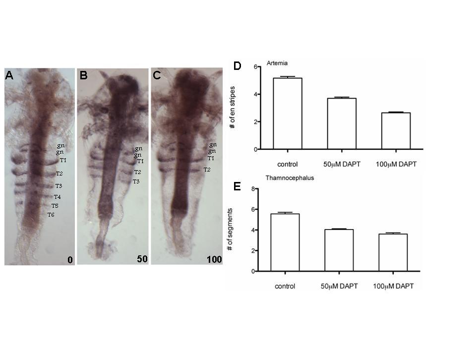
73 73
74 Figure 2.4: DAPT treatment does not affect growth in (A) Artemia, and causes only a small decrease in body size in (B) Thamnocephalus. 74
75 75
76 Figure 2.5: Ubx/abdA expression in the posterior growth zone of DAPT treated larvae. (A) Untreated 96 hour animals. (B) 100 µm DAPT treated animals. Numbers designate the thoracic segments. 76
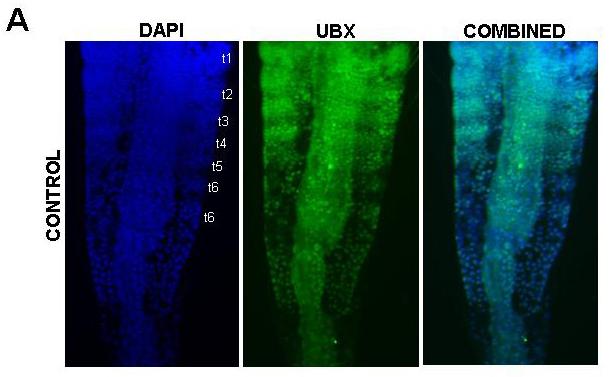
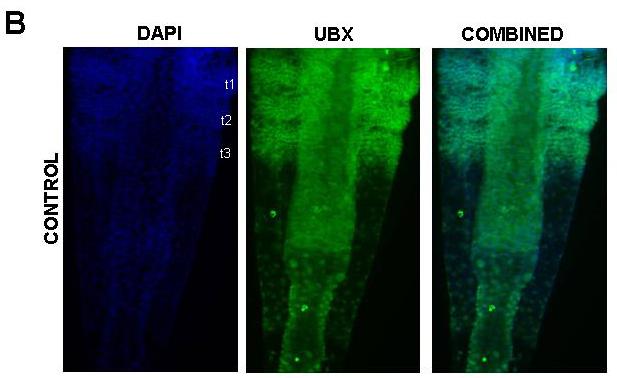
77 77
78 Table 2.1: The comparison of the effects of the loss of Notch signaling on segment patterning in vertebrate and arthropod species. 78
79 79
80 80 CHAPTER 3: SUMMARY OF CONCLUSIONS AND FUTURE DIRECTIONS Since γ-secretase inhibits the formation of segments in Artemia and Thamnocephalus, it is likely that Notch signaling plays a role in segmentation in these two branchiopod crustaceans. Experiments that inhibit Notch signaling during segmentation, such as RNAi, can verify this result. In light of this finding, it would be interesting to see the expression of Notch signaling targets, such as hairy. As previously discussed, hairy is part of a molecular clock in chordates, and its expression oscillates along the PSM in time with the addition of segments (Paleirim et al, 1997; Bessho et al, 2003; Holley et al, 2000; Oates and Ho, 2002). If such a molecular clock exists in Artemia, a dynamic expression pattern of Notch pathway components is expected. Growth of the Artemia larva was not affected by γ-secretase inhibitor treatment, as the treated larvae continued to grow at the same rate as the controls. These data suggest that even though growth is necessary for the formation of segments, the two processes are regulated independently. Interestingly, window experiments suggest that in the presence of growth but absence of segmentation, cells remain in a permissive state, allowing segmentation to resume when γ-secretase mediated signaling is restored. The window treatment experiments were problematic, because complete elimination of the drug post treatment could not be confirmed. However, this approach can be used to establish the timing between segment determination and segment formation by treating the larvae with the drug at progressively later times and assaying the affected segments.
81 81 In sequentially segmented arthropods, segments are added one at a time from anterior to posterior as the larva develops. The timing of the addition of segments has not been assayed previously, and was assumed to be linear. In the case of Thamnacephalus, this assumption is correct, as segments are added at regular intervals. However, Artemia segments are not added at regular intervals. It appears that the addition of the first 5 thoracic segments is linear, but the subsequent segments are added at much longer intervals. It is possible that this slowing of segmentation in Artemia is due to nutritional deficiencies during the incubation period. However, this is unlikely due to depleting food sources as food was refreshed daily and there is no corresponding daily spike in the rate of segment addition in response to fresh food. It is also a possibility that there is a naturally occurring supplement that Artemia larvae need as they continue to develop, which is not as abundant or even absent in lab media. Growing Artemia larvae in sea water from sources where these larvae naturally occur would address this issue. Therefore, to gain an accurate understanding of the rates of segmentation in Artemia, and confirm that segmentation is indeed not linear, it would be best to grow larvae in mesh enclosures in their natural habitat. Interestingly, small changes in temperature do not affect the rate of Artemia segmentation, which is consistent with previous findings that the growth of these larvae is quite resilient to shifts in temperature and salinity (Vanhaecke, 1984; Wear, 1986). This is not surprising considering that Artemia is an ectothermic organism, and hence relies on
82 82 environmental conditions for optimal developmental temperatures. Hence, such an organism would have to be adapted to withstand shifts in temperatures to ensure its survival. By contrast, endothermic larvae develop in very consistent temperature conditions, are not adapted to withstand temperature shifts, and hence are extremely susceptible to temperature variation during development (Feast, 1998). The finding that Notch may be involved in the patterning of segments in branchiopod crustaceans, as well as similar findings in other arthropods, suggest that the common ancestor to arthropods was not only segmented, but also likely utilized Notch signaling to pattern segments. The fact that Notch signaling is also at the heart of vertebrate segmentation strengthens the argument that the common bilaterian ancestor was segmented. In addition to offering more evidence to the debate surrounding the evolution of segments in the three segmented phyla, comparative experiments can also be useful to the overall understanding of segmentation mechanisms in all segmented species. However the debate regarding whether the Urbilaterian was in fact segmented is far from over. At this time, there are certainly more differences between the molecular mechanisms of segmentation between the segmented phyla than there are similarities. For example, evidence for the involvement of Wnt and FGG signaling in arthropod segmentation is lacking, even though these two pathways play such an integral role in vertebrate segmentation. If these two pathways are in fact not involved in the segmentation of most arthropods, then it is likely that more differences than similarities
83 83 will be found between segmentation mechanisms of these two phyla. If this is the case, then it is likely that the Notch signaling pathway was co-opted to pattern segments in arthropods independently of its use to pattern chordate segments.
84 84 APPENDIX A: MORPHOLOGY OF SEGMENTATION Trunk Segments form sequentially from Anterior to Posterior Based on these detailed descriptions of cell rearrangements during the early stages of segment patterning (Freeman, 1986; Freeman et al, 1992) I was able to determine that the anterior-most trunk segments are added sequentially. Artemia larvae hatch out of the dehydrated cyst with three out of five head segments and no morphologically visible trunk segments and no EN expression corresponding to the trunk segments (Figure A1.1A). The anterior nuclei in the trunk form columns along the anterior-posterior axis of the trunk, which precedes the first evidence of segmentation (Freeman, 1992). As the larva continues to grow and develop, the first morphological evidence of segmentation is the arrangement of nuclei in rows across the trunk, which is followed by expression of EN in the forming segment. At 4 hours after hatching, the cells in the anterior-most region of the trunk begin to show the first morphological evidence of segment patterning the nuclei arrange into rows across the trunk (Figure A1.1B). 4 hours later, at 8 hours after hatching, there are more rows of cells, indicating the first evidence of progressive segmental patterning from anterior to posterior (Figure A1.1C). This suggests that anterior segments are not patterned simultaneously, but instead are patterned sequentially, with a short time interval between them. By 12 hours after hatching, the first trunk EN stripe is clearly formed, and there is low level of EN expression where the second stripe is forming (Figure A1.1D,E). The fact that EN does not come on at the same time in the first two segments further implies that these two segments are indeed patterned
85 85 consecutively and not simultaneously. The first two trunk segments show EN expression at 18 hours after hatching (Figure A1.1F,G). These segments continue to develop as more posterior trunk segments are added. As such, the anterior segments are more developed than their posterior counterparts and are first to develop appendages (Figure A.1.1C). Interestingly, the two posterior-most gnathal EN stripes appear more or less at the same time as the first trunk stripe. That is, I have never seen a larva that has the gnathal EN stripes, but not the first trunk stripe. This suggests that these three segments are patterned at the same time. However, due to the overlap in tissues in the head region, the nuclear morphology is difficult to resolve. In sum, our findings show that all Artemia trunk segments are patterned sequentially from anterior to posterior. The EN stripes for the first two segments come on in quick succession, but EN comes on in the first segment before it does in the second. A close morphological analysis, more specifically nuclear arrangements in the anterior trunk in the hours just after the larva hatches out of its cyst confirms this observation. Nuclei are arranged in rows that form from anterior to posterior over time, as opposed to being arranged simultaneously. According to Freeman (1986), the first morphological evidence of an emerging segment in Artemia is the reorganization of cells from columns that run along the anteriorposterior axis of the posterior growth zone to rows of cells that are perpendicular to that axis. These cells then are grouped together, and the ectoderm constricts around them in the region where the limb buds will eventually form (Freeman, 1989). Previous studies have shown that EN expression is initiated before the ectodermal constriction clearly
86 86 delineates the morphological borders between segments (Manzanares et al, 1993). However, we have shown that nuclear organization into rows precedes EN expression, suggesting that EN is a downstream component of the regulatory network that patterns Artemia segments. This is not surprising considering that EN has been shown to give posterior identity to already patterned Drosophila embryonic segments, a role that seems to have been conserved among all arthropods (Patel et al, 1989; Scholtz et al, 1994; (Dearden and Akam, 2001; Hughes and Kaufman, 2002a)Janssen et al, 2004). The appearance of ectodermal constrictions after EN expression is initiated suggests that EN is involved in maintaining, and perhaps even delineating, the border between the posterior of a newly formed segment, and the anterior of the next segment. Segment Morphology Does Not Account for the Difference in the Time of Addition of Trunk Segments The first five segments are patterned on average every 7 hours. Segment 6 takes 18 hours to develop, which is more than twice the time. There is not size difference between segments 5 and 6 to account for such a drastic difference in the timing of their patterning (Figure A1.2). Furthermore, nuclear organization proceeds as normal during the addition of the 6 th segment. Nuclei are organized in rows down to just posterior of the last stripe of EN expression. Posterior to that, nuclei are organized in columns, which is characteristic of unsegmented posterior growth zone just posterior of the last segment (Freeman, 1992). In sum, there is no morphological evidence to account for the increase in the time each segment is patterned segment 6 and all the remaining trunk segments.
87 87 Figure A1.1: (A) Newly hatched larva has no evidence of trunk segments. Nuclei are not yet organized into rows across the trunk even in the most anterior region of the trunk. Instead, columns of nuclei instead extend to the anterior of the trunk (arrow). The bracket designates the trunk region pictured in the subsequent panels. (B) At 4 hours, anterior-most nuclei begin to organize into rows (arrow), but there is still no EN expression. (C) By 8 hours several more rows of nuclei suggesting initiation of segment patterning are apparent (arrows). (A -C ) Duplicate images of A-C with overlaying colored dots to demarcate nuclei and show patterns. The purple dots overlay nuclei arranged in rows of cells as segment patterning begins, and the blue dots show columns of nuclei in the more posterior unsegmented region. (D) A 12 hour larva has its first trunk EN stripe and the second stripe begins to form. There is also EN expression in the gnathal region. Rows of nuclei span the anterior region of the trunk, until just posterior of the first trunk stripe (open arrow). Posterior to EN expression, nuclei are organized in columns (closed arrow). The bright field photo of this animal in panel (E) shows a clearer view of EN expression. (G) Bright field photo showing the EN expression in the 18 hour larva pictured in (F). At 18 hours the initial patterning of the first two trunk segments is complete. There are only 2 rows of nuclei in the trunk, just posterior to the posterior-most EN stripe (open arrow). Posterior to that, nuclei are organized in columns (closed arrow). All embryos were stained with EN and the nuclei were marked with DAPI.
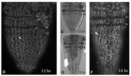
88 88
89 Figure A1.2: There is no morphological difference between segments five and six to account for the increased length of time that it takes to form segment six. (A) Larva with 5 EN stripes and early EN expression in the 6th segment (arrow). (B) Higher magnified view of DAPI stained trunk of the larva in panel A. (C) Larva with 6 EN stripes the 5th and 6th segments, and corresponding EN stripes do not vary in size or cellular organization. (D) DAPI staining of trunk of larva in panel C showing nuclear organization. 89
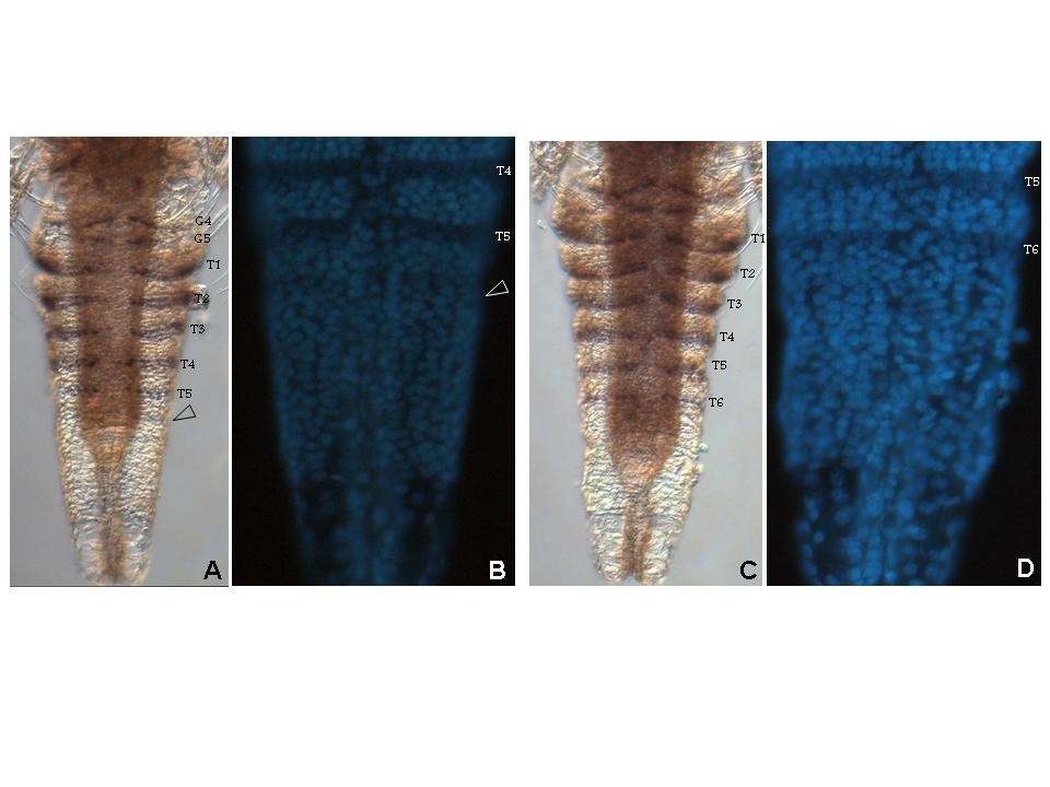
90 90
91 91 APPENDIX B: MORPHOLOGY OF DAPT MUTANTS DAPT treatment disrupts cellular organization within the growth zone In untreated Artemia larvae, the nuclear organization of the growth zone posterior to the last segment can be split into three distinct domains (Figure A2.1A) (Freeman, 1986). The posterior-most nuclei do not exhibit any organization, more anterior nuclei are organized into columns and nuclei just posterior to the last morphologically detectable segment are organized into rows (Figure A2.1A). Thus segmentation is prefigured by the organization of the cells. The onset of EN expression also occurs prior to any visible manifestation of segmentation and is detected first in the cells that have organized into rows. I looked at the organization of nuclei in the posterior growth zone to determine whether DAPT treatment affects the formation of segments, and not just EN expression. DAPT treated larvae not only have fewer EN stripes, but also do not show the organization of cells into rows, which suggests that no segments are patterned posterior to the last EN stripe that forms (Figure A2.1B). There are only a few rows of cells past the posterior most EN stripe and in the remainder of the posterior trunk the cells remain disorganized. Therefore, the decrease in EN stripes in DAPT treated larvae corresponds to a loss of segments as well as the cellular organization that precedes segmentation in the posterior growth zone, and not just a decrease in EN expression.
92 Figure A2.1: Cellular organization in the posterior of DAPT treated larvae. (A) control and (B) 100 µm DAPT treated larvae. Open arrows indicate columns of cells in the unsegmented posterior growth zone. Filled arrows indicate rows of cells posterior to the last distinguishable segmented region. (C and D) Duplicate images of A and B with overlaying colored dots to demarcate nuclei and show patterns. The purple dots overlay nuclei arranged in rows of cells as segment patterning begins posterior to the last patterned segment and the blue dots show columns of nuclei in the more posterior unsegmented region. The dashed lines mark borders of already patterned segments in the anterior of the growth zone. 92
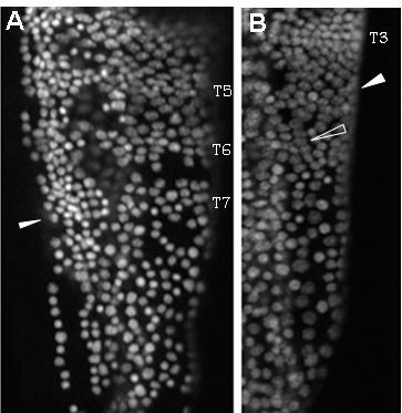
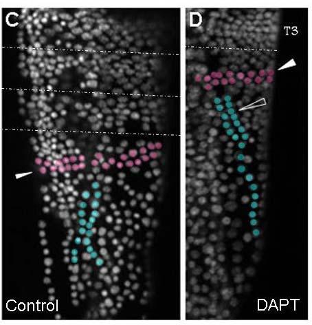
93 93
94 94 Notch Cloning APPENDIX C: PARTIAL SEQUENCE OF ARTEMIA NOTCH Artemia Notch was cloned from a pool of Artemia embryos ranging in age from newly hatched to 96 hours after hatching using degenerate primers (5 CCNGGNMYRCCGAAGTGGGGAGCTACG; GGTGCTGCTGGACCACTTCKCNAAYMGNGA) designed by the COnsensus- DEgenerate Hybrid Oligonucleotide Primers (CODEHOP) program (Rose et al., 2003).
95 Figure A3.1: Partial Artemia Notch amino acid sequence the same portion of the sequence of the protein in other invertebrates. 95
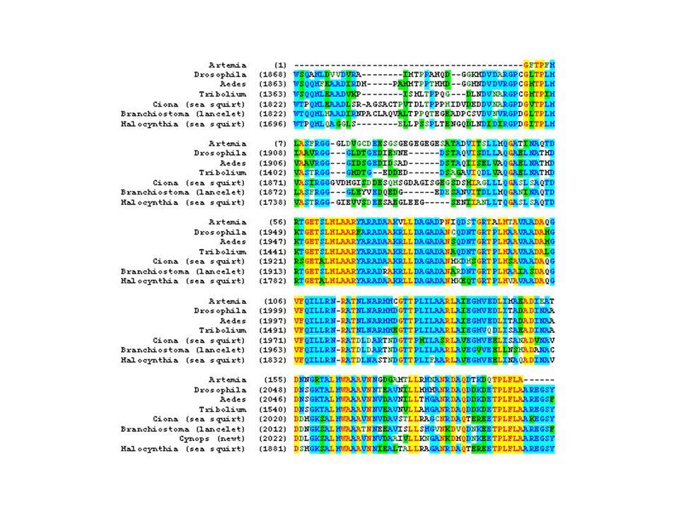
96 96
Axis determination in flies. Sem 9.3.B.5 Animal Science
 Axis determination in flies Sem 9.3.B.5 Animal Science All embryos are in lateral view (anterior to the left). Endoderm, midgut; mesoderm; central nervous system; foregut, hindgut and pole cells in yellow.
Axis Specification in Drosophila
 Developmental Biology Biology 4361 Axis Specification in Drosophila November 2, 2006 Axis Specification in Drosophila Fertilization Superficial cleavage Gastrulation Drosophila body plan Oocyte formation
Axis Specification in Drosophila
 Developmental Biology Biology 4361 Axis Specification in Drosophila November 6, 2007 Axis Specification in Drosophila Fertilization Superficial cleavage Gastrulation Drosophila body plan Oocyte formation
Drosophila Life Cycle
 Drosophila Life Cycle 1 Early Drosophila Cleavage Nuclei migrate to periphery after 10 nuclear divisions. Cellularization occurs when plasma membrane folds in to divide nuclei into cells. Drosophila Superficial
Lecture 7. Development of the Fruit Fly Drosophila
 BIOLOGY 205/SECTION 7 DEVELOPMENT- LILJEGREN Lecture 7 Development of the Fruit Fly Drosophila 1. The fruit fly- a highly successful, specialized organism a. Quick life cycle includes three larval stages
MOLECULAR CONTROL OF EMBRYONIC PATTERN FORMATION
 MOLECULAR CONTROL OF EMBRYONIC PATTERN FORMATION Drosophila is the best understood of all developmental systems, especially at the genetic level, and although it is an invertebrate it has had an enormous
Axis Specification in Drosophila
 Developmental Biology Biology 4361 Axis Specification in Drosophila July 9, 2008 Drosophila Development Overview Fertilization Cleavage Gastrulation Drosophila body plan Oocyte formation Genetic control
Midterm 1. Average score: 74.4 Median score: 77
 Midterm 1 Average score: 74.4 Median score: 77 NAME: TA (circle one) Jody Westbrook or Jessica Piel Section (circle one) Tue Wed Thur MCB 141 First Midterm Feb. 21, 2008 Only answer 4 of these 5 problems.
A pair-rule gene circuit defines segments sequentially in the short-germ insect Tribolium castaneum
 A pair-rule gene circuit defines segments sequentially in the short-germ insect Tribolium castaneum Chong Pyo Choe, Sherry C. Miller, and Susan J. Brown* Division of Biology, Kansas State University, Manhattan,
RALF JANSSEN* Uppsala University, Department of Earth Sciences, Palaeobiology, Uppsala, Sweden
 Int. J. Dev. Biol. 58: 343-347 (2014) doi: 10.1387/ijdb.140058rj www.intjdevbiol.com Gene expression suggests double-segmental and single-segmental patterning mechanisms during posterior segment addition
A systems approach to biology
 A systems approach to biology SB200 Lecture 7 7 October 2008 Jeremy Gunawardena jeremy@hms.harvard.edu Recap of Lecture 6 In phage lambda, cooperativity leads to bistability and hysteresis In HIV-1, sequestration
Chapter 18 Lecture. Concepts of Genetics. Tenth Edition. Developmental Genetics
 Chapter 18 Lecture Concepts of Genetics Tenth Edition Developmental Genetics Chapter Contents 18.1 Differentiated States Develop from Coordinated Programs of Gene Expression 18.2 Evolutionary Conservation
Developmental genetics: finding the genes that regulate development
 Developmental Biology BY1101 P. Murphy Lecture 9 Developmental genetics: finding the genes that regulate development Introduction The application of genetic analysis and DNA technology to the study of
This thesis is protected by copyright which belongs to the author.
 A University of Sussex DPhil thesis Available online via Sussex Research Online: http://sro.sussex.ac.uk/ This thesis is protected by copyright which belongs to the author. This thesis cannot be reproduced
Morphogens in biological development: Drosophila example
 LSM5194 Morphogens in biological development: Drosophila example Lecture 29 The concept of morphogen gradients The concept of morphogens was proposed by L. Wolpert as a part of the positional information
Development of Drosophila
 Development of Drosophila Hand-out CBT Chapter 2 Wolpert, 5 th edition March 2018 Introduction 6. Introduction Drosophila melanogaster, the fruit fly, is found in all warm countries. In cooler regions,
Why Flies? stages of embryogenesis. The Fly in History
 The Fly in History 1859 Darwin 1866 Mendel c. 1890 Driesch, Roux (experimental embryology) 1900 rediscovery of Mendel (birth of genetics) 1910 first mutant (white) (Morgan) 1913 first genetic map (Sturtevant
Expression of myriapod pair rule gene orthologs
 RESEARCH Open Access Expression of myriapod pair rule gene orthologs Ralf Janssen 1*, Graham E Budd 1, Nikola-Michael Prpic 2, Wim GM Damen 3 Abstract Background: Segmentation is a hallmark of the arthropods;
Unicellular: Cells change function in response to a temporal plan, such as the cell cycle.
 Spatial organization is a key difference between unicellular organisms and metazoans Unicellular: Cells change function in response to a temporal plan, such as the cell cycle. Cells differentiate as a
From DNA to Diversity
 From DNA to Diversity Molecular Genetics and the Evolution of Animal Design Sean B. Carroll Jennifer K. Grenier Scott D. Weatherbee Howard Hughes Medical Institute and University of Wisconsin Madison,
v Scientists have identified 1.3 million living species of animals v The definition of an animal
 Biosc 41 9/10 Announcements BIOSC 041 v Genetics review: group problem sets Groups of 3-4 Correct answer presented to class = 2 pts extra credit Incorrect attempt = 1 pt extra credit v Lecture: Animal
Outline. v Definition and major characteristics of animals v Dividing animals into groups based on: v Animal Phylogeny
 BIOSC 041 Overview of Animal Diversity: Animal Body Plans Reference: Chapter 32 Outline v Definition and major characteristics of animals v Dividing animals into groups based on: Body symmetry Tissues
Supporting Information
 Supporting Information Pueyo et al. 10.1073/pnas.0804093105 SI Text Periplaneta americana (Delta GenBank Accession Number FJ222590). MR- WTQQTRVQGAVVVVILAALQQVCCSGVFELRLKSF- INDYGKDSVGQCCSGTPSPGTKACSGPCRTRFRVCL-
Biosc 41 9/10 Announcements
 Biosc 41 9/10 Announcements v Genetics review: group problem sets Groups of 3-4 Correct answer presented to class = 2 pts extra credit Incorrect attempt = 1 pt extra credit v Lecture: Animal Body Plans
Caudal Regulates the Spatiotemporal Dynamics of Pair-Rule Waves in Tribolium
 Caudal Regulates the Spatiotemporal Dynamics of Pair-Rule Waves in Tribolium Ezzat El-Sherif 1, Xin Zhu 2, Jinping Fu 2, Susan J. Brown 2 * 1 Genetics Program, Kansas State University, Manhattan, Kansas,
8/23/2014. Introduction to Animal Diversity
 Introduction to Animal Diversity Chapter 32 Objectives List the characteristics that combine to define animals Summarize key events of the Paleozoic, Mesozoic, and Cenozoic eras Distinguish between the
Developmental Biology 3230 Midterm Exam 1 March 2006
 Name Developmental Biology 3230 Midterm Exam 1 March 2006 1. (20pts) Regeneration occurs to some degree to most metazoans. When you remove the head of a hydra a new one regenerates. Graph the inhibitor
Chapter 11. Development: Differentiation and Determination
 KAP Biology Dept Kenyon College Differential gene expression and development Mechanisms of cellular determination Induction Pattern formation Chapter 11. Development: Differentiation and Determination
Drosophila melanogaster- Morphogen Gradient
 NPTEL Biotechnology - Systems Biology Drosophila melanogaster- Morphogen Gradient Dr. M. Vijayalakshmi School of Chemical and Biotechnology SASTRA University Joint Initiative of IITs and IISc Funded by
of teloblastic cells (Shankland and Seaver, 2000). Anne- Institut für Genetik der Universität zuköln
. Anne- Institut für Genetik der Universität zuköln") Developmental Cell, Vol. 7, 301 312, September, 2004, Copyright 2004 by Cell Press Segmentation Review Diethard Tautz* of teloblastic cells (Shankland and Seaver, 2000). Anne- Institut für Genetik der
Developmental Cell, Vol. 7, 301 312, September, 2004, Copyright 2004 by Cell Press Segmentation Review Diethard Tautz* of teloblastic cells (Shankland and Seaver, 2000). Anne- Institut für Genetik der
Live Imaging of Segmentation Clock Dynamics in Zebrafish. Nathan Pui-Yin Shih. A dissertation submitted in partial satisfaction of the
 Live Imaging of Segmentation Clock Dynamics in Zebrafish By Nathan Pui-Yin Shih A dissertation submitted in partial satisfaction of the requirements for the degree of Doctor of Philosophy in Molecular
Expression of pair rule gene orthologs in the blastoderm of a myriapod: evidence for pair rule-like mechanisms?
 Janssen et al. BMC Developmental Biology 2012, 12:15 RESEARCH ARTICLE Expression of pair rule gene orthologs in the blastoderm of a myriapod: evidence for pair rule-like mechanisms? Ralf Janssen 1*, Wim
Gaining New Insights into Primitive Strategies for Embryonic Axis Specification Using the Wasp Nasonia
 Functional Development and Embryology 2007 Global Science Books Gaining New Insights into Primitive Strategies for Embryonic Axis Specification Using the Wasp Nasonia Eugenia C. Olesnicky Claude Desplan
Principles of Experimental Embryology
 Biology 4361 Developmental Biology Principles of Experimental Embryology June 16, 2008 Overview What forces affect embryonic development? The embryonic environment: external and internal How do forces
BIS &003 Answers to Assigned Problems May 23, Week /18.6 How would you distinguish between an enhancer and a promoter?
 Week 9 Study Questions from the textbook: 6 th Edition: Chapter 19-19.6, 19.7, 19.15, 19.17 OR 7 th Edition: Chapter 18-18.6 18.7, 18.15, 18.17 19.6/18.6 How would you distinguish between an enhancer and
Delimiting the conserved features of hunchback function for the trunk organization of insects
 RESEARCH ARTICLE 881 Development 135, 881-888 (2008) doi:10.1242/dev.018317 Delimiting the conserved features of hunchback function for the trunk organization of insects Henrique Marques-Souza 1, *, Manuel
Autonomous concentration-dependent activation and repression of Krüppel by hunchback in the Drosophila embryo
 Development 120, 3043-3049 (1994) Printed in Great Britain The Company of Biologists Limited 1994 3043 Autonomous concentration-dependent activation and repression of Krüppel by hunchback in the Drosophila
Revision Based on Chapter 25 Grade 11
 Revision Based on Chapter 25 Grade 11 Biology Multiple Choice Identify the choice that best completes the statement or answers the question. 1. A cell that contains a nucleus and membrane-bound organelles
9/4/2015 INDUCTION CHAPTER 1. Neurons are similar across phyla Thus, many different model systems are used in developmental neurobiology. Fig 1.
 INDUCTION CHAPTER 1 Neurons are similar across phyla Thus, many different model systems are used in developmental neurobiology Fig 1.1 1 EVOLUTION OF METAZOAN BRAINS GASTRULATION MAKING THE 3 RD GERM LAYER
Vertebrate Segmentation: From Cyclic Gene Networks to Scoliosis
 Leading Edge Review Vertebrate Segmentation: From Cyclic Gene Networks to Scoliosis Olivier Pourquié 1, * 1 Institut de Génétique et de Biologie Moléculaire et Cellulaire (IGBMC), CNRS (UMR 7104), Inserm
1. What are the three general areas of the developing vertebrate limb? 2. What embryonic regions contribute to the developing limb bud?
 Study Questions - Lecture 17 & 18 1. What are the three general areas of the developing vertebrate limb? The three general areas of the developing vertebrate limb are the proximal stylopod, zeugopod, and
Biology 11. The Kingdom Animalia
 Biology 11 The Kingdom Animalia Objectives By the end of the lesson you should be able to: Describe the 5 ways we classify animals Symmetry Germ layers Body plan Segmentation Animal Evolution Hank Video
Developmental processes Differential gene expression Introduction to determination The model organisms used to study developmental processes
 Date Title Topic(s) Learning Outcomes: Sept 28 Oct 3 1. What is developmental biology and why should we care? 2. What is so special about stem cells and gametes? Developmental processes Differential gene
Development Team. Developmental Biology Axis Specification in Drosophila. Head, Department of Zoology, University of Delhi
 Paper No. : 11 Module : 6 Development Team Principal Investigator: Prof. Neeta Sehgal Head, Department of Zoology, University of Delhi Paper Coordinator: Prof. Namita Agrawal Department of Zoology, University
18.4 Embryonic development involves cell division, cell differentiation, and morphogenesis
 18.4 Embryonic development involves cell division, cell differentiation, and morphogenesis An organism arises from a fertilized egg cell as the result of three interrelated processes: cell division, cell
The segmentation clock mechanism moves up a notch
 Review The segmentation clock mechanism moves up a notch Sarah Gibb, Miguel Maroto and J. Kim Dale College of Life Sciences, University of Dundee, Dundee, DD1 5EH, Scotland, UK The vertebrate segmentation
Evolutionary Developmental Biology
 Evolutionary Developmental Biology a.k.a. EVO-DEVO Paedomorphosis is common among salamanders. Note how this hellbender (top) and mudpuppy (bottom) both have gills, paddle tails, and weaker limbs... Top:
3/8/ Complex adaptations. 2. often a novel trait
 Chapter 10 Adaptation: from genes to traits p. 302 10.1 Cascades of Genes (p. 304) 1. Complex adaptations A. Coexpressed traits selected for a common function, 2. often a novel trait A. not inherited from
Principles of Experimental Embryology
 Biology 4361 Developmental Biology Principles of Experimental Embryology September 19, 2006 Major Research Questions How do forces outside the embryo affect its development? (Environmental Developmental
Cellular automata for exploring gene regulation in Drosophila segmentation
 Cellular automata for exploring gene regulation in Drosophila segmentation Matthew J. Berryman a, Andrew Allison a, and Derek Abbott a a Centre for Biomedical Engineering and School of Electrical and Electronic
Suppressor of Hairless and Presenilin phenotypes imply involvement of canonical Notch-signalling in segmentation of the spider Cupiennius salei
 Developmental Biology 280 (2005) 211 224 www.elsevier.com/locate/ydbio Suppressor of Hairless and Presenilin phenotypes imply involvement of canonical Notch-signalling in segmentation of the spider Cupiennius
MBios 401/501: Lecture 14.2 Cell Differentiation I. Slide #1. Cell Differentiation
 MBios 401/501: Lecture 14.2 Cell Differentiation I Slide #1 Cell Differentiation Cell Differentiation I -Basic principles of differentiation (p1305-1320) -C-elegans (p1321-1327) Cell Differentiation II
Report. Wnt8 Is Required for Growth-Zone Establishment and Development of Opisthosomal Segments in a Spider
 Current Biology 18, 1619 1623, October 28, 2008 ª2008 Elsevier Ltd All rights reserved DOI 10.1016/j.cub.2008.08.045 Wnt8 Is Required for Growth-Zone Establishment and Development of Opisthosomal Segments
Drosophila Somatic Anterior-Posterior Axis (A-P Axis) Formation
 Formation") Home Biol 4241 Luria-Delbruck 1943 Hershey-Chase 1952 Meselson-Stahl 1958 Garapin et al. 1978 McClintock 1953 King-Wilson 1975 Sanger et al. 1977 Rothberg et al. 2011 Jeffreys et al. 1985 Bacterial Genetics
Home Biol 4241 Luria-Delbruck 1943 Hershey-Chase 1952 Meselson-Stahl 1958 Garapin et al. 1978 McClintock 1953 King-Wilson 1975 Sanger et al. 1977 Rothberg et al. 2011 Jeffreys et al. 1985 Bacterial Genetics
Evolution of Transcription factor function: Homeotic (Hox) proteins
 proteins") Evolution of Transcription factor function: Homeotic (Hox) proteins Hox proteins regulate morphology in cellular zones on the anterior-posterior axis of embryos via the activation/repression of unknown
Evolution of Transcription factor function: Homeotic (Hox) proteins Hox proteins regulate morphology in cellular zones on the anterior-posterior axis of embryos via the activation/repression of unknown
Homeotic Genes and Body Patterns
 Homeotic Genes and Body Patterns Every organism has a unique body pattern. Although specialized body structures, such as arms and legs, may be similar in makeup (both are made of muscle and bone), their
UNIVERSITY OF YORK BIOLOGY. Developmental Biology
 Examination Candidate Number: UNIVERSITY OF YORK BSc Stage 2 Degree Examinations 2017-18 Department: BIOLOGY Title of Exam: Developmental Biology Desk Number: Time allowed: 1 hour and 30 minutes Total
Patterning embryos with oscillations: structure, function and dynamics of the vertebrate segmentation clock
 REVIEW 625 Development 139, 625-639 (2012) doi:10.1242/dev.063735 2012. Published by The Company of Biologists Ltd Patterning embryos with oscillations: structure, function and dynamics of the vertebrate
THE MAKING OF THE SOMITE: MOLECULAR EVENTS IN VERTEBRATE SEGMENTATION. Yumiko Saga* and Hiroyuki Takeda
 THE MAKING OF THE SOMITE: MOLECULAR EVENTS IN VERTEBRATE SEGMENTATION Yumiko Saga* and Hiroyuki Takeda The reiterated structures of the vertebrate axial skeleton, spinal nervous system and body muscle
Animal Origins and Evolution
 Animal Origins and Evolution Common Features of Animals multicellular heterotrophic motile Sexual reproduction, embryo Evolution of Animals All animals are multicellular and heterotrophic, which means
purpose of this Chapter is to highlight some problems that will likely provide new
 119 Chapter 6 Future Directions Besides our contributions discussed in previous chapters to the problem of developmental pattern formation, this work has also brought new questions that remain unanswered.
REVIEW ARTICLE. Segmentation: Painting Stripes From Flies to Vertebrates DEVELOPMENTAL GENETICS 23:1 10 (1998) 1998 WILEY-LISS, INC.
 1998 WILEY-LISS, INC.") DEVELOPMENTAL GENETICS 23:1 10 (1998) REVIEW ARTICLE Segmentation: Painting Stripes From Flies to Vertebrates LESLIE PICK* Brookdale Center for Developmental and Molecular Biology, Mt. Sinai School of
DEVELOPMENTAL GENETICS 23:1 10 (1998) REVIEW ARTICLE Segmentation: Painting Stripes From Flies to Vertebrates LESLIE PICK* Brookdale Center for Developmental and Molecular Biology, Mt. Sinai School of
MCB 141 Midterm I Feb. 14, 2012
 Write your name and student ID# on EVERY PAGE of your exam MCB 141 Midterm I Feb. 14, 2012 Question #1 Question #2 Question #3 Question #4 BONUS / 28 pts / 27 pts / 25 pts / 20 pts / 1 pt TOTAL / 100 pts
AP Biology Gene Regulation and Development Review
 AP Biology Gene Regulation and Development Review 1. What does the regulatory gene code for? 2. Is the repressor by default active/inactive? 3. What changes the repressor activity? 4. What does repressor
Metameric body plans are found in diverse metazoan phyla,
 Expression patterns of hairy, even-skipped, and runt in the spider Cupiennius salei imply that these genes were segmentation genes in a basal arthropod Wim G. M. Damen*, Mathias Weller, and Diethard Tautz
2/23/09. Regional differentiation of mesoderm. Morphological changes at early postgastrulation. Segments organize the body plan during embryogenesis
 Regional differentiation of mesoderm Axial Paraxial Intermediate Somatic Splanchnic Chick embryo Morphological changes at early postgastrulation stages Segments organize the body plan during embryogenesis
Phenotypic and dynamical transitions in model genetic networks II. Application to the evolution of segmentation mechanisms
 EVOLUTION & DEVELOPMENT 3:2, 95 103 (2001) Phenotypic and dynamical transitions in model genetic networks II. Application to the evolution of segmentation mechanisms I. Salazar-Ciudad, a,b R. V. Solé,
!!!!!!!! DB3230 Midterm 2 12/13/2013 Name:
 1. (10 pts) Draw or describe the fate map of a late blastula stage sea urchin embryo. Draw or describe the corresponding fate map of the pluteus stage larva. Describe the sequence of gastrulation events
Biology 4361 Developmental Biology The Genetics of Axis Specification in Drosophila November 2, 2006
 Biology 4361 Developmental Biology The Genetics of Axis Specification in Drosophila November 2, 2006 EARLY DROSOPHILA DEVELOPMENT Fertilization 1) Drosophila egg activation occurs at ovulation - eggs are
An Introduction to Animal Diversity
 Chapter 32 An Introduction to Animal Diversity PowerPoint Lectures for Biology, Seventh Edition Neil Campbell and Jane Reece Lectures by Chris Romero Overview: Welcome to Your Kingdom The animal kingdom
Lesson Overview. Gene Regulation and Expression. Lesson Overview Gene Regulation and Expression
 13.4 Gene Regulation and Expression THINK ABOUT IT Think of a library filled with how-to books. Would you ever need to use all of those books at the same time? Of course not. Now picture a tiny bacterium
Neural development its all connected
 Neural development its all connected How do you build a complex nervous system? How do you build a complex nervous system? 1. Learn how tissue is instructed to become nervous system. Neural induction 2.
Sonic hedgehog (Shh) signalling in the rabbit embryo
 signalling in the rabbit embryo") Sonic hedgehog (Shh) signalling in the rabbit embryo In the first part of this thesis work the physical properties of cilia-driven leftward flow were characterised in the rabbit embryo. Since its discovery
Sonic hedgehog (Shh) signalling in the rabbit embryo In the first part of this thesis work the physical properties of cilia-driven leftward flow were characterised in the rabbit embryo. Since its discovery
arxiv: v1 [q-bio.qm] 23 Apr 2007
![arxiv: v1 [q-bio.qm] 23 Apr 2007](/thumbs/95/124440410.jpg "arxiv: v1 [q-bio.qm] 23 Apr 2007") Converting genetic networ oscillations into somite spatial pattern K. I. Mazzitello, 1,2 C. M. Arizmendi, 2 and H. G. E. Hentschel 3 1 CONICET 2 Facultad de Ingeniería, Universidad Nacional de Mar del
Converting genetic networ oscillations into somite spatial pattern K. I. Mazzitello, 1,2 C. M. Arizmendi, 2 and H. G. E. Hentschel 3 1 CONICET 2 Facultad de Ingeniería, Universidad Nacional de Mar del
Development of Developmental System System(Mathematical Topics in Biolo. Citation 数理解析研究所講究録 (1993), 827:
, 827:") Title Development of Developmental System System(Mathematical Topics in Biolo Author(s) Takeda, Yasuhiko Citation 数理解析研究所講究録 (1993), 827: 57-74 Issue Date 1993-03 URL http://hdl.handle.net/2433/83294 Right
Title Development of Developmental System System(Mathematical Topics in Biolo Author(s) Takeda, Yasuhiko Citation 数理解析研究所講究録 (1993), 827: 57-74 Issue Date 1993-03 URL http://hdl.handle.net/2433/83294 Right
Animal Diversity. Animals are multicellular, heterotrophic eukaryotes with tissues that develop from embryonic layers 9/20/2017
 Animal Diversity Chapter 32 Which of these organisms are animals? Animals are multicellular, heterotrophic eukaryotes with tissues that develop from embryonic layers Animals share the same: Nutritional
Questions in developmental biology. Differentiation Morphogenesis Growth/apoptosis Reproduction Evolution Environmental integration
 Questions in developmental biology Differentiation Morphogenesis Growth/apoptosis Reproduction Evolution Environmental integration Representative cell types of a vertebrate zygote => embryo => adult differentiation
Paraxial and Intermediate Mesoderm
 Biology 4361 Paraxial and Intermediate Mesoderm December 7, 2006 Major Mesoderm Lineages Mesodermal subdivisions are specified along a mediolateral axis by increasing amounts of BMPs more lateral mesoderm
BILD7: Problem Set. 2. What did Chargaff discover and why was this important?
 BILD7: Problem Set 1. What is the general structure of DNA? 2. What did Chargaff discover and why was this important? 3. What was the major contribution of Rosalind Franklin? 4. How did solving the structure
Analysis of the Wnt gene repertoire in an onychophoran provides new insights into the evolution of segmentation
 Hogvall et al. EvoDevo 2014, 5:14 RESEARCH Analysis of the Wnt gene repertoire in an onychophoran provides new insights into the evolution of segmentation Open Access Mattias Hogvall 1, Anna Schönauer
Homeotic genes in flies. Sem 9.3.B.6 Animal Science
 Homeotic genes in flies Sem 9.3.B.6 Animal Science So far We have seen that identities of each segment is determined by various regulators of segment polarity genes In arthopods, and in flies, each segment
Early Development in Invertebrates
 Developmental Biology Biology 4361 Early Development in Invertebrates October 25, 2006 Early Development Overview Cleavage rapid cell divisions divisions of fertilized egg into many cells Gastrulation
PRACTICE EXAM. 20 pts: 1. With the aid of a diagram, indicate how initial dorsal-ventral polarity is created in fruit fly and frog embryos.
 PRACTICE EXAM 20 pts: 1. With the aid of a diagram, indicate how initial dorsal-ventral polarity is created in fruit fly and frog embryos. No Low [] Fly Embryo Embryo Non-neural Genes Neuroectoderm Genes
Animal Diversity. Features shared by all animals. Animals are multicellular, heterotrophic eukaryotes with tissues that develop from embryonic layers
 Animal Diversity Animals are multicellular, heterotrophic eukaryotes with tissues that develop from embryonic layers Nutritional mode Ingest food and use enzymes in the body to digest Cell structure and
ASpatio-TemporalModelofNotchSignallinginthe Zebrafish Segmentation Clock: Conditions for Synchronised Oscillatory Dynamics
 ASpatio-TemporalModelofNotchSignallinginthe Zebrafish Segmentation Clock: Conditions for Synchronised Oscillatory Dynamics Alan J. Terry 1 *, Marc Sturrock 1, J. Kim Dale 2, Miguel Maroto 2, Mark A. J.
Section 4 Professor Donald McFarlane
 Characteristics Section 4 Professor Donald McFarlane Lecture 11 Animals: Origins and Bauplans Multicellular heterotroph Cells lack cell walls Most have nerves, muscles, capacity to move at some point in
Paraxial and Intermediate Mesoderm
 Biology 4361 Paraxial and Intermediate Mesoderm December 6, 2007 Mesoderm Formation Chick Major Mesoderm Lineages Mesodermal subdivisions are specified along a mediolateral axis by increasing amounts of
Role of Organizer Chages in Late Frog Embryos
 Ectoderm Germ Layer Frog Fate Map Frog Fate Map Role of Organizer Chages in Late Frog Embryos Organizer forms three distinct regions Notochord formation in chick Beta-catenin localization How does beta-catenin
Paraxial and Intermediate Mesoderm
 Biology 4361 Paraxial and Intermediate Mesoderm December 6, 2007 Mesoderm Formation Chick Major Mesoderm Lineages Mesodermal subdivisions are specified along a mediolateral axis by increasing amounts of
Kingdom Animalia. Zoology the study of animals
 Kingdom Animalia Zoology the study of animals Summary Animals are multicellular and eukaryotic. consume and digest organic materials thereby being heterotrophs. Most are motile at some time in their lives.
Segmentation in animals
 R991 Consequently, the mummy is now housed in a special museum in South Tyrol s capital Bolzano. Err, is there any genetic evidence to back this up? Well, you know that there is no such thing as Italian
Conclusions. The experimental studies presented in this thesis provide the first molecular insights
 C h a p t e r 5 Conclusions 5.1 Summary The experimental studies presented in this thesis provide the first molecular insights into the cellular processes of assembly, and aggregation of neural crest and
10/03/2014. Eukaryotic Development. + Differentiation vs. Development. Differentiation. Development
 Differentiation vs. Development What comes to mind when you think of differentiation? Eukaryotic Development What about development? Presented by: Sean, Daria, Emily, and Maggie Example: Human Development
Two distinct mechanisms for differential positioning of gene expression borders involving the Drosophila gap protein giant
 Development 125, 3765-3774 (1998) Printed in Great Britain The Company of Biologists Limited 1998 DEV5218 3765 Two distinct mechanisms for differential positioning of gene expression borders involving
Developmental Biology
 Developmental Biology 376 (2013) 99 112 Contents lists available at SciVerse ScienceDirect Developmental Biology journal homepage: www.elsevier.com/locate/developmentalbiology Genomes and Developmental
Chapter 32 Introduction to Animal Diversity. Copyright 2008 Pearson Education, Inc., publishing as Pearson Benjamin Cummings
 Chapter 32 Introduction to Animal Diversity Welcome to Your Kingdom The animal kingdom extends far beyond humans and other animals we may encounter 1.3 million living species of animals have been identified
ANIMAL DIVERSITY AND THE EVOLUTION OF BODY PLANS
 ANIMAL DIVERSITY AND THE EVOLUTION OF BODY PLANS GENERAL FEATURES OF ANIMALS Heterotrophy - obtain energy and organic molecules by ingesting other organisms Multicellularity - Many have complex bodies
Cell-Cell Communication in Development
 Biology 4361 - Developmental Biology Cell-Cell Communication in Development October 2, 2007 Cell-Cell Communication - Topics Induction and competence Paracrine factors inducer molecules Signal transduction
Genes, Development, and Evolution
 14 Genes, Development, and Evolution Chapter 14 Genes, Development, and Evolution Key Concepts 14.1 Development Involves Distinct but Overlapping Processes 14.2 Changes in Gene Expression Underlie Cell
Thoracic Patterning by the Drosophila Gap Gene hunchback
 Developmental Biology 237, 79 92 (2001) doi:10.1006/dbio.2001.0355, available online at http://www.idealibrary.com on Thoracic Patterning by the Drosophila Gap Gene hunchback Xuelin Wu,* Vikram Vasisht,*