Reduce, Reuse, Recycle: The tale of two Wnts and the lone C. elegans Syndecan, SDN-1
|
|
|
- Allison Lindsey
- 6 years ago
- Views:
Transcription
1 Reduce, Reuse, Recycle: The tale of two Wnts and the lone C. elegans Syndecan, SDN-1 BY Copyright 2015 Samantha N. Hartin Submitted to the graduate degree program in Molecular Biosciences and the Graduate Faculty of the University of Kansas in partial fulfillment of the requirements for the degree of Doctor of Philosophy. Chairperson Dr. Brian D. Ackley Dr. Erik Lundquist Dr. Stuart Macdonald Dr. Robert Ward Dr. Yoshiaki Azuma Dr. Elias Michaelis Date defended: May 1, 2015
2 The dissertation committee for Samantha N. Hartin certifies that this is the approved version of the following dissertation: Reduce, Reuse, Recycle: The tale of two Wnts and the lone C. elegans Syndecan, SDN-1 Chairperson Dr. Brian D. Ackley Date approved: May 1, 2015 ii
3 Abstract Heparan sulfate proteoglycans (HSPGs) are cell adhesion molecules that have been shown to be involved in a myriad of different aspects of development such as embryogenesis, dorsal-ventral axon guidance and cell migration. Despite knowing the phenotypes caused by mutations in HSPGs, little is known about the ligands working with HSPGs during development. Identification of HSPG ligands will provide insight into how HSGPs function during embryogenesis and later in neural development. Chapter II describes two Caenorhabditis elegans cell adhesion proteins SDN- 1/Syndecan and PTP-3/LAR-RPTP as important regulators of polarization and cell migration during embryogenesis. Loss-of-function (LOF) mutations in either ptp-3 or sdn-1 resulted in low penetrance embryonic developmental defects. We used double mutant analysis to test whether ptp-3 and sdn-1 function in a linear genetic pathway during C. elegans embryogenesis and found that double mutants of sdn-1 and ptp-3 exhibited a highly penetrant synthetic lethality (SynLet), with only a small percentage of animals surviving to adulthood. Analysis of the survivors demonstrated that these animals had a synergistic increase in the penetrance of embryonic developmental defects. Taken together, these data strongly suggested that PTP-3 and SDN-1 function in parallel during embryogenesis. We subsequently used RNAi to knockdown the function of ~3,600 genes predicted to encode secreted and/or transmembrane molecules to identify genes that interacted with ptp-3 or sdn-1. We found that the Wnt ligand, lin-44, was SynLet with sdn-1 but not ptp-3. We used 4-dimensional time-lapse analysis to characterize the interaction between lin- 44 and sdn-1. We found evidence that loss of lin-44 caused defects in the polarization iii
4 and migration of endodermal precursors during gastrulation, a previously undescribed role for lin-44 that is strongly enhanced by the loss of sdn-1. In chapter III the interaction between SDN-1, LIN-44 and EGL-20 during DD/VD (Dorsal D-type and Ventral D-type motorneurons, respectively) axon outgrowth and termination is described. Double mutant analysis between sdn-1, lin- 44 and egl-20 suggests SDN-1 acts extrinsically to inhibit the activation of BAR-1/βcatenin during axon outgrowth of the D-type motorneurons specifically, DD6 and VD13. Then SDN-1 acts intrinsically within the Wnt signaling pathway to ensure proper outgrowth termination of those axons. These results show for the first time that the same Wnt signaling pathways are both positively and negatively regulated by SDN-1 during axon growth. Altogether, our genetic analysis suggest that axon outgrowth and termination occurs in two steps with Wnt ligands acting with SDN-1 in a combinatorial fashion and are not simply working in a parallel manner as previously reported. iv
5 Acknowledgements First, I would like to thank my parents for their constant love and encouragement. Despite not coming from science backgrounds both my parents took an active role in learning about my studies. My father went so far as to look up videos of key note speakers from the conferences I attended. Both always made sure to tell everyone that their baby girl was getting her PhD! Their pride and complete faith in me, always served to boost my spirits when I was overwhelmed. To my parents, Steve and Sandy, I love you and thank you so much. To my older brother Brett, I win! To Anthony, thank you for being there through undergraduate and graduate school. I would like to thank my mentor, Dr. Brian Ackley. Thank you for putting up with me for six and a half years. You let me be my best and sometimes not so best self in lab. I consider myself lucky to have been able to join your lab and grow as a scientist under your tutelage. You even trusted me enough to pass my knowledge and skills on to undergraduate researchers. I also want to thank Dr. Martin Hudson for his invaluable contributions to the embryogenesis work. I would also like to thank members of the Ackley and Lundquist lab for all their help, input, support, and friendship during my graduate school experience. Especially Dr. Erik Lundquist for his advice and insight during our joint lab meetings. Finally, I d like to thank Angie Fowler and Lakshmi Sundararajan, two friends that have been there since the beginning. To my lab mate, Vi Leitenberger, thank you for your friendship, support, endless supply of gossip and shenanigans throughout graduate school. v
6 Table of Contents Page number Abstract... iii... Acknowledgements... v List of Figures... viii List of Tables... x Chapter I: Introduction Figures Chapter II: A synthetic lethal screen identifies a role for lin-44/wnt in C. elegans embryogenesis Abstract Introduction Results Discussion Materials and Methods Figures Tables vi
7 Chapter III: C. elegans sdn-1 modulates Wnt signaling to ensure proper axon outgrowth and termination in motorneurons Abstract Introduction Results Discussion Materials and Methods Figures Chapter IV: Concluding remarks and future directions Concluding remarks Future directions Figures References vii
8 List of Figures Figure Page number 1.1 DD/VD GABAergic motorneurons grow to stereotyped positions along the anterior-posterior body axis LIN-44 and EGL-20 work in parallel pathways for axon termination Canonical Wnt signaling pathway in C. elegans Genomic and protein structure of ptp-3 and sdn Loss of function in ptp-3 and sdn-1 results in low penetrance embryonic and larval defects The dual loss of ptp-3 and sdn-1 results in synergistic defects during embryogenesis Epidermal junctions can be maintained in cell-adhesion mutants and RNAi treated animals sdn-1 mutations enhance lin-44 gastrulation defects D-type motorneurons grow at stereotyped positions along the anteriorposterior axis sdn-1 is needed for D-type axon outgrowth and termination SDN-1 is a determinant of anterior-posterior axon guidance and termination in C. viii
9 elegans SDN-1 functions both cell autonomously and non-autonomously during axon outgrowth and termination sdn-1 functions with Wnt ligands for axon outgrowth and termination sdn-1 functions with canonical Wnt signaling effectors mom-2 does not act redundantly with lin-44 or egl-20 during D-type motorneuron axon outgrowth and termination sdn-1 heparan sulfate side chain modifications are important for Wnt interactions Genetic model for D-type axon outgrowth and termination Potential molecular model for the two step growth of the D-type motorneurons SDN-1 and LIN-17 are present at the approximate termination point of the D-type motorneurons in the dorsal nerve cord ix
10 List of Tables Table Page number 2.1 Lethality by genotype analyses SynLet genes by genotype affected AJM-1::GFP analysis by genotype x
11 Chapter I Introduction 1
12 Predictions indicate that around 20-30% of the human genome encodes for proteins that are present in the extracellular matrix (ECM) [1, 2]. Predictions for other organisms are similar, suggesting that these numbers are accurate [3, 4]. Understanding how the proteins present in the ECM function during development in a spatio-temporal manner is vital to understanding how these same molecules can contribute to different developmental diseases such as, osteogenesis imperfecta, Ehlers-Danlos Syndromes, Marfan syndrome, fibrosis, chondrodysplasias and some cancers [5]. Secreted and transmembrane molecules have multiple roles in the development and maintenance of all cell types. Proteins present in the ECM provide adhesion and support as well as positional and instructional cues to cells throughout development, particularly during nervous system development. Through the ECM and particularly with the help of cell adhesion molecules, cells are able to gather and respond to extracellular cues that are critical for cell differentiation and migration. During nervous system development, when a neuron has migrated to the correct position it must then respond to more instructional cues in order to extend an axon to the proper location and form a synapse. The dynamics of the ECM are tightly regulated to ensure proper development. Abnormal ECM dynamics can lead to improper cell processes that can in turn lead to disease [5]. The ECM is a heterogeneous network that allows for cellular communication and is comprised of interlocking fibrous proteins and glycoproteins. The basal lamina, or basement membrane of the ECM is of particular importance because it provides an attachment substrate for cells during development. The composition of 2
13 the basal lamina varies upon developmental timing and location, but it is mainly composed of laminins, collagens, nidogens and heparan sulfate proteoglycans (HSPGs) [2]. These proteins not only provide trophic support and adhesion but positional and instructional cues as well. Cell adhesion molecules (CAMs) provide multiple functions during the development and homeostasis of an organism. Research on molecules present in the ECM has identified cell adhesion molecules as a class of proteins that are needed for multiple critical aspects of development [6]. Cell adhesion proteins allow cells to communicate with one another and their surrounding environment. They control the complex signaling pathways in which alterations can lead to substantial changes in cell behavior including division, differentiation and migration. LAR/PTP-3 and syndecan/sdn-1 [7-9], among others, have been identified to contribute during embryonic development and later to neural development. LAR/PTP-3 is a member of the type IIa family of tyrosine phosphatases [10]. LAR family members, including C. elegans PTP- 3, are involved in epidermal closure, axon guidance and synaptogenesis [9, 11-14]. Additionally, the cell-surface associated HSPGs, syndecans have been implicated in a broad range of developmental events such as, epidermal closure, ventral neuroblast migration, axon guidance and neuron migration [15-17]. In this work I will discuss our findings regarding the interaction of ptp-3 and sdn-1 during embryogenesis and how their synthetic lethal (SynLet) 3
14 relationship can help to identify other potential genes involved in embryonic development. We found that ptp-3 and sdn-1 function in parallel signaling pathways during C. elegans embryogenesis. ptp-3; sdn-1 double mutants exhibit a highly penetrant SynLet phenotype, with development arresting during embryogenesis or in the first larval stage (L1). We conducted an RNAi screen using the SynLet phenotype to identify potential ptp-3 and sdn-1 interactors from a library of clones encoding for molecules either attached to the membrane or secreted into the extracellular space. One candidate gene, lin-44 displayed a strong SynLet phenotype with sdn-1 but not ptp-3. Further analysis showed that lin-44 works in the same pathway as ptp-3 during early gastrulation events and in parallel to sdn-1. LIN-44 is a member of the highly conserved Wnt family of secreted glycoproteins. Wnt signaling has been shown to be involved in neuronal migrations, cell polarity, axon guidance and synapse formation along the anterior-posterior (AP) axis, all of which are important in neural circuit assembly in C. elegans, Drosophila and mice [18-22]. Organisms that have mutations in Wnt pathways exhibit a range of developmental defects such as axis duplication, AP patterning defects, embryonic lethality, central nervous system defects, limb defects, and cancers [23, 24]. C. elegans D-type motorneurons as a model for axon outgrowth and termination Work in Chapter III of this dissertation is centered on understanding how SDN-1 and Wnt signaling genetically interact during the outgrowth and termination 4
15 of the DD/VD motorneurons. Wnts provide both long and short range signals that contribute to the formation and patterning of complex body structures like the metazoan nervous system. As with other tissues, cells of the nervous system must be able to respond to the appropriate cues throughout development. Alterations in this developmental signaling can lead to neurological disorders and even cancer [23, 24]. Therefore, it is important to understand the signaling pathways that control the development and maintenance of the nervous system. Like the human nervous system, the C. elegans nervous system consists of cells with specific segmental or tiling identity, where some neurons grow to the boundary and then terminate growth, while others grow into other segments [25-28]. This neighborhood hypothesis proposed by White et al., 1983 and 1986, supposes that C. elegans axons grow by following neighborhood-specific surface molecules as recognition cues [25, 29]. The conserved family of Wnt ligands contribute to the tiling of the nervous system and have been implicated in neurological disease and cancer. Wnt ligands are expressed in a gradient along the anterior-posterior (AP) axis of the C. elegans body and are capable of signaling through multiple receptors such as the HSPG, Syndecan, among others. In this context, Syndecans function as an activator or an inhibitor of Wnt signaling in a cell type dependent manner during vertebrate and invertebrate development [30-35]. However, in vertebrates and invertebrates we don t understand how cells of the same type are able to respond differently to extracellular cues to form the segmental pattern of the nervous system. 5
16 We utilize the green fluorescently labeled marker, juis76 in the model organism C. elegans to study the effects of disruptions in Wnt signaling and sdn-1 function to better understand how these genes genetically interact. The juis76 strain contains GFP driven by the GABAergic specific promoter unc-25, which has been widely used as a marker for DD/VD motorneurons [36-39]. The C. elegans nervous system is made up of 302 neurons. Nematode nervous system development is highly conserved with the nervous system development in humans, making it an ideal model for studying neurodevelopmental disorders [25]. The same directional growth decisions (anterior-posterior and dorsal-ventral) made by neurons in the mammalian brain must also be made in C. elegans neural development. The D-type motorneuron cell bodies are located along the ventral nerve cord (VNC). The 6 DD neurons form during embryogenesis followed by the 13 VD neurons during the L1/L2 larval stages. These neurons innervate body wall muscles and are responsible for reciprocal inhibition during locomotion. The DD/VD neurons extend a process anteriorly, bifurcate and send a process dorsally. Once they reach the dorsal nerve cord (DNC), they bifurcate again to send a process anteriorly and posteriorly (Figure 1.1). The work in this dissertation focuses on the development of the two most posterior DD/VD cells, DD6 and VD13 (Figure 1.1). In the dorsal nerve cord, the DD/VD axons fasciculate to form a bundle with only two axons overlapping at a time, terminating at stereotyped positions along the anterior-posterior axis. Furthermore, the DD6 and VD13 axon form a bundle in which the DD6 axon terminates at the posterior edge of the corresponding cell body on the ventral nerve 6
17 cord and the VD13 axon extends slightly farther posteriorly, terminating at the posterior edge of the VD13 cell body (Figure 1.1) [40]. Despite these data, the molecular events underpinning these stereotyped morphologies are not well understood. Because of these stereotyped neuronal tracts, the D-type motorneurons in C. elegans present an opportunity to study how a neuron differentially responds to the same cues for axon outgrowth and termination. Axon outgrowth is combinatorially controlled by SDN-1 and the Wnt ligands, LIN-44 and EGL-20 As stated above, during our SynLet genetic screen we identified lin-44 as a potential ligand for ptp-3, working in a parallel pathway to sdn-1 [41]. Because of this early interaction with lin-44 we sought to determine whether ptp-3 or sdn-1 interacted with lin-44 in axon guidance. LIN-44 is one of five Wnt ligands encoded by the C. elegans genome and is expressed primarily in hypodermal cells located in the tail [42-45]. Wnts are one family of secreted molecules that serve to pattern the embryo and also provide axon guidance cues in later developmental stages [46]. Loss of lin-44 in C. elegans causes the DD6 and/or VD13 axons to over-extend into the posterior region of the animal (Figure 1.2) [40]. Traditionally, Wnt/LIN-44 was thought to control the transcription of target genes via canonical Wnt signaling pathway that includes Frizzled/LIN-17, Dishevelled/MIG-5, Axin/PRY-1, GSK3β/GSK-3 and β- catenin/bar-1 (Figure 1.3). EGL-20, another posteriorly expressed Wnt ligand was previously thought to work in a redundant pathway to ensure proper axon 7
18 termination due to the synergistic phenotype seen in lin-44;egl-20 double mutants [40]. However, in this work we show that the mechanism for axon termination is not as simple as a one-to-one parallel pathway and indeed sdn-1 plays a pivotal role in the posterior growth and termination of the D-type motorneurons. We found that LOF mutations in sdn-1 caused the DD6 and/or VD13 axons to over-extend as well as under-extend. This phenotype was similar to lin-17/frizzled and mig-5/dishevelled, mutations in the canonical Wnt pathway [40]. Complete LOF mutations in sdn-1 suppress the over-extension exhibited in the lin-44 single mutants comparable to sdn-1. However, lin-44;sdn-1 double mutants display underextension defects similar to those in sdn-1 LOF mutants. These data suggest that sdn- 1 is epistatic to lin-44 in a genetic pathway needed for both axon outgrowth and termination. Since egl-20 has been shown to have a synergistic relationship with lin-44 for axon termination we analyzed whether sdn-1 interacts with egl-20 [40]. EGL-20 is another Wnt ligand that is produced from hypodermal cells located anterior to where LIN-44 is expressed. EGL-20 can act as a long range and short range signal for Q neuroblast and distal tip cell migration [31, 45]. In contrast to what was previously observed in egl-20 mutants, we found that total loss of egl-20 causes the axons to over-extend in a higher percentage of animals than in the weak LOF allele. This difference is most likely due to the fact a weak LOF allele was used before instead of a null allele [40]. Double mutant analysis of sdn-1 and a putative egl-20 null allele revealed that sdn-1 acts downstream of egl-20 in a pathway that is 8
19 important for axon outgrowth and that they both have smaller roles in axon termination alongside LIN-44. The side chains of SDN-1 are important for axon guidance, cell migration and early development [15, 17, 47-52]. After side chain addition by EXT enzymes, the chains are modified by an N-deactylase-N-sulfotransferase. Then an epimerase (HSE-5) and two sulfotransferases (HST-2 and HST-6) modify the sugar moieties that regulate specific receptor-ligand interactions [52, 53]. Single mutants of each gene displayed variable phenotypes similar in either under or over-extension to sdn- 1 mutants. When HS modifier genes were mutated in combination with either Wnt ligand the HS mutants did not fully recapitulate the phenotypes seen in sdn-1;wnt double mutants. These suggest there may be another proteoglycan and/or modifying enzyme involved in these two processes. Further analysis will be needed to confirm this hypothesis. Since SDN-1 is expressed in both the nervous system and surrounding tissues we sought to determine where SDN-1 is functioning during axon outgrowth and termination. We employed the use of two separate Mos-SCI (Mos1 mediated Single Copy transgene Insertion) strains, each containing a single copy of the fulllength sdn-1 gene under its endogenous promoter. Both strains were able to rescue the under-extension of the axons but not the over-extension, which may be due to missing regulatory elements. Next we used a cell specific construct to determine if SDN-1 is acting specifically in the DD/VD neurons. We found that at two separate concentrations our construct decreased the penetrance of the over-extension phenotype but not under-extension as seen with the Mos-SCI strains. 9
20 In summary, we created a screening paradigm that allows for the identification of signaling molecules that may be missed in traditional forward genetic screens. The findings from our screen allowed us to identify a novel function for LIN-44 during gastrulation. Based on our later axon guidance studies, we determined that SDN-1 and LIN-44 act together along with downstream Wnt effectors to ensure proper DD/VD axon outgrowth and termination. By using null mutations, we have revealed new insights as to how the original pathways are thought to function in these processes. Specifically, we found that LIN-44 and EGl-20 do not simply function in a one-to-one signaling pathway but function combinatorially during these two separate processes. 10
21 Section 1.1 Figures Figure 1.1 A. DD VD B. * * * juis76 11
22 Figure 1.1 DD/VD GABAergic motorneurons grow to stereotyped positions along the anterior-posterior body axis (A) A schematic of the DD/VD motorneurons. Blue indicates the approximate location of the six DD motorneurons. White indicates the approximate location of the VD motorneurons. (B) The posterior most motorneurons, VD12, DD6 and VD13 (asterisks) are visualized with juis76. The arrowhead indicates the termination of the DD6 axon in the dorsal nerve cord. The arrow indicates the stereotyped termination point for the VD13 axon at the poster edge of the cell bodies located in the ventral nerve cord. 12

23 Figure 1.2 A. B. 13
24 Figure 1.2 LIN-44 and EGL-20 work in parallel pathways for axon termination (A) Over-extension defects occur in lin-44(n1792) and egl-20(n585) mutants. The lin- 44(n1792) over-extension is quantitatively more severe than what is seen in egl- 20(n585) (not shown). (B) lin-44(n1792);egl-20(n585) double mutants display the most severe over-extension defects suggesting they work in parallel for axon termination. wyis75 [Punc-47::DsRed] was used to visualize the DD/VD motorneurons. Arrow indicates stereotyped termination point. Asterisk indicates mutant termination point [40]. 14
25 Figure 1.3 A. B. Frizzled/LIN-17 L R P Wnt/LIN-44 L R P Frizzled/LIN-17 Dishevelled/ MIG-5 GSK3β/GSK-3 Axin/PRY-1 GSK3β/GSK-3 Axin/PRY-1 β-catenin/bar-1 Dishevelled/ MIG-5 APC/APR-1 0 β-catenin/bar-1 APC/APR-1 P P P β-catenin/bar-1 β-catenin/bar-1 Target genes repressed β-catenin/bar-1 Target genes activated 15
26 Figure 1.3 Canonical Wnt signaling pathway in C. elegans (A) In the absence of a Wnt ligand the Axin/APC/GSK3β complex phosphorylates β-catenin and targets it for degradation. Transcription of Wnt target genes is repressed. (B) In the presence of a Wnt ligand, the Frizzled receptor activates Dishevelled which then inhibits the Axin/APC/GSK3β complex from phosphorylating β-catenin. β-catenin then builds up in the cytoplasm and enters the nucleus to initiate the transcription of Wnt target genes. 16
27 Chapter II A synthetic lethal screen identifies a role for lin-44/wnt in Caenorhabditis elegans embryogenesis 17
28 Section 2.1 Abstract Cell adhesion molecules such as the Leukocyte-common antigen related receptor, LAR/PTP-3 and the heparan sulfate proteoglycan (HSPG) syndecan, SDN- 1 have been shown to be involved in multiple developmental events, including but not limited to neurogenesis in mammals, Drosophila and C. elegans [15, 16, 54-60]. Previously, we have demonstrated that the ptp-3b isoform is needed for cell migration and proper axon guidance, while the ptp-3a isoform regulates synapse formations and lacks other phenotypes associated with alleles common to all ptp-3 isoforms. Syndecans are short single pass transmembrane proteins with short intracellular domains. The extracellular domains are decorated with heparan sulfate and chondroitin sulfate side chains and the intracellular domain links to cytoplasmic signaling via a PDZ like motif. The C. elegans genome encodes for one syndecan, sdn- 1, which is most similar the human syndecan-2. Syndecan has been shown to bind to DLar with high affinity during embryogenesis and synaptic development in Drosophila [7, 58]. LAR family members have also been shown to physically and genetically interact with HSPGs in Zebrafish trigeminal and Rohon-Beard neuron development [61]. In C. elegans, both ptp-3 and sdn-1 have been shown to function in multiple developmental events, including but not limited to, cell migration, synapse development and axon guidance [7, 15, 47, 62]. We found that in C. elegans ptp-3 and sdn-1 genetically function in parallel, and do not simply function in a one-to-one signaling pathway as in other organisms. 18
29 Although ptp-3 and sdn-1 single mutants each have a low level of embryonic or larval lethality ptp-3;sdn-1 double mutants exhibit a highly penetrant synthetic lethality (SynLet). The majority of ptp-3;sdn-1 double mutants fail to enclose during embryogenesis or arrest during early larval stages. This combinatorial effect suggests that ptp-3 and sdn-1 function in parallel pathways essential for development. Making use of the commercially available Ahringer RNAi library [63], we conducted an enhancer screen to identify secreted proteins using a SynLet approach. We screened ~3,600 predicted to encode for proteins present in the extracellular matrix or cell membrane using two strains containing a mutation in the C. elegans LAR homolog, ptp-3 or the syndecan homolog, sdn-1. Using this reverse genetic approach allowed for the identification of genes that may have been missed in previous forward genetic screens due to partially penetrant phenotypes and/or redundant gene function. Of the genes searched we isolated 25 genes that were SynLet with ptp-3 alone, sdn-1 alone, both ptp-3 and sdn-1, and with our control strain alone. In particular, the Wnt ligand, lin-44 displayed synthetic lethality with sdn-1. We also identified defects that have not been previously described in the organization and migration of endodermal precursor cells during gastrulation. These defects can be enhanced by the loss of sdn-1, suggesting that lin-44 functions in a genetic pathway with ptp-3 and in parallel to sdn-1. 19
30 Section 2.2 Introduction Cell adhesion molecules (CAMs) provide multiple functions during the development and homeostasis of an organism. In C. elegans, multiple CAMs contribute to early embryonic development and loss-of-function (LOF) mutations in these can result in cellular, tissue and/or organismal abnormalities [64-67]. Interestingly, these molecules often appear to act in semi-redundant ways, where input from multiple CAMs are required for the fidelity of a specific developmental event [12, 14, 66, 68]. This can be best observed when LOF in a single gene has a modest effect on viability, but LOF in two genes in combination can have severe effects leading to highly penetrant lethality or arrest. This synergistic effect, known as Synthetic Lethality (SynLet), can be harnessed to uncover genetic interactions between functional pathways [69-75]. We and others have previously described developmental defects associated with ptp-3, the C. elegans Leukocyte-common antigen related (LAR)-like receptor protein tyrosine phosphatase (RPTP) [11-14, 76]. LAR is a member of the type IIa family of tyrosine phosphatases [10]. Vertebrates have three type IIa family members; LAR (PTPRF), RPTPσ (PTPRD) and RPTPδ (PTPRS) [77]. LAR-like RPTPs have been implicated in multiple aspects of nervous system development [78-85]. LAR is also required for proper mammary gland development in mice [86] and the LAR genomic locus is frequently deleted in breast, colon, and other cancers of epithelial origin [87]. Together, the pleiotropic nature of these observations highlights the 20
31 importance of LAR-like receptor tyrosine phosphatases in organismal development and homeostasis. LAR-like RPTPs are receptors for extracellular matrix molecules, including laminin, chondroitin sulfate proteoglycans and heparan sulfate proteoglycans (CSPGs and HSPGs, respectively) [82, 88-90]. In Drosophila DLAR has been shown to bind syndecan and glypican, two cell-surface associated HSPGs [7, 58], consistent with reports from vertebrates demonstrating that LAR binds HSPG molecules [88, 90]. LAR family members have also been shown to physically and genetically interact with HSPGs in Zebrafish trigeminal and Rohon-Beard neuron development [61]. Importantly, Drosophila genetic studies, in addition to mammalian sensory neuron explant assays demonstrate that competition between distinct HSPG/CSPG ligands for LAR-like RPTPs can exert opposing effects on neural development [7, 90]. Syndecans are cell-surface associated HSPGs that have been implicated in a broad range of developmental events, and have been linked to the modulation of several secreted morphogens including Wnts and Fibroblast Growth Factors (FGFs) [91-98]. The extracellular domain of syndecans can be post-translationally modified with HS- or CS-side chains and the intracellular domain can interact with cytoplasmic signaling effectors via a PDZ binding motif. The C. elegans genome encodes a single syndecan, SDN-1, which, by sequence, is most similar to human syndecan-2 (SDC2) [15, 31, 99]. Here we provide genetic evidence that ptp-3b and sdn-1 function in parallel signaling pathways during C. elegans embryonic development. ptp-3; sdn-1 double 21
32 mutants exhibit a highly penetrant SynLet phenotype, with development arresting during embryogenesis or in the first larval stage (L1). A small percentage of animals do progress to adulthood, but exhibit sterility or low fecundity with all offspring arresting during development. Using an RNAi library comprised primarily of predicted secreted proteins, we screened for genes that exhibited a SynLet phenotype with worms homozygous for either ptp-3 or sdn-1 LOF mutations. From a screen of 3,652 clones, we isolated 25 candidate SynLet genes, and several additional candidate genes displayed an increase in lethality or slow growth phenotype in either the ptp-3 or sdn-1 background, but did not meet our threshold criteria for synthetic lethality. Among the candidate genes, we found that the Wnt ligand, lin-44, was strongly SynLet with sdn-1, but not ptp-3. Using a time-lapse microscopy approach we found defects in the ingression of the endodermal precursor cells Ea and Ep in lin-44 mutants. This phenotype was significantly enhanced when sdn-1 was also removed. This is the first data indicating that LIN-44 contributes to gastrulation events in C. elegans and demonstrates the power of using double mutant analyses to uncover novel roles for well-characterized genes. 22
33 Section 2.3 Results Dual loss of ptp-3 and sdn-1 results in synthetic lethality Both ptp-3 and sdn-1 have previously described roles in C. elegans embryonic development [12, 14-16]. The ptp-3 locus encodes three distinct transcripts, each with independent promoters [11] (Figure 2.1). Mutations in ptp-3b exhibit a low level of embryonic (Emb) and larval lethality (Lva) as well as Variable-abnormal body morphology defects (Vab) (Figure 2.2 and Table 2.1). However, most ptp-3 LOF mutants are superficially normal in appearance. Similarly, sdn-1 mutants exhibit low levels of Emb and Lva offspring (Table 2.1), but most grow to adulthood, where they exhibit uncoordinated movement (Unc) and egg-laying defects (Egl). Based on the low penetrance viability defects in ptp-3 mutants, and previous observations that the Drosophila LAR receptor, DLAR, can bind syndecan [7, 58], we asked whether ptp-3 and sdn-1 could be interacting genetically during C. elegans development. If PTP-3 and SDN-1 were acting as a ligand-receptor pair, we would have expected that ptp-3; sdn-1 double mutants would exhibit embryonic lethality at a rate similar to the single mutant genetic backgrounds. In contrast we found that animals lacking both sdn-1 and ptp-3 were essentially inviable (Table 2.1). To score the development of these animals we generated sdn-1(zh20) homozygotes where a strong ptp-3 LOF mutation, mu245, was balanced with a chromosomal inversion, min1, which is marked with a recessive mutation (dpy-10), and a dominant pharyngeal GFP insertion, mis14 (phgfp) (see Materials and methods). We found an increase in the embryonic and larval lethality in offspring from sdn-1(zh20); ptp-3(mu245)juis76/min1mis14 mothers compared to sdn-1(zh20); 23
34 juis76 or ptp-3(mu245)juis76 alone. Compared to the expected 25% of the brood, only 12.6% of live hatchlings were ptp-3; sdn-1 (126/1000 offspring from a total of 5 mothers). Further, when these broods were analyzed on the first day of adulthood, only 2% (2/114 offspring) of the adults were sdn-1(zh20); ptp-3(mu245)juis76). Surviving sdn-1(zh20); ptp-3(mu245)juis76 adults appeared sickly, were largely paralyzed and had no viable offspring (78 Emb and 5 Lva L1s from 4 mothers). SynLet phenotypes were also observed when we tested other strong ptp-3 LOF mutations including ptp-3(mu256) and ptp-3(op147), and a second deletion allele in sdn-1, ok449. None of the ptp-3; sdn-1 double mutant combinations were viable as homozygous strains, and each had to be maintained as balanced ptp-3/min1mis14 heterozygotes for propagation. In addition, RNAi targeting sdn-1 was lethal in ptp- 3(mu245) animals, and RNAi targeting ptp-3 was lethal in sdn-1(zh20) animals. These data confirm that the SynLet phenotypes observed are due to the alleles in question and not caused by closely linked background mutations. We more closely analyzed the sdn-1(zh20); ptp-3(mu245) homozygous animals to determine the point at which they were arresting. We found instances of early embryonic arrest (pre-morphogenesis), embryonic rupture during epidermal enclosure, and arrest at the L1 stage as misshapen larvae. These are similar arrest points to those observed in embryos from sdn-1(zh20); ptp-3(mu245)/min1mis14 mothers. However, the higher rate of embryonic arrest in the offspring of sdn- 1(zh20); ptp-3(mu245) animals than from sdn-1(zh20); ptp-3(mu245)/min1mis14 mothers strongly suggests a maternal contribution of ptp-3 to embryonic development. Since the offspring of sdn-1(zh20); ptp-3(mu245) animals, which 24
35 should lack any maternal contribution, demonstrated a variable arrest point, we concluded there are likely several stages of development to which both SDN-1 and PTP-3 contribute, in partially compensatory ways. However, we cannot completely discount that defects that occur early in development may present as variable arrest phenotypes. Overall these results indicate that ptp-3 and sdn-1 have overlapping function during embryogenesis and that loss of both genes results in dire consequences for organismal survival. We previously demonstrated that two of the isoforms produced by the ptp-3 locus have differential localization [11, 12]. PTP-3A localization is restricted to synapses, while PTP-3B is associated with cell-cell junctions during embryogenesis but also localizes to axons during axon outgrowth. Consistent with their different localizations, PTP-3A and PTP-3B appear to function as distinct genetic units as loss of ptp-3a has no embryonic patterning or axon guidance defects, yet exhibits a fully penetrant synaptic morphology defect that is equivalent to a complete loss of function for the ptp-3 locus [11]. Similarly, PTP-3B is capable of rescuing the embryonic development, cell migration and axon outgrowth defects associated with ptp-3 LOF mutations [9, 12, 13, 64]. The ptp-3(mu245) lesion specifically affects the PTP-3A and PTP-3B isoforms, and we did not observe a SynLet phenotype when we used a LOF mutation in ptp-3 that specifically affects the PTP-3A isoform, ptp- 3A(ok244) (Figure 2.1). This is consistent with our previous results suggesting that PTP-3A does not obviously contribute to epidermal development, while PTP-3B does. 25
36 To better understand what might be contributing to ptp-3; sdn-1 lethality during embryogenesis, we used 4D time-lapse microscopy to observe cell division and migration during the first 10 hours of embryonic development. The first cell migration event that occurs during C. elegans development is the onset of gastrulation, where the gut precursor cells, Ea and Ep, ingress into the center of the embryo [29]. In wild-type embryos, Ea and Ep ingress in concert, side-by-side, with the space vacated by their ingression filled by movements from six surrounding cells [100]. The next major developmental event occurs when cells of the endodermal and mesodermal lineage begin to ingress at the posterior end, leaving a transient gastrulation cleft on the ventral surface (Figure 2.3) [101]. Onset of cleft opening was phenotypically normal in the embryos observed (n = 10) although one embryo showed a gastrulation cleft opening at the anterior end. However, the relative timing of gastrulation cleft opening was significantly delayed in ptp-3 embryos when compared to Ea/Ep ingression and comma stage. The gastrulation cleft is flanked by neuroblasts; in wild-type embryos, these normally migrate towards the midline of the ventral surface, closing the cleft in about 55 minutes [16]. These subsequently form a substrate for epithelial cell migration, which intercalate and extend from the dorsal and lateral surfaces to enclose the embryo during epiboly. In ptp-3; sdn-1 mutants, 8/14 embryos showed gastrulation clefts that persisted until epithelial extension and ventral enclosure (Figure 2.3). All of these embryos ruptured prior to comma stage, at the onset of embryonic elongation (class I phenotype, [102]). Two large cells were frequently seen in the center of these 26
37 enlarged clefts that showed no adhesion to the cells surrounding them. Based on previous cell lineage experiments, we tentatively identified these as the germ line precursor cells Z2 and Z3 (M. L. Hudson, unpublished observations). Additional embryos showed gross disorganization during development, with lateral loss of cell cell contacts on the embryo surface prior to cleft opening. In the embryos that survived ventral enclosure, defects were also observed in tail morphology. While gastrulation cleft opening appears to be significantly delayed in ptp-3; sdn-1 mutants compared to ptp-3 alone, only 3/10 ptp-3; sdn-1 mutants could be scored for this phenotype as most embryos rupture prior to reaching comma stage, and hence cannot be scored for the final developmental timeline marker. As such, this apparent suppression of ptp-3 developmental timing defects may be misleading. Overall, the most common cause of embryonic lethality was failure to close the gastrulation cleft prior to ventral enclosure. These data confirm previously identified roles for SDN-1 and PTP-3 in embryonic morphogenesis [12, 16], and suggest that SDN-1 and PTP-3 function in parallel, part-redundant pathways to control either cell adhesion, neuroblast migration or both. An RNAi screen for SynLet interactions with ptp-3 and sdn-1 The observation that simultaneous LOF in ptp-3 and sdn-1 resulted in a highly penetrant SynLet phenotype suggested that we could use these backgrounds to identify additional genes that contribute to embryonic development [69]. We employed RNAi knockdown to systematically screen for genes that lead to a synergistic SynLet phenotype in either ptp-3(mu256) and/or sdn-1(zh20) mutant 27
38 backgrounds. RNAi clones that were identified from the first round of screening were retested at least four times to confirm the results. The threshold for declaring an interaction as synthetic lethal was >75% Emb. We also identified genes that were SynLet with both ptp-3 and sdn-1, suggesting these may function in yet another independent parallel pathway, or may contribute in overlapping fashion to both the ptp-3 and sdn-1 pathways. We found 11 genes that showed a SynLet phenotype in ptp-3(mu256) animals, but only limited or no lethality in a wild type background (Table 2.2, Supplemental Table 1). Two of these genes, vab-1 and unc-40, have previously been found to be SynLet with ptp-3, indicating that our screen was capable of identifying relevant genetic interactions [12-14, 31]. We also found genes that are known to be individually lethal via complete loss-of-function mutations, including bli-3 and mek- 2. However, in our assays, RNAi knockdown in the wild type background was insufficient to cause highly penetrant lethality. We conclude that this approach enabled us to identify genetic interactions that might be missed using traditional loss-of-function alleles. Seven of the genes found to be SynLet in the ptp-3 background had an attenuated effect when knocked down in the sdn-1 background (Table 2.2). These genes are candidates to function in sdn-1 mediated development. Of these, several have been associated with the formation or function of the nervous system (including unc-40, vab-1, mek-2 and C27C7.5). Because syndecans are associated with neural development it suggests either failures in neural development can interfere with normal embryogenesis, or that these molecules perform non-neural 28
39 developmental functions, with more evidence for the latter [65, 103]. A second theme that emerged when analyzing the list of candidate genes is that several of the genes have an association with gametogenesis or the germline. For example, perm-4 is expressed in oocytes, and regulates an interaction with sperm, while the C04F12.7 gene is co-expressed with several sperm-specific genes. VAB-1 has also been linked to the function of germline maintenance [104]. Finally, the ZC190.5 has been previously identified as a suppressor of the egl-9 locus [105]. EGL-9 encodes a proline hydroxylase that negatively regulates HIF-1 signaling [106], but also participates in neural development [107]. Thus, it will be interesting to determine whether the identification of these genes implicates syndecan in oxygen sensing or germline development/function. We isolated 15 clones that generated a SynLet phenotype in sdn-1 mutants, but had no effect, or an attenuated one, when knocked down in ptp-3 animals. One of the most interesting candidates to emerge was the Wnt ligand, lin-44 (see below). Wnt ligands contribute to multiple facets of organismal development throughout the animal kingdom. Interestingly, a recent paper describes an interaction between sdn- 1 and another C. elegans Wnt ligand, mom-2, where SDN-1 concentrates the MIG- 5/Dishevelled protein in early embryogenesis [30]. We also identified several genes that may contribute to post-translational modifications of proteins, possibly including Wnts, such as an O-acyltransferase (oac-9), a mannosidase (mans-1), a pterin-4-β-carbinolamine dehydratase (pcbd-1), a putative sugar transporter (B0041.5), a subtilisin-like endoprotease (bli-4) and a dual-oxidase (bli-3). It should be possible to use the lin-44 sdn-1 interaction we 29
40 describe below to tease out potential contributions of these molecules to Wntdependent functions. Two of the genes are likely involved in mitochondrial function (stl-1, and F43G9.3), although it is unclear whether this function is contributing to the embryonic lethality when knocked down in sdn-1. An unanticipated outcome of the RNAi SynLet screen was the discovery of three genes where RNAi was lethal to our control strain yet showed incomplete penetrance in ptp-3 or sdn-1 mutant backgrounds. Two of those genes, C18B12.4 and mnp-1 are reported to cause lethality when mutated, or when knocked down in wild-type animals via RNAi [ ]. The third gene, F17B5.6, is predicted to code for a glycosyl transferase and has not previously been reported to have a role in embryonic development. mnp-1, which encodes a 781 amino acid protein related to the M1 family of metalloproteinases, is required during embryonic development to facilitate muscle cell migrations from lateral to dorsal and ventral positions [111]. Previous work has demonstrated that mnp-1 genetically interacts with the Eph Receptor vab-1, which is SynLet with ptp-3, suggesting that the interactions between these genes maybe more complicated than previously suggested. Interestingly, mnp- 1 is predicted to be catalytically inactive due to the lack of three of four essential zinc-binding amino acids, and thus may function in more of a structural role, perhaps by occluding peptidase sites to promote structural integrity. In the course of our screen we found other genes that interacted genetically with ptp-3 and/or sdn-1, but these interactions were either too variable, or did not repeat in multiple assays, to formally conclude their role in embryonic and larval development (Supplemental Table 1). Some of these caused slowed growth (Gro) or 30
41 an apparent sickness (Sck) that lead to decreased viability over the experimental window, while others may have caused increased changes in the body morphology defects (Bmd or Vab) (Supplemental Table 1). Although these were not included in the list of SynLet candidates, the genes may merit analysis in the future when attempting to further understand how PTP-3 or SDN-1 contribute to morphogenesis. Epidermal junction defects do not correlate with SynLet phenotypes PTP-3B is associated with cell junctions during the cellular migrations and tissue rearrangements that occur during embryogenesis, and ptp-3b mutants exhibit low-penetrance epidermal morphology (Vab) defects [12]. SDN-1 also localizes to the plasma membrane, being concentrated at cell-cell junctions in early embryos [30]. We hypothesized that lethality might arise from disruption of epidermal junction formation or patterning. To assay this we examined a marker for epidermal junctions, AJM-1::GFP, in sdn-1 or ptp-3 mutants alone and when grown on enhancer gene RNAi expressing bacteria. In wild type animals, AJM-1::GFP can be seen accumulating at cell junctions outlining the epidermal cells, starting around the lima bean stage of embryogenesis, and persisting throughout development (Figure 2.4). Epidermal cell junctions in wild-type animals are well organized, and only rarely display gaps or misshapen cells. In contrast, we found that both ptp-3 and sdn-1 mutants had apparent cellshape changes consistent with defects in either cell positioning or cell polarity (Figure 2.4, Table 2.3). 31
42 Interestingly, the presence of disorganized cells as visualized by AJM-1::GFP did not correlate with the embryonic or larval lethality in the various mutant backgrounds assayed. For example, only 4% (4/100) ptp-3 mutants had obvious defects in the AJM-1::GFP pattern (Table 2.3). In contrast, 87% (87/100) of sdn-1 mutants exhibited abnormalities in AJM-1::GFP expression. Despite this, the rate of embryonic lethality in these two backgrounds is similar. Also, when we used RNAi to knock down gene expression in our SynLet screen, we found no correlation between the effect of RNAi on embryonic development and changes in the expression pattern of the epidermal junction marker. Overall this suggests that while these genes may contribute to the positioning of epidermal cells or the organization of epidermal junctions, the proper localization of AJM-1::GFP to these sites is insufficient to explain the lethality observed in the different genetic backgrounds. This is similar to previous reports for cell adhesion molecules in C. elegans, e.g. loss of the E-cadherinlike HMR-1, results in lethality, but animals can form and maintain intact epidermal junctions [112]. The Wnt ligand LIN-44 functions in gastrulation One of the strongest SynLet interactions uncovered in our screen was between the Wnt ligand lin-44 and sdn-1 (Figure 2.3, Table 2.1). Wnts and syndecan have been shown to function together in multiple developmental contexts in both C. elegans and other systems [31, 98, 113, 114]. The C. elegans genome encodes five Wnt ligands; cwn-1, cwn-2, egl-20, lin-44 and mom-2. Of these only mom-2 has been shown to function in embryonic development, as loss of function in mom-2 results in 32
43 maternal-effect embryonic lethality [100], although loss of multiple Wnts can result in a more penetrant lethality [115]. In our screen, knockdown of lin-44 caused a robust increase in the embryonic and larval lethality of sdn-1 mutants, but not in ptp-3 or wild-type animals. We did not observe lethality in sdn-1 animals treated with egl-20 or cwn-2 RNAi, while mom-2 knockdown caused embryonic lethality in all backgrounds tested. cwn-1 was not present in our RNAi library hence was not assayed. To better understand the morphogenetic defects behind the lin-44 and sdn-1 interaction, we built and analyzed a double LOF line, lin-44(n1792); sdn-1(zh20). lin- 44(n1792) is predicted to be a complete loss of function mutation at the lin-44 locus, hence this strain was genetically null for both genes in question. We found that sdn- 1; lin-44 double mutants showed a significant increase in the penetrance of embryonic lethality compared to either single mutant (Table 2.1). Using time-lapse video microscopy, we found highly penetrant defects in the migration of endodermal precursor cells Ea and Ep, in lin-44; sdn-1 double mutants at the 24-cell stage of development (Figure 2.5). In wild type animals gastrulation begins when Ea and Ep rotate and then ingress from the surface of the embryo to the center [51]. In lin-44; sdn-1 double mutants, 48% of embryos (11/24) showed defective Ea/Ep ingression, compared to 21% of lin-44 (5/23) and 20% of sdn-1 (3/15) single mutant embryos. As a comparison, we also examined ptp-3; sdn-1 embryos for Ea/Ep ingression failure. 29% (2/7) embryos showed defects in this process. In one embryo, the Ea/Ep cells completely failed to ingress, while in another, Ep ingressed before Ea. These data are not significantly different from sdn- 33
44 1 mutants alone, suggesting that ptp-3 has no obvious role in early gastrulation. These defects in Ea/Ep ingression often resulted in endodermal cells appearing on the embryo s surface later in development (Gut on the exterior, or Gex phenotype), with catastrophic consequences for subsequent epithelial cell migrations (Figure 2.5). Further analysis of our time-lapse data revealed that 15% (3/20) of lin-44 embryos showed defects in neuroblast migration as manifested by an increase in gastrulation cleft duration and failure of epithelial cells to enclose the embryo (Figure 2.5). This is likely due to mis-positioned gut cells inhibiting or blocking epithelial cell migrations, or causing defects in overall embryonic organization. While the lin-44; sdn-1 Ea/Ep ingression phenotypes appear to be additive when compared to each single mutant, the increase in embryonic lethality is clearly synergistic (Table 2.1). As such, it appears that small defects in Ea/Ep ingression early in development lead to severe consequences in later developmental events. Together these results indicate that both lin-44 and sdn-1 contribute to the normal migration of Ea and Ep cells at the onset of gastrulation, and that ptp-3 has no obvious role in this process. In addition, both lin-44 and sdn-1 may also be involved in controlling neuroblast migration and gastrulation cleft closure later in embryogenesis, although in the case of lin-44, we cannot rule out that defects at later time points are a consequence of earlier defects in Ea/Ep migration. In other contexts LIN-44 has been shown to signal through the LIN-17 Frizzled-like receptor. We made double mutants of lin-17 with sdn-1, but found no significant increase in the Emb phenotype compared to sdn-1 animals alone (Table 2.1). lin-17; sdn-1 double mutant adults were strongly Unc, and often died early, 34
45 around day 2-3 of adulthood, compared to sdn-1 adults, which live for ~7-10 days (B.D. Ackley unpublished observations). This suggests that LIN-44 affects gastrulation via a different receptor, and does not specifically function via LIN-17. Finally, we examined whether ptp-3 loss of function also synergized with lin- 44. We found that double mutants of ptp-3(mu245); lin-44(n1792) exhibited Emb lethality at a rate similar to ptp-3(mu245) single mutants (Table 2.1), although there was a slight increase in larval lethality in the double mutants compared to each single mutant background. This suggests that lin-44 and ptp-3 may be functioning in the same genetic pathway during embryonic development. Overall, our results are consistent with ptp-3 contributing to lin-44-dependent gastrulation, and that at least one of the potential reasons that ptp-3; sdn-1 double mutants die is because of a disruption in the lin-44 / ptp-3 signaling pathway. 35
46 Section 2.4 Discussion Embryonic development requires an orchestrated set of cell migrations and rearrangements, and the proper modulation of cell adhesion is critical to this process. Here we have demonstrated that the type IIa RPTP, ptp-3 and the syndecan ortholog, sdn-1, contribute to parallel genetic pathways during C. elegans embryogenesis. The dual loss of these molecules results in developmental failure due to defects in two major cellular rearrangements, gastrulation and epiboly. Both sdn-1 and ptp-3 have roles at the onset of gastrulation, which is the first major cellular rearrangement seen in C. elegans development. In addition, both are clearly required for cell migration events later in development at gastrulation cleft closure. Failure to close the gastrulation cleft leads to subsequent defects in hypodermal enclosure and developmental failure via ventral rupture at the onset of embryonic elongation. At first glance our results seem at odds with data from other organisms, where LAR-RPTPs and syndecans have been found to function as a ligand-receptor pair [7, 58]. In the Drosophila neuromuscular junction, cis-interactions on the neural membrane between DLar and syndecan interfere with trans-interactions between neural DLar and muscle-derived glypican. The binding of DLar to syndecan reduces the adhesion at the NMJ and serves to permit expansion of the structure. However, the C. elegans isoform most similar to DLar, shown to bind syndecan, is PTP-3A, which is genetically and functionally distinct from the isoform we found to cause embryonic defects, PTP-3B. In our assays ptp-3a had no effect on embryonic 36
47 viability, either alone or in combination with sdn-1, suggesting that the differences between our observations and others are due to the isoform being analyzed. The PTP-3B isoform that our data implicate as functioning in parallel to SDN- 1 appears to be conserved evolutionarily in vertebrates, but is not obviously found in Drosophila. In vertebrates a short isoform of the LAR/PTP-3F receptor has been identified that has the same domain architecture as PTP-3B, being comprised of 5 Fibronectin type III domains in the extracellular portion and two tandem phosphatase domains intracellularly (Uniprot - H0Y4H1). Thus, it is possible that the interaction we have observed between LAR and syndecan is conserved in vertebrates as well. Our results suggest that in C. elegans embryonic development, the syndecan and LAR proteins have overlapping roles, and can partially compensate for the absence of each other. Further, we found that animals lacking both ptp-3 and sdn-1 exhibited variable points of developmental arrest. This was most apparent in the animals that lacked any maternal ptp-3 contribution, which arrested embryogenesis during epiboly or at hatching, after completing embryogenesis. The variable arrest points of the sdn-1; ptp-3 double mutants suggests that there are likely multiple phases of development in which SDN-1 and PTP-3 function in parallel to provide essential functions. However, an alternative hypothesis is that an early defect at the onset of gastrulation can lead to a variable arrest point later in development. Based on our time-lapse analysis, our data favor the former, but it is possible that subtle defects during early development are occurring. To address this point we will need 37
48 to identify the mechanism(s) that underlie the lethality of the ptp-3; sdn-1 double mutant animals. Additional factors likely provide some compensatory function in the absence of both PTP-3 and SDN-1, permitting animals to get through critical developmental periods. For example, some sdn-1; ptp-3 embryos fail to complete epiboly; if this were the earliest function of PTP-3 and SDN-1, and they could not be compensated for during this process, we would expect 100% of the embryos to arrest at that point in development. However, we find that some animals complete epiboly, but arrest at a later time point. The variable arrest points seen in the double mutants suggest that there are potentially multiple proteins that could partially compensate for the loss of ptp-3 and/or sdn-1 during development, including the Eph-ephrin, and slit-robo pathways, which have previously described roles in this process [14, 68, 102]. The SynLet phenotype observed in ptp-3; sdn-1 double mutants provided us with a powerful platform to identify novel genetic interactions required for embryonic development. We found that we can induce lethality in either the ptp-3 or sdn-1 mutant backgrounds by knockdown of orthogonal genes using RNA interference (RNAi). Like most screens using RNAi we observed some variability in the efficacy, compared to known loss-of-function mutations. While this may complicate the interpretation of epistatic relationships, we found RNAi had a robust effect for the purposes of discovery. Based on the results of our SynLet screen, we propose that there are at least three, and likely more, different cell-adhesion pathways functioning semiredundantly during C. elegans development, at least two of which utilize ptp-3 and 38
49 sdn-1. This is not surprising, as development requires an integrated symphony of cell movements, wherein adhesion must be transiently changed in an orchestrated fashion. However, it is interesting to note that some cell adhesion proteins, e.g. laminin, perlecan, collagen IV and integrins are categorically essential to embryonic viability, whereas others, like ptp-3 or sdn-1, have a more flexible requirement. As laminin, perlecan and collagen IV are all components of the basal lamina, and integrins receptors for some of these proteins, it suggests that the basal lamina is a crucial reference point for cell migrations. Cell adhesion, via proteins like PTP-3B or SDN-1, appears to be a more redundant process, with multiple proteins capable of contributing to this role. Our SynLet screen has identified multiple genes with previously undiscovered roles in embryonic development. The identification of mnp-1 as a suppressor of ptp-3 and sdn-1 lethality suggests a complex interplay between cell adhesion and matrix remodeling proteins during embryogenesis. The Eph receptor vab-1 has previously been shown to function in parallel with mnp-1 during C. elegans embryonic muscle cell migration [111]. vab-1 also functions in parallel with ptp-3 to regulate epidermal migration during morphogenesis [12]. The genetic interactions we observe suggest that loss of ptp-3 was protective to animals where mnp-1 had been knocked down. Thus, it will be interesting to determine if this reveals a previously unknown role for ptp-3 in muscle cell migration, or a role for mnp-1 in epidermal cell migration. One of the interactions we uncovered using our SynLet RNAi approach was a genetic interaction between sdn-1 and lin-44, one of the five Wnt ligands encoded by 39
50 the C. elegans genome [44]. Prior to this, mom-2 was the only Wnt ligand that had been demonstrated to affect gastrulation. However, it has been shown that in other Wnt ligands can influence this as cwn-1 and cwn-2 mutations enhance the mom-2 lethal phenotype. The fact that the cwn-1;cwn-2;mom-2 triple mutants exhibit a fully penetrant lethality indicates at least some functional redundancy of Wnt ligands in embryogenesis [115]. mom-2 contributes both to endodermal specification and gastrulation through partially overlapping functions [100]. Ultimately, MOM-2, functioning through the frizzled-like receptor MOM-5, results in the phosphorylation of myosin light chain to induce constriction of the apical surfaces of the Ea and Ep cells to induce their internalization. Recent work has also uncovered a novel role for SDN-1 in embryogenesis, where it functions in a MOM-2-dependent pathway to control the orientation of the mitotic spindle earlier in embryogenesis (6 to 8 cell stage of development) [30]. Here we find that lin-44 also affects the internalization of the Ea and Ep cells, albeit to a lesser extent than mom-2. The loss of sdn-1 significantly enhances the penetrance of Ea and Ep ingression defects, and synergistically causes a highly penetrant embryonic lethality. Further, genetic evidence suggests that the LIN-17 frizzled-like receptor does not function in this event, although previous reports have generally found that LIN-44 signals through LIN-17 [ ]. Previous work suggested that LIN-44 appears to prime cells for other Wntsignals. For example, in the PLM mechanosensory neurons, LIN-44 activity was required to induce asymmetric localization of LIN-17 which then acted as a receptor for the EGL-20 Wnt ligand [117, 120]. One possibility is that LIN-44 also primes the 40
51 Ea and Ep cells to respond to the MOM-2 ligand, although this remains to be determined. The use of parallel genetic backgrounds to identify SynLet interactions allowed us to immediately assign a candidate gene into a genetic pathway, based on the outcome of the screen. For instance, lin-44 RNAi knockdown strongly enhanced sdn-1 LOF phenotypes, yet showed no synergistic phenotypes in a ptp-3 mutant background. This suggests that ptp-3 somehow functions in the Wnt pathway during C. elegans embryonic development. Interestingly though the lin-44; ptp-3 double mutants did have an increase in larval lethality over the single mutant backgrounds, although it did not result in complete synthetic lethality as nearly 70% of the animals survived to adulthood. This may indicate though that during larval development ptp-3 and lin-44 function in parallel pathways. Our future studies harness sdn-1 synergistic effects to allow us to further explore the mechanisms by which lin-44 and ptp-3 affect gastrulation in C. elegans. 41
52 Section 2.5 Materials and Methods Genetics The following alleles were used in this report: N2 (var. Bristol), sdn-1(zh20), sdn-1(ok449), ptp-3(mu254), ptp-3(mu256), ptp-3a(ok244), ptp-3(op147), lin- 17(n671), lin-44(n1792), eri-1(mg366), juis76 [Punc-25::gfp], jcis1 [Pajm-1::AJM- 1::GFP] and min1mis14. Strains were maintained at ºC, using standard maintenance techniques as described [121]. All lethality counts were conducted with animals maintained at 20 ⁰C. To score the synthetic lethality of the ptp-3; sdn-1 double mutant animals we generated sdn-1(zh20) homozygotes where ptp-3(mu245), was balanced with a chromosomal inversion, min1, which is marked with a recessive mutation (dpy-10), and a dominant pharyngeal GFP insertion, mis14 (phgfp). We further marked the mu245 lesion by linking it to a GABAergic neuronal marker, juis76 [Punc-25::gfp] (ngfp). Progeny from the sdn-1(zh20); ptp-3(mu245)juis76/min1mis14 mothers were expected to define the following three phenotypic classes and their corresponding genotypes: Dpy+phGfp - (sdn-1(zh20); min1mis14/min1mis14) phgfp+ngfp+nondpy - (sdn-1(zh20); ptp-3(mu245)juis76/min1mis14) NonDpy+NonphGfp+nGfp - sdn-1(zh20); ptp-3(mu245)juis76. RNAi feeding control strains used RNAi clones were compiled from the Ahringer library [63]. Three RNAi controls were used during each round of screening, an empty vector (L4440), and 42
53 clones targeting ptp-3 (II-7J03; Overlapping CDS: C09D8.1) or sdn-1 (pevl202). The sdn-1 fragment designated for RNAi was obtained by polymerase chain reaction (PCR) from N2 genomic DNA. The fragment was then cloned into the pcr8 TOPO cloning vector (Life Technologies) and recombined via an LR reaction into a L4440 feeding vector. The resulting plasmid was transformed into the HT115(DE3) RNase III-deficient E. coli strain. The following primer pairs were used for PCR amplification of sdn-1: forward 5 -TTTTCTTTTAGAACCCTTTTGC-3 reverse: 3 - CATCAATTTATCATCTCGCAAC-5. RNAi feeding assay (6-well format) HT115 bacterial strains, containing the RNAi clones of interest, were grown overnight at 37 C in 1.5 ml LB plus ampicillin, tetracycline and nystatin. 100 μl of the overnight cultures were aliquoted on single 6-well plates of NGM containing carbenicillin, tetracycline, and 1mM IPTG and grown overnight at 37 C. Approximately 3-4 L4 worms were dispensed in M9 into the top wells (1, 2 and 3) on the 6-well plates. The plates were left at approximately 20 C for five days before the worms were scored for phenotypes and 3-4 L4 worms were transferred to the bottom wells (4, 5 and 6). The bottom wells were then scored after six more days. The experimenter was blind to the RNAi clone being tested in all assays. All clones that exhibited any level of SynLet were rescreened as described above to verify the interaction. Clones were then rescreened if they displayed SynLet or slow growth with any of the query strains. We categorized SynLet as embryonic lethal (Emb), larval lethal (Lvl) or adult lethal (Adl). The following additional phenotypes were 43
54 also scored: Lethal (Let), body morphology defects (Bmd), uncoordinated (Unc), sickness (Sck), sterility (Ste), slow growing (Gro), protruding vulva (Pvl), dumpy (Dpy) and/or egg laying defective (Egl). All phenotypes were compared between the strains on that 6-well plate alone. RNAi clone sequencing Clones isolated from the screen were grown as single clones in liquid LB cultures containing ampicillin, tetracycline and nystatin. Cultures were purified using a Qiagen spin mini-prep kit then sequenced using a modified forward T7 primer (5 -ACTCACTATAGGGAGACCGG-3 ). Time-lapse microscopy of embryonic development 4-dimensional video microscopy was carried out using an Olympus BX61 microscope equipped with a 63x magnification oil immersion objective and motorized z-axis stage. Z-stacks (27-35 slices) were collected every 2 minutes at 1 micron spacing using a Retiga camera (Q-Imaging). Data sets were analyzed via the Bioformats-Importer and 4D Browser plugins in ImageJ [122, 123]. The following time points were collected; Ea/Ep ingression, gastrulation cleft opening, cleft closure, and comma stage. Analysis of developmental time points was normalized relative to Ea/Ep ingression and comma stage. Embryonic lethal phenotypes were categorized according to [102]. Ea/Ep ingression behavior was scored as follows; I, normal (Ea/Ep ingress together); II, no ingression (Ea/Ep remain on the outer surface of the embryo); III, skewed (either Ea or Ep ingresses before its partner); IV, 44
55 unclassified (Ea/Ep migration obscured or unclear). (4d analysis was conducted by Dr. Martin Hudson at Kennesaw State University.) Lethality analysis A single L4 hermaphrodite was placed on a single NGM plate to initiate the assay. Every 24 hours the hermaphrodite was transferred to a new plate. 24 hours after the removal of the hermaphrodite the plate was analyzed for embryos (Emb) or dead L1 larvae (Lva). The plate was rescreened 24 hours later for later larval lethality (Lva). Adults were transferred until they died, or until they stopped giving rise to offspring. All animals were maintained at 20 ⁰C, except during scoring. Experimenter was blind to the genotype during scoring. (Lethality analysis was conducted by Dr. Brian Ackley.) Epithelial morphology assays Epithelial tight junctions were visualized with jcis1 [ajm-1::gfp], which localizes to epithelial junctions, essentially outlining all epithelial cells from about the lima bean stage onwards. Multiple embryos were harvested by bleaching and > 100 assayed for epithelial morphology. Embryos that had cells that were obviously mispositioned, e.g. dorsal cells on ventral side, or misshapen in a way that was strikingly different from wild-type, e.g. rounded cells along lateral aspect where cuboidal cells are normally present, were scored as a mutant. We also recorded time-lapse movies of embryonic development using a Zeiss LSM700 confocal microscope. (Time-lapse 45
56 data was recorded by Dr. Martin Hudson and epithelial analysis conducted by Curtis Yingling.) 46

57 Section 2.6 Figures Figure
58 Figure 2.1 Genomic and protein structure of ptp-3 and sdn-1 A schematic of the gene and protein structures of ptp-3 and sdn-1 are presented with lesions used in this report indicated. (A) The ptp-3 genomic locus gives rise to at least three independently generated transcripts, ptp-3a, ptp-3b and ptp-3c. The ptp-3(ok244) deletion specifically affects ptp-3a, ptp-3(mu245) is a premature stop that affects ptp-3a and ptp-3b, ptp-3(op147) is a Tc1 transposon insertion and ptp-3(mu256) is a frameshift and premature stop that affects all three transcripts. (B) The three PTP-3 protein isoforms differ by the composition of the extracellular domains. See key at bottom of figure for domain architecture. (C) The sdn-1 genomic locus produces a single transcript. The two deletion alleles, sdn-1(zh20) and sdn-1(ok449), both remove a large portion of the sdn-1 coding segment and are strong loss of function alleles. (D) The SDN-1 protein has three SG motifs (vertical lines) that are predicted to be targets for the addition of heparan sulfate side chains. 48

59 Figure
60 Figure 2.2 Loss of function in ptp-3 and sdn-1 results in low penetrance embryonic and larval defects (A) ptp-3(mu245) mutant animals can hatch as normal looking L1 larvae (arrow), but can also die during embryogenesis (asterisk shows an embryo that ruptured at elongation). (B) ptp-3 mutants can exhibit defects in ventral neuroblast migration (B1 outlined area), which result in a persistent gastrulation cleft on the ventral surface. When these fail to close, internal cells extrude through the opening (arrowheads) during elongation, leading to embryonic lethality. (C) ptp-3 mutants can also exhibit variably abnormal herniations in body morphology (empty arrowhead). (D) sdn-1(zh20) mutants also exhibit low penetrance embryonic lethality (asterisks show ruptured embryos). (E) Gastrulation cleft closure defects are observed in some sdn-1(zh20) mutant embryos (outlined area). Again, this can lead to embryonic rupture at ventral enclosure (arrowhead shows cells oozing from within). (Image acquisition and analysis done by Dr. Martin Hudson.) 50

61 Figure
62 Figure 2.3 The dual loss of ptp-3 and sdn-1 results in synergistic defects during embryogenesis (A) Using 4D time-lapse microscopy we monitored embryogenesis in N2 wild-type, ptp-3, sdn-1, lin-44 single mutants, and lin-44; sdn-1 and ptp-3; sdn-1 double mutants. The gastrulation clefts (outlined regions) present in the single mutants are more likely to close during development than the clefts in the double mutants. The arrowhead indicates cells that have extruded from the internal region of the embryo through the open gastrulation cleft (panel 418 minutes). (B) Terminal fates of the embryos plotted by genotype. (Image acquisition and analysis done by Dr. Martin Hudson.) 52

63 Figure
64 Figure 2.4 Epidermal junctions can be maintained in cell-adhesion mutants and RNAi treated animals We used an AJM-1::GFP transgene (jcis1) to examine epidermal morphology in animals being tested. (A) In wild-type jcis1 animals, AJM- 1::GFP is localized to cell junctions (arrow) and outlines epidermal cells. Here a view of the dorsal epidermal cells is visible. (B) A dorsal view of an sdn-1(zh20) animal showing disorganized epidermal cells in the posterior half of the embryo. (C) Ventro-lateral view of a wild-type embryo just after ventral enclosure. Asterisks mark the hexagon-shaped lateral seam cells. Note the regular morphology. (D) A sdn-1(zh20) animal treated with lin-44 RNAi. While some lateral seam cells (asterisks) are correctly. (Experiments and analysis were carried out by Curtis Yingling and Samantha N. Hartin.) 54

65 Figure
66 Figure 2.5 sdn-1 mutations enhance lin-44 gastrulation defects. (A) In C. elegans, gastrulation is initiated by the inward migration of the endodermal precursor cells Ea and Ep (black asterisks). In sdn-1 and ptp-3 mutant animals, the cells ingress, become completely surrounded by neighboring cells, then divide laterally (white asterisks). Note that Ea and Ep are completely internalized prior to the lateral cell division. In lin-44 and sdn-1 mutants, Ea/Ep ingression is often asynchronous. In addition, some lin-44 embryos show a more severe phenotype, where the Ea/Ep cells completely fail to ingress. The subsequent lateral cell division positions two of the daughter cells onto the surface of the embryo, generating a Gut on the exterior, or Gex phenotype [77]. Similar but more penetrant defects are observed in lin-44; sdn-1 double mutants. (B) Summary of Ea and Ep cell ingression behavior by genotype. The lin-44; sdn-1 double mutants exhibit a higher rate of Ea and Ep ingression defects than either single mutant. (C) Relative timing of developmental milestones as a function of genotype. Note the lin-44; sdn-1 double mutants have a significantly longer period in which the gastrulation cleft is open. (Image acquisition and analysis done by Dr. Martin Hudson.) 56

67 Section 2.7 Tables Table 2.1 Lethality by genotype analyses a Listed genotype is the maternal genotype b average total brood size of five mothers c percent of animals displaying phenotype from mothers of indicated genotype d total number of offspring analyzed Significantly different from sdn-1(zh20) p<0.01 (Students t-test) Significantly different from lin-44(n1792) p<0.01 (Students t-test) 57

68 Table 2.2 SynLet Genes by Genotype Affected 58

69 Table 2.3 AJM-1::GFP analysis by genotype N/D not determined 59
70 Chapter III C. elegans sdn-1 modulates Wnt signaling to ensure proper axon outgrowth and termination in motorneurons 60
71 Section 3.1 Abstract Heparan sulfate proteoglycans (HSPGs), such as syndecan, regulate many aspects of neural development including cell migration, axon guidance and synapse formation. HSPGs have also been shown to be important modulators of extracellular growth factors including Wnts. Wnts are among the long and short range signaling molecules that contribute to the formation and patterning of complex body structures like the nervous system. However, many questions remain about how HSPGs and Wnts interact in vivo. The nervous system of C. elegans has a well-defined, stereotyped pattern of axon outgrowth, including the positions where axon growth is terminated. Previous work has shown that loss-of-function (LOF) in either lin-44 or egl-20 Wnt ligands results in an over-extension of the DD6 and/or VD13 axon into the posterior region of the animal where the Wnt ligands are expressed, consistent with the Wnts acting as repulsive cues. lin-44;egl-20 double mutants have a more significant over-extension phenotype suggesting partial redundancy between these ligands. We have identified genetic interactions between sdn-1, the C. elegans syndecan HSPG, and the lin-44 and egl-20 Wnt ligands. In sdn-1 null mutants the posterior growing DD6/VD13 axons exhibit both under-extension and over-extension phenotypes. Unlike the Wnt ligand single mutants, which have only over-extended axons, the lin-44;sdn-1 and egl-20;sdn-1 double mutants exhibit both under- and over-extension of these axons. We propose a two-step model in which SDN-1 interacts with LIN-44 and EGL-20 for proper axon outgrowth and termination. We hypothesize that SDN-1 controls 61
72 Wnt signaling non-autonomously during axon outgrowth and then autonomously for proper axon termination. Understanding how SDN-1 interacts with these Wnt ligands will provide a better mechanistic view of the relationships that exist between HSPGs and Wnt growth factors. 62
73 Section 3.2 Introduction The development of the nervous system is a highly ordered and complex process that requires multiple molecular cues to form functional circuits. During development axonal growth cones are guided to their correct position by external cues presented in the extracellular matrix (ECM). The nematode C. elegans provides a valuable model in which to study nervous system development. There are a plethora of genetic tools that can be used in C. elegans including but not limited to, chromosomal balancers, gene knock-outs, transgenic manipulations and fluorescent reporter proteins. C. elegans have 959 somatic cells of which, 302 are neurons that can be visualized in vivo with fluorescently labeled proteins. During the L1 larval stage the DD motorneurons are born along the ventral nerve cord and extend an axon anteriorly, which then bifurcates and sends a process dorsally. When the process reaches the dorsal nerve cord it bifurcates a second time and grows anteriorly and posteriorly. During late the L1/early L2 stage the VD motorneurons develop in the same manner, fasiculating with the DD motorneurons in along the nerve cords to form bundles. The growth and termination of these GABAergic motorneurons has been long studied in C. elegans with multiple molecules being implicated [26, 37, 124, 125]. Heparan sulfate proteoglycans (HSPGs) have been shown to act as modulators of signals that are crucial for proper axon guidance [15, 17, 62, ]. HSPGs are present in the ECM as secreted proteins or are bound to the cell membrane. The extracellular domain is decorated with heparan sulfate side chains [17, 62, 130]. The individual sugars on these side chains are modified by 63
74 sulfotransferases and an epimerase before being transported to the cell membrane [47, 131]. These side chains bind multiple signaling molecules such as Wnts, and BMPs (Bone morphogenetic proteins), and can also act as co-receptors for other components of the ECM [34, 48, 60, 88, 129, 132, 133]. One of the major families of HSPGs are syndecans. These HSPGs not only have HS side chains but also feature an intracellular domain that contains a PDZ-like motif, suggesting that syndecans can also link to cytoplasmic signaling [134]. Most mammalian genomes encode for four syndecan proteins, while the C. elegans genome only encodes one, named sdn-1. In C. elegans, SDN-1 functions autonomously in the Hermaphrodite Specific Neurons (HSNs) to control cell migration and axon outgrowth, and in DDs and VDs to guide commissure growth along the dorsal-ventral axis [15]. SDN-1 has also been shown to regulate the guidance of non-neural tissues including the distal tip cells (DTC) that migrate along the anterior-posterior axis to form the gonad during larval development [31]. DTC guidance by SDN-1 is also mediated by the Wnt ligand, EGL-20, and recent reports have demonstrated genetic interactions between SDN-1 and Wnt ligands [30, 31, 113]. In early embryogenesis SDN-1 functions in a MOM-2/Wntdependent signaling event to direct cell specification [30]. We previously demonstrated that sdn-1 and lin-44 function in genetically-redundant pathways during gastrulation. Together these data indicate that SDN-1 is likely to function in Wnt growth factor signaling in C. elegans as has been demonstrated in vertebrates. Here we show that SDN-1 along with three of the five C. elegans Wnts work in a combinatorial manner to control the termination of GABAergic motor neurons 64
75 within the dorsal nerve cord along the anterior-posterior body axis. Rescue experiments suggest the effects of SDN-1 on axon termination were both cellautonomous and non-autonomous. Additional epistasis experiments suggested that sdn-1 was genetically downstream of Wnt ligands, but upstream of Wnt effectors, e.g. Dishevelled or β-catenin. Similar results have previously been demonstrated in Wnt signaling for the CAM-1/Ror tyrosine kinase [ ]. Our results suggested that SDN-1 is likely to be functioning in a co-receptor or Wnt patterning role to regulate D-type neuron axon termination in the dorsal nerve cord. 65
76 Section 3.3 Results Syndecan is an anterior/posterior axon guidance factor The GABAergic D-type motorneuron cell bodies are located along the VNC. The six DD and thirteen VD cell bodies extend a neuronal process anteriorly, bifurcates and send a process dorsally, then bifurcates again at the DNC and sends a process anteriorly and posteriorly. In the DNC, the DD processes fasciculate with those of the VD neurons and terminate at stereotyped positions along the anteriorposterior axis [25]. The most posterior DD and VD processes extend as a bundle that terminates at the anterior-posterior position superior to the locations of the DD6 and VD13 cell bodies, which are located on the ventral side[40](figure 3.1). It is this stereotyped development that allows for easy detection of aberrant axon outgrowth. We observed that sdn-1(zh20) mutants had defects in the termination point of the GABAergic axons in the dorsal nerve cord (DNC), similar to what was previously characterized by Maro, Klassen et al., We found that in 39.5% of sdn-1(zh20) mutants (a sdn-1 null allele) the D-type axons failed to reach the correct termination point, and that in 51% of sdn-1(zh20) mutants the axons extended further into the tail (posteriorly) when compared to wild-type animals (Figure 3.2). We also tested a second LOF allele, sdn-1(ok449) which results in an in-frame deletion that produces a truncated form of SDN-1. This shortened form lacks two of the major conserved heparan sulfate attachment sites in the extracellular domain [99]. We found that animals with the ok449 mutation had similar defects to those found with the zh20 allele. The remainder of our study was conducted using the 66
77 zh20 allele since that has been demonstrated to be a genetic and molecular null [15, 30]. The VD neurons are formed at the end of the first larval stage (L1) and finish their initial outgrowth during the beginning of the second larval stage (L2). We initially scored animals in the fourth larval stage (L4). Between the L2 and L4 stages animals approximately double in length. To determine if sdn-1 axon outgrowth errors were due to developmental failures or a failure to keep up with the growth of the animal we scored DNC termination in zh20 animals at the L2 stage. We found that like those scored at later stages, zh20 animals at the L2 stage had both overand under-extended GABAergic axons in the DNC, suggesting that the initial formation of the GABAergic axons is defective, although we cannot completely rule out a secondary role whereby SDN-1 also ensures allometric axon growth with body length. It is worth noting that in other genetic contexts where D-axon termination has been investigated, it is common to observe both under- and over-extension defects in the same genotypes [40, 142]. To determine if the effects of axon guidance along the anterior-posterior axis were limited to the D-type neurons we examined the axons of the six mechanosensory neurons using an integrated GFP reporter, muis32. In wild-type animals the Anterior Lateral Mechanosensory (ALMs) and the Posterior Lateral Mechanosensory (PLMs) neurons extend anteriorly-directed axons from their cell bodies and have a characteristic termination point, with the PLMs terminating just posterior to the ALM cell bodies and the ALMs terminating just posterior to the nose 67
78 of the animal (Figure 3.3). In addition, the axons are largely parallel to the dorsal and/or ventral nerve cords, and do not appear to deviate in the dorsal-ventral axis, except where they make branches into the ventral nerve cord (PLMs) or the nerve ring (ALMs). When we examined the ALM and PLM neurons in sdn-1(zh20) mutant animals we found significant disruptions in the outgrowth of theses neurons. As had been previously reported the ALM cell bodies were frequently displaced posteriorly, likely due to incomplete cell migration. As such we did not further examine axon outgrowth errors in the ALM axons. The PLM neurons do not undergo long-range cell migrations, and the cell bodies were in grossly normal positions in zh20 mutants. We found that PLM axons frequently terminated prematurely, but also showed evidence of failure to terminate growth, and could hook toward the ventral nerve cord [37, 142] We also observed defects in the outgrowth of the AVM and PVM axons. In wild-type animals, the AVM and PVM axons grow towards the ventral midline before turning anteriorly. In sdn-1(zh20) mutants we observed some AVM and PVM axons that were first directed anteriorly prior to making a ventral turn (Figure 3.3). Together with previously published results and our data for the D- type neurons we conclude that SDN-1 is a determinant of anterior-posterior axon guidance and termination in C. elegans. 68
79 Syndecan function is required in multiple tissues to regulate D-type neuron outgrowth We were able to partially rescue the outgrowth phenotypes associated with the zh20 lesion by reintroducing SDN-1. We obtained a Mos1 single copy insertion (Mos-SCI), jusi119, in which a GFP-tagged version of SDN-1 under the regulation of endogenous sdn-1 promoter elements had been inserted on the second chromosome (LG II) (Figure 3.4). The zh20;jusi119 animals had no undergrowth in the DNC (6% vs. 39.5% in zh20, P<0.0001), but still had a high rate of DNC overgrowth (56%). To determine if this was due to the GFP insertion we obtained a second Mos-SCI, wpsi12, which is inserted in the same chromosomal position, but is not GFP tagged. We observed equivalent rescue of the under-extension phenotype (1% vs. 39.5% in zh20, P<0.0001). We generated zh20 homozygotes in which jusi119 was heterozygous (zh20;jusi119/+) and found no change in the ability of the transgene to rescue the under-extension phenotype (36% vs 39.5% in zh20), but a partial rescue of the over-extension phenotype (22% vs. 51% in zh20). Thus we are able to rescue both aspects of the outgrowth phenotype, both under- and over-extension of the GABAergic axons in the DNC, by endogenous sdn-1 expression (Figure 3.4). To test whether sdn-1 could be functioning cell-autonomously we drove the expression of sdn-1 specifically in the GABAergic neurons using the unc-25 promoter (injected at 1 ng/μl). We found that when we cell-specifically expressed sdn-1 we were able to partially rescue the over-extension phenotype in zh20 (21% vs. 51%, P<0.0001), but the rate of under-extended axons was unchanged (30% vs. 39.5%). 69
80 We generated a second transgene at a higher concentration (5 ng/μl) and found no differences in rescue. We were still unable to rescue the under-extension defects (34% vs 39.5) and there was no increase in the efficiency of rescuing the overextension defects (19% vs. 51%) (Figure 3.4). However, at the higher concentration we did observe an increase in the axon guidance defects in other aspects of outgrowth suggesting we were likely to be reaching an over-expression effect. These data suggest that the over-extension of axons is due, at least in part, to the cellautonomous loss of sdn-1, and that the under-extension likely requires sdn-1 function in other tissues. The SDN-1::GFP expressed by jusi119 is present broadly throughout the animal, including in the DNC and tissues surrounding it, including the muscle, epidermis and intestinal cells in the posterior of the animal. We further examined whether SDN-1 was expressed in the D-type neurons using a cytoplasmic RFP (Punc-25::mCherry). In L1 animals, after DD neurons have formed, but prior to the formation of the VD neurons, SDN-1GFP was present in the nervous system, but we found no evidence of expression in the D-neurons. However, later in development, after the VD neurons have formed, SDN-1::GFP was clearly being expressed in the GABAergic motorneurons. This is consistent with a model whereby SDN-1 functions early to promote axon outgrowth non-autonomously, but functions, cell autonomously later in development to restrict growth. 70
81 sdn-1 is required for Wnt-dependent axon outgrowth and termination Previous work has demonstrated that Wnt signaling can regulate axon termination of the D-type neurons [40]. Consistent with those observations we also found that animals with mutations in the most posteriorly expressed Wnt ligand, lin- 44(n1792), caused the DNC axons to over-extend in 97% of animals (Figure 3.5). Previously we have found that sdn-1 and lin-44 exhibit a synthetic lethal genetic interaction in early development, with ~75% of double mutants dying during embryogenesis [41]. To determine whether sdn-1 was functioning in lin-44 dependent outgrowth we generated double mutants. We were able to score the axons in the surviving animals, and found that they exhibited both under and overextended DNCs that were not significantly different from sdn-1 single mutants (P<.3, N=200). Another Wnt ligand, egl-20, has also been found to affect DNC termination. We obtained two putative null alleles of egl-20, lq42 (pers. comm. Erik A Lundquist) and gk from the Million Mutation Project [143]. Both lq42 and gk create premature stop codons in the egl-20 mrna, unlike the reference allele, n585, which is a missense mutation. We found that in both egl-20(lq42) and egl- 20(gk453010) the DNC axons over-extended into the posterior in about 69% of the animals (Figure 3.5). Qualitatively, we observed that the extent to which the axons extended past the expected termination point was lower in egl-20 mutants than in lin-44 mutants. 71
82 We generated lin-44; egl-20 double mutants, and found that DNC axons extended past the wild-type termination point, and that consistent with previous reports, the axons grew further posteriorly than in either single mutant. However, we also observed that complete loss of both Wnt ligands also resulted in axons of the DNC appearing to be under-extended, terminating prior to the expected position (Figure 3.5). These results suggest there are two Wnt-mediated events in the growth of the GABAergic neurons in the DNC. First, a growth promoting function, which ensures that axons grow from the commissure to the normal termination point, and second, growth restricting function, which prevents axons from extending past the normal termination point. Based on our results from the zh20 rescue we inferred that the mechanisms of SDN-1-dependent axon extension from the commissure to the normal termination point is distinct from those that prevent the axon from growing past the termination point. The former appears to be a cell non-autonomous function of SDN- 1, while the latter appears to be at least partially cell-autonomous for the GABAergic neurons. Thus, to simplify the analysis we first focused on the combined role of Wnts and SDN-1 on the outgrowth of axons from the commissure to the normal termination point, failures in which caused the DNC to appear under-extended. sdn-1 functions in a Wnt signaling pathway to promote axon extension We found that the rate of axon under-extension was equivalent in the sdn-1 single mutants to the lin-44; egl-20 double mutants. These data argue that the Wnt ligands are both partially redundant and promote axon outgrowth from the 72
83 commissure. Wnt signaling has been well studied, and in a canonical signaling pathway, activation of Frizzled receptors by Wnt ligands activates the cytosolic protein Dishevelled. Dishevelled then inhibits the GSK3, Axin, APC destruction complex. This inhibition allows β-catenin to activate the transcription of target genes with the help of TCF-LEF transcription factors [144]. Consistent with Wnt signaling being both growth promoting and restricting, we found that mutations in other Wnt signaling components, including lin- 17/Frizzled (24%) and mig-5/dishevelled (44%) can result in under-extension of the DNC, as had been reported [40]. We created double mutants of sdn-1 with lin-17 and mig-5 and found these animals exhibited under-extension (lin-17; sdn-1 31% (P = 0.08), mig-5; sdn-1 44% (P >0.05)) that were not significantly different from the single mutants. In contrast, the loss of bar-1/β-catenin, results in a highly penetrant under-extension phenotype (96% - our observations); [40]). Removing sdn-1 from bar-1 mutants had no effect on axon growth as 95% of the double mutants exhibited an under-extension phenotype. From these we conclude that for axon extension sdn-1 is functioning in the Wnt pathway at a level equivalent with the Frizzled and Dishevelled proteins, upstream of β-catenin. Since loss of lin-44; egl-20 resulted in only a 45% under-extension phenotype we asked whether a third Wnt ligand, mom-2, could also function in this pathway. The mom-2 gene is expressed in the L1 stage, in the posterior of the animal near the region where the DNC is forming. We examined mom-2(or77) mutants and found no evidence of under-extension in the DNC, although we did see over-extension (see 73
84 below). Double mutants of mom-2 with lin-44 (0%) and egl-20 (3%) did not result in under-extension defects. Triple mutants were sick and difficult to obtain, so we used RNAi to knockdown mom-2 in lin-44; egl-20 double mutants. Although mom-2 RNAi caused a highly penetrant embryonic lethality we were able to obtain sufficient numbers of escapers to analyze the DNCs. Compared to the double mutants alone we actually observed a decrease in the rate of under-extension (9% vs. 46.5% in lin- 44;egl-20) in escapers. Thus, it is unlikely that MOM-2 is functioning redundantly with LIN-44 and EGL-20 to activate BAR-1 to promote axon extension from the commissure to the termination point. sdn-1 functions in a Wnt signaling pathway to inhibit axon extension Interestingly, loss of function in sdn-1 and Wnt signaling genes, with the exception of bar-1, also result in axons that grow past the normal termination. A necessary first step in the DNCs being able to be over-extended could be that they reach the wild-type termination point. Thus, to evaluate over-extension we counted the DNCs that were extended past the normal termination point as a fraction of those that were not under-extended. Thus, in sdn-1 mutants 51% of all animals had over-extended DNC, but when we removed animals with under-extension defects we found that 84% of axons that reached the wild-type termination point were over-extended. When we removed either lin-44 or egl-20 or both from sdn-1 animals we observed similar rates of over-extension (lin-44;egl % (P=.42 vs sdn-1) lin-44; sdn % (P>.29 vs sdn-1), egl-20; sdn-1 66% (P>0.003 vs sdn-1), lin- 44; egl-20; sdn-1 67% (P>0.002 vs sdn-1)). These suggest that the Wnt ligands and 74
85 SDN-1 are in the same pathway to promote axon termination at the wild-type position. We subsequently examined over-extension in the lin-17/frizzled and mig- 5/Dishevelled mutants. The lin-17 mutants had an over-extension phenotype in 56% of animals, and removal of sdn-1 in that background resulted in over-extension 47.7% of the time (P= vs lin-17, P= vs sdn-1). When corrected for multiple tests, we concluded that there is no significant difference between the double mutants and either single mutants. For mig-5 we observed over-extension in 46% of single mutants and in 35.6% of animals lacking both mig-5 and sdn-1 (P= vs mig-5, P=0.004 vs sdn-1). The penetrance of these is suggestive that mig-5 is required for sdn-1 mutants to be over-extended, likely placing it downstream of sdn-1. HS modifications are important for SDN-1 to interact with LIN-44 and EGL-20 in a context dependent manner. The glycosaminoglycan side chains on SDN-1 are modified by two sulfotransferases, HST-2 and HST-6 and an epimerase, HSE-5 [62, 145]. Each enzyme has been shown to be involved in multiple aspects of neural development through the specific modifications each makes [15, 17, 47, 127]. We examined the loss of function mutation for each of the modifying enzymes to determine which heparan sulfate sugar modifications are important for the interaction between SDN- 1, LIN-44 and EGL-20. We found that all three enzymes are need for both processes (Figure 3.8). 75
86 HSE-5 is a C5-epimerase that is predicted to catalyze the chain-modifying epimerization of glucuronic acid to iduronic acid during heparan sulfate biosynthesis. HSE-5 is expressed in the hypodermis and intestine [47]. Previously, HSE-5 has been shown to function in parallel to SDN-1 for D-type motorneuron dorsally directed commissure outgrowth and VNC fasciculation [15]. We found that hse-5 LOF significantly decreased the penetrance of over-extension seen in sdn-1 (29% lin-17 vs. 51% sdn-1, P<.0001) but did not significantly change the penetrance of under-extension defects (43.5% lin-17 vs. 35.5% sdn-1) (Figure 3.8). Suggesting that another proteoglycan that is modified by hse-5 is involved in the termination process and can partially compensate for the loss of sdn-1. These data also suggest the initial modification of SDN-1 by HSE-5 may be important for axon outgrowth. When mutated with lin-44, hse-5 did not significantly enhance the penetrance of under-extension seen in lin-44;sdn-1 double mutants (37% lin-44;hse-5 vs. 34% lin- 44;sdn-1, P=.06) suggesting HSE-5 is important for the interaction between SDN-1 and LIN-44 during axon outgrowth. When both hse-5 and egl-20 are mutated together the penetrance of over-extension is not significantly different from egl-20 alone. Loss of hse-5 and egl-20 is not significantly different than the loss of egl-20 and sdn-1 (62%-over, 7%-under egl-20;hse-5 vs. 66%-over, 14%-under egl-20;sdn- 1), suggesting egl-20 interacts with the side chains of sdn-1 during both processes. Two heparan sulfotransferases hst-2 and hst-6 are needed to modify syndecan side chains after hse-5. During neuronal development hst-6 is expressed in neuronal tissues and hst-2 is expressed in the hypodermis [47]. When mutated alone both show a lower penetrance of under-extension defects that of sdn-1 76
87 mutants (23% hst-2 and 13% hst-6) (Figure 3.8). Loss of either hst-2 or hst-6 with lin-44 does not recapitulate the under-extension phenotypes seen in lin-44;sdn-1 (11% and 0% respectively vs. 34% lin-44;sdn-1) suggesting other modifying enzymes and/or other molecules play a role during axon outgrowth (Figure 3.8). Interestingly, the penetrance of under-extension seen in egl-20;hst-2 was not significantly different from egl-20;sdn-1 (16.5% egl-20;hst-2 vs. 14% egl-20;sdn-1). Over-extension defects seen in hst-2 or hst-6 are not significantly different than sdn-1 (46% hst-2 and 55% hst-6) suggesting they are important for axon termination. Loss of lin-44 with hst-2 is significantly different than lin-44;sdn-1 (78.5% vs 58%). This increase suggests that hst-2 could be functioning in a parallel pathway. In fact, when we tested the LOF in both hst-2 and egl-20 we discovered that the loss of both is not significantly different that the loss of both egl-20 and sdn- 1 (57% vs. 66%). Loss of hst-6 also showed similar results with egl-20 (72% vs.66%). However, loss of hst-6 and lin-44 did not recapitulate the phenotype seen in lin-44;sdn-1 (97.5% vs 58%) but was more similar to the loss of lin-44 alone (97.5% double vs. 97% lin-44). Taken together these data suggest that different modifications of the SDN-1 side chains function in a combinatorial fashion with either LIN-44, EGL-20 and some yet unidentified molecule during axon outgrowth and termination. 77
88 Section 3.4 Discussion During nervous system formation multiple molecular cues provide instructive signals that guide axons to their destinations. Here we demonstrated that the single C. elegans syndecan protein functions as both a positive and negative regulator of axon outgrowth along the anterior-posterior axis. Syndecans have been implicated in growth factor signaling and demonstrated to function co-operatively with Frizzled receptors, however their role in Wnt-mediated axon guidance has not been specifically demonstrated. Here we find that loss of function in sdn-1 is similar to the simultaneous loss of two Wnt ligands, lin-44 and egl-20, suggesting sdn-1 is a positive regulator of Wnt signaling. Consistent with that we find that bar-1/βcatenin is necessary for sdn-1 mutants to display over-extension in the DNC, suggesting that SDN-1 is functioning via β-catenin. SDN-1 functions in anterior-posterior axon outgrowth and termination. In C. elegans the D-type neurons use self-avoidance to tile their axons along the anterior-posterior axis, but the most posterior cells (DD6, VD13) have no D-type neurons located to their posterior. Further, the dorsal nerve cord in C. elegans does not specifically terminate where the D-type neurons do, rather it is contiguous along the dorsal side of the animal, and thus these axons lack obvious landmarks to instruct them where to stop growing. Similarly, the PLM axons do not appear to have an obvious physical landmark (cell, axon, etc., that instructs the axon where to terminate Loss of sdn-1 function in C. elegans results in D-type motorneuron and 78
89 PLM axons that stop short of their normal termination point, as well as axons that extend past their normal termination point. We conclude that molecular cues, such as SDN-1 provide contextual information to stop axon growth at the appropriate position. SDN-1 function has been investigated in D-type neurons previously [15]. Most germane to our data was a study when D-axons were severed by laser axonotomy. In that context, loss of sdn-1 appeared to inhibit regeneration suggesting that SDN-1 can stabilize the growth cone of the regenerating axons. This would be consistent with our observation that axons in sdn-1 mutants terminate prematurely. In the study of regenerating axons, sdn-1(-) axons had more periods of unproductive growth, where they did not extend toward the dorsal nerve cord [36]. Thus, if the same events occurred in the DNC during development we would expect axons to fail to reach the normal termination point. Since we found that the percentage of animals with premature termination points as the same in L2 animals as seen later in development, we would conclude that the deficit in extension likely occurred during the earliest stages of axon outgrowth. SDN-1 functions both autonomously and non-autonomously in D-type motorneurons. One question is how can SDN-1 both promote and inhibit axon outgrowth? Our results suggest that these functions are likely mediated by different cells expressing SDN-1. SDN-1 is expressed in multiple different tissues including the hypodermis and nervous system [15]. Previous work has suggested that axon 79
90 guidance by SDN-1 was largely non-autonomous, most likely from the hypodermal cells that form one of the growth substrates for the axons [36]. When we replaced SDN-1 function broadly in the animals we found that we could only rescue the under-extension of axons, but could partially rescue the under-extension when we decreased the levels of SDN-1 by making the integrated transgene heterozygous. We were able to partially rescue the over-growth phenotype by specifically expressing SDN-1 in the D-type neurons. This suggests that SDN-1 can function cellautonomously, and in the D-type neurons it functions to restrict outgrowth. Unlike constructs where SDN-1 was expressed using its endogenous promoter, we found that increasing the concentration of DNA in the transgenes resulted in defects in the axonal migration of the D-type neurons, with no appreciable increase in rescue of the termination phenotype in the DNC. It is unclear why the integrated SDN-1 transgenes, being under the control of the endogenous promoter elements, could not fully rescue the axon outgrowth defects. However, it is worth noting that previous studies using SDN-1 integrants at this chromosomal location were unable to fully rescue an sdn-1 loss-of-function phenotype [36]. It is possible that the integrations we are using are missing longdistance regulatory elements. Our heterozygous integrant rescue data suggest this is not the complete explanation. It is also possible that the integration position in the chromosome alters the levels of expression from the endogenous promoter. We do not see a pronounced dominant effect of the transgene on D-type neuron growth in wild-type animals, suggesting the overall levels of SDN-1 expression in vivo are tightly regulated. 80
91 The SDN-1::GFP protein is present in the animals very early in development, with broad tissue distribution. We also observed an aggregation of SDN-1::GFP near the termination point of the D-type neurons in the DNC, although the axons do not appear to specifically terminate at this position, rather they extend a short distance past the point where SDN-1 was concentrated. Based on its colocalization with a marker specific for the D-type axons, we infer that the SDN-1::GFP is present in those cells, although the focused concentration we observe may not be limited to the D-neuron process. Similarly we find LIN-17 to be concentrated near the normal termination point of the D-axon, consistent with previous results [40]. We found that the foci of LIN-17 and SDN-1 are coincident near the termination point of the D- axons. Syndecans are known to regulate the local concentration of Wnt ligands, and in C. elegans, loss of sdn-1 results in the diffusion of EGL-20 away from source cells [31]. Thus, one possibility is that SDN-1 is acting to promote Wnt signaling, likely via LIN-17, at the termination point of the D-type neurons, to inhibit outgrowth. These results are consistent with our genetic data in that the loss of sdn-1 and lin-17 are both associated with outgrowth errors. Heparan sulfate modification of SDN-1 is partially required for function SDN-1 is decorated with heparan sulfate and chondroitin sulfate side chains that are modified by such enzymes has HSE-5, HST-2 and HST-6. Our results suggest that specific sugar modifications on the side chains of SDN-1 regulate different ligand interactions during axon outgrowth and termination separately. This type of 81
92 side chain modification specificity has been demonstrated before using the C. elegans HSN neuron [146]. In that context SDN-1 in the migrating neurons is modified by hst-3.1 and hst-6. Conversely, hst-2 and hse-5 were found to modify other HSPGs involved in HSN migration. Interestingly, our data indicated that lin-44 and egl-20 might interact with sdn-1 via a specific combination of side chain modifications during axon outgrowth and then switch to other modified sites for axon termination. Our double mutant analysis suggests that during axon outgrowth lin-44 is interacting with sdn-1 through the sites that are modified by hse-5. While egl-20 interacts with sdn-1 at sites modified by hse-5 and hst-2. Both modifying enzymes, hst-2 and hst-6 seem to be interacting with another yet unknown HSPG for this process since the loss of either with a Wnt ligand does not fully recapitulate the penetrance seen in sdn- 1/Wnt double mutants. For axon termination hse-5 seems to be important for the interaction between sdn-1 and lin-44, while egl-20 interacts with sdn-1 via all three types of modifications. Again it is possible that these enzymes modify other proteins involved in both these processes and that other HSPGs and/ or ligands are modified. Further investigation could tease out the interactions and help us to further understand the mechanism behind axon outgrowth and termination. Axon extension relies on proteins that are also involved in synaptogenesis The incomplete effect on axon outgrowth in all of our mutant combinations, with the exception of bar-1, argues that multiple pathways function in a partially redundant fashion to regulate axon termination. For example, recent work has 82
93 indicated that the E3 ubiquitin ligase, RPM-1, and the synaptic scaffold protein, SYD- 2, are also involved in axon termination [37, 147]. Similar to our results with lin-44 and egl-20, the singular loss of rpm-1 or syd-2 did not result in under-extended axons, but double mutant animals did have shortened DNCs. There have been a number of recent papers linking axon extension to synaptic development [142]. Multiple molecules function iteratively in these processes, e.g. rpm-1, lin-44, etc., and, in C. elegans, disruption of the synapse of PLM neurons can stimulate axon outgrowth. Interestingly, at the Drosophila neuromuscular junction, syndecan adhesion can gate the developmental switch between synaptic stability and synaptic growth that occurs with organismal development. Going forward it will be important to determine if the axon outgrowth defects we observe are due to syndecan interacting with different ligands, or if they are entirely mediated through regulation of Wnt-dependent signaling. 83
94 Section 3.5 Materials and Methods Strains and Genetics N2 (var. Bristol) was used as the wild-type reference strain in all experiments. Strains were maintained at ºC, using standard maintenance techniques as described [121]. Alleles used in this report include: LGI, lin-44(n1792), lin-44(gk360814), lin-17(n671); LGII, mig-5(rh94); LGIV, egl-20(lq42) (Erik Lundquist), egl-20(gk453010), LGV, mom-2(or77); LGX bar-1(ga80), sdn-1(ok449), sdn-1(zh20), hse-5(tm472), hse-5(lq49), hst-2(ok595), hst-6(ok273). The following transgenic lines were used: lhis47 [Punc-25::mCherry], juis76 [Punc-25::gfp], muis32 [Pmec-7::gfp], jusi119 [Psdn-1::SDN-1cDNA::GFP::sdn-1 3'UTR] [30], wpsi12 [36], lqis80 [Pscm::gfp] (Erik Lundquist). To generate hemizygous Mos-SCI we crossed N2 males to sdn-1(zh20); jusi119 to generate sdn-1/0; jusi119/+ males and crossed them with sdn- 1(zh20);juIs76. The jusi119 insertion rescues the male mating behavior absent from zh20/0 males. We scored the resulting sdn-1(zh20); jusi119/juis76 animals. The mom-2 RNAi experiments were conducted using a clone obtained from the Ahringer library (V-6A13;Overlapping CDS: F38E1.7) [63]. Plasmid Construction sdn-1 cdna was odtained by amplifying from a cdna library, initially transcribed by random hexamers and Superscript III (Life Technologies). The primers used to amplify the cdna were as follows: sdn-1cdnaf1 5 caggtgattacaccaacaagac 3 and sdn-1cdna R1 5 - cagataagtgccatcagaaacc 3. The resulting PCR product was cloned into the pcr8/gw/topo entry vector (Life 84
95 Technologies) and recombined into the Punc-25 destination vector using LR recombinase (Life Technologies) using the manufacterer s protocol to generate pevl449. The sdn-1 cdna was sequenced prior to recombination to ensure no errors were created by PCR. Fluorescence microscopy Axon termination in DD/VD motorneurons was visualized using the reporter strains, juis76 or lhis47 using an Olympus FV1000 confocal microscope. L4 larva and if necessary, as in the case with strains carrying egl-20(lq42)lqis80, adult animals were assayed for proper axon termination. Animals where the DNC axons did not grow to the posterior edge of the cell bodies located on the VNC were considered under-extended. Animals in which the DNC axons grew past the cell bodies located on the VNC, were considered to be over-extended. Statistics A Fisher exact test was used to evaluate the statistical significance between various mutations and wild type. Significance values were calculated with GraphPad QuickCalcs ( 85
96 Section 3.6 Figures Figure 3.1 A. B. juis76 86
97 Figure 3.1 D-type motorneurons grow at stereotyped positions along the anterior-posterior axis (A) The most posterior D-type cells and their axons are visualized with juis76 (Punc-25::GFP) in wild-type animals. The axons of DD6 and VD13 form a thick bundle that terminates just anterior to the position of the cell bodies in the VNC. The thin bundle consist of the VD13 axon that terminates at the posterior edge of the cell bodies in the VNC. (B) Schematic representation of D-type motor neurons. The VD12, DD6 and VD13 cell bodies (green ovals) in the ventral nerve cord. The arrow head marks the thick bundle termination and the arrow marks the thin bundle termination point. 87

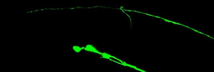
98 Figure 3.2 A. * B. * sdn-1(zh20);juis76 sdn-1(zh20);juis76 C. 88
99 Figure 3.2 sdn-1 is needed for D-type axon outgrowth and termination (A) Visualization of the under-extension phenotype seen in sdn-1 LOF mutants. (B) Visualization of the over-extension phenotype seen in sdn-1 LOF mutants. The arrow indicates the stereotypic termination point and the asterisk indicates the mutant termination point. The presence of both phenotypes indicates sdn-1 functions in both axon outgrowth and axon termination. (C) Quantification of the phenotypes seen in sdn-1 LOF mutants. Error bars represent the Standard Error of the Mean (SEM). 89

100 Figure 3.3 A. B. C. D. 90
101 Figure 3.3 SDN-1 is a determinant of anterior-posterior axon guidance and termination in C. elegans (A, C) DIC and (B, D) fluorescent images of an L2 sdn- 1(zh20) animal with muis32 (integrated GFP reporter expressed in the mechanosensory neurons ALM and PLM). (B) The PLM axons frequently terminated prematurely (not shown), but also failed to terminate growth. (D) The PLM axon would hook toward the ventral nerve cord. Arrow head indicates normal termination point and arrow indicates mutant termination point. 91


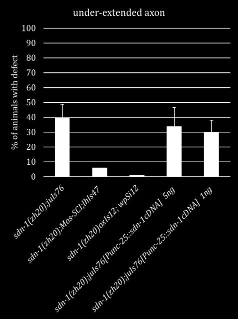
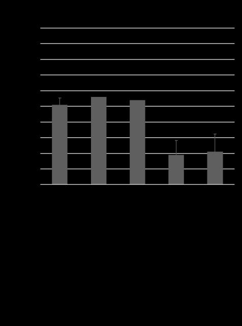
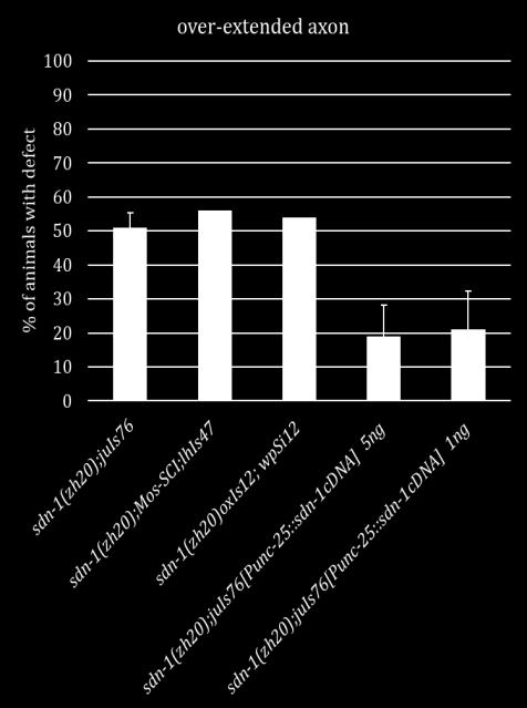
102 Figure 3.4 A. B. n.s. n.s. n.s. *** *** *** *** 92
103 Figure 3.4 SDN-1 functions both cell autonomously and non-autonomously during axon outgrowth and termination (A) Quantification of the under-extension observed in SDN-1 Mos-SCI lines and Punc-25::sdn-1cDNA in sdn-1(zh20) mutants. (B) Quantification of the over-extension observed in SDN-1 Mos-SCI lines and Punc- 25::sdn-1cDNA in sdn-1(zh20) mutants. N for sdn-1(zh20);juis76= 200 animals, SDN- 1::GFP Mos-SCI= 100 animals, wpsi12 Mos-SCI= 100 animals, Punc-25::sdn-1cDNA= 60 animals and Punc-25::sdn-1cDNA= 100 animals. ***P= Statistical significance was calculated by using the Fisher s Exact Test. Error bars represent the Standard Error of the Mean (SEM). (C) Visualization of SDN-1::GFP in a L1 animal. The majority of expression is observed in the nervous system and hypodermal tissues. (D) Visualization of the D-type motorneurons using lhis47 (Punc-25::mCherry). (E) Merge of the SDN-1::GFP and lhis47. (D) Enlargement of the E panel. SDN-1:GFP can be seen co-expressed with lhis47 in the VNC, DNC and cell bodies. 93


104 Figure 3.5 A. B. n.s. n.s. n.s. n.s. ** n.s. ** n.s. ** *** ** 94
105 Figure 3.5 sdn-1 functions with Wnt ligands for axon outgrowth and termination (A) Quantification of the under-extension phenotype seen in sdn-1 as compared to Wnt single and double mutant animals. The under-extension seen in sdn- 1 is not significantly different than lin-44(n1792);sdn-1(zh20) and lin-44(n1792);egl- 20(lq42)lqIs80;lhIs47. (B) Quantification of the over-extension phenotype seen in sdn- 1 as compared to Wnt single and double mutant animals. The over-extension seen in sdn-1 is not significantly different than lin-44(n1792);sdn-1(zh20) and lin- 44(n1792);egl-20(lq42)lqIs80;lhIs47. ***P=.0001, **P=.002. N= 200 animals. Statistical significance was calculated by using the Fisher s Exact Test. Error bars represent the Standard Error of the Mean (SEM). L4 animals were analyzed except when lqis80 was present then adult animals were scored. 95


106 Figure 3.6 A. B. *** *** n.s. n.s. *** n.s. n.s. n.s. n.s. n.s. n.s. ** *** *** 96
107 Figure 3.6 sdn-1 functions with canonical Wnt signaling effectors (A) Quantification of the under-extension phenotype seen in sdn-1 as compared to Wnt effector single and double mutant animals. The under-extension seen in sdn-1 is not significantly different than lin-17(n671);sdn-1(zh20), mig-5(rh94) and mig- 5(rh94);sdn-1(zh20). ***P<.0009 N>100 (B) Quantification of the over-extension phenotype seen in sdn-1 as compared to Wnt effector single and double mutant animals. The over-extension seen in sdn-1 is not significantly different than lin- 17(n671), lin-17(n671);sdn-1(zh20), and mig-5(rh94). ***P=.0001 **P=.004 N>100. Statistical significance was calculated by using the Fisher s Exact Test. Error bars represent the Standard Error of the Mean (SEM). 97


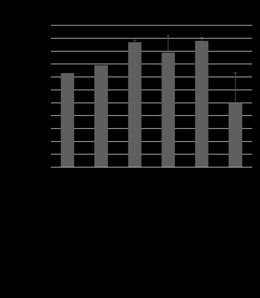
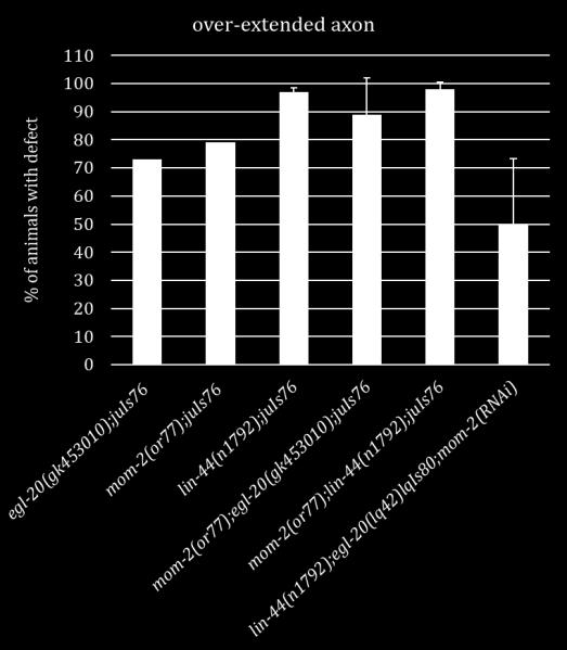
108 Figure 3.7 A. B. *** * *** ** 98
109 Figure 3.7 mom-2 does not act redundantly with lin-44 or egl-20 during D-type motorneuron axon outgrowth and termination (A) Quantification of the underextension phenotype seen in Wnt ligand single and double genetic mutant animals and lin-44;egl-20 animals grown on bacteria expressing mom-2 dsrna. We found no evidence of under-extension in mom-2(or77). Double mutants of mom-2(or77) with lin-44(n1792) (0%) and egl-20(gk453010) (3%) did not result in under-extension defects. The under-extension observed in the lin-44;egl-20;mom-2(rnai) (40%) was not significantly different than lin-44;egl-20 mutants (45%). (B) Quantification of the over-extension phenotype seen in Wnt ligand single and double genetic mutant animals and lin-44;egl-20 animals grown on bacteria expressing mom-2 dsrna. LOF of mom-2 via RNAi caused a significant decrease in over-extension defects seen in lin- 44(n1792) or egl-20(gk453010). However, the loss of mom-2 in a lin-44;egl-20 background (50%) was not significantly different from lin-44;egl-20 (46.5%). ***P=.0002, **P=.004, *P=.02 N>60. Statistical significance was calculated by using the Fisher s Exact Test. Error bars represent the Standard Error of the Mean (SEM). 99


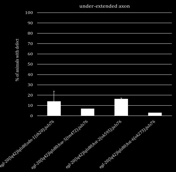
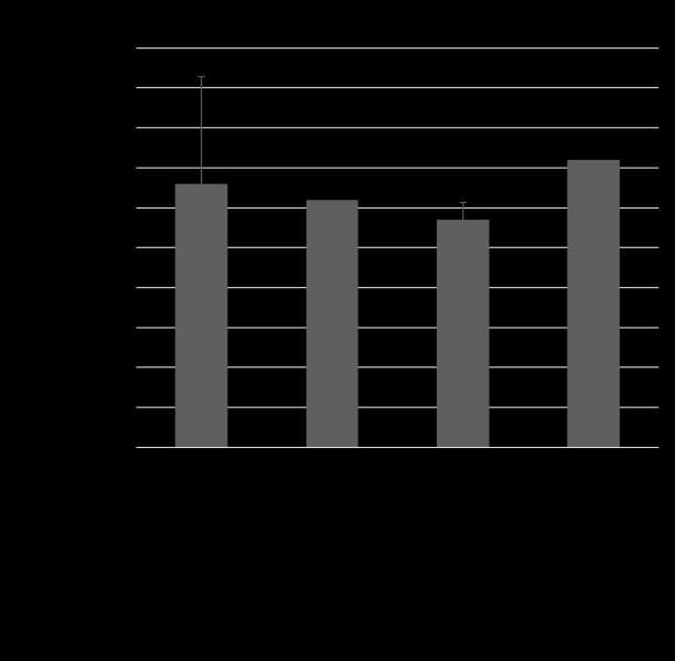
110 Figure 3.8 A. n.s. n.s. n.s. *** ** *** B. C. *** *** n.s. *** *** *** D. E. n.s. n.s. *** 100
111 Figure 3.8 sdn-1 heparan sulfate side chain modifications are important for Wnt interactions (A) Quantification of under- and over-extension defects observed in hse-5(tm472), hst-2(ok595) and hst-6(ok273) compared to sdn-1(zh20). Loss of either hst-2 (23%) or hst-6 (13%) does not fully recapitulate the under-extension defects seen in sdn-1 (39.5%). While hse-5 (43.5%), under-extension is not significantly different than sdn-1 (39.5%), the over-extension is significantly decreased (29% vs. 51%). (B-C) Quantification of under- and over-extension defects observed in lin-44(n1792), hse-5(tm472), hst-2(ok595) and hst-6(ok273) double mutants compared to lin-44(n1792);sdn-1(zh20). (D-E) Quantification of under- and over-extension defects observed in egl-20(lq42), hse-5(tm472), hst- 2(ok595) and hst-6(ok273) double mutants compared to egl-20(lq42);sdn-1(zh20). N>100. ***P=.0001, **P<.03. Statistical significance was calculated by using the Fisher s Exact Test. Error bars represent the Standard Error of the Mean (SEM). Experiments and analysis were conducted by Samantha Hartin and Riley Roberts. 101

112 Figure 3.9 Stage 1: Outgrowth Stage 2: Termination 102
113 Figure 3.9 Genetic model for D-type axon outgrowth and termination (Stage 1: Outgrowth) SDN-1 is most likely inhibiting Wnt signaling and the downstream effectors to allow for proper axon outgrowth. (Stage 2: Termination) Since we were able to partially rescue the over-growth phenotype by specifically expressing SDN-1 in the D-type neurons. This suggests that SDN-1 can in the D-type motorneurons to activate the Wnt signaling pathway and allow for proper axon termination. This is consistent with our genetic data that the loss of sdn-1 and lin-17 are both associated with outgrowth errors. 103
114 Chapter IV Concluding remarks and future directions 104
115 Section 4.1 Concluding remarks The findings in Chapter II reveal roles for PTP-3 and SDN-1 during C. elegans embryogenesis and larval development. Here using double mutant and 4D time lapse analysis we found that PTP-3 and SDN-1 work in parallel pathways to ensure proper epidermal development. Using the SynLet phenotype seen in the ptp-3;sdn-1 double mutants, we conducted an RNAi screen for other ECM molecules that could interact with either gene during development. Our sensitized backgrounds allowed for identification of molecules that may have been missed in traditional screens due to low phenotypic penetrance. It is important to note that we may have missed certain genes during our screen due to the nature of RNAi (Off target effects, reduced induction or inherent resistance). Regardless, identification of genes previously shown to be involved with either ptp-3 or sdn-1 suggests that our method is a valid way to identify genetic interactions using genes of interest. One such gene lin-44, a Wnt ligand, was identified as been SynLet with sdn-1 but not ptp-3 suggesting that it may function in a linear pathway with ptp-3 that functions in parallel to sdn-1. Again using double mutants and 4D time lapse analysis we found that complete LOF mutations in lin-44 and sdn-1 cause the majority of animals to arrest at the embryo stage. This arrest is due to the improper migration of endodermal precursor cells Ea and Ep at the 24-cell stage of development. Normally LIN-44 signals through the Frizzled receptor LIN-17. However, we believe that during this process LIN-44 does not function through its canonical Frizzled receptor, LIN-17, due to the level of embryonic lethality observed in lin-17;sdn-1 double mutants. Double mutant analysis of ptp-3 and lin-44 revealed no significant increase in 105
116 embryonic lethality, although the level of larval lethality increased slightly when compared to each single mutant. This suggests that lin-44 and ptp-3 may be functioning in the same genetic pathway during embryonic development. Our results are consistent with the idea that ptp-3 contributes to lin-44- dependent embryogenesis. Furthermore, it is likely that at least one of the potential reasons that ptp-3; sdn-1 double mutants exhibit SynLet is because of a disruption in a lin-44 / ptp-3 signaling pathway during gastrulation. Chapter III identifies later functions for SDN-1, LIN-44 and EGL-20 during motorneuron axon outgrowth and termination. Additionally, our results show that another yet unidentified signaling molecule functions during both these processes. Our studies complement previous genetic data suggesting that LIN-44 and EGL-20 work together for proper axon termination of the most posterior DD/VD motorneurons in the C. elegans tail. However, our complete LOF double mutant analysis also suggests that both genes function in DD/VD axon outgrowth, but the importance of each molecule differs during the two processes. Here, we found that SDN-1 functions in both processes. During the first process, SDN-1 is required nonautonomously to regulate Wnt signaling either by controlling the diffusion/distribution of the Wnt ligands or by disrupting the Wnt-Frizzled interaction (Figure 4.1). For proper axon termination, SDN-1 acts cell autonomously to inhibit Wnt signaling, perhaps by binding to MIG-5 to disrupt its ability to inhibit the β-catenin destruction complex (Figure 4.1). Further genetic analysis of the downstream Wnt effectors indicate that SDN-1 functions at the level of the Frizzled receptor LIN-17 and possibly with Dishevelled/MIG-5 during both processes. 106
117 Interestingly, loss of bar-1 causes the axons to under-extend in 95% of animals suggesting that BAR-1 activity is negatively regulated for axon extension through SDN-1 inhibition of proper Wnt signaling. The HS side chains of SDN-1 are important for ligand interactions. By analyzing the enzymes that modify the side chains (HSE-5, HST-2 and HST-6) we found that they have functions in both processes. However, none of them were able to recapitulate the phenotypic penetrance seen in sdn-1 single or sdn-1;wnt double mutants. These data suggest that there are other modifying enzymes that function during these processes, and possibly other proteoglycans as well. Overall, our genetic analyses indicate that SDN-1 is needed for both DD/VD motorneuron axon extension and termination because LOF sdn-1 mutants display both under and over-extension defects. We propose a two stage model where EGL-20 and LIN-44 signal through LIN-17, SDN-1, MIG-5 and BAR-1 to regulate axon extension. Here SDN-1 acts as a negative regulator of β-catenin dependent Wnt signaling. Additionally, our findings regarding the extent of over-extension phenotypes lead us to hypothesize that EGL-20 plays a larger role with SDN-1 than LIN-44 during this initial stage. When the axon reaches the proper termination point LIN-44 and EGL-20 again use LIN-17, SDN-1 and MIG-5 to signal for axon termination. During this second stage we believe that LIN-44 is acting as the primary molecule used for termination in a parallel pathway to EGL-20 and SDN-1, and these two pathways converge on the same molecule at some point. Since none of the complete LOF mutations recapitulate the phenotypes seen in the triple mutant we believe there 107
118 is another molecule involved in axon extension and termination. Further studies will be needed to confirm this hypothesis. In conclusion, the results presented in this dissertation describe how known cell adhesion molecules function initially in parallel pathways during gastrulation and later function in the same pathway during neural development. Both the identification of the role of LIN-44 during gastrulation alongside PTP-3 and SDN-1 and the later characterization of the SDN-1/Wnt interaction add to the current knowledge of how these molecules are reused during development. We also identified an effective way to search for genes of interest using a sensitized reverse genetics approach. 108
119 Section 4.2 Future Directions The findings presented in this dissertation provide new insights into the reuse of cell adhesion molecules during development. However, some questions remain unanswered. Going forward, addressing the following questions will further our knowledge of how SDN-1 and the Wnt pathways intersect during embryogenesis and axon guidance. Question 1: Where is SDN-1 functioning during axon outgrowth and termination? In chapter III we demonstrated that LOF mutations in sdn-1 cause both under and over-extension of the DD6 and/or VD13 axons in the dorsal nerve cord, suggesting SDN-1 is involved in both axon extension and termination. Our genetic analysis of SDN-1 and the Wnt pathway suggest that SDN-1 works in combination with both LIN-44 and EGL-20 during both processes. However, we do not know where SDN-1 is functioning in these processes. Previous studies of Wnt receptors showed that they can work both cell autonomously and non-autonomously to modulate Wnt signaling. We hypothesize that SDN-1 has both a cell autonomous and nonautonomous role and switches between these roles during axon outgrowth and axon termination in response to the different Wnt signals. SDN-1 is expressed in multiple tissues throughout C. elegans development including the nervous system, hypodermis and pharynx [15, 99]. Expression of full length SDN-1 in either the nervous system or hypodermis can rescue different phenotypes seen in LOF sdn-1 mutants [15] 109
120 We found that under-extension of the DD/VD axons can be rescued by reintroducing a single copy of the full length SDN-1 gene back into sdn-1 mutants. However, the over-extension phenotype is not fully rescued. This partial phenotypic rescue has also been reported by others [30, 36]. This partial result suggests that there are long distance regulatory elements and/or genomic positional requirements that have yet to be identified, which are necessary for full rescue of the sdn-1 phenotypes. Since we did not obtain full rescue with two separate SDN-1 Mos-SCI lines, we made a SDN-1 cdna construct that is expressed in the VD/DD motorneurons by the unc-25 promoter. We analyzed two separate injection concentrations and found that neither fully rescued either phenotype but we were able to significantly decrease penetrance of the over-extension defects seen in sdn-1 LOF mutants. Next, we made SDN-1 cdna constructs driven by the myo-3 promoter, which drives expression in the muscles. With this construct we were able to significantly decrease the penetrance of over-extended axons however the penetrance of under-extended axons increased significantly. We will need to determine whether the defects can be rescued by the SDN-1 cdna alone and if it can rescue our phenotypes under its endogenous promoter. We will also need to express our SDN-1 cdna from the hypodermis to determine if SDN- 1 functions there during either process. However, similar constructs have not been able to fully rescue all sdn-1 phenotypes including axon guidance [15, 36]. Mosaic analysis may also be employed to determine where SDN-1 is functioning during axon outgrowth and termination. 110
121 Question 2: Does SDN-1 physically interact with components of the Wnt pathway? According to the genetic data presented in this dissertation, SDN-1 acts at the level of LIN-17/Frizzled and/or MIG-5/Dishevelled during each stage. However, it is unknown whether these molecules are physically interacting during signaling. Interactions between HSPGs and Frizzled receptors are reported to be important in the positive and negative regulation of Wnt signaling [30-33, 35]. We have imaged and analyzed animals with SDN-1::GFP and LIN-17::RFP and found that both are broadly localized in the animal. SDN-1::GFP is prominent in the dorsal nerve cord and LIN-17::RFP is also present in the DNC. Closer analysis show SDN-1::GFP and LIN- 17::RFP signals at the axon termination point (Figure 4.2). Our initial FRET (Fluorescence resonance energy transfer) analysis of SDN-1 and LIN-17 suggest that they are closely situated in the both the dorsal and ventral nerve cords but, their association is more prevalent in the dorsal nerve cord. These data suggest that they may physically interact in those axons. To determine whether SDN-1 and LIN-17 are physically interacting we could conduct CO-IP assays in wild-type and mutant strains. SDN-1 may also be physically interacting with MIG-5/Dishevelled via its PDZ domain. To determine if this is important for proper Wnt signaling we could repeat the above assays substituting LIN-17 reagents for MIG-5 reagents. 111


122 Section 4.3 Figures Figure 4.1 A. SDN-1 EGL-20 EGL-20 LIN-44 LIN-44 LIN-17 EGL-20 LIN-44 LIN-44 B. EGL-20 LIN-44 EGL-20 LIN-44 LIN-44 EGL-20 EGL-20 LIN
123 Figure 4.1 Potential molecular model for the two step growth of the D-type motorneurons Our results suggest that these functions are likely mediated by different cells expressing SDN-1. (A) When we replaced SDN-1 function broadly using Mos-SCI constructs we found that we could only rescue the under-extension of axons, but could partially rescue the under-extension when we decreased the levels of SDN-1 by making the integrated transgene heterozygous. These results indicate that SDN-1 is acting outside the D-type motorneurons to inhibit Wnt signaling. Possibly by restricting the distribution of the ligands. (B) We were able to partially rescue the over-growth phenotype by specifically expressing SDN-1 in the D-type neurons. This suggests that SDN-1 can function cell-autonomously in the D-type neurons to restrict outgrowth. It is possible that SDN-1 is acting to promote Wnt signaling, via LIN-17, at the termination point of the D-type neurons, to inhibit further growth. 113

124 Figure
Chapter 4 Evaluating a potential interaction between deltex and git in Drosophila: genetic interaction, gene overexpression and cell biology assays.
 Evaluating a potential interaction between deltex and git in Drosophila: genetic interaction, gene overexpression and cell biology assays. The data described in chapter 3 presented evidence that endogenous
Nature Biotechnology: doi: /nbt Supplementary Figure 1. Overexpression of YFP::GPR-1 in the germline.
 Supplementary Figure 1 Overexpression of YFP::GPR-1 in the germline. The pie-1 promoter and 3 utr were used to express yfp::gpr-1 in the germline. Expression levels from the yfp::gpr-1(cai 1.0)-expressing
Chapter 18 Lecture. Concepts of Genetics. Tenth Edition. Developmental Genetics
 Chapter 18 Lecture Concepts of Genetics Tenth Edition Developmental Genetics Chapter Contents 18.1 Differentiated States Develop from Coordinated Programs of Gene Expression 18.2 Evolutionary Conservation
Conclusions. The experimental studies presented in this thesis provide the first molecular insights
 C h a p t e r 5 Conclusions 5.1 Summary The experimental studies presented in this thesis provide the first molecular insights into the cellular processes of assembly, and aggregation of neural crest and
1. What are the three general areas of the developing vertebrate limb? 2. What embryonic regions contribute to the developing limb bud?
 Study Questions - Lecture 17 & 18 1. What are the three general areas of the developing vertebrate limb? The three general areas of the developing vertebrate limb are the proximal stylopod, zeugopod, and
SUPPLEMENTARY INFORMATION
 Supplementary Discussion Rationale for using maternal ythdf2 -/- mutants as study subject To study the genetic basis of the embryonic developmental delay that we observed, we crossed fish with different
Chapter 11. Development: Differentiation and Determination
 KAP Biology Dept Kenyon College Differential gene expression and development Mechanisms of cellular determination Induction Pattern formation Chapter 11. Development: Differentiation and Determination
Midterm 1. Average score: 74.4 Median score: 77
 Midterm 1 Average score: 74.4 Median score: 77 NAME: TA (circle one) Jody Westbrook or Jessica Piel Section (circle one) Tue Wed Thur MCB 141 First Midterm Feb. 21, 2008 Only answer 4 of these 5 problems.
Lecture 7. Development of the Fruit Fly Drosophila
 BIOLOGY 205/SECTION 7 DEVELOPMENT- LILJEGREN Lecture 7 Development of the Fruit Fly Drosophila 1. The fruit fly- a highly successful, specialized organism a. Quick life cycle includes three larval stages
Supplementary Materials for
 www.sciencesignaling.org/cgi/content/full/6/301/ra98/dc1 Supplementary Materials for Regulation of Epithelial Morphogenesis by the G Protein Coupled Receptor Mist and Its Ligand Fog Alyssa J. Manning,
Axis Specification in Drosophila
 Developmental Biology Biology 4361 Axis Specification in Drosophila November 2, 2006 Axis Specification in Drosophila Fertilization Superficial cleavage Gastrulation Drosophila body plan Oocyte formation
Unicellular: Cells change function in response to a temporal plan, such as the cell cycle.
 Spatial organization is a key difference between unicellular organisms and metazoans Unicellular: Cells change function in response to a temporal plan, such as the cell cycle. Cells differentiate as a
Caenorhabditis elegans
 Caenorhabditis elegans Why C. elegans? Sea urchins have told us much about embryogenesis. They are suited well for study in the lab; however, they do not tell us much about the genetics involved in embryogenesis.
Exam 1 ID#: October 4, 2007
 Biology 4361 Name: KEY Exam 1 ID#: October 4, 2007 Multiple choice (one point each) (1-25) 1. The process of cells forming tissues and organs is called a. morphogenesis. b. differentiation. c. allometry.
Bio 127 Section I Introduction to Developmental Biology. Cell Cell Communication in Development. Developmental Activities Coordinated in this Way
 Bio 127 Section I Introduction to Developmental Biology Cell Cell Communication in Development Gilbert 9e Chapter 3 It has to be EXTREMELY well coordinated for the single celled fertilized ovum to develop
MBios 401/501: Lecture 14.2 Cell Differentiation I. Slide #1. Cell Differentiation
 MBios 401/501: Lecture 14.2 Cell Differentiation I Slide #1 Cell Differentiation Cell Differentiation I -Basic principles of differentiation (p1305-1320) -C-elegans (p1321-1327) Cell Differentiation II
Cell Cell Communication in Development
 Biology 4361 Developmental Biology Cell Cell Communication in Development June 25, 2008 Cell Cell Communication Concepts Cells in developing organisms develop in the context of their environment, including
Cell-Cell Communication in Development
 Biology 4361 - Developmental Biology Cell-Cell Communication in Development June 23, 2009 Concepts Cell-Cell Communication Cells develop in the context of their environment, including: - their immediate
Nature Neuroscience: doi: /nn.2717
 Supplementary Fig. 1. Dendrite length is not secondary to body length. Dendrite growth proceeds independently of the rate of body growth and decreases in rate in adults. n 20 on dendrite measurement, n
Bypass and interaction suppressors; pathway analysis
 Bypass and interaction suppressors; pathway analysis The isolation of extragenic suppressors is a powerful tool for identifying genes that encode proteins that function in the same process as a gene of
9/4/2015 INDUCTION CHAPTER 1. Neurons are similar across phyla Thus, many different model systems are used in developmental neurobiology. Fig 1.
 INDUCTION CHAPTER 1 Neurons are similar across phyla Thus, many different model systems are used in developmental neurobiology Fig 1.1 1 EVOLUTION OF METAZOAN BRAINS GASTRULATION MAKING THE 3 RD GERM LAYER
16 The Cell Cycle. Chapter Outline The Eukaryotic Cell Cycle Regulators of Cell Cycle Progression The Events of M Phase Meiosis and Fertilization
 The Cell Cycle 16 The Cell Cycle Chapter Outline The Eukaryotic Cell Cycle Regulators of Cell Cycle Progression The Events of M Phase Meiosis and Fertilization Introduction Self-reproduction is perhaps
Cell-Cell Communication in Development
 Biology 4361 - Developmental Biology Cell-Cell Communication in Development October 2, 2007 Cell-Cell Communication - Topics Induction and competence Paracrine factors inducer molecules Signal transduction
Reading: Chapter 5, pp ; Reference chapter D, pp Problem set F
 Mosaic Analysis Reading: Chapter 5, pp140-141; Reference chapter D, pp820-823 Problem set F Twin spots in Drosophila Although segregation and recombination in mitosis do not occur at the same frequency
Cellular Neurobiology BIPN 140 Fall 2016 Problem Set #8
 Cellular Neurobiology BIPN 140 Fall 2016 Problem Set #8 1. Inductive signaling is a hallmark of vertebrate and mammalian development. In early neural development, there are multiple signaling pathways
Axis Specification in Drosophila
 Developmental Biology Biology 4361 Axis Specification in Drosophila November 6, 2007 Axis Specification in Drosophila Fertilization Superficial cleavage Gastrulation Drosophila body plan Oocyte formation
Why Flies? stages of embryogenesis. The Fly in History
 The Fly in History 1859 Darwin 1866 Mendel c. 1890 Driesch, Roux (experimental embryology) 1900 rediscovery of Mendel (birth of genetics) 1910 first mutant (white) (Morgan) 1913 first genetic map (Sturtevant
Reading. Lecture VI. Making Connections 9/17/12. Bio 3411 Lecture VI. Making Connections. Bio 3411 Monday September 17, 2012
 Lecture VI. Making Connections Bio 3411 Monday September 17, 2012!! 1! Reading NEUROSCIENCE: 5 th ed, pp!507?536! 4 th ed, pp 577-609 Bentley, D., & Caudy, M. (1983). Nature, 304(5921), 62-65. Dickson,
purpose of this Chapter is to highlight some problems that will likely provide new
 119 Chapter 6 Future Directions Besides our contributions discussed in previous chapters to the problem of developmental pattern formation, this work has also brought new questions that remain unanswered.
RACK-1 Acts with Rac GTPase Signaling and UNC-115/ ablim in Caenorhabditis elegans Axon Pathfinding and Cell Migration
 RACK-1 Acts with Rac GTPase Signaling and UNC-115/ ablim in Caenorhabditis elegans Axon Pathfinding and Cell Migration Rafael S. Demarco, Erik A. Lundquist* Programs in Genetics and Molecular, Cellular,
Axon guidance I. Paul Garrity March 15, /9.013
 Axon guidance I Paul Garrity March 15, 2004 7.68/9.013 Neuronal Wiring: Functional Framework of the Nervous System Stretch reflex circuit Early theories of axonogenesis Schwann: many neurons link to form
Neural development its all connected
 Neural development its all connected How do you build a complex nervous system? How do you build a complex nervous system? 1. Learn how tissue is instructed to become nervous system. Neural induction 2.
Developmental Biology Lecture Outlines
 Developmental Biology Lecture Outlines Lecture 01: Introduction Course content Developmental Biology Obsolete hypotheses Current theory Lecture 02: Gametogenesis Spermatozoa Spermatozoon function Spermatozoon
Mesoderm Induction CBT, 2018 Hand-out CBT March 2018
 Mesoderm Induction CBT, 2018 Hand-out CBT March 2018 Introduction 3. Books This module is based on the following books: - 'Principles of Developement', Lewis Wolpert, et al., fifth edition, 2015 - 'Developmental
Development of Drosophila
 Development of Drosophila Hand-out CBT Chapter 2 Wolpert, 5 th edition March 2018 Introduction 6. Introduction Drosophila melanogaster, the fruit fly, is found in all warm countries. In cooler regions,
Cell Death & Trophic Factors II. Steven McLoon Department of Neuroscience University of Minnesota
 Cell Death & Trophic Factors II Steven McLoon Department of Neuroscience University of Minnesota 1 Remember? Neurotrophins are cell survival factors that neurons get from their target cells! There is a
Axon Guidance. Multiple decision points along a growing axon s trajectory Different types of axon guidance cues:
 Axon Guidance Multiple decision points along a growing axon s trajectory Different types of axon guidance cues: Contact mediated - requires direct contact by growth cone Long range - growth cone responds
Green Fluorescent Protein (GFP) Today s Nobel Prize in Chemistry
 Today s Nobel Prize in Chemistry") In the news: High-throughput sequencing using Solexa/Illumina technology The copy number of each fetal chromosome can be determined by direct sequencing of DNA in cell-free plasma from pregnant women Confession:
In the news: High-throughput sequencing using Solexa/Illumina technology The copy number of each fetal chromosome can be determined by direct sequencing of DNA in cell-free plasma from pregnant women Confession:
Axis Specification in Drosophila
 Developmental Biology Biology 4361 Axis Specification in Drosophila July 9, 2008 Drosophila Development Overview Fertilization Cleavage Gastrulation Drosophila body plan Oocyte formation Genetic control
Developmental genetics: finding the genes that regulate development
 Developmental Biology BY1101 P. Murphy Lecture 9 Developmental genetics: finding the genes that regulate development Introduction The application of genetic analysis and DNA technology to the study of
Chapter 10 Development and Differentiation
 Part III Organization of Cell Populations Chapter Since ancient times, people have wondered how organisms are formed during the developmental process, and many researchers have worked tirelessly in search
AP Biology Gene Regulation and Development Review
 AP Biology Gene Regulation and Development Review 1. What does the regulatory gene code for? 2. Is the repressor by default active/inactive? 3. What changes the repressor activity? 4. What does repressor
MCDB 4777/5777 Molecular Neurobiology Lecture 29 Neural Development- In the beginning
 MCDB 4777/5777 Molecular Neurobiology Lecture 29 Neural Development- In the beginning Learning Goals for Lecture 29 4.1 Describe the contributions of early developmental events in the embryo to the formation
Developmental processes Differential gene expression Introduction to determination The model organisms used to study developmental processes
 Date Title Topic(s) Learning Outcomes: Sept 28 Oct 3 1. What is developmental biology and why should we care? 2. What is so special about stem cells and gametes? Developmental processes Differential gene
SUPPLEMENTARY INFORMATION
 doi:10.1038/nature11589 Supplementary Figure 1 Ciona intestinalis and Petromyzon marinus neural crest expression domain comparison. Cartoon shows dorsal views of Ciona mid gastrula (left) and Petromyzon
7.013 Problem Set
 7.013 Problem Set 5-2013 Question 1 During a summer hike you suddenly spot a huge grizzly bear. This emergency situation triggers a fight or flight response through a signaling pathway as shown below.
Big Idea 3: Living systems store, retrieve, transmit and respond to information essential to life processes. Tuesday, December 27, 16
 Big Idea 3: Living systems store, retrieve, transmit and respond to information essential to life processes. Enduring understanding 3.B: Expression of genetic information involves cellular and molecular
MCN. Complex Genetic Interactions among Four Receptor Tyrosine Phosphatases Regulate Axon Guidance in Drosophila
 MCN Molecular and Cellular Neuroscience 17, 274 291 (2001) doi:10.1006/mcne.2000.0939, available online at http://www.idealibrary.com on Complex Genetic Interactions among Four Receptor Tyrosine Phosphatases
A complementation test would be done by crossing the haploid strains and scoring the phenotype in the diploids.
 Problem set H answers 1. To study DNA repair mechanisms, geneticists isolated yeast mutants that were sensitive to various types of radiation; for example, mutants that were more sensitive to UV light.
Exam 2 ID#: November 9, 2006
 Biology 4361 Name: KEY Exam 2 ID#: November 9, 2006 Multiple choice (one point each) Circle the best answer. 1. Inducers of Xenopus lens and optic vesicle include a. pharyngeal endoderm and anterior neural
5- Semaphorin-Plexin-Neuropilin
 5- Semaphorin-Plexin-Neuropilin 1 SEMAPHORINS-PLEXINS-NEUROPILINS ligands receptors co-receptors semaphorins and their receptors are known signals for: -axon guidance -cell migration -morphogenesis -immune
Developmental Biology 3230 Midterm Exam 1 March 2006
 Name Developmental Biology 3230 Midterm Exam 1 March 2006 1. (20pts) Regeneration occurs to some degree to most metazoans. When you remove the head of a hydra a new one regenerates. Graph the inhibitor
10/2/2015. Chapter 4. Determination and Differentiation. Neuroanatomical Diversity
 Chapter 4 Determination and Differentiation Neuroanatomical Diversity 1 Neurochemical diversity: another important aspect of neuronal fate Neurotransmitters and their receptors Excitatory Glutamate Acetylcholine
Cells to Tissues. Peter Takizawa Department of Cell Biology
 Cells to Tissues Peter Takizawa Department of Cell Biology From one cell to ensembles of cells. Multicellular organisms require individual cells to work together in functional groups. This means cells
Supplementary Figure 1: Mechanism of Lbx2 action on the Wnt/ -catenin signalling pathway. (a) The Wnt/ -catenin signalling pathway and its
 The Wnt/ -catenin signalling pathway and its") Supplementary Figure 1: Mechanism of Lbx2 action on the Wnt/ -catenin signalling pathway. (a) The Wnt/ -catenin signalling pathway and its transcriptional activity in wild-type embryo. A gradient of canonical
Supplementary Figure 1: Mechanism of Lbx2 action on the Wnt/ -catenin signalling pathway. (a) The Wnt/ -catenin signalling pathway and its transcriptional activity in wild-type embryo. A gradient of canonical
Nature Neuroscience: doi: /nn.2662
 Supplementary Figure 1 Atlastin phylogeny and homology. (a) Maximum likelihood phylogenetic tree based on 18 Atlastin-1 sequences using the program Quicktree. Numbers at internal nodes correspond to bootstrap
Zool 3200: Cell Biology Exam 5 4/27/15
 Name: Trask Zool 3200: Cell Biology Exam 5 4/27/15 Answer each of the following short answer questions in the space provided, giving explanations when asked to do so. Circle the correct answer or answers
Bio 3411, Fall 2006, Lecture 19-Cell Death.
 Types of Cell Death Questions : Apoptosis (Programmed Cell Death) : Cell-Autonomous Stereotypic Rapid Clean (dead cells eaten) Necrosis : Not Self-Initiated Not Stereotypic Can Be Slow Messy (injury can
Regulation and signaling. Overview. Control of gene expression. Cells need to regulate the amounts of different proteins they express, depending on
 Regulation and signaling Overview Cells need to regulate the amounts of different proteins they express, depending on cell development (skin vs liver cell) cell stage environmental conditions (food, temperature,
Introduction Principles of Signaling and Organization p. 3 Signaling in Simple Neuronal Circuits p. 4 Organization of the Retina p.
 Introduction Principles of Signaling and Organization p. 3 Signaling in Simple Neuronal Circuits p. 4 Organization of the Retina p. 5 Signaling in Nerve Cells p. 9 Cellular and Molecular Biology of Neurons
Drosophila melanogaster- Morphogen Gradient
 NPTEL Biotechnology - Systems Biology Drosophila melanogaster- Morphogen Gradient Dr. M. Vijayalakshmi School of Chemical and Biotechnology SASTRA University Joint Initiative of IITs and IISc Funded by
Massachusetts Institute of Technology Harvard Medical School Brigham and Women s Hospital VA Boston Healthcare System 2.79J/3.96J/BE.
 Massachusetts Institute of Technology Harvard Medical School Brigham and Women s Hospital VA Boston Healthcare System 2.79J/3.96J/BE.441/HST522J INTEGRINS I.V. Yannas, Ph.D. and M. Spector, Ph.D. Regulator
Drosophila Life Cycle
 Drosophila Life Cycle 1 Early Drosophila Cleavage Nuclei migrate to periphery after 10 nuclear divisions. Cellularization occurs when plasma membrane folds in to divide nuclei into cells. Drosophila Superficial
RPM-1 is localized to distinct subcellular compartments and regulates axon length in GABAergic motor neurons
 Opperman and Grill Neural Development 14, 9:1 RESERCH RTICLE Open ccess RPM-1 is localized to distinct subcellular compartments and regulates axon length in Gergic motor neurons Karla J Opperman and rock
Seattle-Vancouver Worm Meeting
 The Second Annual Seattle-Vancouver Worm Meeting Friday, March 28 th, 2008 1:00 pm Michael Smith Building Lecture Theatre 12:50 pm Introduction Itinerary: Update from the NRRR Committee 1:00 pm Session
CHAPTER 13 PROKARYOTE GENES: E. COLI LAC OPERON
 PROKARYOTE GENES: E. COLI LAC OPERON CHAPTER 13 CHAPTER 13 PROKARYOTE GENES: E. COLI LAC OPERON Figure 1. Electron micrograph of growing E. coli. Some show the constriction at the location where daughter
Amneh Auben. Abdulrahman Jabr. Diala Abu-Hassan
 21 Amneh Auben Abdulrahman Jabr Diala Abu-Hassan Matrix polysaccharides Extracellular matrix (ECM): It s a collection of components that fills the spaces outside the cell or between the cells. ---------
Life Sciences 1a: Section 3B. The cell division cycle Objectives Understand the challenges to producing genetically identical daughter cells
 Life Sciences 1a: Section 3B. The cell division cycle Objectives Understand the challenges to producing genetically identical daughter cells Understand how a simple biochemical oscillator can drive the
Genes required for axon pathfinding and extension in the C. elegans nerve ring
 Development 126, 3679-3692 (1999) Printed in Great Britain The Company of Biologists Limited 1999 DEV8610 3679 Genes required for axon pathfinding and extension in the C. elegans nerve ring Jennifer A.
Role of Organizer Chages in Late Frog Embryos
 Ectoderm Germ Layer Frog Fate Map Frog Fate Map Role of Organizer Chages in Late Frog Embryos Organizer forms three distinct regions Notochord formation in chick Beta-catenin localization How does beta-catenin
UNIVERSITY OF YORK BIOLOGY. Developmental Biology
 Examination Candidate Number: UNIVERSITY OF YORK BSc Stage 2 Degree Examinations 2017-18 Department: BIOLOGY Title of Exam: Developmental Biology Desk Number: Time allowed: 1 hour and 30 minutes Total
C. elegans L1 cell adhesion molecule functions in axon guidance
 C. elegans L1 cell adhesion molecule functions in axon guidance Biorad Lihsia Chen Dept. of Genetics, Cell Biology & Development Developmental Biology Center C. elegans embryogenesis Goldstein lab, UNC-Chapel
Predictive Modeling of Signaling Crosstalk... Model execution and model checking can be used to test a biological hypothesis
 Model execution and model checking can be used to test a biological hypothesis The model is an explanation of a biological mechanism and its agreement with experimental results is used to either validate
Introduction. Gene expression is the combined process of :
 1 To know and explain: Regulation of Bacterial Gene Expression Constitutive ( house keeping) vs. Controllable genes OPERON structure and its role in gene regulation Regulation of Eukaryotic Gene Expression
MOLECULAR CONTROL OF EMBRYONIC PATTERN FORMATION
 MOLECULAR CONTROL OF EMBRYONIC PATTERN FORMATION Drosophila is the best understood of all developmental systems, especially at the genetic level, and although it is an invertebrate it has had an enormous
BIS &003 Answers to Assigned Problems May 23, Week /18.6 How would you distinguish between an enhancer and a promoter?
 Week 9 Study Questions from the textbook: 6 th Edition: Chapter 19-19.6, 19.7, 19.15, 19.17 OR 7 th Edition: Chapter 18-18.6 18.7, 18.15, 18.17 19.6/18.6 How would you distinguish between an enhancer and
Questions in developmental biology. Differentiation Morphogenesis Growth/apoptosis Reproduction Evolution Environmental integration
 Questions in developmental biology Differentiation Morphogenesis Growth/apoptosis Reproduction Evolution Environmental integration Representative cell types of a vertebrate zygote => embryo => adult differentiation
Early Development in Invertebrates
 Developmental Biology Biology 4361 Early Development in Invertebrates October 25, 2006 Early Development Overview Cleavage rapid cell divisions divisions of fertilized egg into many cells Gastrulation
Natural Variation, Development, and Adaptive Phenotypes in C. elegans
 Natural Variation, Development, and Adaptive Phenotypes in C. elegans Bradly Alicea http://publish.illinois.edu/bradly-alicea Fly/Worm Club, UIUC, April 22, 2015 with input from Nathan Schroeder, Rebecca
Axis determination in flies. Sem 9.3.B.5 Animal Science
 Axis determination in flies Sem 9.3.B.5 Animal Science All embryos are in lateral view (anterior to the left). Endoderm, midgut; mesoderm; central nervous system; foregut, hindgut and pole cells in yellow.
with%dr.%van%buskirk%%%
 with%dr.%van%buskirk%%% How$to$do$well?$ Before$class:$read$the$corresponding$chapter$ Come$to$class$ready$to$par9cipate$in$Top$Hat$ Don t$miss$an$exam!!!!!!!!!!!!!!!!!!!!!!!!!!$ But$I m$not$good$with$science
Chapter 3. Results rho-1 regulates epidermal elongation in C. elegans embryogenesis
 Chapter 3. Results 3.1. rho-1 regulates epidermal elongation in C. elegans embryogenesis 3.1.1 Zygotic rho-1 is required for elongation Elongation is driven by actin-myosin contraction in the seam cells,
Open Syntaxin Docks Synaptic Vesicles
 Marc Hammarlund 1,2[, Mark T. Palfreyman 1,2[, Shigeki Watanabe 1,2[, Shawn Olsen 1,2, Erik M. Jorgensen 1,2* PLoS BIOLOGY 1 Department of Biology, University of Utah, Salt Lake City, Utah, United States
Drosophila Somatic Anterior-Posterior Axis (A-P Axis) Formation
 Formation") Home Biol 4241 Luria-Delbruck 1943 Hershey-Chase 1952 Meselson-Stahl 1958 Garapin et al. 1978 McClintock 1953 King-Wilson 1975 Sanger et al. 1977 Rothberg et al. 2011 Jeffreys et al. 1985 Bacterial Genetics
Home Biol 4241 Luria-Delbruck 1943 Hershey-Chase 1952 Meselson-Stahl 1958 Garapin et al. 1978 McClintock 1953 King-Wilson 1975 Sanger et al. 1977 Rothberg et al. 2011 Jeffreys et al. 1985 Bacterial Genetics
Multiple levels of regulation specify the polarity of an asymmetric cell division in C. elegans
 Development 127, 4587-4598 (2) Printed in Great Britain The Company of Biologists Limited 2 DEV4424 4587 Multiple levels of regulation specify the polarity of an asymmetric cell division in C. elegans
BIOLOGY STANDARDS BASED RUBRIC
 BIOLOGY STANDARDS BASED RUBRIC STUDENTS WILL UNDERSTAND THAT THE FUNDAMENTAL PROCESSES OF ALL LIVING THINGS DEPEND ON A VARIETY OF SPECIALIZED CELL STRUCTURES AND CHEMICAL PROCESSES. First Semester Benchmarks:
Cell Biology Review. The key components of cells that concern us are as follows: 1. Nucleus
 Cell Biology Review Development involves the collective behavior and activities of cells, working together in a coordinated manner to construct an organism. As such, the regulation of development is intimately
!!!!!!!! DB3230 Midterm 2 12/13/2013 Name:
 1. (10 pts) Draw or describe the fate map of a late blastula stage sea urchin embryo. Draw or describe the corresponding fate map of the pluteus stage larva. Describe the sequence of gastrulation events
Chapter 18 Regulation of Gene Expression
 Chapter 18 Regulation of Gene Expression Differential gene expression Every somatic cell in an individual organism contains the same genetic information and replicated from the same original fertilized
ADAM FAMILY. ephrin A INTERAZIONE. Eph ADESIONE? PROTEOLISI ENDOCITOSI B A RISULTATO REPULSIONE. reverse. forward
 ADAM FAMILY - a family of membrane-anchored metalloproteases that are known as A Disintegrin And Metalloprotease proteins and are key components in protein ectodomain shedding Eph A INTERAZIONE B ephrin
Signal Transduction. Dr. Chaidir, Apt
 Signal Transduction Dr. Chaidir, Apt Background Complex unicellular organisms existed on Earth for approximately 2.5 billion years before the first multicellular organisms appeared.this long period for
 Cell-cell communication in development Review of cell signaling, paracrine and endocrine factors, cell death pathways, juxtacrine signaling, differentiated state, the extracellular matrix, integrins, epithelial-mesenchymal
Segment boundary formation in Drosophila embryos
 Segment boundary formation in Drosophila embryos Development 130, August 2003 Camilla W. Larsen, Elizabeth Hirst, Cyrille Alexandre and Jean Paul Vincent 1. Introduction: - Segment boundary formation:
- interactions between alleles. - multiple phenotypic effects of one gene. - phenotypic variability in single genes. - interactions between genes
 Genotype Phenotype - interactions between alleles - dominance - multiple phenotypic effects of one gene - phenotypic variability in single genes - interactions between genes amount of product threshold
PRACTICE EXAM. 20 pts: 1. With the aid of a diagram, indicate how initial dorsal-ventral polarity is created in fruit fly and frog embryos.
 PRACTICE EXAM 20 pts: 1. With the aid of a diagram, indicate how initial dorsal-ventral polarity is created in fruit fly and frog embryos. No Low [] Fly Embryo Embryo Non-neural Genes Neuroectoderm Genes
18.4 Embryonic development involves cell division, cell differentiation, and morphogenesis
 18.4 Embryonic development involves cell division, cell differentiation, and morphogenesis An organism arises from a fertilized egg cell as the result of three interrelated processes: cell division, cell
SUPPLEMENTARY INFORMATION
 DOI: 10.1038/ncb3267 Supplementary Figure 1 A group of genes required for formation or orientation of annular F-actin bundles and aecm ridges: RNAi phenotypes and their validation by standard mutations.
AP Curriculum Framework with Learning Objectives
 Big Ideas Big Idea 1: The process of evolution drives the diversity and unity of life. AP Curriculum Framework with Learning Objectives Understanding 1.A: Change in the genetic makeup of a population over
[Frontiers in Bioscience 14, , January 1, 2009] The assembly and maintenance of epithelial junctions in C. elegans
![[Frontiers in Bioscience 14, , January 1, 2009] The assembly and maintenance of epithelial junctions in C. elegans](/thumbs/78/78790227.jpg "[Frontiers in Bioscience 14, , January 1, 2009] The assembly and maintenance of epithelial junctions in C. elegans") [Frontiers in Bioscience 14, 1414-1432, January 1, 2009] The assembly and maintenance of epithelial junctions in C. elegans Allison M. Lynch 1 and Jeff Hardin 1,2 1 Program in Genetics, University of Wisconsin-Madison,
[Frontiers in Bioscience 14, 1414-1432, January 1, 2009] The assembly and maintenance of epithelial junctions in C. elegans Allison M. Lynch 1 and Jeff Hardin 1,2 1 Program in Genetics, University of Wisconsin-Madison,
Allele interactions: Terms used to specify interactions between alleles of the same gene: Dominant/recessive incompletely dominant codominant
 Biol 321 Feb 3, 2010 Allele interactions: Terms used to specify interactions between alleles of the same gene: Dominant/recessive incompletely dominant codominant Gene interactions: the collaborative efforts